Article

i2 Signaling Regulates Neonatal Respiratory Adaptation

by
Veronika Leiss
1,
Katja Pexa
2,†,
Andreas Nowacki
1,†,
James P. Bridges
3,4,†,
Benedikt Duckworth-Mothes
1,5,
Susanne Ammon-Treiber
6,
Ana Novakovic
1,
Franziska Zeyer
7,
Hartwig Wolburg
8,
Petra Fallier-Becker
8,
Roland P. Piekorz
2,
Matthias Schwab
9,10,
Letizia Quintanilla-Martínez
8,
Sandra Beer-Hammer
1,* and
Bernd Nürnberg
1,2,*
1
Department of Pharmacology, Experimental Therapy and Toxicology, Institute of Experimental and Clinical Pharmacology and Pharmacogenomic, Interfaculty Center for Pharmacogenomics and Drug Research, Eberhard Karls University Tübingen and University Clinic, 72076 Tübingen, Germany
2
Institute for Biochemistry and Molecular Biology II, Medical Faculty, Heinrich Heine University Düsseldorf, 40225 Düsseldorf, Germany
3
Department of Medicine, National Jewish Health, Denver, CO 80222, USA
4
Department of Medicine, Division of Pulmonary Sciences and Critical Care Medicine, University of Colorado Anschutz Medical Campus, Aurora, CO 80045, USA
5
Institute of Clinical Anatomy and Cell Analysis, University of Tübingen, 72076 Tübingen, Germany
6
Department of Pharmacology, Toxicology and Clinical Pharmacy, Institute for Pharmacy, University of Tübingen, 72076 Tübingen, Germany
7
Interdisciplinary Paediatric Intensive Care Unit 34, Clinic for Paediatrics and Adolescent Medicine, University Clinic Tübingen, 72016 Tübingen, Germany
8
Institute of Pathology and Neuropathology, Comprehensive Cancer Center, Eberhard Karls University Tübingen and University Clinic, 72074 Tübingen, Germany
9
Dr. Margarete Fischer-Bosch Institute of Clinical Pharmacology, 70376 Stuttgart, Germany
10
Department of Clinical Pharmacology, Institute of Experimental and Clinical Pharmacology and Pharmacogenomic, Interfaculty Center for Pharmacogenomics and Drug Research, Eberhard Karls University Tübingen and University Clinic, 72074 Tübingen, Germany
*
Authors to whom correspondence should be addressed.
These authors contributed equally to this work.
Int. J. Mol. Sci. 2025, 26(21), 10655; https://doi.org/10.3390/ijms262110655 (registering DOI)
Submission received: 5 August 2025 / Revised: 15 October 2025 / Accepted: 30 October 2025 / Published: 1 November 2025

Abstract

Heterotrimeric Gi proteins are crucial modulators of G protein-coupled receptor signaling, with Gαi2 ubiquitously expressed and implicated in diverse physiological processes. Previous reports described partial lethality in Gnai2-deficient mice, but the timing and mechanism of death remained unclear. Here, we demonstrate that impaired neonatal respiratory adaptation contributes to mortality in Gnai2-deficient neonates. Despite normal Mendelian distribution at birth and no overt malformations, at least 20% of the expected Gnai2-deficient neonates died within minutes after birth, displaying abnormal breathing, cyanosis, and features resembling neonatal respiratory distress syndrome (RDS). Histological and ultrastructural analyses revealed reduced alveolar surface area, thickened septa, increased mesenchymal tissue, and impaired surfactant ultrastructure, despite unaltered alveolar surfactant phospholipid levels. These findings suggest that Gαi2 modulates the structural deployment and functional organization of surfactant within alveoli, although the incomplete phenotype and survival of some neonates indicate a regulatory rather than indispensable role of Gαi2. Our data underscore the complexity of neonatal respiratory adaptation and highlight potential systemic and intercellular mechanisms underlying alveolar stabilization.

1. Introduction

Heterotrimeric G proteins (Gαβγ) are pivotal signal transducers activated by G protein-coupled receptors (GPCRs) and other regulatory proteins [for review see [1,2,3,4]]. They mediate the selective coupling of an impressive variety of membrane receptors to intracellular effector systems, thereby eliciting specific cellular responses. This functional specificity is conferred by the diversity of G protein isoforms [5,6]. The Gα subunits are subdivided into families based on sequence homology and functional properties and determine receptor coupling specificity [7]. In contrast, while Gα subunits directly regulate only a limited number of cellular effectors, Gβγ dimers are primarily responsible for selecting and modulating downstream effectors such as enzymes, ion channels, and transporters [8].
Gi proteins, whose Gα subunits exist in three isoforms (Gαi1, Gαi2, Gαi3), are widely expressed and transmit signals from numerous GPCRs involved in development and maintaining homeostasis of the organism [9]. Studies using genetically modified mouse models lacking individual Gαi proteins have provided important insights into their physiological and pathophysiological roles [10,11,12,13,14]. Gi proteins contribute to critical functions in the cardiovascular, endocrine and immune systems, the central nervous system, and in sensory organs [15,16,17,18,19,20,21,22,23,24,25,26].
Among the three Gi isoforms, Gαi2 is unique in being ubiquitously expressed and highly abundant across tissues. Although global deletion of the Gnai2 gene does not prevent embryonic development, the initial characterization of Gnai2-deficient mice indicated a significantly reduced number of homozygous offspring by postnatal day 28 [13]. A follow-up study confirmed that genotype distribution at embryonic day 14 was as expected, but only approximately half of Gnai2-deficient pups survived to postnatal day 21, suggesting increased lethality [10]. This phenotype contrasts with that of mice deficient in Gαi1 or Gαi3, which do not exhibit comparable mortality [10,12]. The underlying cause and timing of death in Gnai2-deficient mice remain unclear. Interestingly, mice expressing a constitutively active knock-in mutant of Gαi2 also show increased perinatal mortality, indicating that both loss and dysregulation of Gαi2 activity can compromise neonatal survival [27].
Potential causes of lethality during the prenatal, perinatal, and early postnatal periods in Gnai2-deficient mice may stem from maternal factors—such as impaired uterine contractility or placental insufficiency—or from fetal abnormalities. The latter may involve cardiovascular or neurological defects, immune dysfunction, or impaired lung development resulting in insufficient respiratory adaptation [14,28].
The aim of this study was to elucidate the role of Gαi2 in the perinatal mortality observed in a subset of Gnai2-deficient mice. To this end, we addressed two critical questions: (1) at what developmental stage do Gnai2-deficient mice die, and (2) what the underlying cause of death is. Our findings demonstrate that a significant proportion of Gnai2-deficient mice die within minutes after birth. Morphological analysis of lung tissue revealed reduced alveolar expansion and altered alveolar surfactant ultrastructure, consistent with impaired respiratory function and early postnatal respiratory failure of Gnai2-deficient mice. Interestingly, isolated lungs from Gnai2-deficient mice reaching adulthood revealed normal or even slightly improved lung function, which might be caused by a compensatory upregulation of the quantitatively minor expressed Gαi3 isoform.

2. Results

2.1. Gnai2-Deficient Mice Exhibit Early Perinatal Lethality

In a previous study, we reported that the survival rate of Gnai2-deficient mice was reduced by approximately 50% between embryonic day 14 (E14) and postnatal day 21 (P21) [10]. To further pinpoint the window of increased mortality, we analyzed offspring from heterozygous intercrosses (Gnai2+/− × Gnai2+/− breeding) at defined developmental stages: E14, E18, postnatal days 5–8 (P5–8), and P21. At both E14 and E18, the proportion of homozygous Gnai2 knockout (Gnai2−/−) fetuses was consistent with Mendelian expectations of 25% (Table 1). However, by P5–8 and P21, Gnai2−/− mice were markedly underrepresented, accounting for only 11% and 9.7% of the population, respectively. This was accompanied by a reduction in average litter size compared to wild-type (wt) mating (Supplementary Table S1). No gender-specific differences were observed, nor was it dependent on the genetic background of the mice (i.e., C57BL/6 or Sv129) (Supplementary Table S2). Concurrently, Gnai3-deficient mice—previously shown not to exhibit increased lethality [10]—displayed normal Mendelian ratios at P21 and no reduced average litter sizes following heterozygous intercrosses (Supplementary Tables S3 and S4).
Since birth poses a series of physiological challenges and risks, we speculated that the increased mortality observed in Gnai2-deficient mice might be caused perinatally. To address this, we first examined whether the expected number of Gnai2-deficient offspring was already reduced at the time of birth and whether Gnai2-deficient neonates were initially viable. To this end, we analyzed the number and vitality of neonates derived from Gnai2+/− × Gnai2–/– mating delivered by cesarean section at E19.5. A total of 78 neonates were obtained, of which 41 were heterozygous and 37 were homozygous Gnai2-deficient. This distribution was consistent with the expected Mendelian ratio (1:1), indicating that the frequency of Gnai2-deficient neonates at the time of birth was normal and that prenatal embryonic lethality was negligible. Moreover, all 78 neonates displayed clear signs of initial breathing behavior immediately after delivery. Heterozygous mice served as controls, as previous studies had shown no increased mortality in this genotype (see Table 1). As part of this initial exploratory study, we assessed the postnatal viability of Gnai2-deficient mice over a 2 h observation period. A total of 40.5% of homozygous Gnai2-deficient neonates died shortly after cesarean delivery. Unexpectedly, 14.6% of heterozygous control neonates also exhibited early postnatal lethality (Figure 1a,b), possibly reflecting limited procedural experience at this early stage of the project and/or increased vulnerability of pups delivered by cesarean section. Despite this, neonatal mortality in the knockout group was 25.9% higher than in heterozygous controls. Moreover, the average time to death was significantly shorter in Gnai2-deficient neonates (12.3 min) compared to heterozygous controls (17.8 min) (Figure 1c). Mean birth weight was similar across groups (1.0 ± 0.1 g), and no gross morphological abnormalities were observed.
Having identified the immediate postnatal period as critical for survival, we next aimed to clarify the underlying cause by analyzing spontaneously delivered Gnai2-deficient newborns and their wild-type (wt) littermates. Both groups were derived from the same breeding colony, with Gnai2-deficient neonates obtained from homozygous Gnai2–/– × Gnai2–/– mating and wt controls from homozygous Gnai2+/+ × Gnai2+/+ mating. This breeding strategy allowed us to obtain sufficient numbers of Gnai2-deficient offspring in a reasonable time frame.
Consistent with our initial observations, a subset of Gnai2-deficient neonates died shortly after birth, with 10 of 49 pups (20.4%) exhibiting early postnatal lethality during the 2 h observation period (Figure 1d, e). This lethality rate closely matched the 25.9% increase observed in our initial cohort compared with controls. Notably, all deceased Gnai2-deficient neonates displayed gasping at birth but failed to establish a regular breathing rhythm before death, in contrast to the survivors. The median survival time of Gnai2-deficient neonates that died postnatally was 22 min, with a mean survival time of 29 ± 21 min (Figure 1f). Among 17 wild-type controls, one stillbirth occurred, while all remaining neonates survived the full 2 h observation period.
Taken together, these data demonstrate that the frequency of Gnai2-deficient mice at birth is normal, but a significant proportion dies shortly thereafter, indicating early postnatal lethality. This is in line with reduced litter sizes at P21 from homozygous Gnai2-deficient mating compared not only to wt mating but also to heterozygous mating (see Supplementary Table S1).
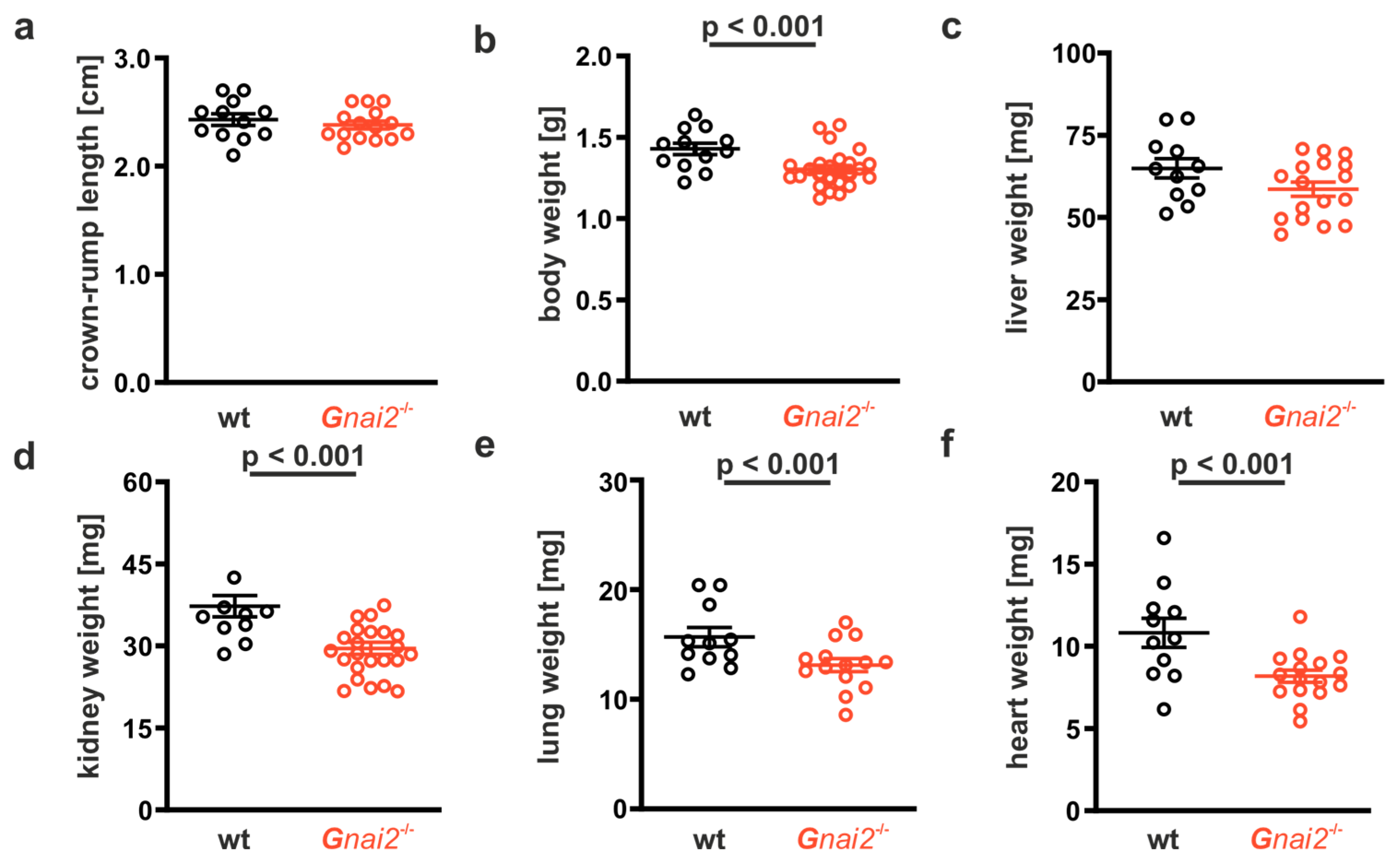
The cause of death could not be attributed to any obvious macroscopic abnormalities. Somatometric and organometric analysis of the neonatal mice revealed no differences in crown–rump length between Gnai2-deficient and wt controls; however, body weights were significantly lower in Gnai2-deficient neonates (Figure 2a,b). Organ weights for the liver were comparable between groups, whereas organ weights for the kidney, lung, and heart were significantly lighter in Gnai2-deficient neonates (Figure 2c–f). To further explore potential causes of death, we analyzed body and organ weights separately in deceased (Gnai2–/–(d)) and surviving Gnai2-deficient (Gnai2–/–(s)) littermates. No differences in crown–rump length, body or specific organ weights were observed between deceased Gnai2−/−(d) and surviving Gnai2–/–(s) neonates (Supplementary Figure S1a–c).

2.2. Gnai2-Deficiency Results in Lung Dysfunction in a Subset of Mice

Under physiological conditions, the respiratory pattern of newborns undergoes a rapid transition within the first hour of life—from initial irregular, wheezing-like efforts to a stable and vigorous rhythm characterized by an increased respiratory rate. During this adaptation phase, the early wheezing persists transiently alongside deep inspiratory efforts that gradually become faster and more synchronized [29]. In contrast, all deceased Gnai2–/– newborns exhibited the characteristic initial wheezing, but failed to establish a regular respiratory rhythm. Instead, these newborns continued to take deep breaths accompanied by subcostal and abdominal retractions. These were interrupted by increasingly long episodes of apnea (lasting up to one minute) and progressive cyanosis. This clinical presentation resembled neonatal respiratory distress syndrome (RDS) in humans [30,31]. However, close inspection of the affected Gnai2-deficient neonates revealed that lung borders and expansion were visible through the thin skin, suggesting that the lungs of those that later died were initially inflated (Figure 3a–c). In contrast, none of the wt neonates, nor the majority of surviving Gnai2-deficient neonates displayed overt abnormalities in breathing patterns. In these animals, the typical postnatal transition from initial deep gasps to a high-frequency breathing pattern occurred within minutes after delivery.

2.3. Gnai2-Deficiency Reduces Alveolar Surface Area in Deceased Newborn Mice

Our findings indicate that the elevated lethality of Gnai2-deficient offspring can be largely attributed to neonatal death occurring immediately after birth. This mortality appears to result from insufficient lung function consistent with features of neonatal respiratory distress syndrome (RDS). Potential causes of lung dysfunction include morphological abnormalities such as lung hypoplasia or congenital alveolar dysplasia, as well as surfactant deficiency or defects [32].
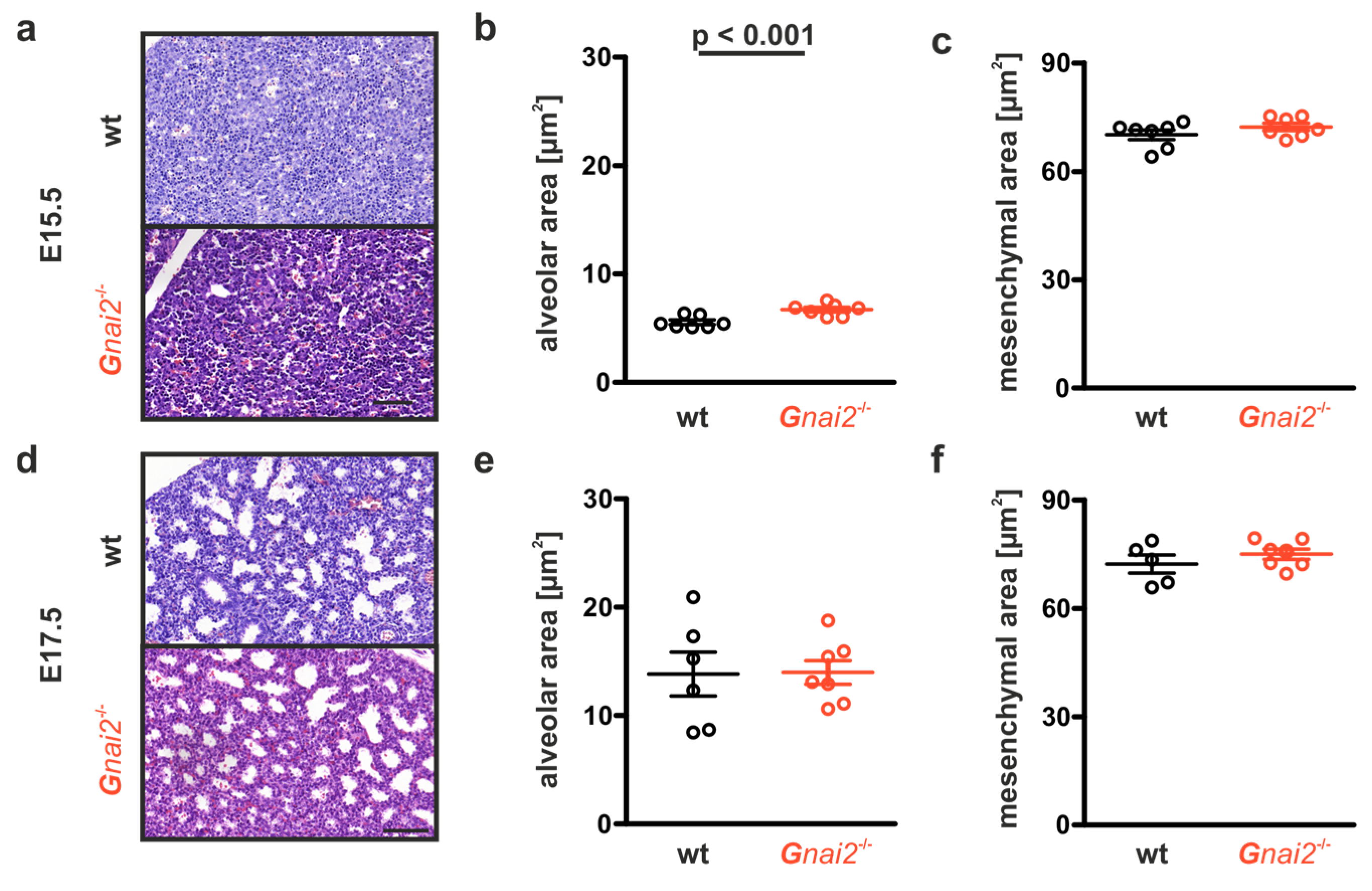
To address this, we examined lung development histologically in Gnai2-deficient embryos at embryonic days (E) 15.5 and 17.5, compared with wt controls. At E15.5, the lungs are in the glandular stage, where the bronchial tree forms and develops through extensive branching [33].
Gnai2-deficient embryos exhibited a cell-rich mesenchyme with numerous primitive bronchi and branching points (Figure 4a). The area fraction of primitive bronchi was significantly higher in Gnai2-deficient embryos (6.7 ± 0.6%, n = 7) than in wt controls (5.5 ± 0.5%, n = 7; p < 0.001; Figure 4b). By E17.5, this fraction increased to 14 ± 2.9% in Gnai2-deficient embryos (n = 7), now comparable to wt controls (13.5 ± 5.5%, n = 5; Figure 4d,e). At this stage, the lungs corresponded histologically to the canalicular phase, where the air–blood barrier begins to form and primitive epithelial sacs appear. The proportion of mesenchymal cell areas did not differ significantly between genotypes at either time point (Figure 4c,f).
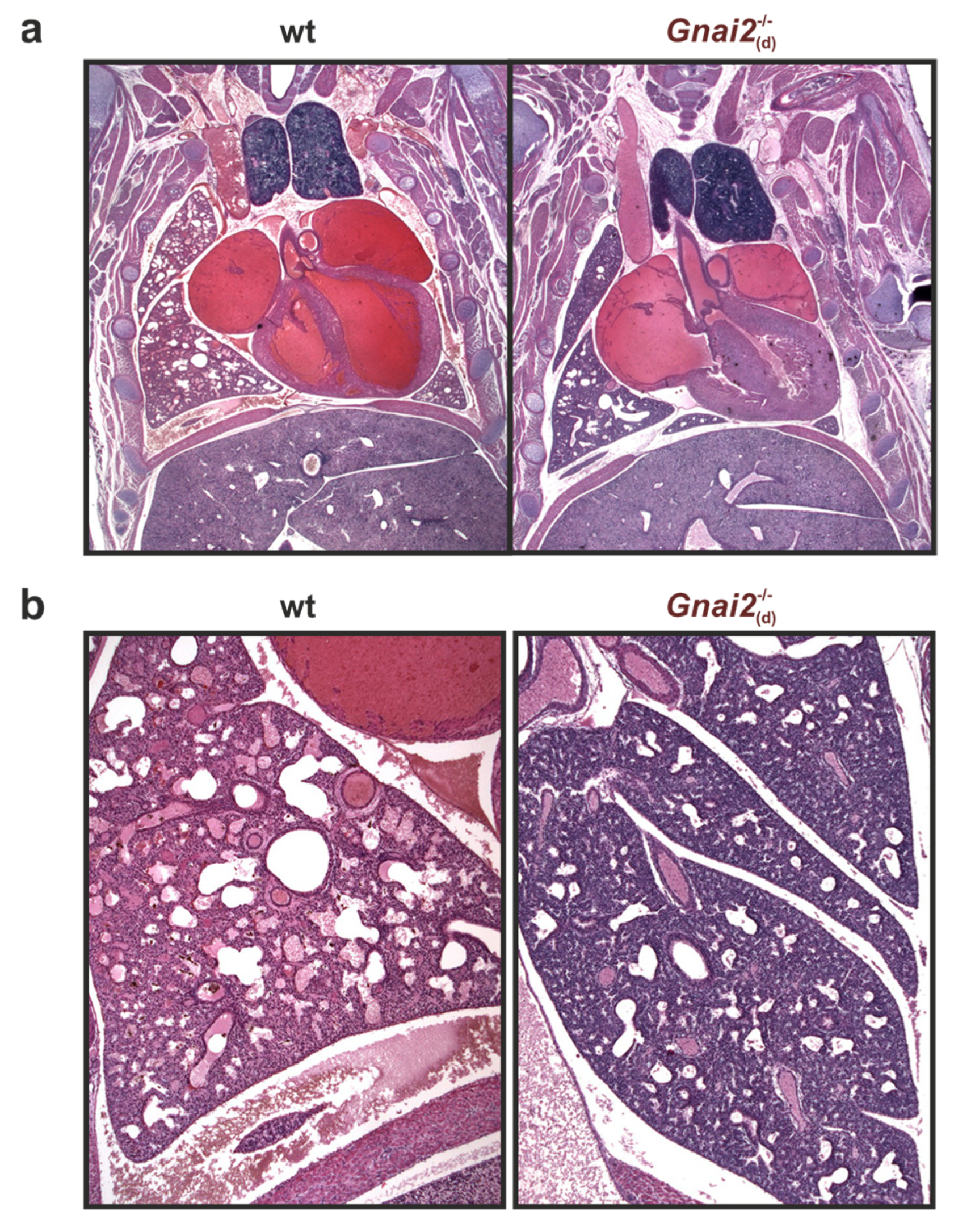
Although embryonic lung development did not reveal persistent structural abnormalities in deceased Gnai2-deficient mice (Figure 5), hematoxylin and eosin (H&E) staining of paraffin-embedded lung tissue from newborns demonstrated marked differences in alveolar and mesenchymal areas (Figure 6a).
While alveolar areas were similar between wt and surviving Gnai2−/−(s) neonates, deceased Gnai2−/−(d) neonates exhibited significantly reduced alveolar areas compared with wt (Figure 6b). Furthermore, lungs of Gnai2−/−(d) neonates appeared underdeveloped, with thickened interalveolar septa (Figure 6a, right). Consistently, the mesenchymal area was significantly increased in Gnai2−/−(d) neonates (Figure 6a, right) compared to both wt (Figure 6a, left) and Gnai2−/−(s) (Figure 6a, middle) neonates (Figure 6c).

2.4. Secreted Alveolar Surfactant Fails to Properly Unfold in Deceased Newborn Gnai2-Deficiency Mice

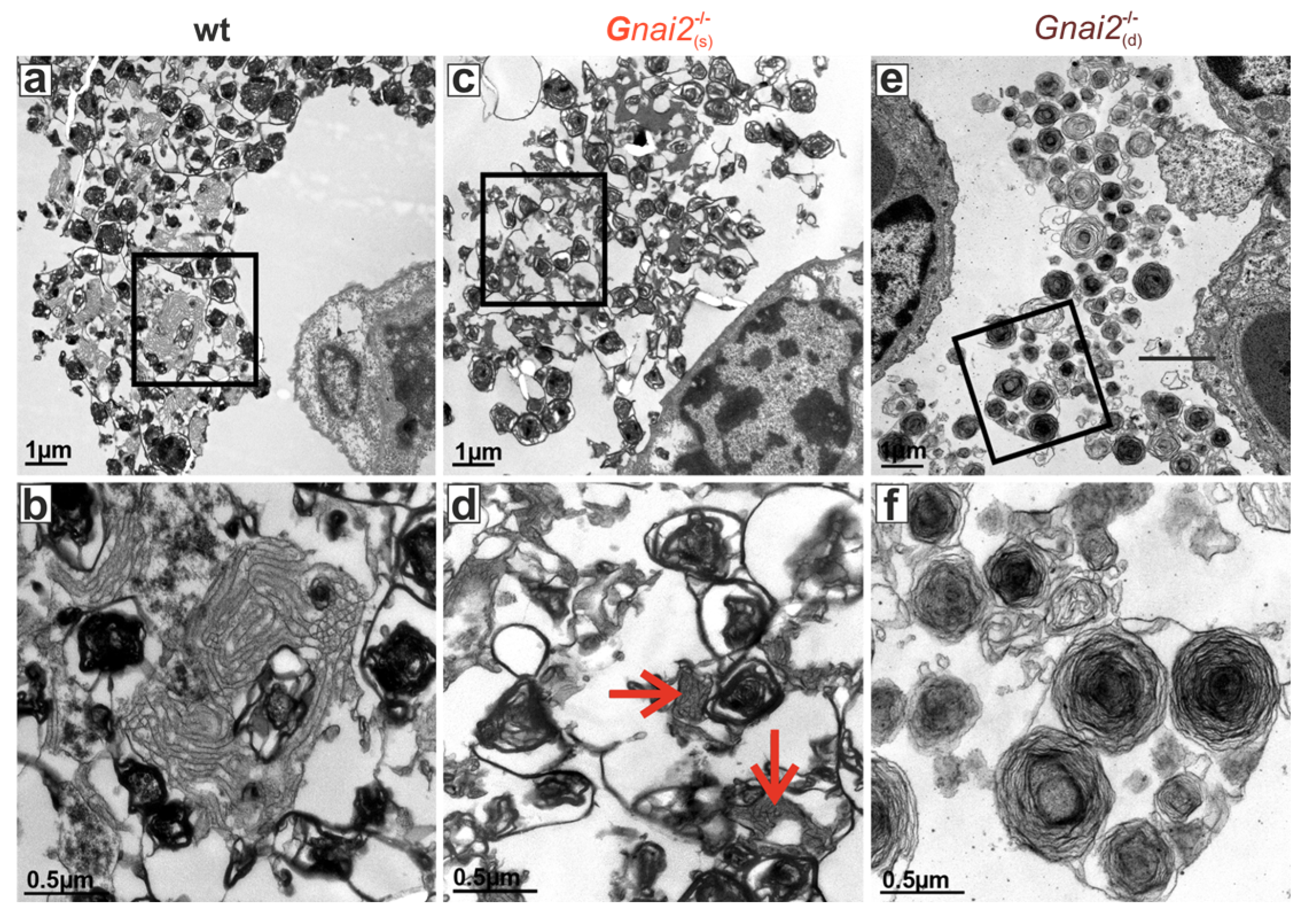
The observed breathing defect together with the histological findings in deceased Gnai2−/− neonates prompted us to test the hypothesis if impaired surfactant secretion is the underlying cause of the respiratory failure. Notably, proper lung function at birth depends on adequate production and secretion of pulmonary surfactant. Surfactant is synthesized by alveolar type II (ATII) cells, stored in intracellular lamellar bodies (LBs), and secreted into the alveolar space, where it is essential for normal breathing immediately after birth [34,35]. A deficiency or functional impairment of surfactant results in alveolar collapse after initial inflation due to insufficient reduction in surface tension, along with increased mesenchyme-rich, compact lung tissue and thickened interalveolar septa—consistent with the observations in Gnai2−/−(d) neonates (Figure 6). We therefore examined lungs from these animals by electron microscopy to assess LBs and the alveolar surfactant lattice structures (Figure 7). In WT mice, typical intracellular LBs and well-formed extracellular tubular myelin, representing structured surfactant reserves in the alveolar fluid, were readily identifiable (Figure 7a,b). A similar pattern was observed in Gnai2−/−(s) neonates (Figure 7c,d). In contrast, Gnai2−/−(d) neonates showed altered ultrastructure of alveolar surfactant unfolding and failure to form a phospholipid monolayer at the air–liquid interface, as evidenced by the absence or disruption of tubular myelin structures (Figure 7e,f).
The unfolding of LBs into a functional monolayer is facilitated by the hydrophobic surfactant proteins SP-B and SP-C, which promote lipid insertion and film stabilization [35]. In addition, SP-A, in a calcium-dependent manner, organizes and aggregates surfactant lipids into functional structures. To exclude a general surfactant deficiency as the cause, we quantified saturated phosphatidylcholine (SatPC), the most abundant surface-active phospholipid species in surfactant. In whole-lung tissue from Gnai2-deficient embryos at E18.5, SatPC levels did not differ from wt controls (Supplementary Figure S2).
To determine whether loss of Gαi2 specifically in ATII or mesenchymal cells was sufficient to reproduce the phenotype, we generated transgenic mice with cell-specific deletion of Gαi2 using Cre recombinase driven by either the Shh promoter (ATII-specific) or the Dermo1 promoter (mesenchymal-specific). Shh-Cre mice and Dermo1-Cre mice have been used extensively in lung development research due to high deletion efficiency in pulmonary epithelial cells and lung mesenchymal cells, respectively [36,37,38,39,40]. Offspring from both lines exhibited normal survival after birth and showed no evidence of respiratory distress syndrome. Furthermore, double deficiency of Gαi2 and the closely related Gαi3 isoform in ATII cells unexpectedly resulted in the expected Mendelian ratios at birth and normal survival beyond 2 h postpartum (Supplementary Table S5). Consistent with these findings, preliminary analyses of SatPC and total phospholipid content in BALF and lamellar body homogenates from ATII-specific Gnai2; Gnai3 double-deficient mice showed no significant differences compared to controls (Supplementary Figure S3), excluding a primary defect in surfactant synthesis or secretion by these cells. Thus, potential extrapulmonary effects warrant further investigation to clarify the cause of the described postnatal lethality in global Gnai2-deficient mice.

2.5. Improved Lung Function in Adult Gnai2-Deficient Mice

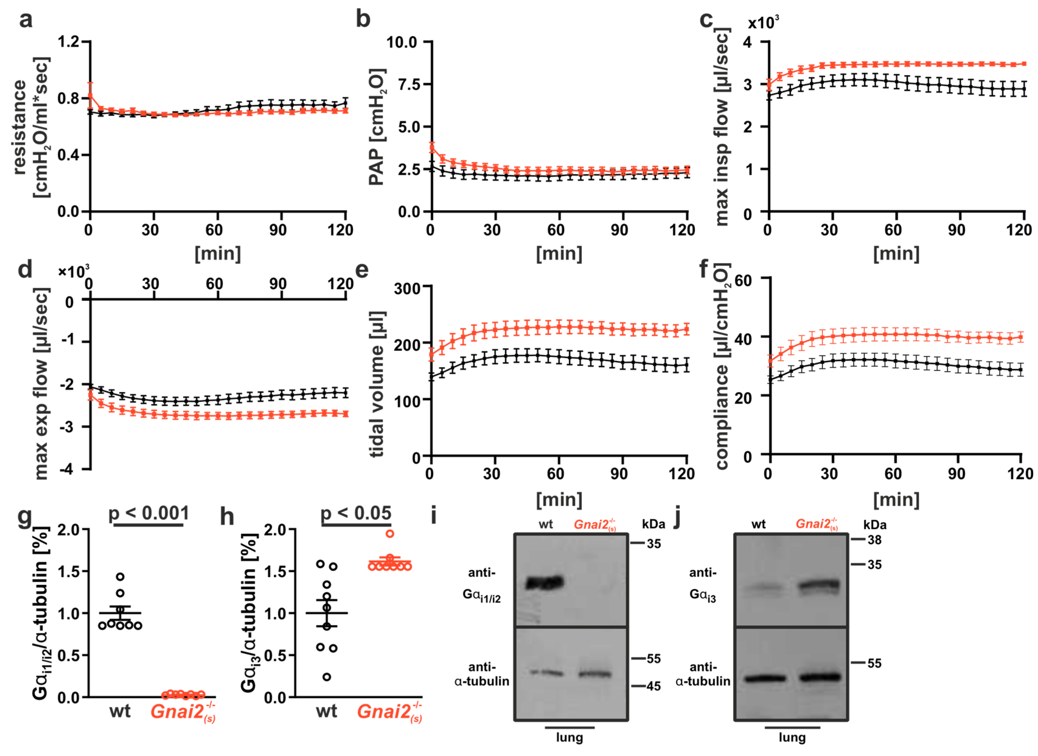
To validate if the lack of Gαi2 in surviving Gnai2−/− mice has any impact on lung function, we used the ex vivo isolated, perfused and ventilated lung model (IPL), which allows for measuring lung parameters under a constant pressure. Adult Gnai2−/− animals displayed normal to improved lung function (Figure 8 and Supplementary Table S6).
While airway resistance and pulmonary artery pressure (PAP) were comparable between genotypes (Figure 8a,b), the inspiratory and expiratory flow rates, tidal volume, and compliance were significantly increased in Gnai2-deficient animals (Figure 8c–f).
Given the high homology and partially redundant functions of Gαi isoforms within specific cell types, we hypothesized that upregulation of the closely related Gαi3 isoform might compensate for the loss of Gαi2. To examine this, lung homogenates were analyzed by immunoblotting for Gαi2 and Gαi3 expression. As expected, Gαi2 protein was readily detectable in WT lungs but absent in Gnai2-deficient tissue (Figure 8g,i). Interestingly, Gαi3 expression was significantly increased in lungs of Gnai2-deficient mice (Figure 8h,i). Although individual cell types were not analyzed in this study, these findings suggest a compensatory role of Gαi3 in maintaining proper lung function in adult Gnai2-deficient mice.

3. Discussion

Our findings demonstrate that the lack of Gαi2 compromises neonatal respiratory adaptation, resulting in increased early postnatal mortality. A substantial proportion of Gnai2-deficient neonates failed to establish regular breathing and died within minutes of birth, presenting with cyanosis and a clinical pattern reminiscent of neonatal respiratory distress syndrome (RDS) [30,31]. Importantly, these neonates were born at expected Mendelian ratios, exhibited normal vitality at delivery, and had no gross morphological abnormalities, indicating that the lethality is based on postnatal functional deficits rather than developmental malformations.
Postnatal RDS with a lethal outcome can arise from several pathophysiological mechanisms that compromise neonatal lung function, including structural lung abnormalities, such as pulmonary hypoplasia or congenital alveolar dysplasia. Histological analyses of embryonic lungs at E15.5 and E17.5 revealed no abnormalities, confirming that lung morphogenesis proceeds normally in the absence of Gαi2. However, at birth, lungs of deceased neonates showed reduced alveolar surface area, thickened interalveolar septa, and increased mesenchymal tissue, consistent with impaired alveolar inflation and collapse. Despite macroscopically inflated lungs, electron microscopy demonstrated disrupted tubular myelin and inadequate surfactant organization at the air–liquid interface, while levels of saturated phosphatidylcholine and surfactant proteins were unaltered. This dissociation between normal surfactant production and impaired deployment highlights the complexity of surfactant biology in vivo.
The inability of surfactant to properly unfold and spread within alveoli, despite intact synthesis and secretion, suggests that Gαi2 plays a previously unknown regulatory role in the post-secretory phase of surfactant function rather than in the regulation of adequate production of pulmonary surfactant by alveolar type II (ATII) epithelial cells [30,34]. This agrees with the current understanding that several signaling pathways other than Gi-dependent, including those involving Gs and Gq proteins, adenylyl cyclases, phospholipases, and protein kinases (PKA, PKC, and Ca2+/calmodulin-dependent kinases), have been implicated in surfactant secretion [41,42]. However, receptor-mediated and receptor-independent pathways involving Gi proteins have also been demonstrated to modulate surfactant release [43,44]. The transformation of lamellar body content into a functional monolayer and tubular myelin lattice is a multistep process dependent on biophysical and biochemical factors. Interestingly, the picture of large lamellar bodies and impaired surfactant processing is reminiscent of the pathophysiology of Hermansky–Pudlak syndrome (HPS) in humans. In this severe genetic disease, defective lysosome-related organelles lead to intracellular formation of giant lamellar bodies in AT II cells and accumulation of surfactant. In HPS pathophysiology, the chitinase-like-protein CHI3L1 and its impaired interplay with its receptor CRTH, a non-chemokine chemoattractant GPCR with a preference for Gαi proteins, seems to play an important role, although the exact causal molecular pathways in HPS are still understudied [45]. Previous studies have shown that this process is influenced by the alveolar microenvironment, extracellular ionic composition, surfactant protein interactions, and alveolar lining fluid properties [30,35]. Our data suggest that Gαi2 signaling may modulate one or more of these factors, potentially by affecting epithelial–mesenchymal crosstalk, alveolar surface fluid composition, or calcium-regulated surfactant organization [41,42]. An alternative explanation for the respiratory failure in Gnai2-deficient neonates could be an impairment of neuromuscular control rather than an intrinsic pulmonary dysfunction. However, several observations argue against this possibility: Gnai2-deficient neonates were able to take initial deep breaths and exhibited normal spontaneous movements before death. Moreover, adult Gnai2-deficient mice showed no overt motor deficits, and lung function was normal or even slightly improved. Collectively, these findings make a neuromuscular origin unlikely and instead support a primarily lung-intrinsic mechanism, although secondary extrapulmonary effects cannot be fully excluded.
Interestingly, conditional deletion of Gnai2 in alveolar type II (ATII) or mesenchymal cells did not replicate the lethal phenotype, suggesting involvement of other lung-resident cell types [46,47]. Endothelial cells and alveolar macrophages are known to influence surfactant turnover, alveolar surface tension, and inflammatory signaling—all of which could impair surfactant ultrastructure and spreading without altering its production. Dysregulated inflammatory mediators or calcium signaling pathways, both linked to G protein-coupled receptor activity, are plausible mechanisms underlying the observed defect [34]. The incomplete penetrance of the phenotype, with ~20% mortality among Gnai2-deficient neonates, suggests possible compensatory mechanisms and/or strain-specific genetic modifiers, as reported for other knockout models [48,49,50,51]. Differences in lethality between breeding strategies support this interpretation, though early procedural variability seems more likely. The lethality rate observed in the second study (~20%) aligns with the ~25% increase seen initially, including the unexpected mortality among heterozygous controls. Due to ethical constraints, these factors could not be fully disentangled.
We also observed a slight tendency toward increased kidney weight in deceased neonates (see Supplementary Figure S1c), possibly reflecting systemic responses to hypoxia, acute cardiopulmonary failure with fluid retention and congestion, or additional effects of Gnai2-deficiency in other organs. Although the significance of this finding remains speculative, it highlights the complex interaction between pulmonary and extrapulmonary factors during neonatal adaptation. Although, to the best of our knowledge, no germline mutations in GNAI2 have been directly linked to lung disease in humans to date. Recent studies have identified GNAI2 mutations, which lead to impaired GTPase activity of Gαi2 followed by an enhanced suppression of cAMP production and are associated with immune system dysregulation [52]. Despite these functions, GNAI2 remains to be identified as a significant locus in recent genome-wide association studies of lung function traits [53].
Although approximately 20% of Gnai2-deficient mice died from postnatal respiratory failure due to immature lungs and insufficient surfactant unfolding, those that survived this critical period exhibited unexpectedly improved lung functions compared with wild-type controls, including higher flow rates, tidal volume, and compliance. Upregulation of the remaining Gαi3 isoform has previously been reported in various tissues of Gnai2-deficient mice [54,55], and likely represents a compensatory mechanism that may contribute to the enhanced pulmonary performance observed here. In addition to potential Gαi3-mediated compensation, other—yet unidentified—factors could also facilitate postnatal survival by functionally substituting for missing Gαi2 activity. While most G-protein signaling mechanisms are classically attributed to the α subunits, it is now well established that βγ dimers also mediate numerous cellular functions [1]. Thus, a possible contribution of βγ-dimer-dependent signaling to this complex compensatory network should also be considered.
Taken together, our results highlight a previously unappreciated regulatory role of Gαi2 in neonatal respiratory physiology. We propose that Gαi2 facilitates the structural organization of surfactant, likely through effects on the alveolar microenvironment and intercellular communication among epithelial, endothelial, mesenchymal, and immune cells. Further investigation into these pathways—including the roles of calcium signaling, inflammatory mediators, and genetic modifiers—will be essential to fully elucidate the mechanisms underlying this phenotype and may inform therapeutic approaches for neonatal RDS.

3.1. Conclusions

i2 serves as a regulatory factor in neonatal respiratory adaptation by facilitating surfactant deployment and alveolar stabilization. Its deficiency leads to an incompletely penetrant phenotype, likely due to compensatory mechanisms and genetic modifiers. These findings emphasize the complexity of neonatal respiratory physiology and the need for precise regulation of surfactant function and intercellular communication.

3.2. Future Directions

Future research should elucidate how Gαi2 signaling integrates with epithelial, endothelial, mesenchymal, and immune pathways to regulate surfactant deployment and alveolar mechanics. Investigating calcium signaling, inflammatory mediators, and genetic modifiers will be crucial to understanding the incomplete phenotype. Such insights may pave the way for novel interventions in neonatal RDS and related disorders.

4. Materials and Methods

Experimental animals. The generation and phenotypic characterization of Gnai2-deficient mice (Gnai2/−), has been described [13]. Gnai2-deficient mice were backcrossed on a C57BL6/N background for more than 11 generations. To study postnatal mortality of Gnai2-deficient mice, newborn homozygous Gnai2-deficient mice were intercrossed and compared to homozygous wild-type litters. For the generation and analysis of mesenchymal cell-specific Gnai2-deficient mice, floxed Gnai2 (Gnai2fl/fl) mice were crossed with Dermo-Cre mice [38]. In addition, alveolar type II (ATII)-specific double-deficient floxed Gnai2; Gnai3-deficient mice (Gnai2fl/fl; Gnai3+/fl) [56] were crossed with Shh-Cre mice [39]. Absence of Gαi2 and Gαi3 protein was approved by immunoblot analysis (Supplementary Figure S4).
For isolated lung perfusion experiments, 8-to-18-week old mice from homozygous and heterozygous breeding were analyzed. Animal experiments were conducted in accordance with the recommendations in the Guide for the Care and Use of Laboratory Animals (FELASA). Animals were kept in standard cages with enrichment under specific pathogen-free conditions (SPF) according to national guidelines for animal care at the animal facility of the University of Düsseldorf and Tübingen. All animals were kept with a 12 h light/dark cycle and had access to food and water ad libitum. The physical condition was monitored daily both before and during the experiments. No animal became severely ill or died before the experimental endpoint. Protocols were approved by the committee on the Ethics of Animal Experiments of local authorities. All surgeries were performed under anesthesia, and all efforts were made to minimize suffering. For IPL mice were anesthetized with Pentobarbital (120 mg/kg body weight).
Analysis of postnatal survival. Pregnancy in female mice was confirmed by the presence of a vaginal plug and designated as embryonic day 0.5 (E0.5). For cesarean section, pregnant females at gestational day 19.5 were sacrificed by cervical dislocation, whereas for spontaneous delivery, the pregnant mice were observed during the expected period of birth until the pups were born. In case of cesarian section, the abdominal cavity was opened by a transverse incision, and the uterine horns were excised. Fetuses were rapidly removed from the uterus and embryonic membranes. Spontaneous onset of labor typically occurred at E20.5. The postnatal survival of newborn mice was monitored for up to 120 min and correlated with their respiratory behavior. The time of death was defined as the point at which breathing ceased and respiratory arrest persisted without recovery.
Histology of lung tissue. For hematoxylin and eosin staining, lungs were removed from newborn mice, fixed in 4% formaldehyde, embedded in paraffin according to standard procedures, and longitudinal 6 µm serial sections were obtained. Alveolar and mesenchymal areas were quantified by hand. Morphometric analysis was performed using Adobe Photoshop (version Photoshop CC 2014) and ImageJ (version ImageJ2).
Electron Microscopy. For electron microscopy, lungs were fixed in 2.5% glutaraldehyde in 0.1 M cacodylate buffer (pH 7.4). The embedded lungs were sectioned into 1–2 mm2 small blocks and post-fixed in 1% OsO4 in 0.1 M cacodylate. For contrast enhancement, specimens were transferred to 70% ethanol saturated with 3% uranyl-acetate, dehydrated in a graded ethanol series, and embedded in Araldite. Ultrathin sections (50 nm) were contrasted with lead citrate and examined using a Zeiss electron microscope (EM 10CR).
Surfactant phospholipid analysis. Saturated phosphatidylcholine and total phospholipids were quantified from cell-free bronchoalveolar lavage fluid and whole lung tissue as previously described [57].
Isolated perfused mouse lung preparation. To determine adult mouse lung functions the Isolated Perfused Lung Size 1, Hugo-Sachs-Elektronik (HSE), March Hugstetten, Germany, was used as described [58]. Lungs were perfused in a non-circulating manner through the pulmonary artery at a constant flow of 1 mL/min. The perfusion medium (RPMI 1640) was supplemented with 1% L-Alanyl-L-Glutamine (200 mM), 4% Albumine bovine Fraction V and 0.12% NaCl. Lungs were ventilated by negative pressure (−8.5 to −3 cm H2O) with 90 breaths/min, resulting in a tidal volume in the range of 100 to 300 μL. A deep inflation (−25 cm H2O) was performed every 5 min to prevent the lungs from atelectasis. All data were transferred to a computer and analyzed by the Pulmodyn software (HSE) (version 1.1.1.202).
Immunoblot analyses. Dissected lungs were homogenized in lysis buffer containing 100 mM NaCl, 20 mM Tris–HCl, 2.5 mM EDTA and protease inhibitor (Complete Roche; Roche, Mannheim, Germany) to generate total protein lysates for subsequent immunoblot analyses. The proteins were separated by molecular weight via 12% SDS gels containing 6 M urea and subsequently transferred onto polyvinylidene difluoride membranes (PVDF; Merck Millipore, Darmstadt, Germany). The membranes were blocked in 5% milk–TBST (tris-buffered saline (TBS)–Tween 20), incubated with the indicated antibodies and developed with enhanced chemiluminescence (ECL; GE Healthcare, Buckinghamshire, UK). Gαi2 protein expression was visualized by immunodetection using either rabbit polyclonal anti-Gαi1/i2 or anti-Gαi3 antibodies (1:2000) and horseradish peroxidase (HRP)-conjugated anti-rabbit IgG secondary antibodies (1:2000) (Cell Signaling Technology, Danvers, MA, USA). Equal loading was verified with rabbit anti-α-tubulin (1:4000) (Cell signaling Technology, Cambridge, UK). Gαi2 and Gαi3 protein levels were quantified by densitometry using the ImageJ software (NIH, Bethesda, MD, USA) and were normalized to α-tubulin levels of the same samples.
Statistical analysis. Data are presented as means ± SED of individual data points. Data were analyzed using Students’ t-test for unpaired groups or one-way ANOVA following Bonferroni’s Multiple Comparison Test when comparing wild type, surviving and dead Gnai2-deficient mice. A p-value of less than 0.05 was considered statistically significant.

Supplementary Materials

The following supporting information can be downloaded at: https://www.mdpi.com/article/10.3390/ijms262110655/s1.

Author Contributions

Conceptualization, S.B.-H., J.P.B., B.N., R.P.P.; methodology, S.A.-T., J.P.B., B.D.-M., P.F.-B., V.L., A.N. (Ana Novakovic), A.N. (Andreas Nowacki), K.P., L.Q.-M., M.S., H.W.; validation, B.D.-M., A.N. (Ana Novakovic), A.N. (Andreas Nowacki), R.P.P.; formal analysis, J.P.B., B.D.-M., V.L., B.N., A.N. (Ana Novakovic), A.N. (Andreas Nowacki); investigation, S.A.-T., J.P.B., B.D.-M., P.F.-B., V.L., A.N. (Ana Novakovic), A.N. (Andreas Nowacki), K.P., H.W.; writing—original draft preparation, S.B.-H., V.L., F.Z.; writing—review and editing, S.B.-H., J.P.B., V.L., B.N., F.Z., R.P.P.; visualization, V.L.; supervision, S.B.-H., V.L., B.N., R.P.P.; project administration, B.N.; funding acquisition, B.N., R.P.P., and M.S. All authors have read and agreed to the published version of the manuscript.

Funding

B.D.-M. got support from DFG-funded Research Training Group (RTG). M.S. was in part supported by the Robert Bosch Stiftung Stuttgart, Germany. This research was funded by Deutsche Forschungsgemeinschaft (DFG) to B.N.: SFB 612, RTGs 1089 and 2816, and Nu53/13-1.

Institutional Review Board Statement

The animal study protocol was approved by the Bezirksregierung Düsseldorf and Regierungspräsidium Tübingen.

Data Availability Statement

Research data will be shared on request.

Acknowledgments

We gratefully acknowledge Renate Riehle for technical assistance in breeding of mice. During the preparation of this manuscript, the authors used DeepL (free version, 2025) for AI-assisted translation of selected passages originally written in German and ChatGPT (versions 4 and 5) for language editing to improve readability and linguistic clarity. These tools were not used for content generation or autonomous writing. The authors have reviewed and edited the output and take full responsibility for the content of this publication.

Conflicts of Interest

The authors declare no conflicts of interest.

Abbreviations

The following abbreviations are used in this manuscript:
ATalveolar-type epithelial cells
GPCRG-protein coupled receptors
HPSHermansky–Pudlak syndrome
RDSRespiratory distress syndrome
TMtubular myelin
wtwild-type

References

  1. Nürnberg, B.; Beer-Hammer, S.; Reisinger, E.; Leiss, V. Non-canonical G protein signaling. Pharmacol. Ther. 2024, 255, 108589. [Google Scholar] [CrossRef]
  2. Weis, W.I.; Kobilka, B.K. The molecular basis of g protein-coupled receptor activation. Annu. Rev. Biochem. 2018, 87, 897–919. [Google Scholar] [CrossRef] [PubMed]
  3. Lin, L.; Li, S.; Zhang, C.; Shen, J.M.; Liu, J. Gpcr heterodimers: Implications for biased signaling. Handb. Exp. Pharmacol. 2025. [Google Scholar] [CrossRef]
  4. Zhou, J.C.; Li, A.; Long, Z.; Kahsai, A.W. Biased allosteric modulation in gpcr drug discovery. Handb. Exp. Pharmacol. 2025. [Google Scholar] [CrossRef]
  5. Simon, M.I.; Strathmann, M.P.; Gautam, N. Diversity of G proteins in signal transduction. Science 1991, 252, 802–808. [Google Scholar] [CrossRef]
  6. Aydogdu, S.; Pepanian, A.; Rathod, D.C.; Schulz, F.; Imhof, D. Physiological functions and structural features of galpha12/13 proteins. Biomed. Pharmacother. 2025, 191, 118523. [Google Scholar] [CrossRef]
  7. Inoue, A.; Raimondi, F.; Kadji, F.M.N.; Singh, G.; Kishi, T.; Uwamizu, A.; Ono, Y.; Shinjo, Y.; Ishida, S.; Arang, N.; et al. Illuminating G-Protein-Coupling Selectivity of GPCRs. Cell 2019, 177, 1933–1947. [Google Scholar] [CrossRef]
  8. Qayum, S.; Petrin, D.; Tanny, J.C.; Hebert, T.E. Gbetagamma signaling: Lessons across the cellular multiverse. Annu. Rev. Pharmacol. Toxicol. 2025. [Google Scholar] [CrossRef] [PubMed]
  9. Wilkie, T.M.; Gilbert, D.J.; Olsen, A.S.; Chen, X.N.; Amatruda, T.T.; Korenberg, J.R.; Trask, B.J.; de Jong, P.; Reed, R.R.; Simon, M.I.; et al. Evolution of the mammalian G protein alpha subunit multigene family. Nat. Genet. 1992, 1, 85–91. [Google Scholar] [CrossRef] [PubMed]
  10. Gohla, A.; Klement, K.; Piekorz, R.P.; Pexa, K.; vom Dahl, S.; Spicher, K.; Dreval, V.; Haussinger, D.; Birnbaumer, L.; Nürnberg, B. An obligatory requirement for the heterotrimeric G protein Gi3 in the antiautophagic action of insulin in the liver. Proc. Natl. Acad. Sci. USA 2007, 104, 3003–3008. [Google Scholar] [CrossRef]
  11. Jantzen, H.M.; Milstone, D.S.; Gousset, L.; Conley, P.B.; Mortensen, R.M. Impaired activation of murine platelets lacking G alpha(i2). J. Clin. Investig. 2001, 108, 477–483. [Google Scholar] [CrossRef]
  12. Plummer, N.W.; Spicher, K.; Malphurs, J.; Akiyama, H.; Abramowitz, J.; Nürnberg, B.; Birnbaumer, L. Development of the mammalian axial skeleton requires signaling through the Gαi subfamily of heterotrimeric G proteins. Proc. Natl. Acad. Sci. USA 2012, 109, 21366–21371. [Google Scholar] [CrossRef]
  13. Rudolph, U.; Finegold, M.J.; Rich, S.S.; Harriman, G.R.; Srinivasan, Y.; Brabet, P.; Boulay, G.; Bradley, A.; Birnbaumer, L. Ulcerative colitis and adenocarcinoma of the colon in Gαi2-deficient mice. Nat. Genet. 1995, 10, 143–150. [Google Scholar] [CrossRef]
  14. Wettschureck, N.; Offermanns, S. Mammalian G proteins and their cell type specific functions. Physiol. Rev. 2005, 85, 1159–1204. [Google Scholar] [CrossRef]
  15. Beer-Hammer, S.; Lee, S.C.; Mauriac, S.A.; Leiss, V.; Groh, I.A.M.; Novakovic, A.; Piekorz, R.P.; Bucher, K.; Chen, C.; Ni, K.; et al. Gαi Proteins are Indispensable for Hearing. Cell Physiol. Biochem. 2018, 47, 1509–1532. [Google Scholar] [CrossRef]
  16. Devanathan, V.; Hagedorn, I.; Köhler, D.; Pexa, K.; Cherpokova, D.; Kraft, P.; Singh, M.; Rosenberger, P.; Stoll, G.; Birnbaumer, L.; et al. Platelet Gi protein Galphai2 is an essential mediator of thrombo-inflammatory organ damage in mice. Proc. Natl. Acad. Sci. USA 2015, 112, 6491–6496. [Google Scholar] [CrossRef]
  17. Ezan, J.; Lasvaux, L.; Gezer, A.; Novakovic, A.; May-Simera, H.; Belotti, E.; Lhoumeau, A.C.; Birnbaumer, L.; Beer-Hammer, S.; Borg, J.P.; et al. Primary cilium migration depends on G protein signalling control of subapical cytoskeleton. Nat. Cell Biol. 2013, 15, 1107–1115. [Google Scholar] [CrossRef] [PubMed]
  18. Keller, K.; Maass, M.; Dizayee, S.; Leiss, V.; Annala, S.; Koth, J.; Seemann, W.K.; Muller-Ehmsen, J.; Mohr, K.; Nurnberg, B.; et al. Lack of Galphai2 leads to dilative cardiomyopathy and increased mortality in beta1-adrenoceptor overexpressing mice. Cardiovasc. Res. 2015, 108, 348–356. [Google Scholar] [CrossRef] [PubMed]
  19. Köhler, D.; Devanathan, V.; Bernardo de Oliveira Franz, C.; Eldh, T.; Novakovic, A.; Roth, J.M.; Granja, T.; Birnbaumer, L.; Rosenberger, P.; Beer-Hammer, S.; et al. Gαi2- and Gαi3-deficient mice display opposite severity of myocardial ischemia reperfusion injury. PLoS ONE 2014, 9, e98325. [Google Scholar] [CrossRef] [PubMed]
  20. Leiss, V.; Schönsiegel, A.; Gnad, T.; Kerner, J.; Kaur, J.; Sartorius, T.; Machann, J.; Schick, F.; Birnbaumer, L.; Haring, H.U.; et al. Lack of Galpha(i2) proteins in adipocytes attenuates diet-induced obesity. Mol. Metab. 2020, 40, 101029. [Google Scholar] [CrossRef]
  21. Wiege, K.; Le, D.D.; Syed, S.N.; Ali, S.R.; Novakovic, A.; Beer-Hammer, S.; Piekorz, R.P.; Schmidt, R.E.; Nürnberg, B.; Gessner, J.E. Defective macrophage migration in Gαi2- but not Gαi3-deficient mice. J. Immunol. 2012, 189, 980–987. [Google Scholar] [CrossRef]
  22. Bai, J.Y.; Li, Y.; Xue, G.H.; Li, K.R.; Zheng, Y.F.; Zhang, Z.Q.; Jiang, Q.; Liu, Y.Y.; Zhou, X.Z.; Cao, C. Requirement of galphai1 and galphai3 in interleukin-4-induced signaling, macrophage m2 polarization and allergic asthma response. Theranostics 2021, 11, 4894–4909. [Google Scholar] [CrossRef]
  23. Jarysta, A.; Tadenev, A.L.; Day, D.M.; Krawchuk, B.; Low, B.E.; Wiles, M.V.; Tarchini, B. Inhibitory g proteins play multiple roles to polarize sensory hair cell morphogenesis. Elife 2024, 12. [Google Scholar] [CrossRef]
  24. Katnahji, N.; Matthes, J. Opposite effects of galpha(i2) or galpha(i3) deficiency on reduced basal density and attenuated beta-adrenergic response of ventricular ca(2+) currents in myocytes of mice overexpressing the cardiac beta(1)-adrenoceptor. Naunyn Schmiedebergs Arch. Pharmacol. 2025, 398, 12543–12549. [Google Scholar] [CrossRef]
  25. van den Bos, E.; Ambrosy, B.; Horsthemke, M.; Walbaum, S.; Bachg, A.C.; Wettschureck, N.; Innamorati, G.; Wilkie, T.M.; Hanley, P.J. Knockout mouse models reveal the contributions of g protein subunits to complement c5a receptor-mediated chemotaxis. J. Biol. Chem. 2020, 295, 7726–7742. [Google Scholar] [CrossRef]
  26. Xie, H.; Ji, J.; Liu, Z.; Lu, N.; Wei, Y.; Zhou, A.; Liu, J.; Jiao, Q. Galphai1/3 signaling mediates il-5-induced eosinophil activation and type 2 inflammation in eosinophilic chronic rhinosinusitis. Front. Immunol. 2024, 15, 1460104. [Google Scholar] [CrossRef]
  27. Huang, X.; Fu, Y.; Charbeneau, R.A.; Saunders, T.L.; Taylor, D.K.; Hankenson, K.D.; Russell, M.W.; D’Alecy, L.G.; Neubig, R.R. Pleiotropic phenotype of a genomic knock-in of an RGS-insensitive G184S Gnai2 allele. Mol. Cell Biol. 2006, 26, 6870–6879. [Google Scholar] [CrossRef] [PubMed]
  28. He, W.; Cowan, C.W.; Wensel, T.G. RGS9, a GTPase accelerator for phototransduction. Neuron 1998, 20, 95–102. [Google Scholar] [CrossRef] [PubMed]
  29. Ramirez, S.C.; Koschnitzky, J.E.; Youngquist, T.M.; Baertsch, N.A.; Smith, C.V.; Ramirez, J.M. Perinatal Breathing Patterns and Survival in Mice Born Prematurely and at Term. Front. Physiol. 2019, 10, 1113. [Google Scholar] [CrossRef] [PubMed]
  30. Whitsett, J.A.; Wert, S.E.; Weaver, T.E. Alveolar surfactant homeostasis and the pathogenesis of pulmonary disease. Annu. Rev. Med. 2010, 61, 105–119. [Google Scholar] [CrossRef]
  31. Bos, L.D.J.; Ware, L.B. Acute respiratory distress syndrome: Causes, pathophysiology, and phenotypes. Lancet 2022, 400, 1145–1156. [Google Scholar] [CrossRef]
  32. Speier, R.; Cotten, C.M. Pulmonary Hypoplasia. In Manual of Neonatal Respiratory Care; Donn, S.M., Mammel, M.C., van Kaam, A.H.L.C., Eds.; Springer International Publishing: Cham, Switzerland, 2022; pp. 699–703. [Google Scholar]
  33. Bartman, C.M.; Matveyenko, A.; Prakash, Y.S. It’s about time: Clocks in the developing lung. J. Clin. Investig. 2020, 130, 39–50. [Google Scholar] [CrossRef]
  34. Milad, N.; Morissette, M.C. Revisiting the role of pulmonary surfactant in chronic inflammatory lung diseases and environmental exposure. Eur. Respir. Rev. 2021, 30, 210077. [Google Scholar] [CrossRef]
  35. Possmayer, F.; Zuo, Y.Y.; Veldhuizen, R.A.W.; Petersen, N.O. Pulmonary Surfactant: A Mighty Thin Film. Chem. Rev. 2023, 123, 13209–13290. [Google Scholar] [CrossRef]
  36. Ustiyan, V.; Bolte, C.; Zhang, Y.; Han, L.; Xu, Y.; Yutzey, K.E.; Zorn, A.M.; Kalin, T.V.; Shannon, J.M.; Kalinichenko, V.V. FOXF1 transcription factor promotes lung morphogenesis by inducing cellular proliferation in fetal lung mesenchyme. Dev. Biol. 2018, 443, 50–63. [Google Scholar] [CrossRef] [PubMed]
  37. Yin, Y.; Wang, F.; Ornitz, D.M. Mesothelial- and epithelial-derived FGF9 have distinct functions in the regulation of lung development. Development 2011, 138, 3169–3177. [Google Scholar] [CrossRef] [PubMed]
  38. Harfe, B.D.; Scherz, P.J.; Nissim, S.; Tian, H.; McMahon, A.P.; Tabin, C.J. Evidence for an expansion-based temporal Shh gradient in specifying vertebrate digit identities. Cell 2004, 118, 517–528. [Google Scholar] [CrossRef]
  39. Sosic, D.; Richardson, J.A.; Yu, K.; Ornitz, D.M.; Olson, E.N. Twist regulates cytokine gene expression through a negative feedback loop that represses NF-kappaB activity. Cell 2003, 112, 169–180. [Google Scholar] [CrossRef]
  40. Harris, K.S.; Zhang, Z.; McManus, M.T.; Harfe, B.D.; Sun, X. Dicer function is essential for lung epithelium morphogenesis. Proc. Natl. Acad. Sci. USA 2006, 103, 2208–2213. [Google Scholar] [CrossRef] [PubMed]
  41. Bridges, J.P.; Safina, C.; Pirard, B.; Brown, K.; Filuta, A.; Panchanathan, R.; Bouhelal, R.; Reymann, N.; Patel, S.; Seuwen, K.; et al. Regulation of pulmonary surfactant by the adhesion GPCR GPR116/ADGRF5 requires a tethered agonist-mediated activation mechanism. elife 2022, 11, e69061. [Google Scholar] [CrossRef]
  42. Andreeva, A.V.; Kutuzov, M.A.; Voyno-Yasenetskaya, T.A. Regulation of surfactant secretion in alveolar type II cells. Am. J. Physiol. Lung Cell Mol. Physiol. 2007, 293, L259–L271. [Google Scholar] [CrossRef]
  43. Pian, M.S.; Dobbs, L.G. Activation of G proteins may inhibit or stimulate surfactant secretion in rat alveolar type II cells. Am. J. Physiol. 1994, 266, L375–L381. [Google Scholar] [CrossRef] [PubMed]
  44. Pian, M.S.; Dobbs, L.G. Lipoprotein-stimulated surfactant secretion in alveolar type II cells: Mediation by heterotrimeric G proteins. Am. J. Physiol. 1997, 273, L634–L639. [Google Scholar] [CrossRef] [PubMed]
  45. Dell’Angelica, E.C.; Shotelersuk, V.; Aguilar, R.C.; Gahl, W.A.; Bonifacino, J.S. Altered trafficking of lysosomal proteins in Hermansky-Pudlak syndrome due to mutations in the beta 3A subunit of the AP-3 adaptor. Mol. Cell 1999, 3, 11–21. [Google Scholar] [CrossRef]
  46. Anciuc-Crauciuc, M.; Cucerea, M.C.; Tripon, F.; Crauciuc, G.A.; Banescu, C.V. Descriptive and Functional Genomics in Neonatal Respiratory Distress Syndrome: From Lung Development to Targeted Therapies. Int. J. Mol. Sci. 2024, 25, 649. [Google Scholar] [CrossRef]
  47. Nieto, M.A.; Huang, R.Y.; Jackson, R.A.; Thiery, J.P. Emt: 2016. Cell 2016, 166, 21–45. [Google Scholar] [CrossRef]
  48. Doetschman, T.; Gregg, R.G.; Maeda, N.; Hooper, M.L.; Melton, D.W.; Thompson, S.; Smithies, O. Targetted correction of a mutant HPRT gene in mouse embryonic stem cells. Nature 1987, 330, 576–578. [Google Scholar] [CrossRef]
  49. Liao, J.; Aggarwal, V.S.; Nowotschin, S.; Bondarev, A.; Lipner, S.; Morrow, B.E. Identification of downstream genetic pathways of Tbx1 in the second heart field. Dev. Biol. 2008, 316, 524–537. [Google Scholar] [CrossRef] [PubMed]
  50. Nadeau, J.H. Modifier genes in mice and humans. Nat. Rev. Genet. 2001, 2, 165–174. [Google Scholar] [CrossRef]
  51. Conlon, R.A.; Reaume, A.G.; Rossant, J. Notch1 is required for the coordinate segmentation of somites. Development 1995, 121, 1533–1545. [Google Scholar] [CrossRef]
  52. Ham, H.; Jing, H.; Lamborn, I.T.; Kober, M.M.; Koval, A.; Berchiche, Y.A.; Anderson, D.E.; Druey, K.M.; Mandl, J.N.; Isidor, B.; et al. Germline mutations in a G protein identify signaling cross-talk in T cells. Science 2024, 385, eadd8947. [Google Scholar] [CrossRef] [PubMed]
  53. Shrine, N.; Guyatt, A.L.; Erzurumluoglu, A.M.; Jackson, V.E.; Hobbs, B.D.; Melbourne, C.A.; Batini, C.; Fawcett, K.A.; Song, K.; Sakornsakolpat, P.; et al. New genetic signals for lung function highlight pathways and chronic obstructive pulmonary disease associations across multiple ancestries. Nat. Genet. 2019, 51, 481–493. [Google Scholar] [CrossRef] [PubMed]
  54. Wiege, K.; Ali, S.R.; Gewecke, B.; Novakovic, A.; Konrad, F.M.; Pexa, K.; Beer-Hammer, S.; Reutershan, J.; Piekorz, R.P.; Schmidt, R.E.; et al. Gαi2 is the essential Gαi protein in immune complex-induced lung disease. J. Immunol. 2013, 190, 324–333. [Google Scholar] [CrossRef]
  55. Leiss, V.; Flockerzie, K.; Novakovic, A.; Rath, M.; Schönsiegel, A.; Birnbaumer, L.; Schürmann, A.; Harteneck, C.; Nürnberg, B. Insulin secretion stimulated by arginine and its metabolite ornithine depends on Gαi2. Am. J. Physiol. Endocrinol. Metab. 2014, 307, E800–E812. [Google Scholar] [CrossRef] [PubMed]
  56. Mauriac, S.A.; Hien, Y.E.; Bird, J.E.; Carvalho, S.D.; Peyroutou, R.; Lee, S.C.; Moreau, M.M.; Blanc, J.M.; Geyser, A.; Medina, C.; et al. Defective Gpsm2/Gαi3 signalling disrupts stereocilia development and growth cone actin dynamics in Chudley-McCullough syndrome. Nat. Commun. 2017, 8, 14907. [Google Scholar] [CrossRef]
  57. Bridges, J.P.; Ikegami, M.; Brilli, L.L.; Chen, X.; Mason, R.J.; Shannon, J.M. LPCAT1 regulates surfactant phospholipid synthesis and is required for transitioning to air breathing in mice. J. Clin. Investig. 2010, 120, 1736–1748. [Google Scholar] [CrossRef]
  58. Mays, L.E.; Ammon-Treiber, S.; Mothes, B.; Alkhaled, M.; Rottenberger, J.; Müller-Hermelink, E.S.; Grimm, M.; Mezger, M.; Beer-Hammer, S.; von Stebut, E.; et al. Modified Foxp3 mRNA protects against asthma through an IL-10-dependent mechanism. J. Clin. Invest. 2013, 123, 1216–1228. [Google Scholar] [CrossRef]
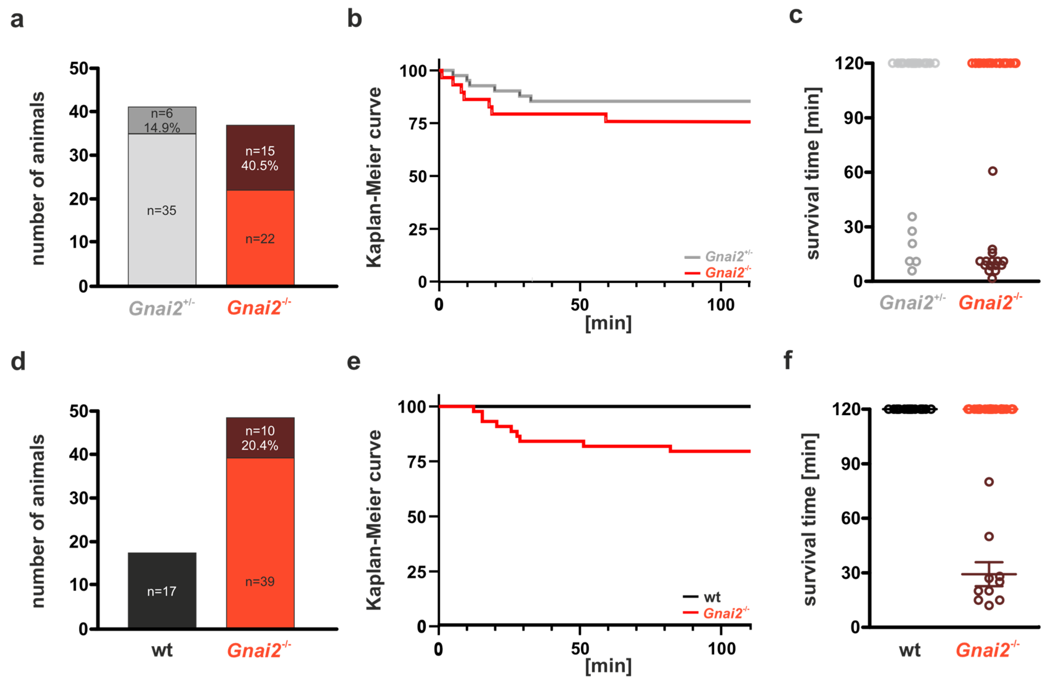
Figure 1. Perinatal lethality of Gnai2-deficient newborns. (a) Percentage of surviving (light grey, red) and deceased (grey, brown) Gnai2+/− and Gnai2−/− neonates delivered by cesarean section. (b) Kaplan–Meier survival curves of the corresponding Gnai2+/ and Gnai2/ neonates over a 2 h observation period. (c) Individual survival times of Gnai2+/− and Gnai2−/− neonates. (d) Percentage of surviving (black, red) and deceased (brown) wild-type (wt) and Gnai2-/- neonates after spontaneous delivery. (e) Kaplan–Meier survival curves of wt and Gnai2/ neonates showing a significant difference (log-rank test, p < 0.05) within the 2 h observation period. (f) Individual survival times of wt and Gnai2−/− neonates. Exact time points of death for Gnai2/ neonates are indicated as brown data points in panels (c) and (f), while surviving Gnai2/ neonates are shown in red.
Ijms 26 10655 g001
Figure 2. Somatometric and organometric parameters of newborn mice. (a) Crown–rump length was comparable between wild-type (wt) and Gnai2/ neonates. (b) Body weight was significantly reduced in Gnai2/ compared with wt neonates. (c) Liver weight was similar between genotypes. (df) Kidney, lung, and heart weights were significantly lower in Gnai2/ neonates than in wt controls. Data represent mean ± SEM from 12–21 animals per group.
Ijms 26 10655 g002
Figure 3. Sequential phases of a respiratory cycle in a mouse. Representative images illustrating distinct stages of a respiratory cycle during spontaneous breathing of a newborn Gnai2-deficient mouse that died: (a) before inspiration—the skin below the rib cage (black dotted line) appears smooth with minimal folding, (b) deep inspiration—prominent skin folds become visible beneath the rib cage, reflecting thoracic movement and negative intrathoracic pressure, (c) after expiration—the skin below the rib cage appears again smooth with minimal folding.
Ijms 26 10655 g003
Figure 4. Lung tissue structure of wt and Gnai2-deficient embryonic lungs at E15.5 and E17.5. (a) Representative H&E staining of paraffin-embedded 5 µm lung sections obtained from whole body sections of wt and Gnai2-deficient embryos at E15.5. Quantification of (b) alveolar and (c) mesenchymal area derived from wt and Gnai2-deficient E15.5 embryonic lungs. (d) Representative H&E- stained lung sections from E17.5 wt and Gnai2-deficient embryos. Statistical analysis of (e) alveolar and (f) mesenchymal area quantified from E17.7 wt and Gnai2-deficient lungs. Data are mean ± SEM from 5 to 7 mice. Scale bars 100 µm.
Ijms 26 10655 g004
Figure 5. Lung tissue structure of wt and deceased Gnai2-deficient (Gnai2−/−(d)) neonates. (a) Representative H&E staining of paraffin-embedded 5 µm thorax sections obtained from a wt newborn that survived the 2 h observation period and a deceased Gnai2-deficient neonate. (b) Representative H&E-stained lung sections from wt and deceased Gnai2-deficient neonates.
Ijms 26 10655 g005
Figure 6. Morphometric structure of lung tissue from Gnai2/(d) mice at P0. (a) Representative H&E staining of lung sections from wt, surviving Gnai2−/−(s) and deceased Gnai2−/−(d) animals. Scale bar 1000 µm (left panel). Magnifications (right panel) show significantly reduced alveolar areas in Gnai2−/−(d) lungs. Scale bar 200 µm. Quantification of (b) alveolar and (c) mesenchymal area of wt (black), Gnai2−/−(s) (red) and Gnai2−/−(d) (brown) lungs. Gnai2−/−(d) lungs have significantly decreased alveolar and significantly increased mesenchymal area. Mean ± SEM.
Ijms 26 10655 g006
Figure 7. Surfactant structure at P0. Representative ultra-thin sections of lung tissue from wt (left, a, b), Gnai2−/−(s) (middle, c,d) and Gnai2−/−(d) (right, e,f) mice (upper panel). Scale bars 1 µm and 0.5 µm. Lower panels show magnifications of secreted surfactant and the newly formed tubular myelin (TM). Surfactant of Gnai2−/−(d) lungs does not unravel as shown for wt and Gnai2−/−(s) surfactant. In the upper panels (a,c,e), regions outlined by black boxes indicate areas that are shown at higher magnification in the lower panels (b,d,f) below.Red arrows in (d) indicate unraveled surfactant. Scale bars 1 and 0.5 µm.
Ijms 26 10655 g007
Figure 8. Lung function of adult Gnai2/(s) in comparison to wt animals analyzed by isolated perfused lung (IPL). Gnai2-deficient mice (red) have normal (a) airway resistance and (b) pulmonary artery pressure (PAP), whereas (c) maximal inspiratory, (d) maximal expiratory flow rate, (e) tidal volume and (f) lung compliance were significantly improved compared to wt (black) animals. Significances for AUC levels are shown in Supplementary Table S5. Protein levels of (g) Gαi2 and (h) Gαi3 in lung tissue from wt and Gnai2-deficient mice. Protein levels of Gαi3 were significantly higher in Gnai2-deficient mice. Representative immunoblots for (i) Gαi2 and (j) Gαi3 expression in lung tissue from wt and Gnai2-deficient mice are depicted. Data are mean ± SEM from 9 to 12 animals.
Ijms 26 10655 g008
Table 1. Mendelian ratio of littermates born from heterozygous Gnai2-matings. Statistics are based on Chi-square analyses (n.s.—not significant).
GenotypeE14E18P5-10P21% Expected
Gnai2+/+7 (15.9%)13 (22%)137 (26.9%)365 (31.1%)25
Gnai2+/−29 (66%)34 (57.7%)316 (62.1%)695 (59.2%)50
Gnai2−/−8 (18.1%)12 (20.3%)56 (11%)114 (9.7%)25
44
(6 litter)
59
(8 litter)
509
(92 litter)
1174
(210 litter)
p valuen.s.n.s.<0.001<0.001
Disclaimer/Publisher’s Note: The statements, opinions and data contained in all publications are solely those of the individual author(s) and contributor(s) and not of MDPI and/or the editor(s). MDPI and/or the editor(s) disclaim responsibility for any injury to people or property resulting from any ideas, methods, instructions or products referred to in the content.