Review

Relationship Between Obesity and Depression Considering the Inflammatory Theory

by
Aleksandra Julia Oracz
1,*,†,
Mateusz Zwierz
2,†,
Maciej Naumowicz
2,
Maria Suprunowicz
1 and
Napoleon Waszkiewicz
1
1
Department of Psychiatry, Medical University of Bialystok, pl. Wołodyjowskiego 2, 15-272 Białystok, Poland
2
Faculty of Medicine with the Division of Dentistry and Division of Medical Education in English, Medical University of Bialystok, Jana Kilińskiego 1, 15-089 Białystok, Poland
*
Author to whom correspondence should be addressed.
These authors have contributed equally to this work.
Int. J. Mol. Sci. 2025, 26(11), 4966; https://doi.org/10.3390/ijms26114966
Submission received: 15 March 2025 / Revised: 9 May 2025 / Accepted: 14 May 2025 / Published: 22 May 2025

Abstract

:
Numerous scientific findings indicate that excess adipose tissue, particularly visceral fat, is associated with a chronic inflammatory state manifested by elevated levels of proinflammatory cytokines and an imbalance in the T helper type 1/type 2 (Th1/Th2) response, which carries numerous metabolic consequences. Obesity induces, among other effects, the activation of the kynurenine pathway and a reduction in serotonin synthesis, alterations in adipokine profiles, modifications of the hypothalamic–pituitary–adrenal (HPA) axis, disturbances in fatty acid ratios, oxidative stress, and dysfunction of the gamma-aminobutyric acid (GABA)ergic system. These neuroimmunological and metabolic disturbances, along with obesity-induced neurotransmission abnormalities that may represent a common underlying model of depression, could provide valuable insights into the pathomechanisms of depression, allowing for prediction of disease progression and individualized therapeutic strategies in overweight patients. Furthermore, the analysis of inflammation-associated biomarkers opens up new therapeutic perspectives, suggesting that interventions aimed at reducing inflammation might lead to potential advances in the treatment of depression.

1. Introduction

Depression and obesity are two conditions that are correlated; however, it is essential to clearly distinguish issues of causality. Statistics unequivocally indicate that, over the years, both diseases have been diagnosed in an increasing proportion of the population—according to World Health Organization (WHO) data, the global prevalence of obesity more than doubled between 1990 and 2022 [1]. Additionally, between 2015 and 2019, a widespread increase in depression diagnoses was observed without a corresponding rise in treatment response [2]. Moreover, individuals with obesity have a 32% higher risk of developing depression compared to those with normal body weight [3].
It is important to emphasize that the etiology of depression is multifactorial. Factors include disturbances in the neurotransmission of serotonin, noradrenaline, gamma-aminobutyric acid (GABA), glutamate, and glycine, as well as theories highlighting the significant role of thyroid dysfunction [4] and abnormalities in neuronal plasticity [5]. Recently, increasing attention has been devoted to the inflammatory basis of depression, which appears especially relevant in the context of obesity, considered a state of chronic low-grade inflammation.
Furthermore, obesity is associated with low self-esteem, a distorted body image, and overall dissatisfaction with one’s appearance, which can increase vulnerability to mood disorders and intensify the tendency to develop maladaptive eating behaviors that further contribute to weight gain [6]. However, the most significant link seems to be the aforementioned association between increased adipose tissue and the resulting inflammation, which may induce or exacerbate depressive symptoms [7]. Numerous studies have demonstrated that depression is related to a chronic inflammatory state, elevated levels of T helper type 1(Th1) markers, dysregulation of the T helper type 1/T helper type 2 (Th1/Th2) ratio, and disturbances of the hypothalamic–pituitary–adrenal (HPA) axis [8].
The aim of this narrative review is to describe the relationship between excessive adipose tissue and the course of depression. An analysis of retrospective and observational studies related to the inflammatory theory of depression aims to determine the rationale for examining Th1 and Th2 response markers with respect to the relationship between immunological responses and the course of depression. Their potential value as predictors of disease progression is also evaluated, which could potentially lead to earlier medical intervention and treatment in obese patients, thereby reducing the likelihood of dangerous behaviors and improving therapeutic outcomes.

2. Materials and Methods

Since this work is a narrative review, no strict inclusion criteria were established for the selection of articles. Studies included were published between 2000 and 2024. The literature search was conducted on PubMed, Web of Science, and Google Scholar between October and December 2024, covering studies from 2000 to 2024 using relevant search terms, including “obesity, adipokines, depression, inflammatory theory of depression, Th1/Th2 imbalance, inflammation, and metabolic biomarkers”. Both original research and review articles were considered during the initial screening. Zotero software (version 7.0.14, 64-bit) was used to remove duplicates that appeared due to overlapping search terms. Based on titles and abstracts, conference abstracts were excluded. Studies not written in English were also omitted. This narrative review is not systematic, and despite attempts to cover all studies, one should keep in mind significant limitations.

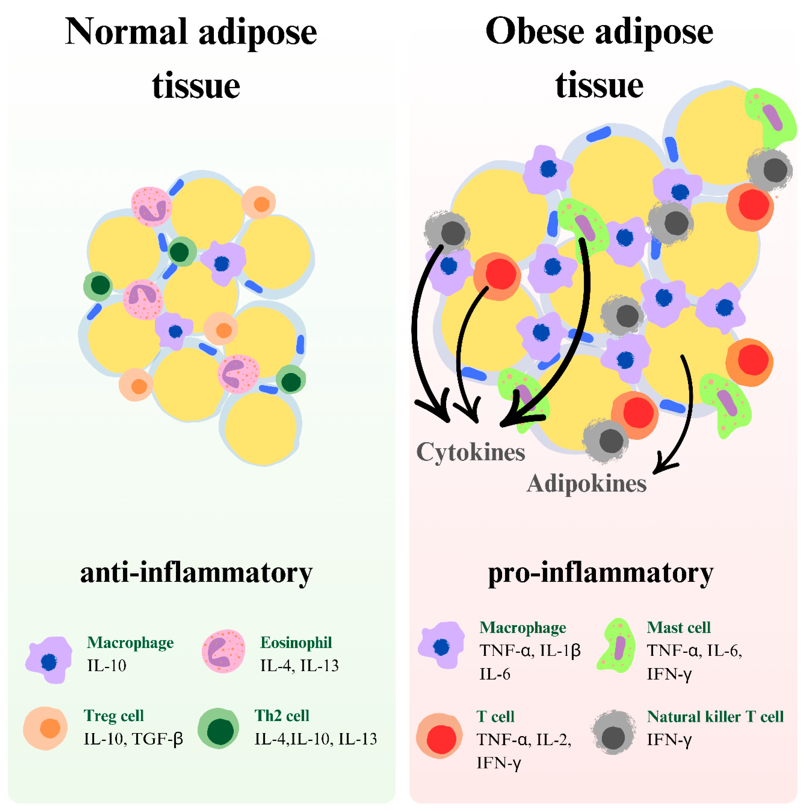
3. Obesity: Causes and Link to Depression

According to the WHO definition, obesity is “abnormal or excessive fat accumulation that may impair health” [9]. It is diagnosed when the body mass index (BMI) is 30 or higher, although this measure may not accurately reflect the condition in some individuals, such as athletes [10]. Globally, it is estimated that 16% of the adult population is obese, a rate that has doubled since 1990. Among teenagers, the obesity incidence has increased four-fold [9].
The development of obesity involves the interaction of genetic factors and environmental influences. While monogenic defects and polygenic variants associated with obesity have been identified, they account for only about 3% of all obesity cases [11]. It is suggested that epigenetic mechanisms, in which environmental factors influence the expression of specific genes, play a more significant role in the development of obesity. Several factors are associated with an increased incidence of obesity, including decreased physical activity (leading to lower energy expenditure), a positive energy balance (due to high-calorie, highly processed foods that are high in sugar and low in fiber), chronic stress (e.g., hormonal imbalance anad chronic inflammation), and social and living conditions (like residing in rural areas) [11,12]. Other contributing factors may include mood disorders, endocrine disorders, hypothalamic damage, and certain medications [12,13] (Figure 1).
In addition to the various complications associated with obesity, such as diabetes, heart disease, kidney disease, hypertension, and neurological disorders, obesity is also linked to an increased risk of depression [14].

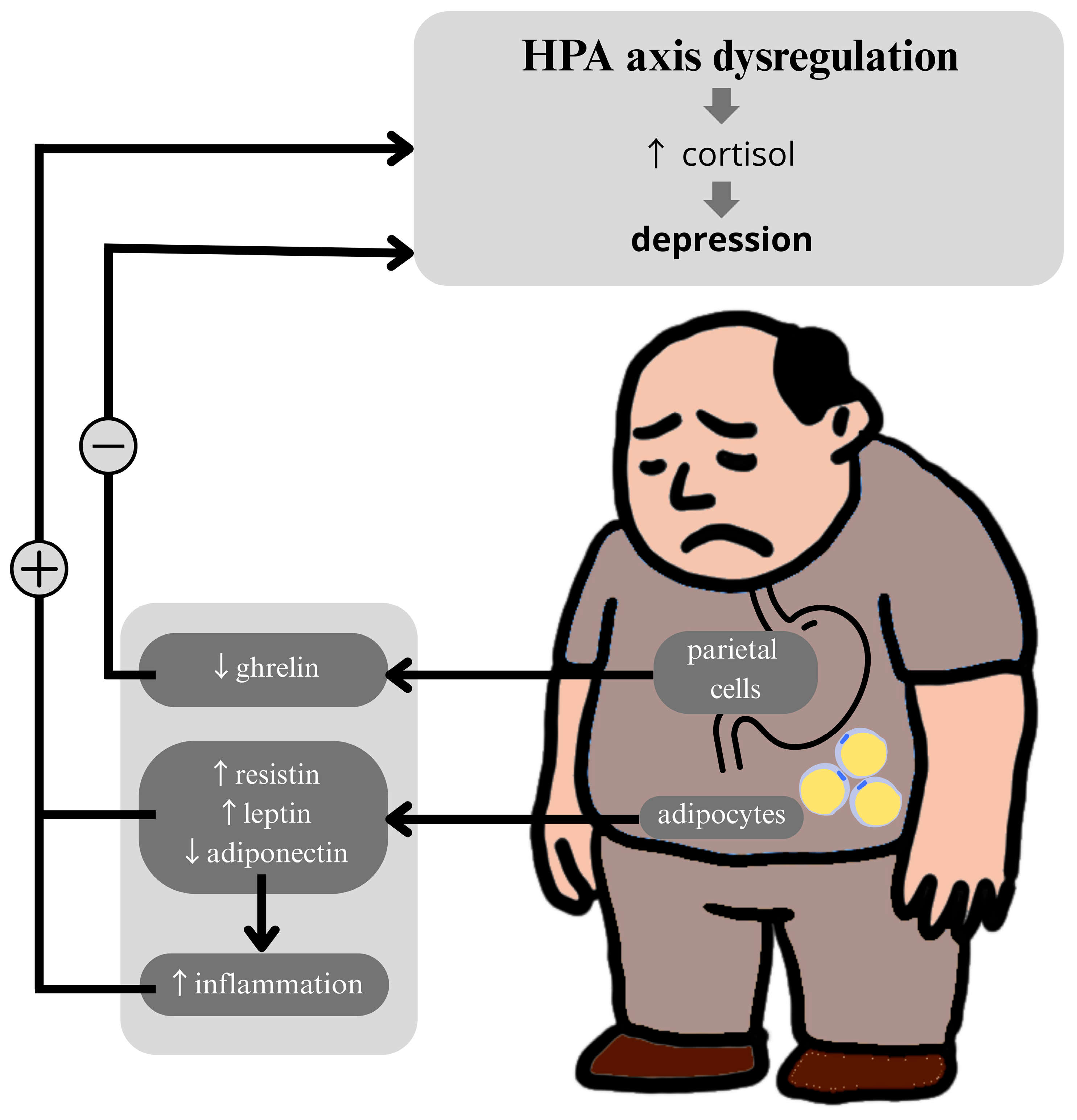
4. Metabolic Biomarkers and the HPA Axis

Available evidence suggests that some of the aforementioned biomarkers (leptin [15,16], adiponectin [17], resistin [18], and ghrelin [19,20]) may modulate the HPA axis, which is known to be hyperactive in patients with severe depression [21,22]. Hyperactivity of the HPA axis in depression manifests as excessive secretion of corticoliberin (CRH), elevated cortisol levels, an increased cortisol response to adrenocorticotropic hormone (ACTH), impaired negative feedback mechanisms mediated by dexamethasone (DEX), and hypertrophy of the pituitary and adrenal glands [21]. This is particularly important because the HPA axis regulates the body’s response to stress, and its hyperactivity may lead to a range of depressive symptoms [16,23].
The literature emphasizes the key role of glucocorticoid secretion as a consequence of HPA axis activation, which is closely linked to the processes leading to the development of depression, as shown in studies in animal models [24], including mice subjected to chronic, unpredictable mild stress during pregnancy [25]. From the perspective of depression, the ability of these biomarkers to regulate the HPA axis is crucial, as it may help alleviate depressive symptoms associated with its hyperactivity [26]. Adiponectin, according to the hypothesis, inhibits the production of tumor necrosis factor-alpha (TNF-α), a proinflammatory cytokine that activates the HPA axis [27]. HPA axis hyperactivity has been identified as a potential contributing factor in the pathogenesis of depression [28], which may result from dysfunction of the kynurenine pathway responsible for converting tryptophan into serotonin and melatonin, compounds that regulate mood [29].
Disruption of HPA axis function due to low adiponectin levels manifests in two ways: first, as a lack of sensitivity to DEX suppression, and second, as an excessive CRH response following DEX administration [17]. The literature indicates that the administration of exogenous adiponectin produces antidepressant effects [17], suggesting that it may serve as a potential therapeutic target in the treatment of depression.
The relationship between resistin levels and HPA axis activity was investigated in a study by Weber-Hamann et al., which observed a positive correlation between serum resistin levels and free cortisol concentrations in the saliva of depressed patients [18]. This suggests that resistin may be regulated by cortisol or may influence its levels, which is supported by in vivo studies in humans [30].
The literature also indicates limited knowledge regarding the direct impact of fetuin-A (FetA) and chemerin on the HPA axis. However, their roles in neuroprotection and the modulation of inflammatory processes [31], as well as their correlation with depressive disorders [32,33], may suggest a potential indirect influence on the HPA axis. Further research is necessary to better understand these relationships.
Ghrelin also exerts a stimulatory effect on the HPA axis [20], which is mediated by the central nervous system (CNS) [19]. In the work of Van Loenen et al., it was emphasized that in stress-induced obesity, ghrelin may act in two ways: directly by stimulating ACTH in the HPA axis and indirectly by influencing hypothalamic neurons. This underscores the multifaceted impact of ghrelin on the HPA axis [20] (Figure 2).

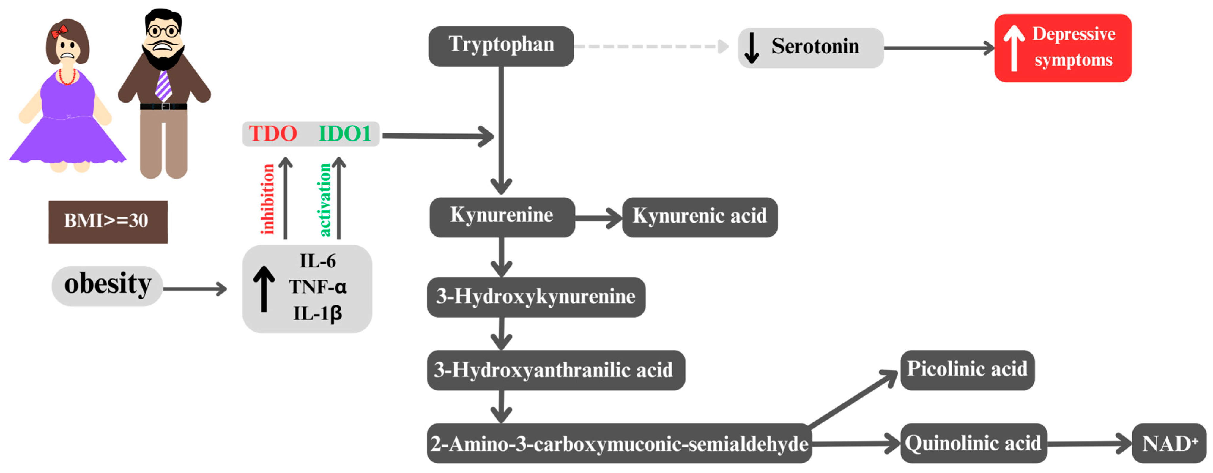
5. From Obesity to Depression: The Role of the Kynurenine Pathway

Proinflammatory cytokines, whose levels are elevated in obese individuals, affect the dysfunction of the kynurenine pathway, which plays a key role in the metabolism of tryptophan (TRP). This explains why adults with obesity are more susceptible to developing depression than individuals with a normal body mass index [34,35,36]. It has been demonstrated that elevated levels of proinflammatory cytokines lead to the activation of indoleamine 2,3-dioxygenase 1 (IDO1) [37,38] and the suppression of tryptophan 2,3-dioxygenase (TDO), resulting in a shift in tryptophan metabolism [39]. Consequently, the metabolism of serotonin and melatonin is altered, leading to increased production of kynurenine (KYN) [40,41,42]. This phenomenon is consistent with the monoamine hypothesis, which postulates that depression results from a deficiency of monoamine neurotransmitters, including serotonin and noradrenaline [43]. As a result, the availability of tryptophan for serotonin synthesis is reduced, which may lead to decreased serotonin levels in the brain and, consequently, be associated with depressive symptoms [40]. Moreover, there is a strong correlation between this process and suicidal ideation and behavior, even when accounting for depression severity [44]. Such changes are particularly evident in the context of obesity, where an increased kynurenine/tryptophan ratio is observed [45], potentially contributing to an inflammatory reaction related to interferon-gamma (IFN-γ) activation [46]. Indeed, the elevated activity of proinflammatory IDO1 is closely linked with the modulation of immune and metabolic responses, as shown in studies conducted in a mouse model of induced lipopolysaccharide (LPS) and depression-like behavioral symptoms [47]. A reduction in the production of serotonin (5-HT), the main neurotransmitter, is significant for both metabolic processes and neuropsychiatric function [48]. The monoamine deficiency hypothesis suggests that a decrease in brain serotonin levels is a primary factor in the etiology of depressive disorders [49]. The ultimate consequence of the disruption of the kynurenine pathway is a reduction in the production of nicotinamide adenine dinucleotides (NAD+), which are essential for cellular energy metabolism [37]. It is noteworthy that the expression of genes encoding enzymes of the kynurenine pathway is significantly upregulated in obese individuals [44], indicating its potential adaptive or pathophysiological significance in this condition. Considering the multifaceted influence of the kynurenine pathway on immunological, metabolic [50], and neuropsychiatric processes [51], its dysfunction may serve as a potential biomarker for the development of depression in obese individuals. It is crucial to consider how inflammatory processes affect TRP metabolism, which may contribute to the onset of depressive disorders [52]. The aforementioned theories were confirmed in a study conducted by Delgado et al., which demonstrated that in more severe cases of depression, elevated levels of C-reactive protein (CRP) were associated with reduced TRP concentrations and its indole pathway metabolites, including indole-3-carboxaldehyde (IAld) [52].
It is worth noting at the outset that CRP may serve as an inflammatory marker useful in the personalized treatment of depression [53]. Large, randomized trials have shown that an increase in CRP levels above 1 mg/L is associated with a less favorable response to selective serotonin reuptake inhibitors (SSRIs) compared to catecholaminergic antidepressants [54]. Moreover, in patients who did not respond to multiple trials of conventional antidepressant treatments, elevated levels were observed not only for CRP but also for interleukin-6 (IL-6) and TNF-α [55] (Figure 3).

6. Metabolic Biomarkers of Depression and Obesity

Adipose tissue is a source of over 600 potentially secreted proteins that are involved in the regulation of insulin, appetite, fat distribution, energy expenditure, endothelial function, blood pressure, hemostasis, and inflammation [56].
It is well documented that excess adipose tissue, particularly visceral fat, is a source of adipokines (also known as adipocytokines) [57], which include leptin, adiponectin, chemerin, resistin, and FetA [58]. It is reasonable to assume that the presence of excess fat in the body may contribute to increased levels of inflammation, which in turn may elevate the risk of developing depression [59]. A key aspect of this process is the signaling pathway between adipocytes and the brain, mediated by adipokines [60], which significantly influences the pathophysiology of depression. This link has been demonstrated both in studies in elderly humans with late-life depression (LLD) [61] and in an animal model of chronic social-defeat stress in mice [17].

6.1. Leptin

Leptin, a hormone secreted by adipose tissue, is increasingly recognized as a potential biomarker for assessing depression risk, particularly in the context of obesity [57,62]. Although the primary function of leptin is the regulation of energy metabolism and appetite, it also exhibits proinflammatory effects that may contribute to increased inflammation [63]. The scientific literature reveals discrepancies regarding the relationship between leptin levels and depression [64,65,66,67]. This dual role has prompted investigations into its relevance to mental health, especially depressive disorders.
The current literature, however, presents inconsistent findings regarding the association between leptin levels and depressive symptoms. Some studies report a significant relationship between elevated leptin levels and depression [64,65], whereas others do not [66,67]. For example, one study reported no statistically significant differences in leptin concentrations between patients with depression and healthy controls [66], in contrast to other studies that found elevated leptin levels in depressed individuals, possibly reflecting symptom severity [64]. This suggests that leptin may not only be relevant in the context of obesity but may also serve as a marker for other inflammation-related processes implicated in the pathophysiology of depression [68]. Building on this framework, a number of authors have proposed that leptin resistance may provide a mechanistic link between obesity, metabolic dysfunction, and depressive symptomatology. This hypothesis is particularly relevant in the context of atypical depression, a subtype characterized by increased appetite, weight gain, and fatigue, all of which reflect disturbances in energy regulation [15,62,69].
In the study by Milaneschi et al., and in the study by Zhang et al. [70], patients with acute episodic depression exhibited a positive correlation between elevated leptin levels and symptoms characteristic of atypical depression, including increased appetite, weight gain, and muscle weakness [69]. These symptoms align with leptin’s known metabolic functions, further suggesting a pathophysiological overlap [71]. It is important to note, however, that the study by Milaneschi et al. was cross-sectional [62], thus precluding conclusions about causality. Longitudinal studies are warranted to determine whether changes in leptin levels precede, coincide with, or follow depressive symptom fluctuations over time. Notably, Zhang et al. failed to support the hypothesis that systemic inflammation mediates the relationship between leptin levels and depression [70]. The correlation between leptin levels and brain cortical areas was found to be independent of CRP levels, a common indicator of inflammation [72]. These results suggest that leptin may influence brain function through mechanisms independent of inflammatory processes [70] or through alternative inflammatory pathways that are independent of CRP, as described in the study by Burrows et al. [73].
Additional evidence indicates that elevated leptin levels, often associated with increased adiposity, may interfere with the brain’s reward-processing circuitry [73,74]. This disruption has been implicated in the development of anhedonia—defined as a diminished ability to experience pleasure—a core symptom of major depressive disorder (MDD) [75,76].
In summary, meta-analytic data underscore the heterogeneity of leptin findings in depression. A meta-analysis by Carvalho et al. revealed that these differences were not statistically significant in the general population [59]. However, a higher BMI was positively correlated with elevated leptin levels in depressed patients [59]. Patients with mild to moderate depression exhibited significantly higher leptin levels compared to controls [59], suggesting a possible compensatory role of leptin in early stages of depressive illness [77]. It has been proposed that such compensatory activity may initially counterbalance emerging metabolic dysregulation, but may become impaired over time, particularly with the progression of obesity and the onset of leptin resistance, further exacerbating both metabolic and affective disturbances, as shown in a study in mice [77].
To reconcile the discrepancies between studies, we compared original research that specifically examined the relationship between leptin levels and clinical symptoms of depression (Table 1). The heterogeneity of the findings may be due to several methodological differences. First, positive associations were more frequently observed in studies focusing on atypical depression, whereas studies reporting no significant results often did not distinguish between depressive subtypes. This suggests that the pathophysiological relevance of leptin may be subtype-specific. Second, gender and BMI appear to be critical moderators; for example, Esel et al. reported significant leptin differences only in women, and studies with samples with a higher mean BMI were more likely to detect correlations. Therefore, future studies should stratify analyses by depressive subtype, sex, and metabolic profile to clarify the role of leptin in the pathogenesis of depression.

6.2. Adiponectin

Adiponectin is the most abundant adipokine, secreted exclusively by mature adipocytes and primarily produced in white adipose tissue [78]. It modulates immune responses and proinflammatory cytokines, making it a potential biomarker of inflammation in depression and a differentiator between depressed and healthy individuals [57,79]. The relationship between adiponectin and depressive disorders is thought to stem from the presence of adiponectin receptors in specific brain regions involved in mood regulation, including the hippocampus [70].
Accumulating evidence indicates that individuals with MDD, including those with recurrent depressive episodes, often exhibit lower adiponectin levels [57,80]. This finding may serve as a starting point for further research aimed at improving the precision of monitoring and predicting the course of these conditions. Islam et al. suggested that adiponectin could be a valuable prognostic tool for the early assessment of depression risk [80]. However, an important limitation of these studies is the lack of consideration of potential dietary influences on the parameter being analyzed, which is significant given that adiponectin is predominantly produced by adipose tissue [81]. Future research should account for participants’ dietary habits and explore stratification by BMI and eating behaviors to refine these associations.
Although the meta-analysis by Carvalho et al. primarily addressed leptin, it also examined adiponectin and found no significant difference in adiponectin levels between depressed and non-depressed individuals across BMI categories [59]. This lack of association may reflect limitations inherent in BMI, which is a crude measure of adiposity and does not capture fat distribution [82]. More precise anthropometric assessments, such as waist circumference or body fat percentage, are recommended for future studies to clarify the adiponectin–obesity–depression axis. A recent meta-analysis by Vuong et al. supports the hypothesis that lower adiponectin levels in obese individuals may contribute to the development of inflammation and insulin resistance [83], which are recognized risk factors for depression [84]. However, this analysis did not reveal statistically significant differences in adiponectin levels between depressed and non-depressed individuals in the general population [83].
Some studies report that higher adiponectin levels are associated with lower severity of depressive symptoms [85,86], likely due to its anti-inflammatory actions [87,88]. However, findings regarding absolute adiponectin levels in depression remain inconsistent. While certain reports described reduced levels in depressed patients [80], others demonstrated elevated levels [89], or no significant differences compared to healthy controls [90]. These discrepancies may be attributed to differences in depression subtypes and severity. For example, Jeong et al. reported elevated adiponectin levels exclusively in individuals with subclinical depression, whereas no significant differences were observed in those with moderate or severe MDD compared to healthy controls [89]. In contrast, the studies by Hu et al. and Islam et al. included diagnostically heterogeneous cohorts encompassing various depressive phenotypes, which may have further contributed to the inconsistency of findings [80,90].
A study by Permody-Pachuta et al. presented intriguing data suggesting that adiponectin may be a promising biomarker for assessing the efficacy of treatment in therapy-resistant depression (TRD) [91]. The authors observed an increase in adiponectin levels in TRD patients after electroconvulsive therapy (ECT), particularly in those who showed clinical improvement [91]. This indicates a potential correlation between adiponectin and clinical recovery. In contrast, Benedetti et al. observed a positive correlation between adiponectin levels and the duration of the depressive episode, suggesting that higher hormone levels before treatment may be indicative of a poorer response to conventional pharmacotherapy [92]. Thus, adiponectin could serve as a prognostic biomarker in predicting the clinical course of depression and treatment response.
An intriguing correlation between adiponectin and chemerin was observed in a study by Malujlo-Balcerska et al., which showed decreased levels of both adipokines in patients with recurrent depressive episodes [57]. Chemerin, like adiponectin, may be an underrecognized biomarker of inflammation in depression. However, evidence for this hypothesis is limited to a few studies, including an experiment conducted on male Sprague-Dawley rats using the chronic restraint stress model [32].

6.3. Resistin

Resistin, another key adipokine associated with obesity [93], may play a particular role in exacerbating inflammatory states [94], suggesting a potential link between excess adipose tissue and the etiology of depression [95]. Elevated resistin levels, which correlate with higher BMI, waist circumference, and visceral fat mass [91], contribute to increased levels of proinflammatory cytokines, such as TNF-α and IL-6 [85,96].
Although the role of resistin as a mediator of inflammation in obesity is well documented [97], further research is needed to elucidate its potential direct links with depression. Available literature indicates that resistin may decrease the release of dopamine and noradrenaline in the hypothalamus, leading to reduced levels of these monoamines in synapses, a study conducted on male Wistar rats showed [98]. These effects of resistin suggest that it might increase the incidence of depressive symptoms by affecting the neurotransmission of key mood-regulating transmitters [99]. In this context, other studies have demonstrated a positive correlation between blood resistin levels and the occurrence of atypical and melancholic subtypes of major depression [18,100], which may indicate a significant role for resistin in the pathogenesis of depression. However, study results remain inconclusive. Some research reports elevated resistin levels in obese individuals [101,102], while others suggest lower levels [103]. A meta-analysis even suggested lower resistin levels in depressed patients relative to healthy controls [60]. Notably, Lehto et al. found that resistin was correlated with atypical depressive symptoms but not with typical presentations [99], and Machado-Vieira et al. reported that higher resistin concentrations were positively associated with symptom severity and decreased significantly following antidepressant treatment [85].
Further evidence highlights the potential utility of resistin as a biomarker of immune activation in mood disorders. Malujlo-Balcerska et al. proposed that elevated resistin may reflect heightened inflammatory processes and immune dysregulation in depression [68], a notion corroborated by Rahman et al., who also identified elevated serum resistin as a possible contributor to MDD pathogenesis [104]. In the context of treatment-resistant depression, a study by Permody-Pachuta et al. found that higher pre-treatment resistin levels were predictive of better clinical outcomes following ECT [91]. Of note, this study excluded overweight participants to control for the confounding effects of body weight on adipokine levels [58].
Thus far, studies on resistin have mainly focused on the correlation between overweight/obesity and its serum levels [85,97]. Parallel research continues into the potential of resistin as a marker for depression [18,99,100]. However, comprehensive studies integrating all three components—excess adipose tissue, resistin levels, and depressive symptoms—are lacking. Such analyses could provide valuable information regarding the role of resistin as a potential mediator of the mechanisms linking obesity and depression and could clarify whether a synergy between excess weight and resistin influences the risk of developing mood disorders.

6.4. Fetuin-A

Similar to the previously discussed resistin and leptin, elevated serum levels of FetA have been observed in patients with recurrent depressive disorder compared to controls [57]. FetA, an acute-phase glycoprotein produced by the liver and adipose tissue [104], exerts regulatory effects on inflammatory processes [31], including the inhibition of proinflammatory mediators, such as TNF-α [105]. A reduction in FetA levels may be associated with an exacerbation of inflammatory processes, as shown in studies conducted on fetuin-A-deficient mice [106].
In contrast to these findings, a study by Fanelli et al. demonstrated lower FetA lev-els in depressed patients compared to healthy controls [33]. A strength of that study was its comprehensive adjustment for potential confounders, such as age, sex, and BMI, which allowed for a more precise determination of the relationship between FetA and depressive symptoms.
Although numerous epidemiological studies indicate elevated FetA levels in obese individuals [107] and a positive correlation between FetA concentration and BMI [108], visceral adipose tissue (VAT) [109], and leptin levels [110], the number of studies analyzing the relationship between FetA and depression remains limited. As with resistin, there is a lack of comprehensive studies integrating all aspects—excess adipose tissue, FetA levels, and the severity of depressive symptoms. This gap in data represents a significant research lacuna in understanding the metabolic–inflammatory links in depression.

6.5. Ghrelin

Ghrelin, although not classified as an adipokine due to its primary synthesis in the gastric fundus rather than in adipocytes [111], plays a notable role in energy balance and fat accumulation, thereby indirectly linking it to obesity [112]. It promotes adiposity by reducing lipid oxidation and enhancing fat storage, ultimately contributing to weight gain [113,114].
Some studies suggest that ghrelin levels are higher in depressed individuals compared to controls [115,116], while others do not confirm such differences [117,118]. Interestingly, research by Sempach et al. examined the effect of probiotic supplementation on depressive symptoms and found that increased ghrelin levels following probiotic intake were associated with symptom improvement [119]. These findings suggest a potential mood-regulating role of ghrelin, possibly mediated by its anti-inflammatory effects. Indeed, ghrelin has been shown to suppress proinflammatory cytokines, such as IL-6 and interleukin-1 beta (IL-1β), in both in vivo models (e.g., male Sprague-Dawley rats) [120] and in vitro human T-cell cultures [121]. In contrast, ghrelin concentrations tend to be lower in obese individuals compared to those with normal BMI, a phenomenon interpreted as a compensatory adaptation to prolonged positive energy balance [122,123]. This paradoxical inverse relationship between ghrelin and body weight further complicates its role in the obesity–depression axis. Moreover, increased ghrelin levels have been observed in subgroups of depressed patients, particularly those following suicide attempts [124] and postmenopausal women with severe depression [125], suggesting that specific clinical contexts may modulate ghrelin secretion.
Animal studies have shown that ghrelin administration affects specific proinflammatory cytokines often associated with depression [126,127], leading to lower levels of these cytokines—a finding confirmed in both a clinical trial involving patients with MDD [128], as well as in a study on male Sprague-Dawley rats, in which depression was induced by a high-fat diet (HFD) and disturbed diurnal rhythm [129]. It has also been suggested that a reduction in ghrelin levels following antidepressant treatment may contribute to improved therapeutic outcomes during depressive episodes [130].
However, further studies are necessary to comprehensively analyze the relationship between obesity, depression, and ghrelin levels, as well as changes in ghrelin across different health states and its impact on inflammatory processes. To date, research on this complex interaction has been somewhat fragmented. In order to gain a fuller understanding of ghrelin’s role in modulating both inflammatory processes and depressive symptoms in the context of obesity, integrated clinical studies considering all these aspects are needed.

6.6. Fibroblast Growth Factor 1

Although fibroblast growth factor 1 (FGF1) has not been directly linked to the initiation of metabolic and inflammatory cascades, its potential role in the development of depressive disorders deserves attention. Peripheral administration of exogenous FGF1 exerts strong anti-diabetic effects, mediated by the FGF1 receptor (FGFR1) in adipose tissue—specifically, by lowering blood glucose levels through the inhibition of adipose tissue lipolysis [131]. It has been shown that the adipokine FGF1 enhances metabolic homeostasis in mice fed a high-fat diet, and FGF1 itself exhibits appetite-reducing effects [132].
In a study conducted by Aurbach et al., gradual administration of exogenous fibroblast growth factor 9 (FGF9) led to an increase in anxiety- and depression-like behaviors and simultaneously decreased FGFR1 expression in the dentate gyrus [133]. This may suggest that FGFR1 levels reflect both the effects of administered growth factors and the severity of depressive symptoms in animals. These findings highlight the potential of FGFR1 as a therapeutic target in the treatment of depression.
It is also worth noting that mood-stabilizing drugs, such as valproate and lithium, may regulate FGF1 gene expression [134] (Table 2).

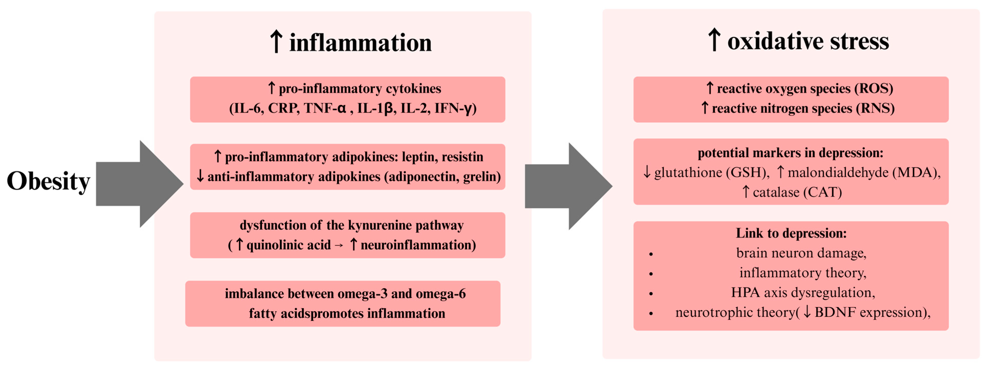
7. Inflammatory Model of Depression

Available evidence supports the hypothesis that inflammation may serve as a key mechanism linking obesity with mood disorders [6]. Research on the immunological response in obesity focuses on the function of Th1 and Th2 cells, given their role in regulating inflammatory processes [135]. Th1 cells are responsible for activating macrophages, which in turn promote the cellular immune response [136]. This process is integral to the host defense mechanisms mediated by phagocytes [137]. In contrast, Th2 cells play a key role in the humoral response and are responsible for host defense mechanisms independent of phagocytes [136]. In individuals with obesity, an increase in proinflammatory cytokine levels suggests the involvement of the Th1 response [138].
Interestingly, Th1 cytokines, including IFN-γ, are elevated in the adipose tissue of obese individuals [139]. In addition, IFN-γ has been shown to facilitate the infiltration of macrophages into adipose tissue, thereby contributing to the development of a local inflammatory response, as confirmed by in vitro studies on cells of human origin [140].
The role of Th2 is to secrete cytokines, such as interleukin-4 (IL-4), interleukin-5 (IL-5), interleukin-10 (IL-10), and interleukin-13 (IL-13), which exert anti-inflammatory effects [141]. Some studies indicate that the Th2 response may have protective effects in obesity, as demonstrated in a diet-induced obesity model in mice [142]. In the early stages of obesity, a simultaneous activation of both Th1 (proinflammatory) and Th2 (anti-inflammatory) responses is observed. The presence of mixed Th1 and Th2 cytokines in the serum of individuals with metabolic syndrome [143,144] indicates the involvement of both the innate and adaptive immune systems [145]. However, as the disease progresses, the levels of both types of cytokines decline, which may indicate a weakening of immune function in advanced obesity [142]. This phenomenon supports the concept that obesity should be regarded as a state of chronic, low-grade inflammation [146]. Such an inflammatory state is characterized by activation of the innate immune system, accumulation of proinflammatory macrophages in adipocytes [147], and elevated levels of acute-phase proteins and cytokines, such as CRP, TNF-α, IL-1β, and IL-6 [135], which are indicators of a Th1 response [148]. The inflammatory and cytokine model of depression suggests that an imbalance in the immune system—such as increased levels of proinflammatory cytokines or decreased production of anti-inflammatory cytokines—may be a key factor in the pathogenesis of clinical depression [149,150,151].
A substantial body of evidence supports the correlation between pathological behaviors—such as anhedonia, social withdrawal, and reduced activity—and elevated levels of proinflammatory cytokines, particularly TNF-α and IL-6 [149,152,153]. These findings were confirmed in a 2009 meta-analysis by Dowlati et al., which reported significantly elevated TNF-α and IL-6 levels in individuals with depressive disorders compared to healthy controls [153]. Interestingly, no significant differences were found for IL-1β or IFN-γ in the same analysis [153], nor for anti-inflammatory cytokines, such as IL-4 and IL-10.
A more recent study by Amerikanou et al. investigated the relationship between inflammatory markers (IL-6 and TNF-α) and mental well-being in obese individuals. Notably, the correlation between TNF-α and depression severity (measured via the Center for Epidemiologic Studies Depression Scale) remained statistically significant only in males [6], which contrasts with previous findings suggesting greater susceptibility of females to inflammation-related psychiatric conditions [154]. This discrepancy may stem from the relatively small sample size in the study, potentially limiting generalizability. Man et al. observed similar cytokine patterns in patients with bone tumors and depression—IL-6 and IL-1β levels were elevated and correlated with Hamilton Depression Rating Scale (HAMD-17) scores, while anti-inflammatory cytokines, such as IL-10 and transforming growth factor beta 1 (TGF-β1), were decreased prior to treatment. After sertraline therapy, proinflammatory cytokines decreased and anti-inflammatory cytokines increased, suggesting a modulatory effect of antidepressants on immune status [155]. However, the lack of a comparison group receiving sertraline without depression remains a limitation.
A meta-analysis conducted by Lombardi et al. confirmed that IL-6 levels tend to be elevated in depressed patients compared to healthy individuals, underscoring the role of inflammation in the pathogenesis of depression [156]. Various therapeutic strategies, including pharmacotherapy, have also demonstrated reductions in IL-6, further supporting its involvement in symptom modulation [156,157,158]. Moreover, proinflammatory cytokines, such as IL-6 and TNF-α, can cross the blood–brain barrier [159], influencing neurotransmission (e.g., GABAergic signaling) and contributing to mood dysregulation [160].
A meta-analysis by Islam et al. revealed that TNF-α levels were elevated in depressed patients compared to healthy subjects [161]. In addition, no significant differences were found in the levels of CRP and IFN-γ between the groups [161]. A potential limitation of this meta-analysis is that it focused on cytokines in peripheral fluids rather than in the CNS, where key processes associated with depression occur. Including CNS cytokines could provide a more comprehensive picture of the issue. Another meta-analysis of 13 studies involving 1123 patients examined the relationship between CRP levels and depressive symptom severity [162]. The analysis revealed a significant correlation between elevated CRP levels and depressive symptom severity, with 10 out of 13 studies demonstrating a positive correlation [162]. This meta-analysis confirmed the correlation, indicating a moderate effect size.
In light of the above studies, it can be stated that among the discussed proinflammatory cytokines, IL-6, CRP, and TNF-α are the most promising candidates as potential biomarkers for depression. This is not only because they have been the subject of the most extensive research but also because their elevated levels have been demonstrated in many of the analyses described above. Moreover, their significance is particularly noteworthy in the context of obesity, where the levels of these cytokines are also elevated [135]. Therefore, IL-6, CRP, and TNF-α may represent promising targets in research on the pathomechanisms of depression and in the search for new diagnostic strategies (Figure 4).

8. Oxidative Stress

An increasing body of evidence indicates that oxidative stress (OS), defined as the overproduction and accumulation of free radicals, plays a key role in the pathogenesis of depression [163]. Analysis of keywords in scientific publications shows that “oxidative stress” has become one of the most important terms in recent years, suggesting growing interest in this issue in the context of depression [164]. At the same time, obesity—a state of chronic, low-grade inflammation [146]—is significantly correlated with elevated levels of oxidative stress markers [165].
Oxidative stress is defined as a pathological condition in which the production of free radicals, such as reactive oxygen species (ROS) and reactive nitrogen species (RNS), exceeds the organism’s capacity to neutralize them [166,167]. The literature indicates that the brain is particularly susceptible to oxidative stress due to its high oxidative metabolism, the presence of polyunsaturated fatty acids in cell membranes, and the limited activity of enzymatic systems responsible for neutralizing ROS [168,169,170,171]. Oxidative stress has been linked with other theories of depression, including HPA axis dysregulation [172,173], the inflammatory theory (where excessive synthesis of proinflammatory cytokines is associated with reduced antioxidant activity, and neuroinflammation induces excessive ROS production) [174,175,176], and the neurotrophic theory (where overproduction of ROS inhibits the expression of BDNF) [175]. The observed correlation between obesity, oxidative stress, and depression suggests a complex mechanism in which excess adipose tissue leads to increased oxidative stress, which in turn may contribute to a higher risk of depression [177]. In line with this, a study by Sanaeifara et al. demonstrated that oxidative stress plays a central role in the link between type 2 diabetes (T2D) and depression, further underscoring the involvement of obesity—a major risk factor for T2D—in this process [177,178]. Scientific literature emphasizes the particular significance of enzymes, such as superoxide dismutase (SOD) and glutathione (GSH), in assessing oxidative stress in patients with depression, serving as markers of oxidative stress in depressive disorders [174,179,180]. A study by Eshkevari et al. found that individuals diagnosed with MDD had lower glutathione levels compared to healthy subjects, suggesting that GSH could serve as a potential biomarker for depression and that its supplementation may provide therapeutic benefits [181]. A study by Duffy et al., investigating the effects of omega-3 fatty acid supplementation on GSH levels in brain tissues of elderly individuals at risk for depression, produced interesting results [182]. These fatty acids possess anti-inflammatory and antioxidant properties [183]. However, supplementation did not lead to the expected increase in GSH levels, and the observed increase in the placebo group was unexpectedly correlated with a worsening of depressive symptoms, as measured by the Patient Health Questionnaire-9 (PHQ-9) [183]. Currently, there are few comprehensive studies that simultaneously analyze BMI as an indicator of obesity, GSH levels, and their relationship with depression. However, intriguing reports in the literature indicate a significant inverse correlation between serum GSH levels and BMI [184]. In a separate study by Foulds et al., examining the relationship between SOD levels and depression, it was observed that serum SOD levels might be decreased, while SOD levels in erythrocytes might be increased [185]. In contrast, Stefanescu et al. reported reduced serum SOD activity [186], whereas Camkurt et al. found decreased SOD activity in erythrocytes [187]. The scientific literature on the relationship between SOD activity and depression is inconclusive—some studies indicated increased SOD activity in depressed patients [188,189,190], while others observed a decrease [186,191] or no differences compared to healthy individuals [192,193]. Nevertheless, the latest meta-analysis did not demonstrate a clear trend in SOD activity in patients with MDD [194]. The potential of malondialdehyde (MDA), a product of lipid peroxidation, as a biomarker of oxidative stress in depression is not widely recognized in the literature [195]. A study by Islam et al. found that serum MDA levels were significantly elevated in individuals diagnosed with MDD compared to controls [196]. These results have been echoed by other studies on lipid peroxidation in MDD and its modulation by antidepressant therapy, which have shown that MDA levels tend to decrease following effective treatment [197,198,199]. The significance of MDA as a potential biomarker of depression is further reinforced by its correlation with body mass, as studies have shown that MDA levels are significantly higher in obese individuals compared to those with normal BMI [200]. These observations suggest a potential link between obesity and increased oxidative stress, emphasizing the need for further studies on the relationship between MDA and obesity [201]. Despite the abundance of studies highlighting the importance of oxidative stress biomarkers—including catalase (CAT), glutathione reductase (GR), and glutathione S-transferase (GST)—in the etiology of bipolar disorders [202], only a few articles address potential associations between CAT and depression [194]. Moreover, there is a paucity of literature examining the correlation between depression and GR or GST. Some studies reported increased CAT activity in depressed patients, a finding supported by meta-analyses [189,190,194,203]. With regard to GR and GST, there is a lack of data. In contrast, findings concerning glutathione peroxidase (GPX) are more varied—some studies indicate decreased GPX in depressed patients [186,204], others report increased levels [184], while additional studies have found no statistically significant differences [189,192] (Figure 5).
When discussing the issue of oxidative stress, it is essential to highlight the role o nuclear factor erythroid 2-related factor 2 (NRF2). In response to oxidative stress, the Keap1–Cul3 complex becomes inactivated, leading to the stabilization and activation of NRF2. The activated NRF2 translocates to the cell nucleus, where it binds to the antioxidant response element (ARE), initiating the expression of genes encoding antioxidant and detoxifying enzymes [205]. Moreover, NRF2 contributes to the suppression of inflammatory responses by inhibiting NF-κB pathway activation and limiting the production of proinflammatory cytokines [206]. Given its pivotal role in inflammation, NRF2 may also influence the development of depressive disorders through its anti-inflammatory mechanisms [207]. This is supported by rodent studies showing that mice exposed to chronic stress exhibit depression-like symptoms [208] along with reduced levels of Keap1 and NRF2 proteins in the CA3 region of the hippocampus, dentate gyrus, and prefrontal cortex compared to healthy controls [209].
The existing literature on oxidative stress parameters, obesity, and depression is inconclusive and often contradictory. There is a clear gap in the literature providing consistent data that definitively elucidates the mechanisms linking these factors, highlighting the need for further research. A better understanding of the role of oxidative stress in these interactions could contribute to a more nuanced comprehension of depression’s pathogenesis and the development of novel diagnostic and therapeutic strategies.

9. Fatty Acids and Cholesterol

A significant correlation has been identified between obesity and depression, with lipid biomarkers—such as polyunsaturated fatty acids (PUFAs) and cholesterol fractions (HDL—high-density lipoprotein, LDL—low-density lipoprotein, and VLDL—very low-density lipoprotein)—emerging as key factors in the genesis of this phenomenon [210]. These biomarkers not only serve structural functions in the body but also play a role in regulating inflammatory processes [211,212]. PUFAs, particularly omega-3 and omega-6 fatty acids, act as precursors to eicosanoids, compounds with potent anti-inflammatory or proinflammatory actions [213]. An imbalance in the omega-3 to omega-6 ratio—a phenomenon frequently observed in individuals with obesity—may exacerbate inflammatory processes, thereby affecting brain function and increasing the risk of depression [214]. The anti-inflammatory action of omega-3 fatty acids largely depends on their incorporation into cell membranes, where they compete with arachidonic acid (AA) for binding sites [215]. AA is a precursor to many proinflammatory mediators, including prostaglandins and leukotrienes [216]. Replacing AA with omega-3 fatty acids, such as eicosapentaenoic acid (EPA) and docosahexaenoic acid (DHA), has been shown to reduce the production of proinflammatory eicosanoids [215] and increase the synthesis of anti-inflammatory compounds, including resolvins and protectins [217]. Multiple studies have shown that individuals with depression exhibit reduced levels of EPA and DHA in erythrocyte membranes, which may play a key role in the pathogenesis and severity of depressive symptoms [218,219]. An observational study found a correlation between national omega-3 fatty acid intake and depression prevalence, indicating that populations with higher consumption levels tend to report lower rates of depressive disorders [220]. Additionally, supplementation with omega-3 fatty acids may be particularly effective in patients with severe depressive episodes, with EPA appearing more effective than DHA in therapeutic outcomes [221]. However, some studies have yielded inconclusive results. In a study by Bidzan-Wiącek et al., although supplementation led to a marked increase in EPA and DHA blood levels—compounds with known anti-inflammatory and neuroprotective effects [222]—it did not affect depressive symptoms. This outcome may be explained by the absence of diagnosed depression among study participants [223].
In addition to examining the function of omega-3 fatty acids, a review article emphasized the importance of omega-6 fatty acids in the context of mental health [172]. A high n-6/n-3 ratio may trigger systemic inflammation, whereas a lower ratio—achieved through higher omega-3 intake—appears to offer protective effects. Therefore, maintaining a low n-6/n-3 dietary ratio is considered a key component in both prevention and treatment of depression [172]. Conversely, a study focusing on omega-6 fatty acid metabolism found no significant differences in omega-6 levels between individuals with a history of mood disorders and healthy controls, suggesting that omega-6 fatty acids may not be a major contributor to depression pathogenesis [224].
Cholesterol, in addition to its role in cell membrane structure [225] and steroid hormone synthesis [226], plays a key role in neuronal function, including myelin synthesis and the modulation of membrane receptors [227]. Alterations in cholesterol metabolism, including changes in the concentrations of its various fractions, are increasingly recognized as significant factors associated with the occurrence of depression [228,229]. Furthermore, elevated levels of LDL cholesterol and total cholesterol, along with reduced HDL cholesterol levels, are frequently observed in individuals with obesity [230]. Such metabolic abnormalities may be significant in the context of mental health, as cholesterol has a proven impact on the integrity of neuronal cell membranes, as demonstrated in in vitro studies conducted primarily on primary cultures of Sprague-Dawley strain rat hippocampus neurons and HEK-293 cell lines [231]. Some studies indicate that low serum cholesterol may be associated with suicidal ideation [212], while others have reported elevated cholesterol levels in individuals with major depression [232]. These inconsistent findings reflect the complexity and controversy in the current literature. For instance, while some reports associate low cholesterol with depression [233], others emphasize the limitations of such studies due to reliance on self-report questionnaires rather than clinical diagnosis.
A notable U-shaped relationship between LDL cholesterol levels and depression risk was observed in a U.S. population study: both very low and very high LDL levels were linked to a higher incidence of depression in men, while in women, low HDL levels—but not LDL—were associated with depression [234]. A study by Wagner et al. found significantly elevated LDL cholesterol levels in patients during a major depressive episode (MDE), with higher baseline levels predicting better treatment outcomes, suggesting LDL may serve as a biomarker for both depression severity and response to therapy [235]. A meta-analysis by Persons and Fiedorowicz supported the relationship between cholesterol and depression, showing lower serum LDL cholesterol levels in individuals with depression, which is consistent with Engelberg’s hypothesis that reduced neuronal membrane cholesterol disrupts serotonin receptor function, impairing neurotransmission [229,236]. Additionally, elevated non-HDL cholesterol has also been correlated with a higher depression risk [237].
In a more detailed analysis, Wysokiński et al. discussed issues related to cholesterol fractions, observing that patients with unipolar depression exhibited higher HDL cholesterol levels and lower LDL cholesterol levels compared to individuals with schizophrenia and bipolar affective disorder [228]. However, it should be noted that this study was limited by the absence of a healthy control group. Including such a control group could help clarify the relationship between cholesterol fractions and psychiatric disorders, thereby enabling the identification of potential biomarkers specific to unipolar depression. Research into the role of triglycerides as potential biomarkers in psychiatric disorders, including depression, is still in its early stages. One of the few studies addressing this issue, conducted by Laederach-Hofmann et al., observed that depressed patients may exhibit elevated triglyceride levels [173].

10. GABAergic System

Dysfunction of the GABAergic system is increasingly recognized as a potential factor linking obesity with psychiatric disorders, including depression, as shown in a study in mice [238]. An expanding body of research indicates that in individuals with obesity, GABA—the primary inhibitory neurotransmitter in the CNS [239]—can modulate inflammatory processes in adipose tissue, as confirmed in both mouse studies and adipose tissue samples from obese individuals [240], as well as in a mouse model of obesity induced by a HFD [241]. This modulation impacts both metabolic processes and nervous system function [242]. Moreover, GABA is implicated in the pathogenesis of obesity by influencing metabolic disturbances and excessive food intake [243]. An animal study using a HFD-induced obesity model showed that the liver in obese individuals produces and releases increased amounts of GABA into the bloodstream, leading to inhibition of vagus nerve activity and promoting the development of hyperinsulinemia and insulin resistance [243]. The anti-inflammatory potential of GABA in adipose tissue has been highlighted by studies demonstrating its ability to reduce macrophage infiltration and proinflammatory cytokine levels (including TNF-α and IL-6), thereby improving insulin sensitivity in diet-induced obese mice [240,244]. However, these effects were observed only in subcutaneous adipose tissue (IAT) and not in visceral adipose tissue (EAT), which, as shown in a study in mice, may indicate a selective sensitivity of IAT to GABA [240]. Nevertheless, other experimental studies, also conducted on mice with HFD-induced obesity, suggest a broader role for GABA in the pathophysiology of obesity. GABA has been shown to reduce visceral fat accumulation while promoting the growth of muscle mass, which may prevent the development of both abdominal and sarcopenic obesity [244]. Furthermore, additional research indicates that GABA exerts anti-obesity effects by inhibiting fat accumulation, increasing energy expenditure, and improving metabolic parameters, as demonstrated in a mouse model of HFD-induced obesity and in vitro studies in adipocyte line 3T3-L1 [245]. GABA plays a critical role in maintaining neurochemical balance, and its dysfunction is increasingly linked to psychiatric disorders, including depression [246]. The potential link between obesity and depression may lie in common inflammatory mechanisms [129,146]. Obesity is defined as a state of chronic low-grade inflammation that affects both adipose tissue [146] and the central nervous system [129]. Proinflammatory cytokines, such as IL-6 and TNF-α, can cross the blood–brain barrier [159], thereby affecting neurotransmission—including GABAergic pathways—which may contribute to the development of mood disorders [160]. A large body of literature supports the notion that depressed patients exhibit lower GABA levels in cerebrospinal fluid [247,248]. These findings were confirmed in a meta-analysis by Romeo et al. involving 26 studies, which demonstrated significantly lower GABA levels in both serum and cerebrospinal fluid in patients experiencing an active episode of major depression compared to controls [249]. A pivotal study by Godfrey et al., utilizing magnetic resonance spectroscopy (MRS), showed a decrease in GABA levels in brain regions associated with depressive disorders [250]. Notably, this study also found that GABA levels returned to normal once the depression subsided [250], suggesting a potential correlation between GABA dysfunction, the pathogenesis of the disorder, and the treatment process. These results are in line with earlier studies reporting similar changes in GABA concentrations in the brains of depressed patients [251,252]. A positron emission tomography (PET) study using the [¹¹C]-flumazenil tracer revealed reduced binding of the GABA-A receptor in patients with depressive disorders in specific brain regions, including the hippocampal gyrus and the right superior temporal cortex [253]. These receptor deficits may be directly related to depressive symptoms, particularly those associated with impaired emotional processing and cognitive function mediated by these brain regions [254]. Other studies have also identified a significant involvement of GABA-A receptors in the mechanisms of depression [255]; however, not all findings are consistent. For example, a similar study by Persson et al. did not demonstrate differences in GABA-A receptor binding between depressed patients and healthy controls [256]. These discrepancies may result from methodological differences or the heterogeneity of depression itself, underscoring the need for further research. Additional evidence for GABA system dysfunction in depression comes from postmortem studies showing reduced expression of glutamate decarboxylase 67 (GAD67), a key enzyme involved in GABA synthesis, in the prefrontal cortex of depressed individuals [257,258]. However, not all studies agree. Some analyses did not find differences in GAD67 expression in the prefrontal cortex between psychiatric patients, including those with depression, and controls [259,260]. These discrepancies may be due to differences in study design, including whether patients were receiving antidepressant treatment or were drug naïve. The hypothesis that GABA system dysfunction is a causative factor in depression has been supported by the efficacy of drugs acting as positive allosteric modulators of GABA-A receptors [261], which have shown beneficial effects in the treatment of depression [262]. Nevertheless, GABA dysfunction is not exclusively associated with depression [263]. Moreover, reduced levels of GABA and related biomarkers have been documented in other psychiatric disorders, including schizophrenia and bipolar affective disorder [264,265]. These findings suggest that GABA system impairment may be a more general factor in the development of psychiatric disorders rather than a specific marker for depression.

11. Limitations

Due to the review nature of the work, systematic inclusion and exclusion criteria were not applied, nor was a critical appraisal of the methodology used in the studies performed. The work includes both small- and large-group studies that are characterized by heterogeneous data. Discrepancies in the results of the cited studies impede a clear interpretation of the role of certain biomarkers in the pathogenesis of depression in individuals with obesity.

12. Summary

An increasing body of scientific evidence indicates that the chronic inflammatory state induced by excess adipose tissue plays a key role in the pathogenesis of psychiatric disorders. The inflammatory and cytokine model of depression suggests that an imbalance in the immune system—characterized by elevated proinflammatory cytokine levels or reduced production of anti-inflammatory cytokines—may be a critical factor in the development of clinical depression. Among the proinflammatory cytokines discussed, IL-6, CRP, and TNF-α appear to be the most promising candidates as potential biomarkers for depression, particularly in the context of obesity, where their levels are also elevated.
Furthermore, excess adipose tissue, especially visceral fat, is associated with increased production of adipokines, which may exacerbate inflammatory processes. Within this group, leptin, which exerts proinflammatory effects, shows inconsistent associations with depression, with some studies reporting elevated levels in depressed individuals and others finding no significant differences. Similarly, adiponectin, which has anti-inflammatory properties and is often lower in obese individuals, may reflect a weakening of protective immune mechanisms, while resistin, which enhances inflammatory processes and modulates neurotransmission, has been identified as a potential biomarker to differentiate depressed individuals from healthy subjects, though study results remain variable. FetA also demonstrates variable findings in relation to depression, indicating a need for further investigation. Interesting observations have also been made regarding ghrelin, an energy-regulating hormone that can influence inflammatory processes and depressive symptoms. Additionally, disturbances in the kynurenine pathway—where activation of IDO1 reduces the availability of tryptophan for serotonin synthesis—may contribute to depression. Another key aspect addressed is the impact of metabolic biomarkers on the HPA axis, whose hyperactivity is associated with depressive symptoms. An imbalance in the omega-3 to omega-6 ratio exacerbates inflammation, thereby increasing the risk of depression. Furthermore, increased oxidative stress—measured through glutathione levels and antioxidant enzyme activities—and GABAergic system dysfunction play crucial roles in the mechanisms linking obesity with mood disorders.
The literature presented suggests that excess adipose tissue may initiate a cascade of metabolic and inflammatory disturbances that indirectly elevate the risk of depression. Discrepancies among study findings may be attributed to differences in methodology, characteristics of the populations studied, or the measures used to assess obesity. Rather than relying solely on BMI, which does not fully capture the distribution of adipose tissue, assessing body fat percentage or waist circumference may be more appropriate. Biomarker analysis provides valuable insights into the underlying mechanisms of these interactions, allowing for prediction of the course of psychiatric disorders and the personalization of therapeutic strategies in patients with excess body weight. The conclusions open new therapeutic perspectives, suggesting that interventions aimed at reducing inflammation may be beneficial in the treatment of depression. A comprehensive approach that considers both metabolic and immunological aspects appears to be crucial for the effective diagnosis and treatment of these disorders.
This review provides a significant extension of the existing literature by not only synthesizing current findings on the inflammatory basis of depression in the context of obesity but also drawing attention to underexplored biomarkers, such as FetA and chemerin. Unlike the widely studied proinflammatory cytokines (IL-6, TNF-α, and CRP), these markers are rarely investigated in psychiatric research, despite their potential as indicators of both the metabolic and inflammatory background of depression. This identified gap highlights the need for studies integrating adipokine levels with mental health status, metabolic parameters, and neuroimaging data.
Furthermore, the article proposes an integrative pathophysiological model that connects immunological mechanisms, metabolic factors, hormonal regulation, neurotransmitter alterations, and oxidative stress into a unified pathway linking obesity and depression. This approach emphasizes the multifactorial nature of the interaction between these two conditions and supports the need for holistic diagnostics and treatment strategies that target not only depressive symptoms but also their metabolic and inflammatory underpinnings (Figure 6).

Author Contributions

Conceptualization, A.J.O. and M.Z.; methodology, M.Z. and A.J.O.; validation, N.W.; resources, N.W.; data curation, A.J.O.; writing—original draft preparation, A.J.O., M.Z., M.N. and M.S.; writing—review and editing, A.J.O., M.Z., M.N., M.S. and N.W.; visualization, M.N.; supervision, N.W.; project administration, A.J.O., M.Z. and M.N.; funding acquisition, N.W. All authors have read and agreed to the published version of the manuscript.

Funding

This research received funding from the Medical University of Bialystok, grant number B.SUB.25.437. The financial sponsor was not involved in the design, implementation, analysis, or interpretation of results.

Conflicts of Interest

The authors declare no conflicts of interest.

References

  1. Ng, M.; Gakidou, E.; Lo, J.; Abate, Y.H.; Abbafati, C.; Abbas, N.; Abbasian, M.; Abd ElHafeez, S.; Abdel-Rahman, W.M.; Abd-Elsalam, S.; et al. Global, Regional, and National Prevalence of Adult Overweight and Obesity, 1990–2021, with Forecasts to 2050: A Forecasting Study for the Global Burden of Disease Study 2021. Lancet 2025, 405, 813–838. [Google Scholar] [CrossRef] [PubMed]
  2. Dobson, K.G.; Vigod, S.N.; Mustard, C.; Smith, P.M. Trends in the Prevalence of Depression and Anxiety Disorders among Canadian Working-Age Adults between 2000 and 2016. Health Rep. 2020, 31, 12–23. [Google Scholar] [CrossRef]
  3. Pereira-Miranda, E.; Costa, P.R.F.; Queiroz, V.A.O.; Pereira-Santos, M.; Santana, M.L.P. Overweight and Obesity Associated with Higher Depression Prevalence in Adults: A Systematic Review and Meta-Analysis. J. Am. Coll. Nutr. 2017, 36, 223–233. [Google Scholar] [CrossRef]
  4. Bains, N.; Abdijadid, S. Major Depressive Disorder. In StatPearls; StatPearls Publishing: Treasure Island, FL, USA, 2025. [Google Scholar]
  5. Christoffel, D.J.; Golden, S.A.; Russo, S.J. Structural and Synaptic Plasticity in Stress-Related Disorders. Rev. Neurosci. 2011, 22, 535–549. [Google Scholar] [CrossRef] [PubMed]
  6. Amerikanou, C.; Valsamidou, E.; Kleftaki, S.A.; Gioxari, A.; Koutoulogenis, K.; Aroutiounova, M.; Stergiou, I.; Kaliora, A.C. Peripheral Inflammation Is Linked with Emotion and Mental Health in People with Obesity. A “Head to Toe” Observational Study. Front. Endocrinol. 2023, 14, 1197648. [Google Scholar] [CrossRef]
  7. Shelton, R.C.; Miller, A.H. Inflammation in Depression: Is Adiposity a Cause? Dialogues Clin. Neurosci. 2011, 13, 41–53. [Google Scholar] [CrossRef] [PubMed]
  8. Gałecki, P.; Talarowska, M. Inflammatory Theory of Depression. Psychiatr. Pol. 2018, 52, 437–447. [Google Scholar] [CrossRef]
  9. World Health Organization. Obesity and Overweight. WHO Fact Sheets 2024. Available online: https://www.who.int/news-room/fact-sheets/detail/obesity-and-overweight (accessed on 18 April 2025).
  10. Sweatt, K.; Garvey, W.T.; Martins, C. Strengths and Limitations of BMI in the Diagnosis of Obesity: What is the Path Forward? Curr. Obes. Rep. 2024, 13, 584–595. [Google Scholar] [CrossRef]
  11. Westbury, S.; Oyebode, O.; van Rens, T.; Barber, T.M. Obesity Stigma: Causes, Consequences, and Potential Solutions. Curr. Obes. Rep. 2023, 12, 10–23. [Google Scholar] [CrossRef]
  12. Wright, S.M.; Aronne, L.J. Causes of obesity. Abdom. Radiol. 2012, 37, 730–732. [Google Scholar] [CrossRef]
  13. Redinger, R.N. The Prevalence and Etiology of Nongenetic Obesity and Associated Disorders. South. Med. J. 2008, 101, 395–399. [Google Scholar] [CrossRef] [PubMed]
  14. Kinlen, D.; Cody, D.; O’Shea, D. Complications of obesity. QJM Int. J. Med. 2018, 111, 437–443. [Google Scholar] [CrossRef] [PubMed]
  15. Lu, X.Y. The Leptin Hypothesis of Depression: A Potential Link between Mood Disorders and Obesity? Curr. Opin. Pharmacol. 2007, 7, 648–652. [Google Scholar] [CrossRef] [PubMed]
  16. Papargyri, P.; Zapanti, E.; Salakos, N.; Papargyris, L.; Bargiota, A.; Mastorakos, G. Links between HPA Axis and Adipokines: Clinical Implications in Paradigms of Stress-Related Disorders. Expert Rev. Endocrinol. Metab. 2018, 13, 317–332. [Google Scholar] [CrossRef]
  17. Liu, J.; Guo, M.; Zhang, D.; Cheng, S.Y.; Liu, M.; Ding, J.; Scherer, P.E.; Liu, F.; Lu, X.Y. Adiponectin Is Critical in Determining Susceptibility to Depressive Behaviors and Has Antidepressant-like Activity. Proc. Natl. Acad. Sci. USA 2012, 109, 12248–12253. [Google Scholar] [CrossRef]
  18. Weber-Hamann, B.; Kratzsch, J.; Kopf, D.; Lederbogen, F.; Gilles, M.; Heuser, I.; Deuschle, M. Resistin and Adiponectin in Major Depression: The Association with Free Cortisol and Effects of Antidepressant Treatment. J. Psychiatr. Res. 2007, 41, 344–350. [Google Scholar] [CrossRef]
  19. Giordano, R.; Picu, A.; Broglio, F.; Bonelli, L.; Baldi, M.; Berardelli, R.; Ghigo, E.; Arvat, E. Ghrelin, Hypothalamus-Pituitary-Adrenal (HPA) Axis and Cushing’s Syndrome. Pituitary 2004, 7, 243–248. [Google Scholar] [CrossRef]
  20. van Loenen, M.R.; Geenen, B.; Arnoldussen, I.A.C.; Kiliaan, A.J. Ghrelin as a Prominent Endocrine Factor in Stress-Induced Obesity. Nutr. Neurosci. 2022, 25, 1413–1424. [Google Scholar] [CrossRef]
  21. Ceruso, A.; Martínez-Cengotitabengoa, M.; Peters-Corbett, A.; Diaz-Gutierrez, M.J. Alterations of the HPA Axis Observed in Patients with Major Depressive Disorder and Their Relation to Early Life Stress: A Systematic Review. Neuropsychobiology 2020, 79, 417–427. [Google Scholar] [CrossRef]
  22. Gillespie, C.F.; Nemeroff, C.B. Hypercortisolemia and Depression. Psychosom. Med. 2005, 67 (Suppl. 1), S26–S28. [Google Scholar] [CrossRef]
  23. Juruena, M.F.; Bocharova, M.; Agustini, B.; Young, A.H. Atypical Depression and Non-Atypical Depression: Is HPA Axis Function a Biomarker? A Systematic Review. J. Affect. Disord. 2018, 233, 45–67. [Google Scholar] [CrossRef]
  24. Wang, D.; Ren, Y.M.; Guo, Y.X.; Zhang, Z.Q.; Sui, H.; Zhang, H.Y. The Effects of Baicalin in Depression: Preclinical Evidence Construction Based on Meta-Analysis. Front. Pharmacol. 2024, 15, 1425094. [Google Scholar] [CrossRef]
  25. Qin, H.; Yu, M.; Han, N.; Zhu, M.; Li, X.; Zhou, J. Antidepressant Effects of Esketamine via the BDNF/AKT/mTOR Pathway in Mice with Postpartum Depression and Their Offspring. Prog. Neuropsychopharmacol. Biol. Psychiatry 2024, 132, 110992. [Google Scholar] [CrossRef]
  26. Zou, X.; Zhong, L.; Zhu, C.; Zhao, H.; Zhao, F.; Cui, R.; Gao, S.; Li, B. Role of Leptin in Mood Disorder and Neurodegenerative Disease. Front. Neurosci. 2019, 13, 378. [Google Scholar] [CrossRef]
  27. Bloemer, J.; Pinky, P.D.; Govindarajulu, M.; Hong, H.; Judd, R.; Amin, R.H.; Moore, T.; Dhanasekaran, M.; Reed, M.N.; Suppiramaniam, V. Role of Adiponectin in Central Nervous System Disorders. Neural Plast. 2018, 2018, 4593530. [Google Scholar] [CrossRef] [PubMed]
  28. Tilg, H.; Wolf, A.M. Adiponectin: A Key Fat-Derived Molecule Regulating Inflammation. Expert Opin. Ther. Targets 2005, 9, 245–251. [Google Scholar] [CrossRef] [PubMed]
  29. Kanchanatawan, B.; Sirivichayakul, S.; Thika, S.; Ruxrungtham, K.; Carvalho, A.F.; Geffard, M.; Anderson, G.; Noto, C.; Ivanova, R.; Maes, M. Physio-Somatic Symptoms in Schizophrenia: Association with Depression, Anxiety, Neurocognitive Deficits and the Tryptophan Catabolite Pathway. Metab. Brain Dis. 2017, 32, 1003–1016. [Google Scholar] [CrossRef] [PubMed]
  30. Sasayama, D.; Hori, H.; Nakamura, S.; Yamamoto, N.; Hattori, K.; Teraishi, T.; Ota, M.; Kunugi, H. Increased Protein and mRNA Expression of Resistin after Dexamethasone Administration. Horm. Metab. Res. 2015, 47, 433–438. [Google Scholar] [CrossRef]
  31. Mori, K.; Emoto, M.; Inaba, M. Fetuin-A: A Multifunctional Protein. Recent Pat. Endocr. Metab. Immune Drug Discov. 2011, 5, 124–146. [Google Scholar] [CrossRef]
  32. Guo, X.; Fu, Y.; Xu, Y.; Weng, S.; Liu, D.; Cui, D.; Yu, S.; Liu, X.; Jiang, K.; Dong, Y. Chronic Mild Restraint Stress Rats Decreased CMKLR1 Expression in Distinct Brain Region. Neurosci. Lett. 2012, 524, 25–29. [Google Scholar] [CrossRef]
  33. Fanelli, G.; Benedetti, F.; Wang, S.M.; Lee, S.J.; Jun, T.Y.; Masand, P.S.; Patkar, A.A.; Han, C.; Serretti, A.; Pae, C.U.; et al. Reduced Plasma Fetuin-A Is a Promising Biomarker of Depression in the Elderly. Eur. Arch. Psychiatry Clin. Neurosci. 2020, 270, 901–910. [Google Scholar] [CrossRef] [PubMed]
  34. Ha, H.; Han, C.; Kim, B. Can Obesity Cause Depression? A Pseudo-Panel Analysis. J. Prev. Med. Public Health 2017, 50, 262–267. [Google Scholar] [CrossRef] [PubMed]
  35. Fuller, N.R.; Burns, J.; Sainsbury, A.; Horsfield, S.; da Luz, F.; Zhang, S.; Denyer, G.; Markovic, T.P.; Caterson, I.D. Examining the Association between Depression and Obesity during a Weight Management Programme. Clin. Obes. 2017, 7, 354–359. [Google Scholar] [CrossRef]
  36. Luppino, F.S.; de Wit, L.M.; Bouvy, P.F.; Stijnen, T.; Cuijpers, P.; Penninx, B.W.; Zitman, F.G. Overweight, Obesity, and Depression: A Systematic Review and Meta-Analysis of Longitudinal Studies. Arch. Gen. Psychiatry 2010, 67, 220–229. [Google Scholar] [CrossRef]
  37. de Lima, J.; Leite, J.A.; Basso, P.J.; Ghirotto, B.; Martins da Silva, E.; Menezes-Silva, L.; Hiyane, M.I.; Goes, C.P.; Coutinho, L.L.; de Andrade Oliveira, V.; et al. Sirtuin 1 Regulates the Phenotype and Functions of Dendritic Cells through Ido1 Pathway in Obesity. Cell Death Dis. 2024, 15, 757. [Google Scholar] [CrossRef]
  38. Liu, Y.N.; Peng, Y.L.; Liu, L.; Wu, T.Y.; Zhang, Y.; Lian, Y.J.; Yang, Y.Y.; Kelley, K.W.; Jiang, C.L.; Wang, Y.X. TNFα Mediates Stress-Induced Depression by Upregulating Indoleamine 2,3-Dioxygenase in a Mouse Model of Unpredictable Chronic Mild Stress. Eur. Cytokine Netw. 2015, 26, 15–25. [Google Scholar] [CrossRef]
  39. Maes, M.; Mihaylova, I.; Ruyter, M.D.; Kubera, M.; Bosmans, E. The Immune Effects of TRYCATs (Tryptophan Catabolites along the IDO Pathway): Relevance for Depression—And Other Conditions Characterized by Tryptophan Depletion Induced by Inflammation. Neuro Endocrinol. Lett 2007, 28, 826–831. [Google Scholar]
  40. Myint, A.M.; Kim, Y.K. Cytokine-Serotonin Interaction through IDO: A Neurodegeneration Hypothesis of Depression. Med. Hypotheses 2003, 61, 519–525. [Google Scholar] [CrossRef] [PubMed]
  41. Sakash, J.B.; Byrne, G.I.; Lichtman, A.; Libby, P. Cytokines Induce Indoleamine 2,3-Dioxygenase Expression in Human Atheroma-Asociated Cells: Implications for Persistent Chlamydophila pneumoniae Infection. Infect. Immun. 2002, 70, 3959–3961. [Google Scholar] [CrossRef]
  42. Comai, S.; Bertazzo, A.; Brughera, M.; Crotti, S. Tryptophan in Health and Disease. Adv. Clin. Chem. 2020, 95, 165–218. [Google Scholar] [CrossRef]
  43. Pastis, I.; Santos, M.G.; Paruchuri, A. Exploring the Role of Inflammation in Major Depressive Disorder: Beyond the Monoamine Hypothesis. Front. Behav. Neurosci. 2023, 17, 1282242. [Google Scholar] [CrossRef] [PubMed]
  44. Achtyes, E.; Keaton, S.A.; Smart, L.; Burmeister, A.R.; Heilman, P.L.; Krzyzanowski, S.; Nagalla, M.; Guillemin, G.J.; Escobar Galvis, M.L.; Lim, C.K.; et al. Inflammation and Kynurenine Pathway Dysregulation in Post-Partum Women with Severe and Suicidal Depression. Brain Behav. Immun. 2020, 83, 239–247. [Google Scholar] [CrossRef]
  45. Kubacka, J.; Staniszewska, M.; Sadok, I.; Sypniewska, G.; Stefanska, A. The Kynurenine Pathway in Obese Middle-Aged Women with Normoglycemia and Type 2 Diabetes. Metabolites 2022, 12, 492. [Google Scholar] [CrossRef] [PubMed]
  46. Engin, A.B.; Engin, A. Tryptophan Metabolism in Obesity: The Indoleamine 2,3-Dioxygenase-1 Activity and Therapeutic Options. Adv. Exp. Med. Biol. 2024, 1460, 629–655. [Google Scholar] [CrossRef]
  47. O’Connor, J.C.; Lawson, M.A.; André, C.; Moreau, M.; Lestage, J.; Castanon, N.; Kelley, K.W.; Dantzer, R. Lipopolysaccharide-Induced Depressive-like Behavior Is Mediated by Indoleamine 2,3-Dioxygenase Activation in Mice. Mol. Psychiatry 2009, 14, 511–522. [Google Scholar] [CrossRef]
  48. Miura, H.; Ozaki, N.; Sawada, M.; Isobe, K.; Ohta, T.; Nagatsu, T. A Link between Stress and Depression: Shifts in the Balance between the Kynurenine and Serotonin Pathways of Tryptophan Metabolism and the Etiology and Pathophysiology of Depression. Stress 2008, 11, 198–209. [Google Scholar] [CrossRef]
  49. Gong, X.; Chang, R.; Zou, J.; Tan, S.; Huang, Z. The Role and Mechanism of Tryptophan-Kynurenine Metabolic Pathway in Depression. Rev. Neurosci. 2023, 34, 313–324. [Google Scholar] [CrossRef] [PubMed]
  50. Kupjetz, M.; Patt, N.; Joisten, N.; Ueland, P.M.; McCann, A.; Gonzenbach, R.; Bansi, J.; Zimmer, P. The Serum Kynurenine Pathway Metabolic Profile Is Associated with Overweight and Obesity in Multiple Sclerosis. Mult. Scler. Relat. Disord. 2023, 72, 104592. [Google Scholar] [CrossRef]
  51. Qin, Y.; Wang, N.; Zhang, X.; Han, X.; Zhai, X.; Lu, Y. IDO and TDO as a Potential Therapeutic Target in Different Types of Depression. Metab. Brain Dis. 2018, 33, 1787–1800. [Google Scholar] [CrossRef]
  52. Delgado, I.; Cussotto, S.; Anesi, A.; Dexpert, S.; Aubert, A.; Aouizerate, B.; Beau, C.; Forestier, D.; Ledaguenel, P.; Magne, E.; et al. Association between the Indole Pathway of Tryptophan Metabolism and Subclinical Depressive Symptoms in Obesity: A Preliminary Study. Int. J. Obes. 2022, 46, 885–888. [Google Scholar] [CrossRef]
  53. Jha, M.K.; Leboyer, M.; Pariante, C.M.; Miller, A.H. Should Inflammation Be a Specifier for Major Depression in the DSM-6? JAMA Psychiatry 2025. [Google Scholar] [CrossRef]
  54. Arteaga-Henríquez, G.; Simon, M.S.; Burger, B.; Weidinger, E.; Wijkhuijs, A.; Arolt, V.; Birkenhager, T.K.; Musil, R.; Müller, N.; Drexhage, H.A. Low-Grade Inflammation as a Predictor of Antidepressant and Anti-Inflammatory Therapy Response in MDD Patients: A Systematic Review of the Literature in Combination With an Analysis of Experimental Data Collected in the EU-MOODINFLAME Consortium. Front. Psychiatry 2019, 10, 458. [Google Scholar] [CrossRef]
  55. Lucido, M.J.; Bekhbat, M.; Goldsmith, D.R.; Treadway, M.T.; Haroon, E.; Felger, J.C.; Miller, A.H. Aiding and Abetting Anhedonia: Impact of Inflammation on the Brain and Pharmacological Implications. Pharmacol. Rev. 2021, 73, 1084–1117. [Google Scholar] [CrossRef]
  56. Fasshauer, M.; Blüher, M. Adipokines in Health and Disease. Trends Pharmacol. Sci. 2015, 36, 461–470. [Google Scholar] [CrossRef]
  57. Małujło-Balcerska, E.; Pietras, T. Adipocytokines Levels as Potential Biomarkers for Discriminating Patients with a Diagnosis of Depressive Disorder from Healthy Controls. J. Psychiatr. Res. 2024, 171, 163–170. [Google Scholar] [CrossRef] [PubMed]
  58. Zorena, K.; Jachimowicz-Duda, O.; Ślęzak, D.; Robakowska, M.; Mrugacz, M. Adipokines and Obesity. Potential Link to Metabolic Disorders and Chronic Complications. Int. J. Mol. Sci. 2020, 21, 3570. [Google Scholar] [CrossRef] [PubMed]
  59. Carvalho, A.F.; Rocha, D.Q.; McIntyre, R.S.; Mesquita, L.M.; Köhler, C.A.; Hyphantis, T.N.; Sales, P.M.; Machado-Vieira, R.; Berk, M. Adipokines as Emerging Depression Biomarkers: A Systematic Review and Meta-Analysis. J. Psychiatr. Res. 2014, 59, 28–37. [Google Scholar] [CrossRef]
  60. Nakamura, K.; Fuster, J.J.; Walsh, K. Adipokines: A Link between Obesity and Cardiovascular Disease. J. Cardiol. 2014, 63, 250–259. [Google Scholar] [CrossRef] [PubMed]
  61. Diniz, B.S.; Teixeira, A.L.; Campos, A.C.; Miranda, A.S.; Rocha, N.P.; Talib, L.L.; Gattaz, W.F.; Forlenza, O.V. Reduced Serum Levels of Adiponectin in Elderly Patients with Major Depression. J. Psychiatr. Res. 2012, 46, 1081–1085. [Google Scholar] [CrossRef]
  62. Milaneschi, Y.; Lamers, F.; Bot, M.; Drent, M.L.; Penninx, B.W. Leptin Dysregulation Is Specifically Associated With Major Depression With Atypical Features: Evidence for a Mechanism Connecting Obesity and Depression. Biol. Psychiatry 2017, 81, 807–814. [Google Scholar] [CrossRef]
  63. Obradovic, M.; Sudar-Milovanovic, E.; Soskic, S.; Essack, M.; Arya, S.; Stewart, A.J.; Gojobori, T.; Isenovic, E.R. Leptin and Obesity: Role and Clinical Implication. Front. Endocrinol. 2021, 12, 585887. [Google Scholar] [CrossRef] [PubMed]
  64. Burrows, K.; Figueroa-Hall, L.K.; Stewart, J.L.; Alarbi, A.M.; Kuplicki, R.; Hannafon, B.N.; Tan, C.; Risbrough, V.B.; McKinney, B.A.; Ramesh, R.; et al. Exploring the Role of Neuronal-Enriched Extracellular Vesicle miR-93 and Interoception in Major Depressive Disorder. Transl. Psychiatry 2024, 14, 199. [Google Scholar] [CrossRef] [PubMed]
  65. Esel, E.; Ozsoy, S.; Tutus, A.; Sofuoglu, S.; Kartalci, S.; Bayram, F.; Kokbudak, Z.; Kula, M. Effects of Antidepressant Treatment and of Gender on Serum Leptin Levels in Patients with Major Depression. Prog. Neuropsychopharmacol. Biol. Psychiatry 2005, 29, 565–570. [Google Scholar] [CrossRef]
  66. Heinen, D.; Heissel, A.; Heinzel, S.; Fydrich, T.; Ströhle, A.; Rapp, M.A.; Vogel, H. Effect of Acute and Long-Term Exercise on Leptin Levels in Depressed Outpatients. BMC Public Health 2023, 23, 2509. [Google Scholar] [CrossRef]
  67. Sohan, M.; Qusar, M.M.A.S.; Shahriar, M.; Islam, S.M.A.; Bhuiyan, M.A.; Islam, M.R. Association of Reduced Serum EGF and Leptin Levels with the Pathophysiology of Major Depressive Disorder: A Case-Control Study. PLoS ONE 2023, 18, e0288159. [Google Scholar] [CrossRef] [PubMed]
  68. Milaneschi, Y.; Sutin, A.R.; Terracciano, A.; Canepa, M.; Gravenstein, K.S.; Egan, J.M.; Vogelzangs, N.; Guralnik, J.M.; Bandinelli, S.; Penninx, B.W.; et al. The Association between Leptin and Depressive Symptoms Is Modulated by Abdominal Adiposity. Psychoneuroendocrinology 2014, 42, 1–10. [Google Scholar] [CrossRef]
  69. Łojko, D.; Rybakowski, J.K. Atypical Depression: Current Perspectives. Neuropsychiatr. Treat. 2017, 13, 2447–2456. [Google Scholar] [CrossRef]
  70. Zhang, Y.; Munshi, S.; Burrows, K.; Kuplicki, R.; Figueroa-Hall, L.K.; Aupperle, R.L.; Khalsa, S.S.; Teague, T.K.; Taki, Y.; Paulus, M.P.; et al. Leptin’s Inverse Association With Brain Morphology and Depressive Symptoms: A Discovery and Confirmatory Study Across 2 Independent Samples. Biol. Psychiatry Cogn. Neurosci. Neuroimaging 2024, 9, 714–725. [Google Scholar] [CrossRef]
  71. Ramos-Lobo, A.M.; Donato, J. The Role of Leptin in Health and Disease. Temperature 2017, 4, 258–291. [Google Scholar] [CrossRef]
  72. Ansar, W.; Ghosh, S. Inflammation and Inflammatory Diseases, Markers, and Mediators: Role of CRP in Some Inflammatory Diseases. In Biology of C Reactive Protein in Health and Disease; Ansar, W., Ghosh, S., Eds.; Springer: New Delhi, India, 2016; pp. 67–107. ISBN 978-81-322-2680-2. [Google Scholar]
  73. Burrows, K.; McNaughton, B.A.; Figueroa-Hall, L.K.; Spechler, P.A.; Kuplicki, R.; Victor, T.A.; Aupperle, R.; Khalsa, S.S.; Savitz, J.B.; Teague, T.K.; et al. Elevated Serum Leptin Is Associated with Attenuated Reward Anticipation in Major Depressive Disorder Independent of Peripheral C-Reactive Protein Levels. Sci. Rep. 2023, 13, 11313. [Google Scholar] [CrossRef]
  74. Gotlib, I.H.; Hamilton, J.P.; Cooney, R.E.; Singh, M.K.; Henry, M.L.; Joormann, J. Neural Processing of Reward and Loss in Girls at Risk for Major Depression. Arch. Gen. Psychiatry 2010, 67, 380–387. [Google Scholar] [CrossRef] [PubMed]
  75. De Fruyt, J.; Sabbe, B.; Demyttenaere, K. Anhedonia in Depressive Disorder: A Narrative Review. Psychopathology 2020, 53, 274–281. [Google Scholar] [CrossRef]
  76. Calabrese, J.R.; Fava, M.; Garibaldi, G.; Grunze, H.; Krystal, A.D.; Laughren, T.; Macfadden, W.; Marin, R.; Nierenberg, A.A.; Tohen, M. Methodological Approaches and Magnitude of the Clinical Unmet Need Associated with Amotivation in Mood Disorders. J. Affect. Disord. 2014, 168, 439–451. [Google Scholar] [CrossRef]
  77. Yamada, N.; Katsuura, G.; Ochi, Y.; Ebihara, K.; Kusakabe, T.; Hosoda, K.; Nakao, K. Impaired CNS Leptin Action Is Implicated in Depression Associated with Obesity. Endocrinology 2011, 152, 2634–2643. [Google Scholar] [CrossRef] [PubMed]
  78. Stern, N.; Osher, E.; Greenman, Y. Hypoadiponectinemia as a Marker of Adipocyte Dysfunction -- Part I: The Biology of Adiponectin. J. Cardiometab. Syndr. 2007, 2, 174–182. [Google Scholar] [CrossRef]
  79. Li, S.; Shin, H.J.; Ding, E.L.; van Dam, R.M. Adiponectin Levels and Risk of Type 2 Diabetes: A Systematic Review and Meta-Analysis. JAMA 2009, 302, 179–188. [Google Scholar] [CrossRef] [PubMed]
  80. Islam, S.; Islam, T.; Nahar, Z.; Shahriar, M.; Islam, S.M.A.; Bhuiyan, M.A.; Islam, M.R. Altered Serum Adiponectin and Interleukin-8 Levels Are Associated in the Pathophysiology of Major Depressive Disorder: A Case-Control Study. PLoS ONE 2022, 17, e0276619. [Google Scholar] [CrossRef]
  81. Pannacciulli, N.; Le, D.S.; Chen, K.; Reiman, E.M.; Krakoff, J. Relationships between Plasma Leptin Concentrations and Human Brain Structure: A Voxel-Based Morphometric Study. Neurosci. Lett. 2007, 412, 248–253. [Google Scholar] [CrossRef]
  82. Han, T.S.; Sattar, N.; Lean, M. ABC of Obesity. Assessment of Obesity and Its Clinical Implications. BMJ 2006, 333, 695–698. [Google Scholar] [CrossRef]
  83. Vuong, E.; Nothling, J.; Lombard, C.; Jewkes, R.; Peer, N.; Abrahams, N.; Seedat, S. Peripheral Adiponectin Levels in Anxiety, Mood, Trauma- and Stressor-Related Disorders: A Systematic Review and Meta-Analysis. J. Affect. Disord. 2020, 260, 372–409. [Google Scholar] [CrossRef]
  84. Leonard, B.E.; Wegener, G. Inflammation, Insulin Resistance and Neuroprogression in Depression. Acta Neuropsychiatr. 2020, 32, 1–9. [Google Scholar] [CrossRef] [PubMed]
  85. Machado-Vieira, R.; Gold, P.W.; Luckenbaugh, D.A.; Ballard, E.D.; Richards, E.M.; Henter, I.D.; De Sousa, R.T.; Niciu, M.J.; Yuan, P.; Zarate, C.A. The Role of Adipokines in the Rapid Antidepressant Effects of Ketamine. Mol. Psychiatry 2017, 22, 127–133. [Google Scholar] [CrossRef] [PubMed]
  86. Hung, Y.J.; Hsieh, C.H.; Chen, Y.J.; Pei, D.; Kuo, S.W.; Shen, D.C.; Sheu, W.H.; Chen, Y.C. Insulin Sensitivity, Proinflammatory Markers and Adiponectin in Young Males with Different Subtypes of Depressive Disorder. Clin. Endocrinol. 2007, 67, 784–789. [Google Scholar] [CrossRef]
  87. Fantuzzi, G. Adiponectin in Inflammatory and Immune-Mediated Diseases. Cytokine 2013, 64, 1–10. [Google Scholar] [CrossRef] [PubMed]
  88. Ghadge, A.A.; Khaire, A.A.; Kuvalekar, A.A. Adiponectin: A Potential Therapeutic Target for Metabolic Syndrome. Cytokine Growth Factor Rev. 2018, 39, 151–158. [Google Scholar] [CrossRef]
  89. Jeong, H.G.; Min, B.J.; Lim, S.; Kim, T.H.; Lee, J.J.; Park, J.H.; Lee, S.B.; Han, J.W.; Choi, S.H.; Park, Y.J.; et al. Plasma adiponectin elevation in elderly individuals with subsyndromal depression. Psychoneuroendocrinology 2012, 37, 948–955. [Google Scholar] [CrossRef]
  90. Hu, Y.; Dong, X.; Chen, J. Adiponectin and Depression: A Meta-Analysis. Biomed. Rep. 2015, 3, 38–42. [Google Scholar] [CrossRef]
  91. Permoda-Pachuta, A.; Malewska-Kasprzak, M.; Skibińska, M.; Rzepski, K.; Dmitrzak-Węglarz, M. Changes in Adipokine, Resitin, and BDNF Concentrations in Treatment-Resistant Depression after Electroconvulsive Therapy. Brain Sci. 2023, 13, 1358. [Google Scholar] [CrossRef]
  92. Benedetti, F.; Branchi, I.; Poletti, S.; Lorenzi, C.; Bigai, G.; Colombo, C.; Zanardi, R. Adiponectin Predicts Poor Response to Antidepressant Drugs in Major Depressive Disorder. Hum. Psychopharmacol. 2021, 36, e2793. [Google Scholar] [CrossRef]
  93. Zhang, J.; Lu, E.; Deng, L.; Zhu, Y.; Lu, X.; Li, X.; Li, F.; Yan, Y.; Han, J.Y.; Li, Y.; et al. Immunological Roles for Resistin and Related Adipokines in Obesity-Associated Tumors. Int. Immunopharmacol. 2024, 142, 112911. [Google Scholar] [CrossRef]
  94. Tripathi, D.; Kant, S.; Pandey, S.; Ehtesham, N.Z. Resistin in Metabolism, Inflammation, and Disease. FEBS J. 2020, 287, 3141–3149. [Google Scholar] [CrossRef] [PubMed]
  95. Lasserre, A.M.; Glaus, J.; Vandeleur, C.L.; Marques-Vidal, P.; Vaucher, J.; Bastardot, F.; Waeber, G.; Vollenweider, P.; Preisig, M. Depression With Atypical Features and Increase in Obesity, Body Mass Index, Waist Circumference, and Fat Mass. JAMA Psychiatry 2014, 71, 880. [Google Scholar] [CrossRef]
  96. Konuk, N.; Tekin, I.O.; Ozturk, U.; Atik, L.; Atasoy, N.; Bektas, S.; Erdogan, A. Plasma Levels of Tumor Necrosis Factor-Alpha and Interleukin-6 in Obsessive Compulsive Disorder. Mediat. Inflamm. 2007, 2007, 65704. [Google Scholar] [CrossRef]
  97. Demirci, S.; Aynalı, A.; Demirci, K.; Arıdoğan, B.C. The Serum Levels of Resistin and Its Relationship with Other Proinflammatory Cytokines in Patients with Alzheimer’s Disease. Clin. Psychopharmacol. Neurosci. 2017, 15, 59–63. [Google Scholar] [CrossRef] [PubMed]
  98. Brunetti, L.; Orlando, G.; Recinella, L.; Michelotto, B.; Ferrante, C.; Vacca, M. Resistin, but Not Adiponectin, Inhibits Dopamine and Norepinephrine Release in the Hypothalamus. Eur. J. Pharmacol. 2004, 493, 41–44. [Google Scholar] [CrossRef]
  99. Lehto, S.M.; Huotari, A.; Niskanen, L.; Tolmunen, T.; Koivumaa-Honkanen, H.; Honkalampi, K.; Ruotsalainen, H.; Herzig, K.H.; Viinamäki, H.; Hintikka, J. Serum Adiponectin and Resistin Levels in Major Depressive Disorder. Acta Psychiatr. Scand. 2010, 121, 209–215. [Google Scholar] [CrossRef]
  100. Zeugmann, S.; Quante, A.; Heuser, I.; Schwarzer, R.; Anghelescu, I. Inflammatory Biomarkers in 70 Depressed Inpatients with and without the Metabolic Syndrome. J. Clin. Psychiatry 2010, 71, 1007–1016. [Google Scholar] [CrossRef] [PubMed]
  101. Degawa-Yamauchi, M.; Bovenkerk, J.E.; Juliar, B.E.; Watson, W.; Kerr, K.; Jones, R.; Zhu, Q.; Considine, R.V. Serum Resistin (FIZZ3) Protein Is Increased in Obese Humans. J. Clin. Endocrinol. Metab. 2003, 88, 5452–5455. [Google Scholar] [CrossRef]
  102. Owecki, M.; Miczke, A.; Nikisch, E.; Pupek-Musialik, D.; Sowiński, J. Serum Resistin Concentrations Are Higher in Human Obesity but Independent from Insulin Resistance. Exp. Clin. Endocrinol. Diabetes 2011, 119, 117–121. [Google Scholar] [CrossRef]
  103. Way, J.M.; Görgün, C.Z.; Tong, Q.; Uysal, K.T.; Brown, K.K.; Harrington, W.W.; Oliver, W.R.; Willson, T.M.; Kliewer, S.A.; Hotamisligil, G.S. Adipose Tissue Resistin Expression Is Severely Suppressed in Obesity and Stimulated by Peroxisome Proliferator-Activated Receptor Gamma Agonists. J. Biol. Chem. 2001, 276, 25651–25653. [Google Scholar] [CrossRef]
  104. Rahman, S.; Shanta, A.A.; Daria, S.; Nahar, Z.; Shahriar, M.; Qusar, M.S.; Islam, S.M.A.; Bhuiyan, M.A.; Islam, M.R. Increased Serum Resistin but Not G-CSF Levels Are Associated in the Pathophysiology of Major Depressive Disorder: Findings from a Case-Control Study. PLoS ONE 2022, 17, e0264404. [Google Scholar] [CrossRef] [PubMed]
  105. Al-Ayadhi, L.Y.; Alghamdi, F.A.; Altamimi, L.A.; Alsughayer, L.Y.; Alhowikan, A.M.; Halepoto, D.M. The Possible Link between Fetuin-A Protein and Neuro-Inflammation in Children with Autism Spectrum Disorder. Pak. J. Med. Sci. 2021, 37, 1166–1171. [Google Scholar] [CrossRef]
  106. Wang, H.; Li, W.; Zhu, S.; Li, J.; D’Amore, J.; Ward, M.F.; Yang, H.; Wu, R.; Jahnen-Dechent, W.; Tracey, K.J.; et al. Peripheral Administration of Fetuin-A Attenuates Early Cerebral Ischemic Injury in Rats. J. Cereb. Blood Flow Metab. 2010, 30, 493–504. [Google Scholar] [CrossRef]
  107. Trepanowski, J.F.; Mey, J.; Varady, K.A. Fetuin-A: A Novel Link between Obesity and Related Complications. Int. J. Obes. 2015, 39, 734–741. [Google Scholar] [CrossRef]
  108. Laughlin, G.A.; Barrett-Connor, E.; Cummins, K.M.; Daniels, L.B.; Wassel, C.L.; Ix, J.H. Sex-Specific Association of Fetuin-A with Type 2 Diabetes in Older Community-Dwelling Adults: The Rancho Bernardo Study. Diabetes Care 2013, 36, 1994–2000. [Google Scholar] [CrossRef]
  109. Ix, J.H.; Wassel, C.L.; Kanaya, A.M.; Vittinghoff, E.; Johnson, K.C.; Koster, A.; Cauley, J.A.; Harris, T.B.; Cummings, S.R.; Shlipak, M.G.; et al. Fetuin-A and Incident Diabetes Mellitus in Older Persons. JAMA 2008, 300, 182–188. [Google Scholar] [CrossRef] [PubMed]
  110. Vörös, K.; Gráf, L.; Prohászka, Z.; Szenthe, P.; Kaszás, E.; Böröcz, Z.; Cseh, K.; Kalabay, L. Serum Fetuin-A in Metabolic and Inflammatory Pathways in Patients with Myocardial Infarction. Eur. J. Clin. Investig. 2011, 41, 703–709. [Google Scholar] [CrossRef] [PubMed]
  111. Makris, M.C.; Alexandrou, A.; Papatsoutsos, E.G.; Malietzis, G.; Tsilimigras, D.I.; Guerron, A.D.; Moris, D. Ghrelin and Obesity: Identifying Gaps and Dispelling Myths. A Reappraisal. In Vivo 2017, 31, 1047–1050. [Google Scholar] [CrossRef]
  112. Radziszewska, M.; Ostrowska, L.; Smarkusz-Zarzecka, J. The Impact of Gastrointestinal Hormones on Human Adipose Tissue Function. Nutrients 2024, 16, 3245. [Google Scholar] [CrossRef]
  113. Nogueiras, R. MECHANISMS IN ENDOCRINOLOGY: The Gut-Brain Axis: Regulating Energy Balance Independent of Food Intake. Eur. J. Endocrinol. 2021, 185, R75–R91. [Google Scholar] [CrossRef]
  114. Abizaid, A.; Horvath, T.L. Ghrelin and the Central Regulation of Feeding and Energy Balance. Indian J. Endocrinol. Metab. 2012, 16, S617–S626. [Google Scholar] [CrossRef]
  115. Ozsoy, S.; Besirli, A.; Abdulrezzak, U.; Basturk, M. Serum Ghrelin and Leptin Levels in Patients with Depression and the Effects of Treatment. Psychiatry Investig. 2014, 11, 167–172. [Google Scholar] [CrossRef] [PubMed]
  116. Kurt, E.; Guler, O.; Serteser, M.; Cansel, N.; Ozbulut, O.; Altinbaş, K.; Alataş, G.; Savaş, H.; Gecici, O. The Effects of Electroconvulsive Therapy on Ghrelin, Leptin and Cholesterol Levels in Patients with Mood Disorders. Neurosci. Lett. 2007, 426, 49–53. [Google Scholar] [CrossRef]
  117. Kluge, M.; Schussler, P.; Schmid, D.; Uhr, M.; Kleyer, S.; Yassouridis, A.; Steiger, A. Ghrelin Plasma Levels Are Not Altered in Major Depression. Neuropsychobiology 2009, 59, 199–204. [Google Scholar] [CrossRef]
  118. Matsuo, K.; Nakano, M.; Nakashima, M.; Watanuki, T.; Egashira, K.; Matsubara, T.; Watanabe, Y. Neural Correlates of Plasma Acylated Ghrelin Level in Individuals with Major Depressive Disorder. Brain Res. 2012, 1473, 185–192. [Google Scholar] [CrossRef] [PubMed]
  119. Sempach, L.; Doll, J.P.K.; Limbach, V.; Marzetta, F.; Schaub, A.C.; Schneider, E.; Kettelhack, C.; Mählmann, L.; Schweinfurth-Keck, N.; Ibberson, M.; et al. Examining Immune-Inflammatory Mechanisms of Probiotic Supplementation in Depression: Secondary Findings from a Randomized Clinical Trial. Transl. Psychiatry 2024, 14, 305. [Google Scholar] [CrossRef] [PubMed]
  120. Wu, R.; Dong, W.; Zhou, M.; Zhang, F.; Marini, C.P.; Ravikumar, T.S.; Wang, P. Ghrelin Attenuates Sepsis-Induced Acute Lung Injury and Mortality in Rats. Am. J. Respir. Crit. Care Med. 2007, 176, 805–813. [Google Scholar] [CrossRef]
  121. Dixit, V.D.; Schaffer, E.M.; Pyle, R.S.; Collins, G.D.; Sakthivel, S.K.; Palaniappan, R.; Lillard, J.W.; Taub, D.D. Ghrelin Inhibits Leptin- and Activation-Induced Proinflammatory Cytokine Expression by Human Monocytes and T Cells. J. Clin. Investig. 2004, 114, 57–66. [Google Scholar] [CrossRef]
  122. Monti, V.; Carlson, J.J.; Hunt, S.C.; Adams, T.D. Relationship of Ghrelin and Leptin Hormones with Body Mass Index and Waist Circumference in a Random Sample of Adults. J. Am. Diet. Assoc. 2006, 106, 822–828; quiz 829–830. [Google Scholar] [CrossRef]
  123. Morin, V.; Hozer, F.; Costemale-Lacoste, J.F. The Effects of Ghrelin on Sleep, Appetite, and Memory, and Its Possible Role in Depression: A Review of the Literature. Encephale 2018, 44, 256–263. [Google Scholar] [CrossRef]
  124. Atescelik, M.; Yilmaz, M.; Korkmaz, S.; Goktekin, M.C.; Gurger, M.; Ilhan, N. The Relationship between Ghrelin and Copeptin Levels, and Anxiety and Depression Levels in Suicide Attempts. Clin. Psychopharmacol. Neurosci. 2017, 15, 256–260. [Google Scholar] [CrossRef] [PubMed]
  125. Štorkánová, H.; Oreská, S.; Špiritović, M.; Heřmánková, B.; Bubová, K.; Komarc, M.; Pavelka, K.; Vencovský, J.; Distler, J.H.W.; Šenolt, L.; et al. Plasma Hsp90 Levels in Patients with Systemic Sclerosis and Relation to Lung and Skin Involvement: A Cross-Sectional and Longitudinal Study. Sci. Rep. 2021, 11, 1. [Google Scholar] [CrossRef]
  126. Beurel, E.; Toups, M.; Nemeroff, C.B. The Bidirectional Relationship of Depression and Inflammation: Double Trouble. Neuron 2020, 107, 234–256. [Google Scholar] [CrossRef] [PubMed]
  127. Ng, A.; Tam, W.W.; Zhang, M.W.; Ho, C.S.; Husain, S.F.; McIntyre, R.S.; Ho, R.C. IL-1β, IL-6, TNF- α and CRP in Elderly Patients with Depression or Alzheimer’s Disease: Systematic Review and Meta-Analysis. Sci. Rep. 2018, 8, 12050. [Google Scholar] [CrossRef] [PubMed]
  128. Das, R.; Emon, M.P.Z.; Shahriar, M.; Nahar, Z.; Islam, S.M.A.; Bhuiyan, M.A.; Islam, S.N.; Islam, M.R. Higher Levels of Serum IL-1β and TNF-α Are Associated with an Increased Probability of Major Depressive Disorder. Psychiatry Res. 2021, 295, 113568. [Google Scholar] [CrossRef]
  129. Pawar, G.R.; Agrawal, Y.O.; Nakhate, K.T.; Patil, C.R.; Sharma, C.; Ojha, S.; Mahajan, U.B.; Goyal, S.N. Ghrelin Alleviates Depression-like Behaviour in Rats Subjected to High-Fat Diet and Diurnal Rhythm Disturbance. Am. J. Transl. Res. 2022, 14, 7098–7108. [Google Scholar]
  130. Ishitobi, Y.; Kohno, K.; Kanehisa, M.; Inoue, A.; Imanaga, J.; Maruyama, Y.; Ninomiya, T.; Higuma, H.; Okamoto, S.; Tanaka, Y.; et al. Serum Ghrelin Levels and the Effects of Antidepressants in Major Depressive Disorder and Panic Disorder. Neuropsychobiology 2012, 66, 185–192. [Google Scholar] [CrossRef]
  131. Sancar, G.; Liu, S.; Gasser, E.; Alvarez, J.G.; Moutos, C.; Kim, K.; van Zutphen, T.; Wang, Y.; Huddy, T.F.; Ross, B.; et al. FGF1 and insulin control lipolysis by convergent pathways. Cell Metab. 2022, 34, 171–183. [Google Scholar] [CrossRef]
  132. Nies, V.J.M.; Struik, D.; Liu, S.; Liu, W.; Kruit, J.K.; Downes, M.; van Zutphen, T.; Verkade, H.J.; Evans, R.M.; Jonker, J.W. Autocrine FGF1 signaling promotes glucose uptake in adipocytes. Proc. Natl. Acad. Sci. USA 2022, 119, e2122382119. [Google Scholar] [CrossRef]
  133. Deng, Z.; Deng, S.; Zhang, M.R.; Tang, M.M. Fibroblast Growth Factors in Depression. Front. Pharmacol. 2019, 10, 60. [Google Scholar] [CrossRef]
  134. Kao, C.Y.; Hsu, Y.C.; Liu, J.W.; Lee, D.C.; Chung, Y.F.; Chiu, I.M. The mood stabilizer valproate activates human FGF 1 gene promoter through inhibiting HDAC and GSK-3 activities. J. Neurochem. 2013, 126, 4–18. [Google Scholar] [CrossRef] [PubMed]
  135. Matia-Garcia, I.; Vadillo, E.; Pelayo, R.; Muñoz-Valle, J.F.; García-Chagollán, M.; Loaeza-Loaeza, J.; Vences-Velázquez, A.; Salgado-Goytia, L.; García-Arellano, S.; Parra-Rojas, I. Th1/Th2 Balance in Young Subjects: Relationship with Cytokine Levels and Metabolic Profile. J. Inflamm. Res. 2021, 14, 6587–6600. [Google Scholar] [CrossRef] [PubMed]
  136. Romagnani, S. Th1/Th2 Cells. Inflamm. Bowel Dis. 1999, 5, 285–294. [Google Scholar] [CrossRef]
  137. Romagnani, S. Understanding the Role of Th1/Th2 Cells in Infection. Trends Microbiol. 1996, 4, 470–473. [Google Scholar] [CrossRef]
  138. Pacifico, L.; Di Renzo, L.; Anania, C.; Osborn, J.F.; Ippoliti, F.; Schiavo, E.; Chiesa, C. Increased T-Helper Interferon-Gamma-Secreting Cells in Obese Children. Eur. J. Endocrinol. 2006, 154, 691–697. [Google Scholar] [CrossRef]
  139. Damsker, J.M.; Hansen, A.M.; Caspi, R.R. Th1 and Th17 Cells: Adversaries and Collaborators. Ann. N. Y. Acad. Sci. 2010, 1183, 211–221. [Google Scholar] [CrossRef] [PubMed]
  140. O’Rourke, R.W.; Metcalf, M.D.; White, A.E.; Madala, A.; Winters, B.R.; Maizlin, I.I.; Jobe, B.A.; Roberts, C.T.; Slifka, M.K.; Marks, D.L. Depot-Specific Differences in Inflammatory Mediators and a Role for NK Cells and IFN-Gamma in Inflammation in Human Adipose Tissue. Int. J. Obes. 2009, 33, 978–990. [Google Scholar] [CrossRef]
  141. O’Garra, A.; Murphy, K. Role of Cytokines in Development of Th1 and Th2 Cells. In Th1 and Th2 Cells in Health and Disease; Romagnani, S., Ed.; S. Karger AG: Basel, Switzerland, 1996; Volume 63, ISBN 978-3-8055-6241-6. [Google Scholar]
  142. Jung, C.; Lichtenauer, M.; Strodthoff, D.; Winkels, H.; Wernly, B.; Bürger, C.; Kamchybekov, U.; Lutgens, E.; Figulla, H.R.; Gerdes, N. Alterations in Systemic Levels of Th1, Th2, and Th17 Cytokines in Overweight Adolescents and Obese Mice. Pediatr. Diabetes 2017, 18, 714–721. [Google Scholar] [CrossRef]
  143. Surendar, J.; Mohan, V.; Rao, M.M.; Babu, S.; Aravindhan, V. Increased Levels of Both Th1 and Th2 Cytokines in Subjects with Metabolic Syndrome (CURES-103). Diabetes Technol. Ther. 2011, 13, 477–482. [Google Scholar] [CrossRef]
  144. Pradhan, A.D.; Manson, J.E.; Rifai, N.; Buring, J.E.; Ridker, P.M. C-Reactive Protein, Interleukin 6, and Risk of Developing Type 2 Diabetes Mellitus. JAMA 2001, 286, 327–334. [Google Scholar] [CrossRef]
  145. Zhou, T.; Hu, Z.; Yang, S.; Sun, L.; Yu, Z.; Wang, G. Role of Adaptive and Innate Immunity in Type 2 Diabetes Mellitus. J. Diabetes Res. 2018, 2018, 7457269. [Google Scholar] [CrossRef] [PubMed]
  146. Calder, P.C.; Ahluwalia, N.; Brouns, F.; Buetler, T.; Clement, K.; Cunningham, K.; Esposito, K.; Jönsson, L.S.; Kolb, H.; Lansink, M.; et al. Dietary Factors and Low-Grade Inflammation in Relation to Overweight and Obesity. Br. J. Nutr. 2011, 106 (Suppl. 3), S5–S78. [Google Scholar] [CrossRef] [PubMed]
  147. Sell, H.; Eckel, J. Adipose Tissue Inflammation: Novel Insight into the Role of Macrophages and Lymphocytes. Curr. Opin. Clin. Nutr. Metab. Care 2010, 13, 366–370. [Google Scholar] [CrossRef]
  148. De Carli, M.; D’Elios, M.M.; Zancuoghi, G.; Romagnani, S.; Del Prete, G. Human Th1 and Th2 Cells: Functional Properties, Regulation of Development and Role in Autoimmunity. Autoimmunity 1994, 18, 301–308. [Google Scholar] [CrossRef]
  149. Dantzer, R.; O’Connor, J.C.; Freund, G.G.; Johnson, R.W.; Kelley, K.W. From Inflammation to Sickness and Depression: When the Immune System Subjugates the Brain. Nat. Rev. Neurosci. 2008, 9, 46–56. [Google Scholar] [CrossRef]
  150. McAfoose, J.; Baune, B.T. Evidence for a Cytokine Model of Cognitive Function. Neurosci. Biobehav. Rev. 2009, 33, 355–366. [Google Scholar] [CrossRef] [PubMed]
  151. Miller, A.H.; Maletic, V.; Raison, C.L. Inflammation and Its Discontents: The Role of Cytokines in the Pathophysiology of Major Depression. Biol. Psychiatry 2009, 65, 732–741. [Google Scholar] [CrossRef]
  152. Mudra Rakshasa-Loots, A.; Vera, J.H.; Laughton, B. Neuroinflammation and Mental Health Outcomes in Adolescents Living with HIV. Curr. Opin. HIV AIDS 2024, 19, 355–360. [Google Scholar] [CrossRef]
  153. Dowlati, Y.; Herrmann, N.; Swardfager, W.; Liu, H.; Sham, L.; Reim, E.K.; Lanctôt, K.L. A Meta-Analysis of Cytokines in Major Depression. Biol. Psychiatry 2010, 67, 446–457. [Google Scholar] [CrossRef]
  154. Slavich, G.M.; Sacher, J. Stress, Sex Hormones, Inflammation, and Major Depressive Disorder: Extending Social Signal Transduction Theory of Depression to Account for Sex Differences in Mood Disorders. Psychopharmacology 2019, 236, 3063–3079. [Google Scholar] [CrossRef]
  155. Ao, M.; Yang, X.; Wang, S.; Li, M.; Zhang, W.; Ou, Q.; Xu, K.; Sun, D. Relationships between Depression Level and Serum Inflammatory Factors and Thyroxine Levels in Patients with Malignant Bone Tumors Associated with Depression. Hum. Exp. Toxicol 2024, 43, 9603271241293119. [Google Scholar] [CrossRef] [PubMed]
  156. Lombardi, A.L.; Manfredi, L.; Conversi, D. How Does IL-6 Change after Combined Treatment in MDD Patients? A Systematic Review. Brain Behav. Immun.-Health 2023, 27, 100579. [Google Scholar] [CrossRef] [PubMed]
  157. Roohi, E.; Jaafari, N.; Hashemian, F. On Inflammatory Hypothesis of Depression: What Is the Role of IL-6 in the Middle of the Chaos? J. Neuroinflammation 2021, 18, 45. [Google Scholar] [CrossRef]
  158. Haapakoski, R.; Mathieu, J.; Ebmeier, K.P.; Alenius, H.; Kivimäki, M. Cumulative Meta-Analysis of Interleukins 6 and 1β, Tumour Necrosis Factor α and C-Reactive Protein in Patients with Major Depressive Disorder. Brain. Behav. Immun. 2015, 49, 206–215. [Google Scholar] [CrossRef]
  159. Yang, J.; Ran, M.; Li, H.; Lin, Y.; Ma, K.; Yang, Y.; Fu, X.; Yang, S. New Insight into Neurological Degeneration: Inflammatory Cytokines and Blood–Brain Barrier. Front. Mol. Neurosci. 2022, 15, 1013933. [Google Scholar] [CrossRef]
  160. Della Vecchia, A.; Arone, A.; Piccinni, A.; Mucci, F.; Marazziti, D. GABA System in Depression: Impact on Pathophysiology and Psychopharmacology. Curr. Med. Chem. 2022, 29, 5710–5730. [Google Scholar] [CrossRef] [PubMed]
  161. Islam, M.R.; Sohan, M.; Daria, S.; Masud, A.A.; Ahmed, M.U.; Roy, A.; Shahriar, M. Evaluation of Inflammatory Cytokines in Drug-Naïve Major Depressive Disorder: A Systematic Review and Meta-Analysis. Int. J. Immunopathol. Pharmacol. 2023, 37, 3946320231198828. [Google Scholar] [CrossRef]
  162. McFarland, D.C.; Doherty, M.; Atkinson, T.M.; O’Hanlon, R.; Breitbart, W.; Nelson, C.J.; Miller, A.H. Cancer-Related Inflammation and Depressive Symptoms: Systematic Review and Meta-Analysis. Cancer 2022, 128, 2504–2519. [Google Scholar] [CrossRef]
  163. Van der Wijk, I.; Belteczki, Z.; Dome, P. The Role of Oxidative Stress in the Pathomechanism of Major Mood Disorders: A Narrative Review with a Special Focus on Uric Acid. Neuropsychopharmacol. Hung. 2024, 26, 105–124. [Google Scholar]
  164. Chen, X.; Wang, X.; Li, C.; Zhang, Y.; Feng, S.; Xu, S. A Scientometric Analysis of Research on the Role of NMDA Receptor in the Treatment of Depression. Front. Pharmacol. 2024, 15, 1394730. [Google Scholar] [CrossRef]
  165. Marseglia, L.; Manti, S.; D’Angelo, G.; Nicotera, A.; Parisi, E.; Di Rosa, G.; Gitto, E.; Arrigo, T. Oxidative Stress in Obesity: A Critical Component in Human Diseases. Int. J. Mol. Sci. 2014, 16, 378–400. [Google Scholar] [CrossRef] [PubMed]
  166. Cecerska-Heryć, E.; Polikowska, A.; Serwin, N.; Roszak, M.; Grygorcewicz, B.; Heryć, R.; Michalczyk, A.; Dołęgowska, B. Importance of Oxidative Stress in the Pathogenesis, Diagnosis, and Monitoring of Patients with Neuropsychiatric Disorders, a Review. Neurochem. Int. 2022, 153, 105269. [Google Scholar] [CrossRef]
  167. Jelinek, M.; Jurajda, M.; Duris, K. Oxidative Stress in the Brain: Basic Concepts and Treatment Strategies in Stroke. Antioxidants 2021, 10, 1886. [Google Scholar] [CrossRef]
  168. Winiarska-Mieczan, A.; Kwiecień, M.; Jachimowicz-Rogowska, K.; Donaldson, J.; Tomaszewska, E.; Baranowska-Wójcik, E. Anti-Inflammatory, Antioxidant, and Neuroprotective Effects of Polyphenols-Polyphenols as an Element of Diet Therapy in Depressive Disorders. Int. J. Mol. Sci. 2023, 24, 2258. [Google Scholar] [CrossRef] [PubMed]
  169. Byrne, J.F.; Healy, C.; Mongan, D.; Susai, S.R.; Zammit, S.; Föcking, M.; Cannon, M.; Cotter, D.R. Transdiagnostic Inflammatory Subgroups among Psychiatric Disorders and Their Relevance to Role Functioning: A Nested Case-Control Study of the ALSPAC Cohort. Transl. Psychiatry 2022, 12, 377. [Google Scholar] [CrossRef] [PubMed]
  170. Alsubaie, N.; Al-Kuraishy, H.M.; Al-Gareeb, A.I.; Alharbi, B.; De Waard, M.; Sabatier, J.M.; Saad, H.M.; Batiha, G.E. Statins Use in Alzheimer Disease: Bane or Boon from Frantic Search and Narrative Review. Brain Sci. 2022, 12, 1290. [Google Scholar] [CrossRef]
  171. Batiha, G.E.; Al-Kuraishy, H.M.; Al-Gareeb, A.I.; Elekhnawy, E. SIRT1 Pathway in Parkinson’s Disease: A Faraway Snapshot but so Close. Inflammopharmacology 2023, 31, 37–56. [Google Scholar] [CrossRef]
  172. Wang, Y.; Dong, L.; Pan, D.; Xu, D.; Lu, Y.; Yin, S.; Wang, S.; Xia, H.; Liao, W.; Sun, G. Effect of High Ratio of N-6/n-3 PUFAs on Depression: A Meta-Analysis of Prospective Studies. Front. Nutr. 2022, 9, 889576. [Google Scholar] [CrossRef]
  173. Laederach-Hofmann, K.; Kupferschmid, S.; Mussgay, L. Links between Body Mass Index, Total Body Fat, Cholesterol, High-Density Lipoprotein, and Insulin Sensitivity in Patients with Obesity Related to Depression, Anger, and Anxiety. Int. J. Eat. Disord. 2002, 32, 58–71. [Google Scholar] [CrossRef]
  174. Ait Tayeb, A.E.K.; Poinsignon, V.; Chappell, K.; Bouligand, J.; Becquemont, L.; Verstuyft, C. Major Depressive Disorder and Oxidative Stress: A Review of Peripheral and Genetic Biomarkers According to Clinical Characteristics and Disease Stages. Antioxidants 2023, 12, 942. [Google Scholar] [CrossRef]
  175. Ji, N.; Lei, M.; Chen, Y.; Tian, S.; Li, C.; Zhang, B. How Oxidative Stress Induces Depression? ASN Neuro 2023, 15, 17590914231181037. [Google Scholar] [CrossRef] [PubMed]
  176. Correia, A.S.; Cardoso, A.; Vale, N. Oxidative Stress in Depression: The Link with the Stress Response, Neuroinflammation, Serotonin, Neurogenesis and Synaptic Plasticity. Antioxidants 2023, 12, 470. [Google Scholar] [CrossRef]
  177. Sanaeifar, F.; Pourranjbar, S.; Pourranjbar, M.; Ramezani, S.; Mehr, S.R.; Wadan, A.S.; Khazeifard, F. Beneficial Effects of Physical Exercise on Cognitive-Behavioral Impairments and Brain-Derived Neurotrophic Factor Alteration in the Limbic System Induced by Neurodegeneration. Exp. Gerontol. 2024, 195, 112539. [Google Scholar] [CrossRef] [PubMed]
  178. Fanelli, G.; Mota, N.R.; Salas-Salvadó, J.; Bulló, M.; Fernandez-Aranda, F.; Camacho-Barcia, L.; Testa, G.; Jiménez-Murcia, S.; Bertaina-Anglade, V.; Franke, B.; et al. The Link between Cognition and Somatic Conditions Related to Insulin Resistance in the UK Biobank Study Cohort: A Systematic Review. Neurosci. Biobehav. Rev. 2022, 143, 104927. [Google Scholar] [CrossRef]
  179. Lievanos-Ruiz, F.J.; Fenton-Navarro, B. Enzymatic Biomarkers of Oxidative Stress in Patients with Depressive Disorders. A Systematic Review. Clin. Biochem. 2024, 130, 110788. [Google Scholar] [CrossRef]
  180. Niedzielska, E.; Smaga, I.; Gawlik, M.; Moniczewski, A.; Stankowicz, P.; Pera, J.; Filip, M. Oxidative Stress in Neurodegenerative Diseases. Mol. Neurobiol. 2016, 53, 4094–4125. [Google Scholar] [CrossRef] [PubMed]
  181. Eshkevari, L.; Sales, M.; Collins, C.; Totoraitis, J.; Donohue, L.; Bowman-Dalley, C.; Bregman, B.; Negro, P.; Gordon, S.; Estrada, C. Efficacy of Addition of the Anti-Inflammatory, IV Glutathione to Standard Ketamine IV Therapy in Major Depressive Disorder. Psychiatry Res. 2024, 337, 115949. [Google Scholar] [CrossRef]
  182. Duffy, S.L.; Lagopoulos, J.; Cockayne, N.; Lewis, S.J.; Hickie, I.B.; Hermens, D.F.; Naismith, S.L. The Effect of 12-Wk ω-3 Fatty Acid Supplementation on in Vivo Thalamus Glutathione Concentration in Patients “at Risk” for Major Depression. Nutrition 2015, 31, 1247–1254. [Google Scholar] [CrossRef]
  183. Tortosa-Caparrós, E.; Navas-Carrillo, D.; Marín, F.; Orenes-Piñero, E. Anti-Inflammatory Effects of Omega 3 and Omega 6 Polyunsaturated Fatty Acids in Cardiovascular Disease and Metabolic Syndrome. Crit. Rev. Food Sci. Nutr. 2017, 57, 3421–3429. [Google Scholar] [CrossRef]
  184. Öngöz Dede, F.; Bozkurt Doğan, Ş.; Balli, U.; Avci, B.; Durmuşlar, M.C.; Baratzade, T. Glutathione Levels in Plasma, Saliva and Gingival Crevicular Fluid after Periodontal Therapy in Obese and Normal Weight Individuals. J. Periodontal Res. 2016, 51, 726–734. [Google Scholar] [CrossRef]
  185. Foulds, P.G.; Mitchell, J.D.; Parker, A.; Turner, R.; Green, G.; Diggle, P.; Hasegawa, M.; Taylor, M.; Mann, D.; Allsop, D. Phosphorylated α-Synuclein Can Be Detected in Blood Plasma and Is Potentially a Useful Biomarker for Parkinson’s Disease. FASEB J. 2011, 25, 4127–4137. [Google Scholar] [CrossRef]
  186. Stefanescu, C.; Ciobica, A. The Relevance of Oxidative Stress Status in First Episode and Recurrent Depression. J. Affect. Disord. 2012, 143, 34–38. [Google Scholar] [CrossRef] [PubMed]
  187. Camkurt, M.A.; Fındıklı, E.; İzci, F.; Kurutaş, E.B.; Tuman, T.C. Evaluation of Malondialdehyde, Superoxide Dismutase and Catalase Activity and Their Diagnostic Value in Drug Naïve, First Episode, Non-Smoker Major Depression Patients and Healthy Controls. Psychiatry Res. 2016, 238, 81–85. [Google Scholar] [CrossRef] [PubMed]
  188. Baek, D.; Park, Y. Association between Erythrocyte N-3 Polyunsaturated Fatty Acids and Biomarkers of Inflammation and Oxidative Stress in Patients with and without Depression. Prostaglandins Leukot. Essent. Fat. Acids 2013, 89, 291–296. [Google Scholar] [CrossRef] [PubMed]
  189. Szuster-Ciesielska, A.; Słotwińska, M.; Stachura, A.; Marmurowska-Michałowska, H.; Dubas-Slemp, H.; Bojarska-Junak, A.; Kandefer-Szerszeń, M. Accelerated Apoptosis of Blood Leukocytes and Oxidative Stress in Blood of Patients with Major Depression. Prog. Neuropsychopharmacol. Biol. Psychiatry 2008, 32, 686–694. [Google Scholar] [CrossRef] [PubMed]
  190. Tsai, M.C.; Huang, T.L. Increased Activities of Both Superoxide Dismutase and Catalase Were Indicators of Acute Depressive Episodes in Patients with Major Depressive Disorder. Psychiatry Res. 2016, 235, 38–42. [Google Scholar] [CrossRef]
  191. Bajpai, A.; Verma, A.K.; Srivastava, M.; Srivastava, R. Oxidative Stress and Major Depression. J. Clin. Diagn. Res. 2014, 8, CC04–CC07. [Google Scholar] [CrossRef]
  192. Caso, J.R.; MacDowell, K.S.; González-Pinto, A.; García, S.; de Diego-Adeliño, J.; Carceller-Sindreu, M.; Sarramea, F.; Caballero-Villarraso, J.; Gracia-García, P.; De la Cámara, C.; et al. Gut Microbiota, Innate Immune Pathways, and Inflammatory Control Mechanisms in Patients with Major Depressive Disorder. Transl. Psychiatry 2021, 11, 645. [Google Scholar] [CrossRef]
  193. Liu, Z.; Zhu, Z.; Zhao, J.; Ren, W.; Cai, Y.; Wang, Q.; Luan, X.; Zhao, K.; He, J. Malondialdehyde: A Novel Predictive Biomarker for Post-Stroke Depression. J. Affect. Disord. 2017, 220, 95–101. [Google Scholar] [CrossRef]
  194. Jiménez-Fernández, S.; Gurpegui, M.; Garrote-Rojas, D.; Gutiérrez-Rojas, L.; Carretero, M.D.; Correll, C.U. Oxidative Stress Parameters and Antioxidants in Adults with Unipolar or Bipolar Depression versus Healthy Controls: Systematic Review and Meta-Analysis. J. Affect. Disord. 2022, 314, 211–221. [Google Scholar] [CrossRef]
  195. Rimti, F.H.; Shahbaz, R.; Bhatt, K.; Xiang, A. A Review of New Insights into Existing Major Depressive Disorder Biomarkers. Heliyon 2023, 9, e18909. [Google Scholar] [CrossRef] [PubMed]
  196. Islam, M.R.; Ahmed, I.; Moktadir, A.A.; Nahar, Z.; Islam, M.S.; Shahid, S.F.B.; Islam, S.N.; Hasnat, A. Elevated Serum Levels of Malondialdehyde and Cortisol Are Associated with Major Depressive Disorder: A Case-Control Study. SAGE Open Med. 2018, 6, 2050312118773953. [Google Scholar] [CrossRef] [PubMed]
  197. Kotan, V.O.; Sarandol, E.; Kirhan, E.; Ozkaya, G.; Kirli, S. Effects of Long-Term Antidepressant Treatment on Oxidative Status in Major Depressive Disorder: A 24-Week Follow-up Study. Prog. Neuropsychopharmacol. Biol. Psychiatry 2011, 35, 1284–1290. [Google Scholar] [CrossRef]
  198. Sarandol, A.; Sarandol, E.; Eker, S.S.; Erdinc, S.; Vatansever, E.; Kirli, S. Major Depressive Disorder Is Accompanied with Oxidative Stress: Short-Term Antidepressant Treatment Does Not Alter Oxidative-Antioxidative Systems. Hum. Psychopharmacol. 2007, 22, 67–73. [Google Scholar] [CrossRef]
  199. Rybka, J.; Kędziora-Kornatowska, K.; Banaś-Leżańska, P.; Majsterek, I.; Carvalho, L.A.; Cattaneo, A.; Anacker, C.; Kędziora, J. Interplay between the Pro-Oxidant and Antioxidant Systems and Proinflammatory Cytokine Levels, in Relation to Iron Metabolism and the Erythron in Depression. Free. Radic. Biol. Med. 2013, 63, 187–194. [Google Scholar] [CrossRef]
  200. Prázný, M.; Skrha, J.; Hilgertová, J. Plasma Malondialdehyde and Obesity: Is There a Relationship? Clin. Chem. Lab. Med. 1999, 37, 1129–1130. [Google Scholar] [CrossRef] [PubMed]
  201. Čolak, E.; Pap, D. The Role of Oxidative Stress in the Development of Obesity and Obesity-Related Metabolic Disorders. J. Med. Biochem. 2021, 40, 1–9. [Google Scholar] [CrossRef]
  202. Jiménez-Fernández, S.; Gurpegui, M.; Garrote-Rojas, D.; Gutiérrez-Rojas, L.; Carretero, M.D.; Correll, C.U. Oxidative Stress Parameters and Antioxidants in Patients with Bipolar Disorder: Results from a Meta-Analysis Comparing Patients, Including Stratification by Polarity and Euthymic Status, with Healthy Controls. Bipolar Disord. 2021, 23, 117–129. [Google Scholar] [CrossRef]
  203. Gałecki, P.; Szemraj, J.; Bieńkiewicz, M.; Florkowski, A.; Gałecka, E. Lipid Peroxidation and Antioxidant Protection in Patients during Acute Depressive Episodes and in Remission after Fluoxetine Treatment. Pharmacol. Rep. 2009, 61, 436–447. [Google Scholar] [CrossRef]
  204. Maes, M.; Mihaylova, I.; Kubera, M.; Uytterhoeven, M.; Vrydags, N.; Bosmans, E. Lower Whole Blood Glutathione Peroxidase (GPX) Activity in Depression, but Not in Myalgic Encephalomyelitis / Chronic Fatigue Syndrome: Another Pathway That May Be Associated with Coronary Artery Disease and Neuroprogression in Depression. Neuro Endocrinol. Lett. 2011, 32, 133–140. [Google Scholar]
  205. Kobayashi, A.; MI, K.; Watai, Y.; KI, T.; Shibata, T.; Uchida, K.; Yamamoto, M. Oxidative and Electrophilic Stresses Activate Nrf2 through Inhibition of Ubiquitination Activity of Keap1. Mol. Cell. Biol. 2006, 26, 221–229. [Google Scholar] [CrossRef] [PubMed]
  206. Li, W.; Khor, T.O.; Xu, C.; Shen, G.; Jeong, W.S.; Yu, S.; Kong, A.N. Activation of Nrf2-antioxidant signaling attenuates NFκB-inflammatory response and elicits apoptosis. Biochem. Pharmacol. 2008, 76, 1485–1489. [Google Scholar] [CrossRef] [PubMed]
  207. Martín-de-Saavedra, M.D.; Budni, J.; Cunha, M.P.; Gómez-Rangel, V.; Lorrio, S.; Del Barrio, L.; Lastres-Becker, I.; Parada, E.; Tordera, R.M.; Rodrigues, A.L.; et al. Nrf2 participates in depressive disorders through an anti-inflammatory mechanism. Psychoneuroendocrinology 2013, 38, 2010–2022. [Google Scholar] [CrossRef] [PubMed]
  208. Golden, S.A.; Covington, H.E.; Berton, O.; Russo, S.J. A standardized protocol for repeated social defeat stress in mice. Nat. Protoc. 2011, 6, 1183–1191. [Google Scholar] [CrossRef]
  209. Hashimoto, K. Essential Role of Keap1-Nrf2 Signaling in Mood Disorders: Overview and Future Perspective. Front. Pharmacol. 2018, 9, 1182. [Google Scholar] [CrossRef]
  210. Parekh, A.; Smeeth, D.; Milner, Y.; Thuret, S. The Role of Lipid Biomarkers in Major Depression. Healthcare 2017, 5, 5. [Google Scholar] [CrossRef]
  211. Milano, W.; Ambrosio, P.; Carizzone, F.; De Biasio, V.; Di Munzio, W.; Foia, M.G.; Capasso, A. Depression and Obesity: Analysis of Common Biomarkers. Diseases 2020, 8, 23. [Google Scholar] [CrossRef] [PubMed]
  212. Sullivan, P.F.; Joyce, P.R.; Bulik, C.M.; Mulder, R.T.; Oakley-Browne, M. Total Cholesterol and Suicidality in Depression. Biol. Psychiatry 1994, 36, 472–477. [Google Scholar] [CrossRef]
  213. Marion-Letellier, R.; Savoye, G.; Ghosh, S. Polyunsaturated Fatty Acids and Inflammation. IUBMB Life 2015, 67, 659–667. [Google Scholar] [CrossRef]
  214. Simopoulos, A.P. An Increase in the Omega-6/Omega-3 Fatty Acid Ratio Increases the Risk for Obesity. Nutrients 2016, 8, 128. [Google Scholar] [CrossRef]
  215. Borsini, A.; Nicolaou, A.; Camacho-Muñoz, D.; Kendall, A.C.; Di Benedetto, M.G.; Giacobbe, J.; Su, K.P.; Pariante, C.M. Omega-3 Polyunsaturated Fatty Acids Protect against Inflammation through Production of LOX and CYP450 Lipid Mediators: Relevance for Major Depression and for Human Hippocampal Neurogenesis. Mol. Psychiatry 2021, 26, 6773–6788. [Google Scholar] [CrossRef] [PubMed]
  216. Granström, E. The Arachidonic Acid Cascade. The Prostaglandins, Thromboxanes and Leukotrienes. Inflammation 1984, 8, S15–S25. [Google Scholar] [CrossRef] [PubMed]
  217. Al-Shaer, A.E.; Buddenbaum, N.; Shaikh, S.R. Polyunsaturated Fatty Acids, Specialized pro-Resolving Mediators, and Targeting Inflammation Resolution in the Age of Precision Nutrition. Biochim. Biophys. Acta Mol. Cell Biol. Lipids 2021, 1866, 158936. [Google Scholar] [CrossRef] [PubMed]
  218. Parletta, N.; Zarnowiecki, D.; Cho, J.; Wilson, A.; Procter, N.; Gordon, A.; Bogomolova, S.; O’Dea, K.; Strachan, J.; Ballestrin, M.; et al. People with Schizophrenia and Depression Have a Low Omega-3 Index. Prostaglandins Leukot. Essent. Fat. Acids 2016, 110, 42–47. [Google Scholar] [CrossRef]
  219. Lin, P.Y.; Huang, S.Y.; Su, K.P. A Meta-Analytic Review of Polyunsaturated Fatty Acid Compositions in Patients with Depression. Biol. Psychiatry 2010, 68, 140–147. [Google Scholar] [CrossRef]
  220. Ciesielski, T.H.; Williams, S.M. Low Omega-3 Intake Is Associated with High Rates of Depression and Preterm Birth on the Country Level. Sci. Rep. 2020, 10, 19749. [Google Scholar] [CrossRef]
  221. Hallahan, B.; Ryan, T.; Hibbeln, J.R.; Murray, I.T.; Glynn, S.; Ramsden, C.E.; SanGiovanni, J.P.; Davis, J.M. Efficacy of Omega-3 Highly Unsaturated Fatty Acids in the Treatment of Depression. Br. J. Psychiatry 2016, 209, 192–201. [Google Scholar] [CrossRef]
  222. Layé, S.; Nadjar, A.; Joffre, C.; Bazinet, R.P. Anti-Inflammatory Effects of Omega-3 Fatty Acids in the Brain: Physiological Mechanisms and Relevance to Pharmacology. Pharmacol. Rev. 2018, 70, 12–38. [Google Scholar] [CrossRef]
  223. Bidzan-Wiącek, M.; Tomczyk, M.; Błażek, M.; Mika, A.; Antosiewicz, J. No Effects of Omega-3 Supplementation on Kynurenine Pathway, Inflammation, Depressive Symptoms, and Stress Response in Males: A Placebo-Controlled Trial. Nutrients 2024, 16, 3744. [Google Scholar] [CrossRef]
  224. Berger, M.E.; Smesny, S.; Kim, S.W.; Davey, C.G.; Rice, S.; Sarnyai, Z.; Schlögelhofer, M.; Schäfer, M.R.; Berk, M.; McGorry, P.D.; et al. Omega-6 to Omega-3 Polyunsaturated Fatty Acid Ratio and Subsequent Mood Disorders in Young People with at-Risk Mental States: A 7-Year Longitudinal Study. Transl. Psychiatry 2017, 7, e1220. [Google Scholar] [CrossRef]
  225. Yeagle, P.L. Cholesterol and the Cell Membrane. Biochim. Biophys. Acta BBA–Rev. Biomembr. 1985, 822, 267–287. [Google Scholar] [CrossRef] [PubMed]
  226. Hu, J.; Zhang, Z.; Shen, W.J.; Azhar, S. Cellular Cholesterol Delivery, Intracellular Processing and Utilization for Biosynthesis of Steroid Hormones. Nutr. Metab. 2010, 7, 47. [Google Scholar] [CrossRef]
  227. Saher, G.; Simons, M. Cholesterol and Myelin Biogenesis. Subcell Biochem. 2010, 51, 489–508. [Google Scholar] [CrossRef]
  228. Wysokiński, A.; Strzelecki, D.; Kłoszewska, I. Levels of Triglycerides, Cholesterol, LDL, HDL and Glucose in Patients with Schizophrenia, Unipolar Depression and Bipolar Disorder. Diabetes Metab. Syndr. 2015, 9, 168–176. [Google Scholar] [CrossRef] [PubMed]
  229. Persons, J.E.; Fiedorowicz, J.G. Depression and Serum Low-Density Lipoprotein: A Systematic Review and Meta-Analysis. J. Affect. Disord. 2016, 206, 55–67. [Google Scholar] [CrossRef] [PubMed]
  230. Rina, S.; Martua, L. Overview of HDL, LDL, Triglycerides, and Total Cholesterol in Obese Patients. In Proceedings of the 2nd Syedza Saintika International Conference on Nursing, Midwifery, Medical Laboratory Technology, Public Health, and Health Information Management (SeSICNiMPH 2021), Padang, Indonesia, 23 September 2021; Atlantis Press: Paris, France, 2021; pp. 12–14. [Google Scholar]
  231. Fernández-Pérez, E.J.; Sepúlveda, F.J.; Peters, C.; Bascuñán, D.; Riffo-Lepe, N.O.; González-Sanmiguel, J.; Sánchez, S.A.; Peoples, R.W.; Vicente, B.; Aguayo, L.G. Effect of Cholesterol on Membrane Fluidity and Association of Aβ Oligomers and Subsequent Neuronal Damage: A Double-Edged Sword. Front. Aging Neurosci. 2018, 10, 226. [Google Scholar] [CrossRef]
  232. Nakao, M.; Yano, E. Relationship between Major Depression and High Serum Cholesterol in Japanese Men. Tohoku J. Exp. Med. 2004, 204, 273–287. [Google Scholar] [CrossRef]
  233. Morgan, R.E.; Palinkas, L.A.; Barrett-Connor, E.L.; Wingard, D.L. Plasma Cholesterol and Depressive Symptoms in Older Men. Lancet 1993, 341, 75–79. [Google Scholar] [CrossRef]
  234. Tedders, S.H.; Fokong, K.D.; McKenzie, L.E.; Wesley, C.; Yu, L.; Zhang, J. Low Cholesterol Is Associated with Depression among US Household Population. J. Affect. Disord. 2011, 135, 115–121. [Google Scholar] [CrossRef]
  235. Wagner, C.J.; Musenbichler, C.; Böhm, L.; Färber, K.; Fischer, A.I.; von Nippold, F.; Winkelmann, M.; Richter-Schmidinger, T.; Mühle, C.; Kornhuber, J.; et al. LDL Cholesterol Relates to Depression, Its Severity, and the Prospective Course. Prog. Neuropsychopharmacol. Biol. Psychiatry 2019, 92, 405–411. [Google Scholar] [CrossRef]
  236. Engelberg, H. Low Serum Cholesterol and Suicide. Lancet 1992, 339, 727–729. [Google Scholar] [CrossRef] [PubMed]
  237. Zhu, X.; Zhao, Y.; Li, L.; Liu, J.; Huang, Q.; Wang, S.; Shu, Y. Association of Non-HDL-C and Depression: A Cross-Sectional Analysis of the NHANES Data. Front. Psychiatry 2023, 14, 1274648. [Google Scholar] [CrossRef] [PubMed]
  238. Xia, G.; Han, Y.; Meng, F.; He, Y.; Srisai, D.; Farias, M.; Dang, M.; Palmiter, R.D.; Xu, Y.; Wu, Q. Reciprocal Control of Obesity and Anxiety-Depressive Disorder via a GABA and Serotonin Neural Circuit. Mol. Psychiatry 2021, 26, 2837–2853. [Google Scholar] [CrossRef]
  239. Watanabe, M.; Maemura, K.; Kanbara, K.; Tamayama, T.; Hayasaki, H. GABA and GABA Receptors in the Central Nervous System and Other Organs. Int. Rev. Cytol. 2002, 213, 1–47. [Google Scholar] [CrossRef]
  240. Hwang, I.; Jo, K.; Shin, K.C.; Kim, J.I.; Ji, Y.; Park, Y.J.; Park, J.; Jeon, Y.G.; Ka, S.; Suk, S.; et al. GABA-Stimulated Adipose-Derived Stem Cells Suppress Subcutaneous Adipose Inflammation in Obesity. Proc. Natl. Acad. Sci. USA 2019, 116, 11936–11945. [Google Scholar] [CrossRef]
  241. Jin, H.; Oh, H.J.; Lee, B.Y. GABA Prevents Age-Related Sarcopenic Obesity in Mice with High-Fat-Diet-Induced Obesity. Cells 2023, 12, 2146. [Google Scholar] [CrossRef] [PubMed]
  242. Ueno, H.; Nakazato, M. Mechanistic Relationship between the Vagal Afferent Pathway, Central Nervous System and Peripheral Organs in Appetite Regulation. J. Diabetes Investig. 2016, 7, 812–818. [Google Scholar] [CrossRef]
  243. Geisler, C.E.; Ghimire, S.; Bruggink, S.M.; Miller, K.E.; Weninger, S.N.; Kronenfeld, J.M.; Yoshino, J.; Klein, S.; Duca, F.A.; Renquist, B.J. A Critical Role of Hepatic GABA in the Metabolic Dysfunction and Hyperphagia of Obesity. Cell Rep. 2021, 35, 109301. [Google Scholar] [CrossRef]
  244. Dantas, W.S.; Zunica, E.R.M.; Heintz, E.C.; Vandanmagsar, B.; Floyd, Z.E.; Yu, Y.; Fujioka, H.; Hoppel, C.L.; Belmont, K.P.; Axelrod, C.L.; et al. Mitochondrial Uncoupling Attenuates Sarcopenic Obesity by Enhancing Skeletal Muscle Mitophagy and Quality Control. J. Cachexia Sarcopenia Muscle 2022, 13, 1821–1836. [Google Scholar] [CrossRef]
  245. Jin, H.; Han, H.; Song, G.; Oh, H.J.; Lee, B.Y. Anti-Obesity Effects of GABA in C57BL/6J Mice with High-Fat Diet-Induced Obesity and 3T3-L1 Adipocytes. Int. J. Mol. Sci. 2024, 25, 995. [Google Scholar] [CrossRef]
  246. Duman, R.S.; Sanacora, G.; Krystal, J.H. Altered Connectivity in Depression: GABA and Glutamate Neurotransmitter Deficits and Reversal by Novel Treatments. Neuron 2019, 102, 75–90. [Google Scholar] [CrossRef] [PubMed]
  247. Fee, C.; Banasr, M.; Sibille, E. Somatostatin-Positive Gamma-Aminobutyric Acid Interneuron Deficits in Depression: Cortical Microcircuit and Therapeutic Perspectives. Biol. Psychiatry 2017, 82, 549–559. [Google Scholar] [CrossRef] [PubMed]
  248. Mann, J.J.; Oquendo, M.A.; Watson, K.T.; Boldrini, M.; Malone, K.M.; Ellis, S.P.; Sullivan, G.; Cooper, T.B.; Xie, S.; Currier, D. Anxiety in Major Depression and Cerebrospinal Fluid Free Gamma-Aminobutyric Acid. Depress. Anxiety 2014, 31, 814–821. [Google Scholar] [CrossRef]
  249. Romeo, B.; Choucha, W.; Fossati, P.; Rotge, J.Y. Meta-Analysis of Central and Peripheral γ-Aminobutyric Acid Levels in Patients with Unipolar and Bipolar Depression. J. Psychiatry Neurosci. 2018, 43, 58–66. [Google Scholar] [CrossRef]
  250. Godfrey, K.E.M.; Gardner, A.C.; Kwon, S.; Chea, W.; Muthukumaraswamy, S.D. Differences in Excitatory and Inhibitory Neurotransmitter Levels between Depressed Patients and Healthy Controls: A Systematic Review and Meta-Analysis. J. Psychiatr. Res. 2018, 105, 33–44. [Google Scholar] [CrossRef] [PubMed]
  251. Hasler, G.; van der Veen, J.W.; Tumonis, T.; Meyers, N.; Shen, J.; Drevets, W.C. Reduced Prefrontal Glutamate/Glutamine and Gamma-Aminobutyric Acid Levels in Major Depression Determined Using Proton Magnetic Resonance Spectroscopy. Arch. Gen. Psychiatry 2007, 64, 193–200. [Google Scholar] [CrossRef]
  252. Price, R.B.; Shungu, D.C.; Mao, X.; Nestadt, P.; Kelly, C.; Collins, K.A.; Murrough, J.W.; Charney, D.S.; Mathew, S.J. Amino Acid Neurotransmitters Assessed by Proton Magnetic Resonance Spectroscopy: Relationship to Treatment Resistance in Major Depressive Disorder. Biol. Psychiatry 2009, 65, 792–800. [Google Scholar] [CrossRef]
  253. Klumpers, U.M.; Veltman, D.J.; Drent, M.L.; Boellaard, R.; Comans, E.F.; Meynen, G.; Lammertsma, A.A.; Hoogendijk, W.J. Reduced Parahippocampal and Lateral Temporal GABAA-[11C]Flumazenil Binding in Major Depression: Preliminary Results. Eur. J. Nucl. Med. Mol. Imaging 2010, 37, 565–574. [Google Scholar] [CrossRef]
  254. Lin, Y.H.; Dhanaraj, V.; Mackenzie, A.E.; Young, I.M.; Tanglay, O.; Briggs, R.G.; Chakraborty, A.R.; Hormovas, J.; Fonseka, R.D.; Kim, S.J.; et al. Anatomy and White Matter Connections of the Parahippocampal Gyrus. World Neurosurg. 2021, 148, e218–e226. [Google Scholar] [CrossRef]
  255. Aihara, M.; Ida, I.; Yuuki, N.; Oshima, A.; Kumano, H.; Takahashi, K.; Fukuda, M.; Oriuchi, N.; Endo, K.; Matsuda, H.; et al. HPA Axis Dysfunction in Unmedicated Major Depressive Disorder and Its Normalization by Pharmacotherapy Correlates with Alteration of Neural Activity in Prefrontal Cortex and Limbic/Paralimbic Regions. Psychiatry Res. 2007, 155, 245–256. [Google Scholar] [CrossRef]
  256. Persson, J.; Wall, A.; Weis, J.; Gingnell, M.; Antoni, G.; Lubberink, M.; Bodén, R. Inhibitory and Excitatory Neurotransmitter Systems in Depressed and Healthy: A Positron Emission Tomography and Magnetic Resonance Spectroscopy Study. Psychiatry Res. Neuroimaging 2021, 315, 111327. [Google Scholar] [CrossRef]
  257. Chattopadhyaya, B.; Di Cristo, G.; Wu, C.Z.; Knott, G.; Kuhlman, S.; Fu, Y.; Palmiter, R.D.; Huang, Z.J. GAD67-Mediated GABA Synthesis and Signaling Regulate Inhibitory Synaptic Innervation in the Visual Cortex. Neuron 2007, 54, 889–903. [Google Scholar] [CrossRef] [PubMed]
  258. Guilloux, J.P.; Douillard-Guilloux, G.; Kota, R.; Wang, X.; Gardier, A.M.; Martinowich, K.; Tseng, G.C.; Lewis, D.A.; Sibille, E. Molecular Evidence for BDNF- and GABA-Related Dysfunctions in the Amygdala of Female Subjects with Major Depression. Mol. Psychiatry 2012, 17, 1130–1142. [Google Scholar] [CrossRef] [PubMed]
  259. Gilabert-Juan, J.; Varea, E.; Guirado, R.; Blasco-Ibáñez, J.M.; Crespo, C.; Nácher, J. Alterations in the Expression of PSA-NCAM and Synaptic Proteins in the Dorsolateral Prefrontal Cortex of Psychiatric Disorder Patients. Neurosci. Lett. 2012, 530, 97–102. [Google Scholar] [CrossRef]
  260. Sibille, E.; Morris, H.M.; Kota, R.S.; Lewis, D.A. GABA-Related Transcripts in the Dorsolateral Prefrontal Cortex in Mood Disorders. Int. J. Neuropsychopharmacol. 2011, 14, 721–734. [Google Scholar] [CrossRef]
  261. Osaka, H.; Kanazawa, T. Emerging Trends in Antipsychotic and Antidepressant Drug Development: Targeting Nonmonoamine Receptors and Innovative Mechanisms. PCN Rep. 2023, 2, e157. [Google Scholar] [CrossRef]
  262. Cutler, A.J.; Mattingly, G.W.; Kornstein, S.G.; Aaronson, S.T.; Lasser, R.; Zhang, H.; Rana, N.; Brown, C.; Levin, S.; Miller, C.; et al. Long-Term Safety and Efficacy of Initial and Repeat Treatment Courses with Zuranolone in Adult Patients with Major Depressive Disorder: Interim Results From the Open-Label, Phase 3 SHORELINE Study. J. Clin. Psychiatry 2023, 85, 50879. [Google Scholar] [CrossRef] [PubMed]
  263. Fogaça, M.V.; Duman, R.S. Cortical GABAergic Dysfunction in Stress and Depression: New Insights for Therapeutic Interventions. Front. Cell Neurosci. 2019, 13, 87. [Google Scholar] [CrossRef]
  264. Schür, R.R.; Draisma, L.W.; Wijnen, J.P.; Boks, M.P.; Koevoets, M.G.; Joëls, M.; Klomp, D.W.; Kahn, R.S.; Vinkers, C.H. Brain GABA Levels across Psychiatric Disorders: A Systematic Literature Review and Meta-Analysis of (1) H-MRS Studies. Hum. Brain Mapp. 2016, 37, 3337–3352. [Google Scholar] [CrossRef]
  265. Chiapponi, C.; Piras, F.; Caltagirone, C.; Spalletta, G. GABA System in Schizophrenia and Mood Disorders: A Mini Review on Third-Generation Imaging Studies. Front. Psychiatry 2016, 7, 61. [Google Scholar] [CrossRef]
Figure 1. Causes of the increase in obesity in society. Genetic factors are rarely the sole cause of obesity. It has been suggested that the obesity pandemic results from the interaction of environmental and genetic factors in epigenetic mechanisms.
Ijms 26 04966 g001
Figure 2. Dysregulation of the hypothalamic–pituitary–adrenal (HPA) axis in obesity. Excessive adipose tissue is responsible for increased concentrations of resistin and leptin and decreased concentrations of the anti-inflammatory adiponectin. This imbalance stimulates the HPA axis to produce cortisol, with elevated cortisol levels being linked to depression. Ghrelin secreted by the gastric parietal cells is also a hormone that stimulates the HPA axis, but its levels decrease in obesity, which may also contribute to HPA axis dysregulation: ↑ (increase), ↓ (decrease), + (stimulation), and – (inhibition).
Ijms 26 04966 g002
Figure 3. Dysregulation of the kynurenine pathway in obesity and its link to depressive symptoms. In individuals with obesity (body mass index, BMI ≥ 30), elevated levels of proinflammatory cytokines, including interleukin-6 (IL-6), tumor necrosis factor-alpha (TNF-α), and interleukin-1 beta (IL-1β), lead to the activation of indoleamine 2,3-dioxygenase 1 (IDO1) and the inhibition of tryptophan 2,3-dioxygenase (TDO). This shift in tryptophan metabolism reduces serotonin synthesis, contributing to depressive symptoms. At the same time, kynurenine is further metabolized into neurotoxic quinolinic acid, which promotes neuroinflammation, while the production of nicotinamide adenine dinucleotide (NAD+), essential for cellular energy metabolism, is altered.
Ijms 26 04966 g003
Figure 4. The inflammatory model posits a link between obesity and depression, proposing that obesity leads to an increase in proinflammatory cytokines, including IL-6, TNF-α, C-reactive protein (CRP), and interleukin-1 beta (IL-1β), while concurrently decreasing levels of anti-inflammatory cytokines, such as interleukin-10 (IL-10) and transforming growth factor beta 1 (TGF-β1). This immune imbalance has been linked to an elevated risk of depression symptoms, thereby supporting the hypothesis that chronic low-grade inflammation may play a role in the development of mood disorders.
Ijms 26 04966 g004
Figure 5. Excessive adipose tissue in obesity promotes inflammation through various mechanisms. Inflammation, in turn, exacerbates oxidative stress, which is associated with depression. Abbreviations used in the figure: interferon-γ (IFN-γ), brain-derived neurotrophic factor (BDNF).
Ijms 26 04966 g005
Figure 6. The graphic depicts the variations in immune cell composition and cytokine patterns between normal and obese adipose tissue. Normal adipose tissue is characterized by an anti-inflammatory environment, with macrophages producing IL-10, eosinophils releasing interleukin-4 (IL-4), IL-10, and interleukin-13 (IL-13), regulatory T cells (Treg) secreting IL-10 and transforming growth factor beta (TGF-β), and Th2 cells producing IL-4, IL-10, and IL-13. In contrast, obese adipose tissue exhibits a proinflammatory profile with macrophages secreting TNF-α, IL-1β, and IL-6, mast cells producing TNF-α, IL-6, and IFN-γ, T cells releasing TNF-α, interleukin-2 (IL-2), and IFN-γ, and natural killer T cells producing IFN-γ.
Ijms 26 04966 g006
Table 1. Summary of studies investigating the relationship between BMI, leptin levels, and depression symptom severity.
Authors and YearBMI DifferencesDepressive Symptom Severity and SubtypeKey Findings
Milaneschi et al., 2017 [62]• Current MDD group: mean BMI = 25.8 ± 5.3 kg/m2.
• CG: mean BMI = 25.1 ± 4.5 kg/m2.
• Three MDD classes identified based on IDS-SR30: severe typical, severe atypical, and moderate.
• Melancholic and atypical subtypes were examined.
• Higher leptin levels were associated with the atypical subtype of MDD.
• No association found between leptin and overall MDD diagnosis or melancholic subtype.
Burrows et al. (2024) [64]• MDD group: mean BMI = 30.41 ± 4.61 kg/m2.
• CG: mean BMI = 25.1 ± 4.5 kg/m²
• Patients with MDD showed moderate to severe depressive symptoms (assessed using PROMIS Depression Score).
• The study did not specify MDD subtypes (e.g., melancholic or atypical).
• Higher serum leptin levels were observed in the MDD group compared to CG.
Esel et al. (2005) [65]• MDD group: mean BMI = 25.68 ± 4.59 kg/m2 (♂ 23.92 ± 3.58; ♀ 26.74 ± 4.88).
• CG: mean BMI = 24.49 ± 3.28 kg/m2 (♂ 23.43 ± 3.33; ♀ 26.13 ± 2.56)
• Patients with MDD showed moderate to severe depressive symptoms (assessed using MADRS).
• The study did not specify MDD subtypes (e.g., melancholic or atypical).
• Women with MDD had significantly higher leptin levels than healthy women in the CG.
• No difference in leptin levels was found between men with MDD and healthy men.
Heinen et al. (2023) [66]• MDD group: mean BMI = 24.7 ± 0.4 kg/m2.
• CG: mean BMI = 24.6 ± 0.7 kg/m2.
• Patients with MDD showed moderate depressive symptoms (assessed using BDI-2).
• The study did not specify MDD subtypes (e.g., melancholic or atypical).
• No significant differences in leptin levels were found between MDD patients and healthy controls.
Sohan et al. (2023) [67]• MDD group: mean BMI = 23.57 ± 0.308 kg/m2.
• CG: mean BMI = 24.45 ± 0.26 kg/m2.
• Patients with MDD showed moderate depressive symptoms (assessed using HAM-D).
• The study did not specify MDD subtypes (e.g., melancholic or atypical).
• No significant differences in serum leptin levels were observed between MDD patients and healthy controls.
Zhang et al. (2024) [70]• MDD group: mean BMI = 28.69 ± 5.47 kg/m2 (sample I); 27.16 ± 5.59 kg/m2 (sample II).
• CG: BMI = 27.90 ± 5.66 kg/m2 (sample I); 24.86 ± 4.76 kg/m2 (sample II).
• Patients in both samples showed at least moderate, and likely severe, depressive symptoms (assessed using PHQ-9 and PROMIS Depression Score).
• The study specifically focused on atypical depressive symptoms, defined as sleep problems, fatigue, and appetite changes.
• Higher leptin levels were correlated with increased severity of atypical depressive symptoms.
The table includes only original research studies referenced in Section 6.1, comparing findings on leptin levels and depressive symptom severity. Abbreviations used in the table: major depressive disorder (MDD); control group (CG); Inventory of Depressive Symptomatology Self-Report (30 items) (IDS-SR30); Patient-Reported Outcomes Measurement Information System Depression Score (PROMIS Depression Score); Montgomery–Åsberg Depression Rating Scale (MADRS); Beck Depression Inventory-II (BDI-2); Hamilton Depression Rating Scale (HAM-D); Patient Health Questionnaire-9 (PHQ-9); ♂ (male); ♀ (female).
Table 2. A comparative summary of key metabolic markers, their mechanisms of action, and their roles in obesity and depression. The table categorizes adipokines as either proinflammatory or anti-inflammatory and outlines their altered levels in obesity and depression.
Metabolic MarkerMechanism of ActionMarker Level in
Obesity
Effect in ObesityMarker Level in
Depression
Leptinproinflammatory↑ [57,62]↑ inflammation↑ [64,65,70],
no significant difference [67]
Adiponectinanti-inflammatory↓ [83]↑ inflammation↓ [57,80,83,86],
no significant difference [59,90],
↑ [87]
Resistinproinflammatory↑ [91]↑ inflammation↑ [18,99,100,101,102,104],
↓ [60,103]
Ghrelinanti-inflammatory↓ [122,123]↑ inflammation↑ [115,116],
no significant difference [117,118]
Fetuin-Aanti-inflammatory↑ [108]↓ inflammation↑ [57],
↓ [33]
Fibroblast growth factor 1 (FGF1) was not included in the table due to the fact that it has not been directly linked to the initiation of metabolic and inflammatory cascades.
Disclaimer/Publisher’s Note: The statements, opinions and data contained in all publications are solely those of the individual author(s) and contributor(s) and not of MDPI and/or the editor(s). MDPI and/or the editor(s) disclaim responsibility for any injury to people or property resulting from any ideas, methods, instructions or products referred to in the content.

