Article

In Vitro Screening of Ecotoxic and Cytotoxic Activities of Ailanthus altissima Leaf Extract against Target and Non-Target Plant and Animal Cells

1
Department of Agricultural Sciences and Food Engineering, “Lucian Blaga” University of Sibiu, 7–9 Dr. Ion Ratiu Street, 550024 Sibiu, Romania
2
Centre of Advanced Research in Bionanoconjugates and Biopolymers, “Petru Poni” Institute of Macromolecular Chemistry, 41A Aleea Grigore Ghica-Voda, 700487 Iasi, Romania
3
Physics of Polymers and Polymeric Materials Department, “Petru Poni” Institute of Macromolecular Chemistry, 41A Grigore Ghica Voda Alley, 700487 Iasi, Romania
4
Institute of Biomolecular Chemistry, Padova Unit, Consiglio Nazionale delle Ricerche (CNR), Via Marzolo 1, 35131 Padova, Italy
5
Department of Chemistry, University of Padova, Via Marzolo 1, 35131 Padova, Italy
*
Author to whom correspondence should be addressed.
Int. J. Mol. Sci. 2024, 25(11), 5653; https://doi.org/10.3390/ijms25115653
Submission received: 17 April 2024 / Revised: 17 May 2024 / Accepted: 21 May 2024 / Published: 22 May 2024
(This article belongs to the Special Issue Investigation of Natural Products as Sources of Bioactive Molecules)

Abstract

:
Ailanthus altissima, an invasive plant species, exhibits pharmacological properties, but also some allergic effects on humans. This study aimed to evaluate the potential toxicity of A. altissima leaves, using a complex approach towards different organisms. The ecotoxic impact of a crude extract was investigated on seeds germination and brine shrimp lethality. Cytotoxicity was studied in vitro using non-target (haemolysis, liposomal model, fibroblast), and target (cancer cells) assays. Leaf extract at 1000 µg/mL significantly inhibited wheat and tomato germination, while no significant effects were found on parsley germination. A slight stimulatory effect on wheat and tomato germination was found at 125 µg/mL. In a brine shrimp-test, the extract showed a low toxicity at 24 h post-exposure (LC50 = 951.04 ± 28.26 μg/mL), the toxic effects increasing with the exposure time and extract concentration. Leaf extract caused low hematotoxicity. The extract was biocompatible with human gingival fibroblasts. No anti-proliferative effect was found within the concentration range of 10–500 µg/mL on malignant melanoma (MeWo) and hepatocellular carcinoma (HepG2). In a liposomal model-test, the extract proved to possess low capability to alter the eukaryotic cell-mimicking membranes within the tested concentration range. Given the low to moderate toxicity on tested organisms/cells, the A. altissima autumn leaves may find useful applications.

1. Introduction

Ailanthus altissima (Mill.) Swingle (Simaroubaceae), also called Tree of Heaven or China-sumac, is a perennial tree native to Asia, which has been introduced into new areas as an ornamental plant. In the course of time, A. altissima has become invasive producing a strong impact on local plant communities and soil characteristics [1]. This species has been included on the List of Invasive Alien Plants by the European and Mediterranean Plant Protection Organization (EPPO) [2] since 2004. The spreading of A. altissima is difficult to control despite the fact that several chemical and mechanical removal strategies have been proposed [3].
On the other hand, a great number of scientific papers have reported the bioactivities of A. altissima confirming several important biological properties of different parts of the plant, e.g., the antimicrobial properties of its leaves [4,5], the neuroprotective effects of its bark [6], the anti-inflammatory properties of its seeds, branches and leaves [7], and the DNA protective effects of its flowers, stem bark and leaves [8]. With regard to its composition, the root barks of A. altissima contain alkaloids, triterpenoids, lignans and coumarins, the stem barks contain mainly terpenoids (e.g., ailanthone) [9,10,11], while the fruits and seeds are rich in terpenoids [12], quassinoid glycosides [13], steroids [14,15] and phenolics [16]. The leaves of A. altissima are known for their high content of polyphenols, molecules with strong antioxidant properties, followed by lower amounts of other compounds, such as alkaloids, sesquiterpene and triterpenoids (ailanthone), proteins, carbohydrates, minerals, etc. [17,18], making leaves of such species excellent candidates for diverse applications (antimicrobials, biopesticides) [19]. However, extracts from such invasive plant species have been less investigated for their level of impact on the environment or human health, generally because natural products are mostly perceived as safe. Yet, it is known that numerous plants synthesize highly toxic metabolites [20].
The particular interest in the pesticidal effects of natural extracts makes imperative the evaluation of their eco-safety to non-target organisms because such products will be intentionally liberated into the environment. Ecotoxicity is usually conducted towards crop plants and organisms from aquatic ecosystems. Ecotoxicological studies have revealed some effects of A. altissima aqueous extract on wheat [21] or other plants’ germination [22,23,24,25,26], and on the mortality of the Daphnia magna crustacean [27]. Cytotoxicity on mammalian cells is usually investigated either on cell cultures exposed to natural products when referring to their safety to non-target organisms, or screening tests on tumor cells when searching for potential bioactive agents. Among cytotoxicological assessments, the in vitro hemolysis assay has been applied as a simple and cheap method to study the disruption of erythrocyte membranes induced by a chemical compound or a natural extract [28]. A similar test but on model lipid membranes has also been studied for medicinal and food applications [29,30]. The cytotoxicity of A. altissima against several tumor cells, such as HepG2 [31,32], HeLa [31,33], 786-O [31], A549 [31,34,35], Jurkat [36], R-HepG2 [37], MCF-7 [34,38], Hep3B [37], MDA-MB-231 [34,39], LAPC4, A375, B16 [40], and SGC-790 [41], has been reported.
Our present study aims to give complete information on the potential toxic effects of A. altissima leaves using a complex approach to different organisms (target/non-target organisms), serving as a key step for future safety considerations of these extracts for diverse applications. The ecotoxic effects of an ethanolic extract of A. altissima leaves on plants (wheat, tomato, parsley) were investigated using the seed germination inhibition test, and on crustaceans using the crustacean Artemia salina lethality test. The cytotoxic effects were studied using different assays, non-target (hemolysis, membrane leakage assay from lipid vesicle by fluorescent spectroscopy, MTS assay on human gingival fibroblast HGF), and target (MTS assay on malignant melanoma and hepatocellular carcinoma). To our knowledge, screening the toxicity of A. altissima leaf extracts using the brine shrimp, hemolysis, membrane leakage, malignant melanoma cells and HGF assays has not been reported so far.

2. Results and Discussion

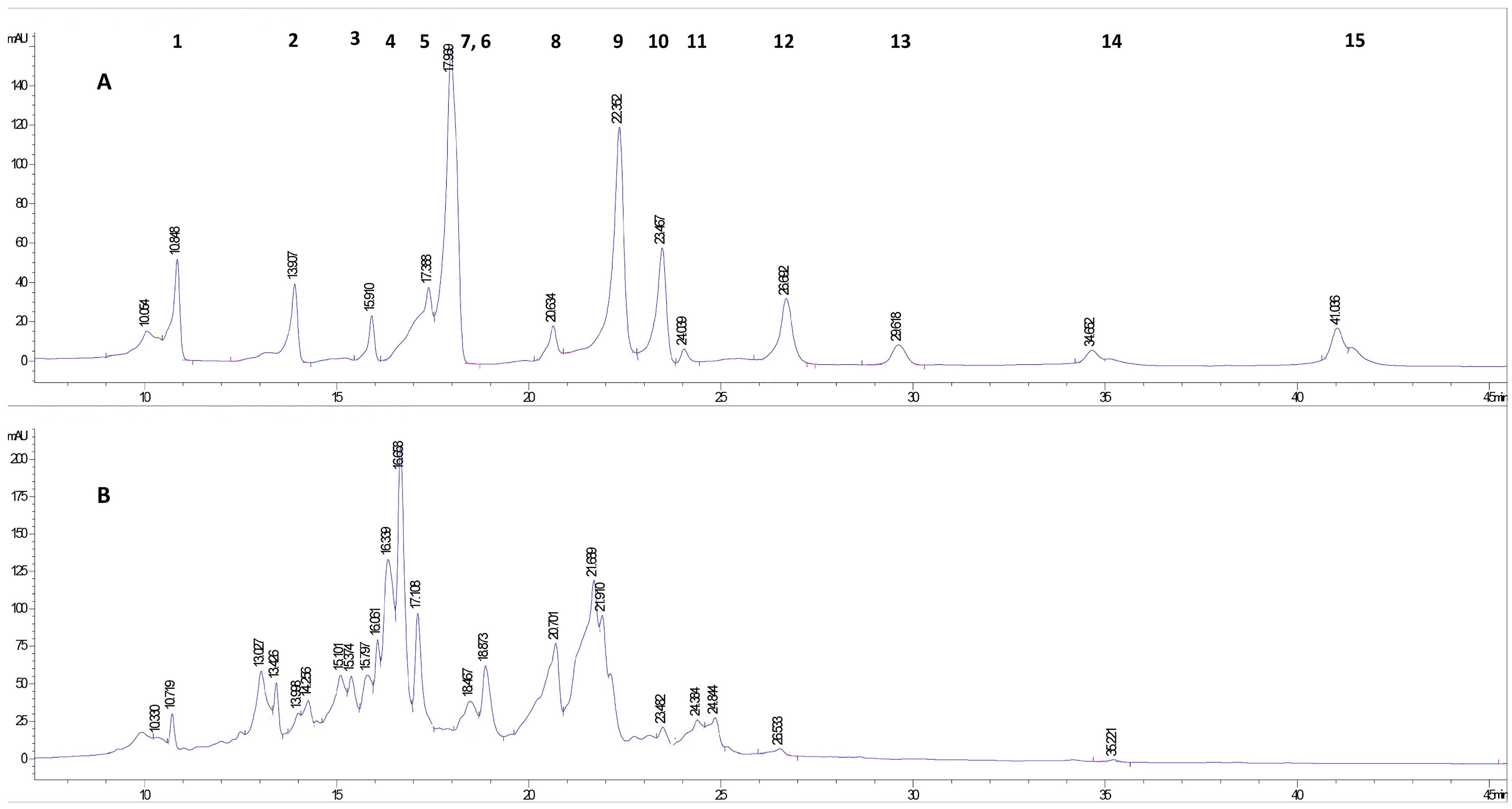
2.1. Characterization of Phenolic Compounds of A. altissima Leaf Extract by HPLC-DAD

In the present work, we investigated the ecotoxic and cytotoxic effects of an A. altissima leaf hydroethanolic extract, which has been characterized by a total content of 6026.31 ± 4.89 mg gallic acid equivalents/100 g DW (phenolics), 575.50 ± 0.10 mg catechin equivalents/100 g DW (tannins), 55.60 ± 0.11 mg β-carotene/100 g DW (carotenoids) and a total antioxidant capacity as measured by FRAP assay of 5043.59 ± 48.11 mg ascorbic acid equivalents/100 g DW.
For the identification of polyphenols in A. altissima leaf extract, stock solutions of 15 polyphenols were prepared with a concentration of 0.5 mg/mL. Using the separation method developed by our research group for HPLC analysis, all 15 polyphenols compounds were analyzed one by one. Retention times (RT) are listed in Table 1. After their analysis, all the 15 polyphenols were mixed in equal proportions and the mixture was analyzed using the same HPLC separation method. Following the separation, as noticed in the HPLC chromatogram (Figure 1), a slight shift in RTs occurred (Table 1) due to the inter-molecular interactions between all 15 compounds in the solution. At the same time, because the RTs were very close for four of the compounds, their peaks overlapped two by two (see Table 1). Even if these compounds’ peaks overlapped in the mixture spectrogram, in the spectrogram obtained for the leaf extract the separation was evident for epicatechin and vanillic acid.
Following the performance of the HPLC analysis, nine polyphenolic compounds were identified in the A. altissima hydroethanolic extract obtained from dried autumn leaves, of which five were phenolic acids (gallic acid, protocatechuic acid, vanillic acid, p-coumaric acid, rosmarinic acid) and four were flavonoids (catechin, rutin, hesperidin, quercetin). All the polyphenolic compounds identified in this study were previously reported by other authors in the leaves of this species [8,42,43].

2.2. Ecotoxicity of A. altissima Leaf Extract

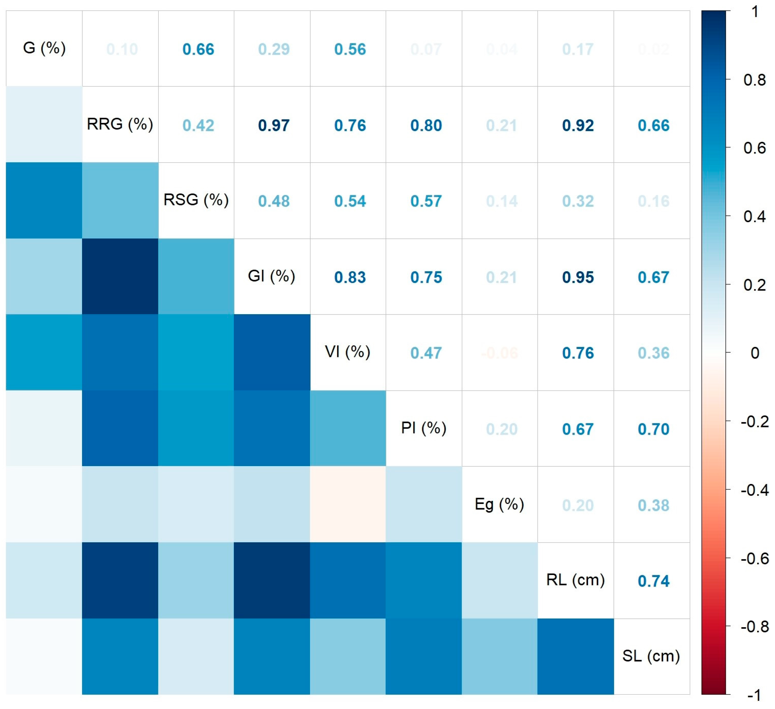
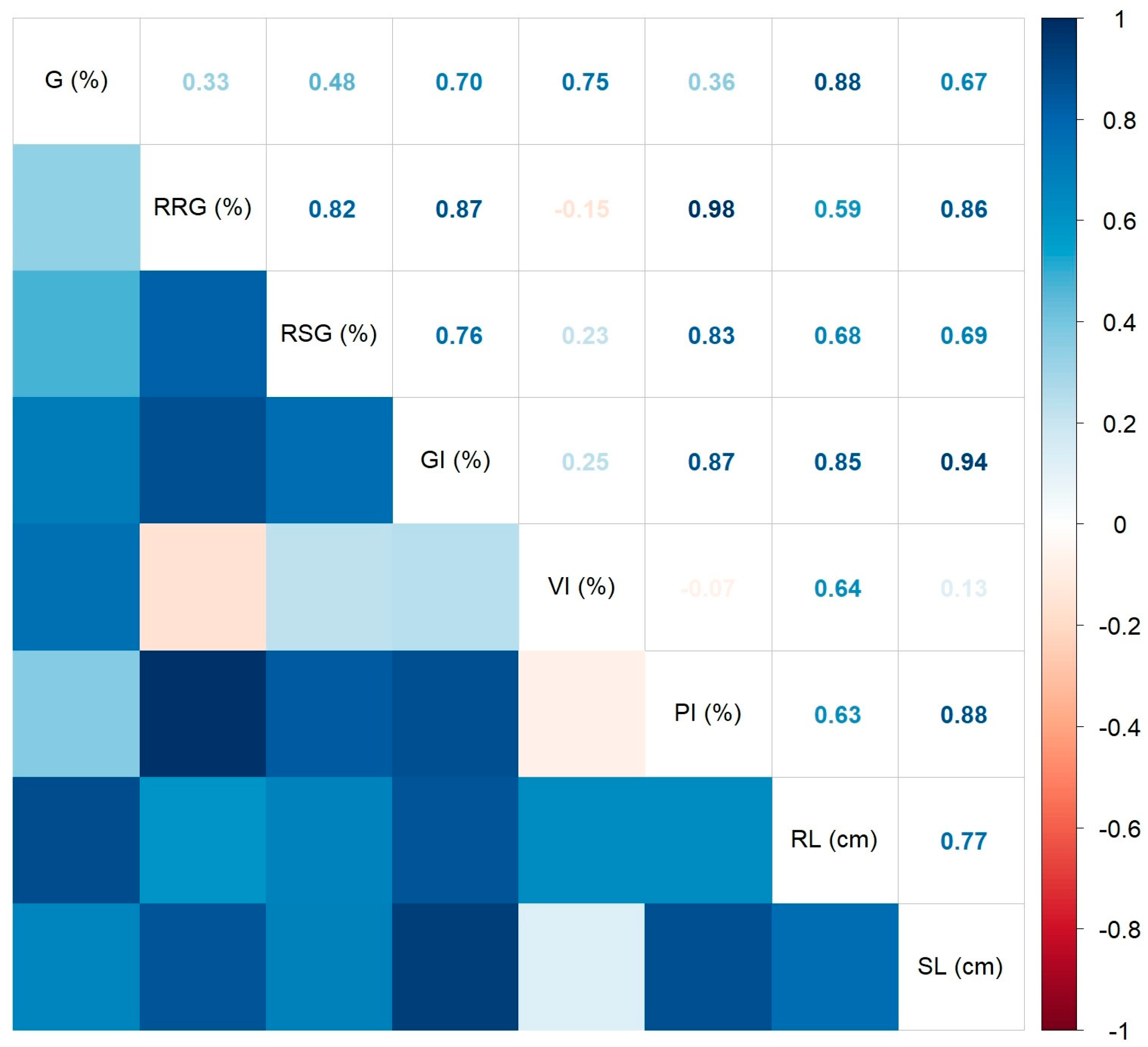
2.2.1. Inhibitory Effects of A. altissima Leaf Extract on Germination of Seeds Used in Agricultural Crops

Ecotoxicity assessments using plant assays are simple approaches providing results suitable for statistical analysis and in connection with those obtained from testing on animal models or even human cells, being used either to identify environmental contaminants or to preliminarily validate drugs and pesticides [44,45]. Wheat and tomato are plant species approved for the ecotoxicity testing of products by the Organization for Economic Cooperation and Development (OECD) and the Food and Drug Administration (FDA) [46].

Wheat Caryopsis Germination Test

Wheat, an important global agricultural crop [47,48], represents one of the most commonly used species for the toxicity assessment of chemical compounds, natural extracts, and nanoparticles [45,49,50].
The results on wheat germination and growth records in the presence of A. altissima ethanolic extract at a concentration range 125–1000 μg/mL are presented in Table 2, comparatively to those for the control.
Most germination parameters excepting the shoot length showed higher mean values in the presence of the lowest extract concentration (125 μg/mL) among all investigated samples. Most germination parameters excepting the germinative energy and shoot length decreased in the presence of the highest extract concentration (1000 μg/mL).
By conducting an ANOVA test, marginally significant differences were observed for GI (%) (p = 0.088) and VI (%) (p = 0.069) (Figure 2) in relation to the extract concentration. According to the Tukey test, the vigor index varied significantly between groups with an added extract at 125 µg/mL and 1000 µg/mL, respectively (p = 0.0490), while the germination index varied marginally (p = 0.0625). Although the ANOVA and Kruskal–Wallis tests did not show statistically significant differences for the other germination parameters related to extract concentrations and to the control (p > 0.1), the most pronounced inhibitory effect of leaf extract on wheat was found at the highest investigated concentration, 1000 μg/mL, while the sample at the lowest concentration (125 μg/mL) actually produced an opposite effect—a slight stimulation of wheat caryopses for all measured indices compared to those of the control and samples with higher concentrations.
The Spearman’s correlation coefficients between various measured/calculated germination indices are presented in Figure 3 by using a correlogram. As noticed, all significant coefficients at p < 0.05 showed positive values. The strongest correlations were found between the germination rate and vigor index (R2 = 0.91, p < 0.01). The weakest significant correlation was identified between the root length and shoot length (R2 = 0.41, p = 0.0484). The germination energy showed no statistically significant correlation with the other variables, while the shoot length indicated only one significant positive correlation with the influence index on the aerial part (R2 = 0.42, p = 0.0437), and a marginally significant one with the relative root growth (R2 = 0.37, p = 0.0778).
Ahmad et al. (2020) [21] observed that an aqueous extract of A. altissima leaves may adversely affect the wheat root length and caryopsis germination. The study of Novak et al. (2021) [51] on A. altissima but using aqueous extracts from roots and an aqueous solution of the pure compound ailanthone, reported very weak inhibitory activity on the wheat germination and root length growth compared to the effect it had on other tested plant seeds (pigweed Amaranthus retroflexus L. and red bristlegrass Setaria pumila L.), at lower concentrations. The authors found that the least significant effect was shown by ailanthone, indicating that an inhibitory effect occurs only in the presence of other root allelochemicals (synergism). According to other published papers, A. altissima root or leaves revealed either an inhibitory effect on Medicago sativa [24,25], Daucus carota [22], Sinapis alba and Brassica napus [23], Lactuca sp. [26], or a stimulating effect on Raphanus sativus L. and Setaria pumila L. [51,52]. The inhibitory effects may be further studied for their potential application as bioherbicides [52].

Tomato Seed Germination Test

The tomato species (Lycopersicum esculentum), belonging to the Solanaceae family, has been used in order to determine the phytotoxic activity of various plant extracts [53,54,55] or antibiotics [56], being considered an appropriate species due to its sensitivity to toxic substances, availability, and good germinating rate [46,57].
Considering the results on the wheat caryopses germination test, two concentrations (125 and 1000 μg/mL, respectively) of the ethanolic leaf extracts of A. altissima were investigated for their potential inhibitory activity on tomato germination. The results are presented in Table 3, comparatively to those for the control.
Most germination parameters excepting the germination rate, relative seed germination, and vigor index, showed the highest mean values in the presence of the lowest extract concentration (125 μg/mL) among all investigated samples. Most germination parameters excepting the germinative energy decreased in the presence of the highest extract concentration (1000 μg/mL).
A boxplot representation of four germination/ growth indices (RRG, GI, VI, RL) which revealed significant differences in relation to extract concentrations and the control is shown in Figure 4.
Data analysis by the ANOVA test and its non-parametric variant, the Kruskal–Wallis test, showed significant differences between mean values of germination indices as a function of extract concentration, indicating a decrease in the investigated indices with the increase in extract concentration. A high statistically significant inhibitory effect was registered for the group with added extract at 1000 μg/mL. No statistically significant differences were found between germination indices of the control and those in the group with added leaf extract at the lowest concentration, 125 μg/mL. Similarly, the Dunn’s test indicated highly statistically significant differences (p < 0.05) for the indices RRG and GI between the groups with added leaf extract at different concentrations, and between the group with the highest extract concentration and the control. The Tukey’s test showed statistically significant higher VI and RL values (p < 0.001) in the control and the group with 125 μg/mL added extract than those in the group at the highest tested concentration, 1000 μg/mL. Although the Kruskal–Wallis/ ANOVA analysis showed no statistically significant differences between groups with added extract at different concentrations for the indices Eg (p = 0.1346), RSG (p = 0.6138), PI (p = 0.5312), G (p = 0. 613), and SL (p = 0.559), slightly lower values were observed at 1000 μg/mL and higher values at 125 μg/mL compared to the control. As an exception, the mean germination energy was slightly higher in the group with the highest extract concentration than in the control, and the mean germination rate showed close values at all concentrations. Our results showed that the root length was more affected by the extract concentration, compared to the aerial part length.
All Spearman’s correlations in the L. esculentum seed germination test were positive and significant (p < 0.05), as shown in the correlogram of Figure 5. The strongest statistically significant correlations were found between the RRG and GI (R2 = 0.97, p < 0.01), and between the GI and RL (R2 = 0.95, p < 0.01). The weakest significant correlation was identified between the RSG and VI (R2 = 0.54, p = 0.0370). All correlations of the Eg parameter were not statistically significant.
In the study of Heisey and Heisey (2003) [58], the compound ailanthone isolated from A. altissima, which was applied to the tomatoes in the field, did not significantly influence the production of tomatoes or their biomass.

Parsley Seed Germination Test

Parsley (Petroselinum crispum (Mill.) var. crispum) represents an important herb for the food industry and gastronomy [59,60], known as a fast-growing species [61]. Most reported studies have focused on improving its germination characteristics because of the low germination rate of these seeds [62,63,64].
Similarly to the experimental runs on the tomato seed germination test, two concentrations (125 and 1000 μg/mL, respectively) of ethanolic leaf extract of A. altissima were investigated for their potential inhibitory activity on parsley seed germination. The results are presented in Table 4, comparatively to those for the control.
All the investigated germination parameters in samples with added leaf extracts were lower than those in the control sample.
A boxplot representation of four germination/growth indices (RRG, RSG, GI, RL) in relation to extract concentrations and the control is shown in Figure 6.
As shown in Figure 6, no statistically significant differences (p ˃ 0.05) were found between values of the investigated germination indices between control and groups with added leaf extracts, and within groups with different concentrations of extracts, by using ANOVA and Kruskal–Wallis tests. Despite the lower values of germination indices in the presence of the plant extract, compared to those in the control, no significant inhibitory effect of ethanolic extract of A. altissima leaves was identified on parsley seed germination.
All Spearman’s correlations in the parsley seed germination test were significant and positive (Figure 7). The strongest significant correlations were found between the RRG and PI (R2 = 0.98, p < 0.01), and between the GI and SL (R2 = 0.94, p < 0.01). The weakest significant correlation was found between the RRG and RL (R2 = 0.59, p = 0.0414).
The effect of A. altissima extracts on parsley seed germination has not been reported so far. Therefore, our results could not be compared to other similar ones.
Regarding the effects of different concentrations of A. altissima leaf extract on the germination of seeds used in agricultural crops (wheat, tomato, parsley), our results indicate an inhibitory effect of the extract at the highest tested concentration (1000 μg/mL) on wheat germination (only marginally significant results, 43–51% for the GI and VI) and tomato germination (statistically significant, 67–72% for the RRG, GI, VI, RL) compared to the control, while no statistically significant effects were found using the parsley germination test. The statistically significant decrease in germination indices (RRG, GI, VI, RL) of the tomato with increased extract concentration might be due to the fact that tomatoes behave like bioindicators and are much more sensitive to the presence of allelochemicals than wheat, as shown by Vidotto et al. (2013) [65]. At the highest investigated extract concentration, the development of seedlings of the three crop plants was also altered, showing a more negative impact on the growth of the root than that of the aerial part. This observation has been also reported by Asgharipour and Armin (2010) [66], in their study on an aqueous extract of Sorghum halepens, which inhibited to a greater extent the root growth of Ocimum basilicum than the shoot length. Using the tomato and wheat germination test, we observed a slight stimulatory effect of 7–8% with respect to the germinative energy in the presence of A. altissima leaf extract.

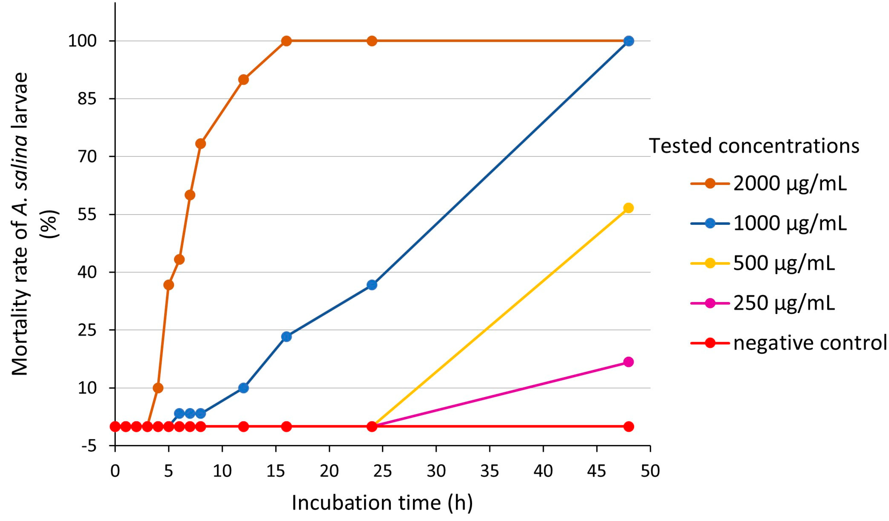
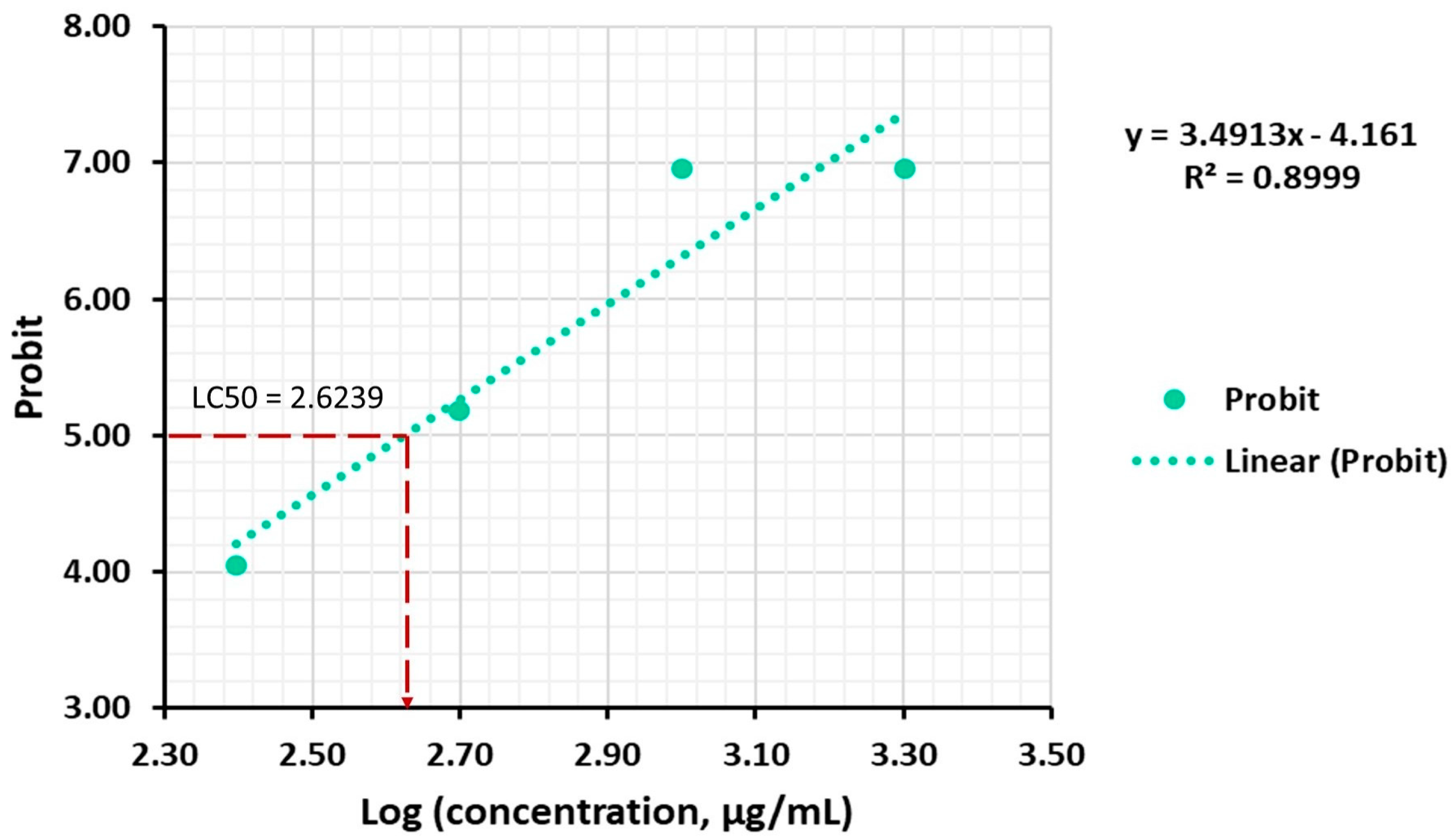
2.2.2. Inhibitory Effects of A. altissima Leaf Extract on A. salina Hatching

The ecotoxicity of products towards aquatic organisms is usually tested against crustaceans, e.g., D. magna, Daphnia pulex, Scapholeberis kingi or A. salina [20], being regulated for the risk assessment of chemicals and materials by international organizations such as the American Society for Testing and Materials (ASTM), the OECD and EU [67,68,69], or for acute toxicity evaluation by the US-EPA [70]. Originally described by Meyer et al. (1982) [71], the brine shrimp larvae (BSL) test becomes over time an easy-to-assess standard technique with promising results in detecting the toxicity and anti-proliferative activity of plant extracts [72,73,74,75] to be applied in the pharmaceutical industry [76].
To further investigate the ecotoxicity of A. altissima leaves, we performed the assay on brine shrimp. The results regarding the evolution of the mortality rate of A. salina larvae incubated with different concentrations (250–2000 μg/mL) of A. altissima leaf extract are presented in Figure 8.
Throughout an incubation period of 48 h, no A. salina individual died in the negative control group, so that no correction by Abbott’s formula was required [77]. The extract concentration of 2000 μg/mL determined the shortest time to onset toxic effects, after 3 h of exposure, followed by the concentration of 1000 μg/mL, at which the larvicidal effects appeared after 5 h. Lower concentrations of extract (250 and 500 μg/mL) produced mortality only after 24 h of exposure.
The values of the median lethal concentration (LC50, μg/mL) of the leaf extracts on BSL using the Log Concentration by Probit analysis are presented in Table 5.
We found a strong positive correlation between the mortality rate and the extract concentration (R2 = 0.8684, p = 0.0681) after 24 h of exposure. The median lethal concentration (LC50) of A. altissima leaf extract, corresponding to a Log concentration of 2.9782, was 951.04 ± 28.26 μg/mL (24 h of exposure). As shown in Figure 9, the equation of the linear regression of the mortality rate expressed in probability units (Probit) against the logarithm of the concentration was Y= 4.4481X − 8.2472.
The lower the LC50 value is, the more toxic the compound/extract is [77]. The toxicity of the ethanolic extract of A. altissima leaves on A. salina at 24 h was low, according to Clarkson’s toxicity criterion: extracts with LC50 ˃ 1 mg/mL, non-toxic; LC50 = 0.5–1 mg/mL, low toxicity; LC50 = 0.1–0.5 mg/mL, medium toxicity; and LC50 = 0–0.1 mg/mL, highly toxic [80].
We also found a strong positive correlation between the mortality rate and the extract concentration (R2 = 0.8999, p = 0.0514) after 48 h of exposure. The median lethal concentration (LC50) of A. altissima leaf extract, corresponding to a Log concentration of 2.6239, was 420.65 μg/mL (48 h), corresponding to a medium toxicity according to Clarkson’s toxicity criterion [80]. As shown in Figure 10, the equation of the linear regression of the mortality rate expressed in probability units (Probit) against the logarithm of the concentration was Y = 3.4913X − 4.161.
To our knowledge, no study regarding the A. altissima toxicity towards A. salina has been published so far. The toxicity of fresh A. altissima leaves litter extracts has been tested towards another aquatic invertebrate, D. magna, showing a median effective concentration (EC50) of 10.1 g/L of air-dried leaf, at 96 h [27]. In our study, the sensitivity of A. salina to A. altissima extract increased with time, the highest one being registered after 48 h, similar to other studies on natural extracts but from different species, e.g., from marine organisms [81]. The mortality of A. salina individuals was positively correlated with the concentration of the extract to which they were exposed, similar to that reported in the study of Krishnaraju et al. (2016) [82] on aqueous extracts of some Indian medicinal plants, including the species Ailanthus excelsa.

2.3. Cytotoxicity of A. altissima Leaf Extract

The study of the interaction between lipid membranes and bioactive molecules or natural extracts allows one either to identify their potential inhibitory effect on microbial and cancer cells, or to evaluate their cytotoxic effects [83]. Hereby, we investigated the interaction of leaf extracts with different cells (erythrocytes, fibroblasts, cancer cells) and with animal cell model membranes, considering it a key initial step to evaluate extract toxicity.

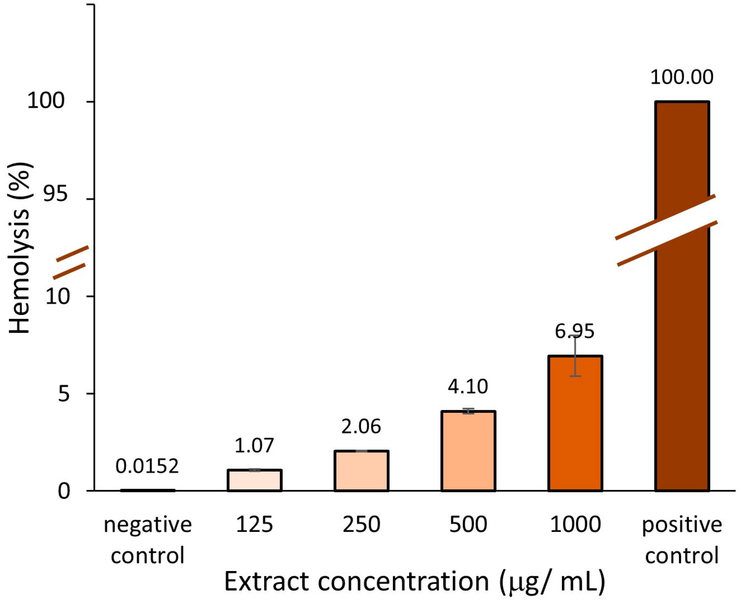
2.3.1. Hemolytic Activity

The effect of different concentrations of A. altissima leaf extract ranging from 125 to 1000 μg/mL, on sheep erythrocyte membrane, was investigated using the erythrocyte viability assay, frequently applied to test the safety of drugs and medicinal nanoparticles [84,85,86]. The results are shown in Figure 11.
The hemolytic activity, HC50, calculated as the value at which 50% of erythrocytes are lysed in the presence of the extract [87], was determined. Based on a regression calculation, according to the equation Y = 0.0067X + 0.414 from plotting hemolysis versus concentration, the HC50 value was 7400.9 µg/mL (equivalent to 7.4%, w/v).
Increasing the concentration of the leaf extract will increase hemolysis. None of the investigated concentrations of leaf extract produced a significant hemolytic activity. The highest hemolysis (6.95 ± 1.04%) occurred in the presence of 1000 μg/mL.
There are no guidelines on the hemolytic properties of natural extracts, but the hemolytic properties of materials used in medical devices, such as nanomaterials, are regulated according to the ASTM based on the percentage of human erythrocyte lysis: <5% (no hemolysis), 5–10% (low hemolysis) ˃10% (marked hemolysis) [88]. Translating these to A. altissima leaf extract, our results indicate a non-hemolytic limit concentration of 500 μg/mL. No hemolytic studies have been reported for A. altissima. In the study of Silva et al. (2023) [89], the synthetic pure coumarin D, a chemical compound found in A. altissima bark being of interest for its antifungal properties, showed a low hemolytic effect (<5%), while several analogs of it determined even less hemolysis. However, the authors indicated that the results cannot exempt the extract from showing toxicity towards other types of cells in the human body. More than being low hemolytic, some natural compounds in particular polyphenols displaying antioxidant properties, showed protective effects on induced erythrocyte hemolysis [84,90]. Caffeic acid and tannins, which are polyphenolic compounds frequently found in plants including A. altissima leaves [5,91], have revealed a strong capacity to inhibit induced hemolysis [92,93].

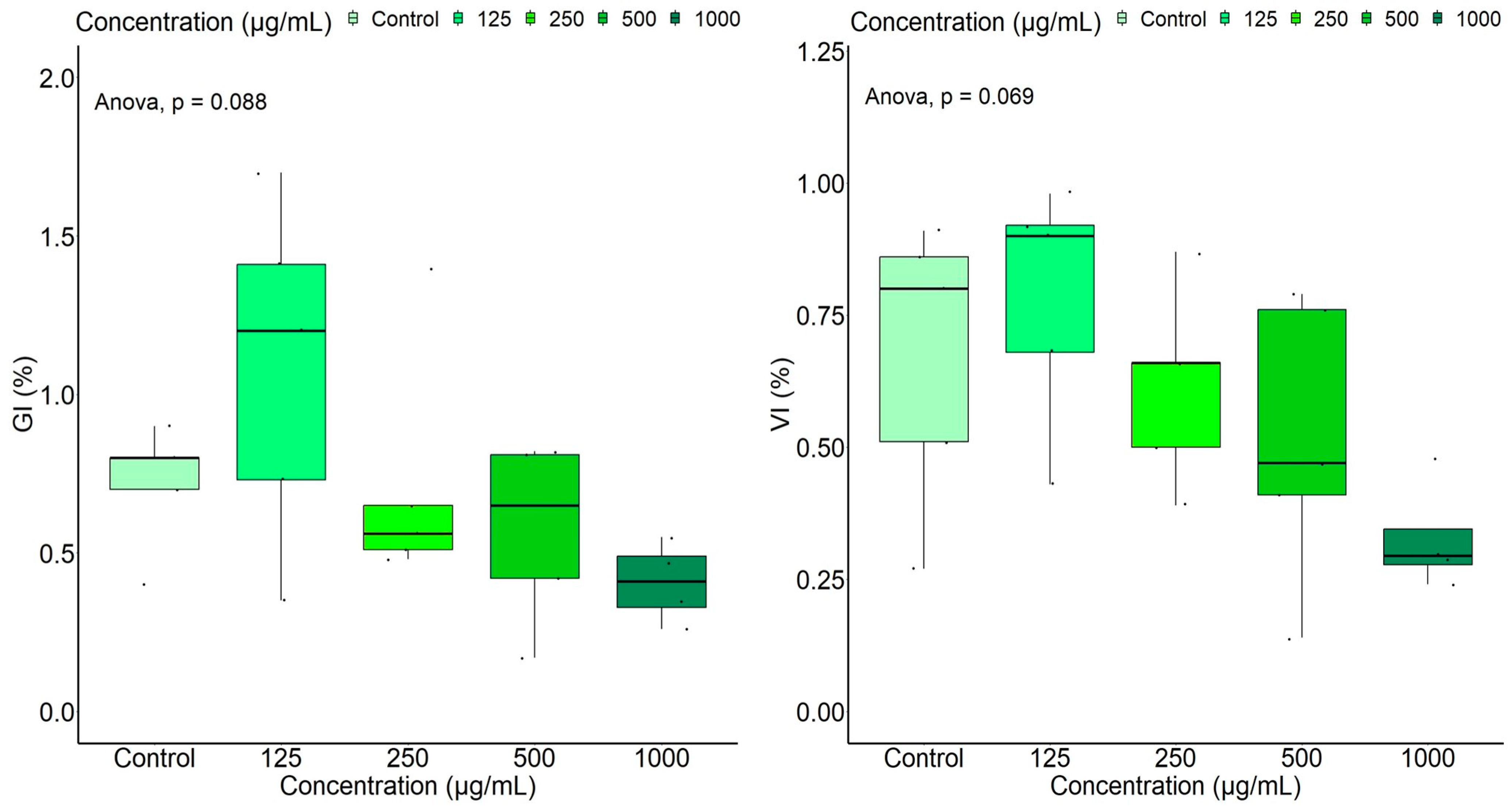
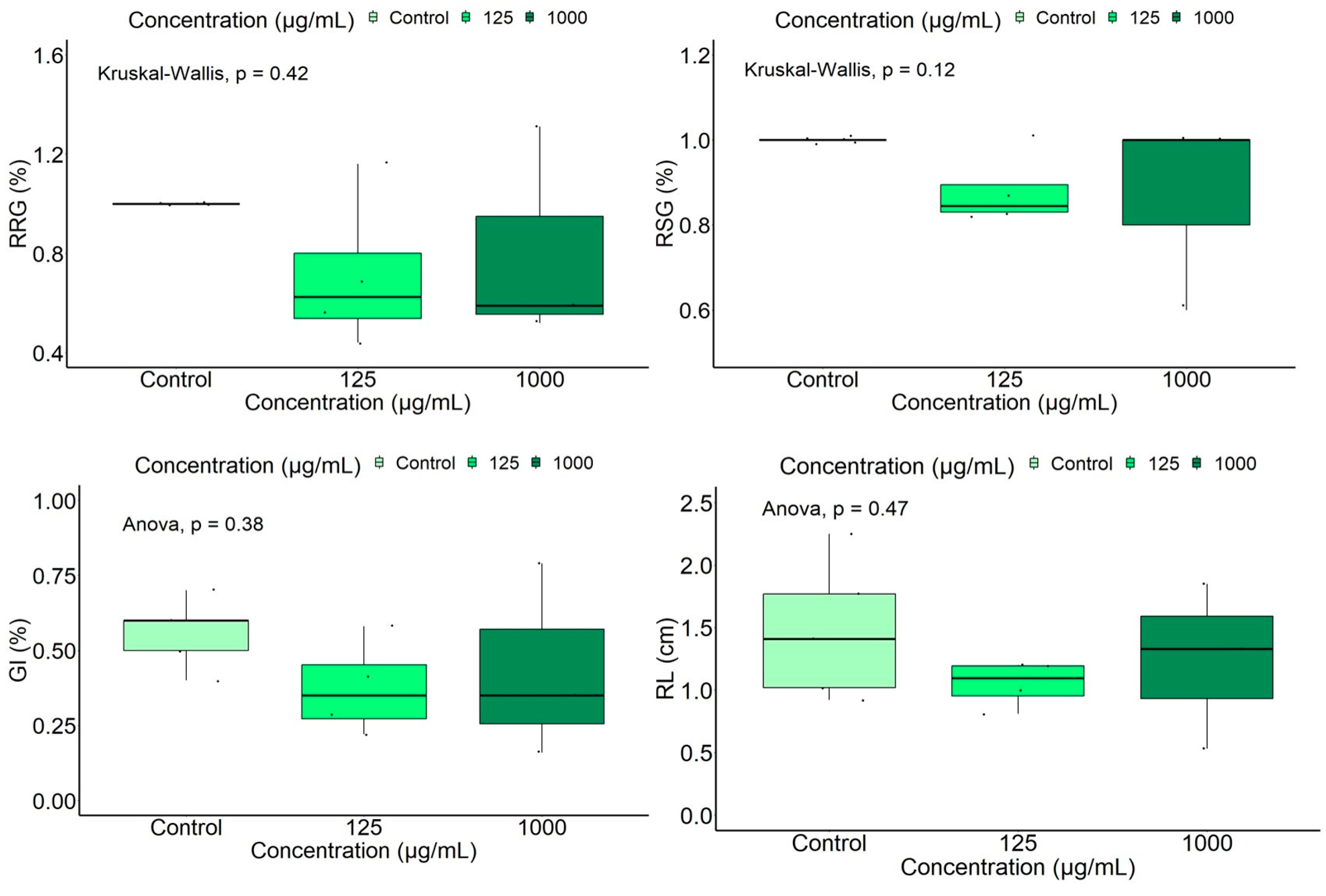
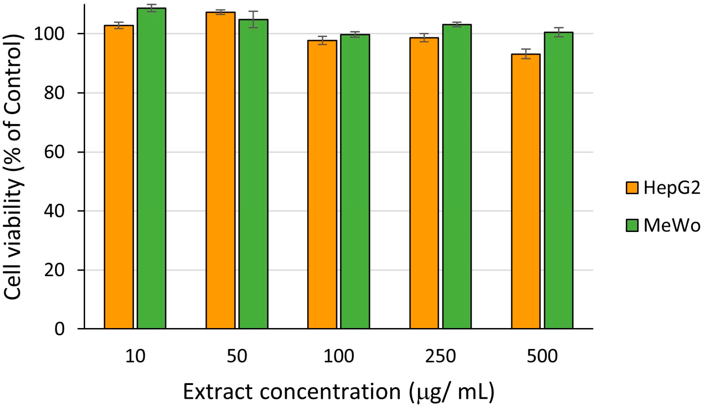
2.3.2. In Vitro Biocompatibility and Cytotoxicity of Leaf Extract at Different Concentrations

Cell viability experiments are important tests in toxicity studies offering information on the cellular response to a toxicant. MTS—a tetrazolium salt-based colorimetric assay—was used in the present study to evaluate the proliferation activity of cells after 24 h incubation.
The results regarding the biocompatibility test of A. altissima leaf extracts in the concentration range 10–500 μg/ mL performed on human gingival fibroblast (HGF) are presented in Figure 12.
In this study, A. altissima leaf extract was biocompatible up to 500 µg/mL. The level of fibroblast viability decreased by increasing the concentration of A. altissima extract. The extract at 10 μg/ mL showed the highest percentage of HGF cell viability at 24 h, 104%. The lowest cell viability value (86%) was registered in the presence of 500 μg/ mL extract.
To our knowledge, this is the first report on the influence of Ailanthus altissima leaf extract on the viability of HGF cells.
Our further cytotoxic study was extended to target organisms and performed on hepatocellular carcinoma (HepG2) and malignant melanoma (MeWo) cells. The results are presented in Figure 13.
The extract had no cytotoxic effect on HepG2 and MeWo cells’ viability over 24 h incubation, indicating no anti-proliferative properties at tested concentrations and on the investigated cell lines. We calculated the half-maximal inhibitory concentration value, IC50, of the extract from the MTS assay for HepG2 and MeWo cells by using the regression equation. IC50 for HepG2 cells was 2441.63 µg/mL, while for MeWo cells it was 5124.07 µg/mL.
Mohamed et al. (2021) [32] also observed a weak cytotoxic effect of the methanolic fraction of a crude extract from A. altissima leaves after 72 h of exposure on HepG2 cells, all the other tested fractions (EtOAc and n-BuOH fractions) exhibiting lower effects than those of doxorubicin, a standard drug [32]. According to the classification proposed by the U.S. National Cancer Institute (NCI) and Geran protocol, which the authors employed in their paper, a half-maximal inhibitory concentration (IC50) between 200 µg/mL and 500 µg/mL was associated with a weak citotoxicity [32]. In the study of Gao et al. (2022) [94], a new steroid with the molecular formula C24H34O4 isolated from leaves of A. altissima demonstrated an important antiproliferative effect on HepG2 cells, with an IC50 = 4.03 μM, more effective than the antiproliferative drug sorafenib. To our knowledge, no studies reported any investigation of A. altissima leaf extract on MeWo cells, but published research showed that quercetin, a polyphenolic compound of flavonoid class, inhibits the signaling and expression of the c-Met receptor in wild-type melanoma MeWo [95].

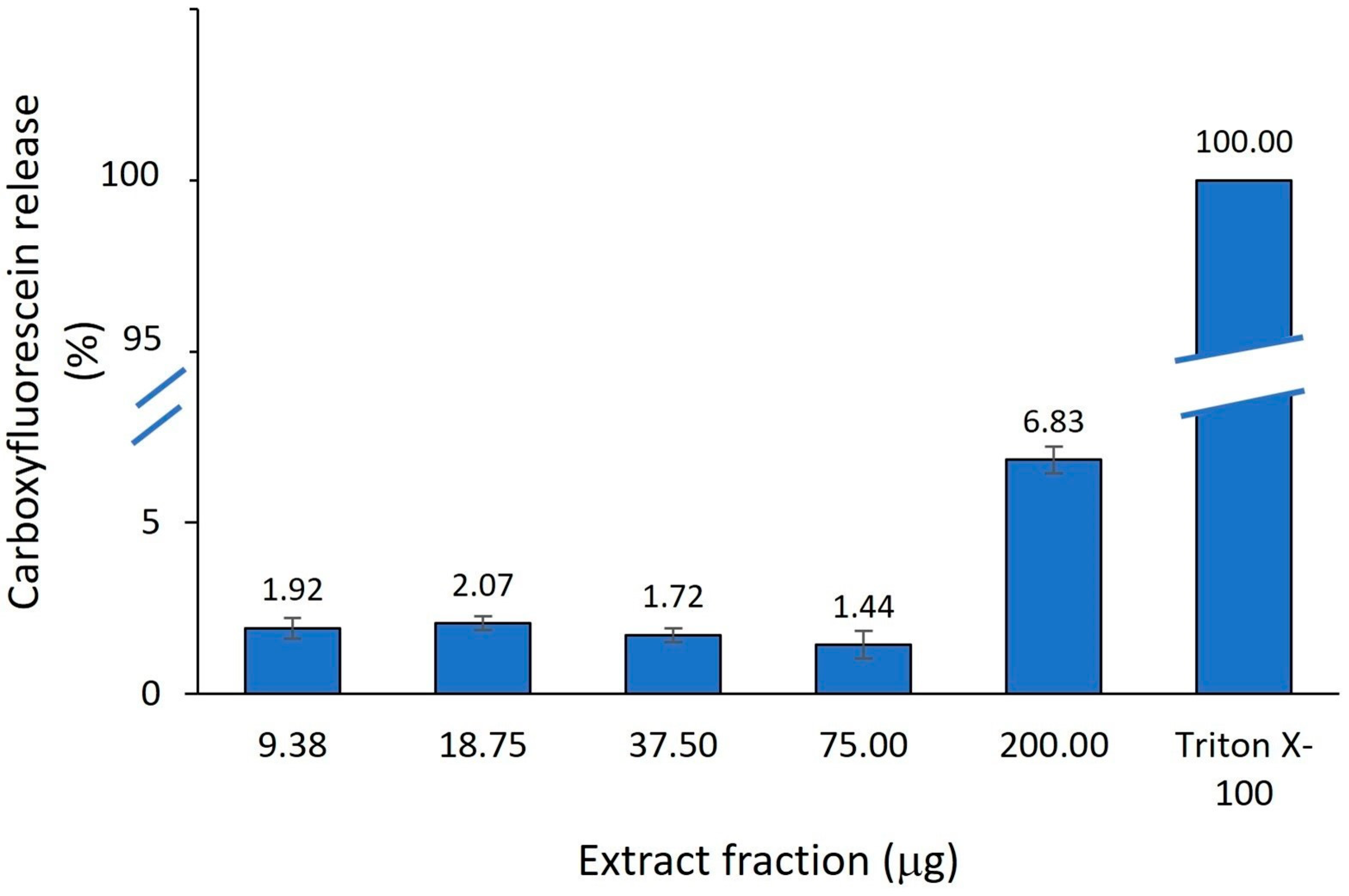
2.3.3. Model Membrane-Modifying Properties of A. altissima Leaf Extract

A simple and relatively stable lipid membrane model of artificial liposomes can be used instead of living cells for studying the interaction of natural extracts with lipid vesicles of various sizes e.g., small unilamellar vesicle (SUV, 20–50 nm in diameter), large unilamellar vesicle (LUV, 100–500 nm in diameter), giant unilamellar vesicle (GUV 10–100 µm in diameter), multilamellar vesicle (MLV, >500 nm in diameter) and multivesicular vesicle (MVV, >500 nm in diameter) [29,30].
In order to corroborate our findings presented above, the permeability of lipid membranes prepared of 1,2-dioleyl-sn-glycero-3-phosphocholine and cholesterol (7/3) was investigated in the presence of different concentrations (125–1000 μg/mL) of A. altissima leaf extract by measuring the induced release of a fluorescent dye (carboxyfluorescein, CF) from small unilamellar liposomes. These liposomes represent a valuable model for eukaryotic cell membranes. The phospholipid concentration was kept constant (0.06 mM) and increasing [extract]/[lipid] molar ratios were obtained by adding aliquots of extract at concentrations between 125 and 1000 μg/mL. The results are presented in Figure 14.
Under the explored conditions, the A. altissima leaf extract did not exhibit any capability to alter or disrupt liposomes within the tested concentration range, even at the highest tested fraction (200 μg) when 6.83% of CF was released. The obtained results indicate a low toxicity of the extract on the investigated liposomal model, a eukaryotic cell-mimicking membrane. No other published papers studying the effect of A. altissima leaf extract on liposome leakage have been identified so far. However, changes in liposome permeability using calcein leakage assay have been recently reported for testing the toxicity of pesticides [96].
One reason why the membrane of liposomes deteriorates is the lipid oxidation [97]. However, the presence of large amounts of polyphenols in the natural extract is not sufficient to explain their possible protective effects on liposomal membranes, according to the observations made by Rodrigues et al. (2016) [98]. Among various phenolic compounds, the leaves of A. altissima contain gallic acid [22,99,100], which has a good capacity to protect membranes from oxidative stress [97].

3. Materials and Methods

3.1. Plant Material and Extract Preparation

Leaflets of A. altissima were collected in the autumn from Vâlcea county, Romania (45°6′34″ (N), 24°22′42″ (E)). The voucher specimen of this plant was deposited at the Herbarium (No. HFS 23 1053, Faculty of Sciences) of the “Lucian Blaga” University of Sibiu. The collected leaves were subjected to hot-air drying with forced air circulation (UFE 400, Memmert, Schwabach, Germany) at 50 °C such as to reach a final moisture content of 5–6% as measured using a moisture analyzer (Mac 210/NP Radwag, Radom, Poland). Dried leaves were ground using a knife mill (Grindomix GM 200, Retsch, Haan, Germany), sieved through a standard sieve (pore size 700 µm) and stored at 4 °C until analysis.
Ethanolic crude extracts of dried leaves were prepared by soaking the powdered plant sample into 70% aqueous ethanol solution at a sample/solvent ratio of 1/10 (w/v), for 6 h at r.t. in darkness. The mixture was centrifuged at 8000 rpm (Universal 320, Hettich, Berlin, Germany) for 10 min at 4 °C. The obtained supernatant was further concentrated using a centrifugal vacuum concentrator (RVC 2-18 CD plus, Christ, Munich, Germany) such as to obtain a soft extract (moisture < 15%).

3.2. Characterization of A. altissima Leaf Extract

The extract was subjected to chemical analysis of polyphenols, such as total phenolics using the Folin–Ciocalteu method [101], total condensed tannins by the vanillin assay [102], and carotenoids [103]. Antioxidant capacity was determined as well, using the Ferric Reducing Antioxidant Power (FRAP) assay described by Benzie and Strain [104].
HPLC-DAD analysis: the polyphenols’ separation and identification were achieved using an Agilent 1260 Infinity Series (Agilent Technologies, Santa Clara, CA, USA), equipped with a UV-Vis DAD detector and a binary pump. The compounds separation was performed using a Mediterranea Sea 18 column (4.6 mm × 150 mm, 5 μm particle size) (Teknokroma Analitica S.A., Barcelona, Spain) and a gradient elution was applied (Table 6) by using 0.1% (v/v) formic acid in water (mobile phase A), and acetonitrile (mobile phase B) (both purchased from Sigma-Aldrich Chemie GmbH, Taufkirchen, Germany), at 0.8 mL/min flow rate. 20 µL of each sample and standard was injected and the separation process was monitored by UV-VIS DAD detector at 280 nm. Fifteen polyphenolic compounds (gallic acid, protocatechuic acid, catechin, vanillic acid, epicatechin, caffeic acid, syringic acid, rutin, ferulic acid, p-coumaric acid, hesperidin, rosmarinic acid, salicylic acid, quercetin, kaempferol, all purchased from Sigma-Aldrich Chemie GmbH, Taufkirchen, Germany) were used as standards, from which 0.5 mg/mL stock solutions were prepared and injected both separately and as a mixture. The mixture was prepared by adding 100 μL of each stock solution, thoroughly homogenized and injected as such.

3.3. Determination of Ecotoxicity of A. altissima Leaf Extract

3.3.1. Germination Bioassay of Wheat (Triticum aestivum L.) Caryopsis

T. aestivum organic seeds (Pronat, Romania) were subjected to sterilization according to the method described by Lindsey III et al. (2017) [105] and Dal Cortivo et al. (2017) [106] with slight modifications. A total of 100 seeds were covered with 500 μL sodium hypochlorite solution (50/50 v/v) for 5 min, after which they were rinsed seven times for 1 min with distilled water. A standard germination test was conducted in five replicates for each extract concentration and control. Ten wheat caryopses and 5 mL leaf extract of different concentrations (soft ethanolic extract re-suspended in distilled water such as to obtain 125, 250, 500 and 1000 μg/mL, respectively) were placed on a double layer of filter paper (79.7 g/m2, 0.165 mm thickness) in a glass Petri dish of 6 cm diameter, which was previously kept in a pre-heated oven at 140 °C for 30 min, in order to remove possible contaminating microorganisms. The control sample was prepared by using 5 mL distilled water. Incubation was conducted in an upright position of dishes at r.t. for 8 days, of which 6 days were monitored using the containers sealed with a tin foil, applied with the purpose of limiting the degradation of the active compounds of extract and the water evaporation, as suggested by Veisz et al. (1996) [107]. The measurements were made on the third day of germination when the number of sprouted caryopses was noticed, and on the eighth day when germinated seeds, root length and aerial length of seedlings were observed.
The following germination indices were evaluated: shoot length (SL, cm), root length (RL, cm), seed germination energy (Eg, %) [108], relative root growth (RRG, %) [109], influence index on the aerial part (PI, %) [110], vigor index (VI, %) and germination index (GI, %) [111], relative seed germination (RSG, %) and germination rate (G, %) [112]. The indices were calculated using the Equations (1)–(7):
E g % = t h e   n u m b e r   o f   g e r m i n a t e d   s e e d s   i n   t h e   t h i r d   d a y t h e   t o t a l   n u m b e r   o f   s e e d s
G % = t h e   n u m b e r   o f   g e r m i n a t e d   s e e d s t h e   t o t a l   n u m b e r   o f   s e e d s × 100
R R G % = a v e r a g e   r o o t   l e n g t h   f o r   t h e   s a m p l e a v e r a g e   r o o t   l e n g t h   f o r   t h e   c o n t r o l × 100
R S G % = t h e   n u m b e r   o f   g e r m i n a t e d   s e e d s   i n   t h e   s a m p l e t h e   n u m b e r   o f   g e r m i n a t e d   s e e d s   i n   t h e   c o n t r o l × 100
G I = G % × R R G % × 100
V I = G % × a v e r a g e   r o o t   l e n g t h + a v e r a g e   s t e m   l e n g t h
P I = G %   s a m p l e t h e   a v e r a g e   l e n g t h   o f   t h e   a e r i a l   p a r t   f o r   t h e   s a m p l e G %   c o n t r o l a v e r a g e   a e r i a l   p a r t   l e n g t h   f o r   c o n t r o l × 100
In case the groups contained fewer germinated caryopses than a minimum of three, they were excluded from the evaluation.
The root length of a seedling was determined by measuring each root strand from the root–shoot junction to the tip and calculated as an average value. The shoot length was obtained in the same way, but the measurement was taken from the culm’s base to the tip of a leaf.

3.3.2. Germination Bioassay of Tomato (Lycopersicum esculentum Mill.) Caryopsis

This bioassay has been performed under similar experimental conditions as described for wheat caryopsis, with slight modifications: 3 mL leaf extract of two different concentrations (125 μg/mL and 1000 μg/mL, respectively) was added to 10 tomato seeds (Starsem, Universal, Romania) in the germination test. The measurements were made on the third and sixth day of germination.

3.3.3. Germination Bioassay of Parsley (Petroselinum crispum (Mill.) var. Crispum) Seed

This bioassay has been performed under similar experimental conditions as described for tomato caryopsis. Parsley seeds from Starsen (Traditional) brand, produced by Agrosel SRL. (Romania), were used in the experiments. The measurements were made on the sixth day of germination, according to Ugolini et al. (2021) [113].

3.3.4. Brine Shrimp (Artemia salina) Assay

The brine shrimp larvae (BSL) test was performed according to the method described by Apetroaei et al. (2018) [114]. For the experiment, commercially purchased Artemia salina eggs (Artemia Hobby; Manufacturer: DAJANA PET) were used. The eggs were hatched using a saline solution (38% w/v) according to McLaughlin et al. (1998) [74]. 500 mL saline solution and 5 g of A. salina eggs were added to a system consisting of two transparent plastic containers, the smaller one having a perforated base and being the one in which the eggs were placed and kept at 24 °C under light over 48 h. Larvae were transferred to identical transparent plastic containers from the hatching vessel using a pipette. Based on the description of McLaughlin et al. (1998) [74], 10 individuals/test container were counted. Although hatched larvae can survive 48 h without nutrient feeding [81], 50 μL of yeast suspension was added to each sample to limit the risk of larval death due to starvation [115]. To the vials containing brine shrimp, aliquots of leaf extracts obtained from A. altissima dried leaves were added at the following concentrations: 250, 500, 1000, and 2000 μg/mL. Experiments were carried out in triplicate per vial. The mortality rate was recorded up to 8 h on an hourly basis, and at 12, 16, 24, and 48 h, respectively. Larvae that showed no movement for at least 10 sec were considered dead, as suggested by Manfra et al. (2012) [116]. In the control group, the extract was replaced with distilled water, the experiments being conducted in three replicates.
The mortality rate was calculated according to Lam et al. (2020) [117], using the Equation (8):
M o r t a l i t y   r a t e   % = n u m b e r   o f   t h e   d e a d   s h r i m p s   t o t a l   n u m b e r   o f   e x p e r i m e n t a l   s h r i m p s × 100
The median lethal concentration (LC50) was calculated according to Pohan et al. (2023) [118]. The transformation of mortality percentage into probability units (probits) was performed using a Finney’s table as described by Nigar et al. (2021) [119]. The transformation of 0% and 100% of mortality into probability units was carried out according to Randhawa (2009) [79].

3.4. Determination of Cytotoxicity of A. altissima Leaf Extract

3.4.1. Hemolytic Activity Assay (Erythrocyte Viability)

The hemolytic activity of leaf extracts was determined according to the method described by Yamada et al. (1994) [120]. The experiments were conducted on sheep red blood cells (RBC) collected from farm animals of the Research and Development Institute for Montanology, Sibiu, Romania. The experiments were approved by the Ethics Committee for Scientific Research Involving Human Subjects and/or Animals, of the ”Lucian Blaga” University of Sibiu (process number 21/2024). The whole blood sample was previously centrifugated at 2500 rpm for 5 min to remove the plasma fraction and washed repeatedly with phosphate buffer (PBS) pH 7.4, until the supernatant became colorless. To the resuspended RBC in PBS buffer, aliquots of leaf extracts of different concentrations (serial dilutions of soft extract in PBS such as to obtain: 125, 250, 500 and 1000 μg/mL, respectively) were added. The samples were incubated for 2 h at 37 °C, then centrifuged at 2500 rpm for 5 min at r.t. A negative control sample (RBC in PBS) without extracts was used. Total lysis was obtained by incubation of RBC with distilled water (positive control). Absorbance of supernatants was measured at 576 nm using the spectrophotometer (Specord 200 Plus UV-Vis, Analytik Jena, Germany).
Percentage of hemolysis was calculated using the Equation (9):
%   h e m o l y s i s = 100 × A s a m p l e A c o n t r o l A 100 A c o n t r o l
where
  • Asample = absorbance of the test sample (RBC in the presence of extract)
  • Acontrol= absorbance of the control (RBC and PBS)
  • A100 = absorbance in case of total lysis (RBC and distilled water)

3.4.2. In Vitro Biocompatibility and Cytotoxicity Assessment (MTS Assay)

The in vitro assessment of biocompatibility of extracts was performed on human gingival fibroblasts (HGF), while that of cytotoxicity was conducted on malignant melanoma (MeWo) and hepatocellular carcinoma (HepG2), all from CLS Cell Lines Service GmbH, Eppelheim, Germany, using the CellTiter 96® AQueous One Solution Cell Proliferation Assay kit (Promega, Madison, WI USA). Cells were seeded in 96-well plates at 5 × 10⁴ cells/well (HGF) or 105 cells/well (MeWo and HepG2) in alpha-MEM medium (PAN-Biotech GmbH, Aidenbach, Germany) supplemented with 10% fetal bovine serum (FBS) and 1% Penicillin-Streptomycin-Amphotericin B mixture (both from Gibco, Thermo Fisher Scientific, Waltham, MA, USA) and allowed to adhere for 24 h. Samples of A. altissima leaf extract were diluted in complete culture medium at 10, 50, 100, 250 and 500 µg extract/mL. Cells were then incubated for 24 h versus control cells (untreated). Control cells were incubated with a complete cell culture medium (considered 100% cell viability). An FLUOstar® Omega microplate reader (BMG LABTECH, Ortenberg, Germany) was used to record the MTS absorbance at 490 nm 3 h after addition of the reagent. The viability of cells treated with different concentrations of the leaf extract was expressed as percentage (%) of the viability of control cells (means ± standard error of the mean). Experiments were done in triplicate.

3.4.3. Membrane Leakage Assay from Lipid Vesicle

The membrane permeability properties were determined by measuring the induced leakage of 5(6)-carboxyfluorescein (CF) entrapped in small unilamellar vesicles (SUV) [121].

SUV Preparation

1,2-dioleyl-sn-glycero-3-phosphocholine (DOPC) was purchased from Avanti Polar Lipids, Inc. (Alabaster, AL, USA), while cholesterol (Ch) was a Sigma-Aldrich (St. Louis, MO, USA) product.
The lipid mixture DOPC/Ch (7/3) was dissolved in CHCl3 in a test tube, dried under N2, and lyophilized overnight. The lipid film was reconstituted with a solution of CF in 30 mM Hepes buffer (pH 7.4) at r.t. for 1 h. To make SUVs, the resulting multilamellar vesicle suspension was sonicated (GEX400 Ultrasonic Processor, Sigma) on ice until the initially cloudy lipid dispersion became translucent. The excess of fluorescent dye was eliminated by gel filtration on Sephadex G-75 (Sigma). SUVs were diluted to a concentration of 0.06 mM with Hepes buffer (5 mM Hepes, 100 mM NaCl, pH 7.4). The SUVs were stored at 4 °C and used within 24 h.

Leakage from SUV

The extract-induced leakage from SUVs was measured at 293 K using a CF-entrapped vesicle technique and a spectrofluorometer (Perkin Elmer model MPF-66). The phospholipid concentration was kept constant (0.06 mM) and increasing [extract]/[lipid] molar ratios were obtained by adding aliquots of extract solutions. The membrane leakage properties were examined for leaf extracts at 125, 250, 500 and 1000 μg/mL, respectively. The highest concentration (1000 μg/mL) was analyzed for two added volumes, 75 μL, investigated for all the concentrations, and 200 μL. After rapid and vigorous stirring, the time course of the fluorescence change corresponding to CF escape was recorded at 520 nm (6-nm band pass) with λexc 488 nm (3-nm band pass). The percentage of released CF at time t was determined according to formula (10):
%   C F = 100 × F t F 0 F T F 0
where
  • F0 = fluorescence intensity of vesicles in the absence of extract,
  • Ft = fluorescence intensity of vesicles at time t in the presence of extract,
  • FT = total fluorescence intensity determined by disrupting the vesicles by addition of 50 µL of a Triton X-100 solution.
The kinetic experiments were stopped at 20 min.

3.5. Statistical Analysis

Experimental data with values expressed as mean ± SD were subjected to statistical analysis using the R 4.3.1 program [122], ANOVA, Kruskal–Wallis test, Tukey’s test, Dunn’s test for multiple comparisons/dunn.test package R Package Version 1.3.5. [123]. The correlation analysis was performed using the rcorr function of the Hmisc package R Package Version 5.1-0 [124]. The Spearman’s correlation was investigated for data which were not normally distributed. The function ggboxplot of the ggpubr package R Package Version 0.6.0 [125] was used to create representative boxplot graphs, while the correlogram was constructed using the corrplot.mixed function from the corrplot package Version 0.92 [126]. The LC50 values obtained from the brine shrimp (A. salina) assay were determined using the Probit analysis [127], in order to evaluate the toxicity of the leaf extracts. The results were considered statistically significant at p < 0.05.

4. Conclusions

The present study brings novelty and completes the existing information on potential toxicity of A. altissima leaves by using a complex approach towards different target/non-target organisms.
Extract of A. altissima leaves in ethanol solution showed a high content of compounds of polyphenolic structure, and antioxidant activity.
The extract exhibited low ecotoxic effects towards wheat, tomato, and parsley up to 500 μg/mL, and significant inhibitory effects on tomato germination at 1000 μg/mL. Screening A. altissima for ecotoxicity towards brine shrimp indicated a low toxicity at 24 h of exposure (Clarkson’s toxicity criterion based on LC50 values), which increased with exposure time and extract concentration. Cytotoxicity of the extract towards sheep erythrocytes indicate non-hematotoxicity. Moreover, the extract was found biocompatible with human gingival fibroblasts according to the MTS assay. Under the explored conditions, the leaf extract did not exhibit any capability to alter or disrupt liposomes within the tested concentration range, according to experiments on model lipid small unilamellar vesicles.
Toxicity tested against target organisms such as malignant melanoma and hepatocellular carcinoma indicates no anti-proliferative effect within the concentration range of 10 μg/mL to 500 μg/mL.
Given the low to moderate toxicity under the tested conditions, the autumn leaves of A. altissina could find useful practical applications/biopesticides. However, some limitations must be addressed, such as requirements for further studies on other cells than erythrocytes or gingival fibroblasts, and in vivo studies to confirm their safety for humans.

Author Contributions

Conceptualization, S.O.; Data curation, M.D.C. and S.O.; Funding acquisition, S.O.; Investigation, M.D.C., N.S., A.R.P., M.S., B.B., L.L. and S.O.; Methodology, M.D.C., N.S., A.R.P., M.S., B.B., L.L. and S.O.; Project administration, S.O.; Resources, N.S., A.R.P., B.B. and S.O.; Writing—original draft, M.D.C., N.S., A.R.P., B.B., L.L. and S.O.; Writing—review and editing, M.D.C. and S.O. All authors have read and agreed to the published version of the manuscript.

Funding

This research was funded by the Lucian Blaga University of Sibiu (Knowledge Transfer Center) and Hasso Plattner Foundation research grants—grant number LBUS-HPI-ERG-2023-04.

Institutional Review Board Statement

Not applicable.

Informed Consent Statement

Not applicable.

Data Availability Statement

All of the data are contained within the article.

Acknowledgments

We would like to thank Duca Gheorghiță Marius, who provided materials for the hemolytic assay.

Conflicts of Interest

The authors declare no conflicts of interest.

References

  1. Montecchiari, S.; Tesei, G.; Allegrezza, M. Ailanthus altissima forests determine a shift in herbaceous layer richness: A paired comparison with hardwood native forests in sub-Mediterranean Europe. Plants 2020, 9, 1404. [Google Scholar] [CrossRef]
  2. EPPO. EPPO Global Database. 2024. Available online: https://gd.eppo.int (accessed on 21 March 2024).
  3. Brundu, G. Information on Measures and Related Costs in Relation to Species Considered for Inclusion on the Union List: Ailanthus altissima. Technical Note Prepared by IUCN for the European Commission. 2017. Available online: https://circabc.europa.eu (accessed on 21 March 2024).
  4. Huo, Q.; Shao, J.; Lin, Q. Study on the antibacterial and bactericidal effects of Ailanthus altissima leaves extract. Asian J. Chem. 2012, 24, 3545. [Google Scholar]
  5. Poljuha, D.; Sladonja, B.; Šola, I.; Dudaš, S.; Bilić, J.; Rusak, G.; Motlhatlego, K.E.; Eloff, J.N. Phenolic composition of leaf extracts of Ailanthus altissima (Simaroubaceae) with antibacterial and antifungal activity equivalent to standard antibiotics. Nat. Prod. Commun. 2017, 12, 1609–1612. [Google Scholar] [CrossRef]
  6. Rahman, H.M.A.; Javaid, S.; Ashraf, W.; Rasool, M.F.; Saleem, H.; Khan, S.A.; Ul-Haq, Z.; Anjum, S.M.M.; Ahmad, T.; Alqahtani, F.; et al. Effects of long-term Ailanthus altissima extract supplementation on fear, cognition and brain an-tioxidant levels. Saudi. Pharm. J. 2023, 31, 191–206. [Google Scholar] [CrossRef] [PubMed]
  7. Caramelo, D.; Pedro, S.I.; Marques, H.; Simão, A.Y.; Rosado, T.; Barroca, C.; Gominho, J.; Anjos, O.; Gallardo, E. Insights into the bioactivities and chemical analysis of Ailanthus altissima (Mill.) Swingle. Appl. Sci. 2021, 11, 11331. [Google Scholar] [CrossRef]
  8. Andonova, T.; Muhovski, Y.; Slavov, I.; Vrancheva, R.; Georgiev, V.; Apostolova, E.; Naimov, S.; Mladenov, R.; Pavlov, A.; Dimitrova-Dyulgerova, I. Phenolic profile, antioxidant and DNA-protective capacity, and microscopic characters of Ailanthus altissima aerial substances. Plants 2023, 12, 920. [Google Scholar] [CrossRef]
  9. He, Q.; Xiao, H.; Li, J.; Liu, Y.; Jia, M.; Wang, F.; Zhang, Y.; Wang, W.; Wang, S. Fingerprint analysis and pharmacological evaluation of Ailanthus altissima. Int. J. Mol. Med. 2018, 41, 3024–3032. [Google Scholar] [CrossRef] [PubMed]
  10. Kundu, P.; Laskar, S. A brief resume on the genus Ailanthus: Chemical and pharmacological aspects. Phytochem. Rev. 2010, 9, 379–412. [Google Scholar] [CrossRef]
  11. Zhang, D.D.; Bai, M.; Yan, Z.Y.; Huang, X.X.; Song, S.J. Chemical constituents from Ailanthus altissima (Mill.) Swingle and chemotaxonomic significance. Biochem. Syst. Ecol. 2020, 93, 104174. [Google Scholar] [CrossRef]
  12. Kožuharova, E.; Lebanova, H.; Getov, I.; Benbassat, N.; Kochmarov, V. Ailanthus altissima (Mill.) Swingle-a terrible invasive pest in Bulgaria or potential useful medicinal plant. Bothalia J. 2014, 44, 213–230. [Google Scholar]
  13. Shin, Y.; Masami, I.; Takakiko, T.; Takahashi, T.; Matsushita, K. Constituents of Ailanthus altissima Swingle. Isolation and structures of shinjuglycosides A, B, C, and D. Bull. Chem. Soc. Jpn. 1984, 57, 2496–2501. [Google Scholar]
  14. Zhao, C.C.; Shao, J.H.; Li, X.; Xu, J.; Zhang, P. Antimicrobial constituents from fruits of Ailanthus altissima Swingle. Arch. Pharm. Res. 2005, 28, 1147–1151. [Google Scholar] [CrossRef]
  15. Ansari, S.H.; Ali, M. Two new phytosterols from Ailanthus altissima (Mill) Swingle. Acta Hort. 2003, 597, 91–94. [Google Scholar] [CrossRef]
  16. Ni, J.C.; Shi, J.T.; Tan, Q.W.; Chen, Q.J. Phenylpropionamides, piperidine, and phenolic derivatives from the fruit of Ailanthus altissima. Molecules 2017, 22, 2107. [Google Scholar] [CrossRef]
  17. Anfossi, L.; Giovannoli, C.; Di Nardo, F.; Cavalera, S.; Chiarello, M.; Trotta, F.; Baggiani, C. Selective enrichment of ailanthone from leaves of Ailanthus altissima by tandem reverse phase/molecularly imprinted solid phase extraction. Microchem. J. 2020, 158, 105198. [Google Scholar] [CrossRef]
  18. Lee, Y.S. Analysis of components in the different parts of Ailanthus altissima. Korean J. Food Preserv. 2008, 15, 261–268. [Google Scholar]
  19. Cocîrlea, M.D.; Oancea, S. Impact of invasive plant species on ecosystems, biodiversity loss and their potential industrial applications. Curr. Trends Nat. Sci. 2022, 11, 88–103. [Google Scholar] [CrossRef]
  20. Ferraz, C.A.; Pastorinho, M.R.; Palmeira-de-Oliveira, A.; Sousa, A.C. Ecotoxicity of plant extracts and essential oils: A review. Environ. Pollut. 2022, 292, 118319. [Google Scholar] [CrossRef]
  21. Ahmad, Z.; Khan, K.R.; Farooq, M.; Shah, A.H.; Mehmood, A.; Jabeen, T.; Zohra, L. Evaluation of allelopathic potential of agricultural land associated trees on germination attributes of wheat (Triticum aestivum L.). Proc. Int. Acad. Ecol. Environ. Sci. 2020, 10, 38–44. [Google Scholar]
  22. Albouchi, F.; Hassen, I.; Casabianca, H.; Hosni, K. Phytochemicals, antioxidant, antimicrobial and phytotoxic activities of Ailanthus altissima (Mill.) Swingle leaves. S. Afr. J. Bot. 2013, 87, 164–174. [Google Scholar] [CrossRef]
  23. Bostan, C.; Borlea, F.; Mihoc, C.; Selesan, M. Ailanthus altissima species invasion on biodiversity caused by potential allelopathy. Res. J. Agric. Sci. 2014, 46, 95–103. [Google Scholar]
  24. Sladonja, B.; Poljuha, D.; Sušek, M.; Dudaš, S. Herbicidal effect of Ailanthus altissima leaves water extracts on Medicago sativa seeds germination. In Proceedings of the Conference VIVUS: Transmission of Innovations, Knowledge and Practical Experience into Everyday Practice, Naklo, Slovenia, 14–15 November 2014. [Google Scholar]
  25. Tsao, R.; Romanchuk, F.E.; Peterson, C.J.; Coats, J.R. Plant growth regulatory effect and insecticidal activity of the extracts of the tree of heaven (Ailanthus altissima L.). BMC Ecol. 2002, 2, 1. [Google Scholar] [CrossRef] [PubMed]
  26. Lawrence, J.G.; Colwell, A.; Sexton, O.J. The ecological impact of allelopathy in Ailanthus altissima (Simaroubaceae). Am. J. Bot. 1991, 78, 948–958. [Google Scholar] [CrossRef]
  27. Alonso, A.; Vázquez de Aldana, B.R.; Castro-Díez, P.; Medina-Villar, S.; Pérez-Corona, M.E. Effects of leaf litter extracts from four tree species on aquatic invertebrates: An ecotoxicological risk assessment approach. Aquat. Ecol. 2020, 54, 1155–1168. [Google Scholar] [CrossRef]
  28. Sæbø, I.P.; Bjørås, M.; Franzyk, H.; Helgesen, E.; Booth, J.A. Optimization of the hemolysis assay for the assessment of cytotoxicity. Int. J. Mol. Sci. 2023, 24, 2914. [Google Scholar] [CrossRef] [PubMed]
  29. Peetla, C.; Stine, A.; Labhasetwar, V. Biophysical interactions with model lipid membranes: Applications in drug discovery and drug delivery. Mol. Pharm. 2009, 6, 1264–1276. [Google Scholar] [CrossRef]
  30. Ding, H.; Yu, X.; Hang, C.; Gao, K.; Lao, X.; Jia, Y.; Yan, Z. Ailanthone: A novel potential drug for treating human cancer. Oncol. Lett. 2020, 20, 1489–1503. [Google Scholar] [CrossRef] [PubMed]
  31. Bai, W.; Yang, H.Y.; Jiao, X.Z.; Feng, K.N.; Chen, J.J.; Gao, K. Structurally diverse highly oxygenated triterpenoids from the roots of Ailanthus altissima and their cytotoxicity. J. Nat. Prod. 2018, 81, 1777–1785. [Google Scholar] [CrossRef]
  32. Mohamed, H.R.; El-Wakil, E.A.; Abdel-Hameed, E.S.S.; El-Hashash, M.M.; Shemis, M. Evaluation of total phenolics, flavonoids, and antioxidant and cytotoxic potential of Ailanthus altissima (Mill.) swingle leaves. J. Rep. Pharm. Sci. 2021, 10, 130–136. [Google Scholar] [CrossRef]
  33. De Feo, V.; Martino, L.D.; Santoro, A.; Leone, A.; Pizza, C.; Franceschelli, S.; Pascale, M. Antiproliferative effects of tree-of-heaven (Ailanthus altissima Swingle). Phytother. Res. 2005, 19, 226–230. [Google Scholar] [CrossRef]
  34. Yang, X.L.; Yuan, Y.L.; Zhang, D.M.; Li, F.; Ye, W.C. Shinjulactone O, a new quassinoid from the root bark of Ailanthus altissima. Nat. Prod. Res. 2014, 28, 1432–1437. [Google Scholar] [CrossRef]
  35. Hwang, Y.J.; Woo, H.I.; Kim, I.H.; Park, D.S.; Kim, J.H.; Om, A.S.; Hwang, K.A. Effect of apoptosis induction of Ailanthus altissima on human lung carcinoma cells. JALS 2011, 45, 91–96. [Google Scholar]
  36. Rosati, A.; Quaranta, E.; Ammirante, M.; Turco, M.C.; Leone, A.; De Feo, V. Quassinoids can induce mitochondrial membrane depolarisation and caspase 3 activation in human cells. Cell Death Differ. 2004, 11, S216–S218. [Google Scholar] [CrossRef]
  37. Wang, Y.; Wang, W.J.; Su, C.; Zhang, D.M.; Xu, L.P.; He, R.R.; Wang, L.; Zhang, J.; Zhang, X.Q.; Ye, W.C. Cytotoxic quassinoids from Ailanthus altissima. BMCL 2013, 23, 654–657. [Google Scholar] [CrossRef] [PubMed]
  38. Wang, R.; Lu, Y.; Li, H.; Sun, L.; Yang, N.; Zhao, M.; Zhang, M.; Shi, Q. Antitumor activity of the Ailanthus altissima bark phytochemical ailanthone against breast cancer MCF-7 cells. Oncol. Lett. 2018, 15, 6022–6028. [Google Scholar] [CrossRef] [PubMed]
  39. Wang, Y.; Zhong, Z.; Ma, M.; Zhao, Y.; Zhang, C.; Qian, Z.; Wang, B. The role played by ailanthone in inhibiting bone metastasis of breast cancer by regulating tumor-bone microenvironment through the RANKL-dependent pathway. Front. Pharmacol. 2023, 13, 1081978. [Google Scholar] [CrossRef] [PubMed]
  40. Li, X.; Li, Y.; Ma, S.; Zhao, Q.; Wu, J.; Duan, L.; Xie, Y.; Wang, S. Traditional uses, phytochemistry, and pharmacology of Ailanthus altissima (Mill.) Swingle bark: A comprehensive review. J. Ethnopharmacol. 2021, 275, 114121. [Google Scholar] [CrossRef] [PubMed]
  41. Chen, Y.; Zhu, L.; Yang, X.; Wei, C.; Chen, C.; He, Y.; Ji, Z. Ailanthone induces G2/M cell cycle arrest and apoptosis of SGC-7901 human gastric cancer cells. Mol. Med. Rep. 2017, 16, 6821–6827. [Google Scholar] [CrossRef] [PubMed]
  42. Tanasković, S.; Gvozdenac, S.; Kolarov, R.; Bursić, V.; Konstantinović, B.; Prvulović, D. Antifeeding and insecticidal activity of Ailanthus altissima and Morus alba extracts against gipsy moth (Lymantria dispar (L.), Lepidoptera, Lymantridae) larvae under laboratory conditions. J. Entomol. Res. Soc. 2021, 23, 197–212. [Google Scholar] [CrossRef]
  43. Marinaș, I.C.; Dinu, M.; Ancuceanu, R.; Hovaneț, M.V.; Oprea, E.; Geană, E.I.; Lazăr, V. The phenols content and phytotoxic capacity of various invasive plants. Rom. Biotechnol. Lett. 2018, 23, 13887. [Google Scholar]
  44. Jităreanu, A.; Ioana-Cezara, C.A.B.A.; Trifan, A.; Pădureanu, S.; Agoroaei, L. Triticum aestivum assay-a useful tool for environmental monitoring and toxicity assessment. Not. Bot. Horti. Agrobo. 2019, 47, 1005–1018. [Google Scholar] [CrossRef]
  45. Ostroumov, S.A. Toxicity testing of chemicals without use of animals. Russ. J. Gen. Chem. 2016, 86, 2933–2941. [Google Scholar] [CrossRef]
  46. Wang, W.; Keturi, P.H. Comparative seed germination tests using ten plant species for toxicity assessment of a metal engraving effluent sample. Water Air Soil Pollut. 1990, 52, 369–376. [Google Scholar] [CrossRef]
  47. Hou, J.; Liu, G.N.; Xue, W.; Fu, W.J.; Liang, B.C.; Liu, X.H. Seed germination, root elongation, root-tip mitosis, and micronucleus induction of five crop plants exposed to chromium in fluvo-aquic soil. Environ. Toxicol. Chem. 2014, 33, 671–676. [Google Scholar] [CrossRef] [PubMed]
  48. Igrejas, G.; Branlard, G. The importance of wheat. In Wheat Quality for Improving Processing and Human Health; Igrejas, G., Ikeda, T.M., Guzmán, C., Eds.; Springer International Publishing: Cham, Switzerland, 2020; pp. 1–7. ISBN 978-3-030-34162-6. [Google Scholar]
  49. An, J.; Zhou, Q.; Sun, Y.; Xu, Z. Ecotoxicological effects of typical personal care products on seed germination and seedling development of wheat (Triticum aestivum L.). Chemosphere 2009, 76, 1428–1434. [Google Scholar] [CrossRef] [PubMed]
  50. Behboudi, F.; Sarvestani, Z.T.; Kassaee, M.Z.; Sanavi, S.A.M.M.; Sorooshzadeh, A. Phytotoxicity of chitosan and SiO2 nanoparticles to seed germination of wheat (Triticum aestivum L.) and barley (Hordeum vulgare L.) plants. Not. Sci. Biol. 2017, 9, 242–249. [Google Scholar] [CrossRef]
  51. Novak, M.; Novak, N.; Milinović, B. Differences in allelopathic effect of tree of heaven root extracts and isolated ailanthone on test-species. JCEA 2021, 22, 611–622. [Google Scholar] [CrossRef]
  52. Caser, M.; Demasi, S.; Caldera, F.; Dhakar, N.K.; Trotta, F.; Scariot, V. Activity of Ailanthus altissima (Mill.) swingle extract as a potential bioherbicide for sustainable weed management in horticulture. Agronomy 2020, 10, 965. [Google Scholar] [CrossRef]
  53. Mahdavikia, F.; Saharkhiz, M.J. Phytotoxic activity of essential oil and water extract of peppermint (Mentha × piperita L. CV. Mitcham). J. Appl. Res. Med. Aromat. Plants 2015, 2, 146–153. [Google Scholar] [CrossRef]
  54. Lara-Núñez, A.; Sánchez-Nieto, S.; Luisa Anaya, A.; Cruz-Ortega, R. Phytotoxic effects of Sicyos deppei (Cucurbitaceae) in germinating tomato seeds. Physiol. Plant. 2009, 136, 180–192. [Google Scholar] [CrossRef]
  55. Miranda-Arámbula, M.; Reyes-Chilpa, R.; Anaya L, A.L. Phytotoxic activity of aqueous extracts of ruderal plants and its potential application to tomato crop. Bot. Sci. 2021, 99, 487–498. [Google Scholar] [CrossRef]
  56. Bellino, A.; Lofrano, G.; Carotenuto, M.; Libralato, G.; Baldantoni, D. Antibiotic effects on seed germination and root development of tomato (Solanum lycopersicum L.). Ecotoxicol. Environ. Saf. 2018, 148, 135–141. [Google Scholar] [CrossRef]
  57. Yang, Y.; Wang, G.; Li, G.; Ma, R.; Kong, Y.; Yuan, J. Selection of sensitive seeds for evaluation of compost maturity with the seed germination index. Waste Manag. 2021, 136, 238–243. [Google Scholar] [CrossRef] [PubMed]
  58. Heisey, R.M.; Kish Heisey, T. Herbicidal effects under field conditions of Ailanthus altissima bark extract, which contains ailanthone. Plant Soil. 2023, 256, 85–99. [Google Scholar] [CrossRef]
  59. Ganea, M.; Vicaș, L.G.; Gligor, O.; Sarac, I.; Onisan, E.; Nagy, C.; Moisa, C.; Ghitea, T.C. Exploring the Therapeutic Efficacy of Parsley (Petroselinum crispum Mill.) as a Functional Food: Implications in Immunological Tolerability, Reduction of Muscle Cramps, and Treatment of Dermatitis. Molecules 2024, 29, 608. [Google Scholar] [CrossRef] [PubMed]
  60. Mahmood, S.; Hussain, S.; Malik, F. Critique of medicinal conspicuousness of Parsley (Petroselinum crispum): A culinary herb of Mediterranean region. Pak. J. Pharm. Sci. 2014, 27, 193–202. [Google Scholar] [PubMed]
  61. van Dijk, N.; Konings, J.; van der Spek, R. Effects of Micosat F Granulate in Ocimum basilicum (Basil) and Petroselinum crispum (Parsley). 21 January 2015, pp. 1–17. Available online: https://micosat.nl/wp-content/uploads/2021/07/Effects-of-Micosat-F-granulate-in-Ocimum-basilicum-and-Petroselinum-crispum-3.pdf (accessed on 21 March 2024).
  62. Dehkourdi, E.H.; Mosavi, M. Effect of anatase nanoparticles (TiO2) on parsley seed germination (Petroselinum crispum) in vitro. Biol. Trace Elem. Res. 2013, 155, 283–286. [Google Scholar] [CrossRef] [PubMed]
  63. Momeni, K.; Moradi, A.; Mahmoudi, S.; Latif Manesh, H. The effect of biopriming and gibberellin on the quality and germination properties of parsley seed (Petroselinum crispum). Iran. J. Seed Res. 2023, 10, 1–17. [Google Scholar] [CrossRef]
  64. Rabin, J.; Berkowitz, G.A.; Akers, S.W. Field performance of osmotically primed parsley seed. Hortic. Sci. 1988, 23, 554–555. [Google Scholar] [CrossRef]
  65. Vidotto, F.; Tesio, F.; Ferrero, A. Allelopathic effects of Ambrosia artemisiifolia L. in the invasive process. Crop Prot. 2013, 54, 161–167. [Google Scholar] [CrossRef]
  66. Asgharipour, M.R.; Armin, M. Inhibitory effects of Sorghum halepens root and leaf extracts on germination and early seedling growth of widely used medicinal plants. Adv. Environ. Biol. 2010, 4, 316–325. [Google Scholar]
  67. ASTM E1193–97; Standard Guide for Daphnia Magna Life-Cycle Toxicity Tests. Annual Book of ASTM Standards. American Society for Testing and Materials: Philadelphia, PA, USA, 1997.
  68. OECD, 2004. Test No. 202: Daphnia sp. Acute Immobilisation Test. Available online: https://doi.org/10.1787/9789264069947-en (accessed on 21 March 2024).
  69. Regulation (EC) No 1907/2006 of the European Parliament and of the Council of 18 December 2006 Concerning the Registration, Evaluation, Authorisation and Restriction of Chemicals (REACH), Establishing a European Chemicals Agency, Amending Directive 1999/45/EC and Repealing Council Regulation (EEC) No 793/93 and Commission Regulation (EC) No 1488/94 as Well as Council Directive 76/769/EEC and Commission Directives 91/155/EEC, 93/67/EEC, 93/105/EC and 2000/21/EC. Available online: https://eur-lex.europa.eu/legal-content/RO/TXT/HTML/?uri=CELEX:32006R1907 (accessed on 21 March 2024).
  70. Weber, C.I. Methods for Measuring the Acute Toxicity of Effluents and Receiving Waters to Freshwater and Marine Organisms, 4th ed.; US-Environmental Protection Agency: Cincinnati, OH, USA, 1993; pp. 117–254.
  71. Meyer, B.; Ferrigni, N.R.; Putnam, J.E.; Jacobsen, L.B.; Nichols, D.E.; McLaughlin, J.L. Brine Shrimp: A Convinient General Bioassay for Active Plant Constituents. J. Med. Plant Res. 1982, 45, 31–34. [Google Scholar] [CrossRef] [PubMed]
  72. Persoone, G.; Wells, P.G. Artemia in aquatic toxicology: A review. In Artemia Research and Its Applications. Morphology, Genetics, Strain Characterization Toxicology; Sorgeloos, P., Ed.; Universita Press: Wetteren, Belgium, 1987; pp. 259–275. [Google Scholar]
  73. Salawu, K.M.; Ajaiyeoba, E.O.; Ogbole, O.O.; Adeniji, J.A.; Faleye, T.C.; Agunu, A. Antioxidant, brine shrimp lethality, and antiproliferative properties of gel and leaf extracts of Aloe schweinfurthii and Aloe vera. J. Herbs Spices Med. Plants 2017, 23, 263–271. [Google Scholar] [CrossRef]
  74. McLaughlin, J.L.; Rogers, L.L.; Anderson, J.E. The Use of Biological Assays to Evaluate Botanicals. Drug Inf. J. 1998, 32, 513–524. [Google Scholar] [CrossRef]
  75. Manure, J.Y. Evaluation of anticancer activity of leaves of Rumex vesicarius Linn and Symplocos racemosa Roxb. by brine shrimp lethality and (3-(4,5-dimethylthiazol-2-yl)-2, 5-diphenyltetrazolium bromide) methods. IJGP 2017, 11. [Google Scholar] [CrossRef]
  76. Ferraz Filha, Z.S.; Lombardi, J.A.; Guzzo, L.S. Brine shrimp (Artemia salina Leach) bioassay of extracts from Lychnophoriopsis candelabrum and different Lychnophora species. RBPM 2012, 14, 358–361. [Google Scholar] [CrossRef]
  77. Jeyaratnam, N.; Nour, A.H.; Kanthasamy, R.; Nour, A.H.; Yuvaraj, A.R.; Akindoyo, J.O. Essential oil from Cinnamomum cassia bark through hydrodistillation and advanced microwave assisted hydrodistillation. Ind. Crops Prod. 2016, 92, 57–66. [Google Scholar] [CrossRef]
  78. Miller, L.C.; Tainter, M. Estimation of the ED50 and its error by means of logarithmic-probit graph paper. Proc. Soc. Exp. Biol. Med. 1944, 57, 261–264. [Google Scholar] [CrossRef]
  79. Randhawa, M.A. Calculation of LD50 values from the method of Miller and Tainter, 1944. J. Ayub. Med. Coll. Abbottabad 2009, 21, 184–185. [Google Scholar] [PubMed]
  80. Clarkson, C.; Maharaj, V.J.; Crouch, N.R.; Grace, O.M.; Pillay, P.; Matsabisa, M.G.; Bhagwandin, N.; Smith, P.J.; Folb, P.I. In vitro antiplasmodial activity of medicinal plants native to or naturalised in South Africa. J. Ethnopharmacol. 2004, 92, 177–191. [Google Scholar] [CrossRef]
  81. Carballo, J.L.; Hernández-Inda, Z.L.; Pérez, P.; García-Grávalos, M.D. A comparison between two brine shrimp assays to detect in vitrocytotoxicity in marine natural products. BMC Biotechnol. 2002, 2, 17. [Google Scholar] [CrossRef]
  82. Krishnaraju, A.V.; Rao, T.V.; Sundararaju, D.; Vanisree, M.; Tsay, H.S.; Subbaraju, G.V. Biological screening of medicinal plants collected from Eastern Ghats of India using Artemia salina (brine shrimp test). IJASE 2006, 4, 115–125. [Google Scholar] [CrossRef]
  83. Šturm, L.; Poklar Ulrih, N. Basic methods for preparation of liposomes and studying their interactions with different compounds, with the emphasis on polyphenols. Int. J. Mol. Sci. 2021, 22, 6547. [Google Scholar] [CrossRef] [PubMed]
  84. Loganayaki, N.; Siddhuraju, P.; Manian, S. Antioxidant activity and free radical scavenging capacity of phenolic extracts from Helicteres isora L. and Ceiba pentandra L. J. Food Sci. Technol. 2013, 50, 687–695. [Google Scholar] [CrossRef]
  85. Ran, Q.; Xiang, Y.; Liu, Y.; Xiang, L.; Li, F.; Deng, X.; Xiao, Y.; Chen, L.; Chen, L.; Li, Z. Eryptosis indices as a novel predictive parameter for biocompatibility of Fe3O4 magnetic nanoparticles on erythrocytes. Sci. Rep. 2015, 5, 16209. [Google Scholar] [CrossRef]
  86. Greco, I.; Molchanova, N.; Holmedal, E.; Jenssen, H.; Hummel, B.D.; Watts, J.L.; Håkansson, J.; Hansen, P.R.; Svenson, J. Correlation between hemolytic activity, cytotoxicity and systemic in vivo toxicity of synthetic antimicrobial peptides. Sci. Rep. 2020, 10, 13206. [Google Scholar] [CrossRef]
  87. Cuervo-Rodríguez, R.; Muñoz-Bonilla, A.; López-Fabal, F.; Fernández-García, M. Hemolytic and antimicrobial activities of a series of cationic amphiphilic copolymers comprised of same centered comonomers with thiazole moieties and polyethylene glycol derivatives. Polymers 2020, 12, 972. [Google Scholar] [CrossRef] [PubMed]
  88. ASTM. Standard Test Method for Analysis of Hemolytic Properties of Nanoparticles; ASTM: West Conshohocken, PA, USA, 2013. [Google Scholar]
  89. Silva, A.C.; de Moraes, D.C.; do Carmo, D.C.; Gomes, G.C.C.; Ganesan, A.; Lopes, R.S.C.; Ferreira-Pereira, A.; Lopes, C.C. Synthesis of altissimacoumarin D and other prenylated coumarins and their ability to reverse the multidrug resistance phenotype in Candida albicans. J. Fungi. 2023, 9, 758. [Google Scholar] [CrossRef] [PubMed]
  90. Paiva-Martins, F.; Fernandes, J.; Santos, V.; Silva, L.; Borges, F.; Rocha, S.; Belo, L.; Santos-Silva, A. Powerful protective role of 3, 4-dihydroxyphenylethanol− elenolic acid dialdehyde against erythrocyte oxidative-induced hemolysis. J. Agric. Food Chem. 2010, 58, 135–140. [Google Scholar] [CrossRef]
  91. Bashir, A.; Farooq, S.; Bhat, Z.A. Pharmacognostic standardization and phytochemical evaluation of Ailanthus altissima (Mill.) Swingle leaves. JDDT 2019, 9, 179–183. [Google Scholar]
  92. Djeridane, A.; Yousfi, M.; Nadjemi, B.; Vidal, N.; Lesgards, J.F.; Stocker, P. Screening of some Algerian medicinal plants for the phenolic compounds and their antioxidant activity. Eur. Food Res. Technol. 2007, 224, 801–809. [Google Scholar] [CrossRef]
  93. Olchowik-Grabarek, E.; Swiecicka, I.; Andreeva-Kovaleskaya, Z.; Solonin, A.; Bonarska-Kujawa, D.; Kleszczyńska, H.; Mavlyanov, S.; Zamaraeva, M. Role of structural changes induced in biological membranes by hydrolysable tannins from sumac leaves (Rhus typhina L.) in their antihemolytic and antibacterial effects. J. Membr. Biol. 2014, 247, 533–540. [Google Scholar] [CrossRef] [PubMed]
  94. Gao, Z.H.; Duan, Z.K.; Ma, Z.T.; Ye, L.; Yao, G.D.; Huang, X.X.; Song, S.J. Chouchunsteride A–D, four new steroids from the leaves of Ailanthus altissima (Mill.) Swingle. Steroids 2022, 188, 109117. [Google Scholar] [CrossRef]
  95. Salgado, M.T.S.F.; Fernandes e Silva, E.; Nascimento, M.A.D.; Lopes, A.C.; Paiva, L.S.D.; Votto, A.P.D.S. Potential Therapeutic Targets of Quercetin in the Cutaneous Melanoma Model and Its Cellular Regulation Pathways: A Systematic Review. Nutr. Cancer 2023, 75, 1687–1709. [Google Scholar] [CrossRef]
  96. Morita, S.; Sakai, C.; Sakamoto, M.; Nishimoto, M. Evaluating pesticide ecotoxicity using a stimuli-response model in liposomes. JCIS Open 2023, 10, 100082. [Google Scholar] [CrossRef]
  97. Silva, D.R.; Cabello, M.C.; Severino, D.; Baptista, M.D.S. Performance of cosmetic ingredients evaluated by their membrane protection efficiency. JCDSA 2021, 11, 169–185. [Google Scholar] [CrossRef]
  98. Rodrigues, D.; Viotto, A.C.; Checchia, R.; Gomide, A.; Severino, D.; Itri, R.; Baptista, M.S.; Martins, W.K. Mechanism of Aloe Vera extract protection against UVA: Shelter of lysosomal membrane avoids photodamage. PPS 2016, 15, 334–350. [Google Scholar] [CrossRef] [PubMed]
  99. Kim, S.R.; Park, Y.; Li, M.; Kim, Y.K.; Lee, S.; Son, S.Y.; Lee, J.S.; Lee, C.H.; Park, H.H.; Lee, J.Y.; et al. Anti-inflammatory effect of Ailanthus altissima (Mill.) Swingle leaves in lipopolysaccharide-stimulated astrocytes. J. Ethnopharmacol. 2022, 286, 114258. [Google Scholar] [CrossRef] [PubMed]
  100. Luís, Â.; Gil, N.; Amaral, M.E.; Domingues, F.; Duartea, A.P. Ailanthus altissima (Miller) Swingle: A source of bioactive compounds with antioxidant activity. BioResources 2012, 7, 2105–2120. [Google Scholar] [CrossRef]
  101. Singleton, V.L.; Rossi, J.A. Colorimetry of total phenolics with phosphomolyb-dic-phosphotungstic acid reagents. Am. J. Enol. Vitic. 1965, 16, 144–158. [Google Scholar] [CrossRef]
  102. Price, M.L.; Van Scoyoc, S.; Butler, L.G. A critical evaluation of the vanillin reaction as an assay for tannin in sorghum grain. Agric. Food Chem. 1978, 26, 1214–1218. [Google Scholar] [CrossRef]
  103. Scott, K.J. Detection and measurement of carotenoids by UV/VIS spectrophotometry. Curr. Protoc. Food Anal. Chem. 2001, 1, F2.2.1–F2.2.10. [Google Scholar] [CrossRef]
  104. Benzie, I.F.; Strain, J.J. The ferric reducing ability of plasma (FRAP) as a measure of “antioxidant power”: The FRAP assay. Anal. Biochem. 1996, 239, 70–76. [Google Scholar] [CrossRef] [PubMed]
  105. Lindsey, B.E., III; Rivero, L.; Calhoun, C.S.; Grotewold, E.; Brkljacic, J. Standardized method for high-throughput sterilization of Arabidopsis seeds. JoVE 2017, 128, e56587. [Google Scholar] [CrossRef] [PubMed]
  106. Dal Cortivo, C.; Barion, G.; Visioli, G.; Mattarozzi, M.; Mosca, G.; Vamerali, T. Increased root growth and nitrogen accumulation in common wheat following PGPR inoculation: Assessment of plant-microbe interactions by ESEM. Agric. Ecosyst. Environ. 2017, 247, 396–408. [Google Scholar] [CrossRef]
  107. Veisz, O.; Galiba, G.; Sutka, J. Effect of abscisic acid on the cold hardiness of wheat seedlings. J. Plant Physiol. 1996, 149, 439–443. [Google Scholar] [CrossRef]
  108. Amooaghaie, R.; Tabatabaei, F.; Ahadi, A.M. Role of hematin and sodium nitroprusside in regulating Brassica nigra seed germination under nanosilver and silver nitrate stresses. Ecotoxicol. Environ. Saf. 2015, 113, 259–270. [Google Scholar] [CrossRef] [PubMed]
  109. Cudalbeanu, M.; Furdui, B.; Cârâc, G.; Barbu, V.; Iancu, A.V.; Marques, F.; Leitão, J.H.; Sousa, S.A.; Dinica, R.M. Antifungal, antitumoral and antioxidant potential of the Danube Delta Nymphaea alba extracts. Antibiotics 2019, 9, 7. [Google Scholar] [CrossRef] [PubMed]
  110. González-Feijoo, R.; Rodríguez-Seijo, A.; Fernández-Calviño, D.; Arias-Estévez, M.; Arenas-Lago, D. Use of three different nanoparticles to reduce Cd availability in soils: Effects on germination and early growth of Sinapis alba L. Plants 2023, 12, 801. [Google Scholar] [CrossRef] [PubMed]
  111. Gopinathan, M.; Thirumurthy, M. Evaluation of phytotoxicity for compost from organic fraction of municipal solid waste and paper & pulp mill sludge. EREM 2012, 59, 47–51. [Google Scholar]
  112. Luo, Y.; Liang, J.; Zeng, G.; Chen, M.; Mo, D.; Li, G.; Zhang, D. Seed germination test for toxicity evaluation of compost: Its roles, problems and prospects. Waste Manag. 2018, 71, 109–114. [Google Scholar] [CrossRef]
  113. Ugolini, F.; Crisci, A.; Albanese, L.; Cencetti, G.; Maienza, A.; Michelozzi, M.; Zabini, F.; Meneguzzo, F. Effects of Silver Fir (Abies alba Mill.) needle extract produced via hydrodynamic cavitation on seed germination. Plants 2021, 10, 1399. [Google Scholar] [CrossRef] [PubMed]
  114. Apetroaei, M.R.; Pădureţu, C.; Rău, I.; Schroder, V. New-chitosan characterization and its bioassay in different salinity solutions using Artemia salina as bio tester. Chem. Pap. 2018, 72, 1853–1860. [Google Scholar] [CrossRef]
  115. Nerdy, N.; Lestari, P.; Sinaga, J.P.; Ginting, S.; Zebua, N.F.; Mierza, V.; Bakri, T.K. Brine shrimp (Artemia salina Leach.) lethality test of ethanolic extract from green betel (Piper betle Linn.) and red betel (Piper crocatum Ruiz and Pav.) through the soxhletation method for cytotoxicity test. Open Access Maced. J. Med. Sci. 2021, 9, 407–412. [Google Scholar] [CrossRef]
  116. Manfra, L.; Savorelli, F.; Pisapia, M.; Magaletti, E.; Cicero, A.M. Long-term lethal toxicity test with the crustacean Artemia franciscana. JoVE 2012, 62, e3790. [Google Scholar] [CrossRef] [PubMed]
  117. Lam, P.H.; Le, M.T.; Dang, D.M.T.; Doan, T.C.D.; Tu, N.P.C.; Dang, C.M. Safe concentration of silver nanoparticles in solution for white leg shrimp (Litopeaneus vannamei) farming. Biol. Chem. Res. 2020, 7, 35–45. [Google Scholar]
  118. Pohan, D.J.; Marantuan, R.S.; Djojosaputro, M. Toxicity test of strong drug using the BSLT (brine shrimp lethality test) method. IJHSR 2023, 13, 203–209. [Google Scholar] [CrossRef]
  119. Nigar, S.; Gupta, N.; Trivedi, A.; Gupta, V. Lead nitrate induced acute toxicity in the freshwater fishes Channa punctatus and Heteropneustes fossilis. Appl. Ecol. Environ. Sci. 2021, 9, 735–743. [Google Scholar]
  120. Yamada, K.; Natori, S. Characterization of the antimicrobial peptide derived from sapecin B, an antibacterial protein of Sarcophaga peregrina (flesh fly). Biochem. J. 1994, 298, 623–628. [Google Scholar] [CrossRef]
  121. El Hajji, M.; Rebuffat, S.; Le Doan, T.; Klein, G.; Satre, M.; Bodo, B. Interaction of trichorzianines A and B with model membranes and with the amoeba Dictyostelium. BBA-Biomembranes 1989, 978, 97–104. [Google Scholar] [CrossRef]
  122. R Core Development Team. R: A Language and Environment for Statistical Computing, Version 4.3.1; R Foundation for Statistical Computing: Vienna, Austria, 2023; Available online: http://www.r-project.org (accessed on 30 September 2023).
  123. Dinno, A. _dunn.test: Dunn’s Test of Multiple Comparisons Using Rank Sums_. R Package Version 1.3.5. 2017. Available online: https://CRAN.R-project.org/package=dunn.test (accessed on 27 January 2024).
  124. Harrell, F., Jr. _Hmisc: Harrell Miscellaneous_. R Package Version 5.1-0. 2023. Available online: https://CRAN.R-project.org/package=Hmisc (accessed on 27 January 2024).
  125. Kassambara, A. _ggpubr: ‘ggplot2’ Based Publication Ready Plots_. R Package Version 0.6.0. 2023. Available online: https://CRAN.R-project.org/package=ggpubr (accessed on 27 January 2024).
  126. Wei, T.; Simko, V.R. R Package ‘Corrplot’: Visualization of a Correlation Matrix (Version 0.92). 2021. Available online: https://github.com/taiyun/corrplot (accessed on 27 January 2024).
  127. Finney, D.J. Probit Analysis; Cambridge University Press: Cambridge, UK, 1971. [Google Scholar]
Figure 1. The comparative HPLC-DAD chromatograms of polyphenolic compounds; (A)—Standards: 1—Gallic acid, 2—Protocatechuic acid, 3—Catechin, 4—Vanillic acid, 5—Epicatechin, 6—Caffeic acid, 7—Syringic acid, 8—Rutin, 9—Ferulic acid, 10—p-Coumaric acid, 11—Hesperidin, 12—Rosmarinic acid, 13—Salicylic acid, 14—Quercetin, 15—Kaempferol; (B)—crude leaf extract.
Ijms 25 05653 g001
Figure 2. Boxplot representation of the measured/calculated wheat germination indices, according to extract concentration.
Ijms 25 05653 g002
Figure 3. Correlation plot of the Spearman’s correlation coefficients between the measured/calculated wheat germination indices.
Ijms 25 05653 g003
Figure 4. Boxplot representation of the measured /calculated tomato germination indices, according to the extract concentration.
Ijms 25 05653 g004
Figure 5. Correlation plot of the Spearman’s correlation coefficients between the measured/calculated tomato seed germination indices.
Ijms 25 05653 g005
Figure 6. Boxplot representation of the measured/calculated parsley germination indices, according to the extract concentration.
Ijms 25 05653 g006
Figure 7. Correlation plot of the Spearman’s correlation coefficients between the measured/calculated parsley seed germination indices.
Ijms 25 05653 g007
Figure 8. Evolution of the cumulative mortality rate of A. salina larvae incubated with different concentrations of A. altissima leaf extract.
Ijms 25 05653 g008
Figure 9. Probit graph for LC50 of A. altissima leaf extract against A. salina, after 24 h of exposure.
Ijms 25 05653 g009
Figure 10. Probit graph for LC50 value of A. altissima leaf extract against A. salina, after 48 h of exposure.
Ijms 25 05653 g010
Figure 11. Hemolytic activity of leaf extract at different concentrations.
Ijms 25 05653 g011
Figure 12. Biocompatibility of A. altissima leaf extracts on human fibroblasts (HGF) after 24 h; data were represented as means ± standard error of the mean.
Ijms 25 05653 g012
Figure 13. Cytotoxicity of A. altissima leaf extracts on malignant melanoma (MeWo) and hepatocellular carcinoma (HepG2) cells after 24 h; data were represented as means ± standard error of the mean.
Ijms 25 05653 g013
Figure 14. Leaf extract-induced leakage of carboxifluorescein (CF) trapped within phosphatidylcholine/ cholesterol SUVs at 20 min for different [extract]/ [lipid] fractions.
Ijms 25 05653 g014
Table 1. Retention time (RT) of polyphenolic compounds recorded in the individual polyphenol injections, in the polyphenol mixture, and in A. altissima leaf extract.
PolyphenolsRT (min)
Polyphenols as Individual RunPolyphenols in Mixture RunA. altissima Leaf Extract
Gallic acid10.9710.8410.71
Protocatechuic acid13.9813.913.99
Catechin15.6315.9115.79
Vanillic acid16.74overlapped with epicatechin16.65
Epicatechin17.2817.38-
Caffeic acid17.4917.96-
Syringic acid18.09overlapped with caffeic acid-
Rutin20.4220.6320.7
Ferulic acid22.3822.35-
p-Coumaric acid23.123.4623.48
Hesperidin24.0124.0324.38
Rosmarinic acid26.3326.6926.53
Salicylic acid29.3129.61-
Quercetin34.7234.6535.22
Kaempferol39.7141.0-
Table 2. Wheat germination and growth parameters in the presence of A. altissima ethanolic leaf extract and in control sample.
Physiological Parameters *Leaf Extract Concentration (μg/mL)Control
1252505001000
Eg (%)62.00 ± 0.2742.00 ± 0.1940.00 ± 0.1660.00 ± 0.1848.00 ± 0.18
G (%)86.00 ± 0.1576.00 ± 0.1368.00 ± 0.2458.00 ± 0.1772.00 ± 0.19
RL (cm)7.89 ± 2.536.17 ±1.665.57 ± 1.655.10 ± 0.446.94 ± 1.46
SL (cm)8.68 ± 1.767.68 ± 1.257.68 ± 2.398.91 ± 1.277.89 ± 1.65
RRG (%)121.00 ± 0.5491.00 ± 0.2982.00 ± 0.2571.00 ± 0.15100.00 ± 0.00
RSG (%)131.00 ± 0.57121.00 ± 0.73107.00 ± 0.6272.00 ± 0.21100.00 ± 0.00
GI (%)108.03 ± 0.5472.25 ± 0.3957.48 ± 0.2840.77 ± 0.1372.00 ± 0.19
VI (%)78.21 ± 0.2361.59 ± 0.1851.46 ± 0.2733.14 ± 0.1067.02 ± 0.27
PI (%)152.00 ± 0.82125.00 ± 0.87113.00 ± 0.7889.00 ± 0.45100.00 ± 0.00
* Eg—Germinative energy; G—germination rate; RL—root length; SL—shoot length; RRG—relative root growth percentage; RSG—relative seed germination; GI—germination index; VI—vigor index; PI—influence index on the aerial part; Data are shown as the mean ± standard deviation.
Table 3. Tomato seeds germination and growth parameters in the presence of A. altissima ethanolic leaf extract and in control sample.
Physiological Parameters *Leaf Extract Concentration (μg/mL)Control
1251000
Eg (%)66.00 ± 0.0560.00 ± 0.1056.00 ± 0.05
G (%)88.00 ± 0.1384.00 ± 0.1592.00 ± 0.08
RL (cm)3.77 ± 1.051.12 ± 0.453.72 ± 0.69
SL (cm)2.46 ± 0.472.14 ± 0.482.29 ± 0.43
RRG (%)107.87 ± 0.4532.12 ± 0.19100.00 ± 0.00
RSG (%)96.28 ± 0.1791.56 ± 0.16100.00 ± 0.00
GI (%)96.25 ± 0.4526.14 ± 0.1492.00 ± 0.08
VI (%)133.64 ± 0.2744.01 ± 0.14149.96 ± 0.15
PI (%)111.64 ± 0.5187.96 ± 0.34100.00 ± 0.00
* Eg—Germinative energy; G—germination rate; RL—root length; SL—shoot length; RRG—relative root growth percentage; RSG—relative seed germination; GI—germination index; VI—vigor index; PI—influence index on the aerial part; Data are shown as the mean ± standard deviation.
Table 4. Parsley seeds germination and growth parameters in the presence of A. altissima ethanolic leaf extract and in the control sample.
Physiological Parameters *Leaf Extract Concentration (μg/mL)Control
1251000
G (%)52.50 ± 0.0550.00 ± 0.1756.00 ± 0.11
RL (cm)1.05 ± 0.181.24 ± 0.661.47 ± 0.55
SL (cm)0.77 ± 0.250.85 ± 0.650.90 ± 0.22
RRG (%)71.52 ± 0.3180.65 ± 0.44100.00 ± 0.00
RSG (%)88.01 ± 0.0886.67 ± 0.23100.00 ± 0.00
GI (%)37.46 ± 0.1643.19 ± 0.3256.00 ± 0.11
VI (%)73.94 ± 0.1281.54 ± 0.4492.64 ± 0.36
PI (%)78.90 ± 0.4384.36 ± 0.66100.00 ± 0.00
* G—germination rate; RL—root length; SL—shoot length; RRG—relative root growth percentage; RSG—relative seed germination; GI—germination index; VI—vigor index; PI—influence index on the aerial part; Data are shown as the mean ± standard deviation.
Table 5. LC50 values of the brine shrimp exposed to different concentrations of A. altissima leaf extract, determined using the Log Concentration.
Concentration (μg/mL)Concentration Logs (X)Number of OrganismsMortality Rate
(%)
Probit
(Y)
Probit
(Y) Corrected *
LC50
(μg/mL)
24 h48 h24 h48 h24 h48 h24 h48 h
2502.4010017.00 ± 0.052.604.053.04 *4.05951.04 ± 28.26420.65 ± 8.56
5002.7010057.00 ± 0.052.605.183.04 *5.18
10003.001037.00 ± 0.06100.00 ± 0.004.677.404.676.96 *
20003.3010100.00 ± 0.00100.00 ± 0.007.407.406.96 *6.96 *
* corrected values of probit for mortality rates of 0% or 100% according to the method of Miller and Tainter (1944) [78] explained by Randhawa (2009) [79].
Table 6. Gradient elution of the polyphenols separation method using Agilent 1260 Infinity system.
% B1520457010011
Time02104055758090
Disclaimer/Publisher’s Note: The statements, opinions and data contained in all publications are solely those of the individual author(s) and contributor(s) and not of MDPI and/or the editor(s). MDPI and/or the editor(s) disclaim responsibility for any injury to people or property resulting from any ideas, methods, instructions or products referred to in the content.

