Article

Concomitant Activation of OsNAS2 and OsNAS3 Contributes to the Enhanced Accumulation of Iron and Zinc in Rice

1
Center for Plant Aging Research, Institute for Basic Science (IBS), Daegu 42988, Republic of Korea
2
Department of Agricultural Biotechnology, National Institute of Agricultural Science, Jeonju 54896, Republic of Korea
3
Graduate School of Green-Bio Science and Crop Biotech Institute, Kyung Hee University, Yongin 17104, Republic of Korea
4
Department of Global Agricultural Sciences, Graduate School of Agricultural and Life Sciences, The University of Tokyo, Tokyo 113-8655, Japan
5
Research Institute for Bioresources and Biotechnology, Ishikawa Prefectural University, Nonoichi 921-8836, Japan
6
Department of New Biology, DGIST, Daegu 42988, Republic of Korea
*
Authors to whom correspondence should be addressed.
These authors contributed equally to this work.
Current address: Ever Summer Labs for Aging Research, Daegu Catholic University, Kyungsan 38430, Republic of Korea.
Int. J. Mol. Sci. 2023, 24(7), 6568; https://doi.org/10.3390/ijms24076568
Submission received: 27 February 2023 / Revised: 26 March 2023 / Accepted: 29 March 2023 / Published: 31 March 2023
(This article belongs to the Special Issue Biotechnology and Crop Breeding)

Abstract

:
Nicotianamine (NA) is produced by NA synthase (NAS), which contains three genes in rice and is responsible for chelating metals such as iron (Fe) and zinc (Zn), as well as preserving metal homeostasis. In this study, we generated a transgenic plant (23D) that shows simultaneous activation of OsNAS2 and OsNAS3 by crossing two previously identified activation-tagged mutants, OsNAS2-D1 (2D) and OsNAS3-D1 (3D). Concomitant activation of both genes resulted in the highest Fe and Zn concentrations in shoots and roots of the 23D plants grown under normal conditions and Fe and Zn limited growth conditions. Expression of genes for the biosynthesis of mugineic acid family phytosiderophores (MAs) and Fe and Zn uptake were enhanced in 23D roots. Additionally, 23D plants displayed superior growth to other plants at higher pH levels. Importantly, 23D seeds had NA and 2′-deoxymugineic acid (DMA) concentrations that were 50.6- and 10.0-fold higher than those of the WT. As a result, the mature grain Fe and Zn concentrations of the 23D plant were 4.0 and 3.5 times greater, respectively, than those of the WT. Furthermore, 23D plants exhibited the greatest resistance to excess metals. Our research suggests that simultaneous activation of OsNAS2 and OsNAS3 can enhance Fe and Zn accumulation in rice grains while also increasing plant tolerance to growing situations with metal deficiency and excess metal availability.

1. Introduction

For proper growth and development of plants and humans, iron (Fe) and zinc (Zn) are essential micronutrients and accumulate in edible parts of crops that are primary sources of micronutrients for humans [1]. In plants, Fe is required for electron transfer reactions in mitochondria, chlorophyll biosynthesis, and photosynthesis [2,3]. Zn is a structurally important component for almost 10% of all plant proteins; affects protein–protein interactions in plant cells; and is required as a cofactor for the functional activity of various enzymes such as Cu/Zn superoxide dismutases, carbonic anhydrase, and alkaline phosphatase [4,5]. In the human body, Fe takes part in various cellular functions, such as immunity, oxygen transport, cellular respiration, energy metabolism, electron transfer reactions, and cell division and differentiation [6,7]. Zn plays a critical role as an essential component for over 300 enzymes and 2000 transcription factors, where any alteration in its quantity can affect signaling and enzymatic responses intracellularly [8].
Fe and Zn content in rice grains is low, as in other cereal crops such as maize and wheat, and populations that depend on these grains as a staple food experience hidden hunger [2,9,10]. As a result, the prevalence of Fe deficiency anemia (IDA) is believed to be above 33% worldwide, while up to 20% may have zinc deficiency [2]. Biofortification, which uses several strategies such as agronomic practices, traditional plant breeding, and modern biotechnology, is a practical and sustainable method to generate nutritionally enhanced food crops with increased bioavailability to the human population [11]. Transgenic approaches are being used for increasing Fe and Zn concentrations in rice grains, utilizing different genes under the control of constitutive or tissue-specific promoters, either a single gene alone or multiple genes together, which are involved in different regulatory pathways including metal-homeostasis-related genes, metal transporters, the MA pathway, and others [2].
Nicotianamine (NA), a non-proteinogenic amino acid that chelates many metal cations, including Fe, Zn, copper (Cu), and manganese (Mn), is found in all plants examined and plays an important role in the internal transport of the metals to seeds [12]. In addition, it serves as a precursor of phytosiderophore 2′-deoxymugineic acid (DMA), a Fe (III) chelator secreted from rice roots [13]. Thus, it was suggested that enhanced NA could possibly translocate more Fe and Zn into seeds in rice [13]. Three molecules of S-adenosyl methionine are converted into NA by NA synthase (NAS) [14]. In the rice genome, three NAS genes are present, namely, OsNAS1, OsNAS2, and OsNAS3, which are differentially regulated by the availability of the external Fe status [15].
Overexpression of NAS genes in rice resulted in elevated concentrations of NA, DMA, Fe, Zn, and other micronutrients in rice grains with enhanced Fe and Zn bioavailability, and moreover, plants were more resistant to Fe and Zn deficiencies and excess metal toxicity [16,17,18,19,20,21]. For example, increased expression of OsNAS3 in an activation-tagged mutant resulted in enhanced concentrations of Fe (2.9-fold), Zn (2.2-fold), and Cu (1.7-fold) in its grains than wild type (WT). Moreover, NA and DMA levels were also increased, 9.6-fold and 4.0-fold, respectively [17]. Enhanced expression of OsNAS2 in an activation-tagged mutant and maize ubiquitin-promoter-driven OsNAS2 transgenic plants produced seeds with 2.3- to 3.0-fold more Fe, and 2.3- to 3.2-fold more Zn, along with 19.8-fold more NA and 3.5-fold more DMA than the WT seeds [18,19]. When all three rice OsNAS genes were overexpressed using CaMV35S promoter, unpolished grains of transgenic seeds contained 2.4, 3.5, and 2.7 times more Fe, respectively, in comparison to WT, while Zn concentrations were increased by 1.9, 2.5, and 2.1 times, respectively [16,22]. There was also a significant increase in NA content. Overexpression of OsNAS1 under the control of the endosperm-specific promoter OsGluB1 (pGluB1) produced transgenic grains with higher NA content than WT that resulted in higher Fe and Zn concentrations in brown rice, and they were more Fe bioavailable, even after the grains were polished [23]. Transgenic rice plants carrying the barley NAS gene, HvNAS1, under the control of pGluB1 or rice actin1 promoter produced seeds with Fe levels that were 1.5 times or 1.3 times higher, respectively [20,21].
Transgenic rice plants coexpressing NAS with other genes are being generated to further improve Fe and Zn concentrations in rice grains [2]. Fe concentration increased over sixfold in rice endosperm when coexpressing the Ferritin gene from Phaseolus vulgaris (PvFer) and the Arabidopsis thaliana NAS gene (AtNAS1) [24]. Coexpression of OsNAS1 and barley nicotianamine aminotransferase (HvNAATb) genes under maize ubiquitin promoter resulted in a 1.4- to 3.7-fold increase in Fe and 1.2- to 4.2-fold increase in Zn in the rice endosperm [25,26]. Trijatmiko et al. [27] used a single cassette containing OsNAS2 driven by the CaMV35S promoter and the soybean ferritin gene (GmFer-H1) under the control of the endosperm-specific promoter GlutelinA2 (pGluA2) to generate transgenic plants in the IR64 variety, and they found 7.5-fold higher Fe and 2.7- to 3.8-fold higher Zn concentrations in polished grains compared with WT. When several genes were introduced into rice, including a construct containing AtIRT1 that encodes an Iron-Regulated Transporter1, that was driven by its native promoter and another construct containing PvFer that was driven by OsGlB-1 promoter and AtNAS1 driven by CaMV35S promoters, concentrations of Fe and Zn were increased up to 3.8-fold and 1.8-fold, respectively, in polished grains compared to the WT [28]. When a single construct containing the tissue-specific expression of the ZmYS1 transporter driven by the OsHMA2 promoter and the TOM1 transporters by OsFRDL1 was combined with endosperm-specific expression of ferritin and the constitutive expression of NAS, Fe and Zn concentrations were increased up to 9.3- fold and 1.2-fold, respectively, compared with WT [29]. However, none of the studies that employed the gene stacking method included multiple OsNAS genes.
In this study, we generated a transgenic rice line (23D) with simultaneous activation of OsNAS2 and OsNAS3 by crossing two rice activation-tagged mutants, OsNAS2-D (2D) and OsNAS3-D (3D) [17,18,19]. Mature grains of 23D contained increased levels of NA, DMA, Fe, and Zn compared to the 2D, 3D, and WT. In addition, we showed that 23D plants exhibited the highest tolerance to Fe and Zn deficiencies and excess metal toxicities in comparison with the 2D, 3D, and WT plants.

2. Results

2.1. Isolation of Double-Activation-Tagged Mutant and Its Enhanced Tolerance to Fe and Zn Deficiencies

Previously, two activation-tagged mutants of rice NAS genes, OsNAS2-D1 and OsNAS3-D1, were studied to increase micronutrient content, specifically Fe and Zn [17,18,19]. Because simultaneous manipulation of multiple genes to increase micronutrient content is a good practice to enhance the nutritive value of grains, we attempted to increase OsNAS2 and OsNAS3 expressions together. For this aim, we crossed the two activation-tagged mutants, OsNAS2-D1 (2D) and OsNAS3-D1 (3D), and isolated a transgenic line containing both activation-tagged transgenes (23D) by PCR. Then, we analyzed the expressions of OsNAS2 and OsNAS3 in seedling shoots and roots, and flag leaves of 2D, 3D, and 23D along with the WT (Figure S1). Compared to WT, significant expression increases of both genes were observed in all tissues, shoots, and roots of seedlings and flag leaves, tested of 23D, whereas OsNAS3 expression did not alter in 2D and OsNAS2 expression did not alter in 3D (Figure S1).
We raised the mutant seedlings in Fe- and Zn-deficient medium to examine whether the elevated expression of OsNAS2 and/or OsNAS3 could have any effect on growth and metal distribution (Figure 1 and Figure S2). Seedlings of all mutants did not show any significant change in shoot length and chlorophyll content on the control media, Yoshida solution, and a half-strength MS medium (Figure 1a,d,e and Figure S2a,d,e). However, significant differences were observed in all mutant seedlings grown under Fe- and Zn-deficient media compared to those of the WT (Figure 1b–e and Figure S2b–e). 23D seedlings outperformed all other seedlings, with lengths of 69.4 and 56.4% higher than the WT seedlings on Fe- and Zn-deficient media, respectively, while the lengths of 2D seedlings were 21 and 35.6% more, and 3D seedlings were 28.6 and 34.2% more on Fe and Zn deficient media, respectively, compared to the WT seedlings. Moreover, all transgenic seedlings had less chlorosis when Fe was limited, as compared to the WT (Figure 1e and Figure S2e). For example, chlorophyll concentrations for 23D, 2D, and 3D were increased to 159.4%, 153.8%, and 157.6%, respectively, compared to the WT.
To evaluate whether simultaneous activation of OsNAS2 and OsNAS3 affected metal distribution, we measured Fe and Zn levels in shoots and roots of the mutants and WT seedlings (Figure 1f,g). We found that both metals were increased significantly in the mutants, irrespective of external metal supply, compared to that of the WT (Figure 1f,g). It is noteworthy that 23D seedlings had the highest Fe and Zn contents, compared with those of 2D, 3D, and WT (Figure 1f,g). Under the control growth conditions, the Fe content in the shoots of 2D, 3D, and 23D seedlings was increased to 198.6%, 218.9%, and 254.2%, respectively, compared with WT, while the corresponding root levels increased to 168.3%, 172.4%, and 203.5%, respectively (Figure 1f). Under Fe deficiency, Fe content in shoots of 2D, 3D, and 23D seedlings increased to 191.5%, 205.2%, and 232.9%, respectively, compared with WT, whereas the corresponding root values rose to respective values of 154.8%, 167.5%, and 203.7% (Figure 1f). Under Zn deficiency, Fe concentration in shoots and roots of 23D seedlings were highest out of the plants (Figure 1f). That is, the Fe contents in the shoots of 2D, 3D, and 23D seedlings were increased to 206.3%, 216.1%, and 232.9%, respectively, compared with WT, while the corresponding root levels increased to 168.9%, 171.8%, and 203.7%, respectively (Figure 1f).
In addition, compared to WT, Zn concentrations in the shoots of 2D, 3D, and 23D seedlings grown on Zn sufficient medium increased by 187.1%, 208.6%, and 245.8%, respectively, while the corresponding root values increased by 170.3%, 180.5%, and 197.2%, respectively (Figure 1g). Zn contents of 2D, 3D, and 23D seedling shoots increased under Zn deficiency to 179.3%, 188.6%, and 241.6%, respectively, compared to WT, whereas the corresponding root values increased to 187.3%, 179.4%, and 222.4%, respectively (Figure 1g). Even under Fe deficiency, Zn concentrations in shoots and roots of 23D seedlings were higher than that of other plants same as normal and Zn deficiency conditions (Figure 1g).
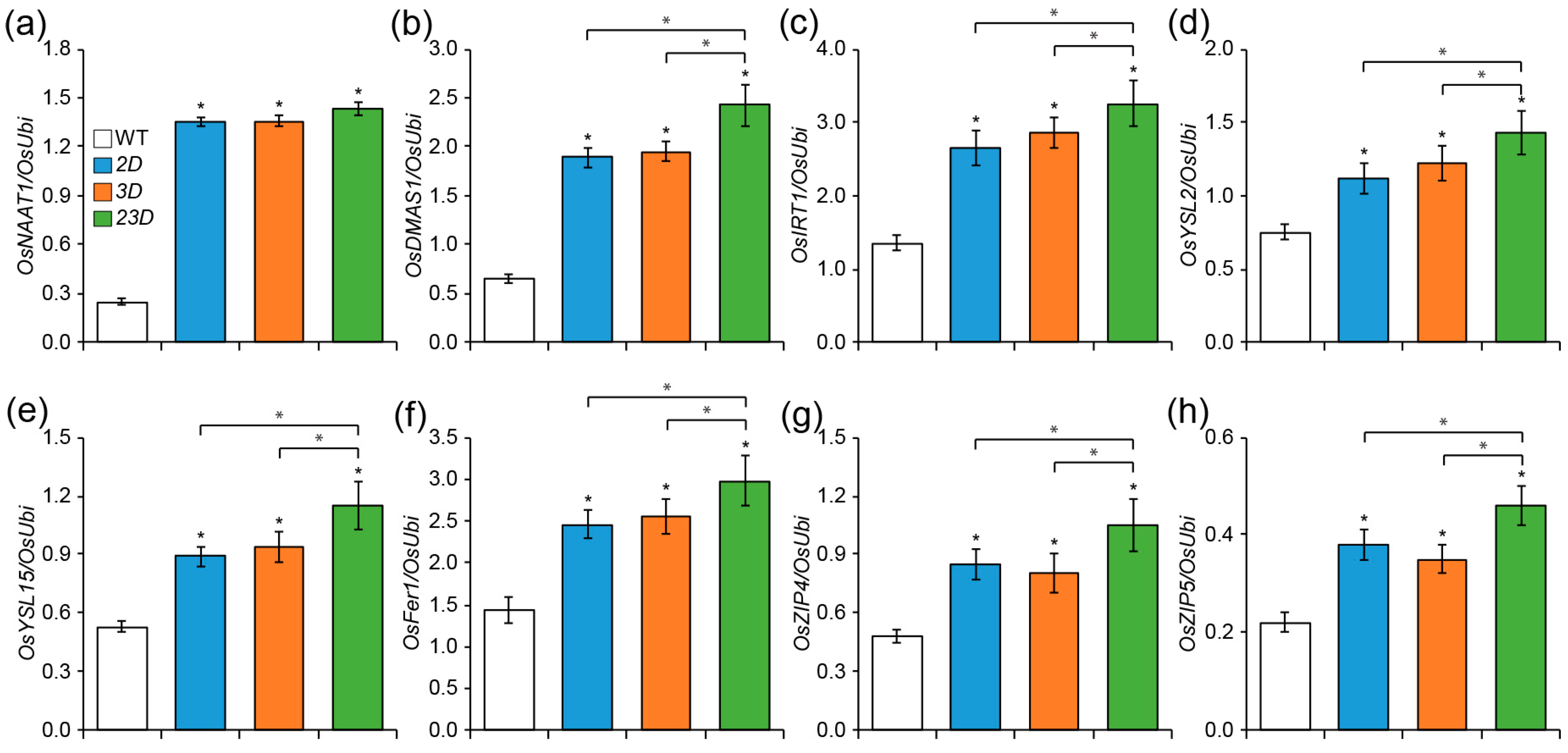
The above data prompted us to investigate the transcript abundance of various genes involved in the homeostasis of Fe and Zn (Figure 2): the nicotianamine aminotransferase gene OsNAAT1 and 2′-deoxymugineic acid synthase gene OsDMAS1, which act downstream of OsNAS genes and are involved in the synthesis of MAs [30,31]; OsIRT1 and OsYSL15 (Yellow Stripe1-Like 15), which uptake Fe from the soil as membrane transporters [32,33,34,35]; OsYSL2, which acts as NA-metal transporter and facilitates Fe translocation [36,37]; rice ferritin1 (OsFer1), which acts as a principal Fe storage protein [38]; and OsZIP4 and OsZIP5, which encode a membrane protein that transports Zn [39,40]. Transcript levels of these genes were found to be significantly higher in the roots of all the mutants compared to the WT (Figure 2). Overall, we found that the roots of 23D had the highest level of all the transcripts we looked at in comparison to others (Figure 2).

2.2. Expression Analysis of Three OsNAS Genes and the Comparison of Seedling Growth under High pH Conditions

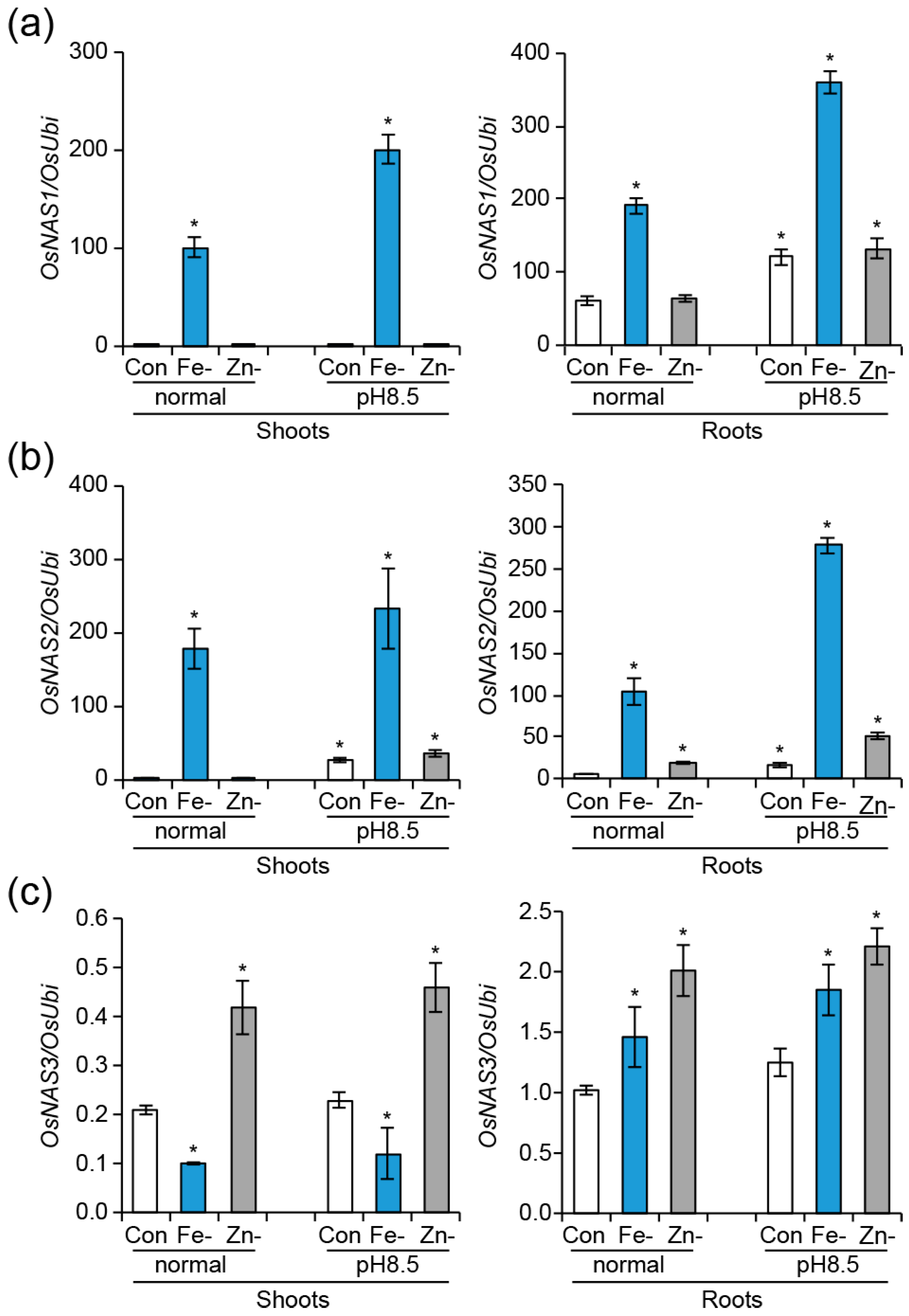
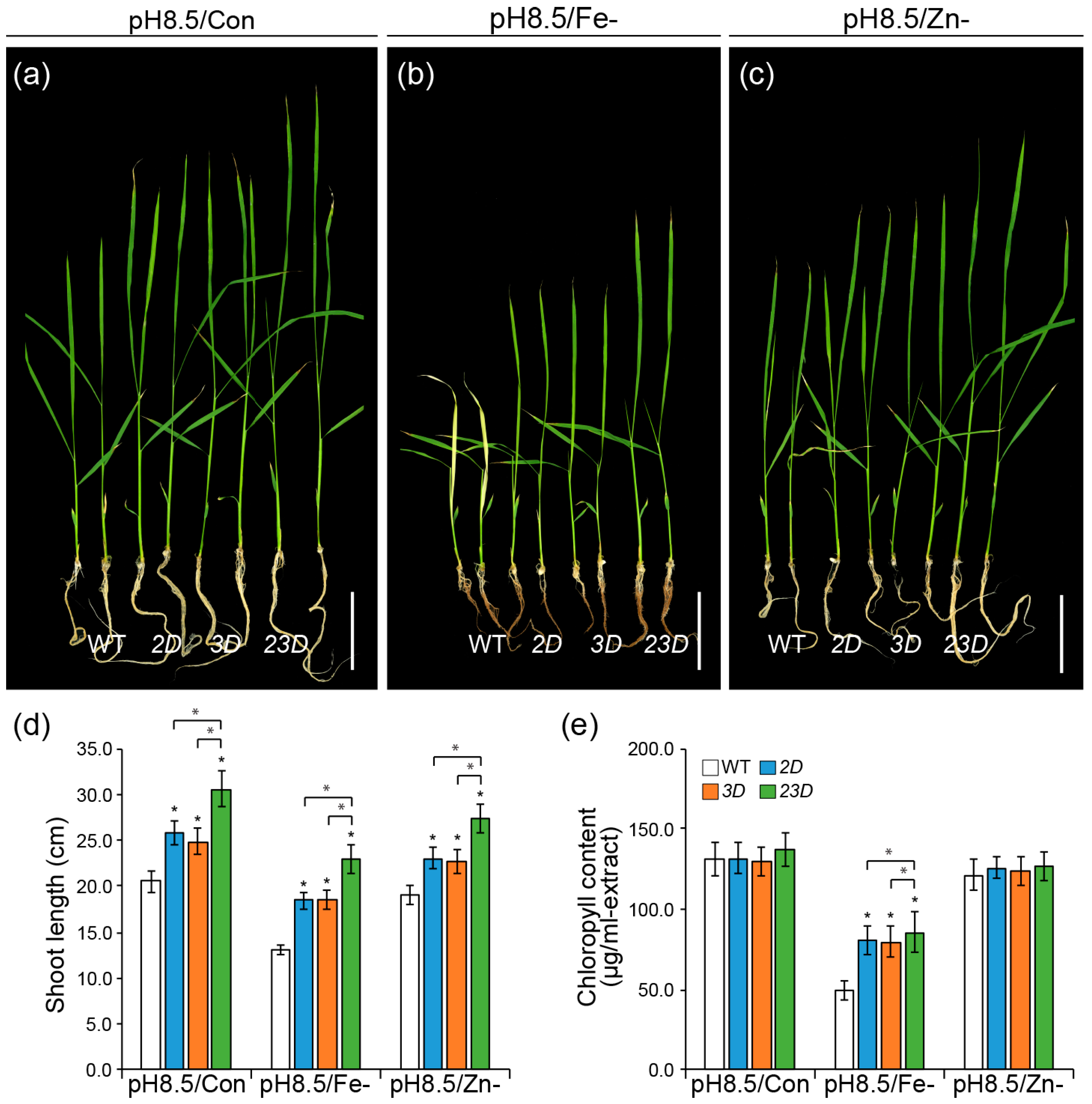
pH influences the solubility and ionic form of several elements in soil and affects nutrient availability for plant growth [41]. For instance, ferric ion precipitates as the utterly insoluble ferric hydroxide in an aerated solution with a pH greater than 8 [19]. Zn solubility reduces by 100-fold for each unit increase in pH [42]. Therefore, we speculated that high pH could affect the expression of OsNASs even under Fe or Zn sufficiency. Then, we analyzed the expression levels of OsNAS genes in shoots and roots of the seedlings grown under high pH (pH 8.5) solution, along with or without Fe or Zn (Figure 3). Transcript levels of OsNAS1 and OsNAS2 were upregulated under alkaline growth solution, as compared to normal growth conditions (pH 5.5) (Figure 3a,b). Even in seedling roots supplemented with Fe and Zn, high pH could promote their expression, indicating that the bioavailability of Fe and Zn decreased due to the alkaline media. High pH without Fe led to the highest levels of OsNAS1 and OsNAS2 expression, respectively, relative to other growth conditions. However, the shoots and roots of the seedlings grown under both growth conditions showed significantly lower OsNAS3 expressions in Fe-deficient media as compared to the seedlings cultivated with Fe (Figure 3c). Under Zn deficiency, no significant expression changes were observed for OsNAS1 in seedling shoots and roots (Figure 3a). In seedling shoots and roots, OsNAS2 expression increased upon Zn exclusion, but high pH raised it more (Figure 3b). Regardless of pH, OsNAS3 rose in Zn-deficient seedlings (Figure 3c). According to these results, Fe or Zn deficiency combined with high pH causes significant expression changes in seedling shoots and roots, and OsNAS gene expression responded differently to high pH. Then, we investigated the growth response of WT, 2D, 3D, and 23D seedlings under high pH Yoshida solution and a half-strength MS medium, with or without Fe or Zn (Figure 4 and Figure S3). In comparison to WT seedlings, all mutant seedlings of 2D, 3D, and 23D displayed a considerable increase in shoot length when grown in a high-pH environment, irrespective of Fe or Zn supply. Significantly, the 23D seedlings outperformed all others. For instance, in high-pH conditions, 2D, 3D, and 23D seedlings increased their shoot lengths by 25.9, 21.0, and 48.8% more than WT seedlings, respectively (Figure 4a,d and Figure S3a,d). At high-pH conditions without Fe, shoot lengths for 2D, 3D, and 23D seedlings increased by 41.2%, 41.2%, and 75.6%, respectively, in comparison to WT seedlings (Figure 4b,d and Figure S3b,d). Moreover, in high-pH conditions without Zn, 2D, 3D, and 23D seedlings’ shoot lengths increased by 21.1%, 20.0%, and 43.7%, respectively, in comparison to WT seedlings (Figure 4c,d and Figure S3c,d). Compared to WT, 2D, and 3D, our results suggest that the double-activation-tagged mutant 23D, which simultaneously expresses higher levels of OsNAS2 and OsNAS3, is better able to counteract the detrimental effects of high pH and high pH without Fe or Zn conditions on plant growth and development.

2.3. Increased Expression of Two OsNAS Genes Boosted Fe and Zn Contents in Mature Seeds

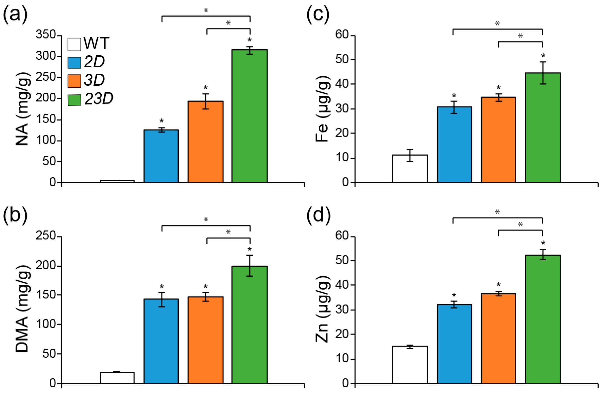
To test if the double activation of OsNAS2 and OsNAS3 might further increase Fe and Zn concentrations, we measured Fe and Zn concentrations in the flag leaves of WT, 2D, 3D, and 23D plants. We found that the mutant flag leaves had higher concentrations of Fe and Zn than the WT flag leaves (Figure S4). Importantly, flag leaves of 23D contained the highest levels of Fe and Zn in comparison with other mutants. For instance, compared to the WT, Fe concentrations were 1.49-, 1.51-, and 1.87-fold elevated in the 2D, 3D, and 23D mutants, respectively (Figure S4a), while Zn concentrations were 1.49-, 1.59-, and 1.94-fold elevated in the 2D, 3D and 23D mutants, respectively (Figure S4b). Elevated amounts of Fe and Zn in flag leaves were correlated with increased amounts of NA and DMA in mature seeds, increasing the accumulation of Fe and Zn [18,19]. Therefore, we measured NA, DMA, Fe, and Zn concentrations in mature seeds (Figure 5). NA concentrations in mature seeds of 2D, 3D, and 23D were found to be 20.2, 30.9, and 50.6 times higher, respectively, compared to WT (Figure 5a). In addition, DMA concentrations in 2D, 3D and 23D seeds were also increased to being 7.2-, 7.4-, and 10.0-fold higher, respectively, compared to the WT (Figure 5b). Following the enhanced NA and DMA amounts, both Fe and Zn were observed to be enhanced (Figure 5c,d). Relative to mature WT seeds, Fe levels in 2D, 3D, and 23D were 2.7, 3.1, and 4.0 times higher, respectively. (Figure 5c). We also performed Perls’ Prussian staining to visualize the accumulation and intensity of the Fe in the mature seeds and embryos (Figure S5). An enhanced blue color was observed in stained 2D, 3D, and 23D mature seeds and embryos compared to the WT (Figure S5a,b). Notably, the strongest color observed visually was evident in mature 23D seeds (Figure S5a) and embryos (Figure S5b), suggesting the highest accumulation of Fe. Furthermore, concentrations of Zn in mature 2D, 3D, and 23D seeds were found to be 2.1, 2.4, and 3.5 times higher than those of WT, respectively (Figure 5d). These data suggest that dual activation of OsNAS2 and OsNAS3 could accumulate more Fe and Zn in rice grains than single-activation mutants of OsNAS2 or OsNAS3.
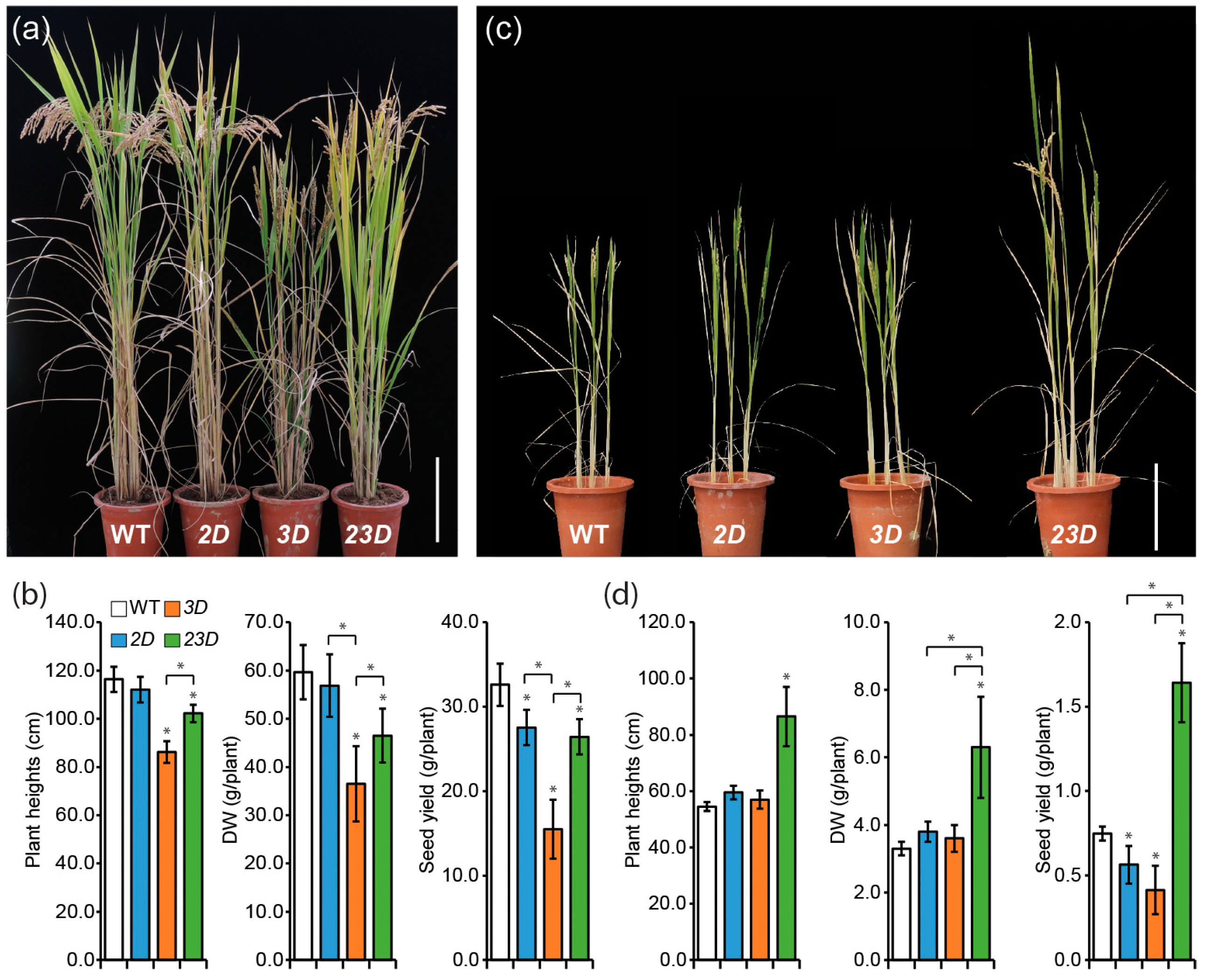
However, negative impacts on agronomic traits were observed for the matured mutant plants compared to WT plants when grown in paddy fields (Figure 6a,b). 2D exhibited similar plant height and biomass as compared to the WT, but less seed yield (84.5% of WT) than the WT. Plant height, aboveground dry weight, and seed yield of 3D were decreased to 74.1%, 61.2%, and 47.5% of WT, respectively. 23D’s agronomic characteristics fell in between 2D and 3D. When the plants were grown in alkaline soil, the single activation-tagged mutants displayed similar plant height and biomass, but less seed yield as compared to WT (Figure 6c,d). Importantly, 23D plants exhibited a highly significant increase in plant height, aboveground mass, and seed yield compared to all others (Figure 6c,d).

2.4. Increased Tolerance to Excess Metals during the Seedling Growth

NA is thought to participate in the detoxification of excess metals. Therefore, we verified the growth response of the mutants under excessive concentrations of various heavy metals (Figure 7 and Figure S6). 2D, 3D, and 23D seedlings exhibited better growth under excessive amounts of Zn (5 mM), Cu (0.3 mM), Fe (1 mM), Ni (0.5 mM), and Co (0.5 mM) compared to the WT. Interestingly, the 23D mutant seedling was the best performer under all the treatments of excess heavy metal concentrations (Figure 7 and Figure S6). For instance, shoot lengths in 2D, 3D, and 23D were 12.4%, 22.9%, and 31.4% higher than the WT, respectively, at a higher Zn level (Figure 7a,d); shoot lengths of 2D, 3D, and 23D were 40.6%, 57.8%, and 89.1% more than the WT, respectively, at an excess Cu level (Figure 7b,d); shoot lengths of 2D, 3D, and 23D were 8.8%, 10.5%, and 13.8% more than the WT, respectively, at an excess Fe level (Figure 7c,d); shoot lengths of 2D, 3D, and 23D were 21.5%, 30.0%, and 35.5% more than the WT, respectively, at an excess Ni level (Figure S6a,c); shoot lengths of 2D, 3D, and 23D were 14.5%, 25.9%, and 42.5% more than the WT, respectively, at an excess Co level (Figure S6b,c). These data suggest that concomitant activation of OsNAS2 and OsNAS3 can improve plant tolerance at elevated heavy metal toxic conditions.

3. Discussion

Transgenic approaches, especially multiple gene expression, have already been proven effective to biofortify enhanced amounts of Fe and Zn in rice grains. Much research on the biofortification of Fe and Zn in rice grains has introduced one of the three OsNAS genes alone or in combination with other genes involved in metal homeostasis, but none of them have introduced two OsNAS genes at once. In this study, we generated a transgenic line (23D) with increased OsNAS2 and OsNAS3 expression to see if simultaneous activation of OsNAS2 and OsNAS3 would improve the accumulation of Fe and Zn in rice grains more than the single-activation-tagged mutants. Compared to the WT and single-activation-tagged mutants, NA and DMA levels were considerably higher in 23D, and as a result, Fe and Zn levels were significantly raised in mature grains. In addition, 23D had much higher concentrations of Fe and Zn in its leaves and roots, suggesting that NA and DMA had also accumulated there. Hence, the mutants were more resistant to Fe and Zn deficiency and high-pH conditions. In particular, 23D plants outperformed all others by demonstrating more tolerance.
Combinatorial effects of OsNAS2 and OsNAS3 resulted in 23D’s improved properties as compared to 2D or 3D. This might have been because OsNAS2 and OsNAS3 have differing physiological functions [15]. In other words, OsNAS2 expression was present in Fe-sufficient root, and under Fe deficiency, its levels significantly increased in roots and leaves. OsNAS3 expression was enhanced in roots but inhibited in leaves in response to Fe deprivation, although the OsNAS3 transcript was present in both Fe-sufficient roots and leaves. In addition, Zn deficiency increased the expression of OsNAS3 in roots and shoots while marginally decreasing the expression of OsNAS2 in roots [31]. OsNAS2 was found to be expressed in companion cells and pericycle cells close to the protoxylem in Fe-sufficient roots, according to promoter-GUS analysis. This suggests that OsNAS2 has a role in DMA secretion for Fe absorption in roots. However, because OsNAS3 is only expressed in the root pericycle and companion cells, it is possible that it is not involved in DMA secretion [15]. Moreover, OsNAS3 expression was seen in the entire leaf tissues of the control plants [15]. Therefore, OsNAS3 may work to produce NA or following parts such as DMA for the distribution of Fe and Zinc in rice plants.
NA is the precursor of DMA, which is synthesized by NAS, and NA is processed by NAAT and DMAS for synthesizing DMA [30,43]. We found that OsNAAT1 and OsDMAS1 were highly expressed in the mutants, with 23D showing the highest expression. This may account for the increased levels of NA and DMA in 23D. In rice, NA chelates Fe(II) and Zn(II) and carries them into the rice grains, and DMA chelates Fe(III) and transports it into root cells [2,13]. Through activating Fe(III)-DMA and Zn(II)-NA chelating processes and, likely, utilizing the YSL and Zn transporter genes, respectively, enhanced NA and DMA promote Fe and Zn accumulation in grains [26]. Moreover, mRNA levels of the Zn transporters (OsZIP4 and OsZIP5), as well as the YSL transporters (OsYSL2 and OsYSL15), were found to be much greater in mutants, which likely explains why transgenic plants and grains accumulate more Fe and Zn. The increased expression of the Fe transporter OsIRT1 and the Fe storage protein OsFer1 in 23D may also be a contributing factor to the elevated levels of Fe and Zn. The mature grains, roots, and leaves of transgenic OsIRT1 overexpressing plants were shown to have increased Fe and Zn concentrations [34].
However, the rice ferritins (OsFer) were not reported to be used for rice biofortification; rather, OsFer genes were found to be necessary for a positive response against Fe-induced oxidative stress [44]. Accordingly, 23D seedlings were more tolerant to Fe toxic conditions, probably due to the elevated expression of OsFer1 in 23D than in 2D, 3D, and WT. Similarly, OsNAS3 knockout plants were sensitive to excess Fe, whereas a transgenic rice line overexpressing barley HvNAS1 with elevated NA production was tolerant of excess Fe [45]. While 23D plants were the best performers under Zn, Cu, Ni, and Co toxicities, 2D and 3D plants also performed better under these conditions. This is likely because 23D plants have improved abilities to chelate more of these metal ions than other plants, which reduces their toxic effects on plant growth.
Because of enhanced accumulation of Fe and Zn in shoots and roots due to higher levels of NA and DMA, transgenic plants were more resistant to Fe- and Zn-deficient conditions as well as high-pH conditions with or without Fe and/or Zn. As a result, 23D plants would grow more successfully than single-activation mutants in alkaline growth conditions where high pH induces nutritional limitation. To maintain the solubility and availability of micronutrients to plants, soil pH is a crucial feature. Alkaline soils have reduced micronutrient availability, which has a detrimental effect on plant growth and yield [46]. For instance, Fe is abundant in soil but not easily soluble, especially in calcareous soils, which make up 30% of the earth’s surface [47]. In addition, the uptake of other metal micronutrients, such as Zn and Mn, can also be inhibited in alkaline soils [48]. The 23D plants exhibited superior tolerance due to the combinatorial effects of OsNAS2 and OsNAS3, as evident from the significant expression enhancement of OsNAS2 and OsNAS3 in the shoots and roots of rice seedlings under normal and high pH environments with or without Fe or Zn. Elevated expressions of OsNAS genes under high pH with or without Fe or Zn also explain the better tolerance of the mutants in alkaline conditions.
Although we measured Fe and Zn concentration only in the dehusked grains of the transgenic rice plants, we speculate that Fe and Zn amounts would be higher in the milled grains as well, with higher bioavailability. Intensified blue colors were observed for the whole transgenic grains and their embryos after Perls’ staining, where the strongest blue color in the 23D grain was obvious. Furthermore, previous studies on the single-activation-tagged mutants, 2D and 3D, showed that the increased Fe and Zn in the milled grains were more bioavailable, as more NA and DMA would bind to more Fe and Zn and transport them into the grain endosperm [17,18,19]. In line with previous reports, 23D seeds may accumulate more Fe and Zn bound to NA and/or DMA than 2D, 3D, or WT seeds, resulting in increased bioavailability. Mouse-feeding experiments can be used to test this in the future. An important point to consider while biofortifying rice is avoiding the negative impacts on normal plant growth and yield. Unfortunately, activation-tagged mutants displayed decreased growth and seed yield. Because they contained the higher amounts of Fe and Zn throughout all growth stages, they might have suffered from the oxidative stress, resulting to poor growth and reduction in yield. Although we did not observe any difference during the seedling stage, the influence increases over time and becomes more pronounced throughout the mature stage, making the negative effect more obvious. As a result, shorter plants with reduced yield were observed for 2D and 3D plants, probably for excess Fe-induced oxidative stress conditions in planta [19]. Similarly, 23D plants showed almost resembling agronomic traits as observed in 2D, but better than 3D. However, when growth under high-pH soil, 23D exhibited the greatest agronomic traits among WT and mutants, indicating the synergistic effect of OsNAS2 and OsNAS3.
Future studies on 23D plants would benefit from their raised biofortified Fe and Zn levels as well as their increased tolerance to alkalinity and metal toxicity. By combining OsNAS2 and OsNAS3 with additional genes, future studies may further raise the Fe and Zn contents in rice grains.

4. Materials and Methods

4.1. Plant Materials and Growth

The Korean rice Dongjin (Oryza sativa L. ssp. japonica) was used as the experimental cultivar in this study. Two activation-tagged mutants (OsNAS2-D1 and OsNAS3-D1) have been described previously [17,18]. Wild-type segregants from each progeny are referred to as wild-type (WT) progeny hereafter. Thirty-day-old seedlings of WT and activation-tagged mutant plants were transplanted into the paddy fields located at the Daegu Gyeongbuk Institute of Science and Technology (36° N) in 2020 under natural conditions. For the growth evaluation in calcareous soil, four-leaf-stage seedlings of WT and activation-tagged mutants were raised in pots (diameter 17 cm, height 18 cm) with nursery soil containing soluble CaCO3 to adjust to pH 8.0 under outdoor growth conditions until harvest. To prevent the soil from drying out, watering was performed frequently.

4.2. Hydroponic Culture for the Growth Test under Fe and Zn Deficiencies

The rice seeds were surface-sterilized, submerged in deionized water for 24 h in the dark, and then allowed to sprout for a further 24 h on wet filter paper. The uniform seedlings were selected and transferred into the containers containing Yoshida hydroponic solution for 15 days. Yoshida solution [49] with the following composition at full strength: NH4NO 1.43 mM, NaH2PO4·2H2O 0.32 mM, K2SO4 0.51 mM, CaCl2·2H2O 1.00 mM, MgSO4·7H2O 1.64 mM, MnCl2·4H2O 9.47 μm, (NH4)6MO7O24·4H2O 0.07 μm, H3BO3 18.88 μm, ZnSO4·7H2O 0.15 μm, CuSO4·5H2O 0.16 μm, FeCl3·6H2O 35.61 μm, citric acid (monohydrate) 70.83 μm. The pH of the working solution was adjusted to 5.5 with 1N sodium hydroxide (NaOH) every two days. To conduct our tests for micronutrient deficiencies, seedlings were moved into the Yoshida solution deficient in FeCl3·6H2O or ZnSO4·7H2O.

4.3. Seedling Growth on MS Solid Medium under Fe and Zn Deficiencies

WT and transgenic rice seeds were germinated on a standard MS agar medium supplemented with 0.1 100 μM Fe (III)-EDTA and 30 μM ZnSO4 as micronutrients [50]. Seedlings were grown for 10 days at 28 °C under continuous light. For our micronutrient deficiency tests, seeds were germinated and grown on an MS medium devoid of ZnSO4 or Fe (III)-EDTA for 10 days.

4.4. RNA Isolation and Quantitative Real-Time PCR

The total RNA was extracted from each tissue type with WelPrep total RNA isolation reagent (WELGENE, Gyeongsan, Republic of Korea), according to the manufacturer’s instructions, and treated with RNase-free DNase I (Takara Bio, Shiga, Japan) to remove the genomic DNA. First-strand cDNA was synthesized from 2 µg of total RNA in a 25 µL reaction mixture using the ImProm II Reverse Transcriptase system kit (Promega, Madison, WI, USA), followed by quantitative real-time PCR (qRT-PCR) analysis to determine gene expression levels (Bio-Rad, CFX96 Touch Real-Time PCR Detection System, USA) using a SYBR premix ExTaq kit (Takara Bio, Shiga, Japan). The rice ubiquitin gene (LOC_Os06g46770) was used to normalize the expression ratio for each gene, and changes in expression were calculated via the ΔΔCt method. The gene-specific primers for PCR are listed in Table S1.

4.5. Measurement of Chlorophyll Concentrations

Chlorophyll was extracted from 0.1 g of leaf samples using 1 mL of 80% acetone [35]. After homogenization, the samples were incubated for 15 min on ice and spun for 10 min at 15,000× g, and then an aliquot of the supernatant fraction was collected for spectrophotometer measurements of A663 and A643. Chlorophyll concentrations, including chlorophyll a and b, were determined according to the method of [51].

4.6. Element Analysis in Plant Tissues

Samples from each tissue were dried at 70 °C until the weight remained constant, and then samples were digested in 1 mL of 11 N HNO3 for 3 days at 180 °C. After dilution, metal concentrations in the solutions were determined by inductively coupled plasma mass spectroscopy (ICP-MS, Varian 820-MS, Varian, Australia).

4.7. Perls’ Staining of the Mature Seeds

After the husk was manually removed, seeds were immersed in ultrapure water for four to five hours before being cut in two lengthwise through the embryo in a Petri dish using a razor blade. The cut seeds were submerged in freshly prepared Perls’ Prussian blue [52] solution (2% (v/v) HCl and 2% (w/v) potassium ferrocyanide), vacuum infiltrated for 15 min (500 mbar), and incubated at room temperature for 30 min. The seeds were then rinsed three times in distilled water and mounted in ultrapure water for visualization on a stereomicroscope.

4.8. Determination of NA and DMA Levels

Mature seeds were crushed into a fine powder with a Multi-Beads Shocker (Yasui Kikai, Osaka, Japan), and then 400 µL of 80% ethanol was added to the samples. The samples were centrifuged at 12,000× g for 5 min and supernatants were collected. Two more centrifugations were performed using the supernatants collected each time. Concentrations of NA and DMA were analyzed using the final supernatants by LC/ESI-TOF-MS, as previously described [53].

4.9. Excess Metal Tolerance

For the excess-metal treatments, seeds were germinated and grown for 10 days on solid media containing half-strength MS salts and supplemented with 5 mM ZnCl2, 0.3 mM CuCl2, 1 mM FeCl3, 0.5 mM NiCl2, or 0.5 mM CoCl2.

Supplementary Materials

The following supporting information can be downloaded at https://www.mdpi.com/article/10.3390/ijms24076568/s1.

Author Contributions

S.L. and J.-S.J. conceived the study; S.L., N.K.N., G.A., H.G.N. and J.-S.J. designed the experiments; S.L., M.M.R. and H.N. performed the experiments; G.A. provided the T-DNA mutants. S.L., M.M.R. and J.-S.J. analyzed the data and wrote the paper. All the authors discussed the results. All authors have read and agreed to the published version of the manuscript.

Funding

This research was supported by the Institute for Basic Science (IBS-R013-D1) from the Ministry of Science, ICT and Future Planning, the National Research Foundation, Korea (2021M3E5E6025387), and the Rural Development Administration, Korea (PJ01690101).

Institutional Review Board Statement

Not applicable.

Informed Consent Statement

Not applicable.

Data Availability Statement

Not applicable.

Conflicts of Interest

The authors declare no conflict of interest.

References

  1. Stangoulis, J.C.R.; Knez, M. Biofortification of major crop plants with iron and zinc—Achievements and future directions. Plant Soil 2022, 474, 57–76. [Google Scholar] [CrossRef]
  2. Wairich, A.; Ricachenevsky, F.K.; Lee, S. A tale of two metals: Biofortification of rice grains with iron and zinc. Front. Plant Sci. 2022, 13, 944624. [Google Scholar] [CrossRef] [PubMed]
  3. Wu, T.-Y.; Gruissem, W.; Bhullar, N.K. Facilitated citrate-dependent iron translocation increases rice endosperm iron and zinc concentrations. Plant Sci. 2018, 270, 13–22. [Google Scholar] [CrossRef] [PubMed]
  4. Clemens, S. The cell biology of zinc. J. Exp. Bot. 2021, 73, 1688–1698. [Google Scholar] [CrossRef] [PubMed]
  5. Colvin, R.A.; Holmes, W.R.; Fontaine, C.P.; Maret, W. Cytosolic zinc buffering and muffling: Their role in intracellular zinc homeostasis. Metallomics 2010, 2, 306–317. [Google Scholar] [CrossRef] [PubMed]
  6. Muñoz, M.; Acheson, A.G.; Auerbach, M.; Besser, M.; Habler, O.; Kehlet, H.; Liumbruno, G.M.; Lasocki, S.; Meybohm, P.; Baikady, R.R.; et al. International consensus statement on the peri-operative management of anaemia and iron deficiency. Anaesthesia 2017, 72, 233–247. [Google Scholar] [CrossRef] [Green Version]
  7. Piskin, E.; Cianciosi, D.; Gulec, S.; Tomas, M.; Capanoglu, E. Iron Absorption: Factors, Limitations, and Improvement Methods. ACS Omega 2022, 7, 20441–20456. [Google Scholar] [CrossRef]
  8. Ozyildirim, S.; Baltaci, S.B. Cardiovascular Diseases and Zinc. Biol. Trace Element Res. 2022, 201, 1615–1626. [Google Scholar] [CrossRef]
  9. Anusha, G.; Rao, D.S.; Jaldhani, V.; Beulah, P.; Neeraja, C.N.; Gireesh, C.; Anantha, M.S.; Suneetha, K.; Santhosha, R.; Prasad, A.S.H.; et al. Grain Fe and Zn content, heterosis, combining ability and its association with grain yield in irrigated and aerobic rice. Sci. Rep. 2021, 11, 10579. [Google Scholar] [CrossRef]
  10. Assunção, A.G.; Cakmak, I.; Clemens, S.; González-Guerrero, M.; Nawrocki, A.; Thomine, S. Micronutrient homeostasis in plants for more sustainable agriculture and healthier human nutrition. J. Exp. Bot. 2022, 73, 1789–1799. [Google Scholar] [CrossRef]
  11. Garg, M.; Sharma, N.; Sharma, S.; Kapoor, P.; Kumar, A.; Chunduri, V.; Arora, P. Biofortified Crops Generated by Breeding, Agronomy, and Transgenic Approaches Are Improving Lives of Millions of People around the World. Front. Nutr. 2018, 5, 12. [Google Scholar] [CrossRef] [PubMed]
  12. Laffont, C.; Arnoux, P. The ancient roots of nicotianamine: Diversity, role, regulation and evolution of nicotianamine-like metallophores. Metallomics 2020, 12, 1480–1493. [Google Scholar] [CrossRef] [PubMed]
  13. Nozoye, T. The Nicotianamine Synthase Gene Is a Useful Candidate for Improving the Nutritional Qualities and Fe-Deficiency Tolerance of Various Crops. Front. Plant Sci. 2018, 9, 340. [Google Scholar] [CrossRef] [PubMed]
  14. Shojima, S.; Nishizawa, N.-K.; Mori, S. Establishment of a Cell-Free System for the Biosynthesis of Nicotianamine. Plant Cell Physiol. 1989, 30, 673–677. [Google Scholar] [CrossRef]
  15. Inoue, H.; Higuchi, K.; Takahashi, M.; Nakanishi, H.; Mori, S.; Nishizawa, N.K. Three rice nicotianamine synthase genes, OsNAS1, OsNAS2, and OsNAS3 are expressed in cells involved in long-distance transport of iron and differentially regulated by iron. Plant J. 2003, 36, 366–381. [Google Scholar] [CrossRef]
  16. Johnson, A.A.T.; Kyriacou, B.; Callahan, D.L.; Carruthers, L.; Stangoulis, J.; Lombi, E.; Tester, M. Constitutive Overexpression of the OsNAS Gene Family Reveals Single-Gene Strategies for Effective Iron- and Zinc-Biofortification of Rice Endosperm. PLoS ONE 2011, 6, e24476. [Google Scholar] [CrossRef] [Green Version]
  17. Lee, S.; Jeon, U.S.; Lee, S.J.; Kim, Y.-K.; Persson, D.P.; Husted, S.; Schjørring, J.K.; Kakei, Y.; Masuda, H.; Nishizawa, N.K.; et al. Iron fortification of rice seeds through activation of the nicotianamine synthase gene. Proc. Natl. Acad. Sci. USA 2009, 106, 22014–22019. [Google Scholar] [CrossRef] [Green Version]
  18. Lee, S.; Persson, D.P.; Hansen, T.H.; Husted, S.; Schjoerring, J.K.; Kim, Y.-S.; Jeon, U.S.; Kim, Y.-K.; Kakei, Y.; Masuda, H.; et al. Bio-available zinc in rice seeds is increased by activation tagging of nicotianamine synthase. Plant Biotechnol. J. 2011, 9, 865–873. [Google Scholar] [CrossRef]
  19. Lee, S.; Kim, Y.-S.; Jeon, U.S.; Kim, Y.-K.; Schjoerring, J.K.; An, G. Activation of Rice nicotianamine synthase 2 (OsNAS2) enhances iron availability for biofortification. Mol. Cells 2012, 33, 269–275. [Google Scholar] [CrossRef]
  20. Masuda, H.; Usuda, K.; Kobayashi, T.; Ishimaru, Y.; Kakei, Y.; Takahashi, M.; Higuchi, K.; Nakanishi, H.; Mori, S.; Nishizawa, N.K. Overexpression of the Barley Nicotianamine Synthase Gene HvNAS1 Increases Iron and Zinc Concentrations in Rice Grains. Rice 2009, 2, 155–166. [Google Scholar] [CrossRef] [Green Version]
  21. Usuda, K.; Wada, Y.; Ishimaru, Y.; Kobayashi, T.; Takahashi, M.; Nakanishi, H.; Nagato, Y.; Mori, S.; Nishizawa, N.K. Genetically engineered rice containing larger amounts of nicotianamine to enhance the antihypertensive effect. Plant Biotechnol. J. 2009, 7, 87–95. [Google Scholar] [CrossRef] [PubMed]
  22. Moreno-Moyano, L.T.; Bonneau, J.P.; Sánchez-Palacios, J.T.; Tohme, J.; Johnson, A.A.T. Association of Increased Grain Iron and Zinc Concentrations with Agro-morphological Traits of Biofortified Rice. Front. Plant Sci. 2016, 7, 1463. [Google Scholar] [CrossRef] [PubMed] [Green Version]
  23. Zheng, L.; Cheng, Z.; Ai, C.; Jiang, X.; Bei, X.; Zheng, Y.; Glahn, R.P.; Welch, R.M.; Miller, D.D.; Lei, X.G.; et al. Nicotianamine, a Novel Enhancer of Rice Iron Bioavailability to Humans. PLoS ONE 2010, 5, e10190. [Google Scholar] [CrossRef] [PubMed]
  24. Wirth, J.; Poletti, S.; Aeschlimann, B.; Yakandawala, N.; Drosse, B.; Osorio, S.; Tohge, T.; Fernie, A.R.; Günther, D.; Gruissem, W.; et al. Rice endosperm iron biofortification by targeted and synergistic action of nicotianamine synthase and ferritin. Plant Biotechnol. J. 2009, 7, 631–644. [Google Scholar] [CrossRef] [PubMed]
  25. Banakar, R.; Fernández, A.A.; Díaz-Benito, P.; Abadia, J.; Capell, T.; Christou, P. Phytosiderophores determine thresholds for iron and zinc accumulation in biofortified rice endosperm while inhibiting the accumulation of cadmium. J. Exp. Bot. 2017, 68, 4983–4995. [Google Scholar] [CrossRef] [PubMed] [Green Version]
  26. Díaz-Benito, P.; Banakar, R.; Rodríguez-Menéndez, S.; Capell, T.; Pereiro, R.; Christou, P.; Abadía, J.; Fernández, B.; Álvarez-Fernández, A. Iron and zinc in the embryo and endosperm of rice (Oryza sativa L.) seeds in contrasting 2′-deoxymugineic acid/nicotianamine scenarios. Front. Plant Sci. 2018, 9, 1190. [Google Scholar] [CrossRef] [Green Version]
  27. Trijatmiko, K.R.; Dueñas, C.; Tsakirpaloglou, N.; Torrizo, L.; Arines, F.M.; Adeva, C.; Balindong, J.; Oliva, N.; Sapasap, M.V.; Borrero, J.; et al. Biofortified indica rice attains iron and zinc nutrition dietary targets in the field. Sci. Rep. 2016, 6, 19792. [Google Scholar] [CrossRef] [Green Version]
  28. Boonyaves, K.; Wu, T.-Y.; Gruissem, W.; Bhullar, N.K. Enhanced grain iron levels in rice expressing an iron-regulated metal transporter, nicotianamine synthase, and ferritin gene cassette. Front. Plant Sci. 2017, 8, 130. [Google Scholar] [CrossRef] [Green Version]
  29. Kawakami, Y.; Gruissem, W.; Bhullar, N.K. Novel rice iron biofortification approaches using expression of ZmYS1 and OsTOM1 controlled by tissue-specific promoters. J. Exp. Bot. 2022, 73, 5440–5459. [Google Scholar] [CrossRef]
  30. Bashir, K.; Inoue, H.; Nagasaka, S.; Takahashi, M.; Nakanishi, H.; Mori, S.; Nishizawa, N.K. Cloning and Characterization of Deoxymugineic Acid Synthase Genes from Graminaceous Plants. J. Biol. Chem. 2006, 281, 32395–32402. [Google Scholar] [CrossRef] [Green Version]
  31. Suzuki, M.; Tsukamoto, T.; Inoue, H.; Watanabe, S.; Matsuhashi, S.; Takahashi, M.; Nakanishi, H.; Mori, S.; Nishizawa, N.K. Deoxymugineic acid increases Zn translocation in Zn-deficient rice plants. Plant Mol. Biol. 2008, 66, 609–617. [Google Scholar] [CrossRef] [PubMed] [Green Version]
  32. Inoue, H.; Kobayashi, T.; Nozoye, T.; Takahashi, M.; Kakei, Y.; Suzuki, K.; Nakazono, M.; Nakanishi, H.; Mori, S.; Nishizawa, N.K. Rice OsYSL15 Is an Iron-regulated Iron(III)-Deoxymugineic Acid Transporter Expressed in the Roots and Is Essential for Iron Uptake in Early Growth of the Seedlings. J. Biol. Chem. 2009, 284, 3470–3479. [Google Scholar] [CrossRef] [PubMed] [Green Version]
  33. Ishimaru, Y.; Suzuki, M.; Tsukamoto, T.; Suzuki, K.; Nakazono, M.; Kobayashi, T.; Wada, Y.; Watanabe, S.; Matsuhashi, S.; Takahashi, M.; et al. Rice plants take up iron as an Fe3+-phytosiderophore and as Fe2+. Plant J. 2006, 45, 335–346. [Google Scholar] [CrossRef] [PubMed]
  34. Lee, S.; An, G. Over-expression of OsIRT1 leads to increased iron and zinc accumulations in rice. Plant. Cell Environ. 2009, 32, 408–416. [Google Scholar] [CrossRef]
  35. Lee, S.; Chiecko, J.C.; Kim, S.A.; Walker, E.L.; Lee, Y.; Guerinot, M.L.; An, G. Disruption of OsYSL15 Leads to Iron Inefficiency in Rice Plants. Plant Physiol. 2009, 150, 786–800. [Google Scholar] [CrossRef] [Green Version]
  36. Ishimaru, Y.; Masuda, H.; Bashir, K.; Inoue, H.; Tsukamoto, T.; Takahashi, M.; Nakanishi, H.; Aoki, N.; Hirose, T.; Ohsugi, R.; et al. Rice metal-nicotianamine transporter, OsYSL2, is required for the long-distance transport of iron and manganese. Plant J. 2010, 62, 379–390. [Google Scholar] [CrossRef]
  37. Koike, S.; Inoue, H.; Mizuno, D.; Takahashi, M.; Nakanishi, H.; Mori, S.; Nishizawa, N.K. OsYSL2 is a rice metal-nicotianamine transporter that is regulated by iron and expressed in the phloem. Plant J. 2004, 39, 415–424. [Google Scholar] [CrossRef]
  38. Harrison, P.M.; Arosio, P. The ferritins: Molecular properties, iron storage function and cellular regulation. Biochim. Biophys. Acta (BBA) Bioenerg. 1996, 1275, 161–203. [Google Scholar] [CrossRef] [Green Version]
  39. Ishimaru, Y.; Suzuki, M.; Kobayashi, T.; Takahashi, M.; Nakanishi, H.; Mori, S.; Nishizawa, N.K. OsZIP4, a novel zinc-regulated zinc transporter in rice. J. Exp. Bot. 2005, 56, 3207–3214. [Google Scholar] [CrossRef] [Green Version]
  40. Lee, S.; Jeong, H.J.; Kim, S.A.; Lee, J.; Guerinot, M.L.; An, G. OsZIP5 is a plasma membrane zinc transporter in rice. Plant Mol. Biol. 2010, 73, 507–517. [Google Scholar] [CrossRef]
  41. Penn, C.J.; Camberato, J.J. Critical Review on Soil Chemical Processes that Control How Soil pH Affects Phosphorus Availability to Plants. Agriculture 2019, 9, 120. [Google Scholar] [CrossRef] [Green Version]
  42. Desta, M.; Broadley, M.; McGrath, S.; Hernandez-Allica, J.; Hassall, K.; Gameda, S.; Amede, T.; Haefele, S. Plant Available Zinc Is Influenced by Landscape Position in the Amhara Region, Ethiopia. Plants 2021, 10, 254. [Google Scholar] [CrossRef] [PubMed]
  43. Higuchi, K.; Suzuki, K.; Nakanishi, H.; Yamaguchi, H.; Nishizawa, N.-K.; Mori, S. Cloning of Nicotianamine Synthase Genes, Novel Genes Involved in the Biosynthesis of Phytosiderophores. Plant Physiol. 1999, 119, 471–480. [Google Scholar] [CrossRef] [Green Version]
  44. Stein, R.J.; Ricachenevsky, F.K.; Fett, J.P. Differential regulation of the two rice ferritin genes (OsFER1 and OsFER2). Plant Sci. 2009, 177, 563–569. [Google Scholar] [CrossRef]
  45. Aung, M.S.; Masuda, H.; Nozoye, T.; Kobayashi, T.; Jeon, J.S.; An, G.; Nishizawa, N.K. Nicotianamine Synthesis by OsNAS3 Is Important for Mitigating Iron Excess Stress in Rice. Front. Plant Sci. 2019, 10, 660. [Google Scholar] [CrossRef] [Green Version]
  46. Gentili, R.; Ambrosini, R.; Montagnani, C.; Caronni, S.; Citterio, S. Effect of soil ph on the growth, reproductive investment and pollen allergenicity of Ambrosia artemisiifolia L. Front. Plant Sci. 2018, 9, 1335. [Google Scholar] [CrossRef] [PubMed] [Green Version]
  47. Chen, Y.; Barak, P. Iron nutrition of plants in calcareous soils. Adv. Agron. 1982, 35, 217–240. [Google Scholar] [CrossRef]
  48. George, E.; Horst, W.J.; Neumann, E. Adaptation of Plants to Adverse Chemical Soil Conditions. In Marschner’s Mineral Nutrition of Higher Plants, 3rd ed.; Marschner, P., Ed.; Academic Press: London, UK, 2012; pp. 409–472. [Google Scholar] [CrossRef]
  49. Yoshida, S.; Forno, D.A.; Cock, J.H.; Gomez, K.A. Laboratory Manual for Physiological Studies of Rice; IRRI: Laguna, Philippines, 1976; p. 83. [Google Scholar]
  50. Murashige, T.; Skoog, F. A Revised Medium for Rapid Growth and Bioassays with Tobacco Tissue Cultures. Physiol. Plant. 1962, 15, 473–497. [Google Scholar] [CrossRef]
  51. Arnon, D.I. Copper enzymes in isolated chloroplasts. Polyphenoloxidase in Beta vulgaris. Plant Physiol. 1949, 24, 1–15. [Google Scholar] [CrossRef] [Green Version]
  52. Vasconcelos, M.; Datta, K.; Oliva, N.; Khalekuzzaman, M.; Torrizo, L.; Krishnan, S.; Oliveira, M.M.; Goto, F.; Datta, S. Enhanced iron and zinc accumulation in transgenic rice with the ferritin gene. Plant Sci. 2003, 164, 371–378. [Google Scholar] [CrossRef]
  53. Kakei, Y.; Yamaguchi, I.; Kobayashi, T.; Takahashi, M.; Nakanishi, H.; Yamakawa, T.; Nishizawa, N.K. A Highly Sensitive, Quick and Simple Quantification Method for Nicotianamine and 2′-Deoxymugineic Acid from Minimum Samples Using LC/ESI-TOF-MS Achieves Functional Analysis of These Components in Plants. Plant Cell Physiol. 2009, 50, 1988–1993. [Google Scholar] [CrossRef] [PubMed] [Green Version]
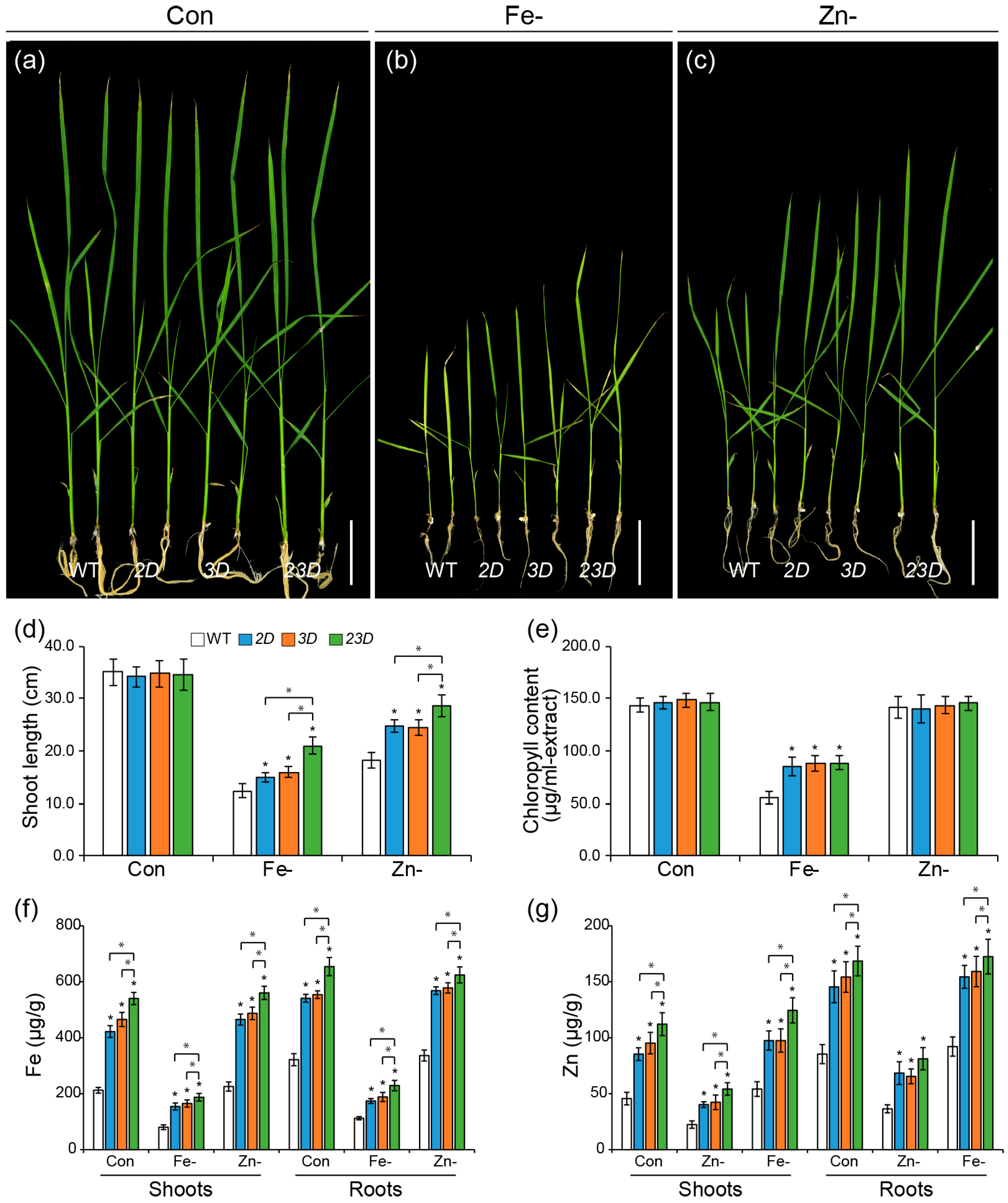
Figure 1. Tolerance of WT and activation-tagged mutants to Fe and Zn deficiencies. (ac) Phenotypes of WT, OsNAS2 activating-tagged mutant (2D), OsNAS3 activating-tagged mutant (3D), and double activation-tagged mutant (23D) grown under control Yoshida solution (Con), Fe-deficient (Fe-) solution or Zn-deficient (Zn-) solution for 15 days. Bars = 5 cm. Shoot lengths (d), total chlorophyll concentrations (e), Fe concentrations (f), and Zn concentrations (g) in shoots and roots of WT and activation-tagged mutants. Data are mean ± SE (n = 10 for length; n = 4 for measurements of chlorophyll contents and Fe and Zn concentrations in shoots and roots). Significant differences among WT and mutant plants are indicated with an asterisk (* p < 0.05 by Student’s t-test).
Ijms 24 06568 g001
Figure 2. Quantitative RT-PCR analysis of Fe and Zn homeostasis-related genes at the seedling roots. Plants were grown under the control Yoshida solution for 10 days. Levels of OsNAAT1 (a), OsDMAS1 (b), OsIRT1 (c), OsYSL2 (d), OsYSL15 (e), OsFer1 (f), OsZIP4 (g), and OsZIP5 (h) in the roots of WT and activation-tagged mutants. Significant differences among WT and mutant plants are indicated with an asterisk (* p < 0.05 by Student’s t-test).
Ijms 24 06568 g002
Figure 3. Elevated expression of OsNAS genes by high pH. Expression of OsNAS1 (a), OsNAS2 (b), and OsNAS3 (c) grown under normal (pH 5.5) or high pH (pH 8.5) Yoshida solution with Fe and Zn (Con), or without Fe (Fe-) or Zn (Zn-). Plants were grown in normal Yoshida solution for 7 days and thereafter were transferred to each growth condition. The seedlings were grown for an additional three days, and shoots and roots from the seedlings were sampled for RNA isolation. Significant differences between the control (pH 5.5) and each growth condition are indicated with an asterisk (* p < 0.05 by Student’s t-test).
Ijms 24 06568 g003
Figure 4. Comparison of seedling growth test under high pH conditions. (ac) Phenotypes of WT, 2D, 3D, and 23D grown on alkaline Yoshida solution ((a) pH 8.5), without Fe ((b) p H8.5/Fe-), or without Zn ((c) p H8.5/Zn-) for 15 days. Bars = 5 cm. Shoot lengths (d) and total chlorophyll concentrations (e) of WT and activation-tagged mutants. Data are mean ± SE (n = 8 for shoot length; n = 4 for chlorophyll content), and significant differences among mutants and WT plants are indicated with an asterisk (* p < 0.05 by Student’s t-test).
Ijms 24 06568 g004
Figure 5. Increment of Fe and Zn concentrations in the mature seeds. Levels of NA (a), DMA (b), Fe (c), and Zn (d) in the unpolished seeds from WT and activation-tagged mutants. Data are means ± SE (n = 4), and significant differences among mutants and WT plants are indicated with an asterisk (* p < 0.05 by Student’s t-test).
Ijms 24 06568 g005
Figure 6. Representative plants of WT, 2D, 3D, and 23D. (a) Comparison of WT, 2D, 3D, and 23D plants grown in a paddy field. To take photographs, plants were transferred to pots from paddy fields before harvest. Scale bar = 20 cm. (b) Plant height, aboveground dry weight (DW), and seed yield of WT, 2D, 3D, and 23D plants grown in a paddy field. Data are means ± SE (n = 4), and significant differences among mutants and WT plants are indicated with an asterisk (* p < 0.05 by Student’s t-test). (c) Morphological comparison of WT, 2D, 3D, and 23D plants grown in alkaline soil (pH 8.0) at the reproductive stage. Scale bar = 20 cm. (d) Plant height, dry weight (DW), and seed yield of WT, 2D, 3D, and 23D plants grown in alkaline soil (pH 8.0). Data are means ± SE (n = 4), and significant differences among mutants and WT plants are indicated with an asterisk (* p < 0.05 by Student’s t-test).
Ijms 24 06568 g006
Figure 7. Growth test under the excess metal treatment. (ac) Phenotypes of WT, 2D, 3D, and 23D grown on half-strength MS solid containing 5 mM ZnSO4 (a), 0.5 mM CuSO4 (b), or 1 mM FeCl3 (c) for 10 days. Bars = 5 cm. Shoot lengths (d) of WT and activation-tagged mutants. Data are mean ± SE (n = 8), and significant differences among mutants and WT plants are indicated with an asterisk (* p < 0.05 by Student’s t-test).
Ijms 24 06568 g007
Disclaimer/Publisher’s Note: The statements, opinions and data contained in all publications are solely those of the individual author(s) and contributor(s) and not of MDPI and/or the editor(s). MDPI and/or the editor(s) disclaim responsibility for any injury to people or property resulting from any ideas, methods, instructions or products referred to in the content.