Article

ARID1A, NOTCH and WNT Signature in Gynaecological Tumours

by
Ieva Vaicekauskaitė
1,2,
Daiva Dabkevičienė
1,2,
Julija Šimienė
1,2,
Diana Žilovič
1,2,
Rūta Čiurlienė
1,
Sonata Jarmalaitė
1,2 and
Rasa Sabaliauskaitė
1,*
1
National Cancer Institute, LT-08660 Vilnius, Lithuania
2
Institute of Biosciences, Life Sciences Center, Vilnius University, LT-08412 Vilnius, Lithuania
*
Author to whom correspondence should be addressed.
Int. J. Mol. Sci. 2023, 24(6), 5854; https://doi.org/10.3390/ijms24065854
Submission received: 4 February 2023 / Revised: 3 March 2023 / Accepted: 17 March 2023 / Published: 19 March 2023
(This article belongs to the Special Issue Notch Signaling in Health and Disease)

Abstract

:
Ovarian cancer (OC) is among the deadliest gynaecologic malignancies in the world. The majority of OC patients are diagnosed at an advanced stage, with high-grade serous OC (HGSOC). The lack of specific symptoms and suitable screening strategies lead to short progression-free survival times in HGSOC patients. The chromatin-remodelling, WNT and NOTCH pathways are some of the most dysregulated in OC; thus their gene mutations and expression profile could serve as diagnostic or prognostic OC biomarkers. Our pilot study investigated mRNA expression of the SWI/SNF chromatin-remodelling complex gene ARID1A, NOTCH receptors, WNT pathway genes CTNNB1 and FBXW7 mRNA expression in two OC cell cultures as well as 51 gynaecologic tumour tissues. A four-gene panel consisting of ARID1A, CTNNB1, FBXW7 and PPP2R1A was used to investigate mutations in gynaecologic tumour tissue. All seven analysed genes were found to be significantly downregulated in OC when compared with non-malignant gynaecologic tumour tissues. NOTCH3 was also downregulated in SKOV3 cells when compared to A2780. Fifteen mutations were found in 25.5% (13/51) of the tissue samples. ARID1A predicted mutations were the most prevalent with alterations detected in 19% (6/32) HGSOC and 67% (6/9) of other OC cases. Thus, ARID1A and NOTCH/WNT-pathway-related changes could be useful diagnostic biomarkers in OC.

1. Introduction

Ovarian cancer (OC) is the second leading cause of death from gynaecologic cancer after cervical cancer [1]. From the other gynaecologic diseases, OC stands out as particularly deadly, as the majority (48%) of cases are high-grade serous ovarian cancer (HGSOC) [2], a pathology often diagnosed in an advanced state (Grade 3, FIGO stage III or IV). The 5-year life expectancy after the diagnosis of HGSOC is only 26% for FIGO stage IV disease [3]. The rest of OC cases are highly heterogeneous with clear-cell, mucinous or endometrial histology, and even germ cell origin (granulosa tumours). The high rate of OC morbidity is attributed to the lack of specific symptoms and sufficient diagnostic techniques. Currently, the only OC biomarker serum CA125 is approved for HGSOC patient monitoring [4]. To compliment CA125, another serum biomarker, human epididymis protein 4 (HE4), has also been approved by the Food and Drug Administration (FDA) for endometrial OC follow-up; however, the biomarker is currently not recommended for clinical use due to inconsistent study results [4]. The OC screening efforts using serum biomarker and transvaginal ultrasound tests have shown no reduction in mortality rates [2]. Thus, OC patients urgently need new diagnostic and predictive biomarkers.
Recent studies have highlighted chromatin-remodeling factors as potential drivers and therapeutic targets of cancer. These complexes involve a substantial collection of proteins that epigenetically govern gene expression and are involved in reparation and replication [5]. ARID1A, coding a SWI/SNF complex protein, is the most-mutated gene in chromatin-remodeling complexes, with alterations detected in 6.2% of all solid tumours [6], and one of the most-mutated genes in OC. ARID1A mutations are mutually exclusive with TP53 and are most indicative of endometriosis-linked OC types [7].
NOTCH and WNT/ β -catenin are closely related pathways involved in female genital tract differentiation and cancerogenesis [8]. The two pathways converge at the primary NOTCH3 receptor ligand Jagged1, whose expression is regulated by the WNT/ β -catenin pathway in OC [9]. The WNT signaling genes CTNNB1, FBXW7 and PPP2R1A [10] are among the most-frequently mutated genes in gynaecologic malignancies [11], while changes in NOTCH family receptors and ligand expression are frequently detected in both malignant and non-malignant gynaecologic disorders [12].
Our pilot study investigated the expression of ARID1A and the NOTCH receptors and WNT components CTNNB1 and FBXW7 in two OC cell cultures, then validated the results in gynaecologic tumour tissues. In addition, a panel of four genes (ARID1A, CTNNB1, FBXW7 and PPP2R1A) was analysed for predicted mutations in gynaecologic tumours.

2. Results

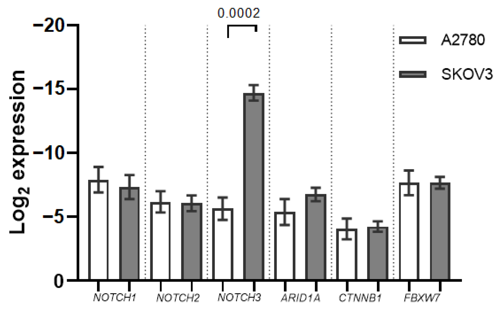
2.1. ARID1A, NOTCH/WNT Pathway Component mRNA Expression in Ovarian Cancer Cell Cultures

Out of the seven mRNAs analysed, all but NOTCH4 expression were detectable in the two OC cell cultures. NOTCH3 mRNA expression was significantly lower in the SKOV3 cell culture when compared with A2780 (−2.6 fold, p = 0.0002). ARID1A expression was also decreased in SKOV3, albeit not significantly (Figure 1).

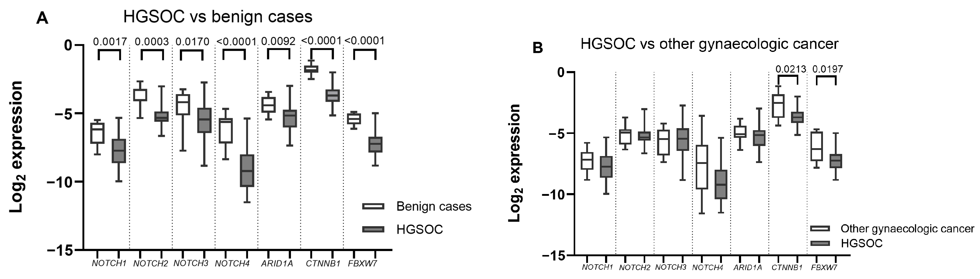
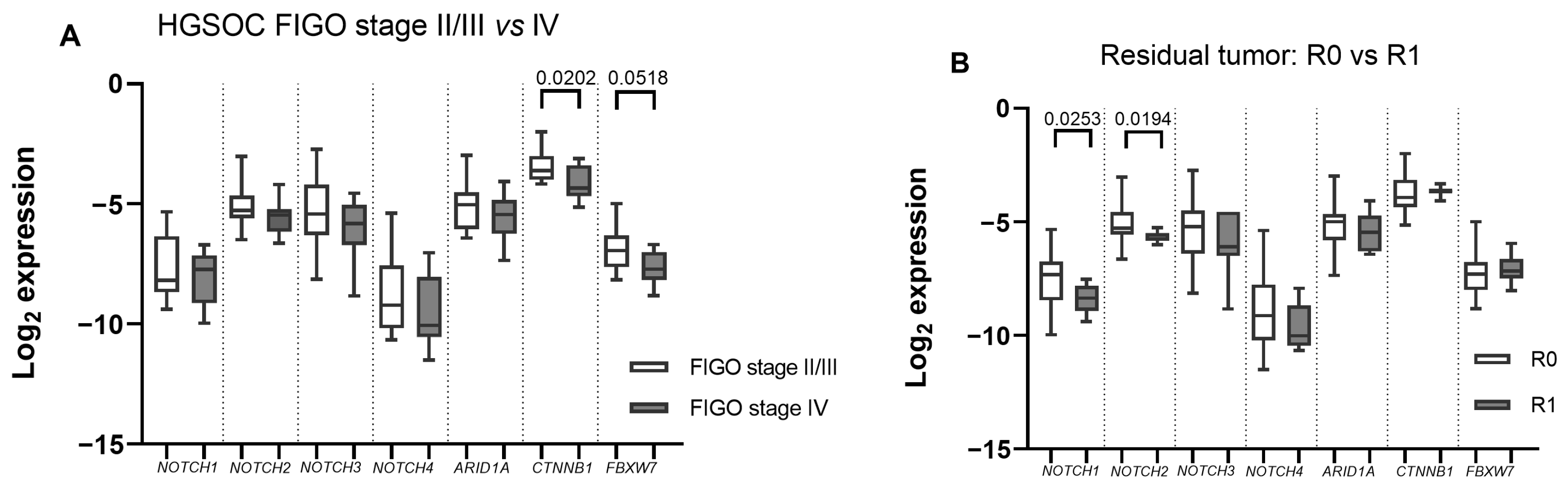
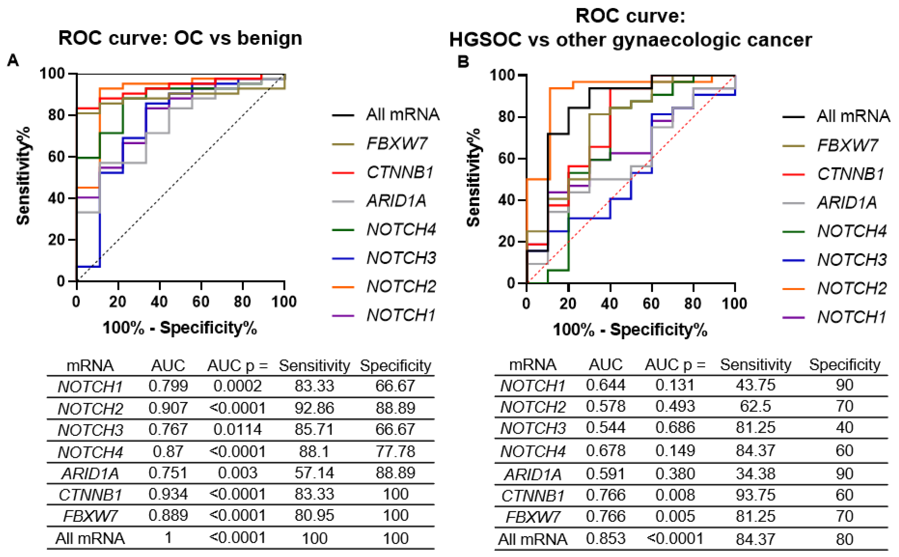
2.2. ARID1A, NOTCH/WNT Pathway Component mRNA Expression in Gynaecological Cancer Tissues

The mRNA expression of all seven analysed genes was significantly downregulated in HGSOC tissues when compared to benign gynaecologic disease tissues (Figure 2A). The NOTCH4 alongside CTNNB1 and FBXW7 mRNA was the most severely altered. However, when compared with non-HGSOC gynaecologic cancer cases, only WNT component downregulation proved significant in HGSOC tissues (Figure 2B).
Next, we evaluated the correlations between mRNA expression and clinical/pathological features in HGSOC samples. In the HGSOC tissue cohort, the CTNNB1 mRNA was significantly downregulated in FIGO stage IV cases when compared with FIGO stage II or III OC (p = 0.02), while the FBXW7 change was of borderline significance (p = 0.05) (Figure 3A). Moreover, NOTCH1 and NOTCH2 expression was significantly reduced in cases with residual tumours (R1) (p = 0.03 and p = 0.02, respectively) (Figure 3B). No correlation between mRNA expression in gynaecologic cancer tissues and age or pre-surgery serum CA125 concentration was found.
ARID1A expression showed an association of borderline significance (p = 0.049) with PFS: OC patients with higher-than-median ARID1A expression showed better PFS (36 vs. 32 months), while other gene expression had no association with PFS.
Overall, all seven mRNAs were good separators of gynaecologic cancer vs. benign disease (Figure 4A), with the CTNNB1 showing the best diagnostic power (area under the curve (AUC) = 0.93). Multiple logistic regression of all seven mRNAs perfectly diagnosed gynaecologic cancer cases from the benign gynaecologic disease (AUC = 1). However, the mRNA expression was less accurate in separating HGSOC cases from other gynaecologic cancers (Figure 4B), with both WNT genes remaining the best separators. The combination of all seven mRNAs showed an acceptable level of HGSOC separation from the other gynaecologic cancers (AUC = 0.85).

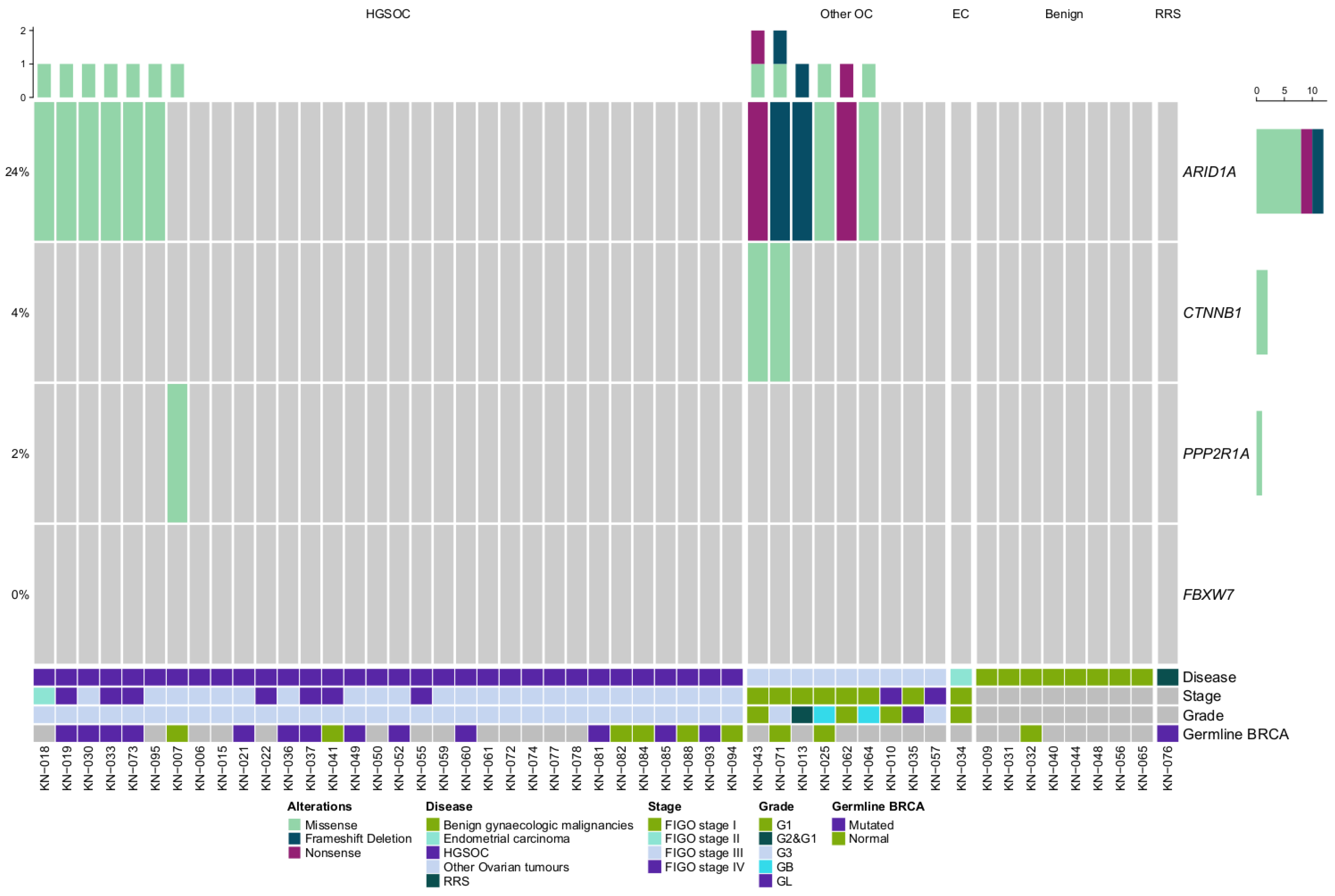
2.3. ARID1A and WNT Pathway Gene Mutations in Gynaecologic Cancer Tissues

The cohort of 51 tissue samples was also investigated for mutations in ARID1A, CTNNB1, FBXW7 and PPP2R1A mutations using targeted NGS. In all, 15 mutations were found in 25.5% (13/51) of the tissue samples (Figure 5). ARID1A alterations were the most prevalent, with 23.5% (12/51) of patients carrying predicted mutations. Only two alterations detected in CTNNB1 and one in PPP2R1A. No mutations were detected in FBXW7. In both cases CTNNB1 co-occurred with ARID1A predicted mutations.
Both CTNNB1 alterations were exclusively detected in non-serous type cancer: one in clear-cell OC and the other in a case with simultaneous endometrioid ovarian and endometrial cancer.
ARID1A alterations were detected in 19% (6/32) of HGSOC and and 67% (6/9) of other OC cases; however, no tissue alterations were found in non-malignant tumours or the sole endometrial cancer case. ARID1A alterations were able to separate OC from benign gynaecologic tumours with 29% sensitivity and 100% specificity. No correlation between the predicted mutation status and the clinical data was found.
Although almost all mRNA expression was lower in cases with ARID1A predicted mutations, no statistically significant correlation between ARID1A alterations and gene expression was found.
None of the detected ARID1A alterations were listed in ClinVar as pathogenic mutations (no information or variants of uncertain significance (VUS)); however, only one CTNNB1 was VUS, and the mutation in PPP2R1A was pathogenic (Table 1). Analysing the VUS alterations using public databases showed that 50% (5/10) of ARID1A alterations are likely to be pathogenic. All but one of these are truncating alterations. All but one ARID1A missense alterations were also predicted “damaging” by the in silico analysis.

3. Discussion

HGSOC is one of the leading causes of female cancer death in the world; thus, means of diagnosis and screening for this gynaecologic cancers are in urgent need. Our pilot study has shown the potential of NOTCH/WNT pathway components as well as the chromatin-remodelling complex member ARID1A as possible biomarkers of OC through an analysis of mRNA transcription and DNA mutations.
First, our small-scale cell culture mRNA expression analysis showed differences in gene expression between a more aggressive type OC cell line (SKOV3), often characterised as clear-cell or serous-type OC (based on TP53 mutation) and a more endometrioid-type OC-representing cell line (A2780) [13]. The cell line analysis provided additional diversity in the types of OC analysed when compared with our tissue cohort. The two OC cell line mRNA expression comparison found differences in all analysed mRNAs; however, only NOTCH3 expression was significantly lower in SKOV3 when compared to A2780. Conversely, Wang et al.’s study found a higher expression of NOTCH components (NOTCH1 and HES1) in A2780 when compared with SKOV3 and three other ovarian cell cultures. The downregulation of NOTCH1 by γ -secretase inhibitors negatively affected cell growth and induced apoptosis [14]. In line with our study, NOTCH3 protein expression was found to be absent from SKOV3, while A2780 expressed NOTCH3 [15,16]. FBXW7 expression was also found to be lower in SKOV3 when compared to A2780 [17]. Interestingly, both cell cultures have ARID1A mutations; however, SKOV3 also has FBXW7 and NOTCH2 mutations [13]. Due to ARID1A nonsense mutations, both OC cell cultures are deficient in full-length ARID1A expression [18]. To our best knowledge no other comparison of ARID1A or CTNNB1 mRNA expression in the two cell cultures has been previously made.
Many studies including data available in the TCGA database find NOTCH1–4, especially NOTCH3, amplifications in a significant percent of HGSOC patients and high NOTCH3 and other NOTCH component upregulation is associated with poor survival [8,16]. Our study showed downregulation of all genes tested. Downregulated NOTCH receptor expression in HGSOC could be related to our control group: in ectopic endometriosis, adenomyosis and other benign gynaecologic diseases, NOTCH receptor expression is increased [12]. We used a mix of various benign gynaecologic conditions (ovarian endometriosis, cystadenomas, myomas) as our control group; thus, the upregulation in HGSOC cases simply could have been less than in benign gynaecologic cancers.
Similarly to our study, significant NOTCH/WNT ubiquitin ligase gene FBXW7 downregulation in serous OC samples was also reflected in Kitade et al.’s study. This study also found associations between lower FBXW7 expression and more advanced OC stages, albeit not clinically significant. The proposed mechanism of this downregulation is hypermethylation of FBXW7 5’-upstream regions, which is related to the high prevalence of TP53 mutations in HGSOC samples causing DNA methyltransferase 1 (DNMT1) overexpression [19].
NOTCH1-2’s downregulated expression was also linked with residual tumour after surgery. In other malignancies, such as gastric cancer and breast cancers, NOTCH receptor expression is a good prognostic indicator of residual disease, where low NOTCH2 expression in residual tumours is correlated with longer survival times [20]. Conversely, in breast cancer, NOTCH signalling is active in residual tumour cells and is related to tumour recurrence [21].
Mutations in CTNNB1 are found in around 43% of endometrioid ovarian carcinomas leading to a loss of β -catenin expression in 51% of cases [22]. Similarly, 18% of endometrioid endometrial carcinomas also possess CTNNB1 mutations [23]. Typically, these mutations are missense alterations affecting the amino-terminal domain required for phosphorylation by GSK3 β to signal degradation of the β -catenin. The activating CTNNB1 mutations render the WNT pathway constantly active [24]. In our study predicted mutations in CTNNB1 were found two types of OC tissues (a case with simultaneous ovarian and endometrial cancers and a clear-cell OC case). However, in OC tissues CTNNB1 expression was the most significantly reduced when compared with benign gynaecologic malignancies and correlated with FIGO stage. The low expression of CTNNB1 in FIGO grade IV HGSOC cases is particularly interesting as there were no associations between β -catenin protein expression and any of the clinical/pathological features of serous OC [25]. More studies are greatly needed in order to further validate the significance of CTNNB1 downregulation in OC.
Protein phosphatase 2A gene PPP2R1A mutation P179R is enriched in high-grade endometrial carcinoma [26]; however, in our study it was found in HGSOC case. The mutation significantly reduces the stability, enzymatic activity and ligand binding to the PP2A catalytic subunit which leads to reduced dephosphorylation of GSK3 β and β -catenin [26].
The ARID1A alterations in our study were found in tissue samples from virtually every type of OC; however, no alterations were detected in benign gynaecologic conditions. Typically, ARID1A loss is indicative of endometrioid-origin cancers; ARID1A mutations are found in 40% of endometrial cancers [27], 32% of endometrioid OCs, 29% of clear-cell OCs, and only 3% of HGSOC cases [28]. The De Leo et al. study, which examined ARID1A mutation’s effect on transcriptomic and proteomic levels of ARID1A, in line with our study results, found no significant correlation between ARID1A mutations and mRNA expression [27]. Although both ARID1A mutations and mRNA downregulation lacked specificity in HGSOC diagnosis, we found borderline significant associations of low ARID1A expression and reduced progression-free survival, showing the potential of ARID1A as a prognostic factor in OC. ARID1A’s loss is associated with PFS as ARID1A downregulation and mutations are highly associated with chemoresistance to platinum-based therapies [29,30]. Synthetic lethality strategies, such as DNA damage response and epigenetic regulation pathway-targeting drugs, should be explored with ARID1A-deficient OC [5].
ARID1A mutations alone are not substantial enough to cause malignancies by themselves [31]; thus, ARID1A mutations are often concomitant with mutations in other genes. In two cases, additional alterations in CTNNB1 were found together with ARID1A mutations. Although none of the ARID1A mutations from our study was annotated in ClinVar (as of January 2023), 40% of them were truncating alterations likely to affect the viability of the ARID1A protein. However, even the non-truncating mutations in ARID1A could still affect the acetylation of histone tails on the nucleosomes and affect target gene expression epigenetically, as ARID1A is a core component of the SWI/SNF chromatin-remodelling complex [32]. More functional studies are greatly needed in order to determine ARID1A mutation pathogenicity and possible implications for OC development.
Due to the pilot study design our analysis was limited by the available tissue samples and OC cell cultures. The small and heterogenic sample cohort limited the statistical power to test associations among mutation, mRNA expression and the clinical data. A larger number of samples should be tested to validate and expand on our results.

4. Materials and Methods

4.1. Cell Culture

Two ovarian cell cultures, SKOV3 and A2780, were tested for the seven genes’ (NOTCH1–4, ARID1A, CTNNB1 and FBXW7) mRNA expression. Both cell cultures are the two best-characterized and most-used OC cell cultures; although they do not closely resemble HGSOC, they were chosen to represent endometrioid OC (A2780) and clear-cell OC (SKOV3), the latter regarded as a more aggressive type of OC (although SKOV3 is frequently assumed to represent serous OC due to mutation in TP53) [33].
Ovarian cancer cells SKOV3 and A2780 were kindly provided by the NCI Immunology laboratory. A2780 cells were cultured in RPMI 1640 + GlutaMax™-I (Gibco, TFS, Grand Island, NY, USA) medium, while SKOV3 cells were cultured in DMEM + GlutaMax™-I (Gibco, TFS, Grand Island, NY, USA) medium. Both cell media were supplemented with 10% fetal bovine serum (Gibco, TFS, Grand Island, NY, USA) and penicillin/streptomycin (Invitrogen Life Technologies, Carlsbad, CA, USA). Cells were cultivated in a monolayer in 37 °C, 5% CO 2 and humidified atmosphere conditions for 48 h until RNA extraction.

4.2. Patient Cohort and Sample Collection

The tissue study cohort consisted of 51 patients who underwent salpingoovarectomy at the Lithuanian National Cancer Institute between 2018 and 2021 for the removal of ovarian or endometrial tumours or benign gynaecologic tumours, including one patient who underwent prophylactic salpingoovarectomy because of a germline BRCA2 mutation (risk-reducing surgery (RSS)). The 51 patients were divided into three groups: HGSOC (32 cases); other gynaecologic tumours (10 cases), which included 9 cases of non-HGSOC ovarian tumours and one case of endometrial cancer. The third group, regarded as controls (9 cases), was made up of 8 benign gynaecologic tumours and one prophylactic salpingoovarectomy case. The clinical features of the tissue sample cohorts are in the Table 2.
During salpingoovarectomy, a small part of the tumour samples were allocated for the study and stored immediately at −80 °C until nucleic acid extraction. The study was approved by the regional bioethics committee (No. 158200-18/5-988-539). All patients were informed about the study and signed written informed consent forms.

4.3. Nucleic Acid Extraction

Prior to nucleic acid extraction, tissue samples were homogenized in liquid nitrogen using a mortar and pestle. 10–15 mg of the resulting tissue was used for nucleic acid extraction. Total RNA extraction for tissue and cell culture samples was performed using TRIzol reagent (Invitrogen, TFS, Carlsbad, CA, USA) using a standard protocol, while for the DNA extraction, tissues first underwent a 16-hour digestion with proteinase K solution (ThermoScientific, TFS, Vilnius, Lithuania) and then standard phenol–chloroform extraction and ethanol precipitation protocols were applied. The nucleic acid samples were stored at −80 °C until further use. The RNA quantity and quality was evaluated using a Nanodrop 2000 spectrophotometer (Thermo Scientific, Wilmingron, DE, USA), while DNA quantification was conducted with a Qubit™ dsDNA HS Assay Kit on a Qubit™ 2.0 Fluorimeter (Invitrogen, TFS, Eugene, OR, USA) according to the manufacturer’s instructions.

4.4. cDNA Synthesis and Quantitative PCR

The RNA samples from ovarian cancer cell cultures and tissue samples were used for the quantitative analysis of NOTCH receptors (NOTCH1-4), FBXW7 and the β -catenin gene CTNNB1, as well as the chromatin-remodelling complex SWI/SNF subunit coding gene ARID1A mRNA transcripts. First, cDNA was synthesised from the total RNA samples using the Maxima First Strand cDNA Synthesis Kit for RT–qPCR with dsDNase (ThermoScientific, TFS, Vilnius, Lithuania) on a ProFlex PCR System (Applied Biosystems, TFS, Singapore). The resulting cDNA was used for quantitative PCR (qPCR) using a Maxima SYBR Green qPCR Master Mix (2X) kit (ThermoScientific, TFS, Vilnius, Lithuania) on a QuantStudio 5 Real-Time PCR System (Applied Biosystems, TFS, Singapore). The primer sequences are provided in Appendix A Table A1. All qPCR reactions were performed in duplicate according to the manufacturer’s protocol. The initial Ct values were gathered using QuantStudio Design & Analysis Software v1.4.3 (Applied Biosystems) with automatic baseline. Then, the data were normalized to a reference gene (GAPDH) and log2 2 Δ Ct values and used for further statistical analysis.

4.5. Targeted Next-Generation Sequencing

The 51 tissue samples were analysed for mutations in the ARID1A, CTNNB1, FBXW7 and PPP2R1A genes using targeted next-generation sequencing (NGS). The library preparation was carried out according to the manufacturer’s protocol using an Ion AmpliSeq™ Library Kit 2.0 and a custom On-Demand Panel (Life Technologies (LT), Carlsbad, CA, USA). Quantification of the final libraries was performed using an Ion Library TaqMan™ Quantification Kit (AB, TFS, Vilnius, Lithuania). An equal amount of each sample library was used for sequencing with the Ion Torrent™ Ion S5™ system. Sequencing data analysis was performed using the Ion Reporter 5.18 tool (LT, Carlsbad, CA, USA). First, the sequence reads were aligned to human reference genome 19 (Genome Reference Consortium GRCh38); then, each alignment was additionally visualized and verified on the Integrative Genomics Viewer 2.4.8 tool. Each mutation was classified according to the ClinVar [34] database as pathogenic or likely pathogenic, benign or likely benign, and mutations with conflicting evidence of pathogenicity and variants not yet included in ClinVar as of January 2023 were classified as variant uncertain significance (VUS). Only pathogenic, likely pathogenic and VUS were included in the analysis. The VUS alterations were predicted pathogenic mutations according to the analysis performed using the Varsome (varsome.com, accessed on January 2023 [35]) and Varcards (varcards.biols.ac.cn, accessed on January 2023 [36]) databases.

4.6. Statistical Analysis

Data normality was tested using the Shapiro–Wilk W test. As appropriate, associations between categorical data variables were determined using a two-sided Chi-square test or Fisher’s exact test, while associations between two independent samples were analysed with a Mann–Whitney U test or Welch’s t test. Receiver operator curves were applied to determine the biomarker sensitivity and specificity. Logistic regression probabilities were used for combining multiple biomarkers into a singular test model. Kaplan–Meier curve analysis were applied for progression-free survival (PFS) analyses. PFS at one year was denoted as the time from treatment (surgery) to progression or death. Results were regarded as statistically significant when p value ≤ 0.050. All statistical data analysis and visualisation was conducted using R x64 4.0.3, GraphPad Prism 8 and MedCalc 14.8.1 softwares.

5. Conclusions

Our pilot study reveals a significant deregulation of NOTCH receptor expression, as well as expression changes and mutations in ARID1A and the WNT pathway genes CTNNB1 and FBXW7 in gynaecologic tumours. Alterations in the chromatin-remodeling and NOTCH/WNT pathways in the future could serve as novel diagnostic or prognostic biomarkers for gynaecologic malignancies. More expansive studies are required for tissue biomarker validation in non-invasive liquid biopsy samples as well as the clinical use of genetic biomerkers for OC detection.

Author Contributions

R.S., D.D., R.Č., D.Ž. and J.Š., conceptualization; R.Č. and D.Ž., sample collection; I.V., sample preparation; I.V., DNA and RNA extraction; I.V. and R.S., NGS and qPCR experiments; J.Š., I.V. and D.D., cell culture experiments; I.V., R.S. and D.D., formal analysis; I.V., visualization and original draft preparation; all authors, writing, review and editing; S.J., supervision; R.S. and D.D., funding acquisition. All authors have read and agreed to the published version of the manuscript.

Funding

This research was funded by the Science Foundation from the National Cancer Institute. The cell culture experiments were funded by the Research Council of Lithuania grant No. 09.3.3-LMT-K-712-22-0300.

Institutional Review Board Statement

This study was conducted in accordance with the Declaration of Helsinki. The study was approved by the Regional Bioethics Committee No. 158200-18/5-988-539.

Informed Consent Statement

Informed consent was obtained from all subjects involved in the study.

Data Availability Statement

Not applicable.

Acknowledgments

We would like to thank Jan Krasko, for providing the cell cultures used in this study.

Conflicts of Interest

The authors declare no conflict of interest.

Abbreviations

The following abbreviations are used in this manuscript:
OCOvarian Cancer
ECEndometrial Cancer
HGSOCHigh-grade serous ovarian cancer
RSSRisk reducing surgery

Appendix A

Table A1. Primer pairs used in the study.
GeneReference Sequence, NCBIPrimerPrimer Sequence
FBXW7NM_001013415.2Forward 3 →5 GTGATAGAACCCCAGTTTCA
Reverse 3 →5 CTTCAGCCAAAATTCTCCAG
ARID1ANM_006015.6Forward 3 →5 CAGTAAGGGAGGGCAAGAAG
Reverse 3 →5 GAGGAGAGAAAGGAGACTGA
CTNNB1NM_001904.4Forward 3 →5 TCTGAGGACAAGCCACAAGATTACA
Reverse 3 →5 TGGGCACCAATATCAAGTCCAA
NOTCH1NM_017617.5Forward 3 →5 CAGCCTCAACATCCCCTACAAG
Reverse 3 →5 GCAGCCCACGAAGAACAGAA
NOTCH2NM_024408.4Forward 3 →5 GTGGATGGGGTCAACACTTACA
Reverse 3 →5 CACTCCAGCCGTTGACACATAC
NOTCH3NM_000435.3Forward 3 →5 CGTGGCTTCTTTCTACTGTGC
Reverse 3 →5 CGTTCACCGGATTTGTGTCAC
NOTCH4NM_004557.4Forward 3 →5 AACTCCTCCCCAGGAATCTG
Reverse 3 →5 CCTCCATCCAGCAGAGGTT
GAPDHNC_000012.12Forward 3 →5 GAAGGTCGGAGTCAACGGATTT
Reverse 3 →5 ATGGGTGGAATCATATTGGAAC

References

  1. Siegel, R.L.; Miller, K.D.; Fuchs, H.E.; Jemal, A. Cancer statistics, 2022. CA Cancer J. Clin. 2022, 72, 7–33. [Google Scholar] [CrossRef]
  2. Koshiyama, M.; Matsumura, N.; Konishi, I. Subtypes of Ovarian Cancer and Ovarian Cancer Screening. Diagnostics 2017, 7, 12. [Google Scholar] [CrossRef] [PubMed] [Green Version]
  3. Torre, L.A.; Trabert, B.; DeSantis, C.E.; Miller, K.D.; Samimi, G.; Runowicz, C.D.; Gaudet, M.M.; Jemal, A.; Siegel, R.L. Ovarian cancer statistics, 2018. CA Cancer J. Clin. 2018, 68, 284–296. [Google Scholar] [CrossRef] [Green Version]
  4. Colombo, N.; Sessa, C.; Bois, A.D.; Ledermann, J.; McCluggage, W.G.; McNeish, I.; Morice, P.; Pignata, S.; Ray-Coquard, I.; Vergote, I.; et al. ESMO-ESGO consensus conference recommendations on ovarian cancer: Pathology and molecular biology, early and advanced stages, borderline tumours and recurrent disease. Ann. Oncol. 2019, 30, 672–705. [Google Scholar] [CrossRef] [Green Version]
  5. Caumanns, J.J.; Wisman, G.B.A.; Berns, K.; van der Zee, A.G.; de Jong, S. ARID1A mutant ovarian clear cell carcinoma: A clear target for synthetic lethal strategies. Biochim. Biophys. Acta-Rev. Cancer 2018, 1870, 176–184. [Google Scholar] [CrossRef] [PubMed]
  6. Jiang, T.; Chen, X.; Su, C.; Ren, S.; Zhou, C. Pan-cancer analysis of ARID1A Alterations as Biomarkers for Immunotherapy Outcomes. J. Cancer 2020, 11, 776–780. [Google Scholar] [CrossRef] [Green Version]
  7. Reske, J.J.; Wilson, M.R.; Holladay, J.; Siwicki, R.A.; Skalski, H.; Harkins, S.; Adams, M.; Risinger, J.I.; Hostetter, G.; Lin, K.; et al. Co-existing TP53 and ARID1A mutations promote aggressive endometrial tumorigenesis. PLoS Genet. 2021, 17, e1009986. [Google Scholar] [CrossRef] [PubMed]
  8. Orzechowska, M.; Anusewicz, D.; Bednarek, A.K. Functional Gene Expression Differentiation of the Notch Signaling Pathway in Female Reproductive Tract Tissues—A Comprehensive Review With Analysis. Front. Cell Dev. Biol. 2020, 8, 592616. [Google Scholar] [CrossRef]
  9. Chen, X.; Stoeck, A.; Lee, S.J.; Shih, I.M.; Wang, M.M.; Wang, T.L. Jagged1 expression regulated by Notch3 and Wnt/β-catenin signaling pathways in ovarian cancer. Oncotarget 2010, 1, 210–218. [Google Scholar] [CrossRef]
  10. Thompson, J.J.; Williams, C.S. Protein Phosphatase 2A in the Regulation of Wnt Signaling, Stem Cells, and Cancer. Genes 2018, 9, 121. [Google Scholar] [CrossRef] [Green Version]
  11. Spaans, V.M.; Trietsch, M.D.; Crobach, S.; Stelloo, E.; Kremer, D.; Osse, E.M.; Haar, N.T.T.; Eijk, R.V.; Muller, S.; Wezel, T.V.; et al. Designing a high-throughput somatic mutation profiling panel specifically for gynaecological cancers. PLoS ONE 2014, 9, e93451. [Google Scholar] [CrossRef] [PubMed]
  12. Moldovan, G.E.; Miele, L.; Fazleabas, A.T. Notch signaling in reproduction. Trends Endocrinol. Metab. TEM 2021, 32, 1044–1057. [Google Scholar] [CrossRef] [PubMed]
  13. Papp, E.; Hallberg, D.; Konecny, G.E.; Bruhm, D.C.; Adleff, V.; Noë, M.; Kagiampakis, I.; Palsgrove, D.; Conklin, D.; Kinose, Y.; et al. Integrated Genomic, Epigenomic, and Expression Analyses of Ovarian Cancer Cell Lines. Cell Rep. 2018, 25, 2617–2633. [Google Scholar] [CrossRef] [Green Version]
  14. Wang, M.; Wu, L.; Wang, L.; Xin, X. Down-regulation of Notch1 by gamma-secretase inhibition contributes to cell growth inhibition and apoptosis in ovarian cancer cells A2780. Biochem. Biophys. Res. Commun. 2010, 393, 144–149. [Google Scholar] [CrossRef]
  15. McAuliffe, S.M.; Morgan, S.L.; Wyant, G.A.; Tran, L.T.; Muto, K.W.; Chen, Y.S.; Chin, K.T.; Partridge, J.C.; Poole, B.B.; Cheng, K.H.; et al. Targeting Notch, a key pathway for ovarian cancer stem cells, sensitizes tumors to platinum therapy. Proc. Natl. Acad. Sci. USA 2012, 109, E2939–E2948. [Google Scholar] [CrossRef] [PubMed] [Green Version]
  16. Hu, W.; Liu, T.; Ivan, C.; Sun, Y.; Huang, J.; Mangala, L.S.; Miyake, T.; Dalton, H.J.; Pradeep, S.; Rupaimoole, R.; et al. Notch3 pathway alterations in ovarian cancer. Cancer Res. 2014, 74, 3282–3293. [Google Scholar] [CrossRef] [PubMed] [Green Version]
  17. Xu, Z.; Zhuang, L.; Wang, X.; Li, Q.; Sang, Y.; Xu, J. FBXW7γ is a tumor-suppressive and prognosis-related FBXW7 transcript isoform in ovarian serous cystadenocarcinoma. Future Oncol. 2020, 16, 1921–1930. [Google Scholar] [CrossRef]
  18. Domcke, S.; Sinha, R.; Levine, D.A.; Sander, C.; Schultz, N. Evaluating cell lines as tumour models by comparison of genomic profiles. Nat. Commun. 2013, 4, 2126. [Google Scholar] [CrossRef] [Green Version]
  19. Kitade, S.; Onoyama, I.; Kobayashi, H.; Yagi, H.; Yoshida, S.; Kato, M.; Tsunematsu, R.; Asanoma, K.; Sonoda, K.; Wake, N.; et al. FBXW7 is involved in the acquisition of the malignant phenotype in epithelial ovarian tumors. Cancer Sci. 2016, 107, 1399–1405. [Google Scholar] [CrossRef] [PubMed] [Green Version]
  20. Bauer, L.; Langer, R.; Becker, K.; Hapfelmeier, A.; Ott, K.; Novotny, A.; Höfler, H.; Keller, G. Expression profiling of stem cell-related genes in neoadjuvant-treated gastric cancer: A NOTCH2, GSK3B and β-catenin gene signature predicts survival. PLoS ONE 2012, 7, e44566. [Google Scholar] [CrossRef] [Green Version]
  21. Abravanel, D.L.; Belka, G.K.; Pan, T.C.; Pant, D.K.; Collins, M.A.; Sterner, C.J.; Chodosh, L.A. Notch promotes recurrence of dormant tumor cells following HER2/neu-targeted therapy. J. Clin. Investig. 2015, 125, 2484–2496. [Google Scholar] [CrossRef] [Green Version]
  22. Zyla, R.E.; Olkhov-Mitsel, E.; Amemiya, Y.; Bassiouny, D.; Seth, A.; Djordjevic, B.; Nofech-Mozes, S.; Parra-Herran, C. CTNNB1 Mutations and Aberrant β-Catenin Expression in Ovarian Endometrioid Carcinoma: Correlation With Patient Outcome. Am. J. Surg. Pathol. 2021, 45, 68–76. [Google Scholar] [CrossRef] [PubMed]
  23. Kim, G.; Kurnit, K.C.; Djordjevic, B.; Singh, C.; Munsell, M.F.; Wang, W.L.; Lazar, A.J.; Zhang, W.; Broaddus, R. Nuclear β-catenin localization and mutation of the CTNNB1 gene: A context-dependent association. Mod. Pathol. 2018, 31, 1553–1559. [Google Scholar] [CrossRef] [PubMed]
  24. Nguyen, V.H.L.; Hough, R.; Bernaudo, S.; Peng, C. Wnt/β-catenin signalling in ovarian cancer: Insights into its hyperactivation and function in tumorigenesis. J. Ovarian Res. 2019, 12, 122. [Google Scholar] [CrossRef] [PubMed] [Green Version]
  25. Sallum, L.F.; Andrade, L.; Costa, L.B.E.D.; Ramalho, S.; Ferracini, A.C.; Natal, R.D.A.; Brito, A.B.C.; Sarian, L.O.; Derchain, S. BRCA1, Ki67, and β-Catenin Immunoexpression Is Not Related to Differentiation, Platinum Response, or Prognosis in Women With Low- and High-Grade Serous Ovarian Carcinoma. Int. J. Gynecol. Cancer 2018, 28, 437–447. [Google Scholar] [CrossRef]
  26. Taylor, S.E.; O’Connor, C.M.; Wang, Z.; Shen, G.; Song, H.; Leonard, D.; Sangodkar, J.; LaVasseur, C.; Avril, S.; Waggoner, S.; et al. The highly recurrent PP2A Aa-subunit mutation P179R alters protein structure and impairs PP2A enzyme function to promote endometrial tumorigenesis. Cancer Res. 2019, 79, 4242–4257. [Google Scholar] [CrossRef] [PubMed]
  27. Leo, A.D.; Ravegnini, G.; Musiani, F.; Maloberti, T.; Visani, M.; Sanza, V.; Angelini, S.; Perrone, A.M.; Iaco, P.D.; Corradini, A.G.; et al. Relevance of ARID1A Mutations in Endometrial Carcinomas. Diagnostics 2022, 12, 592. [Google Scholar] [CrossRef]
  28. Lapke, N.; Chen, C.H.; Chang, T.C.; Chao, A.; Lu, Y.J.; Lai, C.H.; Tan, K.T.; Chen, H.C.; Lu, H.Y.; Chen, S.J. Genetic alterations and their therapeutic implications in epithelial ovarian cancer. BMC Cancer 2021, 21, 499. [Google Scholar] [CrossRef]
  29. Katagiri, A.; Nakayama, K.; Rahman, M.T.; Rahman, M.; Katagiri, H.; Nakayama, N.; Ishikawa, M.; Ishibashi, T.; Iida, K.; Kobayashi, H.; et al. Loss of ARID1A expression is related to shorter progression-free survival and chemoresistance in ovarian clear cell carcinoma. Mod. Pathol. 2012, 25, 282–288. [Google Scholar] [CrossRef] [Green Version]
  30. Yokoyama, Y.; Matsushita, Y.; Shigeto, T.; Futagami, M.; Mizunuma, H. Decreased ARID1A expression is correlated with chemoresistance in epithelial ovarian cancer. J. Gynecol. Oncol. 2014, 25, 58–63. [Google Scholar] [CrossRef] [Green Version]
  31. Guan, B.; Rahmanto, Y.S.; Wu, R.C.; Wang, Y.; Wang, Z.; Wang, T.L.; Shih, I.M. Roles of deletion of Arid1a, a tumor suppressor, in mouse ovarian tumorigenesis. J. Natl. Cancer Inst. 2014, 106, dju146. [Google Scholar] [CrossRef] [PubMed] [Green Version]
  32. Wiegand, K.C.; Hennessy, B.T.; Leung, S.; Wang, Y.; Ju, Z.; McGahren, M.; Kalloger, S.E.; Finlayson, S.; Stemke-Hale, K.; Lu, Y.; et al. A functional proteogenomic analysis of endometrioid and clear cell carcinomas using reverse phase protein array and mutation analysis: Protein expression is histotype-specific and loss of ARID1A/BAF250a is associated with AKT phosphorylation. BMC Cancer 2014, 14, 120. [Google Scholar] [CrossRef] [PubMed] [Green Version]
  33. Barnes, B.M.; Nelson, L.; Tighe, A.; Burghel, G.J.; Lin, I.H.; Desai, S.; McGrail, J.C.; Morgan, R.D.; Taylor, S.S. Distinct transcriptional programs stratify ovarian cancer cell lines into the five major histological subtypes. Genome Med. 2021, 13, 140. [Google Scholar] [CrossRef] [PubMed]
  34. Landrum, M.J.; Lee, J.M.; Benson, M.; Brown, G.R.; Chao, C.; Chitipiralla, S.; Gu, B.; Hart, J.; Hoffman, D.; Jang, W.; et al. ClinVar: Improving access to variant interpretations and supporting evidence. Nucleic Acids Res. 2018, 46, D1062–D1067. [Google Scholar] [CrossRef] [PubMed] [Green Version]
  35. Kopanos, C.; Tsiolkas, V.; Kouris, A.; Chapple, C.E.; Aguilera, M.A.; Meyer, R.; Massouras, A. VarSome: The human genomic variant search engine. Bioinformatics 2018, 35, 1978–1980. [Google Scholar] [CrossRef] [Green Version]
  36. Li, J.; Shi, L.; Zhang, K.; Zhang, Y.; Hu, S.; Zhao, T.; Teng, H.; Li, X.; Jiang, Y.; Ji, L.; et al. VarCards: An integrated genetic and clinical database for coding variants in the human genome. Nucleic Acids Res. 2018, 46, D1039–D1048. [Google Scholar] [CrossRef] [Green Version]
Figure 1. mRNA expression in ovarian cancer (OC) cell cultures A2780 and SKOV3. Boxes indicate the mRNA expression normalized to GAPDH expression data. Whiskers denote standard deviation from the three experimental replicates.
Ijms 24 05854 g001
Figure 2. mRNA levels in ovarian cancer tissues. (A) gynaecologic cancer (n = 42) vs. benign cases (n = 9) (B) high-grade serous ovarian cancer (HGSOC) (n = 32) vs. other gynaecologic cancer cases (n = 10). Boxes indicate the log 2 normalized expression data. Whiskers denote minimum and maximum values.
Ijms 24 05854 g002
Figure 3. mRNA levels in high-grade serous ovarian cancer HGSOC tissues: (A) FIGO stage II/III (n = 20) vs. IV (n = 12); (B) with (R1) (n = 7) vs. without (R0) residual tumour (n = 25). Boxes indicate the log 2 normalized expression data. Whiskers denote minimum and maximum values.
Ijms 24 05854 g003
Figure 4. ROC curve analysis of tissue mRNA biomarkers. (A) gynaecologic cancer (ovarian cancers + one endometrial case) vs. benign cases. (B) HGSOC cases vs. other gynaecologic cancer cases. AUC—area under the curve.
Ijms 24 05854 g004
Figure 5. Oncoprint of gynaecologic tumour tissue sample mutation data. HGSOC—High-grade ovarian cancer, OC—ovarian cancer, EC—endometrial cancer, RRS—risk-reducing surgery.
Ijms 24 05854 g005
Table 1. Observed alterations and their predicted pathogenicity.
No.SamplesHistology/Disease TypeLocusGeneCoding SequenceAmino Acid ChangeVariant EffectClinVardbSNPVarSome VerdictVarCards “Damaging Score”
1KN-007HGSOCchr19:52715971PPP2R1Ac.536C>Gp.Pro179ArgmissenseLikely pathogenicrs786205228Likely pathogenic0.83
2KN-043OC and ECchr3:41266104CTNNB1c.101G>Ap.Gly34GlumissensePathogenic/ Likely pathogenicrs28931589Pathogenic0.83
3KN-043OC and ECchr1:27106364ARID1Ac.5975C>Ap.Ser1992Ternonsense Pathogenic1
4KN-071Clear-cell OCchr3:41266113CTNNB1c.110C>Gp.Ser37CysmissensePathogenic/ Likely pathogenicrs121913403Pathogenic0.83
5KN-071Clear-cell OCchr1:27057961ARID1Ac.1670_1674delAGTCTp.Gln557ProfsTer64frameshift deletion Likely pathogenic-
6KN-013OC and ECchr1:27099367ARID1Ac.3606delGp.Asn1203IlefsTer3frameshift deletion Pathogenic-
7KN-018HGSOCchr1:27099478ARID1Ac.3715G>Ap.Ala1239Thrmissense Likely pathogenic0.64
8KN-073HGSOCchr1:27023961ARID1Ac.1067G>Ap.Arg356Lysmissense Likely benign0.52
9KN-019HGSOCchr1:27106228ARID1Ac.5839C>Gp.Gln1947Glumissense Likely benign0.3
10KN-033HGSOCchr1:27101532ARID1Ac.4814C>Tp.Pro1605Leumissensers375431469VUS0.61
11KN-064Serous borderline
12KN-025Mucinous borderlinechr1:27089762ARID1Ac.2718C>Gp.Asn906LysmissenseUncertain significancers201864573Benign0.65
13KN-095HGSOC
14KN-062Mucinous borderlinechr1:27101504ARID1Ac.4786G>Tp.Glu1596Ternonsense Pathogenic1
15KN-030HGSOCchr1:27105686ARID1Ac.5297A>Tp.Glu1766Valmissense rs1363371199Likely benign0.39
Table 2. Clinical features of the patients with tissue samples.
Clinical/Pathological CharacteristicsNumber of Patients (%)
Disease GroupHGSOCOther Gynaecologic CancersBenign Gynaecologic TumourOverall
n =3210951
Average Age, years (min–max)57.8 (41–82)63.7(49–77)53.6 (43–72)58.3 (41–82)
Average CA125 pre op. concentration U/mL (N/A)888.5 (1 N/A)139.4 (3 N/A)51.35 (2 N/A)641.8 (6 N/A)
FIGO Stage
IA 8 (80.0) 8 (15.6)
IIB1 (3.1) 1 (2.0)
IIIB2 (6.3) 2 (4.0)
IIIC17 (53.1) 17 (33.3)
IVB12 (37.5)2 (20.0) 14 (27.5)
N/A 1 9 (100.0)9 (17.6)
Tumour differentiation grade
G1 4 (40.0) 4 (7.8)
G2 1 (10.0) 1 (2.0)
G332 (100.0)2 (20.0) 4 (66.7)
GB/GL 2 3 (30.0) 3 (5.9)
N/A1 9 (100.0)9 (17.6)
Progressed disease
Yes12 (37.5)1 (10.0) 13 (25.5)
No20 (51.4)9 (90.0)9 (100.0)38 (74.5)
Radical disease after surgery
R024 (47.1)8 (80.0)9 (100.0)41 (80.3)
R16 (18.8)2 (20.0) 8 (15.7)
R31 (3.1) 1 (2.0)
N/A 31 (3.1) 1 (2.0)
1 N/A—benign cases 2 GB—borderline cases, GL—granulosa tumour 3 N/A—needle biopsy.
Disclaimer/Publisher’s Note: The statements, opinions and data contained in all publications are solely those of the individual author(s) and contributor(s) and not of MDPI and/or the editor(s). MDPI and/or the editor(s) disclaim responsibility for any injury to people or property resulting from any ideas, methods, instructions or products referred to in the content.

