Article

Early Pregnancy Regulates Expression of IkappaB Family in Ovine Spleen and Lymph Nodes

School of Life Sciences and Food Engineering, Hebei University of Engineering, Handan 056038, China
*
Author to whom correspondence should be addressed.
These authors contributed equally to this work.
Int. J. Mol. Sci. 2023, 24(6), 5156; https://doi.org/10.3390/ijms24065156
Submission received: 27 January 2023 / Revised: 3 March 2023 / Accepted: 6 March 2023 / Published: 8 March 2023

Abstract

:
Early pregnancy modulates the maternal immune system, including the spleen and lymph nodes, which participate in maternal innate and adaptive immune responses. Methods: Ovine spleens and lymph nodes were sampled at day 16 of the estrous cycle, and at days 13, 16 and 25 of gestation, and qRT-PCR, Western blot and immunohistochemistry analysis were used to analyze the expression of the IκB family, including BCL-3, IκBα, IκBβ, IκBε, IKKγ, IκBNS and IκBζ. Early pregnancy induced expression of BCL-3, IκBα, IκBε, IKKγ and IκBζ, and expression of BCL-3, IκBβ and IκBNS peaked at day 16 of pregnancy in the spleen. However, early pregnancy suppressed the expression of BCL-3 and IκBNS, but stimulated the expression of IκBβ and IκBζ, and expression levels of IκBα, IκBβ, IκBε and IKKγ peaked in lymph nodes at days 13 and/or 16 of pregnancy. Early pregnancy changed the expression of the IκB family in the maternal spleen and lymph node in a tissue-specific manner, suggesting that the modulation of the IκB family may be involved in regulation of maternal functions of the spleen and lymph nodes, which are necessary for the establishment of maternal immune tolerance during early pregnancy in sheep.

1. Introduction

The relationship between mother and fetus is an immunological marvel and enigma, as averting maternal–fetal conflict is continuously required throughout pregnancy [1]. There are significant changes in maternal immune system components that interact with the fetal immune system dynamically and co-operatively to contribute to the modulation of the risk of infection and the course of immunological disease during pregnancy [2]. Ruminant conceptus signaling (interferon-tau, IFNT) modulates the innate immune system in paracrine and endocrine manners, which is essential for successful pregnancy establishment and prevention of rejection against the allogenic conceptus by the mother [3]. The immune system changes in response to the presence of an embryo, and these responses are different in local and peripheral immune tissues during early pregnancy in sheep [4]. Nuclear factor kappa B (NF-κB) is implicated in both physiological and pathological processes; NF-κB regulation is essential during pregnancy, and dysregulation results in premature termination of pregnancy, with bad outcomes for the mother and the fetus [5]. Early pregnancy modulates the expression of NF-κB components in the maternal thymus, spleen, liver and inguinal lymph nodes, which is necessary for embryo implantation and pregnancy maintenance in sheep [6,7,8,9].
The activities of NF-κB proteins are strictly repressed by inhibitors of NF-κB (IκB) proteins, including IκBα, IκBβ, IκBε, IKKγ, B cell leukemia-3 (BCL-3), IκBNS (also known as NFKBID) and IκBζ, and IκBβ plays a crucial role in the regulation of innate immune responses [10]. BCL-3 can either promote or inhibit NF-κB target gene expression, which is dependent on the type of cell, the type of activating stimulus and the type of the NF-κB target gene involved [11]. IκB proteins not only interact with NF-κB to change its transcriptional activity, but also bind to chromatin and control gene expression to participate in immune homeostasis [12]. BaelanChagsangBang (BCB) water extract has strong anti-oxidative and cytoprotective effects in vitro, and in vivo administration of BCB water extract can improve the number of implantation sites in pregnant mice through upregulation of IκBα expression [13]. IKKβ signaling is involved in the prevention of preterm delivery and the improvement of neonatal outcomes through the suppression of production of interleukin-6 (IL-6) and related cytokines [14]. IκB proteins are related to successful pregnancy.
The spleen plays a key role in adaptive immune responses, and the splenic nerve is involved in modulation of the central amygdala and the paraventricular nucleus corticotrophin-releasing hormone-producing neurons [15]. OLT1177 (an orally active β-sulfonyl nitrile molecule) inhibits activation of the nucleotide-binding oligomerization domain (NOD)-like receptor protein 3 inflammasome, which results in a downregulation of phosphorylated NF-κB and IκB kinase levels in spleen cells from OLT1177-treated mice [16]. There is a differential expression of costimulatory molecules on splenic antigen-presenting cells in spleens of female mice during the preimplantation period of pregnancy, which are related to pregnancy outcome and the tolerogenic immune response in mice [17].
Lymph nodes are involved in adaptive immunity through modulating T cell and B cell activation and their differentiation into effector cells [18]. There is a downregulation of the specific alloreactivity during the pre-implantation and implantation stages of pregnancy, but the specific and non-specific alloreactivities are upregulated at mid-pregnancy in para-aortic lymph node cells [19]. The immune functions of the lymph nodes around the reproductive tract and other peripheral lymph nodes are different during pregnancy, which are involved in the modulation of the maternal immune system in ewes [4]. Lymphatic endothelial cells modulate progesterone bioavailability, which is essential for regulating immune tolerance during pregnancy in humans [20].
Previous studies report that interferon-stimulated genes (ISGs), progesterone receptor, progesterone-induced blocking factor, tumor necrosis factor (TNF)-β, IL-2, IL-5, IL-6, IL-10, cyclooxygenase 2, aldo-keto reductase family 1, member B1, melatonin receptor 1 (MT1), gonadotropin-releasing hormone and its receptor are upregulated, but MT2 is downregulated in the maternal spleen of ewes during early pregnancy [21,22,23,24,25,26]. In addition, early pregnancy modulates the expression of Toll-like receptor signaling members and NF-κB components in the ovine maternal spleen [7,27]. Furthermore, the expression of ISGs, progesterone receptor, prostaglandin synthases, Th cytokines, MT1, gonadotropin-releasing hormone and its receptor, and prolactin and its receptor are changed in lymph nodes during early pregnancy in sheep [24,25,28,29,30,31,32]. On the other hand, early pregnancy regulates the expression of Toll-like receptor signaling members, complement components, nuclear factor kappa B family and NOD receptors in the ovine maternal lymph nodes [9,33,34,35].
It is hypothetic that early pregnancy modulates the expression of IκB proteins in the ovine spleen and inguinal lymph nodes. The aim of this study was to analyze the expression of BCL-3, IκBα (NFKBIA), IκBβ (NFKBIB), IκBε (NFKBIE), IKKγ (IKBKG), IκBNS (NFKBID) and IκBζ (NFKBIZ) in the maternal spleen and lymph nodes of sheep, which may be beneficial for understanding the maternal immunomodulation of spleen and lymph nodes during early pregnancy in sheep.

2. Results

2.1. Expression of BCL-3, NFKBIA, NFKBIB, NFKBIE, IKBKG, NFKBID and NFKBIZ mRNA in the Spleen and Lymph Nodes

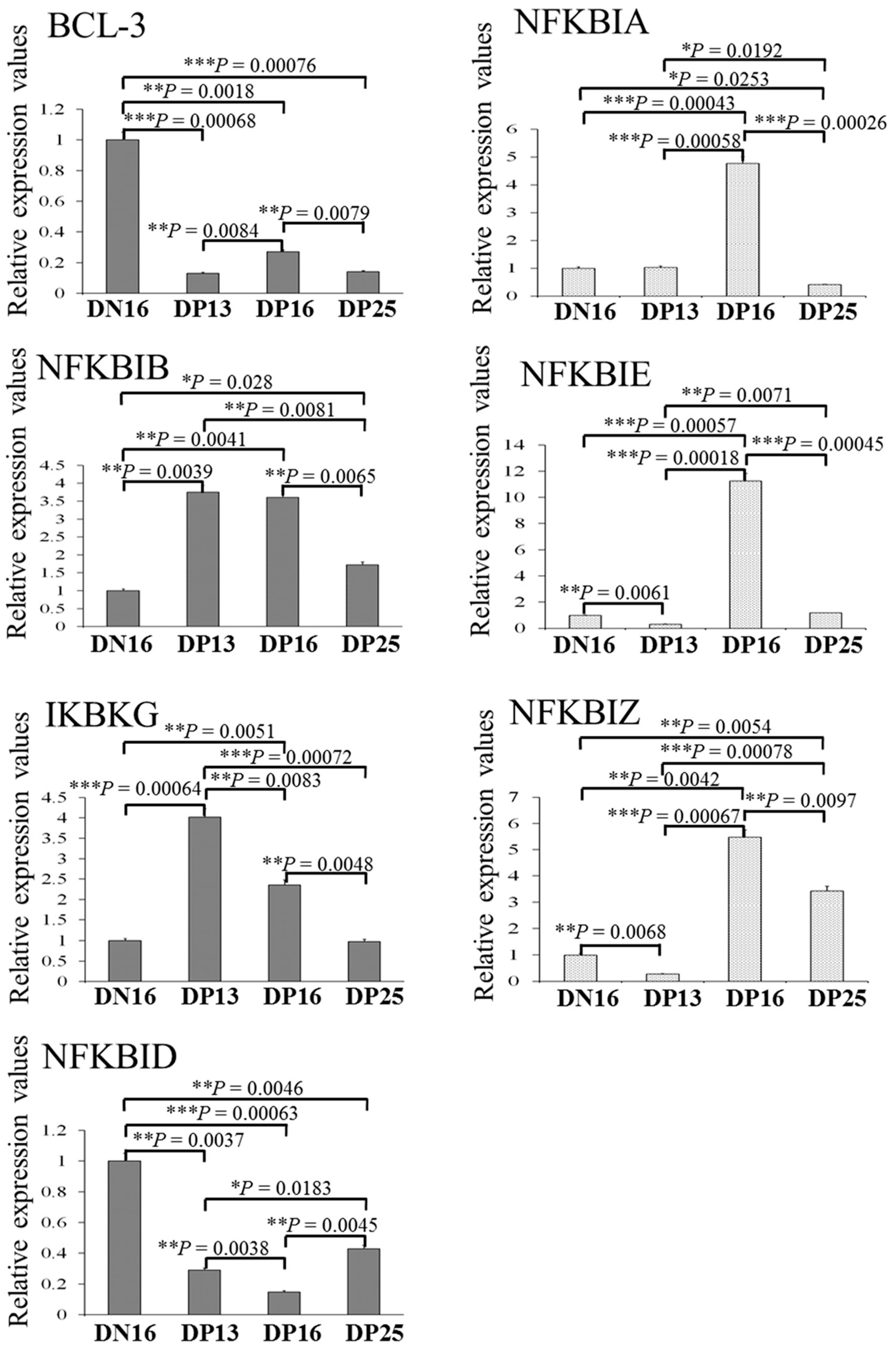
Figure 1 showed that the relative expression level of BCL-3 mRNA was increased in the spleen during early pregnancy compared to day 16 of the estrous cycle, with a peak at day 16 of pregnancy (p < 0.05). The relative expression levels of NFKBIA, NFKBIE, IKBKG and NFKBIZ mRNA gradually increased from day 13 to 25 of pregnancy (p < 0.05). Furthermore, there was a peak in the relative expression levels of NFKBIB and NFKBID mRNA at day 16 of pregnancy, but the expression level of NFKBIB mRNA was the lowest at day 13 of pregnancy (p < 0.05).
In the lymph nodes, early pregnancy suppressed the expression of BCL-3 and NFKBID mRNA, but stimulated the expression of NFKBIB and NFKBIZ mRNA at days 16 and 25 of pregnancy (p < 0.05; Figure 2). In addition, there was a peak in the relative expression levels of NFKBIB and IKBKG mRNA at days 13 and 16 of pregnancy (p < 0.05). Furthermore, expression levels of NFKBIA, NFKBIE and NFKBIZ mRNA peaked at day 16 of pregnancy, but expression level of NFKBIA was the lowest at day 25 of pregnancy (p < 0.05), and expression levels of NFKBIE and NFKBIZ mRNA were the lowest at day 13 of pregnancy among the four groups (p < 0.05).

2.2. Expression of IκB Proteins in the Spleen and Lymph Nodes

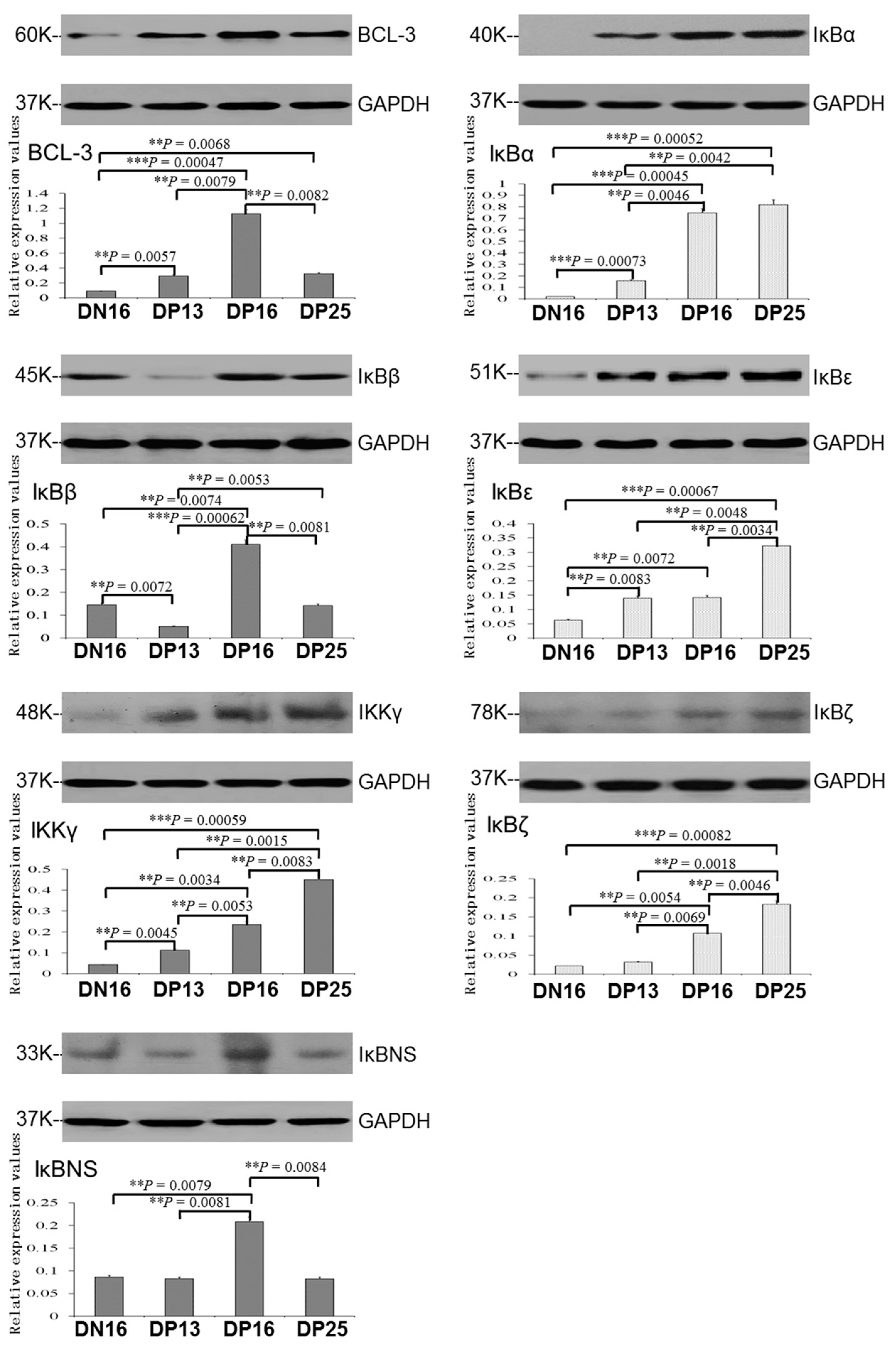
It was revealed in Figure 3 that there was an upregulation of BCL-3 protein during early pregnancy compared to day 16 of the estrous cycle, and the expression level of IκBβ protein was the highest at day 16 of pregnancy (p < 0.05). Early pregnancy induced gradual upregulation of IκBα, IκBε, IKKγ and IκBζ proteins from day 13 to 25 of pregnancy in the spleen (p < 0.05), and IκBα, IKKγ and IκBζ proteins were almost undetected at day 16 of the estrous cycle. In addition, expression of IκBβ and IκBNS proteins was the highest at day 16 of pregnancy (p < 0.05), but IκBβ protein was almost undetected at day 13 of pregnancy.
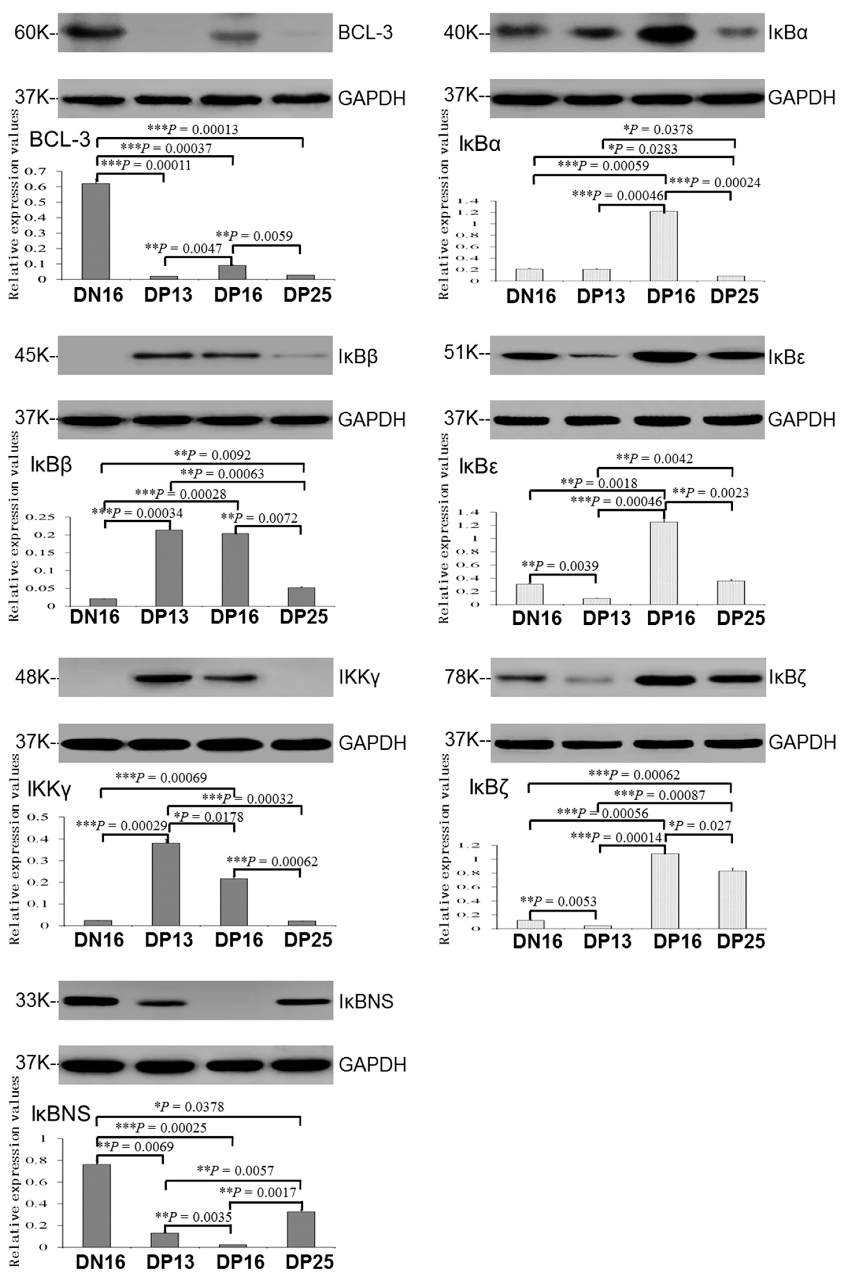
In the lymph nodes, expression of BCL-3 and IκBNS proteins was downregulated during early pregnancy (p < 0.05; Figure 4), and BCL-3 protein was almost undetected at days 13 and 25 of pregnancy, while IκBNS protein was almost undetected at day 16 of pregnancy. In addition, expression levels of IκBβ and IKKγ proteins peaked at days 13 and 16 of pregnancy (p < 0.05), IκBβ protein was almost undetected at day 16 of the estrous cycle and IKKγ protein was almost undetected at day 16 of the estrous cycle and day 25 of pregnancy. Furthermore, expression levels of IκBα, IκBε and IκBζ proteins peaked at day 16 of pregnancy, but IκBα protein level was the lowest at day 25 of pregnancy (p < 0.05), and levels of IκBε and IκBζ proteins were the lowest at day 13 of pregnancy among the four groups (p < 0.05).

2.3. Immunohistochemistry for IκBβ and IKKγ Proteins in the Spleen and Lymph Nodes

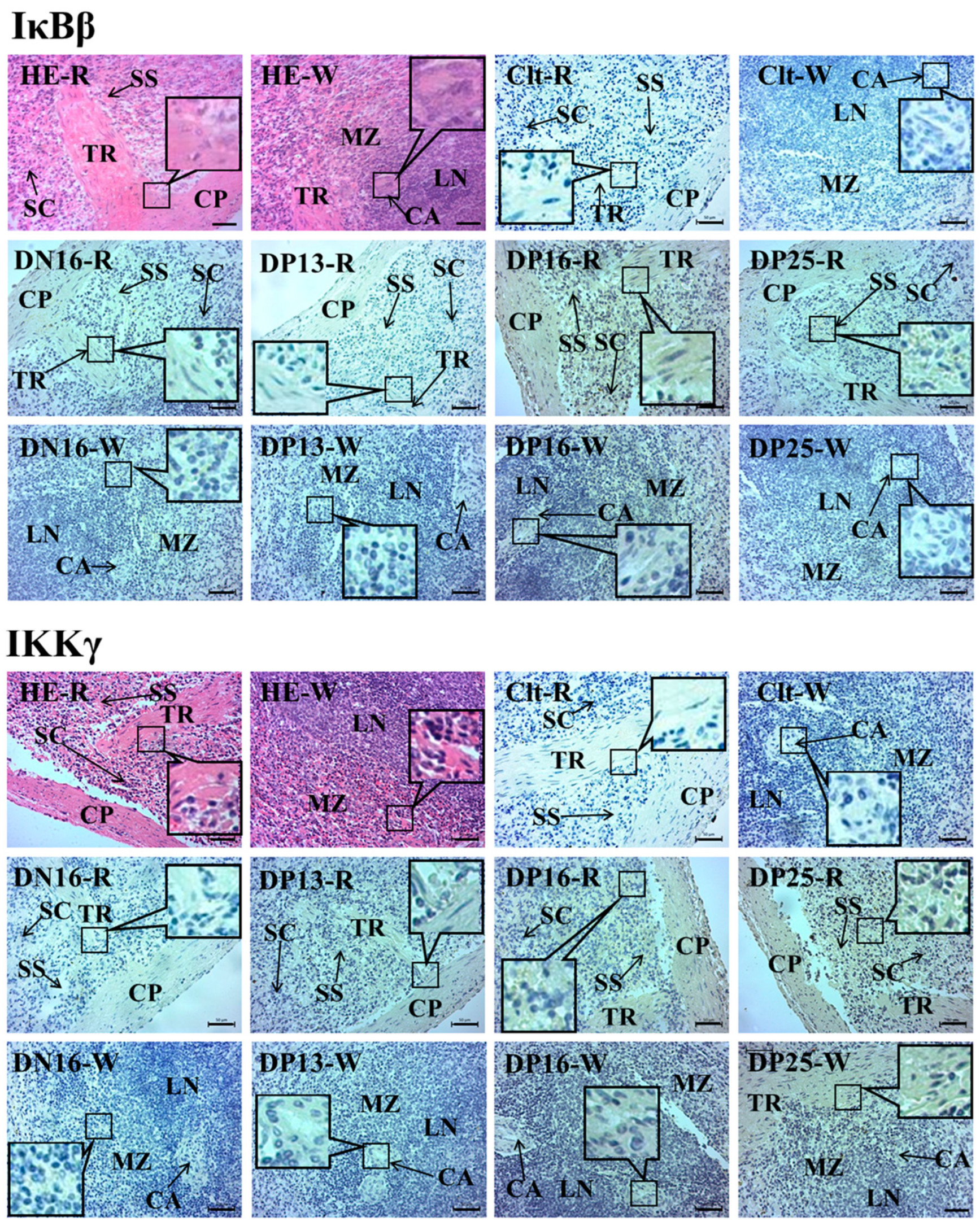
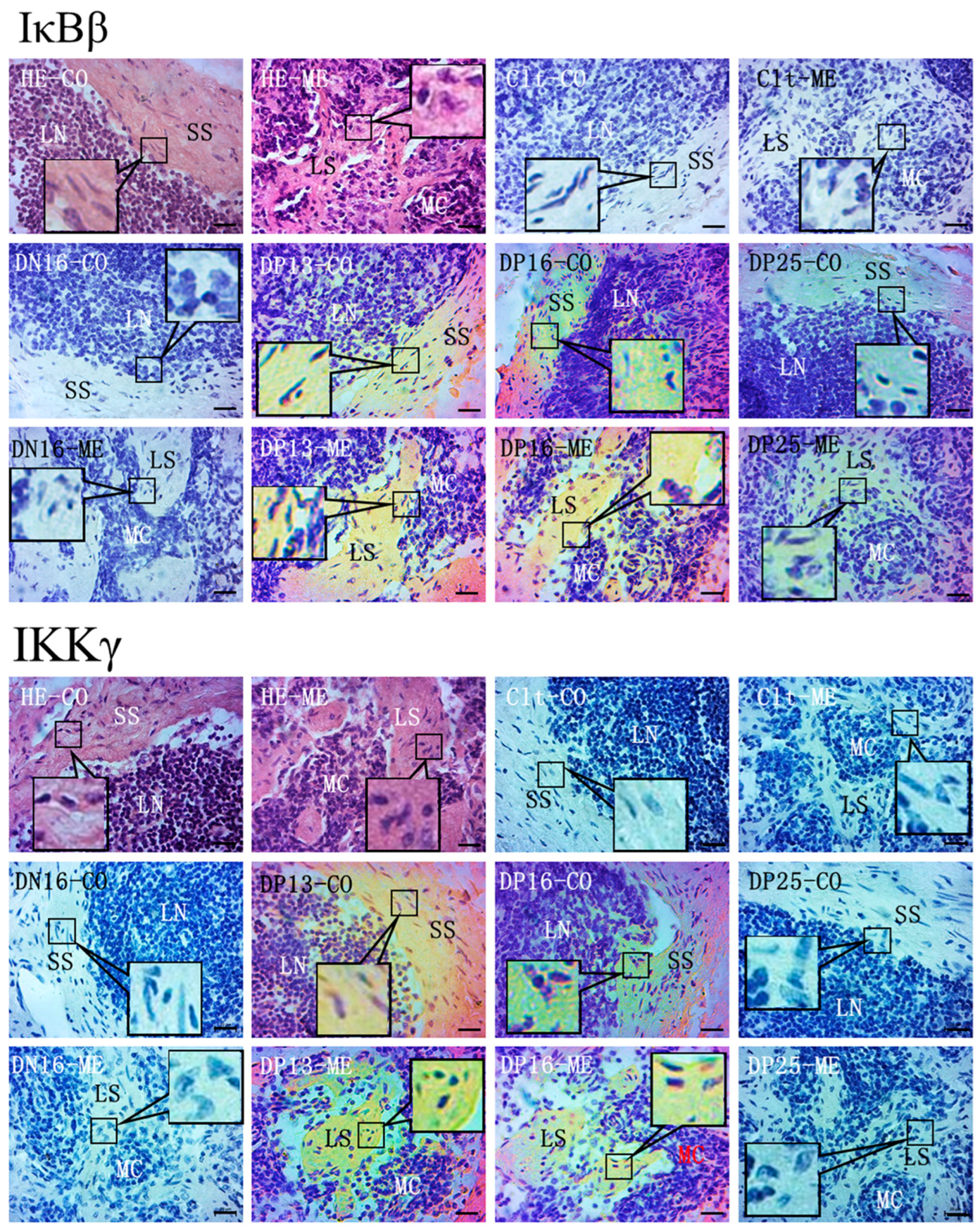
In the spleen, IκBβ and IKKγ proteins were located in the capsule, trabeculae and splenic cords. For the negative control, the spleens from day 16 of the estrous cycle, and spleens from days 13, 16, and 25 of pregnancy, the staining intensities for IκBβ protein were 0, 1, 0, 3 and 1, and the staining intensities for IKKγ protein were 0, 0, 1, 2 and 2, respectively (Figure 5). The staining intensity was as follows: 0 = negative; 1 = weak; 2 = strong; 3 = stronger.
In lymph nodes, IκBβ and IKKγ proteins were limited to the subcapsular sinus and lymph sinuses, but there was almost no immunostaining in the lymphoid nodule and medullary cords (Figure 6). The staining intensities for IκBβ protein were 0, 0, 3, 3 and 1, and the staining intensities for IKKγ protein were 0, 0, 3, 2 and 0, for the negative control, the lymph nodes from day 16 of the estrous cycle and lymph nodes from days 13, 16 and 25 of pregnancy, respectively.

3. Discussion

BCL-3 is involved in the immune response through interactions with the NF-κB subunits, and plays essential roles in germinal center formation and marginal zone B-cell development [36,37]. There is an upregulation of NF-κB1 or NF-κB2 in the maternal spleen during early pregnancy [7], suggesting that BCL-3 participates in the regulation of adaptive immunity through binding homodimers of NF-κB1 or NF-κB2. BCL-3 is required for dendritic cells in effective priming of CD4 and CD8 T cells and generates adaptive immunity in mice [38]. However, BCL-3 is upregulated in human placentas of severe early onset pre-eclampsia cases, which is related to immunology functions [39]. In addition, there is an upregulation of BCL-3 in lymphoid malignancy, and BCL-3-transgenic mice show splenomegaly and an accumulation of mature B cells in lymph nodes [40]. In this study, early pregnancy induced upregulation of BCL-3 mRNA and protein in the maternal spleen, but inhibited expression of BCL-3 in the maternal lymph nodes. Therefore, early pregnancy induced tissue-specific expression of BCL-3 in the maternal spleen and lymph nodes. The upregulation of BCL-3 in the maternal spleen may be associated with modulation of splenic adaptive immunity, but the downregulation of BCL-3 in maternal lymph nodes may be related to immune tolerance.
Circulating fetal DNA activates NF-κB and degrades IκBα in human peripheral blood mononuclear cells (PBMCs), resulting in the production of proinflammatory cytokines, which is related to spontaneous preterm birth [41]. IκBα plays key roles in proper B cell and secondary lymphoid tissue formation, which is related to NF-κB activation potentials in the mature B cells of mice [42]. IκBα level in cytoplasmic fractions from PBMCs of pregnant females is decreased compared with nonpregnant women, but this is not related to NF-κB activation in pregnancy [43]. In addition, IκBα level is downregulated in the lymph nodes of mice afflicted with murine-acquired immunodeficiency syndrome, but this does not lead to upregulation of NF-κB DNA binding activity [44]. Our results revealed that early pregnancy stimulates expression of IκBα in the maternal spleen, and IκBα level in the maternal lymph node peaked at day 16 of pregnancy, but declined at day 25 of pregnancy. Therefore, the upregulation of IκBα in the maternal spleen may be necessary for maturation of splenic B cells and pregnancy maintenance, and changed expression of IκBα in the maternal lymph node may be related to maternal immunoregulation during early pregnancy in sheep.
The E3 ligase ARIH2 causes degradation of IκBβ in the nucleus of dendritic cells, which is essential for embryogenesis [45]. NF-κB activity is suppressed in PBMCs from pregnant females, and IκBβ level is decreased more in pre-eclampsia [43]. It was revealed in this study that IκBβ mRNA and protein levels were declined on day 13 of pregnancy, but upregulated on day 16 of pregnancy in the maternal spleen, and IκBβ protein was located in the capsule, trabeculae and splenic cords. However, pregnancy stimulated the expression of IκBβ in the maternal lymph nodes, with peaks at days 13 and 16 of pregnancy, and IκBβ protein was limited to the subcapsular sinus and lymph sinuses. Therefore, the changing expression of IκBβ in the maternal spleen and lymph nodes in a tissue-specific manner may be involved in the immune regulation of maternal spleen and lymph nodes in sheep.
TNF-α and insulin-like growth factor-I cause pathological changes of placentas, and lead to pre-eclampsia, changing NFKBIE DNA methylation in BeWo cells [46]. As a negative regulator of the transcription factor NF-κB, IκBε interacts with RelA- and cRel-specific dimers to regulate B cell proliferation and survival in a stimulus-specific manner [47]. IκBε deletion results in increased lymph node cellularity and enhanced basal thymidine incorporation by lymphoid cells ex vivo, as well as enhancing the survival of ex vivo splenic B cells, which are regulated via c-Rel-dependent lymphoid responses in murine T- and B cells [48]. IκBε is mainly expressed in T cells in the spleen and lymph nodes, and IκBε deletion results in the reduction of one T cell precursor subspecies in mice, but does not lead to observable augmentation of constitutive nuclear NF-κB/Rel-binding activity [49]. It was found in this study that expression levels of IκBε and IκBα were upregulated in the maternal spleen during early pregnancy. However, the expression level of IκBε was the lowest at day 13 of pregnancy, but the highest at day 16 of pregnancy in the maternal lymph nodes. Therefore, the upregulation in the spleen and changed expression of IκBε in the lymph nodes may participate in regulating the immune functions of the maternal spleen and lymph nodes during early pregnancy in sheep.
NF-κB essential modulator (NEMO), also known as IκB kinase γ (IKKγ), plays a central role in the innate immune system by regulating the IKKα-IKKβ signaling axis [50]. NEMO gene expression level in maternal blood is higher, but lower in placentas of women with pre-eclampsia than healthy controls, suggesting that NEMO gene expression is associated with pre-eclampsia development in a tissue-specific manner [51]. NEMO mutation causes immune deficiency, which impairs lymph node formation in hemizygous mice and men [52]. In addition, NEMO knockout in T-lymphocyte induces the expression of Th17-related cytokines in spleen CD4+ T cells, and interrupted the canonical NF-κB pathway in an experimental nephrotoxic nephritis mouse model [53]. Our data revealed that IKKγ mRNA and protein levels gradually upregulated from day 13 to 25 of pregnancy in the maternal spleen, and IKKγ protein was located in the capsule, trabeculae and splenic cords. However, IKKγ mRNA and protein levels peaked at days 13 and 16 of pregnancy in the maternal lymph nodes, and IKKγ protein was limited to the subcapsular sinus and lymph sinuses. Therefore, the changed tissue-specific expression of IKKγ in maternal spleen and lymph nodes may be involved in the immune regulation of maternal spleen and lymph nodes.
IκBζ has six ankyrin repeats that are conserved in other IκB proteins, and its mRNA expression is induced rapidly following lipopolysaccharide injection in the spleen of mice [54]. IκBζ induces IL-17 production by helper T cells, which is critical for the proliferation of lymph node and splenic stromal cells [55]. IκBζ negatively modulates NF-κB activation in lymph nodes, which is beneficial for inhibiting the transformation and development process of lymphomas [56]. Our results showed that expression of IκBζ mRNA and protein increased at days 16 and 25 of pregnancy in the maternal spleen and lymph nodes. Therefore, the upregulation of IκBζ may be related to negative modulation of NF-κB activation, which may contribute to the immune tolerance of the maternal spleen and lymph nodes during early pregnancy.
IκBNS is a negative nuclear regulator of NF-κB activity that regulates IL-6 and TNF-α transcription, which are important factors in the remodeling of the uterus for blastocyst implantation in mice [57]. IκBNS negatively regulates IL-6 production under transcriptional control of NF-κB that plays an essential role in embryo implantation and the onset of labor during pregnancy in mice [58]. PBMCs in miscarrying women produce significantly higher concentrations of Th1 cytokines compared with normal pregnancy, which indicates that Th1 cytokines are deleterious to successful pregnancy in humans [59]. IκBNS knock-out results in a B cell extrinsic defect in the spleen and inguinal lymph node in mice [60]. Our data revealed that IκBNS mRNA and protein levels peaked at day 16 of pregnancy in the maternal spleen, but IκBNS expression level was the lowest at day 16 of pregnancy in the maternal lymph nodes. Therefore, the peak in the maternal spleen and the lowest level of IκBNS in lymph nodes at day 16 of pregnancy may be involved in the modulation of the maternal functions of the spleen and lymph nodes in a tissue-specific manner during early pregnancy in sheep.
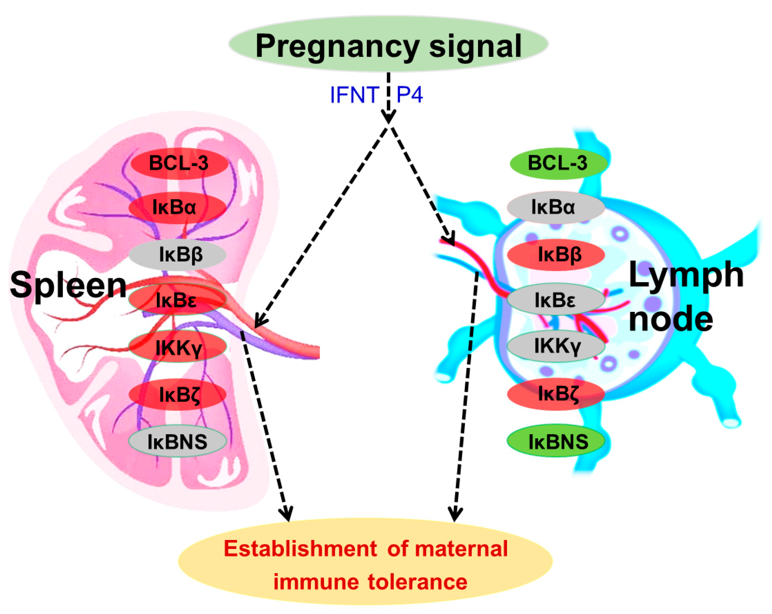
Type I interferons, including IFNT, induce hundreds of through activations of a number of cellular factors, including the IκB kinase epsilon [61]. There is a negative interaction between NF-κB and progesterone receptor [62]. In addition, our previous studies reported that ISGs and progesterone receptor are upregulated in ovine lymph nodes and spleen during early pregnancy, which are related to maternal immune tolerance [21,23,26,28,29,31]. During early pregnancy in sheep, early pregnancy signals (IFNT and progesterone) exert effects on the expression of the IκB family in the maternal spleen and lymph nodes, which upregulate the expression of BCL-3, IκBα, IκBε, IKKγ and IκBζ in the spleen, and IκBβ and IκBζ in the lymph nodes. However, early pregnancy signals inhibit the expression of BCL-3 and IκBNS in the lymph nodes, and also modulate the expression of IκBβ and IκBNS in the spleen and IκBα and IKKγ in the lymph nodes. Therefore, early pregnancy modulates the expression of the IκB family in the maternal spleen and lymph nodes in a tissue-specific manner, which may contribute to the establishment of maternal immune tolerance during early pregnancy (Figure 7).

4. Materials and Methods

4.1. Animal Tissue Collection

All procedures were approved by the Hebei University of Engineering Animal Care and Use Committee (application number 2019-017). Mature Small-tail Han ewes (approximately 18 month old) were purchased from Handan Boyuan Animal Husbandry Co., Ltd., Handan (China), and housed using conventional breeding and nutrition. The ewes with normal estrous cycles were randomly divided into three groups of pregnant ewes (days 13, 16 and 25 of pregnancy) and a group of day 16 of the estrous cycle (n = 6 for each group). After detection of sexual receptivity (day 0 of pregnancy or nonpregnancy) with a vasectomized ram, for the three groups, ewes were bred with intact rams, and the nonpregnant ewes were not mated with an intact ram. Spleens and inguinal lymph nodes were sampled on days 13, 16 and 25 post-estrus after the females were killed. Pregnancy was confirmed through sighting a conceptus in the uterus. Samples of spleens and lymph nodes were immediately fixed in fresh 4% (w/v) paraformaldehyde, and also frozen, stored for following quantitative real-time PCR (qRT-PCR) and Western blot analysis.

4.2. RNA Extraction and qRT-PCR Assay

Total RNA was isolated from the samples of spleens and lymph nodes following the manufacturer’s instructions using TRNzol reagent (Tiangen Biotech Co., Ltd., Beijing, China), and a Nanodrop spectrophotometer (Thermo Fisher Scientific, Wilmington, NC, USA) was applied to measure the concentration of total RNA at 260/280 nm. The cDNA was synthesized according to a FastQuant RT kit with DNase (Tiangen Biotech Co., Ltd., Beijing, China) protocol. The mRNA expression values of BCL-3, NFKBIA, NFKBIB, NFKBIE, IKBKG, NFKBID and NFKBIZ were carried out using a SuperReal PreMix Plus kit (Tiangen Biotech) according to optimized PCR protocols in triplicate, and glyceraldehyde-3-phosphate dehydrogenase (GAPDH) was amplified in parallel with the target genes. The primer sequences (Table 1) were designed and synthesized by Shanghai Sangon Biotech Co., Ltd. China. PCR conditions were 40 cycles of 95 °C for 10 s, 60–62.5 °C (60 °C for BCL-3, 60.5 °C for NFKBIA, NFKBID and NFKBIZ, 61 °C for NFKBIB and NFKBIE, 62.5 °C for IKBKG) for 20 s and 72 °C for 25 s. Melting curve analysis and electrophoresis on 2% agarose gel were used to verify PCR results. The GAPDH gene was used as an internal control, and positive and negative controls were tested every time. The 2−ΔΔCt analysis method [63] was used to calculate relative expression value with GAPDH as a normalization control. The data from the ewes on day 16 of the estrous cycle were used as normalization control.

4.3. Western Blot

The samples of spleens and lymph nodes were prepared as described previously [7,9], and total proteins were separated on polyacrylamide gels. Total proteins were transferred to PVDF membranes (Millipore, Bedford, MA, USA), and the membranes were incubated with primary antibodies overnight at 4 °C. Membranes were blocked with 5% skimmed milk powder. An anti-BCL-3 monoclonal antibody (Santa Cruz Biotechnology, Santa Cruz, CA, USA, sc-32741), an anti-IκBα monoclonal antibody (Santa Cruz Biotechnology, sc-1643), an anti-IκBβ monoclonal antibody (Santa Cruz Biotechnology, sc-390622), an anti-IκBε monoclonal antibody (Santa Cruz Biotechnology, sc-7275), an anti-IKKγ monoclonal antibody (Santa Cruz Biotechnology, sc-166398), an anti-NFKBID polyclonal antibody (Abcam, Cambridge, UK, ab232913, 1:1000) and an anti-IκBζ polyclonal antibody (Abcam, ab155142, 1:1000) were used. Depending on the origin of the primary antibodies, goat anti-mouse IgG-HRP (Biosharp, Hefei, China BL001A) or goat anti-rabbit IgG-HRP (Biosharp, BL003A) were used in 1:10,000 dilution. After washing, the membranes were incubated with an ECL Western blotting detection reagent (Tiangen Biotech), and the signals were detected. The immunospecific bands were analyzed using the Quantity One 4.1 software (Bio-Rad Laboratories, Hercules, CA, USA) with GAPDH as an internal control protein using an anti-GAPDH antibody (Santa Cruz Biotechnology, Inc., sc-20357, 1:1000).

4.4. Immunohistochemistry Analysis

The fixed samples of spleens and lymph nodes were prepared as described previously [7,9]. Several sections were stained by hematoxylin and eosin (HE). Endogenous peroxidase activity of other sections was quenched using 3% H2O2, and nonspecific binding was reduced with 5% normal goat serum. Immunohistochemical localization of IκBβ and IKKγ in the spleen and lymph nodes was performed using the anti-IκBβ monoclonal antibody (Santa Cruz Biotechnology, sc-390622, 1:200) and anti-IKKγ monoclonal antibody (Santa Cruz Biotechnology, sc-166398, 1:200), and a negative control was treated with goat anti-rabbit IgG. The antibody binding sites in the tissue sections was visualized using a DAB kit (Tiangen Biotech), and then nuclear was stained with hematoxylin. The images were captured using a light microscope (Nikon Eclipse E800, Tokyo, Japan) with a digital camera (AxioCam ERc 5s), and the intensity of staining and density of the stained cells were analyzed through the images as described previously [7,9].

4.5. Statistical Analysis

Data for relative expression levels of BCL-3, NFKBIA, NFKBIB, NFKBIE, IKBKG, NFKBID and NFKBIZ mRNA and proteins were analyzed with MIXED procedure in SAS (Version 9.1; SAS Institute, Cary, NC, USA). Relative expression levels of the different groups were compared using Duncan’s method, and data are presented as least-squares means. A p value < 0.05 was considered significantly different.

5. Conclusions

Expression of the IκB family was altered in the maternal spleen and lymph nodes in a tissue-specific manner during early pregnancy, which may be related to the IFNT from conceptus and the progesterone from the corpus lutea. These changes may be associated with establishment of maternal immune tolerance during early pregnancy.

Author Contributions

Conceptualization, L.Y.; methodology, S.F. and C.C.; validation, Y.B.; investigation, S.F. and C.C.; data curation, L.Z.; writing—original draft preparation, L.Y.; writing—review and editing, Y.B.; visualization, L.Z.; supervision, L.Y.; project administration, L.Z.; funding acquisition, L.Y. All authors have read and agreed to the published version of the manuscript.

Funding

This work was supported by the grants from Natural Science Foundation of Hebei Province, China (grant numbers C2021402019 and C2022402038) and Hebei Science and Technology Agency, China (grant number 22326602D).

Institutional Review Board Statement

Not applicable.

Informed Consent Statement

Not applicable.

Data Availability Statement

Data supporting the findings of this study are available within the paper.

Conflicts of Interest

The authors declare no conflict of interest.

References

  1. Deshmukh, H.; Way, S.S. Immunological basis for recurrent fetal loss and pregnancy complications. Annu. Rev. Pathol. 2019, 14, 185–210. [Google Scholar] [CrossRef] [PubMed]
  2. Abu-Raya, B.; Michalski, C.; Sadarangani, M.; Lavoie, P.M. Maternal immunological adaptation during normal pregnancy. Front. Immunol. 2020, 11, 575197. [Google Scholar] [CrossRef] [PubMed]
  3. Rocha, C.C.; da Silveira, J.C.; Forde, N.; Binelli, M.; Pugliesi, G. Conceptus-modulated innate immune function during early pregnancy in ruminants: A review. Anim. Reprod. 2021, 18, e20200048. [Google Scholar] [CrossRef] [PubMed]
  4. Quirke, L.D.; Maclean, P.H.; Haack, N.A.; Edwards, S.J.; Heiser, A.; Juengel, J.L. Characterization of local and peripheral immune system in pregnant and nonpregnant ewes. J. Anim. Sci. 2021, 99, skab208. [Google Scholar] [CrossRef]
  5. Gómez-Chávez, F.; Correa, D.; Navarrete-Meneses, P.; Cancino-Diaz, J.C.; Cancino-Diaz, M.E.; Rodríguez-Martínez, S. NF-κB and its regulators during pregnancy. Front. Immunol. 2021, 12, 679106. [Google Scholar] [CrossRef]
  6. Fang, S.; Zhang, T.; Qiao, H.; Hao, S.; Zhang, L.; Yang, L. Expression of nuclear factor kappa B components in the ovine maternal liver in early pregnancy periods. Anim. Sci. J. 2022, 93, e13724. [Google Scholar] [CrossRef]
  7. Hao, S.; Fang, H.; Fang, S.; Zhang, T.; Zhang, L.; Yang, L. Changes in nuclear factor kappa B components expression in the ovine spleen during early pregnancy. J. Anim. Feed Sci. 2022, 31, 3–11. [Google Scholar] [CrossRef]
  8. Yang, L.; Cai, C.; Fang, S.; Hao, S.; Zhang, T.; Zhang, L. Changes in expression of nuclear factor kappa B subunits in the ovine thymus during early pregnancy. Sci. Rep. 2022, 12, 17683. [Google Scholar] [CrossRef]
  9. Zhang, L.; Zhang, T.; Yang, Z.; Cai, C.; Hao, S.; Yang, L. Expression of nuclear factor kappa B in ovine maternal inguinal lymph nodes during early pregnancy. BMC Vet. Res. 2022, 18, 266. [Google Scholar] [CrossRef]
  10. Yamauchi, S.; Ito, H.; Miyajima, A. IkappaBeta, a nuclear IkappaB protein, positively regulates the NF-kappaB-mediated expression of proinflammatory cytokines. Proc. Natl. Acad. Sci. USA 2010, 107, 11924–11929. [Google Scholar] [CrossRef] [Green Version]
  11. Palmer, S.; Chen, Y.H. Bcl-3, a multifaceted modulator of NF-kappaB-mediated gene transcription. Immunol. Res. 2008, 42, 210–218. [Google Scholar] [CrossRef] [PubMed]
  12. MaruYama, T. The nuclear IκB family of proteins controls gene regulation and immune homeostasis. Int. Immunopharmacol. 2015, 28, 836–840. [Google Scholar] [CrossRef] [PubMed]
  13. Jeon, S.; Liu, Q.F.; Cai, H.; Jeong, H.J.; Kim, S.H.; Kim, D.I.; Lee, J.H. Administration of a herbal formulation enhanced blastocyst implantation via IκB activation in mouse endometrium. Chin. Med. 2020, 15, 112. [Google Scholar] [CrossRef] [PubMed]
  14. Toda, A.; Sawada, K.; Fujikawa, T.; Wakabayashi, A.; Nakamura, K.; Sawada, I.; Yoshimura, A.; Nakatsuka, E.; Kinose, Y.; Hashimoto, K.; et al. Targeting inhibitor of κB kinase β prevents inflammation-induced preterm delivery by inhibiting IL-6 production from amniotic cells. Am. J. Pathol. 2016, 186, 616–629. [Google Scholar] [CrossRef] [PubMed] [Green Version]
  15. Whalley, K. Brain-spleen link tunes immunity. Nat. Rev. Neurosci. 2020, 21, 350–351. [Google Scholar] [CrossRef]
  16. Marchetti, C.; Swartzwelter, B.; Gamboni, F.; Neff, C.P.; Richter, K.; Azam, T.; Carta, S.; Tengesdal, I.; Nemkov, T.; D’Alessandro, A.; et al. OLT1177, a β-sulfonyl nitrile compound, safe in humans, inhibits the NLRP3 inflammasome and reverses the metabolic cost of inflammation. Proc. Natl. Acad. Sci. USA 2018, 115, E1530–E1539. [Google Scholar] [CrossRef] [PubMed] [Green Version]
  17. Slawek, A.; Maj, T.; Chelmonska-Soyta, A. CD40, CD80, and CD86 costimulatory molecules are differentially expressed on murine splenic antigen-presenting cells during the pre-implantation period of pregnancy, and they modulate regulatory T-cell abundance, peripheral cytokine response, and pregnancy outcome. Am. J. Reprod. Immunol. 2013, 70, 116–126. [Google Scholar]
  18. Jalkanen, S.; Salmi, M. Lymphatic endothelial cells of the lymph node. Nat. Rev. Immunol. 2020, 20, 566–578. [Google Scholar] [CrossRef]
  19. Kapovic, M.; Rukavina, D. Kinetics of lymphoproliferative responses of lymphocytes harvested from the uterine draining lymph nodes during pregnancy in rats. J. Reprod. Immunol. 1991, 20, 93–101. [Google Scholar] [CrossRef]
  20. Klossner, R.; Groessl, M.; Schumacher, N.; Fux, M.; Escher, G.; Verouti, S.; Jamin, H.; Vogt, B.; Mohaupt, M.G.; Gennari-Moser, C. Steroid hormone bioavailability is controlled by the lymphatic system. Sci. Rep. 2021, 11, 9666. [Google Scholar] [CrossRef]
  21. Yang, L.; Liu, Y.; Lv, W.; Wang, P.; Wang, B.; Xue, J.; Zhang, L. Expression of interferon-stimulated gene 15-kDa protein, cyclooxygenase (COX) 1, COX-2, aldo-keto reductase family 1, member B1, and prostaglandin E synthase in the spleen during early pregnancy in sheep. Anim. Sci. J. 2018, 89, 1540–1548. [Google Scholar] [CrossRef] [PubMed]
  22. Li, N.; Zhao, Z.; Bai, J.; Liu, B.; Mi, H.; Zhang, L.; Li, G.; Yang, L. Characterization of the Th cytokines profile in ovine spleen during early pregnancy. J. Appl. Anim. Res. 2019, 47, 386–393. [Google Scholar] [CrossRef] [Green Version]
  23. Wang, Y.; Han, X.; Zhang, L.; Cao, N.; Cao, L.; Yang, L. Early pregnancy induces expression of STAT1, OAS1 and CXCL10 in ovine spleen. Animals 2019, 9, 882. [Google Scholar] [CrossRef] [Green Version]
  24. Bai, J.; Zhang, L.; Zhao, Z.; Li, N.; Wang, B.; Yang, L. Expression of melatonin receptors and CD4 in the ovine thymus, lymph node, spleen and liver during early pregnancy. Immunology 2020, 160, 52–63. [Google Scholar] [CrossRef]
  25. Cao, N.; Cao, L.; Gao, M.; Wang, H.; Zhang, L.; Yang, L. Changes in mRNA and protein levels of gonadotropin releasing hormone and receptor in ovine thymus, lymph node, spleen, and liver during early pregnancy. Domest. Anim. Endocrinol. 2021, 76, 106607. [Google Scholar] [CrossRef]
  26. Yang, L.; Guo, R.; Yao, X.; Yan, J.; Bai, Y.; Zhang, L. Expression of progesterone receptor and progesterone-induced blocking factor in the spleen during early pregnancy in ewes. Livest. Sci. 2018, 209, 14–19. [Google Scholar] [CrossRef]
  27. Zhang, L.; Yang, G.; Zhang, Q.; Feng, P.; Gao, M.; Yang, L. Early pregnancy affects expression of Toll-like receptor signaling members in ovine spleen. Anim. Reprod. 2021, 18, e20210009. [Google Scholar] [CrossRef] [PubMed]
  28. Yang, L.; Zang, S.; Bai, Y.; Yao, X.; Zhang, L. Effect of early pregnancy on the expression of progesterone receptor and progesterone-induced blocking factor in ovine lymph node. Theriogenology 2017, 93, 78–83. [Google Scholar] [CrossRef]
  29. Yang, L.; Wang, Q.; Liu, Y.; Zhang, L.; Lv, W.; Liu, B. Expression profiles of interferon-stimulated gene 15 and prostaglandin synthases in the ovine lymph nodes during early pregnancy. Mol. Reprod. Dev. 2019, 86, 100–108. [Google Scholar] [CrossRef]
  30. Yang, L.; Wang, P.; Mi, H.; Lv, W.; Liu, B.; Du, J.; Zhang, L. Comparison of Th1 and Th2 cytokines production in ovine lymph nodes during early pregnancy. Theriogenology 2019, 123, 177–184. [Google Scholar] [CrossRef]
  31. Zhang, L.; Cao, L.; Yang, F.; Han, X.; Wang, Y.; Cao, N.; Yang, L. Relative abundance of interferon-stimulated genes STAT1, OAS1, CXCL10 and MX1 in ovine lymph nodes during early pregnancy. Anim. Reprod. Sci. 2020, 214, 106285. [Google Scholar] [CrossRef]
  32. Feng, P.; Wu, J.; Ren, Y.; Zhang, L.; Cao, J.; Yang, L. Early pregnancy regulates the expression of prolactin and its receptor in the thymus, the liver, the spleen and lymph nodes in sheep. Domest. Anim. Endocrinol. 2022, 81, 106731. [Google Scholar] [CrossRef] [PubMed]
  33. Wu, J.; Zhang, Q.; Zhang, L.; Feng, P.; Gao, M.; Zhao, Z.; Yang, L. Toll-like receptor signaling is changed in ovine lymph node during early pregnancy. Anim. Sci. J. 2021, 92, e13541. [Google Scholar] [CrossRef] [PubMed]
  34. Zhang, L.; Cao, L.; Feng, P.; Han, X.; Yang, L. Complement regulation in ovine lymph nodes during early pregnancy. Exp. Ther. Med. 2022, 23, 166. [Google Scholar] [CrossRef] [PubMed]
  35. Zhao, Z.; Li, Y.; Cao, J.; Fang, H.; Zhang, L.; Yang, L. Early pregnancy modulates expression of the nod-like receptor family in lymph nodes of ewes. Animals 2022, 12, 3285. [Google Scholar] [CrossRef] [PubMed]
  36. Hövelmeyer, N.; Wörns, M.A.; Reissig, S.; Adams-Quack, P.; Leclaire, J.; Hahn, M.; Wörtge, S.; Nikolaev, A.; Galle, P.R.; Waisman, A. Overexpression of Bcl-3 inhibits the development of marginal zone B cells. Eur. J. Immunol. 2014, 44, 545–552. [Google Scholar] [CrossRef] [Green Version]
  37. Zhang, X.; Paun, A.; Claudio, E.; Wang, H.; Siebenlist, U. The tumor promoter and NF-κB modulator Bcl-3 regulates splenic B cell development. J. Immunol. 2013, 191, 5984–5992. [Google Scholar] [CrossRef] [Green Version]
  38. Tassi, I.; Claudio, E.; Wang, H.; Tang, W.; Ha, H.L.; Saret, S.; Ramaswamy, M.; Siegel, R.; Siebenlist, U. The NF-κB regulator Bcl-3 governs dendritic cell antigen presentation functions in adaptive immunity. J. Immunol. 2014, 193, 4303–4311. [Google Scholar] [CrossRef] [Green Version]
  39. Heikkilä, A.; Tuomisto, T.; Häkkinen, S.K.; Keski-Nisula, L.; Heinonen, S.; Ylä-Herttuala, S. Tumor suppressor and growth regulatory genes are overexpressed in severe early-onset preeclampsia—An array study on case-specific human preeclamptic placental tissue. Acta Obstet. Gynecol. Scand. 2005, 84, 679–689. [Google Scholar]
  40. Ong, S.T.; Hackbarth, M.L.; Degenstein, L.C.; Baunoch, D.A.; Anastasi, J.; McKeithan, T.W. Lymphadenopathy, splenomegaly, and altered immunoglobulin production in BCL3 transgenic mice. Oncogene 1998, 16, 2333–2343. [Google Scholar] [CrossRef] [Green Version]
  41. Scharfe-Nugent, A.; Corr, S.C.; Carpenter, S.B.; Keogh, L.; Doyle, B.; Martin, C.; Fitzgerald, K.A.; Daly, S.; O’Leary, J.J.; O’Neill, L.A. TLR9 provokes inflammation in response to fetal DNA: Mechanism for fetal loss in preterm birth and preeclampsia. J. Immunol. 2012, 188, 5706–5712. [Google Scholar] [CrossRef] [PubMed] [Green Version]
  42. Wuerzberger-Davis, S.M.; Chen, Y.; Yang, D.T.; Kearns, J.D.; Bates, P.W.; Lynch, C.; Ladell, N.C.; Yu, M.; Podd, A.; Zeng, H.; et al. Nuclear export of the NF-κB inhibitor IκBα is required for proper B cell and secondary lymphoid tissue formation. Immunity 2011, 34, 188–200. [Google Scholar] [CrossRef] [PubMed] [Green Version]
  43. McCracken, S.A.; Drury, C.L.; Lee, H.S.; Morris, J.M. Pregnancy is associated with suppression of the nuclear factor kappaB/IkappaB activation pathway in peripheral blood mononuclear cells. J. Reprod. Immunol. 2003, 58, 27–47. [Google Scholar] [CrossRef]
  44. Crinelli, R.; Bianchi, M.; Gentilini, L.; Magnani, M.; Hiscott, J. Activation of the ubiquitin proteolytic system in murine acquired immunodeficiency syndrome affects IkappaBalpha turnover. Eur. J. Biochem. 1999, 263, 202–211. [Google Scholar] [CrossRef] [PubMed]
  45. Lin, A.E.; Ebert, G.; Ow, Y.; Preston, S.P.; Toe, J.G.; Cooney, J.P.; Scott, H.W.; Sasaki, M.; Saibil, S.D.; Dissanayake, D.; et al. ARIH2 is essential for embryogenesis, and its hematopoietic deficiency causes lethal activation of the immune system. Nat. Immunol. 2013, 14, 27–33. [Google Scholar] [CrossRef] [PubMed]
  46. Tanaka, K.; Nakabayashi, K.; Kawai, T.; Tanigaki, S.; Matsumoto, K.; Hata, K.; Kobayashi, Y. Gene expression and DNA methylation changes in BeWo cells dependent on tumor necrosis factor-α and insulin-like growth factor-I. Hum. Cell 2020, 33, 37–46. [Google Scholar]
  47. Alves, B.N.; Tsui, R.; Almaden, J.; Shokhirev, M.N.; Davis-Turak, J.; Fujimoto, J.; Birnbaum, H.; Ponomarenko, J.; Hoffmann, A. IκBε is a key regulator of B cell expansion by providing negative feedback on cRel and RelA in a stimulus-specific manner. J. Immunol. 2014, 192, 3121–3132. [Google Scholar] [CrossRef] [PubMed] [Green Version]
  48. Clark, J.M.; Aleksiyadis, K.; Martin, A.; McNamee, K.; Tharmalingam, T.; Williams, R.O.; Mémet, S.; Cope, A.P. Inhibitor of kappa B epsilon (IκBε) is a non-redundant regulator of c-Rel-dependent gene expression in murine T and B cells. PLoS ONE 2011, 6, e24504. [Google Scholar] [CrossRef] [Green Version]
  49. Mémet, S.; Laouini, D.; Epinat, J.C.; Whiteside, S.T.; Goudeau, B.; Philpott, D.; Kayal, S.; Sansonetti, P.J.; Berche, P.; Kanellopoulos, J.; et al. IkappaBepsilon-deficient mice: Reduction of one T cell precursor subspecies and enhanced Ig isotype switching and cytokine synthesis. J. Immunol. 1999, 163, 5994–6005. [Google Scholar] [CrossRef]
  50. Clark, K.; Nanda, S.; Cohen, P. Molecular control of the NEMO family of ubiquitin-binding proteins. Nat. Rev. Mol. Cell Biol. 2013, 14, 673–685. [Google Scholar] [CrossRef]
  51. Sakowicz, A.; Hejduk, P.; Pietrucha, T.; Nowakowska, M.; Płuciennik, E.; Pospiech, K.; Gach, A.; Rybak-Krzyszkowska, M.; Sakowicz, B.; Kaminski, M.; et al. Finding NEMO in preeclampsia. Am. J. Obstet. Gynecol. 2016, 214, 538.e1–538.e7. [Google Scholar] [CrossRef] [PubMed]
  52. Siggs, O.M.; Berger, M.; Krebs, P.; Arnold, C.N.; Eidenschenk, C.; Huber, C.; Pirie, E.; Smart, N.G.; Khovananth, K.; Xia, Y.; et al. A mutation of Ikbkg causes immune deficiency without impairing degradation of IkappaB alpha. Proc. Natl. Acad. Sci. USA 2010, 107, 3046–3051. [Google Scholar] [CrossRef] [Green Version]
  53. Guo, L.; Huang, J.; Chen, M.; Piotrowski, E.; Song, N.; Zahner, G.; Paust, H.J.; Alawi, M.; Geffers, R.; Thaiss, F. T-lymphocyte-specific knockout of IKK-2 or NEMO induces Th17 cells in an experimental nephrotoxic nephritis mouse model. FASEB J. 2019, 33, 2359–2371. [Google Scholar] [CrossRef] [Green Version]
  54. Shiina, T.; Morimatsu, M.; Kitamura, H.; Ito, T.; Kidou, S.; Matsubara, K.; Matsuda, Y.; Saito, M.; Syuto, B. Genomic organization, chromosomal localization, and promoter analysis of the mouse Mail gene. Immunogenetics 2001, 53, 649–655. [Google Scholar] [CrossRef] [PubMed]
  55. Majumder, S.; Amatya, N.; Revu, S.; Jawale, C.V.; Wu, D.; Rittenhouse, N.; Menk, A.; Kupul, S.; Du, F.; Raphael, I.; et al. IL-17 metabolically reprograms activated fibroblastic reticular cells for proliferation and survival. Nat. Immunol. 2019, 20, 534–545. [Google Scholar] [CrossRef]
  56. Ishikawa, C.; Senba, M.; Mori, N. Induction of IκB-ζ by Epstein-Barr virus latent membrane protein-1 and CD30. Int. J. Oncol. 2015, 47, 2197–2207. [Google Scholar] [CrossRef]
  57. Sierra-Mondragón, E.; Gómez-Chávez, F.; Murrieta-Coxca, M.; Vázquez-Sánchez, E.A.; Martínez-Torres, I.; Cancino-Díaz, M.E.; Rojas-Espinosa, O.; Cancino-Díaz, J.C.; Reyes-Sánchez, J.L.; Rodríguez-Muñóz, R.; et al. Low expression of IL-6 and TNF-α correlates with the presence of the nuclear regulators of NF-κB, IκBNS and BCL-3, in the uterus of mice. Mol. Immunol. 2015, 68, 333–340. [Google Scholar] [CrossRef] [PubMed]
  58. Gómez-Chávez, F.; López-Portales, Ó.H.; Baeza-Martínez, D.A.; Cancino-Díaz, J.C.; Murrieta-Coxca, J.M.; Cancino-Díaz, M.E.; Pérez-Tapia, S.M.; Rodríguez-Martínez, S. IκBNS and IL-6 expression is differentially established in the uterus of pregnant healthy and infected mice. Heliyon 2020, 6, e04122. [Google Scholar] [CrossRef] [PubMed]
  59. Raghupathy, R.; Makhseed, M.; Azizieh, F.; Omu, A.; Gupta, M.; Farhat, R. Cytokine production by maternal lymphocytes during normal human pregnancy and in unexplained recurrent spontaneous abortion. Hum. Reprod. 2000, 15, 713–718. [Google Scholar] [CrossRef] [Green Version]
  60. Khoenkhoen, S.; Ádori, M.; Solís-Sayago, D.; Soulier, J.; Russell, J.; Beutler, B.; Pedersen, G.K.; Karlsson Hedestam, G.B. IκBNS expression in B cells is dispensable for IgG responses to T cell-dependent antigens. Front. Immunol. 2022, 13, 1000755. [Google Scholar] [CrossRef] [PubMed]
  61. Rajsbaum, R.; Versteeg, G.A.; Schmid, S.; Maestre, A.M.; Belicha-Villanueva, A.; Martínez-Romero, C.; Patel, J.R.; Morrison, J.; Pisanelli, G.; Miorin, L.; et al. Unanchored K48-linked polyubiquitin synthesized by the E3-ubiquitin ligase TRIM6 stimulates the interferon-IKKε kinase-mediated antiviral response. Immunity 2014, 40, 880–895. [Google Scholar] [CrossRef] [PubMed] [Green Version]
  62. McKay, L.I.; Cidlowski, J.A. Cross-talk between nuclear factor-kappa B and the steroid hormone receptors: Mechanisms of mutual antagonism. Mol. Endocrinol. 1998, 12, 45–56. [Google Scholar] [CrossRef] [PubMed] [Green Version]
  63. Livak, K.J.; Schmittgen, T.D. Analysis of relative gene expression data using real-time quantitative PCR and the 2(-Delta Delta C(T)) method. Methods 2001, 25, 402–408. [Google Scholar] [CrossRef] [PubMed]
Figure 1. Relative expression values of BCL-3, NFKBIA, NFKBIB, NFKBIE, IKBKG, NFKBID and NFKBIZ mRNA in ovine spleen measured by quantitative real-time PCR. Note: DN16 = day 16 of the estrous cycle; DP13 = day 13 of pregnancy; DP16 = day 16 of pregnancy; DP25 = day 25 of pregnancy. Significant differences (p < 0.05) are indicated by different letters within same color columns. p values between 0.001 and 0.01 are shown with two (**) asterisks, and p value less than 0.001 is designated with three (***) asterisks.
Ijms 24 05156 g001
Figure 2. Relative expression values of BCL-3, NFKBIA, NFKBIB, NFKBIE, IKBKG, NFKBID and NFKBIZ mRNA in the lymph nodes from non-pregnant ewes and pregnant ewes. Note: DN16 = day 16 of the estrous cycle; DP13 = day 13 of pregnancy; DP16 = day 16 of pregnancy; DP25 = day 25 of pregnancy. Significant differences (p < 0.05) are indicated by different letters within same color columns. p values between 0.01 and 0.05 are shown with one (*) asterisk, and p values between 0.001 and 0.01 are shown with two (**) asterisks, and p value less than 0.001 is designated with three (***) asterisks.
Ijms 24 05156 g002
Figure 3. Expression of IκB family proteins in ovine spleen analyzed by Western blotting. Note: DN16 = day 16 of the estrous cycle; DP13 = day 13 of pregnancy; DP16 = day 16 of pregnancy; DP25 = day 25 of pregnancy. Significant differences (p < 0.05) are indicated by different superscript letters within the same color column. p values between 0.001 and 0.01 are shown with two (**) asterisks, and p value less than 0.001 is designated with three (***) asterisks.
Ijms 24 05156 g003
Figure 4. Expression of IκB family proteins in lymph nodes from non-pregnant ewes and pregnant ewes. Note: DN16 = day 16 of the estrous cycle; DP13 = day 13 of pregnancy; DP16 = day 16 of pregnancy; DP25 = day 25 of pregnancy. Significant differences (p < 0.05) are indicated by different letters within the same color column. p values between 0.01 and 0.05 are shown with one (*) asterisk, and p values between 0.001 and 0.01 are shown with two (**) asterisks, and p value less than 0.001 is designated with three (***) asterisks.
Ijms 24 05156 g004
Figure 5. Representative immunohistochemical localization of IκBβ and IKKγ proteins in ovine spleen. The spleen is divided into red pulp (R) and white pulp (W), and surrounded by a thickened capsule. Capsule (CP) with several trabeculae (TR) projects into the substance of the spleen. Note: HE = stained by hematoxylin and eosin; Clt = negative control; SS = splenic sinuses; SC = splenic cords; MZ = marginal zone; LN = lymphoid nodule; CA = central arteriole; DN16 = day 16 of the estrous cycle; DP13 = day 13 of pregnancy; DP16 = day 16 of pregnancy; DP25 = day 25 of pregnancy. Bar = 50 µm.
Ijms 24 05156 g005
Figure 6. Representative immunohistochemical localization of IκBβ and IKKγ proteins in the lymph nodes from non-pregnant ewes and pregnant ewes. The lymph node is divided into the outer cortex (CO) and the inner medulla (ME). Lymph enters the convex through the subcapsular sinus (SS) around the lymphoid nodules (LN), and flows into the medulla through the lymph sinus (LS) around the medullary cord (MC). Note: HE = stained by hematoxylin and eosin; Clt = negative control; DN16 = day 16 of the estrous cycle; DP13 = day 13 of pregnancy; DP16 = day 16 of pregnancy; DP25 = day 25 of pregnancy. Bar = 20 µm.
Ijms 24 05156 g006
Figure 7. Proposed sketch of IκB family in maternal spleen and lymph nodes during early pregnancy in sheep. Early pregnancy signals, including interferon-tau (IFNT) and progesterone (P4), induce changed expression of IκB family, including B cell leukemia-3 (BCL-3), IκBα, IκBβ, IκBε, IKKγ, IκBζ and IκBNS, in a tissue-specific manner, which contributes to establishment of maternal immune tolerance during early pregnancy. Note: red, stimulators; green, negative regulators; blue, changed.
Ijms 24 05156 g007
Table 1. Primers used for RT-qPCR.
GenePrimerSequenceSize (bp)Accession Numbers
BCL-3ForwardGCGACCAGAGGCAATTTACTACCAG98XM_027978453.2
ReverseGAGGTGTAGGCAAGTTCAGCAGAG
NFKBIAForwardAGGACGAGGAGTATGAGCAGATGG130NM_001166184.1
ReverseGCCAAGTGCAGGAACGAGTCTC
NFKBIBForwardCCCCAAGACCTACCTCGCTCAG119XM_027978262.2
ReverseTCCAGTCCTCTTCACTCTCATCCTC
NFKBIEForwardGCACTCACGTACATTTCCGAGGAC97XM_042236979.1
ReverseGCAGCAGAGCCAGGCAATACAG
IKBKGForwardGGGCAACCAGAGGGAGGAGAAG146XM_027963334.2
ReverseGGCATGTCTTCAGGCGTTCCAC
NFKBIZForwardGCAAAGGCGTACAATGGAAACACC137NM_001306117.1
ReverseGGCTGCTCGTTCTCCAAGTTCC
NFKBIDForwardACATTCGTGAGCATAAGGGCAAGAC114XM_027977435.2
ReverseGATGGTCAGTGGCATTGGGTTCC
GAPDHForwardGGGTCATCATCTCTGCACCT176NM_001190390.1
ReverseGGTCATAAGTCCCTCCACGA
Disclaimer/Publisher’s Note: The statements, opinions and data contained in all publications are solely those of the individual author(s) and contributor(s) and not of MDPI and/or the editor(s). MDPI and/or the editor(s) disclaim responsibility for any injury to people or property resulting from any ideas, methods, instructions or products referred to in the content.