Review

Adverse Human Health Effects of Chromium by Exposure Route: A Comprehensive Review Based on Toxicogenomic Approach

1
Institute of Environmental Medicine for Green Chemistry, Department of Life Science, Dongguk University Biomedi Campus, 32 Dongguk-ro, Ilsandong-gu, Goyang-si 10326, Republic of Korea
2
Insilicogen Inc., Suwon 441813, Republic of Korea
3
Department of Nano, Chemical & Biological Engineering, College of Natural Science and Engineering, Seokyeong University, Seoul 02173, Republic of Korea
*
Authors to whom correspondence should be addressed.
Int. J. Mol. Sci. 2023, 24(4), 3410; https://doi.org/10.3390/ijms24043410
Submission received: 26 December 2022 / Revised: 31 January 2023 / Accepted: 7 February 2023 / Published: 8 February 2023
(This article belongs to the Special Issue Heavy Metal Toxicity in Humans 2.0)

Abstract

:
Heavy metals are defined as metals with relatively high density and atomic weight, and their various applications have raised serious concerns about the environmental impacts and potential human health effects. Chromium is an important heavy metal that is involved in biological metabolism, but Cr exposure can induce a severe impact on occupational workers or public health. In this study, we explore the toxic effects of Cr exposure through three exposure routes: dermal contact, inhalation, and ingestion. We propose the underlying toxicity mechanisms of Cr exposure based on transcriptomic data and various bioinformatic tools. Our study provides a comprehensive understanding of the toxicity mechanisms of different Cr exposure routes by diverse bioinformatics analyses.

1. Introduction

Heavy metals are defined as metals with relatively high density, atomic weight, or atomic number. Their widespread industrial, domestic, agricultural, medical, and technological applications have raised serious concerns about the environmental impacts and potential human health effects of these elements [1,2]. Due to the ubiquitous characteristic of heavy metals, human exposure can occur through inhalation, ingestion, and dermal routes. Because of the high degree of toxicity, toxicological studies of various heavy metals have been actively conducted and the metals are managed through regulation. Heavy metal exposure also induced diverse cancer. The International Agency for Research on Cancer (IARC) designated arsenic, cadmium, chromium, and nickel as Group 1 carcinogens [3] (i.e., they are carcinogenic to humans).
Chromium is an important micronutrient that is involved in carbohydrate, lipid, and protein metabolism [4]. Cr is an important metal in pigment manufacturing, painting, metal plating, wood treating, and leather tanning, and it is used in metallurgy and refractory material production, among various other applications [5]. Cr is abundant in the Earth’s crust, in which it occurs at a level of 100 μg/g, and its toxicity depends on its chemical state [6,7]. Its two most prevalent oxidation forms are trivalent and hexavalent, but whereas trivalent Cr is largely non-toxic, hexavalent Cr is toxic and carcinogenic in living organisms. The salt form of trivalent and hexavalent Cr compounds, called chromate, generates toxic dust during manufacturing processes [8]. Many studies have investigated the adverse effects of Cr dust on chromate production workers [9,10]. Furthermore, industrial wastes containing Cr are known as major sources of soil and water pollution and, hence, can have a severe impact on public health.
According to the National Institute for Occupational Safety and Health (NIOSH), adverse effects of Cr exposure vary depending on their exposure paths: dermal contact, inhalation, and ingestion [11]. The general population is exposed to Cr from the ingestion of Cr-contaminated food or drinking water, mainly around a specific area contaminated with Cr for a long time [12]. In contrast to exposure via the ingestion route, Cr inhalation and skin contact exposure occurs occupationally, such as in tanneries or timber industries [13]. In addition to the level of exposure, the toxic effects are highly dependent on the exposure path [14].
Because of increased attention to health and biology, a large collection of toxicological data and literature are available. Data-mining techniques and biological analysis tools are useful for organizing “big data” to understand the biological effects of heavy metals [15]. Molecular network analysis is one way to provide a comprehensive view of heavy metal-induced adverse effects and related diseases or potential marker proteins [8,16]. This approach can be undertaken using molecular network analysis software to construct a network about toxic effects on genes and cellular processes and provide disease information.
Gene expression profiling provides the expression level of thousands of mRNAs at the same time, allowing insight into cellular conditions [17]. By analyzing gene expression profiling data, the variation of mRNA level can be used to detect cellular status under certain conditions, such as medicines, toxic substances, or disease status, and explore the underlying mechanisms of chemical toxicity. In addition, network-based hub gene selection and cellular processes contribute to the prediction of specific protein markers for toxicity.
Protein markers are useful for determining whether exposure to certain harmful substances has occurred or to predict the occurrence of certain diseases, but reliance on a single function of a gene/protein to predict toxic effects may lead to misinformation, particularly if it exhibits its toxic effect only after prolonged exposure. Functional enrichment is a widely used tool to identify trends in large-scale biological datasets [18]. It can integrate multiple functional information from various sources to provide a list of genes and the biological processes significantly enriched in these genes [19]. One of the most popular databases of functional enrichment analysis is Gene Ontology, providing information about biological processes, cellular components, and molecular functions.
In this study, we explore the toxic effects of Cr exposure through three exposure routes: dermal contact, inhalation, and ingestion. We summarize the sources of Cr exposure and a undertake a network analysis of public gene expression big data to analyze the toxic effects (Figure 1). A gene ontology term analysis was conducted to predict the biological functions of differentially expressed genes (DEGs), and the toxic effects of Cr exposure were explored through a comparison of the results (Figure 1b). Finally, we present a common and specific toxic effect from all three routes of Cr exposure.

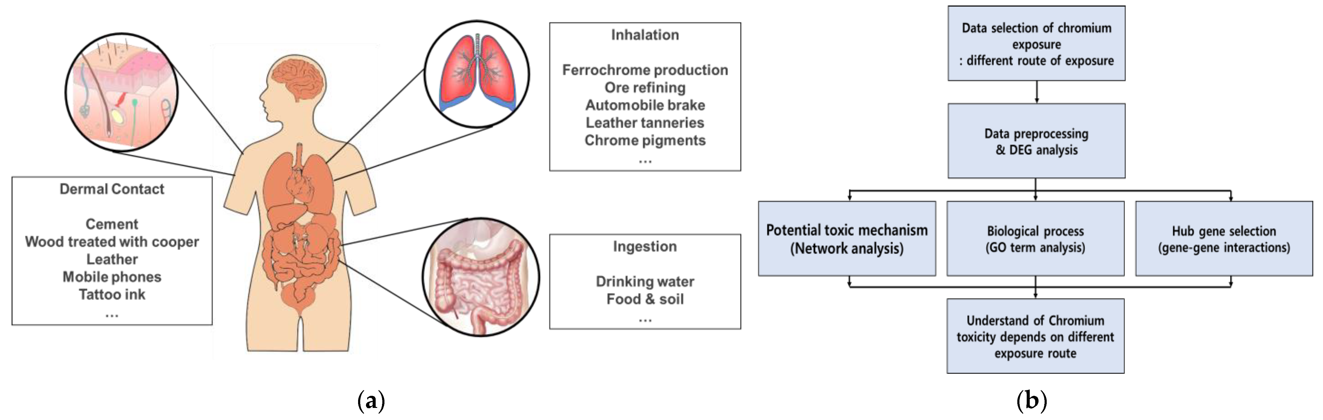
2. Adverse Effects of Cr on Skin

Dermal exposure to Cr for the general public can occur from dermal contact with certain consumer products or soils that contain Cr. Cr compounds can be present in cosmetic products and leather products in the form of both Cr(III) and Cr(VI) [20]. Construction workers can be exposed to Cr (VI) present in cement [21]. Cr can penetrate human skin to some extent, especially if the skin is damaged. Penetrated Cr can bind to immune cells of the skin, causing dermal toxicity [22]. Cr exposure can lead to sensitization and elicitation of contact dermatitis by forming Cr-protein conjugates. In addition, Cr exposure can cause skin irritation that may result in ulceration [22].
To understand the toxic mechanisms of Cr dermal contact, we conducted a molecular network analysis with Pathway Studio software (Version 12.5.0.2) using the gene expression profile dataset obtained from the public gene expression database. We used the microarray dataset GSE16394 derived from the study of human dermal fibroblast exposure to in vitro potassium dichromate from the Gene Expression Omnibus (GEO) database (https://www.ncbi.nlm.nih.gov/geo/ (accessed on 1 November 2022)). Figure 2a shows that exposure to Cr and Cr compounds through dermal contact mainly induces DNA damage, metastasis, and immune system disorders. Skin diseases, such as skin damage and ulceration, allergic reactions, and hypersensitivity, seem to be the effects of skin exposure to Cr. Rudolf et al. revealed that Cr exposure led to DNA damage in human dermal fibroblasts [23]. Our molecular network analysis result showed that dermal exposure to Cr was also related to immune system responses. Thomas et al. identified that chromate exposure caused monocyte and dense T-cell infiltration, pointing to a delayed-type immune reaction [24]. In another study, mRNA expression of IL-4, IL-6, and IFN-Q was increased in skin tissues exposed to chromate [24]. Moreover, dermal exposure to Cr has been implicated in immediate-type or type I hypersensitivity reactions. Cases of asthma with an immediate or delayed response have been observed following occupational exposure to Cr [25].
A molecular pathway analysis has advantages in visualizing major cellular processes and disease information based on hub genes, but biases may exist when determining the toxic effects of Cr from dermal exposure because of confounders in the study and references. We identified the biological processes (BP), cellular components (CC), and molecular functions (MF) associated with dermal exposure to Cr by Gene Ontology (GO) term analysis using all DEGs (Figure 2b). Enriched GO pathways were determined based on the cutoff criterion of adjusted p-value < 0.05. By GO term analysis, we explored the functions of genes whose expression changed after dermal exposure to Cr. The results showed enrichment of several BP terms, including RNA modification, transfer RNA (tRNA) processing, non-coding RNA processing, sterol biosynthesis process, and cholesterol biosynthesis process. According to Pavesi and Moreira, non-coding RNA induced by Cr exposure could trigger epigenetic modifications that may lead to DNA damage and cancer [26]. Guo et al. revealed that skin cell exposure to Cr induced elevated cholesterol biosynthesis and cytotoxic effects [27]. DNA replication and DNA unwinding in DNA replication were major BP terms suppressed by dermal exposure to Cr. Intrinsic apoptotic signaling pathways were also suppressed. Although it is known that Cr increases replication stress-associated DNA damage [28], the association with the decrease in DNA replication is not yet known. Among the CC terms, the transcriptional regulator complex and RNA polymerase II regulator complex were enriched. This was consistent with previous results that Cr disrupted the binding of the transcriptional factor CTCF and subsequently affected transcription regulation [29]. The CMG helicase complex was significantly suppressed. CMG plays a crucial role in the initiation of DNA replication by unwinding duplex DNA. Among the MF terms, GTPase regulatory activity (GTPase regulator activity and small GTPase binding) was enriched, whereas single DNA helicase activity was a downregulated pathway. Enriched GTPase regulatory activity inhibits protein synthesis [30]. DNA helicase activity is associated with nucleotide excision repair; suppressed DNA helicase activity blocks the repair of DNA adducts induced by Cr exposure [31]. As a result of GO term analysis, the impaired synthesis of various biomolecules and the inhibition of DNA damage repair are predicted to be the main toxic effects of Cr dermal contact.
A network-based gene–gene interaction analysis was performed to select the hub genes associated with Cr dermal contact. The gene–gene interaction network was constructed using Cytoscape (Version 3.7.2) to analyze the topological parameters among the genes (Figure 2c). The size of each node represents the edge degree, and the color of the node represents the betweenness centrality; a bright color indicates high centrality. Finally, we identified the hub genes that may contribute to the dermal effects of Cr exposure, and the core network was identified through bioinformatics approaches (Figure 2d). CXCL8 is a chemokine secreted when keratinocytes are exposed to allergens or irritants [32]. CXCL8 contributes significantly to disease-associated processes, including tissue injury, fibrosis, angiogenesis, and tumorigenesis in the skin. PTGS2 participates in the inflammatory pathway and plays an important role in cutaneous aging and damage, as well as in the activation of acute and chronic neurodegenerative pathologies associated with neuronal death [33]. Overexpression of FOS can lead to the development of tumor cells [34]. HMOX1 is the most consistently observed genetic marker induced by skin sensitizers; sensitizers activate the Keap1–Nrf2 pathway to promote HMOX1 expression [35]. Hao et al. revealed that ATF3 might promote skin cancer cell proliferation and enhance skin keratinocyte tumor development through inhibiting p53 expression and then activating Stat3 phosphorylation [36]. The activation of IRS1 has been shown to affect skin formation and development, being one of the main activators of the differentiation process in skin keratinocytes [37]. E2F1 has both oncogenic and tumor-suppressive properties in skin carcinomas and induces benign skin papillomas or p53 deficiency [38]. Taken together, the selected hub genes could be important markers for skin diseases caused by dermal toxicants.

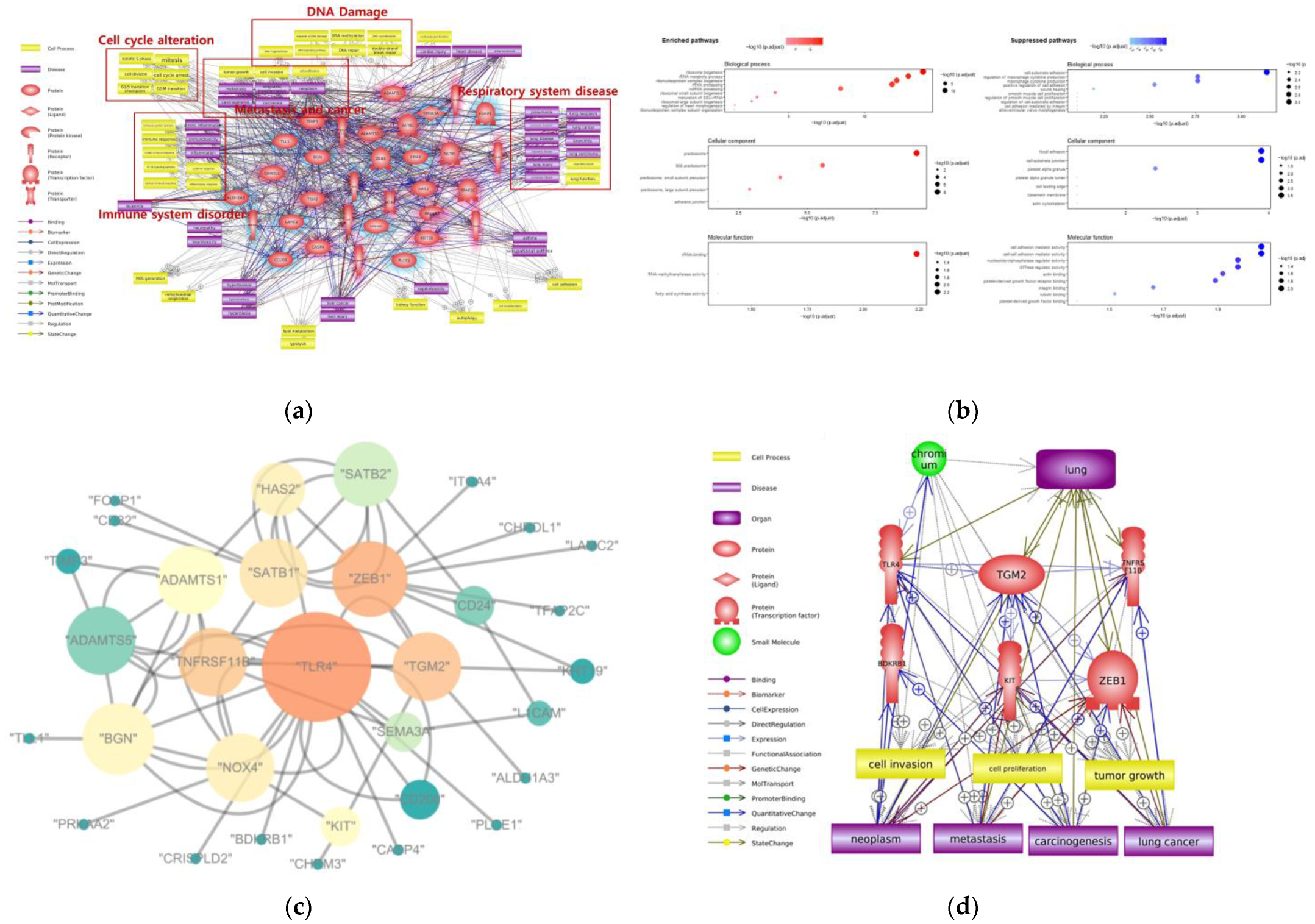
3. Adverse Effects of Cr on Respiratory System

Respiratory exposure to Cr mainly occurs in industrial workers [39]. The inhalation of Cr(VI) is associated with an increased risk in lung cancer, based on numerous observations in occupational epidemiology studies. Among workers with increased lung cancer risk, nasal irritation and perforation have also been reported [40]. Furthermore, lung cancer occurred more often in workers in the chromate-producing industry than in control subjects [41]. Although the mode of action for Cr(VI)-induced lung tumors has not yet been established, these observations would generally support a relationship between inhalation of Cr and lung cancer.
To understand the toxic mechanisms of Cr inhalation, we conducted molecular pathway analysis with Pathway Studio software using the gene expression profile dataset for lung exposure to Cr from the public gene expression database. Microarray datasets GSE24025 and GSE36684 were from the in vitro study of the human bronchial epithelial cell line (BEAS-2B) exposed to Cr(VI) from the GEO database. By the integration and batch effect correction of the different datasets, we investigated the toxic effects of Cr exposure on the respiratory system. Figure 3a shows that exposure to Cr and Cr compounds through inhalation mainly induces DNA damage, cell cycle progression and metastasis, tumor growth, and various respiratory diseases, such as pneumonia, lung neoplasm, pulmonary fibrosis, and lung cancer. Seidler et al. revealed that inhaled Cr(VI) might act by a non-thresholded genotoxic mechanism, form DNA adducts, and induce lung tumor formation [42].
We performed a GO term analysis using all DEGs to predict the biological functions related to Cr inhalation by identifying the significantly enriched BP, CC, and MF terms (Figure 3b). Among the BP terms, ribosomal RNA (rRNA) biogenesis, rRNA metabolic process, and ribonucleoprotein complex biogenesis were significantly enriched. Ye and Shi demonstrated that ribosomal activity was increased in Cr-exposed lung cells, thereby initiating a reconstruction process through the upregulation of protein synthesis [43]. In our study, cell–substrate adhesion and the positive regulation of macrophage cytokine production were suppressed BP terms. The alteration of cell adhesion promotes cancer cell invasion and extravasation in various organs [44,45]. Among the CC terms, pre-ribosome-related terms were enriched, whereas focal adhesion and cell–substrate junction were suppressed. The enrichment of pre-ribosomal processes involved in ribosome biogenesis and protein synthesis supported the enriched BP terms. The suppression of the CC term cellular adhesion is also consistent with the BP result. Among the MF terms, tRNA binding was significantly enriched, while cell adhesion mediator activity was suppressed. The activation of tRNA binding lead to protein synthesis, which is consistent with the BP and CC results. The suppression of cell adhesion mediator activities among the MF terms is also consistent with the previous results. As a result of the GO term analysis, the enrichment of protein synthesis and suppression of cell adhesion were predicted to be the main toxic effects of Cr inhalation.
A network-based gene–gene interaction analysis was performed to select the hub genes associated with Cr inhalation. To analyze the topological parameters among the genes related to Cr inhalation, the gene–gene interaction network was constructed (Figure 3c). We identified the hub genes and core network associated with Cr-induced respiratory effects (Figure 3d). TLR4, TGM2, TNFRSF11B, BDKRB1, KIT, and ZEB1 were the hub genes, and tumorigenic cell processes and diseases were identified as the toxic effects of Cr inhalation. The TLR4 expression in alveolar and bronchial epithelial cells plays a critical role in response to endotoxin [46]. According to Zhang et al., TLR4 deficiency induced lung damage and emphysema [47]. TGM2 is generally overexpressed in lung cancer cells. TGM2-overexpressing cell lines showed high resistance to drugs such as cisplatin in non-small-cell lung cancer [48]. TNFRSF11B, a member of the TNF receptor superfamily, can inhibit tumor apoptosis by binding to a TNF-related apoptosis-inducing ligand. Overexpression of TNFRSF11B can promote tumor growth and metastasis [49]. KIT protein is expressed in small-cell lung cancer, and its kinase activity has been implicated in the pathophysiology of many tumors [50]. KIT expression was associated with advanced disease and a decreased incidence of bone metastasis [51]. According to Larsen et al., increased expression of ZEB1 is associated with tumor grade and metastasis in lung cancer, likely due to its role as a transcription factor in epithelial-to-mesenchymal transition [52]. The selected hub genes could be important markers for respiratory diseases and lung cancer caused by Cr inhalation exposure.

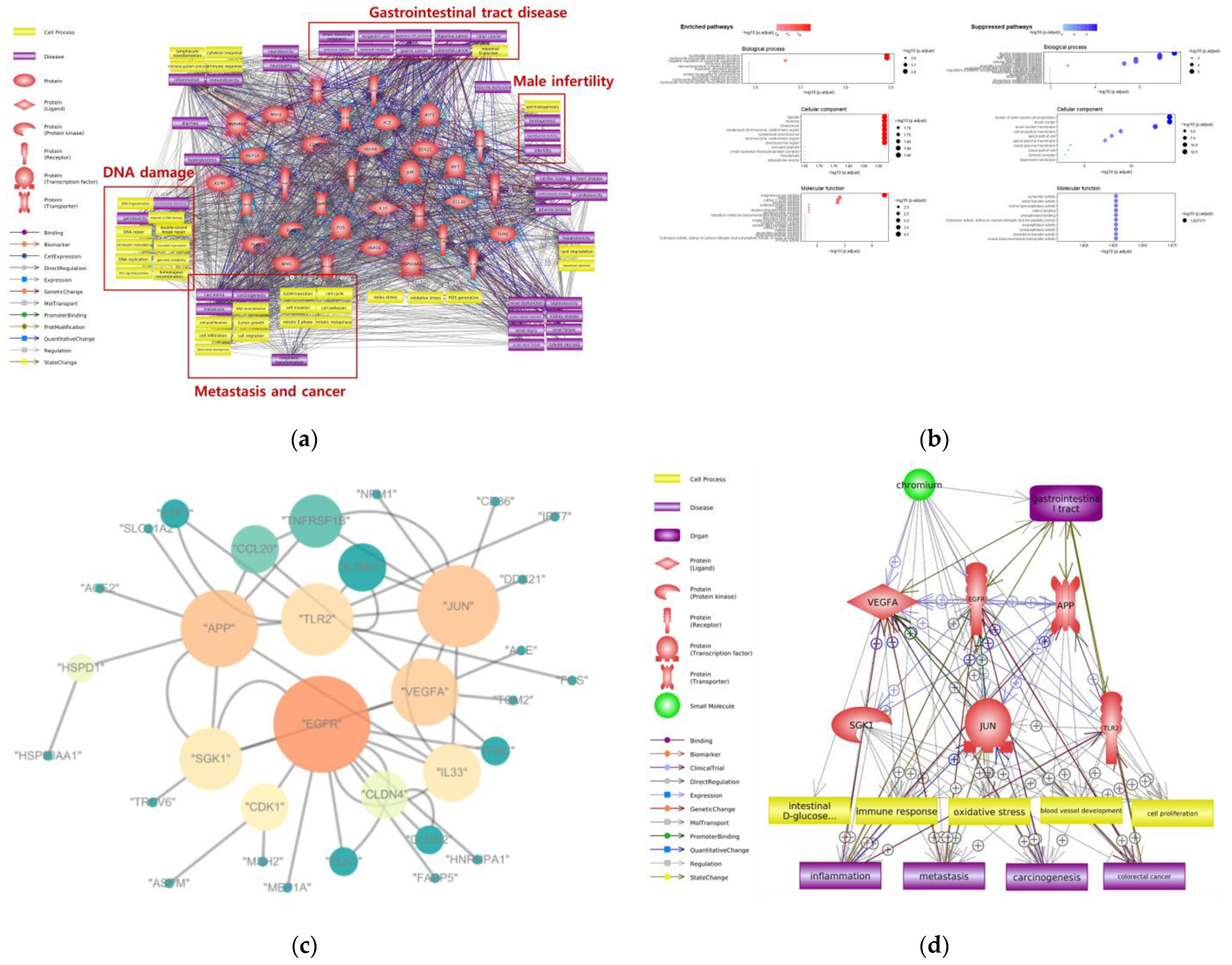
4. Adverse Effects of Cr on Gastrointestinal Tract

Cr is a well-known carcinogen in the respiratory tract and can cause lung cancer in occupational workers. In contrast, the human evidence for the toxicity of Cr ingested orally is limited. Beaumont et al. documented increased cancer risks in a Chinese population exposed to Cr(VI) in drinking water [53]. The primary route of exposure in non-occupational workers is food ingestion. The California Environmental Protection Agency (CalEPA) environmental health hazard assessment set a public health goal for total Cr in drinking water. Results from several genotoxicity assays associated ingested Cr(VI) with forestomach tumors in mice.
To understand the toxic mechanisms of Cr ingestion, we conducted a molecular pathway analysis with Pathway Studio software. Microarray dataset GSE120146 was derived from an in vivo study of mouse duodenum exposed to Cr(VI) from the GEO database. Figure 3a shows that ingestion exposure to Cr and Cr compounds mainly induces DNA damage, metastasis, and tumorigenic cell processes. Male infertility, renal disease, and gastrointestinal tract diseases, such as small intestine neoplasm and squamous cell carcinoma, were associated with Cr ingestion. Ingested Cr is able to induce oxidative stress via multiple pathways. Depending on the levels of reactive oxygen species produced, Cr-induced oxidative stress may lead to cell death or tumor formation [53].
We performed a GO term analysis using all DEGs to predict the biological functions related to Cr ingestion by identifying the BP, CC, and MF terms (Figure 4b). Among the BP terms, nucleotide biosynthesis and nucleoside phosphate biosynthesis process were highly enriched. The upregulation of nucleotide biosynthesis can cause cell proliferation and cancer [54]. The suppressed BP terms were alcohol metabolic process, steroid metabolic process, and lipid metabolic process. Those processes were related to liver function, showing that there was an abnormality in liver function due to gastrointestinal tract exposure [55]. Various cell division-related CC terms such as spindle, midbody, kinetochore, and condensed chromosome were enriched. The acceleration of the cell cycle and cell division can lead to tumor proliferation in various digestive cancers [56]. The cell projection membrane, brush border, and brush border membrane were suppressed by Cr ingestion. The brush border can be found in the small intestine and large intestine, where absorption occurs [57]. Impairment of the brush border membrane can cause dysfunction of the digestive tract, and brush border proteins involved in cell polarity have an important role in intestinal tumor development [58]. Among the MF terms, rRNA binding was enriched, while tough symporter and sterol transfer activity were suppressed. Enrichment of ribosomal RNA binding can lead to protein synthesis, consistent with enriched biosynthesis-related BP terms. Additionally, impaired symporter activity and sterol transfer activity are associated with liver dysfunction, one of the typical toxic effects of digestive toxicity. As a result of the GO term analysis, the enrichment of diverse biosynthesis processes and diverse biological indicators of liver dysfunction are predicted to be the main toxic effects of Cr ingestion.
A network-based gene–gene interaction analysis was performed to select the hub genes associated with Cr ingestion. To analyze the topological parameters among the genes related to Cr ingestion, the gene–gene interaction network was constructed (Figure 4c). We identified the hub genes and core network associated with Cr ingestion (Figure 4d). VEGFA, EGFR, APP, SGK1, JUN, and TLR2 were the hub genes, and oxidative stress, cell proliferation, and blood vessel development were cell processes closely linked with Cr ingestion. According to the molecular network, colorectal cancer can occur due to Cr ingestion. VEGFA is a known stimulator of tumor angiogenesis. The positive correlation between VEGFA expression and gastric cancer was verified, and a high level of VEGFA frequently predicted shorter survival time [59]. EGFR is a well-known growth factor receptor involved in the development and progression of carcinogenesis [60]. In the gastrointestinal tract, EGFR overexpression was a prognostic factor in patients with gastric cancer. APP has been associated with cell adhesion, cell motility, and cell proliferation, and APP proteins are highly expressed in gastrointestinal tumors [61]. SGK1 expression favors the development of intestinal tumors in mice [62]. JUN overexpression is observed in various cancers, such as non-small-cell lung cancer, breast cancer, and vulvar cancer, but there is no direct evidence linking JUN overexpression with gastrointestinal cancer. However, JUN overexpression leading to cell cycle progression and metastasis may generate or accelerate cancer [63]. TLR2 has been reported to activate the PI3K/Akt/GSK3β pathway, leading to anti-inflammatory effects in the gut epithelia [64]. The selected hub genes could be important markers for gastrointestinal tract diseases and colorectal cancer caused by Cr ingestion exposure.

5. Adverse Human Health Effects of Cr Depend on the Exposure Route

We explored the toxic effects of Cr exposure dependent on the exposure route. We endeavored to organize the toxicity mechanisms, GO terms (BP, CC, and MF), and hub genes associated with Cr exposure (Table 1). DNA damage was found to be a common potential toxic mechanism of Cr for all exposure routes. The relationship between Cr exposure and DNA damage has been well-documented. Cr produces free radicals in the cytoplasm and nucleus, and free radicals can bind to DNA [65]. In human fibroblasts, Cr exposure induced DNA damage by the formation of a sub-G1 peak and arrest of the G2/M phase of the cell cycle [66]. Furthermore, lung exposure to Cr induced DNA damage, tissue irritation, inflammation, cytotoxicity, and, ultimately, lung cancer [67]. Ingestion of Cr through drinking water can form Cr-DNA adducts and induce DNA damage [68]. Metastasis, which was also found to be a common potential toxic mechanism of all exposure routes of Cr, is well-known to be involved in the development of various diseases and cancers. In addition, the results of the GO term analysis identified the disruption of cell adhesion-related BP, CC, and MF terms in all exposure routes. Based on previous studies and our analysis result, we present Cr-DNA adducts and metastasis as a common toxic mechanism of Cr exposure.
An allergic reaction is a potential toxicity mechanism specific to Cr dermal exposure. Although it is known that Cr can penetrate skin tissue and induce an allergic reaction and dermatitis, the underlying mechanism has not yet been clearly verified. Various biosynthesis processes that can cause cell proliferation were enriched in response to Cr dermal exposure. The cell cycle alteration is identified as a specific potential mechanism associated with the potential respiratory effects of Cr inhalation. Moreover, the enrichment of the ribosomal biogenesis process and suppression of cell adhesion are strongly associated with cancer development. Our analysis result found no specific toxicity mechanism related to Cr ingestion. However, enrichment of the nucleotide biosynthesis process and liver function failure was detected as a Cr ingestion-related BP terms.

6. Conclusions

Heavy metals have a widespread use in diverse fields, and their environmental impacts and potential health effects have raised serious concerns. Cr is commercially used in various products, and Cr exposure in workers and the general population can occur via various routes [69]. The most prevalent oxidation forms of Cr are Cr(III) and Cr(VI); both forms have been reported as potentially toxic and direct toxicities. In this review, we present the potential toxic mechanism, BP terms, and hub genes for each exposure route by bioinformatic analysis based on public gene expression data. DNA damage was shown as a common potential toxic mechanism of Cr for all three exposure routes, although the detailed BP terms differed. In particular, for the inhalation and ingestion exposure routes, the BP terms identified were associated with cancer, which is intimately linked to the commonly known Cr-induced occupational diseases. We also present hub genes for each Cr exposure route as potential candidates for markers of Cr toxicity. As our analysis was conducted using only partial data, more public genomic data need to be integrated. In summary, diverse bioinformatic analyses to comprehensively understand these complex mechanisms will be beneficial for understanding the Cr-induced toxic effects following various routes of exposure.

Author Contributions

Y.R.S. and E.-M.C. designed the research study. D.Y.S. performed overall analysis procedure and wrote the manuscript. S.M.L., D.Y.S. and Y.J. composed the figures. J.L. and D.Y.S. wrote the manuscript. C.M.L. provided ideas throughout the manuscript. All authors have read and agreed to the published version of the manuscript.

Funding

This work was supported by Korea Environment Industry & Technology Institute(KEITI) through Core Technology Development Project for Environmental Diseases Prevention and Management Program, funded by South Korea’s Ministry of Environment(MOE) (grant number 2022003310012).

Institutional Review Board Statement

Not applicable.

Informed Consent Statement

Not applicable.

Data Availability Statement

No new data were created or analyzed in this study. Data sharing is not applicable to this article.

Conflicts of Interest

The authors declare no conflict of interest. The funders had no role in the design of the study; in the collection, analyses, or interpretation of data; in the writing of the manuscript; or in the decision to publish the result.

References

  1. Pereira, S.C.; Oliveira, P.F.; Oliveira, S.R.; Pereira, M.L.; Alves, M.G. Impact of Environmental and Lifestyle Use of Chromium on Male Fertility: Focus on Antioxidant Activity and Oxidative Stress. Antioxidants 2021, 10, 1365. [Google Scholar] [CrossRef] [PubMed]
  2. Safonov, A.; Popova, N.; Andrushenko, N.; Boldyrev, K.; Yushin, N.; Zinicovscaia, I. Investigation of materials for reactive permeable barrier in removing cadmium and chromium(VI) from aquifer near a solid domestic waste landfill. Environ. Sci. Pollut. Res. Int. 2021, 28, 4645–4659. [Google Scholar] [CrossRef]
  3. Forte, I.M.; Indovina, P.; Costa, A.; Iannuzzi, C.A.; Costanzo, L.; Marfella, A.; Montagnaro, S.; Botti, G.; Bucci, E.; Giordano, A. Blood screening for heavy metals and organic pollutants in cancer patients exposed to toxic waste in southern Italy: A pilot study. J. Cell. Physiol. 2020, 235, 5213–5222. [Google Scholar] [CrossRef] [PubMed]
  4. Zaeemzadeh, N.; Jahanian Sadatmahalleh, S.; Ziaei, S.; Kazemnejad, A.; Movahedinejad, M.; Mottaghi, A.; Mohamadzadeh, N. Comparison of dietary micronutrient intake in PCOS patients with and without metabolic syndrome. J. Ovarian Res. 2021, 14, 10. [Google Scholar] [CrossRef] [PubMed]
  5. Vaiopoulou, E.; Gikas, P. Regulations for chromium emissions to the aquatic environment in Europe and elsewhere. Chemosphere 2020, 254, 126876. [Google Scholar] [CrossRef]
  6. Batyrova, G.; Kononets, V.; Amanzholkyzy, A.; Tlegenova, Z.; Umarova, G. Chromium as a Risk Factor for Breast Cancer: A Meta-Analysis. Asian Pac. J. Cancer Prev. 2022, 23, 3993–4003. [Google Scholar] [CrossRef]
  7. Hossini, H.; Shafie, B.; Niri, A.D.; Nazari, M.; Esfahlan, A.J.; Ahmadpour, M.; Nazmara, Z.; Ahmadimanesh, M.; Makhdoumi, P.; Mirzaei, N.; et al. A comprehensive review on human health effects of chromium: Insights on induced toxicity. Environ. Sci. Pollut. Res. Int. 2022, 29, 70686–70705. [Google Scholar] [CrossRef]
  8. Fontaine, M.; Clement, Y.; Blanc, N.; Demesmay, C. Hexavalent chromium release from leather over time natural ageing vs accelerated ageing according to a multivariate approach. J. Hazard. Mater. 2019, 368, 811–818. [Google Scholar] [CrossRef]
  9. Tsuchiyama, T.; Tazaki, A.; Al Hossain, M.A.; Yajima, I.; Ahsan, N.; Akhand, A.A.; Hashimoto, K.; Ohgami, N.; Kato, M. Increased levels of renal damage biomarkers caused by excess exposure to trivalent chromium in workers in tanneries. Environ. Res. 2020, 188, 109770. [Google Scholar] [CrossRef]
  10. Kozlowska, L.; Gromadzinska, J.; Wasowicz, W. Health risk in transport workers. Part II. Dietary compounds as modulators of occupational exposure to chemicals. Int. J. Occup. Med. Environ. Health 2019, 32, 441–464. [Google Scholar] [CrossRef]
  11. Santonen, T.; Porras, S.P.; Bocca, B.; Bousoumah, R.; Duca, R.C.; Galea, K.S.; Godderis, L.; Goen, T.; Hardy, E.; Iavicoli, I.; et al. HBM4EU chromates study—Overall results and recommendations for the biomonitoring of occupational exposure to hexavalent chromium. Environ. Res. 2022, 204, 111984. [Google Scholar] [CrossRef]
  12. Balali-Mood, M.; Naseri, K.; Tahergorabi, Z.; Khazdair, M.R.; Sadeghi, M. Toxic Mechanisms of Five Heavy Metals: Mercury, Lead, Chromium, Cadmium, and Arsenic. Front. Pharmacol. 2021, 12, 643972. [Google Scholar] [CrossRef]
  13. Oginawati, K.; Susetyo, S.H.; Rosalyn, F.A.; Kurniawan, S.B.; Abdullah, S.R.S. Risk analysis of inhaled hexavalent chromium (Cr6+) exposure on blacksmiths from industrial area. Environ. Sci. Pollut. Res. Int. 2021, 28, 14000–14008. [Google Scholar] [CrossRef]
  14. Ferdous, Z.; Nemmar, A. Health Impact of Silver Nanoparticles: A Review of the Biodistribution and Toxicity Following Various Routes of Exposure. Int. J. Mol. Sci. 2020, 21, 2375. [Google Scholar] [CrossRef]
  15. Niu, Y.; Jiang, X.; Wang, K.; Xia, J.; Jiao, W.; Niu, Y.; Yu, H. Meta analysis of heavy metal pollution and sources in surface sediments of Lake Taihu, China. Sci. Total Environ. 2020, 700, 134509. [Google Scholar] [CrossRef]
  16. Kim, H.S.; Kim, Y.J.; Seo, Y.R. An Overview of Carcinogenic Heavy Metal: Molecular Toxicity Mechanism and Prevention. J. Cancer Prev. 2015, 20, 232–240. [Google Scholar] [CrossRef]
  17. Tsoucas, D.; Dong, R.; Chen, H.; Zhu, Q.; Guo, G.; Yuan, G.C. Accurate estimation of cell-type composition from gene expression data. Nat. Commun. 2019, 10, 2975. [Google Scholar] [CrossRef]
  18. Wijesooriya, K.; Jadaan, S.A.; Perera, K.L.; Kaur, T.; Ziemann, M. Urgent need for consistent standards in functional enrichment analysis. PLoS Comput. Biol. 2022, 18, e1009935. [Google Scholar] [CrossRef]
  19. Raudvere, U.; Kolberg, L.; Kuzmin, I.; Arak, T.; Adler, P.; Peterson, H.; Vilo, J. g:Profiler: A web server for functional enrichment analysis and conversions of gene lists (2019 update). Nucleic Acids Res. 2019, 47, W191–W198. [Google Scholar] [CrossRef]
  20. Zhao, M.; Xu, J.; Li, A.; Mei, Y.; Ge, X.; Liu, X.; Wei, L.; Xu, Q. Multiple exposure pathways and urinary chromium in residents exposed to chromium. Environ. Int. 2020, 141, 105753. [Google Scholar] [CrossRef]
  21. Kan, Y.; Ohashi, T.; Kobayashi, E.; Kumagai, A.; Uhara, H. Cement and Retarder-Associated Systemic Contact Dermatitis in a Chromium-Negative Patch-Tested Patient. Dermatitis 2021, 32, e76–e78. [Google Scholar] [CrossRef] [PubMed]
  22. Hagvall, L.; Pour, M.D.; Feng, J.; Karma, M.; Hedberg, Y.; Malmberg, P. Skin permeation of nickel, cobalt and chromium salts in ex vivo human skin, visualized using mass spectrometry imaging. Toxicol. In Vitro 2021, 76, 105232. [Google Scholar] [CrossRef] [PubMed]
  23. Rudolf, E.; Cervinka, M. The role of intracellular zinc in chromium(VI)-induced oxidative stress, DNA damage and apoptosis. Chem. Biol. Interact. 2006, 162, 212–227. [Google Scholar] [CrossRef] [PubMed]
  24. Thomas, P.; Summer, B.; Sander, C.A.; Przybilla, B.; Thomas, M.; Naumann, T. Intolerance of osteosynthesis material: Evidence of dichromate contact allergy with concomitant oligoclonal T-cell infiltrate and TH1-type cytokine expression in the peri-implantar tissue. Allergy 2000, 55, 969–972. [Google Scholar] [CrossRef]
  25. Leroyer, C.; Dewitte, J.D.; Bassanets, A.; Boutoux, M.; Daniel, C.; Clavier, J. Occupational asthma due to chromium. Respiration 1998, 65, 403–405. [Google Scholar] [CrossRef]
  26. Pavesi, T.; Moreira, J.C. Mechanisms and individuality in chromium toxicity in humans. J. Appl. Toxicol. 2020, 40, 1183–1197. [Google Scholar] [CrossRef]
  27. Guo, L.; Xiao, Y.; Wang, Y. Hexavalent chromium-induced alteration of proteomic landscape in human skin fibroblast cells. J. Proteome Res. 2013, 12, 3511–3518. [Google Scholar] [CrossRef]
  28. Liu, F.J.; Barchowsky, A.; Opresko, P.L. The Werner syndrome protein suppresses telomeric instability caused by chromium (VI) induced DNA replication stress. PLoS ONE 2010, 5, e11152. [Google Scholar] [CrossRef]
  29. VonHandorf, A.; Sanchez-Martin, F.J.; Biesiada, J.; Zhang, H.; Zhang, X.; Medvedovic, M.; Puga, A. Chromium disrupts chromatin organization and CTCF access to its cognate sites in promoters of differentially expressed genes. Epigenetics 2018, 13, 363–375. [Google Scholar] [CrossRef]
  30. Zhao, Y.; Cholewa, J.; Shang, H.; Yang, Y.; Ding, X.; Wang, Q.; Su, Q.; Zanchi, N.E.; Xia, Z. Advances in the Role of Leucine-Sensing in the Regulation of Protein Synthesis in Aging Skeletal Muscle. Front. Cell Dev. Biol. 2021, 9, 646482. [Google Scholar] [CrossRef]
  31. Wise, S.S.; Holmes, A.L.; Wise, J.P., Sr. Hexavalent chromium-induced DNA damage and repair mechanisms. Rev. Environ. Health 2008, 23, 39–57. [Google Scholar] [CrossRef]
  32. Toebak, M.J.; Pohlmann, P.R.; Sampat-Sardjoepersad, S.C.; von Blomberg, B.M.; Bruynzeel, D.P.; Scheper, R.J.; Rustemeyer, T.; Gibbs, S. CXCL8 secretion by dendritic cells predicts contact allergens from irritants. Toxicol. In Vitro 2006, 20, 117–124. [Google Scholar] [CrossRef]
  33. Astakhova, A.; Chistyakov, D.; Thomas, D.; Geisslinger, G.; Brune, B.; Sergeeva, M.; Namgaladze, D. Inhibitors of Oxidative Phosphorylation Modulate Astrocyte Inflammatory Responses through AMPK-Dependent Ptgs2 mRNA Stabilization. Cells 2019, 8, 1185. [Google Scholar] [CrossRef]
  34. Jiang, X.; Xie, H.; Dou, Y.; Yuan, J.; Zeng, D.; Xiao, S. Expression and function of FRA1 protein in tumors. Mol. Biol. Rep. 2020, 47, 737–752. [Google Scholar] [CrossRef]
  35. Ade, N.; Leon, F.; Pallardy, M.; Peiffer, J.L.; Kerdine-Romer, S.; Tissier, M.H.; Bonnet, P.A.; Fabre, I.; Ourlin, J.C. HMOX1 and NQO1 genes are upregulated in response to contact sensitizers in dendritic cells and THP-1 cell line: Role of the Keap1/Nrf2 pathway. Toxicol. Sci. 2009, 107, 451–460. [Google Scholar] [CrossRef]
  36. Hao, Z.F.; Ao, J.H.; Zhang, J.; Su, Y.M.; Yang, R.Y. ATF3 activates Stat3 phosphorylation through inhibition of p53 expression in skin cancer cells. Asian Pac. J. Cancer Prev. 2013, 14, 7439–7444. [Google Scholar] [CrossRef]
  37. Sadagurski, M.; Nofech-Mozes, S.; Weingarten, G.; White, M.F.; Kadowaki, T.; Wertheimer, E. Insulin receptor substrate 1 (IRS-1) plays a unique role in normal epidermal physiology. J. Cell. Physiol. 2007, 213, 519–527. [Google Scholar] [CrossRef]
  38. Xu, J.; Liu, H.; Yang, Y.; Wang, X.; Liu, P.; Li, Y.; Meyers, C.; Banerjee, N.S.; Wang, H.K.; Cam, M.; et al. Genome-Wide Profiling of Cervical RNA-Binding Proteins Identifies Human Papillomavirus Regulation of RNASEH2A Expression by Viral E7 and E2F1. mBio 2019, 10, e02687-18. [Google Scholar] [CrossRef]
  39. Kurt, O.K.; Basaran, N. Occupational Exposure to Metals and Solvents: Allergy and Airway Diseases. Curr. Allergy Asthma Rep. 2020, 20, 38. [Google Scholar] [CrossRef]
  40. Alvarez, C.C.; Bravo Gomez, M.E.; Hernandez Zavala, A. Hexavalent chromium: Regulation and health effects. J. Trace Elem. Med. Biol. 2021, 65, 126729. [Google Scholar] [CrossRef]
  41. Behrens, T.; Ge, C.; Vermeulen, R.; Kendzia, B.; Olsson, A.; Schuz, J.; Kromhout, H.; Pesch, B.; Peters, S.; Portengen, L.; et al. Occupational exposure to nickel and hexavalent chromium and the risk of lung cancer in a pooled analysis of case-control studies (SYNERGY). Int. J. Cancer 2023, 152, 645–660. [Google Scholar] [CrossRef] [PubMed]
  42. Seidler, A.; Jahnichen, S.; Hegewald, J.; Fishta, A.; Krug, O.; Ruter, L.; Strik, C.; Hallier, E.; Straube, S. Systematic review and quantification of respiratory cancer risk for occupational exposure to hexavalent chromium. Int. Arch. Occup. Environ. Health 2013, 86, 943–955. [Google Scholar] [CrossRef] [PubMed]
  43. Ye, J.; Shi, X. Gene expression profile in response to chromium-induced cell stress in A549 cells. Mol. Cell. Biochem. 2001, 222, 189–197. [Google Scholar] [CrossRef] [PubMed]
  44. Laubli, H.; Borsig, L. Altered Cell Adhesion and Glycosylation Promote Cancer Immune Suppression and Metastasis. Front. Immunol. 2019, 10, 2120. [Google Scholar] [CrossRef] [PubMed]
  45. Sousa, B.; Pereira, J.; Paredes, J. The Crosstalk Between Cell Adhesion and Cancer Metabolism. Int. J. Mol. Sci. 2019, 20, 1933. [Google Scholar] [CrossRef] [PubMed]
  46. Shang, J.; Liu, W.; Yin, C.; Chu, H.; Zhang, M. Cucurbitacin E ameliorates lipopolysaccharide-evoked injury, inflammation and MUC5AC expression in bronchial epithelial cells by restraining the HMGB1-TLR4-NF-kappaB signaling. Mol. Immunol. 2019, 114, 571–577. [Google Scholar] [CrossRef]
  47. Zhang, X.; Shan, P.; Jiang, G.; Cohn, L.; Lee, P.J. Toll-like receptor 4 deficiency causes pulmonary emphysema. J. Clin. Investig. 2006, 116, 3050–3059. [Google Scholar] [CrossRef]
  48. Li, M.; Song, D.; Chen, X.; Wang, X.; Xu, L.; Yang, M.; Yang, J.; Kalvakolanu, D.V.; Wei, X.; Liu, X.; et al. RSL3 triggers glioma stem cell differentiation via the Tgm2/AKT/ID1 signaling axis. Biochim. Biophys. Acta Mol. Basis Dis. 2022, 1868, 166529. [Google Scholar] [CrossRef]
  49. Xie, G.; Yang, H.; Ma, D.; Sun, Y.; Chen, H.; Hu, X.; Jiang, Y.Z.; Shao, Z.M. Integration of whole-genome sequencing and functional screening identifies a prognostic signature for lung metastasis in triple-negative breast cancer. Int. J. Cancer 2019, 145, 2850–2860. [Google Scholar] [CrossRef]
  50. Luan, F.; Li, X.; Cheng, X.; Huangfu, L.; Han, J.; Guo, T.; Du, H.; Wen, X.; Ji, J. TNFRSF11B activates Wnt/beta-catenin signaling and promotes gastric cancer progression. Int. J. Biol. Sci. 2020, 16, 1956–1971. [Google Scholar] [CrossRef]
  51. Lopez-Martin, A.; Ballestin, C.; Garcia-Carbonero, R.; Castano, A.; Lopez-Rios, F.; Lopez-Encuentra, A.; Sanchez-Cespedes, M.; Castellano, D.; Bartolome, A.; Cortes-Funes, H.; et al. Prognostic value of KIT expression in small cell lung cancer. Lung Cancer 2007, 56, 405–413. [Google Scholar] [CrossRef]
  52. Larsen, J.E.; Nathan, V.; Osborne, J.K.; Farrow, R.K.; Deb, D.; Sullivan, J.P.; Dospoy, P.D.; Augustyn, A.; Hight, S.K.; Sato, M.; et al. ZEB1 drives epithelial-to-mesenchymal transition in lung cancer. J. Clin. Investig. 2016, 126, 3219–3235. [Google Scholar] [CrossRef]
  53. Beaumont, J.J.; Sedman, R.M.; Reynolds, S.D.; Sherman, C.D.; Li, L.H.; Howd, R.A.; Sandy, M.S.; Zeise, L.; Alexeeff, G.V. Cancer mortality in a Chinese population exposed to hexavalent chromium in drinking water. Epidemiology 2008, 19, 12–23. [Google Scholar] [CrossRef]
  54. Villa, E.; Ali, E.S.; Sahu, U.; Ben-Sahra, I. Cancer Cells Tune the Signaling Pathways to Empower de Novo Synthesis of Nucleotides. Cancers 2019, 11, 688. [Google Scholar] [CrossRef]
  55. Charni-Natan, M.; Aloni-Grinstein, R.; Osher, E.; Rotter, V. Liver and Steroid Hormones-Can a Touch of p53 Make a Difference? Front. Endocrinol. 2019, 10, 374. [Google Scholar] [CrossRef]
  56. Zatulovskiy, E.; Zhang, S.; Berenson, D.F.; Topacio, B.R.; Skotheim, J.M. Cell growth dilutes the cell cycle inhibitor Rb to trigger cell division. Science 2020, 369, 466–471. [Google Scholar] [CrossRef]
  57. Schulthess, G.; Lipka, G.; Compassi, S.; Boffelli, D.; Weber, F.E.; Paltauf, F.; Hauser, H. Absorption of monoacylglycerols by small intestinal brush border membrane. Biochemistry 1994, 33, 4500–4508. [Google Scholar] [CrossRef]
  58. Mazzolini, R.; Dopeso, H.; Mateo-Lozano, S.; Chang, W.; Rodrigues, P.; Bazzocco, S.; Alazzouzi, H.; Landolfi, S.; Hernandez-Losa, J.; Andretta, E.; et al. Brush border myosin Ia has tumor suppressor activity in the intestine. Proc. Natl. Acad. Sci. USA 2012, 109, 1530–1535. [Google Scholar] [CrossRef]
  59. Macedo, F.; Ladeira, K.; Longatto-Filho, A.; Martins, S.F. Gastric Cancer and Angiogenesis: Is VEGF a Useful Biomarker to Assess Progression and Remission? J. Gastric Cancer 2017, 17, 1–10. [Google Scholar] [CrossRef]
  60. Nautiyal, J.; Kanwar, S.S.; Majumdar, A.P. EGFR(s) in aging and carcinogenesis of the gastrointestinal tract. Curr. Protein Pept. Sci. 2010, 11, 436–450. [Google Scholar] [CrossRef]
  61. Arvidsson, Y.; Andersson, E.; Bergstrom, A.; Andersson, M.K.; Altiparmak, G.; Illerskog, A.C.; Ahlman, H.; Lamazhapova, D.; Nilsson, O. Amyloid precursor-like protein 1 is differentially upregulated in neuroendocrine tumours of the gastrointestinal tract. Endocr. Relat. Cancer 2008, 15, 569–581. [Google Scholar] [CrossRef] [PubMed]
  62. Wang, K.; Gu, S.; Nasir, O.; Foller, M.; Ackermann, T.F.; Klingel, K.; Kandolf, R.; Kuhl, D.; Stournaras, C.; Lang, F. SGK1-dependent intestinal tumor growth in APC-deficient mice. Cell. Physiol. Biochem. 2010, 25, 271–278. [Google Scholar] [CrossRef] [PubMed]
  63. Liu, Y.; Zhao, J.; Zhang, W.; Gan, J.; Hu, C.; Huang, G.; Zhang, Y. lncRNA GAS5 enhances G1 cell cycle arrest via binding to YBX1 to regulate p21 expression in stomach cancer. Sci. Rep. 2015, 5, 10159. [Google Scholar] [CrossRef] [PubMed]
  64. Tattoli, I.; Petitta, C.; Scirocco, A.; Ammoscato, F.; Cicenia, A.; Severi, C. Microbiota, innate immune system, and gastrointestinal muscle: Ongoing studies. J. Clin. Gastroenterol. 2012, 46, S6–S11. [Google Scholar] [CrossRef] [PubMed]
  65. Faisal, M.; Hameed, A.; Hasnain, S. Chromium-resistant bacteria and cyanobacteria: Impact on Cr(VI) reduction potential and plant growth. J. Ind. Microbiol. Biotechnol. 2005, 32, 615–621. [Google Scholar] [CrossRef]
  66. Shrivastava, H.Y.; Ravikumar, T.; Shanmugasundaram, N.; Babu, M.; Unni Nair, B. Cytotoxicity studies of chromium(III) complexes on human dermal fibroblasts. Free Radic. Biol. Med. 2005, 38, 58–69. [Google Scholar] [CrossRef]
  67. Rager, J.E.; Suh, M.; Chappell, G.A.; Thompson, C.M.; Proctor, D.M. Review of transcriptomic responses to hexavalent chromium exposure in lung cells supports a role of epigenetic mediators in carcinogenesis. Toxicol. Lett. 2019, 305, 40–50. [Google Scholar] [CrossRef]
  68. Tumolo, M.; Ancona, V.; De Paola, D.; Losacco, D.; Campanale, C.; Massarelli, C.; Uricchio, V.F. Chromium Pollution in European Water, Sources, Health Risk, and Remediation Strategies: An Overview. Int. J. Environ. Res. Public Health 2020, 17, 5438. [Google Scholar] [CrossRef]
  69. Lou, J.; Yu, S.; Feng, L.; Guo, X.; Wang, M.; Branco, A.T.; Li, T.; Lemos, B. Environmentally induced ribosomal DNA (rDNA) instability in human cells and populations exposed to hexavalent chromium [Cr (VI)]. Environ. Int. 2021, 153, 106525. [Google Scholar] [CrossRef]
Figure 1. (a) Sources of Cr exposure based on different exposure paths. (b) Flow chart of the steps used in this review.
Ijms 24 03410 g001
Figure 2. Bioinformatic analysis about Cr toxicity on skin. (a) Molecular network includes gene, cell process, diseases associated with Cr dermal contact. Potential toxicity mechanism highlighted with red color box. (b) Enriched and Suppressed Biological processes, Cellular components, Molecular functions associated with Cr dermal contact (c) Gene–gene interaction network following Cr dermal contact; each node’s color represents the betweenness centrality, each node’s size represents degree of the nodes; (d) Summarized molecular network and hub genes about Cr dermal contact.
Ijms 24 03410 g002
Figure 3. Bioinformatic analysis about Cr toxicity on respiratory system. (a) Molecular network include genes, cell processes, diseases associated with Cr inhalation; potential toxicity mechanism highlighted with red colored box; (b) Enriched and Suppressed biological processes, cellular components, molecular function about Cr inhalation; (c) Gene–gene interaction network following Cr inhalation; each node’s color represents the betweenness centrality, each node’s size represents degree of the nodes; (d) Summarized molecular network and hub genes following Cr inhalation.
Ijms 24 03410 g003
Figure 4. Bioinformatic analysis about Cr toxicity on gastrointestinal tract. (a) molecular network includes genes, cell processes, diseases about Cr ingestion; potential toxicity mechanism highlighted with red colored box. (b) Enriched and Suppressed biological processes, cellular components, molecular functions associated with Cr ingestion. (c) Gene–gene interaction network following Cr ingestion; each node’s color represents betweenness centrality, and each node’s size represents the degree of the nodes; (d) Summarized molecular network and hub genes about Cr ingestion.
Ijms 24 03410 g004
Table 1. Potential toxicity mechanism, biological processes, and hub genes of Cr exposure dependent on different exposure routes.
Exposure RoutePotential Toxicity MechanismBiological Process (BP)Hub Genes
Dermal contactDNA damage, metastasis, immune system disorder, allergic reactionRNA modification, sterol biosynthesis process, DNA replication, intrinsic apoptotic signaling pathwayCXCL8, PTGS2, FOS, HMOX1, ATF3, IRS1, E2F1
InhalationDNA damage, metastasis, cell cycle alteration, immune system disorderRibosome biogenesis, rRNA metabolic process, cell–substrate adhesion, macrophage cytokine productionTLR4, TGM2, TNFRSF11B, BDKRB1, KIT, ZEB1
IngestionDNA damage, metastasisNucleotide biosynthesis process, nucleotide phosphate biosynthesis process, alcohol metabolic process, steroid metabolic processVEGFA, EGFR, APP, SGK1, JUN, TLR2
Disclaimer/Publisher’s Note: The statements, opinions and data contained in all publications are solely those of the individual author(s) and contributor(s) and not of MDPI and/or the editor(s). MDPI and/or the editor(s) disclaim responsibility for any injury to people or property resulting from any ideas, methods, instructions or products referred to in the content.

