Article

Omega-3 Recovers Cannabinoid 1 Receptor Expression in the Adult Mouse Brain after Adolescent Binge Drinking

by
Ane Martín-Llorente
1,†,
Maitane Serrano
1,2,†,
Itziar Bonilla-Del Río
1,2,
Leire Lekunberri
1,2,
Garazi Ocerin
1,2,
Nagore Puente
1,2,
Almudena Ramos
1,2,
Irantzu Rico-Barrio
1,2,
Inmaculada Gerrikagoitia
1,2,* and
Pedro Grandes
1,2,*
1
Laboratory of Ultrastructural and Functional Neuroanatomy of the Synapse, Department of Neurosciences, Faculty of Medicine and Nursing, University of the Basque Country UPV/EHU, 48940 Leioa, Spain
2
Achucarro Basque Center for Neuroscience, Science Park of the UPV/EHU, 48940 Leioa, Spain
*
Authors to whom correspondence should be addressed.
These authors contributed equally to this work.
Int. J. Mol. Sci. 2023, 24(24), 17316; https://doi.org/10.3390/ijms242417316
Submission received: 31 October 2023 / Revised: 7 December 2023 / Accepted: 8 December 2023 / Published: 10 December 2023
(This article belongs to the Special Issue Molecular Relationship between Endocannabinoid System and Disease 2.0)

Abstract

:
Adolescent binge drinking is a social problem with a long-lasting impact on cognitive functions. The cannabinoid type-1 (CB1) receptor of the endocannabinoid system (ECS) is involved in brain synaptic plasticity, cognition and behavior via receptor localization at specific subcellular compartments of the cortical, limbic and motor regions. Alcohol (EtOH) intake affects the ECS, CB1 and their functions. Evidence indicates that binge drinking during adolescence impairs memory via the abrogation of CB1-dependent synaptic plasticity in the hippocampus. However, the impact of EtOH consumption on global CB1 receptor expression in the adult brain is unknown. We studied this using optical density analysis throughout brain regions processed for light microscopy (LM) immunohistotochemistry. CB1 staining decreased significantly in the secondary motor cortex, cerebellum, cingulate cortex, amygdala and nucleus accumbens. Next, as omega-3 (n-3) polyunsaturated fatty acids (PUFAs) rescue synaptic plasticity and improve EtOH-impaired cognition, we investigated whether docosahexaenoic acid (DHA) and eicosapentaenoic acid (EPA) had any effect on CB1 receptors. N-3 intake during EtOH abstinence restored CB1 immunostaining in the secondary motor cortex, cerebellum and amygdala, and ameliorated receptor density in the cingulate cortex. These results show that n-3 supplementation recovers CB1 receptor expression disrupted by EtOH in distinct brain regions involved in motor functions and cognition.

1. Introduction

The ECS system is mainly composed of endocannabinoids (eCBs), the enzymes involved in their synthesis and degradation, as well as cannabinoid receptors [1]. The cannabinoid CB1, the most abundant G-protein-coupled receptor in the brain, is highly expressed in the basal ganglia, cerebral cortex, cerebellum (Cb) and hippocampus, which is consistent with its role in learning and memory, motor control, motivation, reward and emotional processing, among many others [1,2]. Furthermore, CB1 increases and changes its distribution from adolescence to adulthood, is predominantly localized in presynaptic terminals and noticeably present in glial cells and mitochondria, modulates neurotransmitter release and also participates in synaptic plasticity [1,2,3,4,5].
Binge drinking during adolescence has a long-lasting impact on cognition and behavior via interference with brain neurochemical systems, including the ECS which participates in the regulation of motivation and EtOH intake [6,7,8,9]. EtOH exposure alters both CB1 mRNA and protein [10,11], changing the availability and binding of the receptor [12,13]. Conversely, gene deletion and CB1 receptor antagonism affect drinking behavior [14]. In addition, a CB1 mRNA decrease in the adult hippocampus after adolescent binge drinking correlates with CB1 receptor reduction in excitatory terminals of the hippocampal dentate molecular layer. These changes, together with the rise in monoacylglycerol lipase (MAGL) mRNA, the main degrading enzyme of the eCB 2-arachidonoylglycerol (2-AG), disrupt endocannabinoid-dependent long-term depression (LTD) and set off memory impairment [15]. Remarkably, the 2-AG increase elicited by MAGL inhibition recovers synaptic plasticity and cognition [15]. Although pharmacological manipulations are potential therapeutic strategies for treating alcohol-induced long-term cognitive deficits, nutritional supplementation might also stand up as a suitable alternative.
DHA and EPA are n-3 PUFAs that accumulate in brain cell membranes and maintain their structure and fluidity, restore glutathione levels, are anti-inflammatory, reduce oxidative stress and apoptosis, and overcome synaptic plasticity deficits, improving the cognitive impairment caused by prenatal EtOH exposure [16,17,18,19]. DHA is unevenly distributed among brain regions and between neurons and glial cells, and participates in membrane-associated protein functions, cellular signaling, gene expression, lipogenesis, neurogenesis and synaptic pruning [16]. EtOH decreases n-3 via different mechanisms [20,21]. The negative impact of EtOH on DHA alters synaptic plasticity in the hippocampus and medial prefrontal cortex, both enriched in DHA under normal conditions [16]. Noticeably, the enhancement of eCB signaling recovers emotional and cognitive functions as well as reverses the abrogated eCB-dependent synaptic plasticity caused by n-3 PUFA deficiency in brain regions processing mood and cognition [22,23,24]. Moreover, DHA modulates CB1 mRNA and protein expression [25] as well as eCBs [26,27]. In fact, a new class of n-3-derived eCBs has been identified in addition to the most-known n-6-derived 2-AG and anandamide (AEA) [28].
However, despite the close relationship between EtOH, n-3 and CB1 receptors, the direct impact of n-3 PUFAs on brain CB1 receptor expression after EtOH intake remains unknown. In this investigation, we studied the effects of an n-3-enriched diet on CB1 receptor expression in the adult brain after binge drinking during adolescence. In particular, we analyzed CB1 receptor optical density in fourteen brain regions that are sensitive to EtOH damage and are known to express CB1 receptors.

2. Results

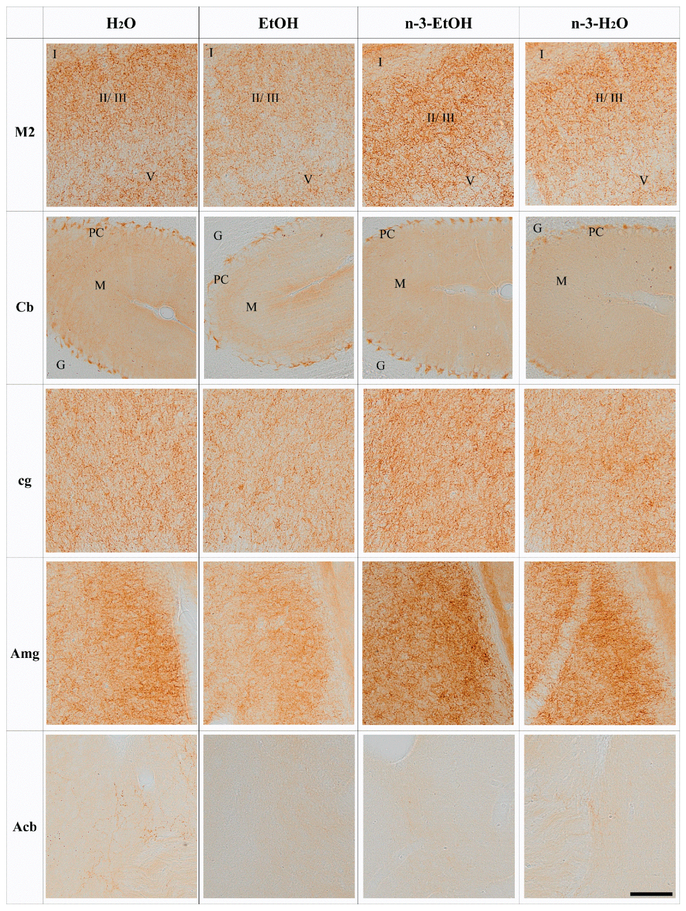
N-3 and Brain CB1 Receptor Expression

The brain and cerebellar CB1 immunostaining patterns in H2O, EtOH, n-3-EtOH and n-3-H2O mice corresponded well with previous observations of the CB1 receptor distribution in the mouse brain and cerebellum. CB1 immunoreactive punctate appeared concentratedin certain brain regions as well as in some cerebral and cerebellar cortical layers. Thus, strong CB1 receptor immunoreactivity was observed in the granule cell layer of the olfactory bulb (OB) [29] and remarkable staining was also revealed in the striatum, cerebral cortex (layers II–III and V–VI), olfactory tubercle, substantia nigra pars reticulata (SN), amygdala (Amg) and hippocampus [30], as well as the cerebellar molecular and Purkinje cell layers. More moderate immunoreactivity was detected in the nucleus accumbens (Acb) [31].
The analysis of the CB1 receptor optical density showed that the receptor distribution pattern varied in some regions depending on EtOH and/or n-3 intake. However, no changes were found in the OB, primary motor cortex (M1), frontal cortex (Fr3), cingular cortex area (Cg1), caudate putamen (CPu), dentate gyrus (DG), hippocampal CA1, SN and entorhinal cortex (Ent) in the four conditions (p > 0.05; Figure 1, Table 1). In contrast, the secondary motor cortex (M2), Cb, cingulum (cg), Amg and Acb were affected by EtOH and n-3 intake (p < 0.05; Figure 2). Thus, a significant reduction in CB1 receptor staining (~20%) was detected in the adult M2 after adolescent EtOH intake relative to the control (EtOH: 82.20 ± 3.732%; H2O: 100.00 ± 3.507% * p < 0.05; Figure 2 and Figure 3 A; Table 1). Remarkably, n-3 supplementation during withdrawal recovered CB1 receptor expression in the M2 to control levels (n-3-EtOH: 106.30 ± 4.604% ** p < 0.01), without the enriched diet having any effect on H2O mice (n-3-H2O: 100.50 ± 5.218% * p < 0.05; Figure 2 and Figure 3A; Table 1). The decrease in CB1 optical density following EtOH exposure during adolescence was more conspicuous in the Cb (EtOH: 76.40 ± 4.445% versus H2O: 100.00 ± 5.267%, ** p < 0.01), particularly in the molecular layer (Figure 2 and Figure 3B; Table 1). The n-3 diet normalized the detrimental effect of EtOH on CB1 values (n-3-EtOH: 98.43 ± 5.627%, * p < 0.05; Figure 2 and Figure 3B; Table 1). Again, n-3 under standard H2O conditions did not modify receptor expression in this region (n-3-H2O: 100.40 ± 4.620%, ** p < 0.01; Figure 2 and Figure 3B; Table 1). A similar effect of EtOH was observed in the cg, where CB1 optical density decreased significantly in adult mice after binge drinking during adolescence (EtOH: 80.77 ± 4.864% versus H2O: 100.00 ± 4.868% * p < 0.05; Figure 2 and Figure 3C; Table 1). However, n-3 was unable to revert significantly the receptor deficit (n-3-EtOH: 97.86 ± 5.274%), in contrast to the dietary effect on H2O (n-3-H2O: 105.20 ± 4.610%) relative to the CB1 decrease in EtOH mice (** p < 0.001; Figure 2 and Figure 3C; Table 1). The values in the Amg were also affected (Figure 2 and Figure 3D; Table 1): the significant CB1 reduction in EtOH mice (EtOH: 82.80 ± 3.468% versus H2O: 100.00 ± 3.594%, ** p < 0.01) returned to normal with n-3 intake (n-3-EtOH: 102.30 ± 3.977%, ** p < 0.01), with no effect of the nutritional supplementation when mice only drank H2O (97.69 ± 5.744%). Finally, CB1 optical density was drastically reduced in the Acb of the mature brain exposed to EtOH during adolescence (EtOH: 54.18 ± 10.81% versus H2O: 100.00 ± 6.028, *** p < 0.001; Figure 2 and Figure 3E; Table 1). However, n-3 intake could not restore CB1 receptor expression in EtOH mice (n-3-EtOH: 60.61 ± 8.153%). In addition, and contrary to the other studied brain regions, n-3 downregulated CB1 staining in H2O mice (n-3-H2O: 66.15 ± 11.04%, * p < 0.05; Figure 2 and Figure 3E; Table 1).

3. Discussion

3.1. Long-Lasting Effect of Adolescent Binge Drinking on CB1 Receptor Expression

We have shown that binge drinking during adolescence reduces CB1 receptor immunostaining in certain regions of the mature mouse brain, in particular, the M2, Cb, cg, Amg and Acb. Interestingly, n-3 supplementation during abstinence restores CB1 receptor expression measured using optical density in the M2, Cb and Amg and ameliorates density levels in the cg.
The CB1 receptor expression pattern matched the brain receptor distribution in the cortical, limbic and motor regions [2,30]. However, long-term changes in CB1 immunostaining after adolescent alcohol intake were restricted to some brain regions and the Cb that seem to correlate with the impact of EtOH intake on brain structure and function [6,32,33]. EtOH alters grey matter throughout the cortex, including the olfactory areas, Amg and Cb [33,34,35]. Also, the mesocorticolimbic system is affected. We observed that the long-lasting decrease in CB1 receptors normally expressed in the Acb [36] did not recover over time after adolescent binge drinking, remaining low even when the animals were under n-3 supplementation. As CB1 receptors intervene in brain maturation, it is plausible that the CB1 receptor expression deficits revealed in the Acb negatively contribute to the shape of the mesocorticolimbic system during the adolescent period, ultimately promoting brain vulnerability and alcohol addiction [35].
This study was conducted using male mice. Though males might be more vulnerable to withdrawal [37], females are more sensitive to EtOH [38,39]. Also, the effectiveness of treatments differs between males and females [40]. It is plausible that the EtOH impact on CB1 receptor expression and the effects of the n-3-enriched diet may vary between males and females, a possibility that will be explored in our future investigations.
The LM immunohistochemistry applied in the present study has some obvious limitations that deserve attention. Immunohistochemical techniques for LM were used in the 1990s to describe the pattern of CB1 receptor-like immunoreactivity in the brain [41,42]. Consequently, the strong immunostaining observed in certain brain regions (motor, limbic, reward, cortical) endorsed the advance in the knowledge of CB1 receptor functions in brain circuits. However, low CB1 receptor expression in cell types cannot be visualized using LM immunohistochemistry [30]. In addition, the tendency to diffusion of the 3-3′diaminobenzidine (DAB) reaction product used as chromogen in LM immunohistochemistry could lead to potential unspecific staining or false positives due to endogenous biotinylated proteins. These pitfalls can only be ruled out by using appropriate controls. In this study, we have used CB1-knockout brain tissue to discard bias, confirming the specificity of the CB1 staining observed throughout the brain in our experimental conditions. Ultimately, high-resolution immunoelectron microscopy that has been shown to be an excellent tool for unveiling the precise subcellular localization of CB1 receptors in the brain, would definitely identify the subcellular compartments and the CB1 receptor pools that were conspicuously reduced by adolescent EtOH intake, and recovered by n-3 in the specific brain regions identified in this study.
Endocannabinoid levels, membrane fluidity and EtOH-degrading enzymatic machinery could contribute to the altered CB1 receptor pattern observed in the adult brain after adolescent binge drinking. In addition, EtOH intake during adolescence causes memory impairment that can last into adulthood [43,44]. Our model of binge drinking during adolescence used in this study has previously been shown to be associated with hippocampal memory deficits in adulthood [15,45]. Although CB1 receptor optical density was not significantly affected by binge drinking, subtle subcellular changes in receptor expression were detected in the dentate molecular layer that should contribute to the abrogation of cannabinoid-dependent synaptic plasticity at the excitatory medial perforant path synapses and related memory loss [15]. Remarkably, the deleterious cognitive binge drinking effects were recovered by increasing the endocannabinoid 2-AG or by environmental conditions [15,45]. Furthermore, the M2 and Cb, both affected by EtOH, are brain regions involved in motor coordination [46,47] and EtOH intake leads to motor incoordination and ataxia [43]. Our present results show a significant decrease in CB1 staining in the cerebellar molecular layer, where the receptor is mostly localized to the excitatory granule cell parallel fiber terminals [48]. However, the lack of CB1 receptors does not seem to cause evident cerebellar motor deficits [49], despite their role in motor learning [50]. Nevertheless, cannabinoid-dependent motor control is also exerted from the cortex [51], where we detected deficits in CB1 receptors upon adolescent binge drinking. Interestingly, young adult mice under enriched environment recovered motor coordination and balance after adolescent binge drinking [45]. It is likely that the ECS participates in this motor improvement, as it is the case in the memory recovery elicited by environmental enrichment via the restoration of endocannabinoid-dependent excitatory synaptic plasticity, in which CB1 receptors, group I metabotropic glutamate receptors and 2-AG were involved [9].
EtOH modifies synaptic membrane fluidity [52] and stimulates arachidonic acid (AA) production from membrane phospholipids by increasing phospholipase A2 (PLA2) [53]. The availability of more AA for AEA synthesis may be responsible for the decrease in CB1 agonist binding and gene expression elicited by chronic EtOH in certain brain regions [54,55]). In fact, the drop in CB1 receptor immunostaining in the cerebellar molecular layer correlates with AEA transport inhibition and a 2-AG increase in granule cells after chronic EtOH [56,57]. Cannabinoids internalize CB1 receptors and reduce their mobility, having an impact on receptor availability at the synapse [58]. We have demonstrated previously that Δ-9-tetrahydrocannabinol (THC) causes a selective CB1 receptor labeling decrease in certain subcellular compartments (excitatory and inhibitory terminals, mitochondria, astrocytes) of several brain regions [59]. This distinct impact of THC could be related to the different THC levels and metabolites detected among brain regions after acute THC administration [60]. A similar phenomenon could befall our model of binge drinking. In fact, brain EtOH metabolism by class III alcohol dehydrogenase (ADH) generates acetaldehyde that accumulates in the hippocampus, cortex and Cb, where the enzyme is more expressed [61]. Interestingly, the enzyme distribution coincides in brain regions with high CB1 receptor expression, such as the cortex and Cb, both strikingly affected by adolescent binge drinking. EtOH decreases glutathione in these same regions, thus increasing oxidative processes and brain damage in a model of prenatal EtOH exposure [17]. As class III ADH is a glutathione-dependent formaldehyde dehydrogenase, it is likely that the glutathione reduction elicited by EtOH and the consequent oxidative state leads to enzyme malfunction, jeopardizing EtOH elimination in regions where the enzyme is more abundant.

3.2. N-3 Recovers CB1 Receptor Expression in the Brain

DHA and AA are major phospholipid components of brain cell membranes [16,19]. EtOH reduces DHA in the brain [20,21] and its deficit impacts on both cell membranes, altering their biophysical properties, and related membrane proteins, such as enzymes and receptors [19]. N-3 deficiency lowers CB1 receptors in different brain regions [62] and impairs endocannabinoid-mediated synaptic plasticity [22]. The Fr3, OB, Cb, hippocampus, midbrain and striatum rank in high-to-low order among the brain regions with more DHA [63]. However, the negative effect on CB1 receptor expression in the adult brain after adolescent binge drinking was particularly outstanding in the M2, Cb and Acb (ventral striatum), with no effect in the hippocampus, dorsal striatum, some cortical areas, OB and SN. A DHA-enriched diet counteracts the low brain n-3 PUFA levels due to EtOH intake [21] and reverses EtOH-induced impairment of synaptic plasticity [18]. It also restores aquaporin-4, PLA-2 and glutathione affected by EtOH [17,21,64,65]. The recovery of CB1 receptor immunostaining by n-3 supplementation during the abstinence period points to the normalization of cell membrane homeostasis.
In conclusion, abusive EtOH consumption during adolescence alters CB1 receptor immunostaining optical density in some brain regions of the adult mouse, and an n-3-enriched diet recovers the reduced CB1 expression in limbic and motor structures following binge drinking. Uncovering the PUFA effects and mechanisms by which the n-3-enriched diet can impact on brain cannabinoid receptor expression (as shown in this paper) and function after adolescent binge drinking, could be an appropriate non-pharmacological approach to counteract the EtOH impact on cannabinoid-dependent synaptic plasticity, cognition and behavior.

4. Material & Methods

4.1. Generation of CB1-KO

CB1-knockout (CB1-KO) mice were generated and genotyped as previously described [66] and formerly collected [30]. They were obtained by crossing CB1f/f mice with CMV-Cre mice (“Cre deleter”). Mice were of a predominant C57BL/6-N background (9–10 back-crossings) and the breeding strategy used was female CB1+/− × male CB1+/− (Table 2).

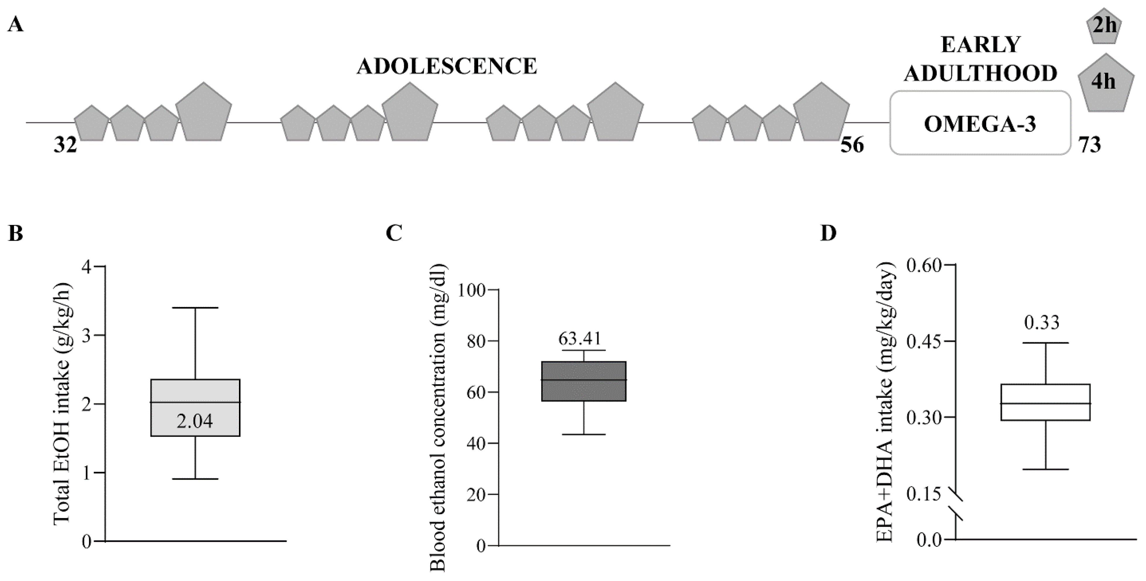
4.2. Animal Treatment

Four-week-old C57BL/6J male mice (Janvier Labs, Le Genest-Saint-Isle, France) were housed in pairs and habituated to a dark cycle (8 a.m.–8 p.m.). Then, they were exposed to water or drinking-in-the-dark (DID) during adolescence, as previously described [45]. Briefly, the mice were individualized and exposed to a bottle of 10 mL tap water or EtOH 20% (Boter S.L., Barcelona, Spain) four days a week during four weeks (postnatal day (PND) 32 to 56). They had free access to the bottle for 2 h the first three days, and 4 h the fourth day. Mice were resting and kept in pairs with food and water ad libitum for the last three days of the week (Figure 4A–C). On PND 56, a blood sample was collected from the lateral tail vein using a capillary tube 30 min after 4 h of EtOH exposure (Sarstedt, Nümbrecht, Germany). Blood samples were analyzed for EtOH concentration using a commercial OH assay kit (Abcam ab65646, Madrid, Spain), following manufacturer instructions. Half of the mice were fed 2% EPA and DHA (2.2% EPA and 1.5% DHA of total fats; SAFE, Augy, France) during withdrawal (PND 56–73). Twice a week, mice and food were weighed to measure EPA and DHA intake (mg/kg/day) (Figure 4D). Three mice/group were culled on PND 73. They were deeply anesthetized using 4% chloral hydrate (10 mL/kg body weight, i.p.) and perfused through the left ventricle with 30 mL phosphate-buffered saline (PBS, 0.1 M, pH 7.4), followed by 80 mL of the fixative (4% formaldehyde depolymerized from paraformaldehyde, 0.2% picric acid, 0.1% glutaraldehyde) prepared in PBS at room temperature (RT). Brains were removed and post-fixed as described elsewhere in detail [59].

4.3. Antibody Characterization

The specificity of the CB1 receptor antibody (Nittobo Medical Co., Ltd., Tokyo, Japan); goat polyclonal, CB1-Go-Af450, RRID AB_2571592, Table 3) has been tested thoroughly [3,4,30]. In addition, CB1 receptor staining was not detected in the CB1-KO mouse brain (Figure 5).

4.4. Immunohistochemistry for Light Microscopy

This was performed following the protocol previously published [30]. Briefly, coronal and sagittal sections cut at 50 μm on a vibratome (Leica VT 1000s, Wetzlar, Germany) were taken rostro-caudally from the whole brain and Cb, respectively, and collected in phosphate buffer (PB 0.1 M, pH 7.4) at RT. They were pre-incubated in a blocking solution for 30 min at RT, and then incubated in goat anti-CB1 receptor antibody (2 μg/mL) diluted in 10% horse serum, 0.1% sodium azide, 0.5% Triton in 1× Tris-HCl-buffered saline (TBS) overnight at RT. After washing in 1% horse serum and 0.5% Triton in 1× TBS the next day, tissue sections were incubated with a horse anti-goat IgG biotinylated antibody (H + L) (1:200, Vector Labs, Newark, CA, USA, cat#BA9500; RRID: AB_2336123) for 1 h at RT. Following several washes, they were incubated in avidin-biotin peroxidase complex (1:50, Vector Labs, Newark, CA, USA, Cat#PK-6100, RRID: AB_2336819) for 1 h at RT. Tissue was washed several times with the last two containing 0.5% Triton in PB, and incubated in 0.05% DAB (Sigma-Aldrich, Merck KGaA, Darmstadt, Germany, Cat#D5637; RRID: AB_2336819) with 0.01% hydrogen peroxide prepared in 0.1 M PB for 3 min. Finally, following five washes with 0.5% Triton in PB, the sections were mounted, dehydrated and coverslipped with DPX (Sigma Aldrich, Merck KGaA, Darmstadt, Germany Cat#44581).

4.5. Semiquantitative Analysis of CB1 Receptor Optical Density

Brain and cortical regions known to express CB1 receptors were selected: OB, M1, M2, Fr3, Cg1, cg, CPu, Acb, Amg, DG, hippocampal CA1, SN, Ent and Cb. Micrographs were taken at 10× using a light microscope (Olympus BX61, Hamburg, Germany) and processed using the Olympus cellSens Dimension using consecutive sections containing the brain regions and cortical areas of interest. For each region, three independent optical density measurements were performed at 10×, and two more were taken in a blank zone to rank background level. As some regions were through several slides, they were analyzed repeatedly in each mouse of the four experimental conditions (Table 4). All measurements were then pooled by mouse. Subsequently, data were normalized to 100% of the H2O group. Image J software (1.8.0_322); NIH; RRID:SCR_003070) and a statistical software package were used (GraphPad Prism 8; RRID: SCR_002798). The Shapiro–Wilk normality test was applied before running one-way ANOVA. Parametric data were analyzed using Holm Sidak’s multiple comparison test and non-parametric data using Dunn’s multiple comparison test. All values are given as mean ± SEM.

Author Contributions

Conceptualization, I.G. and P.G.; Methodology, N.P. and I.R.-B.; Validation, A.R. and P.G.; Formal Analysis, L.L. and G.O.; Investigation, A.M.-L. and M.S.; Resources, P.G.; Data Curation, M.S. and L.L.; Writing—Original Draft Preparation, M.S., P.G. and I.B.-D.R.; Writing—Review & Editing, P.G. and I.B.-D.R.; Supervision, P.G.; Funding Acquisition, P.G. All authors have read and agreed to the published version of the manuscript.

Funding

This work has been supported by The Basque Government (IT1620-22); Red de Investigación en Atención Primaria de Adicciones (RIAPAd), Instituto de Salud Carlos III (RD21/0009/0006); Red de Trastornos Adictivos, Instituto de Salud Carlos III, European Regional Development Funds-European Union (ERDF-EU; RD16/0017/0012); and Ministry of Science and Innovation (PID2019-107548RB-I00). M.S. is in receipt of a PhD contract from The University of the Basque Country (PIF 19/164).

Institutional Review Board Statement

The protocols for animal care and use were approved by the Committee of Ethics for Animal Welfare of the University of the Basque Country (M20-2020-113). They were also in agreement with the European Communities Council Directive of 22 September 2010 (2010/63/EU) and Spanish regulations (Real Decreto 53/2013, BOE 08-02-2013). The number of animals and suffering were controlled and minimized.

Data Availability Statement

The data presented in this study are available on request from the corresponding author.

Acknowledgments

We deeply thank Giovanni Marsicano (INSERM, U1215 Neurocentre Magendie, Endocannabinoids and Neuroadaptation, Bordeaux, France. Université de Bordeaux, France) for providing us with the CB1-knockout mice.

Conflicts of Interest

The authors declare that they have no known competing financial interest or personal relationships that could have appeared to influence the work reported in this paper.

References

  1. Lu, H.-C.; Mackie, K. An introduction to the endogenous cannabinoid system. Biol. Psychiatry 2016, 79, 516–525. [Google Scholar] [CrossRef]
  2. Mechoulam, R.; Parker, L.A. The endocannabinoid system and the brain. Annu. Rev. Psychol. 2013, 64, 21–47. [Google Scholar] [CrossRef] [PubMed]
  3. Gutiérrez-Rodríguez, A.; Bonilla-Del Río, I.; Puente, N.; Gómez-Urquijo, S.M.; Fontaine, C.J.; Egaña-Huguet, J.; Elezgarai, I.; Ruehle, S.; Lutz, B.; Robin, L.M.; et al. Localization of the cannabinoid type-1 receptor in subcellular astrocyte compartments of mutant mouse hippocampus. Glia 2018, 66, 1417–1431. [Google Scholar] [CrossRef] [PubMed]
  4. Hebert-Chatelain, E.; Desprez, T.; Serrat, R.; Bellocchio, L.; Soria-Gomez, E.; Busquets-Garcia, A.; Pagano Zottola, A.C.; Delamarre, A.; Cannich, A.; Vincent, P.; et al. A cannabinoid link between mitochondria and memory. Nature 2016, 539, 555–559. [Google Scholar] [CrossRef] [PubMed]
  5. Katona, I.; Freund, T.F. Multiple functions of endocannabinoid signaling in the brain. Annu. Rev. Neurosci. 2012, 35, 529–558. [Google Scholar] [CrossRef]
  6. Carbia, C.; Cadaveira, F.; Caamaño-Isorna, F.; Rodríguez-Holguín, S.; Corral, M. Binge drinking during adolescence and young adulthood is associated with deficits in verbal episodic memory. PLoS ONE 2017, 12, e0171393. [Google Scholar] [CrossRef] [PubMed]
  7. Lutz, B.; Marsicano, G.; Maldonado, R.; Hillard, C.J. The endocannabinoid system in guarding against fear, anxiety and stress. Nat. Rev. Neurosci. 2015, 16, 705–718. [Google Scholar] [CrossRef]
  8. Pava, M.; Woodward, J. A review of the interactions between alcohol and the endocannabinoid system: Implications for alcohol dependence and future directions for research. Alcohol 2012, 46, 185–204. [Google Scholar] [CrossRef]
  9. Rico-Barrio, I.; Peñasco, S.; Lekunberri, L.; Serrano, M.; Egaña-Huguet, J.; Mimenza, A.; Soria-Gomez, E.; Ramos, A.; Buceta, I.; Gerrikagoitia, I.; et al. Environmental enrichment rescues endocannabinoid-dependent synaptic plasticity lost in young adult male mice after ethanol exposure during adolescence. Biomedicines 2021, 9, 825. [Google Scholar] [CrossRef]
  10. Mitrirattanakul, S.; López-Valdés, H.E.; Liang, J.; Matsuka, Y.; Mackie, K.; Faull, K.F.; Spigelman, I. Bidirectional alterations of hippocampal cannabinoid 1 receptors and their endogenous ligands in a rat model of alcohol withdrawal and dependence. Alcohol. Clin. Exp. Res. 2007, 31, 855–867. [Google Scholar] [CrossRef]
  11. Vinod, K.Y.; Kassir, S.A.; Hungund, B.L.; Cooper, T.B.; Mann, J.J.; Arango, V. Selective alterations of the CB1 receptors and the fatty acid amide hydrolase in the ventral striatum of alcoholics and suicides. J. Psychiatr. Res. 2010, 44, 591–597. [Google Scholar] [CrossRef] [PubMed]
  12. Ceccarini, J.; Hompes, T.; Verhaeghen, A.; Casteels, C.; Peuskens, H.; Bormans, G.; Claes, S.; Van Laere, K. Changes in cerebral CB1 receptor availability after acute and chronic alcohol abuse and monitored abstinence. J. Neurosci. 2014, 34, 2822–2831. [Google Scholar] [CrossRef] [PubMed]
  13. Hirvonen, J.; Zanotti-Fregonara, P.; Umhau, J.C.; George, D.T.; Rallis-Frutos, D.; Lyoo, C.H.; Li, C.T.; Hines, C.S.; Sun, H.; Terry, G.E.; et al. Reduced cannabinoid CB 1 receptor binding in alcohol dependence measured with positron emission tomography. Mol. Psychiatry 2013, 18, 916–921. [Google Scholar] [CrossRef] [PubMed]
  14. Maccioni, P.; Colombo, G.; Carai, M. Blockade of the cannabinoid cb1 receptor and alcohol dependence: Preclinical evidence and preliminary clinical data. CNS Neurol. Disord. Drug Targets 2010, 9, 55–59. [Google Scholar] [CrossRef] [PubMed]
  15. Peñasco, S.; Rico-Barrio, I.; Puente, N.; Fontaine, C.J.; Ramos, A.; Reguero, L.; Gerrikagoitia, I.; Rodríguez de Fonseca, F.; Barrondo, S.; Aretxabala, X.; et al. Intermittent ethanol exposure during adolescence impairs cannabinoid type 1 receptor- dependent long-term depression and recognition memory in adult mice. Neuropsychopharmacology 2020, 45, 309–318. [Google Scholar] [CrossRef]
  16. Joffre, C.; Rey, C.; Layé, S. N-3 polyunsaturated fatty acids and the resolution of neuroinflammation. Front. Pharmacol. 2019, 10, 1022. [Google Scholar] [CrossRef]
  17. Patten, A.R.; Brocardo, P.S.; Christie, B.R. Omega-3 supplementation can restore glutathione levels and prevent oxidative damage caused by prenatal ethanol exposure. J. Nutr. Biochem. 2013, 24, 760–769. [Google Scholar] [CrossRef]
  18. Patten, A.R.; Sickmann, H.M.; Dyer, R.A.; Innis, S.M.; Christie, B.R. Omega-3 fatty acids can reverse the long-term deficits in hippocampal synaptic plasticity caused by prenatal ethanol exposure. Neurosci. Lett. 2013, 551, 7–11. [Google Scholar] [CrossRef]
  19. Serrano, M.; Rico-Barrio, I.; Grandes, P. The effect of omega-3 fatty acids on alcohol-induced damage. Front. Nutr. 2023, 10, 1068343. [Google Scholar] [CrossRef]
  20. Milne, G.L.; Morrow, J.D.; Picklo, M.J. Elevated oxidation of docosahexaenoic acid, 22:6 (n-3), in brain regions of rats undergoing ethanol withdrawal. Neurosci. Lett. 2006, 405, 172–174. [Google Scholar] [CrossRef]
  21. Tajuddin, N.; Moon, K.H.; Marshall, S.A.; Nixon, K.; Neafsey, E.J.; Kim, H.Y.; Collins, M.A. Neuroinflammation and neurodegeneration in adult rat brain from binge ethanol exposure: Abrogation by docosahexaenoic acid. PLoS ONE 2014, 9, e101223. [Google Scholar] [CrossRef] [PubMed]
  22. Lafourcade, M.; Larrieu, T.; Mato, S.; Duffaud, A.; Sepers, M.; Matias, I.; De Smedt-Peyrusse, V.; Labrousse, V.F.; Bretillon, L.; Matute, C.; et al. Nutritional omega-3 deficiency abolishes endocannabinoid-mediated neuronal functions. Nat. Neurosci. 2011, 14, 345–350. [Google Scholar] [CrossRef] [PubMed]
  23. Manduca, A.; Bara, A.; Larrieu, T.; Lassalle, O.; Joffre, C.; Layé, S.; Manzoni, O.J. Amplification of mGlu5-endocannabinoid signaling rescues behavioral and synaptic deficits in a mouse model of adolescent and adult dietary polyunsaturated fatty acid imbalance. J. Neurosci. 2017, 37, 6851–6868. [Google Scholar] [CrossRef]
  24. Thomazeau, A.; Bosch-Bouju, C.; Manzoni, O.; Layé, S. Nutritional n-3 PUFA deficiency abolishes endocannabinoid gating of hippocampal long-term potentiation. Cereb. Cortex 2017, 27, 2571–2579. [Google Scholar] [CrossRef]
  25. Pan, J.P.; Zhang, H.Q.; Wang, W.; Guo, Y.F.; Xiao, N.; Cao, X.H.; Liu, L.J. Some subtypes of endocannabinoid/endovanilloid receptors mediate docosahexaenoic acid-induced enhanced spatial memory in rats. Brain Res. 2011, 1412, 18–27. [Google Scholar] [CrossRef] [PubMed]
  26. Artmann, A.; Petersen, G.; Hellgren, L.I.; Boberg, J.; Skonberg, C.; Nellemann, C.; Hansen, S.H.; Hansen, H.S. Influence of dietary fatty acids on endocannabinoid and N-acylethanolamine levels in rat brain, liver and small intestine. Biochim. Biophys. Acta—Mol. Cell Biol. Lipids 2008, 1781, 200–212. [Google Scholar] [CrossRef]
  27. Wood, J.A.T.; Williams, J.S.; Pandarinathan, L.; Janero, D.R.; Lammi-Keefe, C.J.; Makriyannis, A. Dietary docosahexaenoic acid supplementation alters select physiological endocannabinoid-system metabolites in brain and plasma. J. Lipid Res. 2010, 51, 1416–1423. [Google Scholar] [CrossRef]
  28. Watson, J.E.; Kim, J.S.; Das, A. Emerging class of omega-3 fatty acid endocannabinoids & their derivatives. Prostaglandins Other Lipid Mediat. 2019, 143, 106337. [Google Scholar] [CrossRef]
  29. Soria-Gómez, E.; Bellocchio, L.; Reguero, L.; Lepousez, G.; Martin, C.; Bendahmane, M.; Ruehle, S.; Remmers, F.; Desprez, T.; Matias, I.; et al. The endocannabinoid system controls food intake via olfactory processes. Nat. Neurosci. 2014, 17, 407–415. [Google Scholar] [CrossRef]
  30. Gutiérrez-Rodríguez, A.; Puente, N.; Elezgarai, I.; Ruehle, S.; Lutz, B.; Reguero, L.; Gerrikagoitia, I.; Marsicano, G.; Grandes, P. Anatomical characterization of the cannabinoid CB1 receptor in cell-type–specific mutant mouse rescue models. J. Comp. Neurol. 2017, 525, 302–318. [Google Scholar] [CrossRef]
  31. Winters, B.D.; Krüger, J.M.; Huang, X.; Gallaher, Z.R.; Ishikawa, M.; Czaja, K.; Krueger, J.M.; Huang, Y.H.; Schlüter, O.M.; Dong, Y. Cannabinoid receptor 1-expressing neurons in the nucleus accumbens. Proc. Natl. Acad. Sci. USA 2012, 109, 2717–2725. [Google Scholar] [CrossRef] [PubMed]
  32. Hanson, K.L.; Medina, K.L.; Padula, C.B.; Tapert, S.F.; Brown, S. Impact of adolescent alcohol and drug use on neuropsychological functioning in young adulthood: 10-year outcomes. J. Child Adolesc. Subst. Abus. 2011, 20, 135–154. [Google Scholar] [CrossRef]
  33. Lees, B.; Debenham, J.; Squeglia, L.M. Alcohol and cannabis use and the developing brain. Alcohol Res. Curr. Rev. 2021, 41, 11. [Google Scholar] [CrossRef]
  34. Coleman, L.G.; He, J.; Lee, J.; Styner, M.; Crews, F. Adolescent binge drinking alters adult brain neurotransmitter gene expression, behavior, brain regional volumes, and neurochemistry in mice. Alcohol. Clin. Exp. Res. 2011, 35, 671–688. [Google Scholar] [CrossRef] [PubMed]
  35. Guerri, C.; Pascual, M. Mechanisms involved in the neurotoxic, cognitive, and neurobehavioral effects of alcohol consumption during adolescence. Alcohol 2010, 44, 15–26. [Google Scholar] [CrossRef] [PubMed]
  36. Manzanares, J.; Cabañero, D.; Puente, N.; García-Gutiérrez, M.S.; Grandes, P.; Maldonado, R. Role of the endocannabinoid system in drug addiction. Biochem. Pharmacol. 2018, 157, 108–121. [Google Scholar] [CrossRef] [PubMed]
  37. Flores-Bonilla, A.; Richardson, H. Sex differences in the neurobiology of alcohol use disorder. Alcohol Res. 2020, 40, 04. [Google Scholar] [CrossRef]
  38. Alfonso-Loeches, S.; Pascual, M.; Guerri, C. Gender differences in alcohol-induced neurotoxicity and brain damage. Toxicology 2013, 311, 27–34. [Google Scholar] [CrossRef]
  39. Maynard, M.E.; Barton, E.A.; Robinson, C.R.; Wooden, J.I.; Leasure, J.L. Sex differences in hippocampal damage, cognitive impairment, and trophic factor expression in an animal model of an alcohol use disorder. Brain Struct. Funct. 2018, 223, 195–210. [Google Scholar] [CrossRef]
  40. Moore, C.F.; Lynch, W.J. Alcohol preferring (P) rats as a model for examining sex differences in alcohol use disorder and its treatment. Pharmacol. Biochem. Behav. 2015, 132, 1–9. [Google Scholar] [CrossRef]
  41. Egertova, M.; Giang, D.K.; Cravatt, B.F.; Elphick, M.R. A new perspective on cannabinoid signalling: Complimentary localization of fatty acid amide hydrolase and the CB1 receptor in rat brain. Proc. R. Soc. London Ser. B Biol. Sci. 1998, 265, 2081–2085. [Google Scholar] [CrossRef]
  42. Tsou, K.; Brown, S.; Sañudo-Peña, M.C.; Mackie, K.; Walker, J.M. Immunohistochemical distribution of cannabinoid CB1 receptors in the rat central nervous system. Neuroscience 1998, 83, 393–411. [Google Scholar] [CrossRef]
  43. Lees, B.; Meredith, L.R.; Kirkland, A.E.; Bryant, B.E.; Squeglia, L.M. Effect of alcohol use on the adolescent brain and behavior. Pharmacol. Biochem. Behav. 2020, 192, 172906. [Google Scholar] [CrossRef]
  44. Risher, M.L.; Fleming, R.L.; Boutros, N.; Semenova, S.; Wilson, W.A.; Levin, E.D.; Markou, A.; Swartzwelder, H.S.; Acheson, S.K. Long-term effects of chronic intermittent ethanol exposure in adolescent and adult rats: Radial-arm maze performance and operant food reinforced responding. PLoS ONE 2013, 8, e62940. [Google Scholar] [CrossRef] [PubMed]
  45. Rico-Barrio, I.; Peñasco, S.; Puente, N.; Ramos, A.; Fontaine, C.J.; Reguero, L.; Giordano, M.E.; Buceta, I.; Terradillos, I.; Lekunberri, L.; et al. Cognitive and neurobehavioral benefits of an enriched environment on young adult mice after chronic ethanol consumption during adolescence. Addict. Biol. 2019, 24, 969–980. [Google Scholar] [CrossRef]
  46. Li, N.; Chen, T.W.; Guo, Z.V.; Gerfen, C.R.; Svoboda, K. A motor cortex circuit for motor planning and movement. Nature 2015, 519, 51–56. [Google Scholar] [CrossRef]
  47. Schmahmann, J.D. The cerebellum and cognition. Neurosci. Lett. 2019, 688, 62–75. [Google Scholar] [CrossRef] [PubMed]
  48. Buceta, I.; Elezgarai, I.; Rico-Barrio, I.; Gerrikagoitia, I.; Puente, N.; Grandes, P. Deletion of the cannabinoid CB1 receptor impacts on the ultrastructure of the cerebellar parallel fiber-Purkinje cell synapses. J. Comp. Neurol. 2019, 528, 1041–1052. [Google Scholar] [CrossRef] [PubMed]
  49. Albergaria, C.; Silva, N.T.; Darmohray, D.; Carey, M.R. Cannabinoids modulate associative cerebellar learning via alterations in behavioral state. eLife 2020, 9, e61821. [Google Scholar] [CrossRef]
  50. Kishimoto, Y.; Kano, M. Endogenous cannabinoid signaling through the CB1 receptor is essential for cerebellum-dependent discrete motor learning. J. Neurosci. 2006, 26, 8829–8837. [Google Scholar] [CrossRef]
  51. El Manira, A.; Kyriakatos, A. The role of endocannabinoid signaling in motor control. Physiology 2010, 25, 230–238. [Google Scholar] [CrossRef]
  52. Reddy, V.D.; Padmavathi, P.; Bulle, S.; Hebbani, A.V.; Marthadu, S.B.; Venugopalacharyulu, N.C.; Maturu, P.; Varadacharyulu, N.C. Association between alcohol-induced oxidative stress and membrane properties in synaptosomes: A protective role of vitamin E. Neurotoxicology Teratol. 2017, 63, 60–65. [Google Scholar] [CrossRef]
  53. Moon, K.H.; Tajuddin, N.; Brown, J.; Neafsey, E.J.; Kim, H.Y.; Collins, M.A. Phospholipase A2, Oxidative Stress, and Neurodegeneration in Binge Ethanol-Treated Organotypic Slice Cultures of Developing Rat Brain. Alcohol. Clin. Exp. Res. 2014, 38, 161–169. [Google Scholar] [CrossRef] [PubMed]
  54. Basavarajappa, B.S.; Nagre, N.; Xie, S.; Subbanna, S. Elevation of endogenous anandamide impairs ltp, learning and memory through cb1 receptor signaling in mice. Hippocampus 2014, 24, 808–818. [Google Scholar] [CrossRef] [PubMed]
  55. Ortiz, S.; Oliva, J.M.; Pérez-Rial, S.; Palomo, T.; Manzanares, J. Chronic ethanol consumption regulates cannabinoid CB1 receptor gene expression in selected regions of rat brain. Alcohol Alcohol. 2004, 39, 88–92. [Google Scholar] [CrossRef] [PubMed]
  56. Basavarajappa, B.S.; Saito, M.; Cooper, T.B.; Hungund, B.L. Chronic ethanol inhibits the anandamide transport and increases extracellular anandamide levels in cerebellar granule neurons. Eur. J. Pharmacol. 2003, 466, 73–83. [Google Scholar] [CrossRef] [PubMed]
  57. Ferrer, B.; Bermúdez-Silva, F.J.; Bilbao, A.; Alvarez-Jaimes, L.; Sanchez-Vera, I.; Giuffrida, A.; Serrano, A.; Baixeras, E.; Khaturia, S.; Navarro, M.; et al. Regulation of brain anandamide by acute administration of ethanol. Biochem. J. 2007, 404, 97–104. [Google Scholar] [CrossRef] [PubMed]
  58. Mikasova, L.; Groc, L.; Choquet, D.; Manzoni, O.J. Altered surface trafficking of presynaptic cannabinoid type 1 receptor in and out synaptic terminals parallesls receptor desensitization.pdf. Proc. Natl. Acad. Sci. USA 2008, 105, 18596–18601. [Google Scholar] [CrossRef]
  59. Bonilla-Del Río, I.; Puente, N.; Mimenza, A.; Ramos, A.; Serrano, M.; Lekunberri, L.; Gerrikagoitia, I.; Christie, B.R.; Nahirney, P.C.; Grandes, P. Acute Δ9-tetrahydrocannabinol prompts rapid changes in cannabinoid CB1 receptor immunolabeling and subcellular structure in CA1 hippocampus of young adult male mice. J. Comp. Neurol. 2020, 529, 2332–2346. [Google Scholar] [CrossRef]
  60. Leishman, E.; Manchanda, M.; Thelen, R.; Miller, S.; Mackie, K.; Bradshaw, H.B. Cannabidiol’s Upregulation of N-acyl Ethanolamines in the Central Nervous System Requires N-acyl Phosphatidyl Ethanolamine-Specific Phospholipase D. Cannabis Cannabinoid Res. 2018, 3, 228–241. [Google Scholar] [CrossRef]
  61. Waddell, J.; McKenna, M.C.; Kristian, T. Brain ethanol metabolism and mitochondria. Curr. Top. Biochem. Res. 2022, 23, 1–13. [Google Scholar]
  62. Isaac, A.R.; de Velasco, P.C.; Fraga, K.Y.D.; Tavares-do-Carmo, M.d.G.; Campos, R.M.P.; Iannotti, F.A.; Verde, R.; Martins, D.B.G.; Santos, T.A.; Ferreira, B.K.; et al. Maternal omega-3 intake differentially affects the endocannabinoid system in the progeny`s neocortex and hippocampus: Impact on synaptic markers. J. Nutr. Biochem. 2021, 96, 108782. [Google Scholar] [CrossRef] [PubMed]
  63. Carrié, I.; Clément, M.; De Javel, D.; Francès, H.; Bourre, J.M. Specific phospholipid fatty acid composition of brain regions in mice: Effects of n-3 polyunsaturated fatty acid deficiency and phospholipid supplementation. J. Lipid Res. 2000, 41, 465–472. [Google Scholar] [CrossRef] [PubMed]
  64. Brown, J., III; Achille, N.; Neafsey, E.J.; Collins, M.A. Binge ethanol-induced neurodegeneration in rat organotypic brain slice cultures: Effects of PLA2 inhibitor mepacrine and docosahexaenoic acid (DHA). Neurochem. Res. 2009, 34, 260–267. [Google Scholar] [CrossRef] [PubMed]
  65. Collins, M.A.; Moon, K.; Tajuddin, N.; Neafsey, E.J.; Kim, Y. Docosahexaenoic acid (DHA) prevents binge ethanol-dependent aquaporin-4 elevations while inhibiting neurodegeneration: Experiments in rat adult-age entorhino-hippocampal slice cultures. Neurotox. Res. 2013, 23, 105–110. [Google Scholar] [CrossRef] [PubMed]
  66. Marsicano, G.; Wotjak, C.T.; Azad, S.C. The endogenous cannabinoid system controls extinction of aversive memories. Nature 2002, 418, 530–534. [Google Scholar] [CrossRef]
Figure 1. Normalized (%) CB1 receptor optical density in the OB (A), M1 (B), Fr3 (C), Cg1 (D), CPu (E), DG (F), CA1 (G), SN (H), and Ent (I). Pooled data are expressed as mean ± SEM (one-way ANOVA, Dunn’s and Holm Sidak’s multiple comparisons tests).
Ijms 24 17316 g001
Figure 2. CB1 receptor-like immunoreactivity in the M2, Cb, cg, Amg and Acb of adult male mice exposed to H2O, EtOH, n-3-EtOH or n-3-H2O. Pre-embedding immunoperoxidase method for light microscopy. The typical CB1 staining pattern is observed: abundant dotty elements distributed in the superficial (II–III) and deep (V) layers of the M2 cortex as well as in the cingulum (cg) and amygdala (Amg); uniform immunostaining in the cerebellar molecular (M) layer, strong basket cell terminal labeling around Purkinje cell (PC) bodies in the PC layer, and lack of staining in the granule (G) cell layer; very faint staining in the Acb with only some positive varicose fibers in control (H2O). Scale bar: 200 µm.
Ijms 24 17316 g002
Figure 3. CB1 receptor optical density in adult male mice exposed to H2O, EtOH, n-3-EtOH or n-3-H2O. Normalized (%) CB1 optical density in the M2 (A), Cb (B), cg (C), Amg (D) and Acb (E). Pooled data are expressed as mean ± SEM (one-way ANOVA, Dunn’s multiple comparisons test; * p < 0.05; ** p < 0.01; *** p < 0.001). The n-3 diet during withdrawal recovers the significant decrease in CB1 receptor expression in the M2, Cb and Amg of the adult brain after adolescent binge drinking.
Ijms 24 17316 g003
Figure 4. Schematic timeline of the EtOH procedure, total EtOH intake, BEC, EPA and DHA intake. (A) C57BL/6J male mice were submitted to the DID procedure over four weeks (PND 32–56). They had 2 h free access to H2O or EtOH for the first three days of the week, and 4 h the fourth day. During abstinence (PND 56–73), half of them were fed with n-3 supplementation (EPA and DHA 2%). (B) Average of total EtOH intake during DID of EtOH (2.033 ± 0.5437 g/kg/h, n = 8) and n-3-EtOH (2.051 ± 0.5941 g/kg/h, n = 8) (Student’s t-test, p > 0.05). (C) Average BEC obtained on the last day of EtOH exposure in EtOH (62.96 ± 10.89 mg/dL; n = 8) and n-3-EtOH (63.92 ± 10.08 mg/dL; n = 7) (Student’s t-test, p > 0.05). (D) Average of total EPA and DHA intake during withdrawal in n-3-H2O (0.332 ± 0.0727 mg/kg/day, n = 12) and n-3-EtOH mice (0.324 ± 0.0679 mg/kg/day, n = 12) (Student’s t-test, p > 0.05).
Ijms 24 17316 g004
Figure 5. Specificity of the CB1 antibody tested in brain tissue (hippocampus) lacking CB1 receptors (CB1-KO). Pre-embedding immunoperoxidase method for light microscopy. No trace of staining canbe detected. Scale bar: 200 µm.
Ijms 24 17316 g005
Table 1. Normalized values (% mean ± SEM) of CB1 receptor optical density in olfactory bulb (OB), primary and secondary motor cortex (M1, M2), frontal cortex (Fr3), cingular cortex area 1 (Cg1), cingulum (cg), caudate putamen (CPu), nucleus accumbens (Acb), amygdala (Amg), dentate gyrus (DG), hippocampal CA1, substantia nigra (SN), entorhinal cortex (Ent) and cerebellum (Cb) for each experimental condition (n = 3 mice/group).
H2OEtOHn-3-EtOHn-3-H2O
OB100.00 ± 3.982102.30 ± 5.493108.50 ± 4.499100.40 ± 6.144
M1100.00 ± 4.07489.57 ± 4.427106.80 ± 6.38492.25 ± 5.785
M2100.00 ± 3.50782.20 ± 3.732106.30 ± 4.604100.50 ± 5.218
Fr3100.00 ± 15.57114.10 ± 14.25130.20 ± 12.08116.60 ± 13.00
Cg1100.00 ± 6.69477.99 ± 9.68877.06 ± 14.0890.67 ± 10.22
cg100.00 ± 4.86880.77 ± 4.86497.86 ± 5.274105.20 ± 4.610
CPu100.00 ± 11.3179.01 ± 11.6183.25 ± 12.0390.02 ± 15.31
Acb100.00 ± 6.02854.18 ± 10.8160.61 ± 8.15366.15 ± 11.04
Amg100.00 ± 3.59482.80 ± 3.468102.30 ± 3.97797.69 ± 5.744
DG100.00 ± 7.65192.42 ± 4.13596.72 ± 4.38393.04 ± 3.291
CA1100.00 ± 10.62100.80 ± 10.23103.80 ± 5.83998.02 ± 3.491
SN100.00 ± 4.788111.20 ± 3.457109.10 ± 3.95799.57 ± 3.918
Ent100.00 ± 8.55596.17 ± 4.879104.20 ± 7.367103.70 ± 3.471
Cb100.00 ± 5.26776.40 ± 4.44598.43 ± 5.627100.40 ± 4.620
Table 2. CB1-knockout mice.
NameMouse Line Derived fromBackgroundBreeding Strategy Used
CB1-KOCB1-KO (CB1−/−)
Originally obtained by crossing CB1f/f mice with CMV-Cre mice (“Cre deleter”) [66]
Predominant C57BL/6-N
(9–10 back-crossings)
Female CB1+/−
X
Male CB1+/−
CB1, Type-1 cannabinoid; CB1-KO, Cannabinoid type-1 receptor knockout mouse.
Table 3. Primary antibody used for immunohistochemistry.
AntibodyImmunogenManufacturer, Species, Catalog Number, RridDilutionCharacterization
ANTI-CB1Recognizes the last 31 amino
acids of the C-terminus of the mouse CB1 receptor (NM007726), as provided by the manufacturer: NCBI Reference Sequence: NP_031752.1; 443–473 amino acid residues: MHRAAESCIKSTVKIAKVTMSVSTDTSAEAL
Frontier Institute; Goat
polyclonal; #CB1-Go-Af450, RRID:
AB_2571592
2 µg/mLOn immunoblot, the antibody detects a
single protein band at 52 kDa
Table 4. Number of measurements taken in the olfactory bulb (OB), primary and secondary motor cortex (M1, M2), frontal cortex (Fr3), cingular cortex area 1 (Cg1), cingulum (cg), caudate putamen (CPu), nucleus accumbens (Acb), amygdala (Amg), dentate gyrus (DG), CA1 hippocampus, substantia nigra (SN), entorhinal cortex (Ent) and cerebellum (Cb) for each experimental condition (n = 3 mice/group).
H2OEtOHn-3-EtOHn-3-H2O
OB39544530
M172f694866
M272634866
Fr318181818
Cg118181218
cg1021058470
CPu18181518
Acb18181218
Amg90755460
DG51514536
CA136362721
SN51514236
Ent36362124
Cb45302436
Disclaimer/Publisher’s Note: The statements, opinions and data contained in all publications are solely those of the individual author(s) and contributor(s) and not of MDPI and/or the editor(s). MDPI and/or the editor(s) disclaim responsibility for any injury to people or property resulting from any ideas, methods, instructions or products referred to in the content.

