Article

The Homeodomain–Leucine Zipper Subfamily I Contributes to Leaf Age- and Time-Dependent Resistance to Pathogens in Arabidopsis thaliana

Department of Bioscience and Biotechnology, Fukui Prefectural University, Fukui 910-1195, Japan
*
Author to whom correspondence should be addressed.
Int. J. Mol. Sci. 2023, 24(22), 16356; https://doi.org/10.3390/ijms242216356
Submission received: 28 October 2023 / Revised: 11 November 2023 / Accepted: 13 November 2023 / Published: 15 November 2023
(This article belongs to the Special Issue Signal Transduction Mechanism in Plant Disease and Immunity)

Abstract

:
In Arabidopsis thaliana (Arabidopsis), nonhost resistance (NHR) is influenced by both leaf age and the moment of inoculation. While the circadian clock and photoperiod have been linked to the time-dependent regulation of NHR in Arabidopsis, the mechanism underlying leaf age-dependent NHR remains unclear. In this study, we investigated leaf age-dependent NHR to Pyricularia oryzae in Arabidopsis. Our findings revealed that this NHR type is regulated by both miR156-dependent and miR156-independent pathways. To identify the key players, we utilized rice-FOX Arabidopsis lines and identified the rice HD-Zip I OsHOX6 gene. Notably, OsHOX6 expression confers robust NHR to P. oryzae and Colletotrichum nymphaeae in Arabidopsis, with its effect being contingent upon leaf age. Moreover, we explored the role of AtHB7 and AtHB12, the Arabidopsis closest homologues of OsHOX6, by studying mutants and overexpressors in Arabidopsis–C. higginsianum interaction. AtHB7 and AtHB12 were found to contribute to both penetration resistance and post-penetration resistance to C. higginsianum in a leaf age- and time-dependent manner. These findings highlight the involvement of HD-Zip I AtHB7 and AtHB12, well-known regulators of development and abiotic stress responses, in biotic stress responses in Arabidopsis.

1. Introduction

To initiate a plant disease, several factors must interact including the pathogen, host plant, and environmental conditions [1]. It has been observed that the susceptibility of the host plant can vary with the time of day [2]. The circadian clock in plants plays a significant role in regulating plant physiology by integrating environmental cues [3]. Recent studies have shown that the circadian clock can also impact plant responses to biotic stresses [4,5,6]. This biological clock enables plants to anticipate regular environmental changes, such as light and dark cycles, as well as biotic challenges like pathogens.
The susceptibility of host plants to disease also varies with their developmental stage/developmental phenophase [7]. Aging in plants is a complex process that involves various stages such as leaf development, transitions from juvenile to adult plants, and eventual senescence [8]. These stages are genetically programmed and regulated by intricate pathways. As plants age, changes occur in their organ morphology and chemical composition, including alterations in hormone levels. These changes collectively influence how plants perceive and respond to biotic and abiotic stress signals [9]. The stress resilience of a plant organ, such as a leaf, is determined by the integration of age-related developmental factors and stress response pathways. However, the precise molecular and cellular mechanisms underlying this integration remain unclear. Aging in plants can be viewed in two aspects [9,10]. The first is organ aging, which results from the combination of organ differentiation and growth during the plant’s life cycle. Despite the similarity in developmental processes for all organs, differences emerge over time. For example, in Arabidopsis thaliana (Arabidopsis), the rosette serves as a developmental axis where leaves of varying ages possess distinct morphologies and biochemistries. Such differences result from a dynamic genetic footprint that changes over time and is influenced by environmental cues. The second aspect of plant aging pertains to the transition from a juvenile to an adult vegetative stage, which then moves into a generative or reproductive phase, activating sexual reproduction. These transitions have significant impacts on the structure and chemistry of existing and developing plant organs and are determined by genetic programming and influenced by environmental factors. One crucial factor in the transition from the juvenile to the adult phase is the miR156 gene, which is highly expressed in juvenile leaves but decreases as the plant ages [11]. miR156 inhibits the activity of SQUAMOSA PROMOTER BINDING-LIKE (SPL) transcription factors to maintain the vegetative phase and prevent the initiation of flowering [12]. Overexpression of miR156 delays the transition to the adult phase, while its inactivation results in premature flowering.
Resistance to pathogens of a leaf can vary greatly with its age, position, and the age of the plant [7,10,13]. The phenomenon of leaf stage-associated resistance has been examined in various pathosystems, and it has been found to be linked with phytohormones in a way that varies depending on the particular pathosystem [14]. Age-related resistance (ARR) refers to the gain of disease resistance during shoot or organ maturation, and miR156 regulates the timing of ARR associated with the transition from the juvenile to the adult vegetative phase [15]. Despite these findings, our understanding of the molecular mechanisms underlying age-related resistance is still quite limited.
Rice blast is a destructive fungal disease that affects rice and is caused by Pyricularia oryzae (syn. Magnaporthe oryzae). While rice is a host of P. oryzae, most other plants are not. Nonhost resistance (NHR) is the term used to describe the ability of all genotypes of a plant species to provide resistance to all genotypes of a pathogen species. NHR is expressed by every plant towards the majority of potentially pathogenic microbes. Recent studies have identified several rice blast resistance genes in rice, but the mechanisms underlying NHR to P. oryzae in nonhost plants are not well understood. To investigate the regulation of NHR to P. oryzae in Arabidopsis (a nonhost of P. oryzae), we previously identified several genes, including PENETRATION 2 (PEN2), POWDERY MILDEW RESISTANCE 5 (PMR5), and MILDEW RESISTANCE LOCUS O 2 (MLO2), that are involved in NHR [16,17]. PEN2 encodes an atypical myrosinase that metabolizes indolic glucosinolate (IG) in defense responses [18]. Glucosinolates are secondary metabolites with defensive function in members of the order Brassicales [19]. IGs are derived from tryptophan (Trp), and the first step in IG biosynthesis is catalyzed by CYP79B2 and CYP79B3 [19]. These two P450 monooxygenases convert Trp into indole-3-acetaldoxime (IAOx) [20]. IAOx is a precursor of the IG, camalexin, and indole-3-carboxylic acid derivatives. These indole-type metabolites act in defense responses in Arabidopsis. In fact, cyp79B2 cyp79B3 mutant plants are highly susceptible to many plant fungal pathogens, including P. oryzae [21,22,23,24].
We also found that leaf age and time of inoculation influence NHR in Arabidopsis [25]. Specifically, we discovered that NHR is partially controlled by CIRCADIAN CLOCK ASSOCIATED1 (CCA1) and is thus linked to Arabidopsis’s circadian clock [26]. Additionally, we identified PMR5 as a candidate gene of direct targets of CCA1 and found that a CCA1-PMR5 module in the epidermis contributes to the establishment of time-of-day-specific NHR to P. oryzae in Arabidopsis [27]. However, the role of developmental age in regulating NHR is still unclear.
C. higginsianum is a species of Colletotrichum that belongs to a main phylogenetic clade within the C. destructivum complex. It causes anthracnose disease on a variety of cruciferous plants, including Arabidopsis [28]. Like P. oryzae, Colletotrichum species produce appressoria-containing melanin in their walls, and their infection mechanism is similar. On Arabidopsis, the hemibiotrophic life cycle of C. higginsianum begins with conidia landing on the leaf surface and producing germ tubes that form appressoria to penetrate the leaf surface. Within the breached epidermal cell, the initial narrow hypha from the peg gives rise to a swollen biotrophic hyphae (BH) that enlarges and forms lateral bulbous lobes resembling a haustorium. The fungus establishes itself as a biotroph within 36 h post-infection by forming a multiseptate, multilobed structure that is variable in shape and confined within the initially infected epidermal cells. After 72 h post-infection and subsequent colonization of neighboring cells, there is a switch in both hyphal morphology and the trophic relationship. At the periphery of the lobed BH, outgrowths develop rapidly to produce narrow necrotrophic hyphae (NH). These hyphae radiate from each BH and grow through adjacent cell walls to infect surrounding cells. Narrow NH grow rapidly, and hyphal spread eventually leads to necrotic lesions with the appearance of water-soaked lesions on the surface of the infected host as soon as 84 h post-infection. However, the role of Arabidopsis developmental age in regulating host resistance (HR) to C. higginsianum remains unknown.
According to PlantTFDB [29], Arabidopsis and rice have 2296 and 2408 transcription factors (TFs), respectively. These TFs are classified into families based on their DNA binding domain and then divided into subfamilies based on additional structural and functional characteristics. The homeodomain–leucine zipper (HD-Zip) family is unique to plants and is characterized by the presence of a homeodomain linked to a leucine zipper [30]. The HD-Zip family is divided into four subfamilies (I-IV) based on sequence similarity and the intron/exon patterns of the corresponding genes. Members of subfamily I (HD-Zip I) have been found to interact with the pseudo-palindromic sequence CAAT(A/T)ATTG and have been implicated in the plant’s adaptive response to abiotic stress [31]. Their expression is regulated by various external conditions and hormones such as drought, salt, abscisic acid (ABA), ethylene, jasmonic acid, freezing, and aluminum in different tissues and organs [31,32]. In Arabidopsis, the HD-Zip I subfamily comprises 17 members divided into six groups. AtHB7 and AtHB12 belong to the HD-Zip I subfamily in Arabidopsis. Similarly, in rice, OsHOX6, OsHOX22, and OsHOX24, which are the closest homologues to AtHB7 and AtHB12, are upregulated under water-deficit conditions [33,34,35]. AtHB12 is expressed at higher levels during early Arabidopsis development, while AtHB7 is expressed during later developmental stages [36]. These two TFs affect each other’s expression, and their regulation is dependent on the plant’s developmental stage, as shown by analyses of gene expression in single and double mutants, and in transgenic plants expressing these TFs. Phenotypic analysis of these plants revealed that AtHB12 induces root elongation and leaf development in young plants under standard growth conditions and seed production in water-stressed plants. In contrast, AtHB7 promotes leaf development, chlorophyll levels, and photosynthesis and reduces stomatal conductance in mature plants. Moreover, AtHB7 delays senescence processes in standard growth conditions [36]. Further, AtHB7 and AtHB12 oppositely regulate aluminum resistance by affecting aluminum accumulation in root cell wall [32]. However, the mechanism by which AtHB7 and AtHB12 regulate development and stress responses is not yet fully understood.
Ichikawa et al. developed the Full-length cDNA OvereXpressing (FOX) hunting system as an alternative to activation tagging in Arabidopsis [37]. In this system, transcriptomes of full-length cDNAs from another plant species are ectopically expressed in Arabidopsis. A rice-FOX Arabidopsis population of 23,000 lines was previously generated by introducing 13,000 full-length rice cDNAs under the control of the cauliflower mosaic virus (CaMV) 35S promoter into Arabidopsis ecotype Columbia [38]. Several screenings have been performed on these lines to date, and genes related to heat stress tolerance [39], salt tolerance [40,41], and disease resistance [42] have been identified.
We discovered that the regulation of NHR to P. oryzae involves not only miR156-dependent but also miR156-independent pathways. Using rice-FOX Arabidopsis lines, we identified the OsHOX6 gene from rice which provides strong NHR to P. oryzae and C. nymphaeae in the old leaves of rice-FOX Arabidopsis C2-35 plants. The effect of OsHOX6 expression in Arabidopsis is dependent on the age of the leaves. We also investigated the role of AtHB7 and AtHB12, the closest homologues of OsHOX6 in Arabidopsis, in resistance responses to C. higginsianum by studying mutants (athb7 and athb12) and transgenic overexpressors (AT7 and AT12). Our findings reveal that AtHB7 and AtHB12 function in both penetration resistance and post-penetration resistance to C. higginsianum in a leaf age- and time-dependent manner.

2. Results

2.1. Nonhost Resistance to Pyricularia oryzae in Arabidopsis thaliana pen2 35S::miR156a Plants

In our previous study, we found that penetration 2 (pen2) plants allowed increased penetration into epidermal cells by P. oryzae, which suggests the PEN2-dependent nonhost resistance (NHR) to P. oryzae in Arabidopsis [16]. We also found that old leaves of pen2 plants following pm-inoculation showed significantly increased penetration rates compared to wild-type Col-0 plants (Figure 1) [25]. These findings suggest that the regulation of NHR in Arabidopsis pen2 plants is dependent on both leaf age and the time of day when the plant is inoculated.
We hypothesized that the NHR changes in leaves could be related to phase transitions since the decreased expression of NHR corresponds to the onset of the transition from juvenile to adult leaves in Arabidopsis pen2 plants (Figure 1) [25]. To investigate this idea, we utilized transgenic Arabidopsis plants overexpressing the miRNA miR156a, which prolongs the expression of juvenile traits [11]. We created pen2 35S::miR156a plants and exposed their young and old leaves (leaf numbers 13 and 8, respectively) to P. oryzae at two different times, 10:00 a.m. (am-inoculation) and 5:00 p.m. (pm-inoculation), and measured cell penetration (Figure 1). Following am-inoculation, we found that young leaves of pen2 35S::miR156a plants showed significantly increased penetration rates compared to pen2 plants, and the penetration rate in young leaves of pen2 35S::miR156a plants was higher than that of old leaves of pen2 plants (Figure 1). Following pm-inoculation, we found that young leaves of pen2 35S::miR156a plants did not show any significant differences compared to pen2 plants, and the penetration rate in young leaves of pen2 35S::miR156a plants was less than that of old leaves of pen2 plants (Figure 1). These findings suggest that miR156-dependent vegetative phase changes mainly influence NHR following am-inoculation. In contrast, leaf age, rather than vegetative phase change, likely controls NHR following pm-inoculation. Therefore, both miR156-dependent vegetative phase changes and miR156-independent leaf age contribute to the establishment of NHR.
We also found that old leaves of pen2 35S::miR156a plants showed a significantly increased penetration rate compared to that of pen2 plants after pm-inoculation (Figure 1). This result suggests that the miR156-dependent pathway and time of inoculation would synergistically regulate NHR in Arabidopsis.

2.2. Nonhost Resistance to Pyricularia oryzae in Rice-FOX Arabidopsis C2-35 Plants

In a previous study, we demonstrated that Arabidopsis cyp79b2 cyp79b3 plants have reduced NHR to P. oryzae compared to pen2 plants, which suggests that the CYP79B2 CYP79B3-dependent indole-type metabolites act in NHR to P. oryzae in Arabidopsis [24]. In order to discover genes related to NHR regulation against P. oryzae, particularly in relation to leaf age and time, we developed rice-FOX Arabidopsis lines by introducing rice full-length cDNAs under the control of the cauliflower mosaic virus (CaMV) 35S promoter into Arabidopsis cyp79b2 cyp79b3 plants.
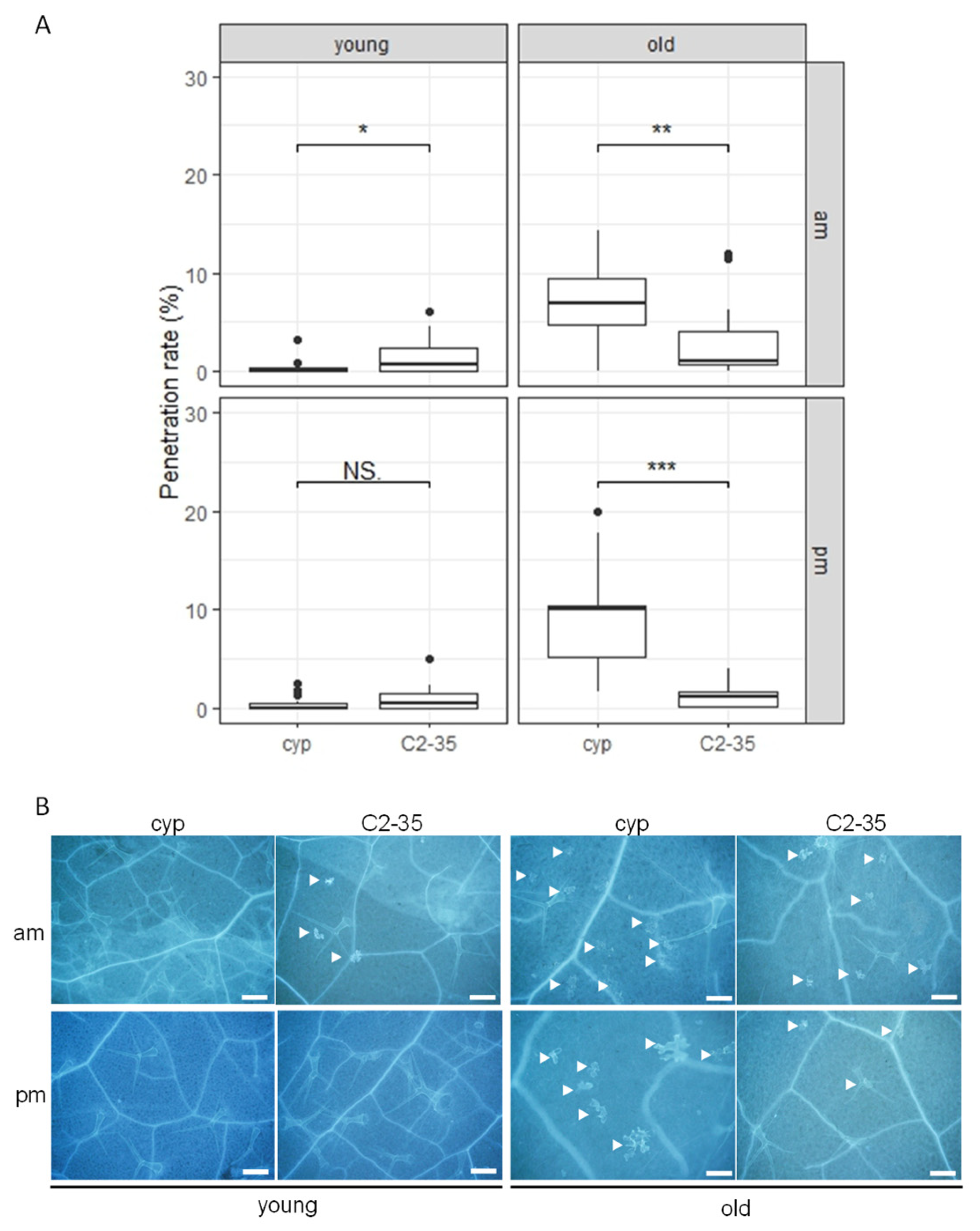
In our study, we first inoculated young rosette leaves of Arabidopsis with P. oryzae in the morning (10:00 a.m., am-inoculation) and assessed cell penetration. We screened 500 rice-FOX Arabidopsis cyp79b2 cyp79b3 lines and identified one line, C2-35, which exhibited decreased NHR to P. oryzae compared to the control plants (Figure 2).
To investigate how NHR is regulated in C2-35 plants depending on leaf age and time, we conducted experiments where we inoculated young and old leaves of the plants with P. oryzae at 10:00 a.m. (am-inoculation) and 5:00 p.m. (pm-inoculation). Our results show that am-inoculation led to decreased NHR in young leaves but not pm-inoculation (Figure 2). However, both am- and pm-inoculation led to increased NHR in old leaves, which is different from what we observed in young leaves (Figure 2). In contrast to cyp79b2 cyp79b3 plants, C2-35 plants exhibited a consistently steady penetration rate to P. oryzae under various inoculation conditions. These findings suggest that C2-35 plants experience a compromise in leaf age- and time-dependent NHR to P. oryzae. We also identified the overexpressed gene in the C2-35 line, which was found to be a full-length rice cDNA AK103160 (Os09g0528200) that encodes rice OsHOX6. These findings indicate that the rice OsHOX6 plays a crucial role in enhancing NHR in old leaves of cyp79b2 cyp79b3 plants, while not having the same effect on young leaves. In conclusion, our results demonstrate that the regulation of NHR by rice OsHOX6 is dependent on both leaf age and time in Arabidopsis.

2.3. Nonhost Resistance to Colletotrichum nymphaeae in Rice-FOX Arabidopsis C2-35 Plants

We inoculated young and old leaves of Arabidopsis plants with C. nymphaeae at 5:00 p.m. and quantified cell penetration. This pathogen, isolated from a Japanese flowering cherry, is nonadapted to Arabidopsis [43]. We found that young and old leaves of cyp79b2 cyp79b3 plants showed significantly increased penetration rates compared to wild-type Col-0 plants (Figure 3A and Figure S1A,B). This result suggests that penetration resistance to C. nymphaeae was severely compromised in cyp79b2 cyp79b3 plants. Further, to assess post-penetration resistance in penetrated epidermal cells, we examined fungal growth in the cells with bright-field microscopy at 72 h post-inoculation (hpi). We measured the severe fungal growth region in the inoculated area of young and old leaves and found that the cyp79b2 cyp79b3 plants exhibited significantly increased severe fungal growth compared to Col-0 plants (Figure S1D). This result indicates that post-penetration resistance to C. nymphaeae was severely compromised in cyp79b2 cyp79b3 plants compared to Col-0 plants. We also noticed that the severe fungal growth of C. nymphaeae in cyp79b2 cyp79b3 plants damaged infected cells and led to the accumulation of autofluorescent material (Figure S1B).
Then, we tested the effectiveness of rice OsHOX6 overexpression against nonadapted fungal pathogen Colletotrichum nymphaeae by examining the C2-35 lines for NHR. The old leaves of C2-35 lines exhibited increased penetration resistance to C. nymphaeae, but not the young leaves (Figure 3A and Figure S1C). This finding indicates that rice OsHOX6 can also provide robust NHR to cyp79b2 cyp79b3 plants and that the effect is age-dependent on the leaves, similar to NHR to P. oryzae in C2-35 plants (Figure 2). In contrast, the growth of infection hyphae in C2-35 plants was comparable to that of cyp79b2 cyp79b3 plants, suggesting the same level of post-penetration resistance between cyp79b2 cyp79b3 and C2-35 plants (Figure S1D).
During the experiment, we observed a significant reduction in the incidence of melanized appressoria in the nonadapted C. nymphaeae, while host-adapted C. higginsianum develops typical melanized appressoria on Arabidopsis. To further understand the infection structure of C. nymphaeae, we classified individual germinated spores into three groups. Class I sporelings developed darkly melanized appressoria, while class II sporelings developed an appressorium with slight pigmentation or one without detectable pigmentation. Lastly, class III sporelings produced a germ tube without a recognizable appressorium or developed a small swollen structure at the hyphal tip (Figure 3B). We noticed that the class III hyphal development resembles hyphal tip-based entry (HTE), previously reported in nonadapted C. gloeosporioides [44].
We analyzed the proportion of appressoria (AP) classes during C. nymphaeae penetration in Arabidopsis. We found that cyp79b2 cyp79b3 and C2-35 plants exhibited a significant decrease in the incidence of class II sporelings (class II, Figure 3C) and a significant increase in class III sporelings (class III, Figure 3C) compared to wild-type Col-0 plants in young and old leaves (Figure 3C). We could not detect any significant differences between cyp79b2 cyp79b3 and C2-35 plants (Figure 3C). This result suggests that overexpression of the rice OsHOX6 gene did not affect the proportion of AP classes during C. nymphaeae penetration in Arabidopsis.

2.4. Host Resistance to Colletotrichum higginsianum in Rice-FOX Arabidopsis C2-35 Plants

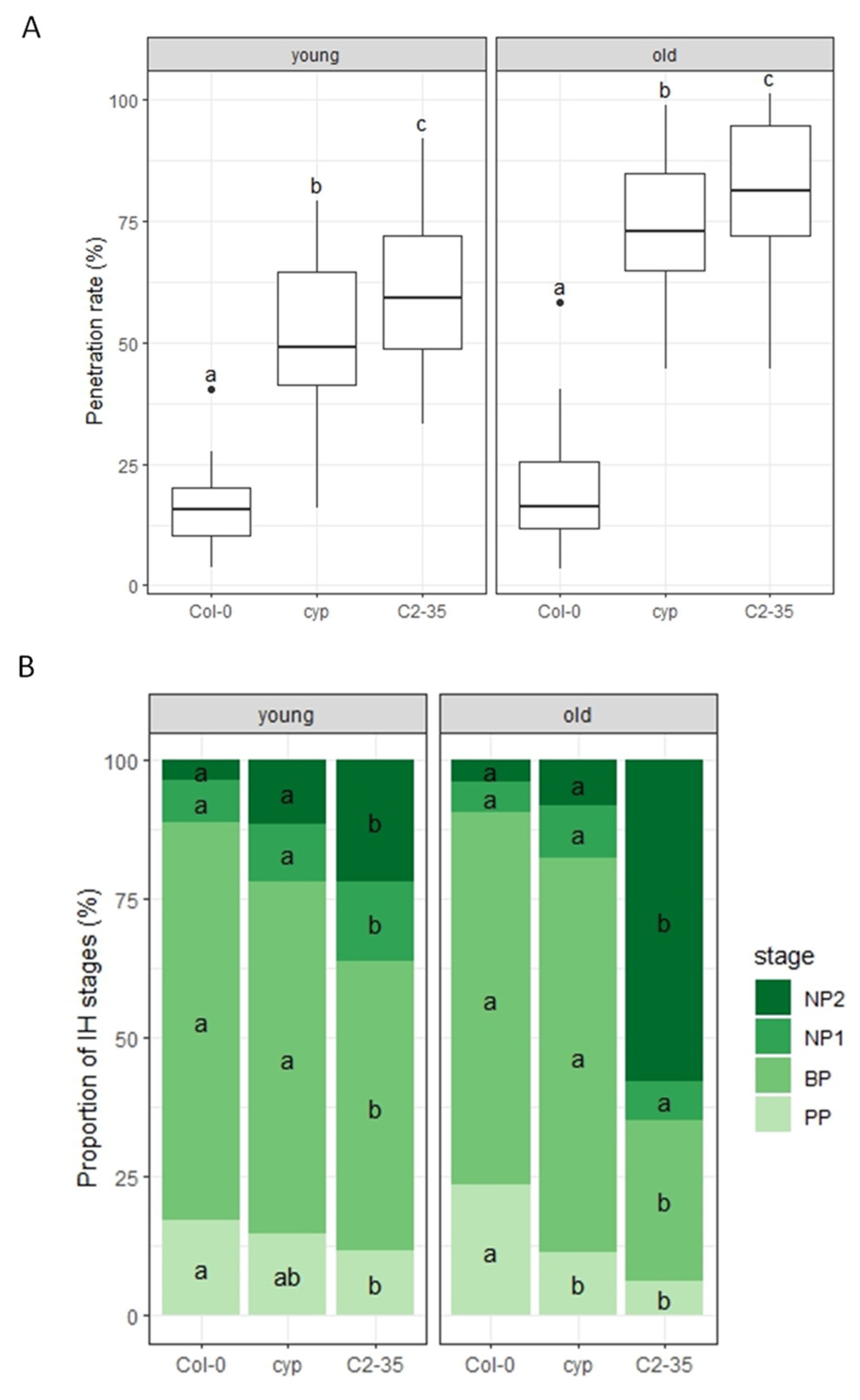
To test if overexpression of the rice OsHOX6 gene could confer resistance to other pathogens, we investigated its effect against the host-adapted C. higginsianum in C2-35 plants. We inoculated conidial suspensions of C. higginsianum on young and old leaves of Arabidopsis plants at 5:00 p.m. We found that both young and old leaves of cyp79b2 cyp79b3 plants exhibited a significant decrease in penetration resistance to C. higginsianum compared to wild-type Col-0 plants (Figure 4A and Figure S2). Further, we found that both young and old leaves of C2-35 plants exhibited a significant decrease in penetration resistance to C. higginsianum compared to cyp79b2 cyp79b3 plants (Figure 4A and Figure S2). These findings suggest that the overexpression of the rice OsHOX6 gene can reduce the penetration resistance of young and old leaves of cyp79b2 cyp79b3 plants against C. higginsianum.
We observed that the host-adapted C. higginsianum forms specialized infection structures, such as melanized appressoria, penetrating hyphae, biotrophic hyphae, and necrotrophic hyphae, during its infection process on Arabidopsis. Based on this, we divided the process into four stages: the penetration phase (PP), biotrophic phase (BP), necrotrophic phase with NH confined within the initially penetrated epidermal cells (NP1), and necrotrophic phase with NH spreading into the surrounding cells (NP2). Next, we analyzed the proportion of infection stages of penetrated sporelings in Arabidopsis. We could not find any significant differences between Col-0 and cyp79b2 cyp79b3 plants, except decreased IH in the PP stage in old leaves of cyp79b2 cyp79b3 plants compared to Col-0 plants (Figure 4B and Figure S2). However, C2-35 plants had significantly higher IH of the NP2 stage in young and old leaves, while significantly lower IH in the BP stage compared to cyp79b2 cyp79b3 plants (Figure 4B and Figure S2). These results indicate that the overexpression of the rice OsHOX6 gene significantly reduced post-penetration resistance to C. higginsianum in young and old leaves of cyp79b2 cyp79b3 plants.

2.5. Host Resistance to C. higginsianum in Col-0 Plants

The rice OsHOX6 gene is a member of the HD-Zip I family, with its closest homologues being AtHB7 (At2g46680) and AtHB12 (At3g61890) in Arabidopsis. The HD-Zip I family has several members across plant species that regulate development in response to environmental changes. For instance, AtHB5, AtHB6, AtHB7, and AtHB12 in Arabidopsis are mainly induced by water deficit, salt, and abscisic acid (ABA) [31]. In this study, we found that the rice OsHOX6 gene plays a role in host resistance (HR) and NHR in rice-FOX Arabidopsis C2-35 plants. This result suggests that AtHB7 and AtHB12 may also regulate resistance responses in Arabidopsis. Furthermore, AtHB7 and AtHB12 are induced by various pathogens according to BAR (Bio Analytic Resource for Plant Biology: http://bar.utoronto.ca/, accessed on 11 November 2023), which also implies their involvement in biotic stress responses. However, the precise functions of AtHB7 and AtHB12 in pathogen attack remain poorly understood.
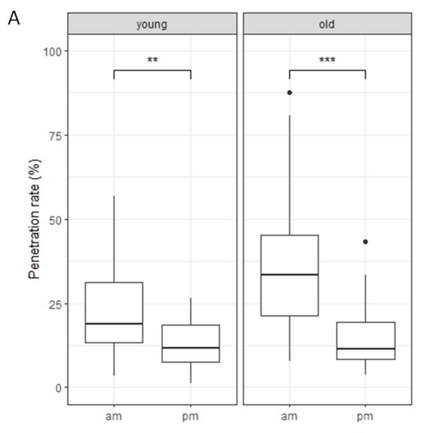
We first investigated the interaction between the host-adapted pathogen, C. higginsianum, and Arabidopsis Col-0 plants. To do so, we inoculated the plants with conidial suspensions of C. higginsianum on young and old leaves of rosettes at two different times, 10:00 a.m. (am-inoculation) and 5:00 p.m. (pm-inoculation). Our findings revealed that Col-0 plants showed a significantly increased penetration rate in young and old leaves after am-inoculation compared to pm-inoculation (Figure 5A). These results suggest that the penetration resistance to C. higginsianum in Col-0 plants is mainly regulated in a time-dependent manner.
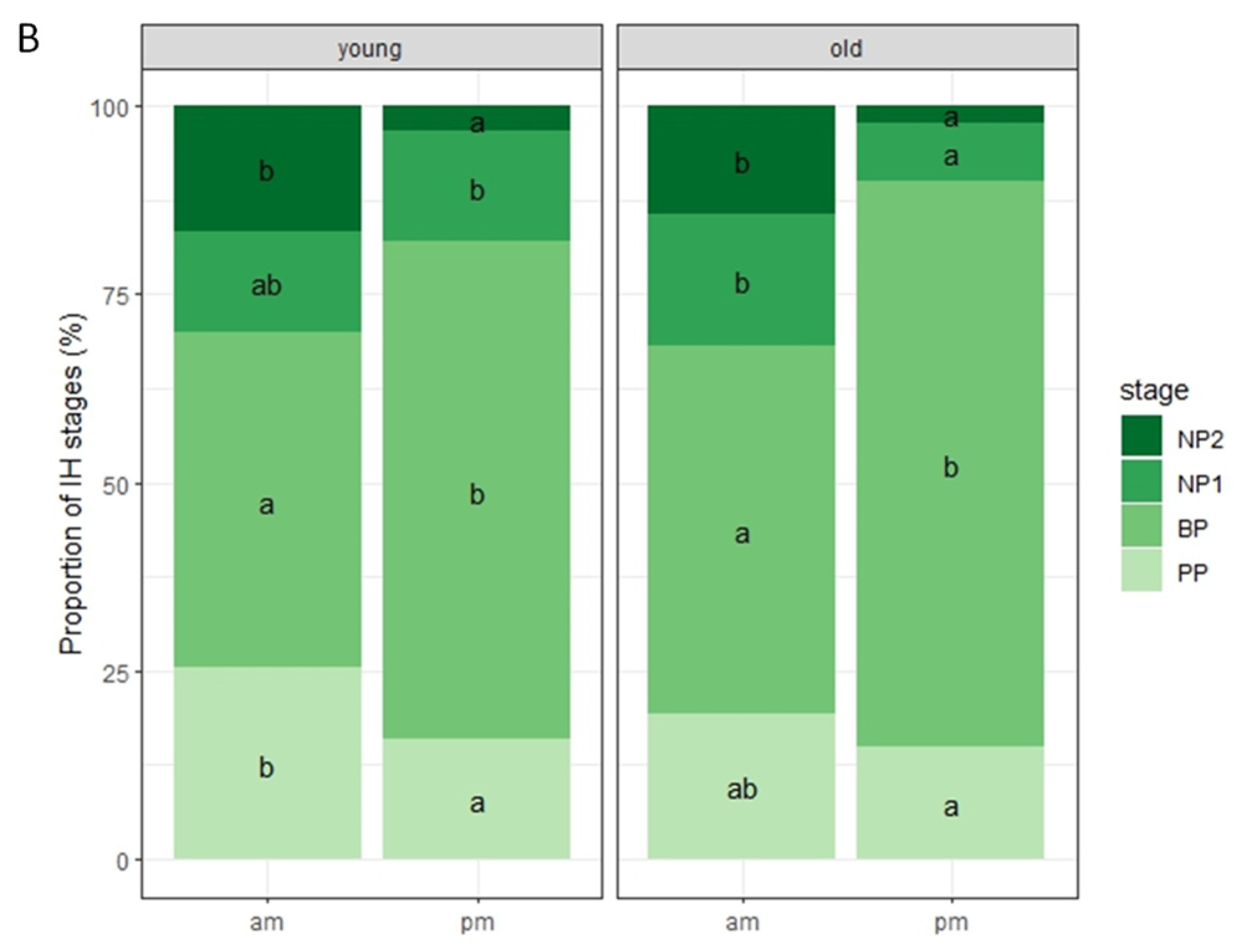
Following this, we analyzed the proportion of IH stages of penetrated sporelings. Our observations showed that young and old leaves of Col-0 plants had significantly increased IH in the NP2 stage following am-inoculation compared to pm-inoculation (Figure 5B). This indicates that post-penetration resistance to C. higginsianum in Col-0 plants is also mainly regulated in a time-dependent manner.

2.6. C. higginsianum Growth in Arabidopsis Mutant Plants

To investigate the role of AtHB7 and AtHB12 in Arabidopsis resistance to C. higginsianum, mutants (athb7 and athb12) and overexpressors of each gene (AT7 and AT12) were analyzed. Arabidopsis plants were inoculated with C. higginsianum conidial suspensions at 10:00 a.m. (am-inoculation) and 5:00 p.m. (pm-inoculation) on young and old leaves of rosettes.
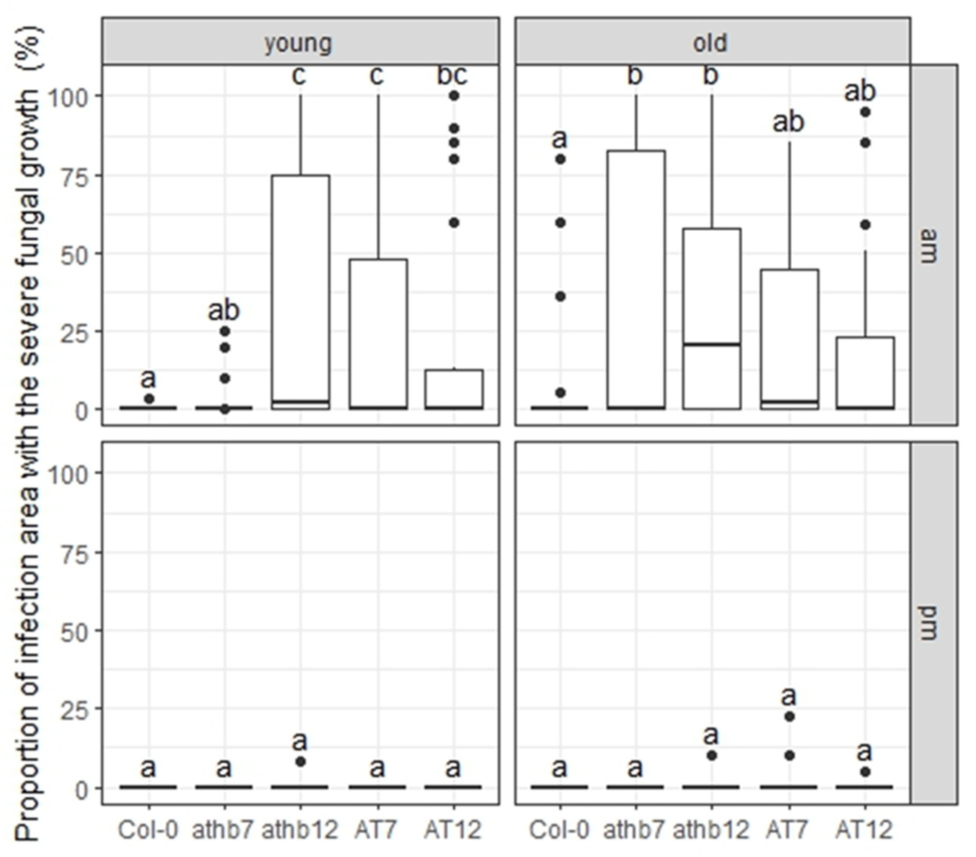
To assess fungal growth in penetrated epidermal cells, we examined each cell with bright-field microscopy at 72 h post-inoculation (hpi). However, in severe fungal growth regions, it was difficult to distinguish individual epidermal cells due to damage caused by C. higginsianum necrotic growth in later phases. Therefore, we first measured the severe fungal growth region in the inoculated area, where penetrated cells were in the NP2 stage, with bright-field microscopy at 72 hpi. After am-inoculation, young leaves of athb12, AT7, and AT12 plants exhibited significantly increased severe fungal growth regions compared to Col-0 plants, and old leaves of athb7 and athb12 plants also showed significantly increased regions compared to Col-0 plants (Figure 6 and Figure S3). However, no severe fungal growth regions were observed in either young and old leaves following pm-inoculation (Figure 6 and Figure S3). These results suggest that AtHB7 and AtHB12 are involved in time-dependent HR to C. higginsianum.

2.7. Host Resistance to C. higginsianum in Arabidopsis Mutant Plants

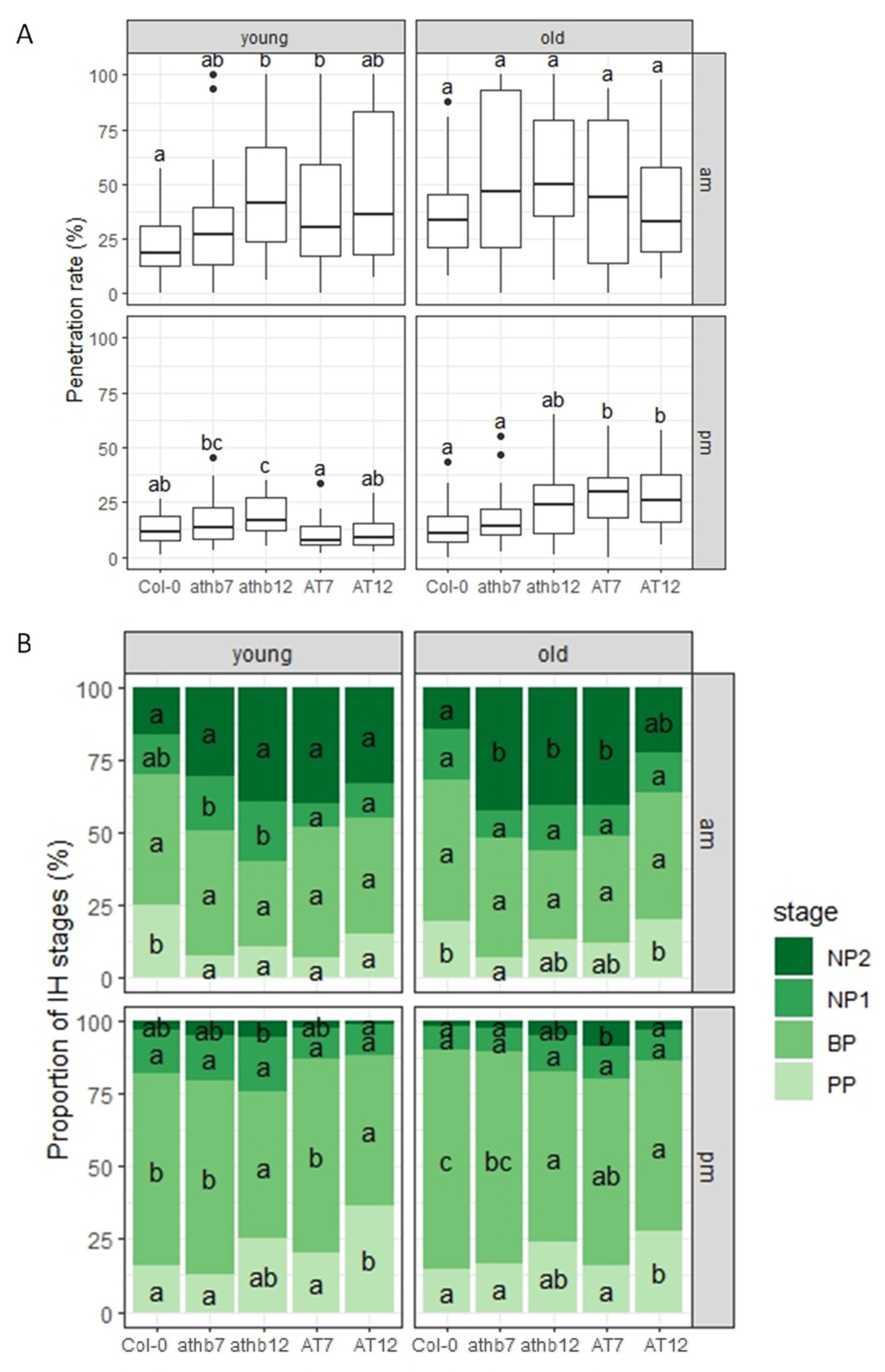
To measure the degree of cell penetration, we conducted an experiment in which we inoculated Arabidopsis plants with conidial suspensions of C. higginsianum at 10:00 a.m. (am-inoculation) and 5:00 p.m. (pm-inoculation) on both young and old leaves of the rosettes of Arabidopsis plants. We examined germinated fungal sporelings that had developed appressoria, and we found that the young leaves of athb12 and AT7 plants showed a significantly increased penetration rate following am-inoculation compared to control Col-0 plants. However, we did not observe any significant differences in the old leaves (Figure 7A and Figure S3). In addition, we found that the young leaves of athb12 plants and the old leaves of AT7 and AT12 plants showed a significantly increased penetration rate following pm-inoculation compared to control Col-0 plants (Figure 7A and Figure S3). These findings suggest that the AtHB7 and AtHB12 genes function in penetration resistance to C. higginsianum in a leaf age- and time-dependent manner.
To quantify post-penetration resistance to C. higginsianum, we investigated the proportion of different IH stages of penetrated sporelings. We found that following am-inoculation, young leaves of mutants (athb7 and athb12) and overexpressors (AT7 and AT12) showed significantly decreased proportions of the PP stage, while old leaves of mutants (athb7 and athb12) and the overexpressor (AT7) showed significantly increased proportions of the NP2 stage compared to control plants (Figure 7B and Figure S3). These results confirmed the severe fungal growth observed in the previous experiment (Figure 6). Furthermore, following pm-inoculation, significant differences in the proportion of the BP stage of penetrated sporelings were observed in young leaves between mutant plants (athb12 and AT12) and control plants, and in old leaves between mutant plants (athb12, AT7, and AT12) and control plants (Figure 7B and Figure S3). These findings suggest that AtHB7 and AtHB12 play a role in post-penetration resistance to C. higginsianum in a leaf age- and time-dependent manner.

3. Discussion

In this research, we discovered that leaf age-dependent nonhost resistance (NHR) to P. oryzae is not only regulated by miR156-dependent but also miR156-independent pathways. Using rice-FOX Arabidopsis thaliana (Arabidopsis) lines, we identified the rice OsHOX6 gene, which imparts strong NHR to P. oryzae and C. nymphaeae in the old leaves of Arabidopsis C2-35 plants. The impact of rice OsHOX6 expression in Arabidopsis is dependent on leaf age. AtHB7 and AtHB12, the Arabidopsis closest homologues of OsHOX6, were then examined in the context of Arabidopsis–C. higginsianum interaction by studying mutants (athb7 and athb12) and transgenic overexpressors (AT7 and AT12). Our findings revealed that AtHB7 and AtHB12 play a role in both penetration and post-penetration resistance to C. higginsianum, in a manner dependent on leaf age and time.
Age-related changes in immunity are observed in both animals and plants. In plants, age-related resistance (ARR) refers to an increase in disease resistance during the maturation of shoots or organs [10]. This trait is particularly significant during the vegetative phase change, which marks the transition from the juvenile to adult stage. ARR contributes to resistance against multiple pathogens, and recent research by Hu et al. has shown that miR156 plays a crucial role in regulating the timing of ARR [15]. The coordinated development of maturation and the acquisition of disease resistance is achieved through the action of miR156-controlled SPL transcription factors with distinct functions. Specifically, a subset of these factors (SPL2, SPL10, and SPL11) promotes resistance by activating key genes involved in defense signaling [15]. On the other hand, Barens et al. has shown that leaf age controls abscisic acid–salicylic acid cross talk independently of vegetative phase change [45]. Our previous research has revealed that penetration resistance to P. oryzae in pen2 plants is significantly decreased in older leaves following pm-inoculation, as compared to young leaves following am-inoculation [25]. This finding suggests that the circadian clock and developmental age play important roles in NHR to P. oryzae in Arabidopsis. In the present study, we have discovered that leaf age-dependent NHR is regulated not only by miR156-dependent pathways but also by miR156-independent pathways (Figure 1).
We have identified the rice OsHOX6 gene as a key regulator of NHR to P. oryzae (Figure 2) and C. nymphaeae (Figure 3) in old leaves of rice-FOX Arabidopsis C2-35 plants. Interestingly, we observed that the expression of OsHOX6 leads to a significant decrease in the HR to C. higginsianum in both young and old leaves of C2-35 plants (Figure 4). Our findings suggest that OsHOX6 can function as a regulator of both HR and NHR in Arabidopsis, and its effect on these processes may vary depending on the type of resistance involved. We also investigated the involvement of the closest homologues of OsHOX6 in Arabidopsis, namely, AtHB7 and AtHB12, in leaf age-dependent resistance responses. Previous studies have detected the expression of AtHB7 and AtHB12 in meristems, root tips, and flowers and have shown that their expression is strongly upregulated under osmotic or drought stress and when young 14-day-old plants are treated with ABA or NaCl [46,47]. It has been suggested that AtHB7 and AtHB12 act as negative developmental regulators in response to drought [47], and AtHB12 has been assigned a role as a regulator of shoot growth in standard growth conditions [48]. However, the ectopic expression of AtHB7 in tomato has been shown to confer drought tolerance to this species [49]. Furthermore, loss-of-function athb7 and athb12 mutants have been found to activate clade A protein phosphatases 2C (PP2C) genes and repress ABA signaling [50]. In our study, we have demonstrated that AtHB7 and AtHB12 play a role in the plant defense response against C. higginsianum in Arabidopsis. Interestingly, the involvement of these TFs is dependent on leaf age and time, as shown in Figure 6 and Figure 7. These findings support the idea that HD-Zip I OsHOX6, AtHB7, and AtHB12 are important in mediating resistance responses in plants. Furthermore, our study highlights the usefulness of rice-FOX Arabidopsis lines in identifying defense-related genes, indicating a shared defense mechanism between monocots and dicots.
The FOX hunting system typically involves the overexpression of full-length cDNA to produce a dominant gain-of-function mutant phenotype. However, in the case of rice-FOX Arabidopsis lines, the resulting plant phenotypes may not accurately reflect the true functions of the overexpressed genes. This is because proteins can be regulated differently in their respective genomic backgrounds due to the difference between amino acid sequences. As a result, overexpressing a foreign gene may lead to different phenotypes than overexpressing the corresponding endogenous gene. Therefore, caution must be exercised when interpreting the phenotypes observed in rice-FOX Arabidopsis lines. To investigate the function of OsHOX6 homologues in Arabidopsis, we examined the role of AtHB7 and AtHB12 in the Arabidopsis–C. higginsianum interaction using knockout mutants (athb7 and athb12) and transgenic plants overexpressing these TFs (AT7 and AT12). In this study, we defined knockout mutants as loss-of-function and overexpressors as gain-of-function. Typically, opposite phenotypes are observed between knockout mutants and overexpressors. However, when we examined the function of AtHB7 and AtHB12, we did not observe such opposite phenotypes. Contrary to expectation, both the knockout mutants and overexpressors displayed similar defects in penetration resistance and post-penetration resistance in the Arabidopsis–C. higginsianum interaction (Figure 6 and Figure 7). Based on our findings, it appears that the regulation of resistance responses by AtHB7 and AtHB12 is a complex process. The unexpected phenotypes we observed suggest that the expression of these genes involves intricate mechanisms. Recent research by Re et al. has shown that AtHB12 is highly expressed during the early development of Arabidopsis, while AtHB7 is expressed more strongly in later developmental stages [36]. This study also demonstrated that these genes have overlapping yet specific roles in various developmental processes. By examining the expression of AtHB7 and AtHB12 in single and double mutants, as well as in transgenic plants expressing these genes, the researchers discovered a complex mechanism that depends on the developmental stage of the plant and in which the expression of AtHB7 and AtHB12 affects each other [36]. Therefore, to ensure precise function of these transcription factors, it is necessary to fine-tune their expression levels with respect to each other. In knockout mutants and overexpressors, disrupting the expression level of one AtHB would also disrupt the expression of the other AtHB. This leads to unexpected regulation of AtHB7 and AtHB12 and makes it difficult to draw precise conclusions from the analysis of these plants. Nonetheless, our results indicate that AtHB7 and AtHB12 play a role in regulating plant immunity in Arabidopsis. To understand the mechanisms involved, it is important to identify the target genes of AtHB7 and AtHB12 in the context of age- and time-dependent regulation in leaves.
In a previous study, we discovered that Arabidopsis plants exhibited a strong NHR to P. oryzae in young leaves after being am-inoculated, while showing a weak NHR in old leaves after being pm-inoculated [25]. However, in our current study, we found that Arabidopsis plants displayed a strong HR to C. higginsianum after being pm-inoculated but a weak HR after being am-inoculated. This time-dependent response is different from that seen in the Arabidopsis–P. oryzae interaction, despite the similar infection mechanisms used by both pathogens. Our study further revealed that AtHB7 and AtHB12 play a role in resistance against C. higginsianum in a time-dependent as well as leaf age-dependent manner, by functioning in both penetration and post-penetration resistance. Additionally, we found that the expression of AtHB7 and AtHB12 exhibits a circadian rhythm (CAST-R: https://nagellab.shinyapps.io/CASTR-v1/, accessed on 11 November 2023). These results suggest that the circadian expression of AtHB7 and AtHB12 could be responsible for regulating time-dependent HR and NHR in Arabidopsis.
To conclude, the resistance of plants to certain pathogens is affected by the age of leaves and the time of inoculation. In our research, we discovered that the expression of OsHOX6, a type of HD-Zip I gene found in rice, can affect NHR against P. oryzae and C. nymphaeae and HR against C. higginsianum, depending on leaf age and time in rice-FOX Arabidopsis C2-35 plants. This implies that the Arabidopsis HD-Zip I genes, AtHB7 and AtHB12, are likely involved in regulating the age- and time-dependent resistance responses in Arabidopsis. Actually, our findings suggest that AtHB7 and AtHB12 are involved in the age- and time-dependent regulation of HR against C. higginsianum. Prior studies have indicated that AtHB7 and AtHB12 are associated with the development and responses to abiotic stress in Arabidopsis [36]. Consequently, it would be fascinating to explore how these genes balance the trade-offs between development, abiotic stress, and biotic stress responses in Arabidopsis. To fully comprehend the genetic and mechanistic requirements of AtHB7 and AtHB12 in Arabidopsis, further investigations will be needed. Our study’s insights into the role of AtHB7 and AtHB12 in plant immunity may be used to boost defense responses in plants.

4. Materials and Methods

4.1. Plant Materials and Growth Conditions

The plants used in this study were Col-0 (wild-type), pen2 [18], pen2 35S::miR156a, cyp79b2 cyp79b3 [20], athb7, athb12, AT7 (35S::AtHB7), and AT12 (35S::AtHB12) [36], which are all on the Col-0 background. We used pen2 and 35S::miR156a [11] plants to generate pen2 35S::miR156a plants. Arabidopsis thaliana (Arabidopsis) plants were grown on Murashige and Skoog plates in a growth room for three weeks in short-day conditions (9:15 L:D) at 22 °C (in 100 μmol m−2s−1 fluorescent illumination). Then, the plants were transferred to soil and grown in a growth chamber for 4 weeks, where they continued to grow in short-day conditions (9:15 L:D) at 22 °C (in 100 μmol m−2s−1 fluorescent illumination).

4.2. Fungal Strains and Media

We obtained Pyricularia oryzae isolate Hoku 1 (race 007) from H. Koga (Ishikawa Prefectural University). Colletotrichum higginsianum (MAFF305635) and Colletotrichum nymphaeae (MAFF240037) were obtained from the Ministry of Agriculture, Forestry and Fisheries GenBank, Japan. P. oryzae culture was maintained on oatmeal medium at 25 °C in the dark. Cultures of fungal isolates of Colletotrichum were maintained on PDA medium at 25 °C in the dark. For inoculation, P. oryzae and C. nymphaeae were cultured under a 9 h light/15 h dark cycle.

4.3. Fungal Inoculation

To measure the penetration rates of fungal pathogens, a conidial suspension of each fungus (P. oryzae, 5 × 10⁴ conidia/mL; Colletrichum, 1 × 10⁵ conidia/mL) was inoculated onto leaves (young leaf, leaf number 13; old leaf, leaf number 8) of rosettes on Arabidopsis (i.e., leaves numbered from oldest to youngest) in the morning (10:00 a.m., am-inoculation) and the evening (5:00 p.m., pm-inoculation). Inoculated plants were maintained in a growth chamber with saturating humidity in short-day conditions (9 h:15 h light:dark) at 22 °C (in 100 μmol m−2s−1 fluorescent illumination). Inoculated leaves were harvested at 72 h post-inoculation (hpi).
To quantify cell penetration for P. oryze and C. higginsianum, we examined germinated fungal sporelings that had developed appressoria (six leaves from six independent plants per experiment and genotype). We evaluated a minimum of 100 appressoria/leaves. We detected successful penetration of fungal pathogens by observing autofluorescence or hyphal elongation at infection sites with fluorescence and bright-field microscopy. Each plant genotype was quantified in three independent experiments.
To quantify cell penetration for C. nymphaeae, we examined germinated fungal sporelings. We also examined the appressoria classes that had penetrated into Arabidopsis leaves. We detected successful penetration of C. nymphaeae by observing autofluorescence or hyphal elongation at infection sites with fluorescence and bright-field microscopy. Each plant genotype was quantified in three independent experiments.
To quantify severe fungal growth region for C. nymphaeae and C. higginsianum in Arabidopsis leaves, we examined the inoculated area and measured the severe fungal growth region with bright-field microscopy (six leaves from six independent plants per experiment and genotype). Each plant genotype was quantified in three independent experiments.

4.4. Rice-FOX Arabidopsis cyp79b2 cyp79b3 Lines and P. oryzae Screening

The Agrobacterium library of rice full-length cDNAs was obtained from RIKEN [38]. Arabidopsis cyp79b2 cyp79b3 plants were transformed using the Agrobacterium library, and the transformed cyp79b2 cyp79b3 plants expressing rice full-length cDNAs (rice-FOX Arabidopsis cyp79b2 cyp79b3 lines) were generated. We inoculated the rice-FOX Arabidopsis cyp79b2 cyp79b3 lines with P. oryzae by applying 5 μL droplets (5 × 10⁴ spores/mL) of P. oryzae onto young leaves (leaf number 13) of rosettes on Arabidopsis (i.e., leaves numbered from oldest to youngest) in the morning (10:00 a.m.). Then, inoculated plants were maintained in a growth chamber with saturating humidity in short-day conditions (9 h:15 h light:dark) at 22 °C (in 100 μmol m−2s−1 fluorescent illumination). Inoculated leaves were harvested at 72 hpi. To quantify cell penetration, we examined germinated fungal sporelings that had developed appressoria. We evaluated a minimum of 100 appressoria/leaves. We detected successful penetration of P. oryzae by observing autofluorescence or hyphal elongation at infection sites with fluorescence and bright-field microscopy.
We identified the candidate NHR-related lines with a penetration rate different from the rate of control cyp79b2 cyp79b3 plants from a screen of approximately 500 rice-FOX Arabidopsis cyp79b2 cyp79b3 lines. Screening of the candidate NHR-related lines was repeated thrice for verification. For further examination, the selected candidate lines were inoculated with P. oryzae at 10:00 a.m. (am-inoculation) and 5:00 p.m. (pm-inoculation) on young and old leaves (young leaf, leaf number 13; old leaf, leaf number 8) of rosettes on Arabidopsis. Each plant was quantified in three independent experiments.

Supplementary Materials

The following supporting information can be downloaded at: https://www.mdpi.com/article/10.3390/ijms242216356/s1.

Author Contributions

A.I. designed the research. N.M., F.M., T.N., A.F., H.I., Y.K. and A.I. performed the research. A.I. wrote the manuscript. All authors have read and agreed to the published version of the manuscript.

Funding

This work was supported by JSPS KAKENHI (Grant No. 22K05656).

Informed Consent Statement

Not applicable.

Data Availability Statement

Data are contained within the article and supplementary materials.

Acknowledgments

We acknowledge the Arabidopsis Biological Resource Center for providing seeds of 35S::miR156a. We thank R. Chan (Instituto de Agrobiotecnología del Litoral) for providing seeds of athb7, athb12, AT7 (35S::AtHB7), and AT12 (35S::AtHB12), H. Koga (Ishikawa Prefectural University) for providing the P. oryzae isolate, and P. Shulze-Lefert (Max Planck Institute for Plant Breeding Research) for seeds of pen2. We also thank researchers of the Institute of Physical and Chemical Research (RIKEN), Research Institute for Biological Sciences (RIBS), and National Agriculture and Food Research Organization (NARO), who constructed and provided us with the rice full-length cDNA library for the FOX hunting system.

Conflicts of Interest

The authors declare no conflict of interest.

References

  1. Scholthof, K.B. The disease triangle: Pathogens, the environment and society. Nat. Rev. Microbiol. 2007, 5, 152–156. [Google Scholar] [CrossRef] [PubMed]
  2. Roden, L.C.; Ingle, R.A. Lights, rhythms, infection: The role of light and the circadian clock in determining the outcome of plant-pathogen interactions. Plant Cell 2009, 21, 2546–2552. [Google Scholar] [CrossRef] [PubMed]
  3. Seo, P.J.; Mas, P. STRESSing the role of the plant circadian clock. Trends Plant Sci. 2015, 20, 230–237. [Google Scholar] [CrossRef] [PubMed]
  4. Bhardwaj, V.; Meier, S.; Petersen, L.N.; Ingle, R.A.; Roden, L.C. Defence responses of Arabidopsis thaliana to infection by Pseudomonas syringae are regulated by the circadian clock. PLoS ONE 2011, 6, e26968. [Google Scholar] [CrossRef]
  5. Wang, W.; Barnaby, J.Y.; Tada, Y.; Li, H.; Tor, M.; Caldelari, D.; Lee, D.U.; Fu, X.D.; Dong, X. Timing of plant immune responses by a central circadian regulator. Nature 2011, 470, 110–114. [Google Scholar] [CrossRef]
  6. Zhang, C.; Xie, Q.; Anderson, R.G.; Ng, G.; Seitz, N.C.; Peterson, T.; McClung, C.R.; McDowell, J.M.; Kong, D.; Kwak, J.M.; et al. Crosstalk between the circadian clock and innate immunity in Arabidopsis. PLoS Pathog. 2013, 9, e1003370. [Google Scholar] [CrossRef]
  7. Develey-Riviere, M.P.; Galiana, E. Resistance to pathogens and host developmental stage: A multifaceted relationship within the plant kingdom. New Phytol. 2007, 175, 405–416. [Google Scholar] [CrossRef]
  8. Thomas, H.; Ougham, H.J.; Wagstaff, C.; Stead, A.D. Defining senescence and death. J. Exp. Bot. 2003, 54, 1127–1132. [Google Scholar] [CrossRef]
  9. Rankenberg, T.; Geldhof, B.; van Veen, H.; Holsteens, K.; Van de Poel, B.; Sasidharan, R. Age-Dependent Abiotic Stress Resilience in Plants. Trends Plant Sci. 2021, 26, 692–705. [Google Scholar] [CrossRef]
  10. Hu, L.; Yang, L. Time to Fight: Molecular Mechanisms of Age-Related Resistance. Phytopathology 2019, 109, 1500–1508. [Google Scholar] [CrossRef]
  11. Wu, G.; Poethig, R.S. Temporal regulation of shoot development in Arabidopsis thaliana by miR156 and its target SPL3. Development 2006, 133, 3539–3547. [Google Scholar] [CrossRef] [PubMed]
  12. Wang, J.W.; Czech, B.; Weigel, D. miR156-regulated SPL transcription factors define an endogenous flowering pathway in Arabidopsis thaliana. Cell 2009, 138, 738–749. [Google Scholar] [CrossRef] [PubMed]
  13. Li, P.; Lu, Y.J.; Chen, H.; Day, B. The Lifecycle of the Plant Immune System. CRC Crit. Rev. Plant Sci. 2020, 39, 72–100. [Google Scholar] [CrossRef]
  14. Xu, Y.P.; Lv, L.H.; Xu, Y.J.; Yang, J.; Cao, J.Y.; Cai, X.Z. Leaf stage-associated resistance is correlated with phytohormones in a pathosystem-dependent manner. J. Integr. Plant Biol. 2018, 60, 703–722. [Google Scholar] [CrossRef] [PubMed]
  15. Hu, L.; Qi, P.; Peper, A.; Kong, F.; Yao, Y.; Yang, L. Distinct function of SPL genes in age-related resistance in Arabidopsis. PLoS Pathog. 2023, 19, e1011218. [Google Scholar] [CrossRef] [PubMed]
  16. Maeda, K.; Houjyou, Y.; Komatsu, T.; Hori, H.; Kodaira, T.; Ishikawa, A. AGB1 and PMR5 contribute to PEN2-mediated preinvasion resistance to Magnaporthe oryzae in Arabidopsis thaliana. Mol. Plant-Microbe Interact. 2009, 22, 1331–1340. [Google Scholar] [CrossRef]
  17. Nakao, M.; Nakamura, R.; Kita, K.; Inukai, R.; Ishikawa, A. Non-host resistance to penetration and hyphal growth of Magnaporthe oryzae in Arabidopsis. Sci. Rep. 2011, 1, 171. [Google Scholar] [CrossRef]
  18. Lipka, V.; Dittgen, J.; Bednarek, P.; Bhat, R.; Wiermer, M.; Stein, M.; Landtag, J.; Brandt, W.; Rosahl, S.; Scheel, D.; et al. Pre- and postinvasion defenses both contribute to nonhost resistance in Arabidopsis. Science 2005, 310, 1180–1183. [Google Scholar] [CrossRef]
  19. Pastorczyk, M.; Bednarek, P. The Function of Glucosinolates and Related Metabolites in Plant Innate Immunity. Glucosinolates 2016, 80, 171–198. [Google Scholar] [CrossRef]
  20. Zhao, Y.D.; Hull, A.K.; Gupta, N.R.; Goss, K.A.; Alonso, J.; Ecker, J.R.; Normanly, J.; Chory, J.; Celenza, J.L. Trp-dependent auxin biosynthesis in Arabidopsis: Involvement of cytochrome P450s CYP79B2 and CYP79B3. Genes Dev. 2002, 16, 3100–3112. [Google Scholar] [CrossRef]
  21. Sanchez-Vallet, A.; Ramos, B.; Bednarek, P.; Lopez, G.; Pislewska-Bednarek, M.; Schulze-Lefert, P.; Molina, A. Tryptophan-derived secondary metabolites in Arabidopsis thaliana confer non-host resistance to necrotrophic Plectosphaerella cucumerina fungi. Plant J. 2010, 63, 115–127. [Google Scholar] [CrossRef]
  22. Hiruma, K.; Fukunaga, S.; Bednarek, P.; Pislewska-Bednarek, M.; Watanabe, S.; Narusaka, Y.; Shirasu, K.; Takano, Y. Glutathione and tryptophan metabolism are required for Arabidopsis immunity during the hypersensitive response to hemibiotrophs. Proc. Natl. Acad. Sci. USA 2013, 110, 9589–9594. [Google Scholar] [CrossRef]
  23. Kosaka, A.; Pastorczyk, M.; Pislewska-Bednarek, M.; Nishiuchi, T.; Ono, E.; Suemoto, H.; Ishikawa, A.; Frerigmann, H.; Kaido, M.; Mise, K.; et al. Tryptophan-derived metabolites and BAK1 separately contribute to Arabidopsis postinvasive immunity against Alternaria brassicicola. Sci. Rep. 2021, 11, 1488. [Google Scholar] [CrossRef]
  24. Shimizu, S.; Yamauchi, Y.; Ishikawa, A. Photoperiod Following Inoculation of Arabidopsis with Pyricularia oryzae (syn. Magnaporthe oryzae) Influences on the Plant-Pathogen Interaction. Int. J. Mol. Sci. 2021, 22, 5004. [Google Scholar] [CrossRef]
  25. Yamauchi, Y.; Makihara, M.; Ishikawa, A. Leaf age and time of inoculation contribute to nonhost resistance to Pyricularia oryzae in Arabidopsis thaliana. Plant Biotechnol. 2017, 34, 207–210. [Google Scholar] [CrossRef] [PubMed]
  26. Yamaura, S.; Yamauchi, Y.; Makihara, M.; Yamashino, T.; Ishikawa, A. CCA1 and LHY contribute to nonhost resistance to Pyricularia oryzae (syn. Magnaporthe oryzae) in Arabidopsis thaliana. Biosci. Biotechnol. Biochem. 2020, 84, 76–84. [Google Scholar] [CrossRef] [PubMed]
  27. Maeda, N.; Noguchi, T.; Nakamichi, N.; Suzuki, T.; Ishikawa, A. Epidermal CCA1 and PMR5 contribute to nonhost resistance in Arabidopsis. Biosci. Biotechnol. Biochem. 2022, 86, 1623–1630. [Google Scholar] [CrossRef] [PubMed]
  28. Yan, Y.Q.; Yuan, Q.F.; Tang, J.T.; Huang, J.B.; Hsiang, T.; Wei, Y.D.; Zheng, L. Colletotrichum higginsianum as a Model for Understanding Host-Pathogen Interactions: A Review. Int. J. Mol. Sci. 2018, 19, 2142. [Google Scholar] [CrossRef]
  29. Jin, J.P.; Tian, F.; Yang, D.C.; Meng, Y.Q.; Kong, L.; Luo, J.C.; Gao, G. PlantTFDB 4.0: Toward a central hub for transcription factors and regulatory interactions in plants. Nucleic Acids Res. 2017, 45, D1040–D1045. [Google Scholar] [CrossRef]
  30. Ariel, F.D.; Manavella, P.A.; Dezar, C.A.; Chan, R.L. The true story of the HD-Zip family. Trends Plant Sci. 2007, 12, 419–426. [Google Scholar] [CrossRef] [PubMed]
  31. Perotti, M.F.; Ribone, P.A.; Chan, R.L. Plant transcription factors from the homeodomain-leucine zipper family I. Role in development and stress responses. IUBMB Life 2017, 69, 280–289. [Google Scholar] [CrossRef]
  32. Liu, Y.; Xu, J.; Guo, S.; Yuan, X.; Zhao, S.; Tian, H.; Dai, S.; Kong, X.; Ding, Z. AtHB7/12 Regulate Root Growth in Response to Aluminum Stress. Int. J. Mol. Sci. 2020, 21, 4080. [Google Scholar] [CrossRef]
  33. Agalou, A.; Purwantomo, S.; Overnas, E.; Johannesson, H.; Zhu, X.; Estiati, A.; de Kam, R.J.; Engstrom, P.; Slamet-Loedin, I.H.; Zhu, Z.; et al. A genome-wide survey of HD-Zip genes in rice and analysis of drought-responsive family members. Plant Mol. Biol. 2008, 66, 87–103. [Google Scholar] [CrossRef]
  34. Zhang, S.; Haider, I.; Kohlen, W.; Jiang, L.; Bouwmeester, H.; Meijer, A.H.; Schluepmann, H.; Liu, C.M.; Ouwerkerk, P.B. Function of the HD-Zip I gene Oshox22 in ABA-mediated drought and salt tolerances in rice. Plant Mol. Biol. 2012, 80, 571–585. [Google Scholar] [CrossRef]
  35. Bhattacharjee, A.; Sharma, R.; Jain, M. Over-Expression of OsHOX24 Confers Enhanced Susceptibility to Abiotic Stresses in Transgenic Rice via Modulating Stress-Responsive Gene Expression. Front. Plant Sci. 2017, 8, 628. [Google Scholar] [CrossRef] [PubMed]
  36. Re, D.A.; Capella, M.; Bonaventure, G.; Chan, R.L. Arabidopsis AtHB7 and AtHB12 evolved divergently to fine tune processes associated with growth and responses to water stress. BMC Plant Biol. 2014, 14, 150. [Google Scholar] [CrossRef]
  37. Ichikawa, T.; Nakazawa, M.; Kawashima, M.; Iizumi, H.; Kuroda, H.; Kondou, Y.; Tsuhara, Y.; Suzuki, K.; Ishikawa, A.; Seki, M.; et al. The FOX hunting system: An alternative gain-of-function gene hunting technique. Plant J. 2006, 48, 974–985. [Google Scholar] [CrossRef]
  38. Kondou, Y.; Higuchi, M.; Takahashi, S.; Sakurai, T.; Ichikawa, T.; Kuroda, H.; Yoshizumi, T.; Tsumoto, Y.; Horii, Y.; Kawashima, M.; et al. Systematic approaches to using the FOX hunting system to identify useful rice genes. Plant J. 2009, 57, 883–894. [Google Scholar] [CrossRef] [PubMed]
  39. Yokotani, N.; Ichikawa, T.; Kondou, Y.; Matsui, M.; Hirochika, H.; Iwabuchi, M.; Oda, K. Expression of rice heat stress transcription factor OsHsfA2e enhances tolerance to environmental stresses in transgenic Arabidopsis. Planta 2008, 227, 957–967. [Google Scholar] [CrossRef] [PubMed]
  40. Yokotani, N.; Ichikawa, T.; Kondou, Y.; Maeda, S.; Iwabuchi, M.; Mori, M.; Hirochika, H.; Matsui, M.; Oda, K. Overexpression of a rice gene encoding a small C2 domain protein OsSMCP1 increases tolerance to abiotic and biotic stresses in transgenic Arabidopsis. Plant Mol. Biol. 2009, 71, 391–402. [Google Scholar] [CrossRef]
  41. Yokotani, N.; Ichikawa, T.; Kondou, Y.; Iwabuchi, M.; Matsui, M.; Hirochika, H.; Oda, K. Role of the rice transcription factor JAmyb in abiotic stress response. J. Plant Res. 2013, 126, 131–139. [Google Scholar] [CrossRef] [PubMed]
  42. Dubouzet, J.G.; Maeda, S.; Sugano, S.; Ohtake, M.; Hayashi, N.; Ichikawa, T.; Kondou, Y.; Kuroda, H.; Horii, Y.; Matsui, M.; et al. Screening for resistance against Pseudomonas syringae in rice-FOX Arabidopsis lines identified a putative receptor-like cytoplasmic kinase gene that confers resistance to major bacterial and fungal pathogens in Arabidopsis and rice. Plant Biotechnol. J. 2011, 9, 466–485. [Google Scholar] [CrossRef]
  43. Irieda, H.; Takano, Y. Epidermal chloroplasts are defense-related motile organelles equipped with plant immune components. Nat. Commun. 2021, 12, 2739. [Google Scholar] [CrossRef] [PubMed]
  44. Hiruma, K.; Onozawa-Komori, M.; Takahashi, F.; Asakura, M.; Bednarek, P.; Okuno, T.; Schulze-Lefert, P.; Takano, Y. Entry mode-dependent function of an indole glucosinolate pathway in Arabidopsis for nonhost resistance against anthracnose pathogens. Plant Cell 2010, 22, 2429–2443. [Google Scholar] [CrossRef] [PubMed]
  45. Berens, M.L.; Wolinska, K.W.; Spaepen, S.; Ziegler, J.; Nobori, T.; Nair, A.; Kruler, V.; Winkelmuller, T.M.; Wang, Y.; Mine, A.; et al. Balancing trade-offs between biotic and abiotic stress responses through leaf age-dependent variation in stress hormone cross-talk. Proc. Natl. Acad. Sci. USA 2019, 116, 2364–2373. [Google Scholar] [CrossRef] [PubMed]
  46. Lee, Y.H.; Chun, J.Y. A new homeodomain-leucine zipper gene from Arabidopsis thaliana induced by water stress and abscisic acid treatment. Plant Mol. Biol. 1998, 37, 377–384. [Google Scholar] [CrossRef]
  47. Olsson, A.S.; Engstrom, P.; Soderman, E. The homeobox genes ATHB12 and ATHB7 encode potential regulators of growth in response to water deficit in Arabidopsis. Plant Mol. Biol. 2004, 55, 663–677. [Google Scholar] [CrossRef]
  48. Son, O.; Hur, Y.S.; Kim, Y.K.; Lee, H.J.; Kim, S.; Kim, M.R.; Nam, K.H.; Lee, M.S.; Kim, B.Y.; Park, J.; et al. ATHB12, an ABA-inducible homeodomain-leucine zipper (HD-Zip) protein of Arabidopsis, negatively regulates the growth of the inflorescence stem by decreasing the expression of a gibberellin 20-oxidase gene. Plant Cell Physiol. 2010, 51, 1537–1547. [Google Scholar] [CrossRef]
  49. Mishra, K.B.; Iannacone, R.; Petrozza, A.; Mishra, A.; Armentano, N.; La Vecchia, G.; Trtilek, M.; Cellini, F.; Nedbal, L. Engineered drought tolerance in tomato plants is reflected in chlorophyll fluorescence emission. Plant Sci. 2012, 182, 79–86. [Google Scholar] [CrossRef]
  50. Valdes, A.E.; Overnas, E.; Johansson, H.; Rada-Iglesias, A.; Engstrom, P. The homeodomain-leucine zipper (HD-Zip) class I transcription factors ATHB7 and ATHB12 modulate abscisic acid signalling by regulating protein phosphatase 2C and abscisic acid receptor gene activities. Plant Mol. Biol. 2012, 80, 405–418. [Google Scholar] [CrossRef]
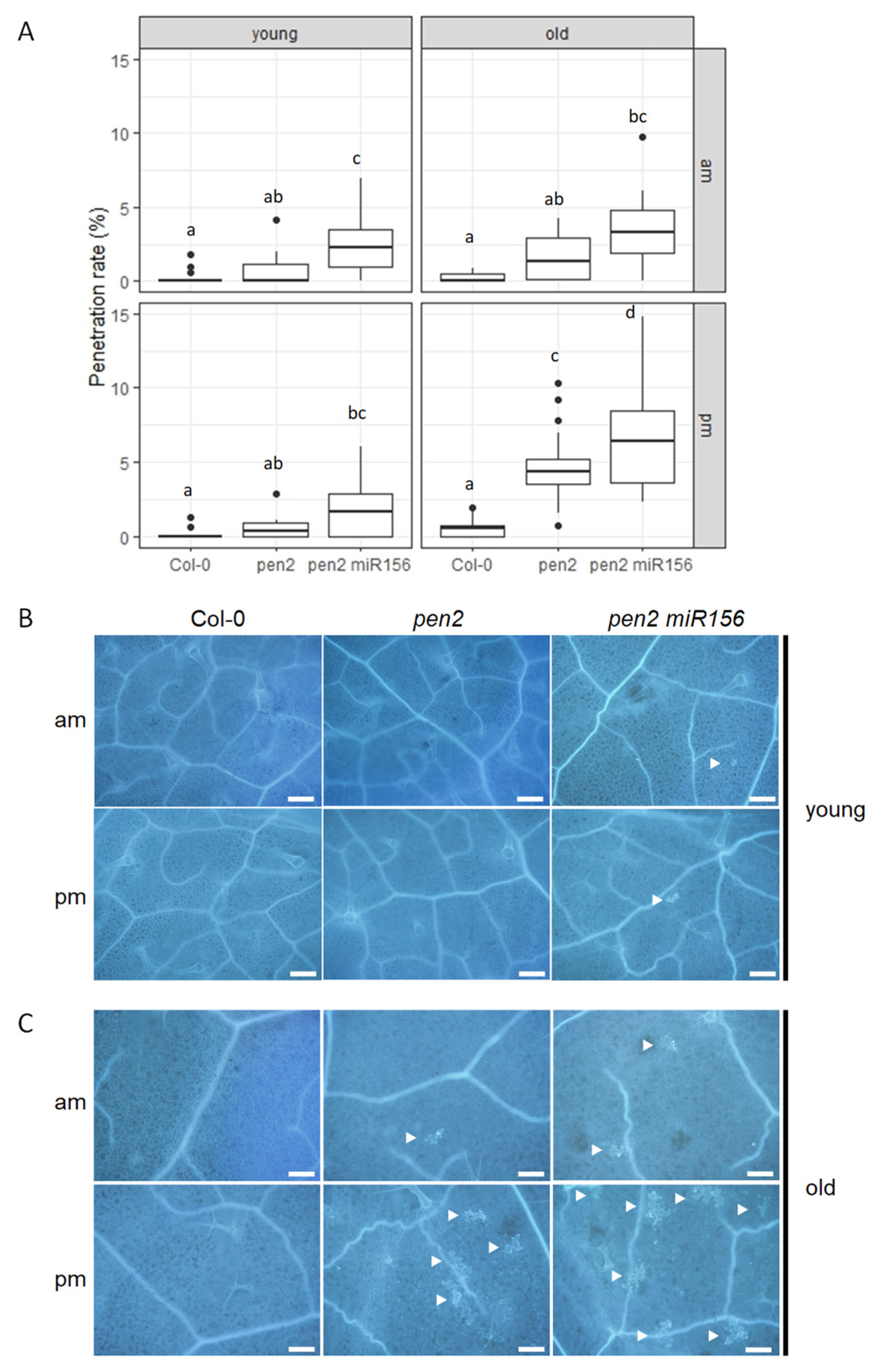
Figure 1. Nonhost resistance to Pyricularia oryzae is regulated by miR156-dependent and miR156-independent pathways in Arabidopsis thaliana (Arabidopsis). (A) Penetration rate of P. oryzae into Col-0, pen2, and pen2 35S::miR156a (pen2 miR156) plants at 72 h post-inoculation (hpi) expressed as the percentage of the total number of infection sites. Arabidopsis plants were inoculated at 10:00 a.m. (am) and 5:00 p.m. (pm) on young and old leaves. Values are from three independent experiments, each containing six biological replicates. Significantly different statistical groups of genotypes indicated by the analyses of variance (Tukey’s test; p < 0.05) are shown with lowercase letters in each inoculation time. (B) Col-0, pen2, and pen2 35S::miR156a (pen2 miR156) plants were inoculated with P. oryzae at 10:00 a.m. (am) and 5:00 p.m. (pm) on young leaves. (C) Col-0, pen2, and pen2 35S::miR156a (pen2 miR156) plants were inoculated with P. oryzae at 10:00 a.m. (am) and 5:00 p.m. (pm) on old leaves. Cell-death-associated autofluorescence at infection sties of Arabidopsis plants at 72 hpi as visualized by fluorescence microscopy. The white arrowhead indicates a penetrated epidermal cell. Bars, 0.2 mm.
Ijms 24 16356 g001
Figure 2. C2-35 plants show increased NHR to P. oryzae in old leaves. (A) Penetration rate of P. oryzae into cyp79b2 cyp79b3 (cyp) and C2-35 plants at 72 hpi expressed as the percentage of the total number of infection sites. Arabidopsis plants were inoculated at 10:00 a.m. (am) and 5:00 p.m. (pm) on young and old leaves. Values are from three independent experiments, each containing six biological replicates. The Student’s t-test was used for statistical analysis; NS, not significant; *, p < 0.05; **, p < 0.01; ***, p < 0.001. (B) Arabidopsis cyp79b2 cyp79b3 (cyp) and C2-35 plants were inoculated with P. oryzae at 10:00 a.m. (am) and 5:00 p.m. (pm) on young and old leaves. Cell-death-associated autofluorescence at infection sites of Arabidopsis plants at 72 hpi as visualized by fluorescence microscopy. The white arrowhead indicates a penetrated epidermal cell. Bars, 0.2 mm.
Ijms 24 16356 g002
Figure 3. C2-35 plants show increased NHR to C. nymphaeae in old leaves. (A) Penetration resistance to C. nymphaeae in C2-35 plants. Penetration rate of C. nymphaeae into Col-0, cyp79b2 cyp79b3 (cyp), and C2-35 plants at 72 hpi expressed as the percentage of the total number of infection sites. Arabidopsis plants were inoculated at 5:00 p.m. on young and old leaves. Values are from three independent experiments, each containing six biological replicates. Significantly different statistical groups of genotypes indicated by the analyses of variance (Tukey’s test; p < 0.05) are shown with lowercase letters. (B) Micrographs showing the development of C. nymphaeae categorized to different classes (class I–III). Conidial suspensions of C. nymphaeae were inoculated on Arabidopsis plants, and penetrated conidia were examined at 72 hpi. Class I, well-melanized appressoria; Class II, slight melanized appressoria or appressoria without detectable pigmentation; Class III, tiny appressoria without detectable pigmentation or penetrated conidia without swollen structures. Bars, 10 μm. Black arrowhead, appressorium; white arrowhead, penetration site of class III. (C) Classification of penetrated C. nymphaeae appressoria (AP) development. Conidial suspensions of C. nymphaeae were inoculated on Col-0, cyp79b2 cyp79b3 (cyp), and C2-35 plants, and penetrated conidia were examined at 72 hpi. Class I, well-melanized appressoria; Class II, slight melanized appressoria or appressoria without detectable pigmentation; Class III, tiny appressoria without detectable pigmentation or penetrated conidia without swollen structures. Values are expressed as mean from three independent experiments, each containing six biological replicates. Significantly different statistical groups of genotypes indicated by the analyses of variance (Tukey’s test; p < 0.05) are shown with lowercase letters.
Ijms 24 16356 g003
Figure 4. C2-35 plants show decreased host resistance to C. higginsianum. (A) Penetration resistance to C. higginsianum in C2-35 plants. Penetration rate of C. higginsianum into Col-0, cyp79b2 cyp79b3 (cyp), and C2-35 plants at 72 hpi expressed as the percentage of the total number of infection sites. Arabidopsis plants were inoculated at 5:00 p.m. on young and old leaves. Values are from three independent experiments, each containing six biological replicates. Significantly different statistical groups of genotypes indicated by the analyses of variance (Tukey’s test; p < 0.05) are shown with lowercase letters. (B) Classification of infection hyphae (IH) of C. higginsianum. Conidial suspensions of C. higginsianum were inoculated on Col-0, cyp79b2 cyp79b3 (cyp), and C2-35 plants, and IH were examined at 72 hpi. The infection process was classified into four stages: penetration phase (PP), biotrophic phase (BP), necrotrophic phase with NH, which are confined within the initially penetrated epidermal cells (NP1), and necrotrophic phase with NH, which spread into the surrounding cells (NP2). Values are expressed as mean from three independent experiments, each containing six biological replicates. Significantly different statistical groups of genotypes indicated by the analyses of variance (Tukey’s test; p < 0.05) are shown with lowercase letters.
Ijms 24 16356 g004
Figure 5. Penetration resistance to Colletotrichum higginsianum in Col-0 plants is regulated in a time-dependent manner. (A) Penetration resistance to C. higginsianum in Col-0 plants. Penetration rate of C. higginsianum into Col-0 plants at 72 hpi expressed as the percentage of the total number of infection sites. Arabidopsis plants were inoculated at 10:00 a.m. (am) and 5:00 p.m. (pm) on young and old leaves. Values are from three independent experiments, each containing six biological replicates. The Student’s t-test was used for statistical analysis; **, p < 0.01; ***, p < 0.001. (B) Classification of IH of C. higginsianum. Conidial suspensions of C. higginsianum were inoculated on Arabidopsis, and infection hyphae were examined at 72 hpi. The infection process was classified into four stages: penetration phase (PP), biotrophic phase (BP), necrotrophic phase with NH, which are confined within the initially penetrated epidermal cells (NP1), and necrotrophic phase with NH, which spread into the surrounding cells (NP2). Values are expressed as mean from three independent experiments, each containing six biological replicates. Significantly different statistical groups of genotypes indicated by the analyses of variance (Tukey’s test; p < 0.05) are shown with lowercase letters.
Ijms 24 16356 g005aIjms 24 16356 g005b
Figure 6. AtHB7 and AtHB12 are involved in time-dependent HR to C. higginsianum. Arabidopsis plants, Col-0, athb7, athb12, AT7, and AT12, were inoculated at 10:00 a.m. (am) and 5:00 p.m. (pm) on young and old leaves with C. higginsianum. The proportion of the infection area with the severe fungal growth was measured under microscopy at 72 hpi. Values are from three independent experiments, each containing six biological replicates. Significantly different statistical groups of genotypes indicated by the analyses of variance (Tukey’s test; p < 0.05) are shown with lowercase letters.
Ijms 24 16356 g006
Figure 7. AtHB7 and AtHB12 function in penetration and post-penetration resistance to C. higginsianum in a leaf age- and time-dependent manner. (A) Penetration rate of C. higginsianum into Col-0, athb7, athb12, AT7, and AT12 plants at 72 hpi expressed as the percentage of the total number of infection sites. Arabidopsis plants were inoculated at 10:00 a.m. (am) and 5:00 p.m. (pm) on young and old leaves. Values are from three independent experiments, each containing six biological replicates. Significantly different statistical groups of genotypes indicated by the analyses of variance (Tukey’s test; p < 0.05) are shown with lowercase letters. (B) Classification of IH of C. higginsianum. Conidial suspensions of C. higginsianum were inoculated on Col-0, athb7, athb12, AT7, and AT12 plants, and infection hyphae were examined at 72 hpi. The infection process was classified into four stages: penetration phase (PP), biotrophic phase (BP), necrotrophic phase with NH, which are confined within the initially penetrated epidermal cells (NP1), and necrotrophic phase with NH, which spread into the surrounding cells (NP2). Values are expressed as mean from three independent experiments, each containing six biological replicates. Significantly different statistical groups of genotypes indicated by the analyses of variance (Tukey’s test; p < 0.05) are shown with lowercase letters.
Ijms 24 16356 g007
Disclaimer/Publisher’s Note: The statements, opinions and data contained in all publications are solely those of the individual author(s) and contributor(s) and not of MDPI and/or the editor(s). MDPI and/or the editor(s) disclaim responsibility for any injury to people or property resulting from any ideas, methods, instructions or products referred to in the content.