Article

Oxygen Vacancies and Surface Wettability: Key Factors in Activating and Enhancing the Solar Photocatalytic Activity of ZnO Tetrapods

1
Smart Materials Laboratory, Dagestan State University, 367000 Makhachkala, Russia
2
Federal Research Center “Crystallography and Photonics”, Russian Academy of Sciences, 119333 Moscow, Russia
3
Institute of Radiation Problems of Azerbaijan National Academy of Sciences, AZ1143 Baku, Azerbaijan
4
Department of Physics, Faculty of Electrical Engineering and Communication, Brno University of Technology, 61600 Brno, Czech Republic
*
Authors to whom correspondence should be addressed.
Int. J. Mol. Sci. 2023, 24(22), 16338; https://doi.org/10.3390/ijms242216338
Submission received: 4 October 2023 / Revised: 6 November 2023 / Accepted: 13 November 2023 / Published: 15 November 2023
(This article belongs to the Section Physical Chemistry and Chemical Physics)

Abstract

:
This paper reports on the high photocatalytic activity of ZnO tetrapods (ZnO-Ts) using visible/solar light and hydrodynamic water flow. It was shown that surface oxygen defects are a key factor in the photocatalytic activity of the ZnO-Ts. The ability to control the surface wettability of the ZnO-Ts and the associated concentration of surface defects was demonstrated. It was demonstrated that the photocatalytic activity during the MB decomposition process under direct and simulated sunlight is essentially identical. This presents excellent prospects for utilizing the material in solar photocatalysis.

Graphical Abstract

1. Introduction

A global environmental problem is the presence of organic substances as pollutants in industrial wastewater, household waste, and landfills. Over many decades, new alternative methods of wastewater treatment have been developed. One of the most attractive methods in terms of cost-effectiveness, efficiency, and simplicity of technology is photocatalytic water purification [1,2,3]. TiO2 and ZnO are widely used in photocatalysis due to their high physicochemical stability, low toxicity, cost-effectiveness, and availability [4]. However, they have some drawbacks, such as a wide bandgap and the fast recombination of photoinduced charges. To improve the photoresponse of zinc oxide and prevent the recombination of e and h+, it undergoes extensive modifications, including metal and non-metal doping, the deposition of noble metals, and the construction of heterojunctions [5]. The popularity of ZnO is explained by its variety of morphological forms with different optical properties and types of defects such as nanoparticles [6], nanorods [7], nanotubes [8], nanosheets [9], and tetrapods [10]. These branched nanostructured materials predominantly high surface areas and good dispersion, which prevents them from forming aggregates and improves their photocatalytic characteristics [7].
One of the methods of increasing photocatalytic activity is the controlled synthesis of materials, allowing for the creation of intrinsic defects in the structural matrix without introducing impurities. Zinc oxide is known for numerous defect states, such as zinc vacancies (VZn), oxygen vacancies (VO), interstitial zinc (Zni), oxygen incorporation (Oi), zinc anti-sites (ZnO), and oxygen anti-sites (OZn) [11]. Among them, oxygen vacancies are of particular interest due to their ability to enhance light absorption in the visible range by forming isolated energy sublevels in the bandgap [12,13], suppressing the recombination of photo-generated charges and increasing O2 adsorption, which, in turn, enhances the generation of superoxide radicals ·O2 [14].
Wettability is an important component of photocatalysis, indicating the physical interaction between a liquid and the surface of a material. It depends on the chemical composition, surface free energy, and geometric structure of the surface [15]. In recent years, one of the simplest and most popular methods for controlling wettability has been UV irradiation, which allows for reversible transitions between superhydrophilicity and superhydrophobicity [16,17,18]. In [19], the reversible wettability of ZnO thin films under light irradiation was investigated. Schematically, wettability switching under these conditions can be described as follows: upon illumination, formed holes react with lattice oxygen to create oxygen vacancies, which can react with water molecules or oxygen to form hydroxyl groups, increasing hydrophilicity [20]. Recent studies indicate that improving control over the wettability of a catalyst’s surface can lead to an increase in its photocatalytic activity [21,22,23]. Often, surface modification using compounds such as hexamethyldisilazane, perfluorodecyltriethoxysilane, 3-(methacryloxy) propyltrimethoxysilane, and trimethylchlorosilane is used to achieve controlled changes in the wettability of photocatalyst surfaces. However, it should be noted that these compounds, by occupying active sites on the surface, may have a negative impact on surface chemical reactions and the interaction of the catalyst with light. Therefore, it is important to develop unmodified photocatalysts with the ability to control surface wettability to achieve optimal catalytic characteristics. In this regard, catalysts with simultaneous control over surface wettability and defect engineering represent a special scientific and practical interest.
This paper presents the results of the carbothermal synthesis of high-defect ZnO microtetrapods which are active in visible light and investigates the influence of surface wettability switching on their photocatalytic activity.

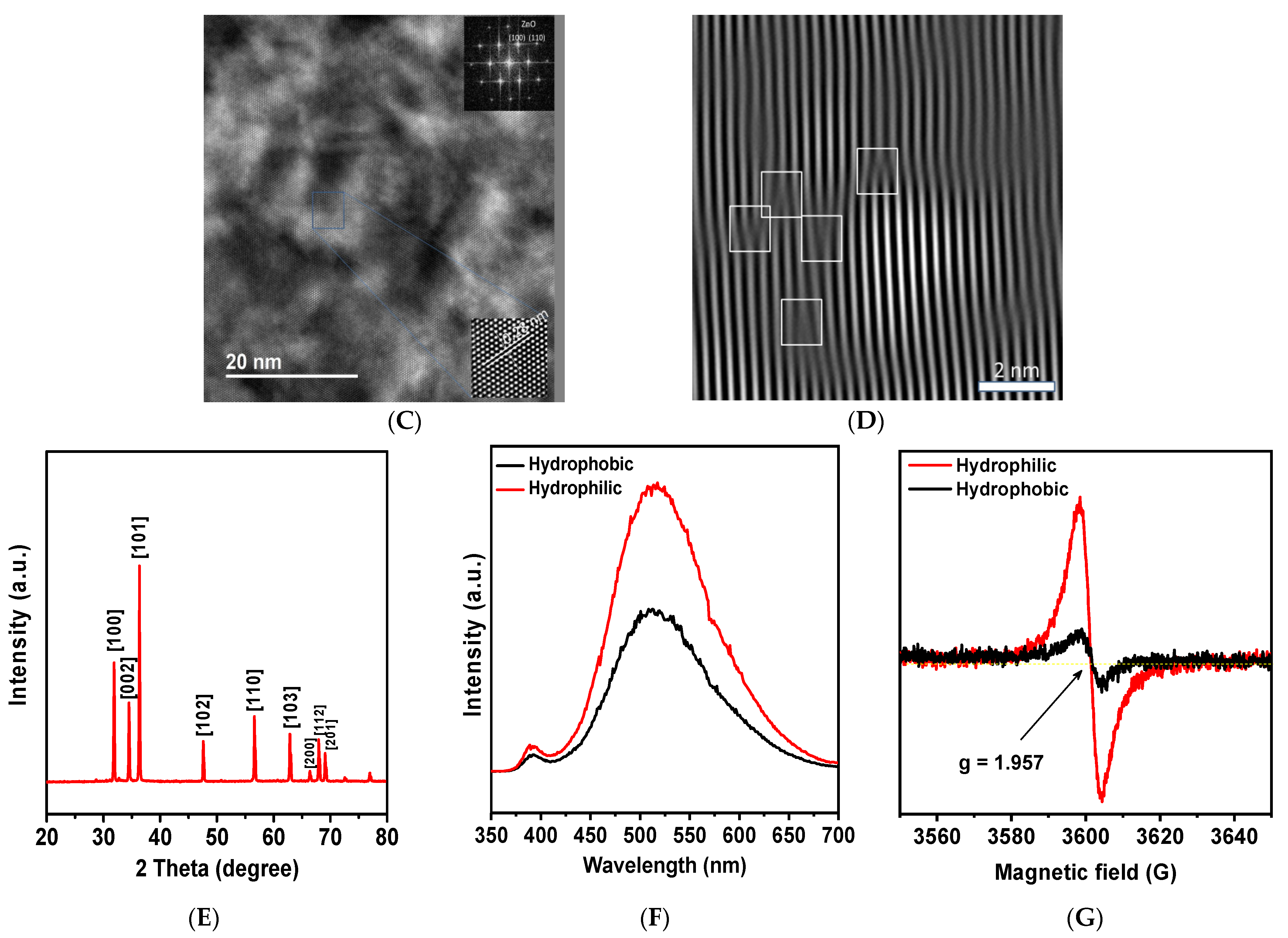
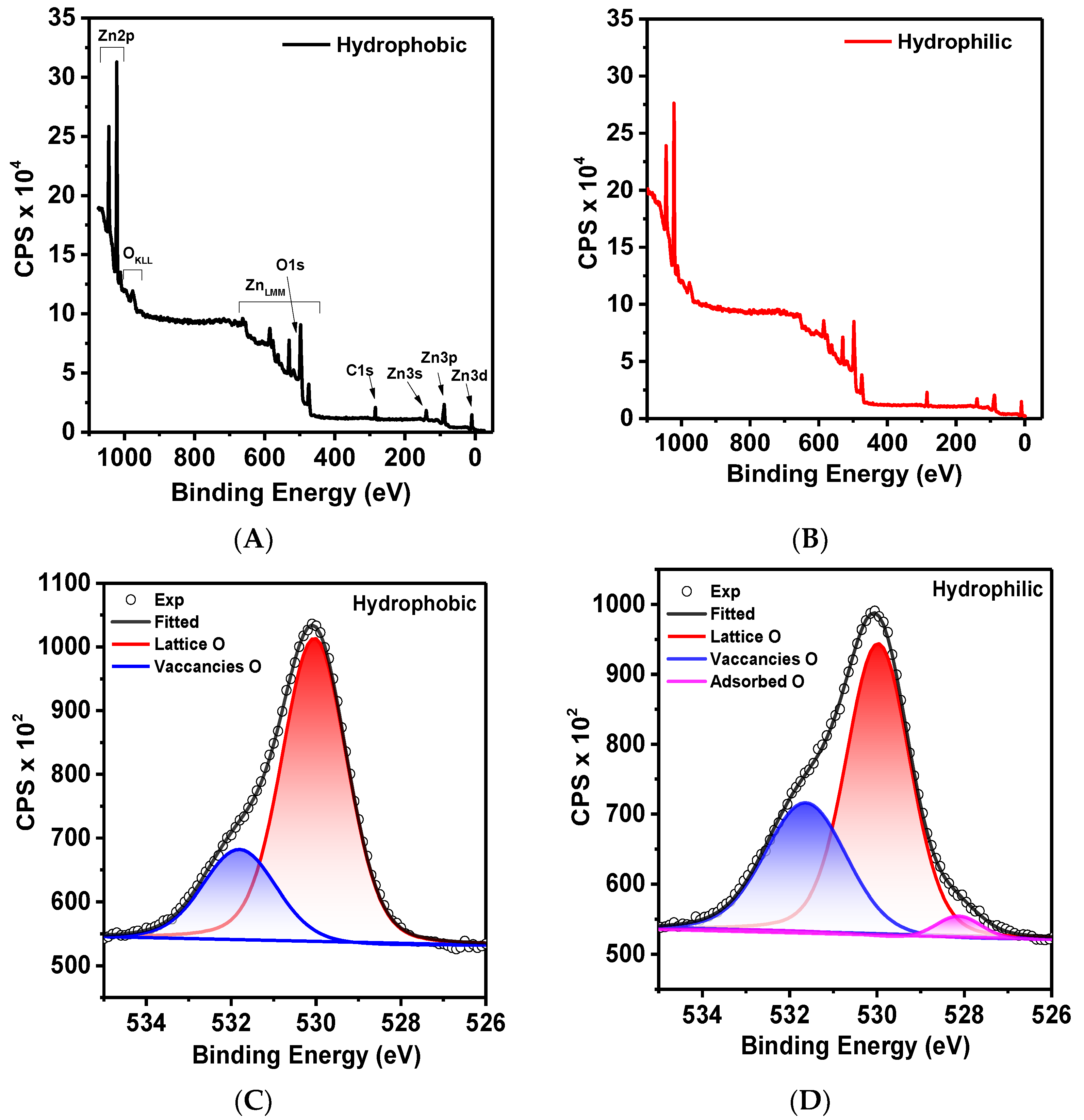
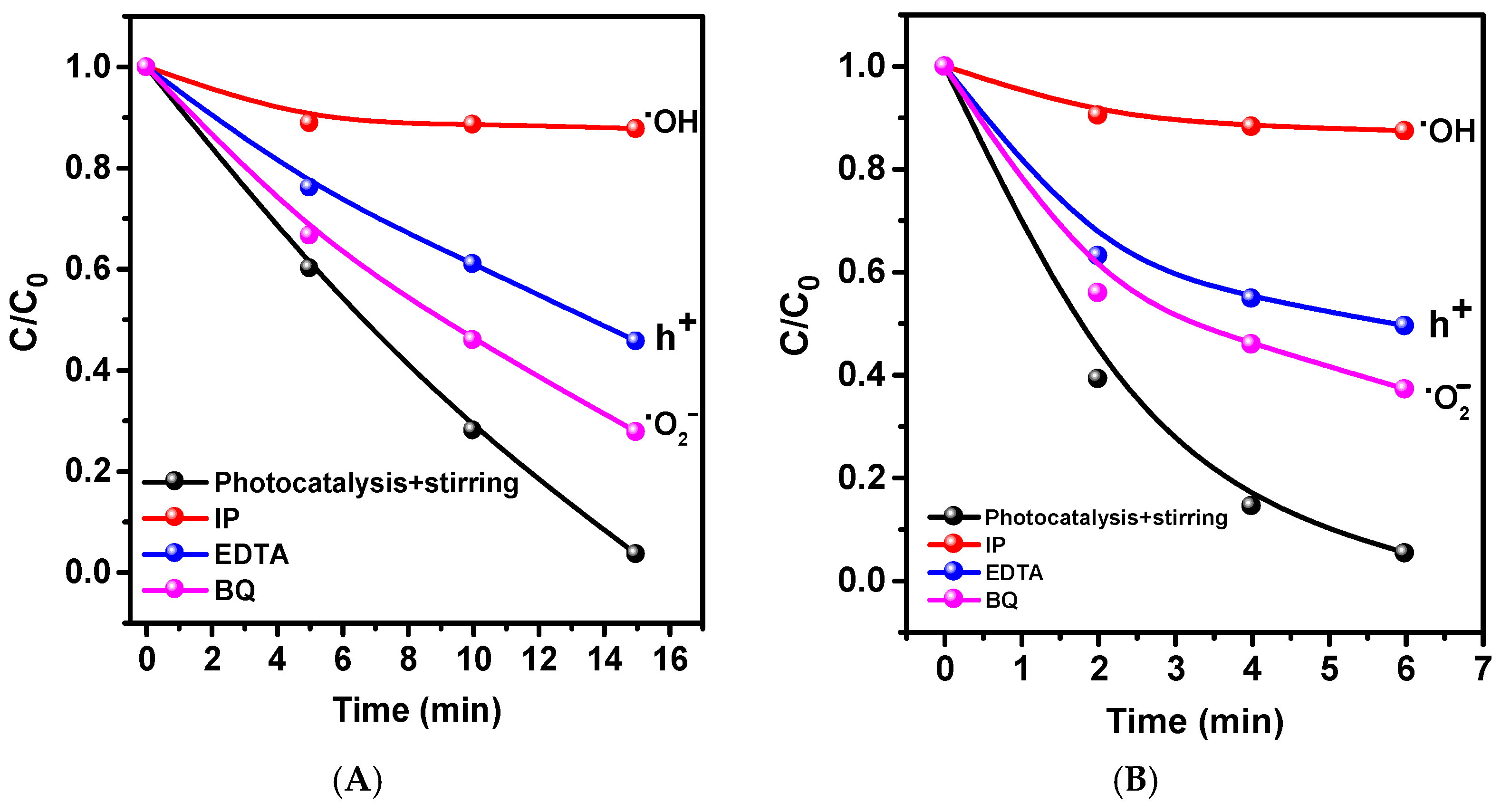
2. Results and Discussion

During the carbothermal synthesis process, tetrapods (self-organized, pseudo-three-dimensional nanostructures characterized by four monocrystalline rods emanating from the vertices of a tetrahedron) of ZnO were formed with “legs” whose lengths (Figure 1A) were up to tens of micrometers.
The EDS spectra (Figure 1B) show the atomic ratios of a ZnO-T in three positions from the center to the tip. From the obtained results, the ratio of the atomic masses of O and Zn changes. In the center, there is a significant predominance of oxygen, approximately 68.8%, while the content of zinc is about 31.2%. In the central region of the tetrapod, this ratio is inversely proportional: 29.3% O and 70.7% Zn. At the tip, zinc dominates, with its content increasing to 88.4%, and the oxygen content is approximately 11.6%. This indicates that there is a high accumulation of oxygen vacancies (VO) at the tips, while at the base of the tetrapod, there is a deficiency of zinc, suggesting the presence of interstitial zinc vacancies (Zni).
According to transmission electron microscopy data (Figure 1C) of a perpendicular cross-section of one of the ZnO-T rods, a monocrystalline structure is formed. An analysis of diffraction patterns and calculations of interplanar distances confirm the wurtzite structure of ZnO-T [JCPDS № 79–0205]. The absence of extended defects demonstrates the high crystalline quality of the ZnO-T. However, an analysis using Fourier transformation and subsequent image filtering (Figure 1D) show the presence of broken planes that are packaging defects and possible dislocation cores. The band structure is distorted around the dislocation core, and an additional level is introduced closer to the center of the forbidden zone. In n-type crystals such as ZnO, dislocations can capture electrons and hinder their recombination, which is also a factor that increases photosensitivity.
Diffraction reflections on the X-ray diffraction spectrum of the tetrapods (Figure 1E) corresponded to the hexagonal wurzite phase of ZnO, with a slight shift toward higher angles compared to the reference sample [JCPDS No. 79-0205], indicating a reduction in the size of the ZnO unit cell. The reduction in ZnO parameters is likely associated with point defects such as oxygen and zinc vacancies. The cathodoluminescence spectra in Figure 1F for hydrophilic and hydrophobic zinc oxide show a narrow band in the near UV region, corresponding to excitonic emission, and a broader and more intense band in the visible region associated with intrinsic or impurity point defects such as oxygen vacancies (VO), oxygen interstitials (Os), zinc vacancies (VZns), and zinc interstitials (Zns) and their complexes. The cathodoluminescence spectra were fitted with Gaussian functions to determine the components. The results are shown in Figure 2. Additionally, from Figure 1F, it can be concluded that the intensity of the defect band increases when transitioning from hydrophobic to hydrophilic states.
Figure 1G shows that the ESR spectrum represents a symmetrical singlet with parameters g = 1.957 and ∆B = 6.11 G, and these parameter values are nearly identical for both samples. The identity of the g-factor values and line width indicates that the same paramagnetic center is formed in all samples in terms of both its chemical nature and the structure of its immediate environment. These paramagnetic centers are attributed to point defects. The fact that the g-factor values are lower than the g-factor value for a free electron (2.0023) suggests that these defects have a hole-like character and carry a positive charge (V-centers). In [11], the signal with g~1.95–1.97 is associated with oxygen vacancies VO specifically in ZnO powders. It is evident that the concentration of defects in the hydrophobic sample is significantly lower compared to the hydrophilic sample, and we can conclude that the concentration of oxygen vacancies has increased. For oxygen vacancies in the neutral VO and 2+ charge states VO2+, a localized occupied state is recognized in the bandgaps at 2.5–2.6 eV and 0.9–1 eV below the conduction-band minimum [24], respectively, and this suggests the activity of the ZnO tetrapods in the visible region of the spectrum.
The XPS spectra (Figure 3) were calibrated using the C1s peak (284.6 eV). From the survey spectra (Figure 3A,B), it can be concluded that the surface is chemically pure and free from impurities. There are no visible differences between the hydrophilic and hydrophobic samples. In the high-resolution Zn2p spectra (Figure 3E,F), two distinct doublet peaks can be observed at 1021.6 eV and 1044.7 eV, corresponding to Zn2p3/2 and Zn2p1/2, respectively. The energy difference of 23.1 eV falls within the standard reference resolution of ZnO, and the visible doublets are attributed to Zn2+ ions. The asymmetric O1s peak is presented in Figure 3C,D. For the hydrophobic state, the spectrum is approximated by two components with maxima at 530.11 eV and 531.28 eV, corresponding to different forms of oxygen. The first peak at 530.11 eV can be attributed to oxygen ions (O2−) in the wurtzite structure of the ZnO. The higher energy peak at 531.28 eV is associated with regions of oxygen deficiency or oxygen vacancies in the matrix [25,26]. In the hydrophilic state, as shown in Figure 3D, an additional peak appears at a binding energy of 528 eV. The presence of peaks in this region is usually attributed to adsorbed oxygen [27]. The adsorption of oxygen on the hydrophilic surface can be explained by the surface’s tendency for charge compensation. Additionally, it is interesting to compare the ratio of the integrated peak areas of lattice oxygen and oxygen vacancies. It can be observed that the ratio changes from 3 to 1.8 when transitioning from the hydrophobic to the hydrophilic state.
The results of the photocatalytic decomposition of methylene blue (MB) using tetrapods in hydrophobic and hydrophilic states and metal halide lamp are presented in Figure 4.
As can be seen, even in a non-dispersed state without stirring, the ZnO-T shows photocatalytic activity, leading to a 26% degradation of MB in 15 min compared to 11% degradation under similar conditions during photolysis. The slight improvement in photolysis activity with stirring (15%) indicates the contribution of mass transfer processes. The photocatalysis with stirring showed that 97% of the dye decomposed in 15 min. The reaction was shown to accelerate by 10 times compared to photocatalysis without stirring (Figure 4B). The results of similar experiments on pre-hydrophilized ZnO-Ts are presented in Figure 4C. The highest activity was observed in the photocatalysis with stirring experiment, in which 95% of the MB was decomposed in 6 min. Without stirring, the catalysis efficiency dropped by almost half to 49.5%, while the degradation efficiency during photolysis was about 4–6%. The rate constants (k), calculated from the kinetic curves in Figure 4D using the pseudo-first-order equation, were 0.0065, 0.0095, 0.1155, and 0.4965 for photolysis, photolysis with stirring, photocatalysis, and photocatalysis with stirring, respectively. In photocatalysis (stirring), the reaction rate increased by 4.3 and 52 times compared to photocatalysis and photolysis, respectively. The significant acceleration of MB degradation on the hydrophilic ZnO-Ts (6.2 times for photocatalysis and 2.6 times for photocatalysis with stirring) compared to hydrophobic ones is due to both an increase in surface defects on the tetrapods and improved wettability, as all photochemical processes occur at the solid/liquid interface.
Considering the negligible amount of UV radiation present in the spectrum of the metal halide lamp, we conducted independent research utilizing cutoff filters to differentiate the impacts of both visible and UV light.
Figure 5 highlights the outcomes of our study, which reveal that exposure to UV light alone results in 96% degradation of MB within a 15 min timeframe. Conversely, cutting off UV light from the source leads to a decline in PC activity, with decomposition levels reduced to 65%. As a result, our findings suggest that ZnO-Ts in both visible and UV light possess PC activity. Photocatalytic activity in visible light can be influenced by surface oxygen vacancies and the photosensitization effect. Hydrophilization confirms the enhancing effect of oxygen vacancies on PC activity, while further experimentation is needed to verify the photosensitization effect.
Tetrapods, due to their unique morphology, are superhydrophobic and did not wet even during intense stirring of the solution in the experiment. For clarity, please refer to Figure 6, which shows a photograph of a water droplet on tetrapod powder on glass.
To understand the difference in the mechanisms of the photocatalytic reaction for hydrophilic and hydrophobic ZnO-Ts, tests were conducted to capture some active redox forms.
From Figure 7, we can observe that hydroxyl radicals, which are produced in the hole–water reaction, significantly contribute to the decomposition mechanism. The holes themselves have a minor role since hydroxyl radicals are also generated through the reactions of superoxide anion radicals with water. The presence of AgNO3, an electron scavenger, enhances catalytic activity. This is because Ag accepts an electron and reduces silver, which can act as a co-catalyst in dye degradation. It is well established that Ag-ZnO composites catalyze the degradation of MB. The application of benzoquinone as a superoxide anion radical scavenger also results in a decrease in activity, indicating their generation and involvement in reactions.
A hydrophilic ZnO-T was tested for its photocatalytic activity with the stirring decomposition of MB under direct sunlight irradiation. Figure 8 presents the results, which show that in just 8 min, 93% of MB decomposed compared to only 25% during photolysis. Furthermore, the activity of the catalyst under sunlight is almost identical to that seen under lamp irradiation, with only a slight slowdown reaction of 1.3 times. Long-term stability tests were carried out under sunlight conditions. Figure 8C demonstrates that the photocatalyst’s activity minimally decreases, indicating exceptional stability.
Additionally, the PC degradation efficiency of these systems is evidently higher compared to previous studies, as shown in Table 1. Considering the studies exploring additional piezo stimulation in PC due to the piezo properties of ZnO, we present a comparative table comparing our PC results with piezophotocatalysis. Table 2 summarizes the findings.
The tables present a comparative analysis indicating the high photocatalytic activity of microtetrapods. Table 1 shows that comparable rate constants in photocatalytic reactions are achieved solely via UV radiation and ZnO modification. It is worth noting, though, that most materials presented in the studies were at a nanoscale, unlike the particles employed in our research, which have dimensions in the micron range. Compared with piezophotocatalysis that utilizes both UV radiation and ultrasonic treatment, as shown in Table 2, our materials exhibit high efficiency in terms of the rate constant.
It is well known that zinc oxide is a wide-bandgap semiconductor that cannot be excited by visible light. Optical investigations presented in Figure 9B,C reveal that the bandgap’s width is 3.16 eV. Furthermore, the material exhibits strong light scattering which surpasses the absorption coefficient throughout the entire wavelength range by an order of magnitude.
To determine the structure of energy zones, VB XPS spectra were obtained, and the data are presented in Figure 9A. The presence of energy state density localized near the Fermi energy is immediately noticeable, confirming the presence of oxygen defect levels in the bandgap. The VBmax energy was estimated to be 2.80 and 2.58 eV for the hydrophobic and hydrophilic states, respectively. Based on this, the CBmin was estimated to be −0.36 and −0.58 eV. The energy required to form surface layer defects is less than that of bulk defects, leading to a diffuse energy band linked to defects within the bandgap.
When light is applied, electron–hole charge states are generated due to localized levels of impurity in the bandgap, mainly caused by oxygen vacancies (VO). Electrons generated by the light and moving from the conduction band to the surface of the material will interact with dissolved oxygen in water, creating superoxide anion radicals which can efficiently oxidize organic pollutants or produce hydroxyl radicals. Upon exposure to light, photogenerated holes on the surface can either react directly with MB in water or produce hydroxyl radicals.
Consequently, the reaction mechanism can be expressed as follows, based on experimental data, and free charge carriers (e/h+) are created:
Z n O + h v + e + h +
The existence of defect levels in the bandgap facilitates the primary capture of carriers and restricts recombination processes. Zinc defects within ZnO serve as electron traps for photoexcited electrons. These electrons can relax through interactions with oxygen molecules on the surface (reaction 3) or recombination. When electrons move toward the ZnO surface and interact with molecular compounds, superoxide radicals ·O2 are formed (reaction 4). In the instance of hydrophilic ZnO-Ts, the involvement of adsorbed molecular oxygen also plays a part in this mechanism.
e + O 2 · O 2
Similarly, holes (h+) are captured by oxygen vacancies and either interact with OH/H2O on the surface to form ·OH (reactions 5, 6) or recombine.
h + + O H · O H
h + + H 2 O H + + · O H
These radicals further oxidize organic pollutants. Thus, the presence of photocatalytic activity under visible light indicates the role of defects, primarily oxygen vacancies, and a significant increase in photocatalytic activity with the addition of mechanical stress indicates the role of surface wettability, which is also influenced by increased surface defects.

3. Materials and Methods

3.1. Synthesis of ZnO Tetrapods

Crystalline powders of zinc oxide were prepared via a modified method of high-temperature pyrolytic synthesis. In the first stage, a filter impregnated with a ZnO precursor was rolled into tubes with a diameter of 5 mm and placed on mesh frames made of corundum rods in a porcelain container. Next, the containers were heated to 1150 °C in a muffle furnace with an air supply at a speed of 9 L/min. Heating was carried out with a temperature gradient of 3.7 °C/min. When the set temperature was reached, the heating was stopped and the system was kept in thermostatic mode for 30 min and then cooled to room temperature. An aqueous solution of zinc acetate Zn(CH3COO)2·2H2O (Alpha Aesar, Bio Aqua Group, Targu Mures, Romania) with a zinc concentration of 70 g/L was used as a ZnO precursor. An ash-free white tape filter with an ash mass of 0.15 wt.% (Himreactive, N. Novgorod, Russia) was used as a paper filter. The filter served as a source of carbon formation; carbon played the role of a reducing agent in the process of producing zinc vapor as an intermediate reaction product for subsequent oxidation and the formation of zinc oxide tetrapods. The resulting reaction products were removed from the frame.

3.2. Characterization of Samples

The surface morphology and chemical composition of the samples were investigated via scanning electron microscopy (SEM), using an FEI Quanta 200 3D microscope with an attached energy-dispersive X-ray spectrometer (EDS) and EDAX Genesis (accelerating voltage 20 kV). To prevent sample charging, the samples were fixed on the microscope stage using a conductive adhesive tape based on graphite.
Sample preparation for transmission electron microscopy (TEM) was performed on a “Scios” scanning electron ion microscope (SEM) (FEI, Lincoln, NE, USA). According to the standard methodology, cross-sections perpendicular to the central growth axis of the ZnO tetrapod rod-like protrusions were prepared. To protect the sample surface during preparation, a technological layer of Pt was applied on all sides of the sample at a thickness of 1–3 μm. The cross-sections were examined using an “Osiris” TEM (FEI, Lincoln, NE, USA) at an accelerating voltage of 200 kV in TEM mode, high-resolution electron microscopy (HRTEM) mode, and scanning transmission electron microscopy (STEM) mode, as well as an energy-dispersive X-ray spectrometer (EDS).
To determine the chemical composition of the zinc oxide and zinc oxide–titanium oxide composite via X-ray photoelectron spectroscopy (XPS), a SPECS XPS spectrometer (Specs, Berlin, Germany) equipped with an Al anode was used. The choice of anode material was made to avoid interference from Auger lines in the useful signal. Spectra were recorded in the binding energy range from 0 to 1200 eV. The calibration of binding energies was performed using the C-C line of the C1s spectrum (E binding = 284.6 eV).
X-ray diffraction patterns were obtained using a Rigaku Miniflex 600 diffractometer (Japan) with Cu-Kα radiation and a β-filter. The diffraction patterns were analyzed using the TOPAS software (Bruker, 2015).
The bandgap parameters were determined using UV/Vis spectroscopy, with a spectrometric complex based on the monochromator. The material powder was placed on a special holder and compacted. Diffuse reflection spectra were recorded in the wavelength range λ from 250 to 800 nm.
Cathodoluminescence (CL) excitation measurements were performed using an electron beam from an EG-75 electronograph, with an electron energy of 40 keV (spot diameter: 1 mm) and an electron beam current of 80 µA. The spectra were analyzed using the AvaSpec-ULS2048x64-USB2 spectrophotometric complex (Avantes, Apeldoorn, The Netherlands). A vacuum optical fiber coupler, an FC-VFT-UV400, was used to extract radiation from the electron column. The angle of incidence of the electron beam on the substrate plane was 45°, and the angle between the axis of the optical fiber coupler and the direction of propagation of the incident electron beam was 90°.
The EPR/ESR spectra of the studied samples were obtained using a Bruker EMX Plus radio spectrometer in the “X” ultra-high-frequency radio wave range (frequency, ~9.8 GHz; wavelength, ~3 cm) at room temperature. For all samples, spectra were recorded over a wide range of magnetic fields (0–6000 G) to examine the presence of all possible signals.
The spectra of the total transmittance Tt and diffuse reflectance Rd for the studied objects were measured in the wavelength range of (300 to 1000) nm using an Avasphere-50 integrating sphere (Avantes, Apeldoorn, the Netherlands). A combined deuterium/halogen lamp AvaLight-DH-S-BAL (Avantes, Apeldoorn, The Netherlands) was used as an illumination source; its radiation was supplied via 600 μm fiber-optic light guides. Photographic signals were registered using an automated spectrometer, an MS3504i (SOL-Instruments, Minsk, Belarus), coupled with a CCD matrix camera, an HS-101H-HR (Hamamatsu, Hamamatsu City, Japan). The final spectrophotometric coefficient data Tt and Rd were determined as follows:
R d e x p = R d s λ R 0 λ R g l λ R 0 λ
T t e x p = T t s λ T 0 λ T g l λ T 0 λ ,
where T t s λ and R d s λ —are the transmission and reflection spectra of the samples; T g l λ and R g l λ —are the spectra of the reference signal measured with quartz plates; T 0 λ —is the signal of the integrating sphere with closed input and open output ports; and R 0 λ —is the signal for the sphere with open optical ports. The spectral dependence of the optical absorption coefficient μa and light scattering coefficient— μ s was calculated using an inverse Monte Carlo numerical modeling method, using the two-flow Kubelka–Munk model.

3.3. Photocatalytic Degradation Analysis

The photocatalytic characteristics of the samples were evaluated based on the photodegradation of methylene blue (MB) in an aqueous solution (2.5 mg L−1). Photocatalytic experiments were conducted in a 50 mL glass beaker. Visible and solar light was used in this case. A 70-watt metal halide lamp (Osram, Munich, Germany) was used as the light source. Activity with and without light filters that cut off wavelengths above and below 400 nm (λ > 400 nm and λ < 400 nm) was examined separately. A constant temperature in the reaction vessel of 26 °C was maintained using ventilation and monitored using a thermometer. For the photocatalytic reaction on hydrophobic particles, 20 mg of the original photocatalyst was added to 20 mL of an aqueous MB solution. Before turning on the light, the cuvette was kept in darkness for 60 min to achieve adsorption–desorption equilibrium. The photocatalysis process was carried out both without stirring and with stirring on a magnetic stirrer (400 rpm). The light source was positioned above the reactor at a distance of 10 cm. Samples (3 mL) were collected at fixed time intervals for each experiment. The particles were separated from the solution by centrifugation at 14,000 rpm for 2 min, using a laboratory centrifuge. The concentration of MB was measured using a spectrophotometer based on the characteristic absorption peak of MB at a wavelength of 663.7 nm. After the measurement, the solution was poured back into the reactor and the process was continued. For comparison, an MB solution was tested under similar conditions without a photocatalyst (photolysis). The concentration of MB was determined using the Beer–Lambert law.
For the photocatalytic reaction on hydrophilic particles, 20 mg of the original photocatalyst was initially poured into a beaker with distilled water (3 mL) and irradiated with a 250-watt high-pressure mercury UV lamp (Philips, Amsterdam, The Netherlands) without any cutoff filters until the complete evaporation of the water and the drying of the powder. The remaining experiment was conducted similarly to the hydrophobic one. Similar experiments were conducted under direct sunlight.

4. Conclusions

ZnO-Ts with surface oxygen defects were found to display exceptional photocatalytic activity in UV light, visible light, and direct sunlight. Controlling surface wettability was shown to regulate this activity, with an increase in surface defects occurring during the transition from hydrophobic to hydrophilic states. Although the optical width of the bandgap was 3.16 eV, the presence of a high density of localized defects in the bandgap led to sufficiently high PC activity in visible light. The experiment exhibited that using hydrophilic powder increases the reaction rate by 2.6 times compared to hydrophobic powder when irradiated with simulated sunlight. Implementing the procedure under direct sunlight results in a negligible reduction in the reaction rate by a factor of 1.3. Activity contributions from UV and visible light were distinguished by using cut-off light filters. In the presence of visible light, 65% of MB decomposes, while under UV light, 96% decomposes in 15 min. ·OH and ·O2 radicals are the main active forms responsible for the degradation process.

Author Contributions

Conceptualization, F.O. and A.L.; methodology, A.M., R.R.G. and V.K. (Vladimir Kanevsky); validation, A.M. and R.R.G.; formal analysis, D.S. (Daud Selimov) and V.K. (Valeriya Krasnova); investigation, A.M., D.S. (Daud Selimov), A.L. and V.K. (Valeriya Krasnova); resources, F.O., V.K. (Vladimir Kanevsky); data curation, A.M. and R.R.G.; writing—original draft preparation, F.O. and A.L.; writing—review and editing, F.O., D.S. (Daud Selimov), V.K. (Vladimir Kanevsky) and R.G.; supervision, F.O. and R.G.; project administration, F.O., V.K. (Vladimir Kanevsky); funding acquisition, D.S. (Dinara Sobola). All authors have read and agreed to the published version of the manuscript.

Funding

This work was performed within the State assignment of Federal Scientific Research Center “Crystallography and Photonics” of the Russian Academy of Sciences.

Institutional Review Board Statement

Not applicable.

Informed Consent Statement

Not applicable.

Data Availability Statement

The data are available upon request.

Conflicts of Interest

The authors declare no conflict of interest.

References

  1. Pan, X.; Yang, X.; Yu, M.; Lu, X.; Kang, H.; Yang, M.-Q.; Qian, Q.; Zhao, X.; Liang, S.; Bian, Z. 2D MXenes polar catalysts for multi-renewable energy harvesting applications. Nat. Commun. 2023, 14, 4183. [Google Scholar] [CrossRef]
  2. Li, J.; Lv, X.; Weng, B.; Roeffaers, M.B.; Jia, H. Engineering light propagation for synergetic photo- and thermocatalysis toward volatile organic compounds elimination. Chem. Eng. J. 2023, 461, 142022. [Google Scholar] [CrossRef]
  3. Feng, W.; Lei, Y.; Wu, X.; Yuan, J.; Chen, J.; Xu, D.; Zhang, X.; Zhang, S.; Liu, P.; Zhang, L.; et al. Tuning the interfacial electronic structure via Au clusters for boosting photocatalytic H2 evolution. J. Mater. Chem. A 2021, 9, 1759–1769. [Google Scholar] [CrossRef]
  4. Abdullah, F.H.; Bakar, N.H.H.A.; Bakar, M.A. Current advancements on the fabrication, modification, and industrial application of zinc oxide as photocatalyst in the removal of organic and inorganic contaminants in aquatic systems. J. Hazard. Mater. 2022, 424, 127416. [Google Scholar] [CrossRef]
  5. Ong, C.B.; Ng, L.Y.; Mohammad, A.W. A review of ZnO nanoparticles as solar photocatalysts: Synthesis, mechanisms and applications. Renew. Sustain. Energy Rev. 2018, 81, 536–551. [Google Scholar] [CrossRef]
  6. Meulenkamp, E.A. Synthesis and Growth of ZnO Nanoparticles. J. Phys. Chem. B 1998, 102, 5566–5572. [Google Scholar] [CrossRef]
  7. Gonzalez-Valls, I.; Lira-Cantu, M. Vertically-aligned nanostructures of ZnO for excitonic solar cells: A review. Energy Environ. Sci. 2008, 2, 19–34. [Google Scholar] [CrossRef]
  8. Tong, Y.; Liu, Y.; Shao, C.; Liu, Y.; Xu, C.; Zhang, J.; Lu, Y.; Shen, D.; Fan, X. Growth and Optical Properties of Faceted Hexagonal ZnO Nanotubes. J. Phys. Chem. B 2006, 110, 14714–14718. [Google Scholar] [CrossRef]
  9. Taniguchi, T.; Yamaguchi, K.; Shigeta, A.; Matsuda, Y.; Hayami, S.; Shimizu, T.; Matsui, T.; Yamazaki, T.; Funatstu, A.; Makinose, Y.; et al. Enhanced and Engineered d0Ferromagnetism in Molecularly-Thin Zinc Oxide Nanosheets. Adv. Funct. Mater. 2013, 23, 3140–3145. [Google Scholar] [CrossRef]
  10. Qiu, Y.; Yang, S. ZnO Nanotetrapods: Controlled Vapor-Phase Synthesis and Application for Humidity Sensing. Adv. Funct. Mater. 2007, 17, 1345–1352. [Google Scholar] [CrossRef]
  11. Janotti, A.; Van de Walle, C.G. Native point defects in ZnO. Phys. Rev. B 2007, 76, 165202. [Google Scholar] [CrossRef]
  12. Devynck, F.; Alkauskas, A.; Broqvist, P.; Pasquarello, A. Charge transition levels of carbon-, oxygen-, and hydrogen-related defects at the SiC/SiO2interface through hybrid functionals. Phys. Rev. B 2011, 84, 235320. [Google Scholar] [CrossRef]
  13. Vanheusden, K.; Warren, W.L.; Seager, C.H.; Tallant, D.R.; Voigt, J.A.; Gnade, B.E. Mechanisms behind green photoluminescence in ZnO phosphor powders. J. Appl. Phys. 1996, 79, 7983–7990. [Google Scholar] [CrossRef]
  14. Kurtz, M.; Strunk, J.; Hinrichsen, O.; Muhler, M.; Fink, K.; Meyer, B.; Wöll, C. Active Sites on Oxide Surfaces: ZnO-Catalyzed Synthesis of Methanol from CO and H2. Angew. Chem. Int. Ed. 2005, 44, 2790–2794. [Google Scholar] [CrossRef]
  15. Liu, K.; Yao, X.; Jiang, L. Recent developments in bio-inspired special wettability. Chem. Soc. Rev. 2010, 39, 3240–3255. [Google Scholar] [CrossRef]
  16. Li, J.; Sun, Q.; Han, S.; Wang, J.; Wang, Z.; Jin, C. Reversibly light-switchable wettability between superhydrophobicity and superhydrophilicity of hybrid ZnO/bamboo surfaces via alternation of UV irradiation and dark storage. Prog. Org. Coatings 2015, 87, 155–160. [Google Scholar] [CrossRef]
  17. Miyauchi, M.; Nakajima, A.; Watanabe, T.; Hashimoto, K. Photocatalysis and Photoinduced Hydrophilicity of Various Metal Oxide Thin Films. Chem. Mater. 2002, 14, 2812–2816. [Google Scholar] [CrossRef]
  18. Watanabe, T.; Yoshida, N. Wettability control of a solid surface by utilizing photocatalysis. Chem. Rec. 2008, 8, 279–290. [Google Scholar] [CrossRef]
  19. Liu, H.; Feng, L.; Zhai, J.; Jiang, L.; Zhu, D. Reversible wettability of a chemical vapor deposition prepared ZnO film between superhydrophobicity and superhydro-philicity. Langmuir 2004, 20, 5659–5661. [Google Scholar] [CrossRef]
  20. Feng, X.; Feng, L.; Jin, M.; Zhai, J.; Jiang, L.; Zhu, D. Reversible Super-hydrophobicity to Super-hydrophilicity Transition of Aligned ZnO Nanorod Films. J. Am. Chem. Soc. 2004, 126, 62–63. [Google Scholar] [CrossRef]
  21. Wang, L.; Zhang, S.; Wu, S.; Long, Y.; Li, L.; Zheng, Z.; Hei, Y.; Zhou, L.; Luo, L.; Jiang, F. Controlling wettability of AgI/BiVO4 composite photocatalyst and its effect on photocatalytic performance. J. Alloy. Compd. 2020, 835, 155367. [Google Scholar] [CrossRef]
  22. Li, H.; Wang, L.; Pi, X.; Ma, M.; Jiang, X.; Ji, S.; Jiang, F.; Luo, L. Effect of the wettability of Ag2MoO4/BiVO4 {010} composite on the photocatalytic degradation for 17α-ethinyl estradiol. J. Alloy. Compd. 2022, 899, 163295. [Google Scholar] [CrossRef]
  23. Zhu, H.; Cai, S.; Liao, G.; Gao, Z.F.; Min, X.; Huang, Y.; Jin, S.; Xia, F. Recent Advances in Photocatalysis Based on Bioinspired Superwettabilities. ACS Catal. 2021, 11, 14751–14771. [Google Scholar] [CrossRef]
  24. Oba, F.; Togo, A.; Tanaka, I.; Paier, J.; Kresse, G. Defect energetics in ZnO: A hybrid Hartree-Fock density functional study. Phys. Rev. B 2008, 77, 245202. [Google Scholar] [CrossRef]
  25. Zheng, J.; Jiang, Q.; Lian, J. Synthesis and optical properties of flower-like ZnO nanorods by thermal evaporation method. Appl. Surf. Sci. 2011, 257, 5083–5087. [Google Scholar] [CrossRef]
  26. Tam, K.H.; Cheung, C.K.; Leung, Y.H.; Djurišić, A.B.; Ling, C.C.; Beling, C.D.; Fung, S.; Kwok, W.M.; Chan, W.K.; Phillips, D.L.; et al. Defects in ZnO nanorods prepared by a hydrothermal method. J. Phys. Chem. B 2006, 110, 20865–20871. [Google Scholar] [CrossRef]
  27. Bukhtiyarov, V.I.; Hävecker, M.; Kaichev, V.V.; Knop-Gericke, A.; Mayer, R.W.; Schlögl, R. Atomic oxygen species on silver: Photoelectron spectroscopy and X-ray absorption studies. Phys. Rev. B 2003, 67, 235422. [Google Scholar] [CrossRef]
  28. Lee, K.; Sahu, M.; Hajra, S.; Abolhassani, R.; Mistewicz, K.; Toroń, B.; Rubahn, H.-G.; Mishra, Y.K.; Kim, H.J. Zinc oxide tetrapod sponges for environmental pollutant monitoring and degradation. J. Mater. Res. Technol. 2023, 22, 811–824. [Google Scholar] [CrossRef]
  29. Chamoli, P.; Shukla, R.K.; Bezbaruah, A.N.; Kar, K.K.; Raina, K. Microwave-assisted rapid synthesis of honeycomb core-ZnO tetrapods nanocomposites for excellent photocatalytic activity against different organic dyes. Appl. Surf. Sci. 2021, 555, 149663. [Google Scholar] [CrossRef]
  30. Heo, S.-G.; Jo, S.-I.; Jeong, G.-H. Revealing the enhanced photocatalytic properties of ZnO tetrapods produced by atmospheric-pressure microwave plasma jet system. Curr. Appl. Phys. 2023, 46, 46–54. [Google Scholar] [CrossRef]
  31. Kumar, S.; Kaushik, R.; Purohit, L. Novel ZnO tetrapod-reduced graphene oxide nanocomposites for enhanced photocatalytic degradation of phenolic compounds and MB dye. J. Mol. Liq. 2021, 327, 114814. [Google Scholar] [CrossRef]
  32. Mourya, A.K.; Singh, R.P.; Kumar, T.; Talmale, A.S.; Gaikwad, G.S.; Wankhade, A.V. Tuning the morphologies of ZnO for enhanced photocatalytic activity. Inorg. Chem. Commun. 2023, 154, 110850. [Google Scholar] [CrossRef]
  33. Qi, F.; Gao, X.; Wang, C.; Shuai, Y.; Yang, L.; Liao, R.; Xin, J.; Peng, S.; Shuai, C. In situ grown silver nanoparticles on tetrapod-like zinc oxide whisker for photocatalytic antibacterial in scaffolds. Mater. Today Sustain. 2022, 19, 100210. [Google Scholar] [CrossRef]
  34. Guo, M.Y.; Ng, A.M.C.; Liu, F.; Djurišić, A.B.; Chan, W.K.; Su, H.; Wong, K.S. Effect of Native Defects on Photocatalytic Properties of ZnO. J. Phys. Chem. C 2011, 115, 11095–11101. [Google Scholar] [CrossRef]
  35. Ferreira, N.S.; Sasaki, J.M.; Silva, R.S., Jr.; Attah-Baah, J.M.; Macêdo, M.A. Visible-Light-Responsive Photocatalytic Activity Significantly Enhanced by Active [VZn+VO+] Defects in Self-Assembled ZnO Nanoparticles. Inorg. Chem. 2021, 60, 4475–4496. [Google Scholar] [CrossRef]
  36. Park, S.J.; Das, G.S.; Schütt, F.; Adelung, R.; Mishra, Y.K.; Tripathi, K.; Kim, T.M. Visible-light photocatalysis by carbon-nano-onion-functionalized ZnO tetrapods: Degradation of 2,4-dinitrophenol and a plant-model-based ecological assessment. NPG Asia Mater. 2019, 11, 8. [Google Scholar] [CrossRef]
  37. Mishra, Y.K.; Modi, G.; Cretu, V.; Postica, V.; Lupan, O.; Reimer, T.; Paulowicz, I.; Hrkac, V.; Benecke, W.; Kienle, L.; et al. Direct Growth of Freestanding ZnO Tetrapod Networks for Multifunctional Applications in Photocatalysis, UV Photodetection, and Gas Sensing. ACS Appl. Mater. Interfaces 2015, 7, 14303–14316. [Google Scholar] [CrossRef]
  38. Ghosh, A.; Guha, P.; Samantara, A.K.; Jena, B.K.; Bar, R.; Ray, S.; Satyam, P.V. Simple Growth of Faceted Au–ZnO Hetero-nanostructures on Silicon Substrates (Nanowires and Triangular Nanoflakes): A Shape and Defect Driven Enhanced Photocatalytic Performance under Visible Light. ACS Appl. Mater. Interfaces 2015, 7, 9486–9496. [Google Scholar] [CrossRef]
  39. Sun, C.; Fu, Y.; Wang, Q.; Xing, L.; Liu, B.; Xue, X. Ultrafast piezo-photocatalytic degradation of organic pollutions by Ag2O/tetrapod-ZnO nanostructures under ultrasonic/UV exposure. RSC Adv. 2016, 6, 87446–87453. [Google Scholar] [CrossRef]
  40. Lv, Y.; Yao, W.; Ma, X.; Pan, C.; Zong, R.; Zhu, Y. The surface oxygen vacancy induced visible activity and enhanced UV activity of a ZnO1−x photocatalyst. Catal. Sci. Technol. 2013, 3, 3136–3146. [Google Scholar] [CrossRef]
  41. Zhu, H.; Zhang, C.; Xie, K.; Li, X.; Liao, G. Photocatalytic degradation of organic pollutants over MoS2/Ag-ZnFe2O4 Z-scheme heterojunction: Revealing the synergistic effects of exposed crystal facets, defect engineering, and Z-scheme mechanism. Chem. Eng. J. 2023, 453, 139775. [Google Scholar] [CrossRef]
  42. Yang, B.; Wang, Z.; Zhao, J.; Sun, X.; Wang, R.; Liao, G.; Jia, X. 1D/2D carbon-doped nanowire/ultra-thin nanosheet g-C3N4 isotype heterojunction for effective and durable photocatalytic H2 evolution. Int. J. Hydrogen Energy 2021, 46, 25436–25447. [Google Scholar] [CrossRef]
  43. Tan, H.; Zhao, Z.; Zhu, W.-B.; Coker, E.N.; Li, B.; Zheng, M.; Yu, W.; Fan, H.; Sun, Z. Oxygen Vacancy Enhanced Photocatalytic Activity of Pervoskite SrTiO3. ACS Appl. Mater. Interfaces 2014, 6, 19184–19190. [Google Scholar] [CrossRef] [PubMed]
  44. Lv, Y.; Zhu, Y.; Zhu, Y. Enhanced photocatalytic performance for the BiPO4-x nanorod induced by surface oxygen vacancy. J. Phys. Chem. C 2013, 117, 18520–18528. [Google Scholar] [CrossRef]
  45. Xu, X.; Ding, X.; Yang, X.; Wang, P.; Li, S.; Lu, Z.; Chen, H. Oxygen vacancy boosted photocatalytic decomposition of ciprofloxacin over Bi2MoO6: Oxygen vacancy engineering, biotoxicity evaluation and mechanism study. J. Hazard. Mater. 2019, 364, 691–699. [Google Scholar] [CrossRef]
  46. Liu, J.; Xie, F.; Li, R.; Li, T.; Jia, Z.; Wang, Y.; Wang, Y.; Zhang, X.; Fan, C. TiO2-x/Ag3PO4 photocatalyst: Oxygen vacancy dependent visible light photocatalytic performance and BPA degradative pathway. Mater. Sci. Semicond. Process. 2019, 97, 1–10. [Google Scholar] [CrossRef]
  47. Bettini, S.; Pagano, R.; Valli, D.; Ingrosso, C.; Roeffaers, M.; Hofkens, J.; Giancane, G.; Valli, L. ZnO nanostructures based piezo-photocatalytic degradation enhancement of steroid hormones. Surfaces Interfaces 2023, 36, 102581. [Google Scholar] [CrossRef]
  48. Xue, X.; Zang, W.; Deng, P.; Wang, Q.; Xing, L.; Zhang, Y.; Wang, Z.L. Piezo-potential enhanced photocatalytic degradation of organic dye using ZnO nanowires. Nano Energy 2015, 13, 414–422. [Google Scholar] [CrossRef]
  49. Ye, Y.; Wang, K.; Huang, X.; Lei, R.; Zhao, Y.; Liu, P. Integration of piezoelectric effect into a Au/ZnO photocatalyst for efficient charge separation. Catal. Sci. Technol. 2019, 9, 3771–3778. [Google Scholar] [CrossRef]
  50. Chimupala, Y.; Phromma, C.; Yimklan, S.; Semakul, N.; Ruankham, P. Dye wastewater treatment enabled by piezo-enhanced photocatalysis of single-component ZnO nanoparticles. RSC Adv. 2020, 10, 28567–28575. [Google Scholar] [CrossRef]
  51. Yu, C.; Yu, X.-X.; Zheng, D.-S.; Yin, H. Piezoelectric potential enhanced photocatalytic performance based on ZnO with different nanostructures. Nanotechnology 2021, 32, 135703. [Google Scholar] [CrossRef] [PubMed]
  52. Kumar, M.; Vaish, R.; Elqahtani, Z.M.; Kebaili, I.; Al-Buriahi, M.; Sung, T.H.; Hwang, W.; Kumar, A. Piezo-photocatalytic activity of Bi2VO5.5 for methylene blue dye degradation. J. Mater. Res. Technol. 2022, 21, 1998–2012. [Google Scholar] [CrossRef]
  53. Masekela, D.; Masekela, D.; Hintsho-Mbita, N.C.; Hintsho-Mbita, N.C.; Ntsendwana, B.; Ntsendwana, B.; Mabuba, N.; Mabuba, N. Thin Films (FTO/BaTiO3/AgNPs) for Enhanced Piezo-Photocatalytic Degradation of Methylene Blue and Ciprofloxacin in Wastewater. ACS Omega 2022, 7, 24329–24343. [Google Scholar] [CrossRef] [PubMed]
  54. Adiba, A.; Waris; Munjal, S.; Khan, M.Z.; Ahmad, T. Piezo-photocatalytic degradation of organic pollutant by a novel BaTiO3–NiO composite. Eur. Phys. J. Plus 2023, 138, 408. [Google Scholar] [CrossRef]
  55. Fu, Y.; Wang, Y.; Zhao, H.; Zhang, Z.; An, B.; Bai, C.; Ren, Z.; Wu, J.; Li, Y.; Liu, W.; et al. Synthesis of ternary ZnO/ZnS/MoS2 piezoelectric nanoarrays for enhanced photocatalytic performance by conversion of dual heterojunctions. Appl. Surf. Sci. 2021, 556, 149695. [Google Scholar] [CrossRef]
  56. Kumar, M.; Vaish, R.; ben Ahmed, S. Piezo-photocatalytic activity of mechanochemically synthesized BiVO4 for dye cleaning. J. Am. Ceram. Soc. 2022, 105, 2309–2322. [Google Scholar] [CrossRef]
  57. Hong, D.; Zang, W.; Guo, X.; Fu, Y.; He, H.; Sun, J.; Xing, L.; Liu, B.; Xue, X. High Piezo-photocatalytic Efficiency of CuS/ZnO Nanowires Using Both Solar and Mechanical Energy for Degrading Organic Dye. ACS Appl. Mater. Interfaces 2016, 8, 21302–21314. [Google Scholar] [CrossRef]
  58. Yan, S.; Chen, Z.; Lu, Z.; Wang, X. Piezoelectric-Enhanced Photocatalytic Performance of BaTi2O5 Nanorods for Degradation of Organic Pollutants. ACS Appl. Nano Mater. 2023, 6, 15721–15733. [Google Scholar] [CrossRef]
  59. Ren, Z.; Li, X.; Guo, L.; Wu, J.; Li, Y.; Liu, W.; Li, P.; Fu, Y.; Ma, J. Facile synthesis of ZnO/ZnS heterojunction nanoarrays for enhanced piezo-photocatalytic performance. Mater. Lett. 2021, 292, 129635. [Google Scholar] [CrossRef]
Figure 1. (A) SEM image. (B) EDX analysis. (C) HR-TEM image of a cross-section of the “legs” of a ZnO-T. Inset: magnified image of the highlighted area and its Fourier transform. (D) FFT image of the “legs” of a ZnO-T. (E) XRD spectra of ZnO-Ts. (F) Cathodoluminescence spectra. (G) ESR spectra.
Ijms 24 16338 g001aIjms 24 16338 g001b
Figure 2. Gauss fitted CL spectra of T-ZnO samples: hydrophobic (A) and hydrophilic (B).
Ijms 24 16338 g002
Figure 3. XPS spectra (A,B) wide XPS (C,D) O1s spectra and (E,F) Zn2p spectra of hydrophobic and hydrophilic ZnO-Ts.
Ijms 24 16338 g003aIjms 24 16338 g003b
Figure 4. Changes in the concentration of MB and kinetic curves during irradiation with a metal halide lamp without cutoff filters for hydrophobic (A,B) and hydrophilic (C,D) ZnO-Ts.
Ijms 24 16338 g004
Figure 5. Changes in the concentration of MB via PC with UV- and Vis-cutoff filters for hydrophilic ZnO-Ts.
Ijms 24 16338 g005
Figure 6. Photograph of a water droplet on the surface of hydrophobic ZnO-T powder.
Ijms 24 16338 g006
Figure 7. Influence of traps on changes in MB concentration during the photocatalysis process for hydrophobic (A) and hydrophilic (B) ZnO-Ts without cutoff filters, using a metal halide lamp.
Ijms 24 16338 g007
Figure 8. Changes in the concentration of MB (A) and kinetic curves (B) during solar photocatalysis on hydrophilic ZnO-Ts and a cyclic experiment (C).
Ijms 24 16338 g008
Figure 9. (A). VB XPS spectra for hydrophobic and hydrophilic ZnO-Ts. (B). Optical absorbance and scattering spectra. (C). Tauc plots for bandgap calculations.
Ijms 24 16338 g009
Table 1. Previously reported work and its comparison with our present work in the field photocatalytic properties of ZnO-based and other materials.
MaterialsPollutantsTime (min)Light SourceDegradation, %Rate Constant (min−1)References
ZnO tetrapod1
ZnO tetrapod2
methyl orange
methylene blue
methyl orange
methylene blue
130UV lamp
100 W
λ = 254 nm
50.8
85.7
61.6
96.4
1.6 × 10⁻⁴
2.9 × 10⁻⁴
1.7 × 10⁻⁴
3.6 × 10⁻⁴
[28]
GNs-ZnO
20 mg
MB (1 mg/L)40UV (125 W)
Vis (125 W)
70.87
17.26
4.5 × 10⁻²
5 × 10⁻³
[29]
ZnO tetrapods
1 mg
MB (10 ppm)70UV light (Philips, 350–400 nm wavelength, 60 W)94.51.02 × 10−1[30]
ZTPGMethylene blue
(20 ppm)
90UV light (60 W, 365 nm)98.050.03[31]
ZnO sample 25 mgV = 100 mL
Rhodamine B (20 ppm)
110UV irradiation (8 W)98.860.036[32]
T-ZnOw/PLLAV = 50 mL
MB (3 × 10⁻⁴ M)
60Visible light
300.0065[33]
T-ZnO
50 mg
V = 50 mL
MB (5 mg/L)
8UV illumination (365 nm, 66.2 mW/cm2, Blak-Ray B-100 AP lamp)100-[34]
Rod-like ZnO nanoparticles
10 mg
V = 50 mL
MB (50 mg/L)
120100 W halogen lamp (with λ > 420 nm and a light intensity of 2.87 W m–2)30.672.9 × 10−3[35]
T-ZnO
T-ZnO-CNO
100 mg
V = 50 mL
DNP (0.1 mM)
14060 W tungsten bulb30
92
0.00274 
0.01834 
[36]
ZnO tetrapods
60 mg
V = 60 mL
MB (1 μmol·L–1)
10UV diode array consisting of four diodes (central wavelength = 370 nm, 170 mW/diode)96-[37]
NWs
TNFs coated Si substrates of area 0.5 cm2
V = 10 mL
RhB (5 × 10⁻⁶ M)
180100 W bulb (with a luminous irradiance of 10 mW/cm2 at the sample) λ ≥ 400 nm95-[38]
T-ZnO
Ag2O/T-ZnO
200 mg
V = 100 mL
MB (5 mg L−1)
2UV lamp, 50 W63
85
-
-
[39]
ZnO1−x
50 mg
V = 100 mL
MB (1 × 10⁻⁵ M)
360Halogen–tungsten lamp (power = 175 W; λmain = 550 nm950.522 h−1 [40]
MoS2/Ag-ZnFe2O4
40 mg
V = 50 mL
TC = 10 mg/L
60300 W Xenon lamp with optical filter (λ ≥ 420 nm)950.04868[41]
2D g-C3N4 nanosheets
20 mg
V = 100mL
TEOA = 10%
240300 W Xenon lamp with optical filter (λ ≥ 420 nm)-7.414 mmol g−1 h−1[42]
SrTiO3
50 mg
120 mL of 25% aqueous methanol solution300UV–visible light
300 W Xenon lamp
-2.2
mmol h–1 g–1
[43]
BiPO4–x
25 mg
V = 50 mL
MB = 1 × 10⁻⁵ M
30UV-light 300 W high-pressure mercury lamp890.300[44]
Bi2MoO6
20 mg
V = 50 mL
CIP = 20 ppm
40300 W Xenon lamp with optical filter (λ ≥ 400 nm)971.7990 
mg m−2 min−1
[45]
TiO2-x/Ag3PO4
100 mg
V = 100 mL
BPA = 10 mg/L
16500 W Xenon lamp with optical filter (λ ≥ 420 nm)95-[46]
70W metal–halogen lamp:
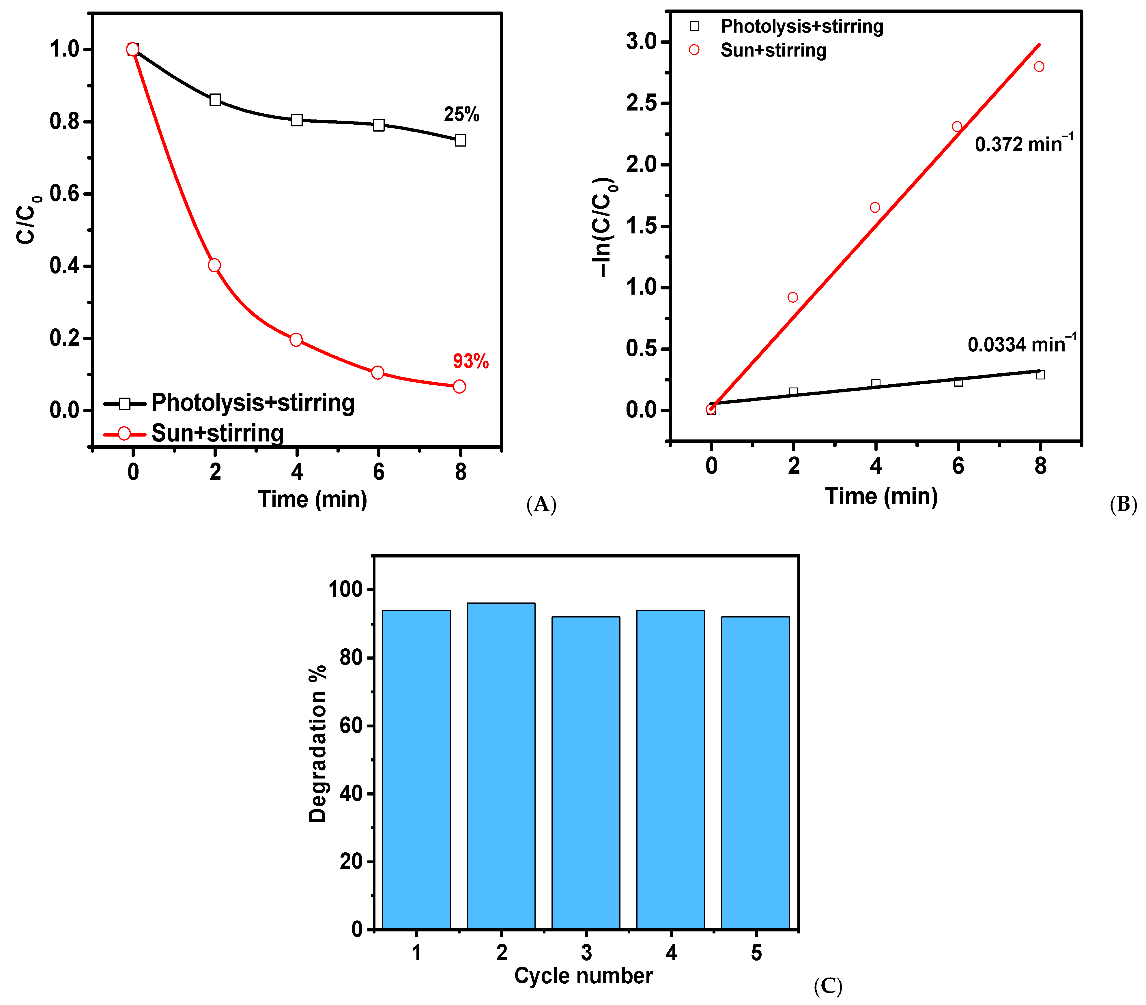
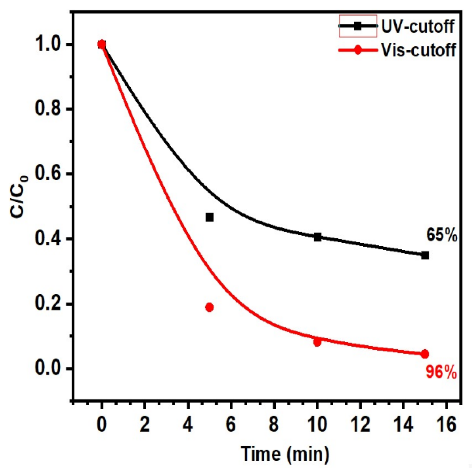
ZnO-T
20 mg
20 mL of MB (2.5 mg/L)6
15
15
8
Without cut-off
λ > 400 nm
λ < 400 nm
Direct sunlight
95
65
96
93
0.496
0.101
0.229
0.372
This work
Table 2. Previously reported work and its comparison with our present work in field of the piezophotocatalytic properties of ZnO-based and other materials.
MaterialsPollutantsTime (min)Light/Mechanical SourceDegradation, %Rate Constant, (min−1)References
ZnO NS/
2.5 mg
V = 10 mL
TST (testosterone)
(5 × 10⁻⁵ M)
45LOT-Oriel Solar S (140 W), 35 kHz501.8 × 10−2[47]
ZnO nanowires/CFs
200 mg
V = 100 mL
MB (C0 = 5 mg/L)
120High-pressure mercury lamp (50 W)/stirring96-[48]
ZnO nanorods
20 mg
V = 50 mL
RhB (10 ppm)
20300 W Xe lamp equipped with a 350 nm bandpass filter/ultrasonic frequency 27 kHz750.0744[49]
calcined ZnOTW-0.20
1g/L
V = 100 mL
MB (5 ppm)
120UVA light with peak wavelength of 365 nm and intensity of 940 μW cm−2/ultrasonic bath (120 W, 40 kHz)90-[50]
T-ZnO nanostructures
200 mg
V = 100 mL
MB (5 mg L−1)
2UV lamp/ultrasonic probe
50 W UV, 200 W ultrasonic
74-[51]
Bi2VO5.5
0.25 g
V = 10 mL
MB = (5 mg/L)
24015W (Havells company) 2 lamp visible light; ultrasonicator (40 kHz, 150 W).820.00528[52]
FTO/BaTiO3
/AgNPs
2 cm × 2 cm
V = 75 mL
MB = (5 mg/L)
18070 W UV lamp;
24 kHz ultrasonic vibration 30 W
900.02329[53]
BaTiO3 –NiO
0.2 g
V = 200 mL
MB = (10 mg/L)
80UV lamp, 125 W;
ultrasonic cleaner, ~40 kHz
900.028[54]
ZnO/ZnS/MoS2
10 mg
V = 50 mL
MB = 10 mg/L
50300 W Xenon lamp to simulate the solar source;
stirring at 1000 rpm
870.0411[55]
BiVO4
0.2 g
V = 10 mL
MB = (5 mg/L)
24015W (Havells company) 2 lamp visible light; ultrasonicator (40 kHz, 70 W).810.00802[56]
CuS/ZnO nanowires on stainless steel mesh
6.0 × 6.0 cm, 100 mg
V = 50 mL
MB = 5 mg/L
20Xenon lamp, 500 W, to simulate the solar source; ultrasonic probe, 200 W980.18236[57]
BaTi2O5
40 mg
V = 60 mL
RhB = 10 mg/L
MB = 10 mg/L
MO = 10 mg/L
50Xenon lamp, 300 W, λ > 400 nm; ultrasonic cleaner, 53 kHz, 100 W
82.5
-
-
0.0353
0.1775
0.0314
[58]
ZnO/ZnSV = 50 mL
MB = 5 mg/L
50300 W
UV irradiation;
180 W sonication, 40 kHz
600.0154[59]
70 W metal–halogen lamp:
ZnO-T
20 mg
V = 20 mL
MB (2.5 mg/L)
6
15
15
8
Without cut-off
λ > 400 nm
λ < 400 nm
Direct sunlight
95
65
96
93
0.496
0.101
0.229
0.372
This work
Disclaimer/Publisher’s Note: The statements, opinions and data contained in all publications are solely those of the individual author(s) and contributor(s) and not of MDPI and/or the editor(s). MDPI and/or the editor(s) disclaim responsibility for any injury to people or property resulting from any ideas, methods, instructions or products referred to in the content.

