Review

A Review of the Molecular Landscape of Adenoid Cystic Carcinoma of the Lacrimal Gland

by
Sarah Kate Powell
1,*,†,
Karina Kulakova
1,2,† and
Susan Kennedy
1,3
1
Research Foundation, Royal Victoria Eye and Ear Hospital, D02 XK51 Dublin, Ireland
2
Department of Biotechnology, Dublin City University, D09 V209 Dublin, Ireland
3
National Ophthalmic Pathology Laboratory, D04 T6F6 Dublin, Ireland
*
Author to whom correspondence should be addressed.
Those authors contributed equally to this work.
Int. J. Mol. Sci. 2023, 24(18), 13755; https://doi.org/10.3390/ijms241813755
Submission received: 25 June 2023 / Revised: 21 August 2023 / Accepted: 31 August 2023 / Published: 6 September 2023
(This article belongs to the Special Issue Advances in Molecular Understanding of Ocular Adnexal Disease)

Abstract

:
Adenoid cystic carcinoma (ACC) has a worldwide incidence of three to four cases per million population. Although more cases occur in the minor and major salivary glands, it is the most common lacrimal gland malignancy. ACC has a low-grade, indolent histological appearance, but is relentlessly progressive over time and has a strong proclivity to recur and/or metastasise. Current treatment options are limited to complete surgical excision and adjuvant radiotherapy. Intra-arterial systemic therapy is a recent innovation. Recurrent/metastatic disease is common due to perineural invasion, and it is largely untreatable as it is refractory to conventional chemotherapeutic agents. Given the rarity of this tumour, the molecular mechanisms that govern disease pathogenesis are poorly understood. There is an unmet, critical need to develop effective, personalised targeted therapies for the treatment of ACC in order to reduce morbidity and mortality associated with the disease. This review details the evidence relating to the molecular underpinnings of ACC of the lacrimal gland, including the MYB–NFIB chromosomal translocations, Notch-signalling pathway aberrations, DNA damage repair gene mutations and epigenetic modifications.

1. Introduction

A rare, biphasic, relentlessly growing and progressing carcinoma of the secretory glands, adenoid cystic carcinoma (ACC), has a worldwide incidence of 3–4 patients per million [1,2]. ACC of the head and neck is an uncommon malignancy, accounting for approximately 1% of all head and neck cancers [1,3]. Although ACC can occur at any age, it most commonly affects patients aged between fifty and sixty years, with the sixth decade of life reported as the most common decade of disease occurrence [4]. A younger mean age at diagnosis has been reported in patients with lacrimal gland ACC (39.5 years) when compared to other anatomical sites [5].
Tumours of the lacrimal gland are extremely rare. A Danish study of lacrimal gland lesions from 1974 to 2007 estimated an incidence of one case per million per year. They accounted for 25% of ocular space-occupying lesions. The tumours are of epithelial origin in over 50% of cases, lymphoid in 33% and mesenchymal or metastatic cancers in the remaining 10–15% of cases [6]. Over 50% of lacrimal gland tumours are malignant. The most frequent malignant tumour in the lacrimal gland is ACC, accounting for 66% of all malignant tumours that arise in the lacrimal gland [7]. A population-based study using the Surveillance, Epidemiology and End Results (SEER) program in the USA that examined 5464 cases of ACC found that lacrimal gland ACC accounted for 1.5% (81 patients) of all ACC cases [5]. Lacrimal gland ACC is associated with poorer prognosis when compared to ACC arising from other anatomical sites [8]. This is due to its propensity for unpredictable growth rates, frequent R/M, retrograde perineural invasion (PNI) and infiltration of both soft-tissue structures and bone [9,10]. PNI is commonly observed in the absence of vascular or lymphatic invasion and is a route for tumour cell propagation [11,12]. Lacrimal gland ACC is associated with a high rate of bone invasion. Williams et al. confirmed histologically that 82% of patients had lacrimal gland fossa invasion, which indicates the need for this to be addressed during initial preoperative planning and surgical management [13]. Five-year survival rates of lacrimal gland ACC have been reported as <50%, further diminishing to 20% at 10 years [14,15].
In 1998, Font et al. analysed 12 cases of ACC of the lacrimal gland specifically and reported a 100% local recurrence rate (mean interval of 3.25 years) and a 60% mortality rate with a mean survival of 5 years [15]. Overall survival rates have been reported as up to 30% over the last decade [16]. Esmaeli et al. analysed the outcomes of 20 lacrimal gland ACC patients and noted that local recurrence occurred in 35% of cases, distant metastasis was present in 80% of patients and 65% of patients had died at the time of the study as a direct consequence of the neoplasm [17].
ACC at all sites has a proclivity to recur and/or metastasise [18]. Over time, distant metastasis is reported to occur in over 50% of all ACC tumours, despite initial curative surgical treatment [11,19]. Distant metastasis is related to several inciting factors, including the primary tumour site, large tumour size, age and lymph node involvement [20]. Lung is the most common site of ACC distant metastasis [20]. Long-term overall survival rates of ACC are grim and have been reported between 23 and 40%, irrespective of treatment modalities [11]. Patients with any lacrimal gland tumour will present with a palpable tumour, proptosis or eye displacement, decreased range of motion and ptosis [21]. Pain is a characteristic feature of ACC, not seen with other tumours. This is due to perineural invasion, which is an almost universal feature of ACC. This can be highlighted on MRI examination [22].
The current gold standard treatment option for ACC patients involves complete surgical excision with adjuvant radiotherapy (RT). To date, no effective nonsurgical systemic therapeutic agents have been identified or developed to treat ACC and/or metastatic ACC effectively. Recurrent and/or metastatic (R/M) ACC tumours are poorly characterised. Their driving molecular alterations and underpinnings remain largely unknown [23]. Metastatic ACC has an extremely poor prognosis and is largely regarded as an incurable disease generally refractive to chemotherapeutic agents [23]. Cytotoxic chemotherapy has low efficacy [2]. Clinical trials have been developed to investigate actionable therapies. A novel phase II trial investigating proton radiation or intensity-modulated RT for the improvement of local control rate and lower toxicities compared to standard RT treating salivary and lacrimal gland malignancies is ongoing [24]. RT is utilised as a local control to lower the rate of local recurrence, but it does not improve overall patient survival rates [2]. Neoadjuvant-targeted intra-arterial chemotherapy has been used recently as a systemic therapy for lacrimal gland ACC in order to improve survival outcomes, although definite improvements have not yet been shown [25].
ACC possesses a deceptively low-grade histological appearance and is generally characterised by indolent yet relentless disease progression. Histologically, the tumour demonstrates characteristic morphology with three distinct histological growth architectural patterns, namely tubular, cribriform and/or solid, which can be seen in various amounts in each individual tumour [26]. Tubular and cribriform patterns are associated with a low proliferative index, few mitoses and little pleomorphism. These are deceptively benign-appearing histologic features. Histologic samples may be graded based on the degree of solid growth component in their tumour. The extent of solid patterns can significantly predict patient outcomes [27,28].
The tubular growth pattern, described in Figure 1A, is the most differentiated histological pattern. The tumour is composed of small well-formed ductal or tubular structures. This morphology has been associated with the most favourable prognosis. Tumours with this pattern alone metastasise less frequently when compared to the other patterns, and longer overall survival rates have been observed (9 years for tubular versus 8 years for cribriform and 5 years for solid [29]). Tubules are lined by inner epithelial luminal cells surrounded by myoepithelial cells with a clear cytoplasm [29].
The cribriform growth pattern is typical of ACC and is shown in Figure 1B. In terms of clinical prognosis and histology, it lies between tubular and solid ACC forms [30]. It is characterised by the formation of cystic structures [27]. The cribriform type has a ‘sieve-like’ or ‘cookie cutter’ appearance with luminal myxoid globules.
The solid growth pattern shown in Figure 1C is associated with the worst prognosis. It is the least-differentiated form of ACC. The cells are pleomorphic, with hyperchromatic nuclei, variable cytoplasm and a high mitotic rate, unlike the cribriform or tubular patterns. If the solid pattern accounts for >30% of the tumour, this is associated with a poorer prognosis [31]. The morphologic features are not specific to ACC and thus may represent a diagnostic challenge, particularly when attempting to differentiate ACC from other salivary gland epithelial tumours in a small biopsy or FNAC [21]. Molecular studies may be complimentary to histology and immunohistochemistry in the diagnostic setting.
There are more than eight hundred cases of lacrimal gland ACC reported in the literature to date [21]. The cellular processes and molecular landscapes that govern the disease pathogenesis are not yet fully elucidated. This paucity of knowledge may be attributed to a lack of large-scale population-based studies, which is a direct consequence of the rarity of the tumour, thereby resulting in a scarcity of clinical cases to evaluate and analyse [32]. There is also a lack of bona fide cell lines and animal disease models available [33]. Understanding the molecular drivers of ACC will undoubtedly be critical in the development of novel therapeutic options in order to halt disease progression and metastasis.
This review aims to explore the molecular landscape of ACC arising in the lacrimal gland and analyse established genetic mutations described in the literature that are key drivers of disease pathology, including fusion translocations, such as the MYB–NFIB fusion, Notch signalling, DNA damage response genes, and epigenetic modifications, and chromatin remodelling genes, such as SMARCA2, CREBBP and KDM6A [12,34].

2. MYB/NFIB Translocation

Genetic alterations, specifically chromosomal translocations, are hallmarks of several forms of cancer, including solid carcinomas and haematological malignancies [35,36]. Chromosomal translocations are characterised by the rearrangement of two nonhomologous chromosomes [35]. Nowell and Hungerford made the first direct link between chromosomal translocations and cancer in 1960 when they discovered the Philadelphia chromosome (translocation between chromosomes 9 and 22) and its association with the haematological malignancy, chronic myeloid leukaemia [37]. Since then, several chromosomal translocations have been associated with cancer and are often considered as the key inciting event that drives cancer pathogenesis, because their occurrence results in aberrant gene expression.
Fusion of the proto-oncogene MYB with the transcription factor NFIB plays a major role in ACC tumorigenesis [38]. It is unique to this tumour type so that its presence is diagnostic when there is histologic uncertainty [39]. The mechanism is either through copy number gain or enhancer hijacking [18,40]. This is a pro-oncogenic phenomenon whereby the upregulation of an oncogene is driven by enhancer repositioning leading to altered chromatin interactions and ultimately increased oncogene expression [41,42]. Primary ACC tumours have relatively quiet genomes with low tumour mutational burdens [43]. A recent study by Bell et al. is the only study to date to show that the oncogenes KRAS, NRAS and MET are mutated in lacrimal gland ACC, thereby identifying the EGFR–RAS–RAF-signalling pathway as a potential inhibitory target [44].
As MYB–NFIB fusion is highly specific for ACC, it is considered a genomic hallmark. It is estimated that over 70% of all ACC patients possess mutations of either the MYB or MYB1 genes [45]. Aberrant MYB gene expression is seen in approximately 60% of all ACC patients, compared to MYBL1, which is reported in approximately 35% of patients who are negative for the MYB–NFIB fusion [46]. Persson et al. were the first group to report recurrent MYB–NFIB fusion as a key oncogenic event in the pathogenesis of ACC, using cytogenetic and karyotype analyses of head, neck and breast ACC, as well as by reverse transcriptase polymerase chain reaction (RT-PCR). They also proposed the notion that MYB–NFIB fusion may be an attractive disease biomarker [47].
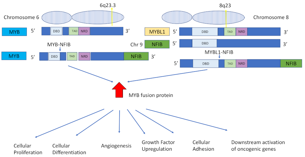
MYB–NFIB gene fusion most commonly occurs via the t(6;9)(q23;p23) translocation [2]. The fusion is supported by an alternative splicing system specific to MYB–NFIB and occurs due to MYBL1 encoding A-MYB protein, structurally similar to MYB. It is currently unknown which mechanism activates MYB in the t(6;9) translocation event. The fusion event causes upregulation of MYB protein expression resulting in highly oncogenic MYB fusion protein in ACC. This upregulation is believed to be the oncogenic driver of ACC [1]. The MYB–NFIB and MYBL1–NFIB gene fusions and downstream cellular sequelae are schematically described in Figure 2.
MYB, a master transcriptional activator found at chromosomal band 6q23 [36], is an oncogene essential for haematopoiesis and colonic crypt renewal, which, when overexpressed, results in increased rates of cellular proliferation, differentiation, angiogenic and growth factor upregulation, cell-cycle control, cellular adhesion and downstream activation of oncogenic genes [16,47]. MYB is not expressed in normal glandular cells but MYB mRNA and protein are highly expressed in ACC. MYB target genes are also overexpressed at the protein level in ACC [49]. Due to the existence of several routes for overexpression, such as selective gain at MYB locus [49], upregulation of the gene can be seen in both fusion-positive and a subset of fusion-negative tumours [1]. Transcription factor NFIB is found at the chromosomal band 9p22-23 [36]. 5′ rearrangements of NFIB have been described in cases of both salivary and breast ACC [46,50].
Currently, chromosomal translocations are most commonly detected using techniques, such as fluorescence in situ hybridisation (FISH) or PCR testing. Using dual-colour ‘break-apart’ probes, FISH analysis analysing MYBL1, MYB and NFIB rearrangements may take place [51]. In an interphase nucleus lacking translocation of the 6q23.2–q23.3 region, two orange and two green signals are expected [47]. This represents two normal, non-rearranged loci as seen in Figure 3A. Separate orange and green signals and the appearance of an orange-green fusion signal are indicative of the MYB–NFIB translocation and are diagnostic of ACC [47,51].
Targeted RNA sequencing can be used for RNA assessment and fusion gene detection, and RT-PCR and subsequent Sanger sequencing can be used to confirm novel fusions [52]. Next-generation sequencing may also be used to map chromosomal breakpoints and identify fusion transcripts and is less time-consuming than the traditional FISH analysis [53,54].
There are some disadvantages associated with these methods, including high failure rates when using archival formalin-fixed paraffin-embedded (FFPE) tissue. FISH may be inaccurate at delineating the exact chromosomal breakpoint [55]. Recently, McIntyre et al. conducted a pilot study that demonstrated NanoString probe technology was efficient in the detection of MYB–NFIB fusion. NanoString probe methodology negates the need for mRNA reverse transcription and thereby may be a more effective technique in detecting MYB–NFIB fusion in ACC patient samples, although more robust, large-scale studies are needed to confirm these preliminary results [56].
Pharmacological research aimed at targeting MYB–NFIB fusion is limited despite being widely proposed as a potential therapeutic target. This is in part due to the lack of defined ACC cell lines possessing the MYB–NFIB fusion [57]. Currently, a phase I single-arm multicentre clinical trial targeting fusion genes as a potential treatment through the use of a TetMYB vaccine and antiprogrammed death 1 (PD1) antibody is ongoing [58]. If the vaccine is found effective, patients with MYB overexpression and MYB–NFIB fusions in both cohorts will be eligible. A recent study identified that MYB–NFIB fusion is regulated by AKT-dependent insulin-like growth factor receptor 1 (IGFR1) signalling, which also has ramifications in terms of potential treatment modalities aimed at inhibiting IGF1R/AKT signalling [33].

2.1. MYB–NFIB Fusion in Salivary Gland ACC

Mitani et al. studied the prevalence of MYB–NFIB fusion in 123 patients with salivary ACC. They reported that the fusion was present in 28% of patients with primary ACC and 35% of patients with metastatic ACC. Importantly, the MYB–NFIB fusion was not detected in other salivary carcinomas, highlighting its specificity for ACC and its potential as a clinical diagnostic biomarker [46]. Fujii et al. evaluated the presence of MYB, MYBL1 and NFIB in 33 patients with ACC of the salivary gland using FISH analysis. They noted that 88% of patients had aberrant MYB, MYB1 or NFIB expression. Furthermore, patients with MYC overexpression had significantly shorter disease-free periods, indicating that MYC overexpression may have a role as a prognostic biomarker [45].
Ferararotto et al. performed a proteogenomic analysis on 38 ACC tumour specimens. They found two ACC molecular subtypes, namely ACC-I and ACC-II. MYB and Notch-activating genes were overexpressed in ACC-1 samples, whereas aberrant TP63 and receptor tyrosine kinases (AXL and EGFR) RNA and protein expression were evident in ACC-II specimens. The proto-oncogene BCL-2 was also upregulated in ACC-1, and its inhibition may be an attractive therapeutic target. These results may have important implications in terms of developing personalised therapeutic targets for each ACC subtype [59].
Using bulk RNA sequencing analysis, Brayer et al. analysed samples from 56 salivary gland ACC patients and subdivided ACC tumours into three distinct cohorts on the basis of their genetic expression profiles [60]. Group 1 (76% of patients) had a median survival of >10 years post-surgical intervention and were characterised by the presence of MYB or MYB1 differential gene expression. Group 2, known as the ‘no MYB group’ (10% of patients), did not express either MYB or MY1B on RNA-seq analyses but had similar survival rates to the main group. Group 3 (14% of patients) had a significantly poorer prognosis when compared to the other two groups. TP63 was significantly underexpressed in this patient group [60]. Frerich et analysed 68 ACC tumour samples using RNA sequencing [61]. SOX4 and EN1 gene overexpression were closely linked with the upregulation of MYB or MYBL1, indicating they may behave as Myb-directed targets. A total of 80% of the patients in this study had MYB or MYBL1 expression, and very different differential gene expression analysis was observed in these patients when compared to patients expressing neither MYB or MYBL1. This study also identified a subgroup of patients who had poor survival outcomes. SOX4 and CTNNB1 were overexpressed in this patient cohort, and PIK3R1 and TP63 were under expressed [49]. Further research is necessary to stratify ACC patients at diagnosis according to their gene expression signatures and to identify those requiring prompt, aggressive treatment [49].

2.2. MYB–NFIB Fusion in Lacrimal Gland ACC

There is limited literature describing MYB–NFIB fusion in lacrimal gland ACC. Chen et al. performed a retrospective review of cases of lacrimal gland ACC at the Mayo Clinic, which comprised 12 cases over 25 years. Using FISH analysis, they noted that MYB rearrangement was present in 58% of cases, but its presence had no significant impact on survival rates [26].
Von Holstein et al. analysed 14 cases of lacrimal gland ACC and found that 50% of patients expressed MYB–NFIB fusion. Lacrimal gland ACC and salivary gland ACC are genetically and clinically similar [62]. The MYBL1–NFIB gene fusion has been described in salivary gland ACC and breast ACC, but as of yet, there are no reports of MYBL1 rearrangements in ACC of the lacrimal gland [50,63].
Larger-scale studies looking at the genetic profile of lacrimal gland ACC are necessary in order to determine whether lacrimal gland ACC can also be subdivided into groups based on their genetic signature, thereby paving the way for precision medicine.

3. Notch-Signalling Pathway

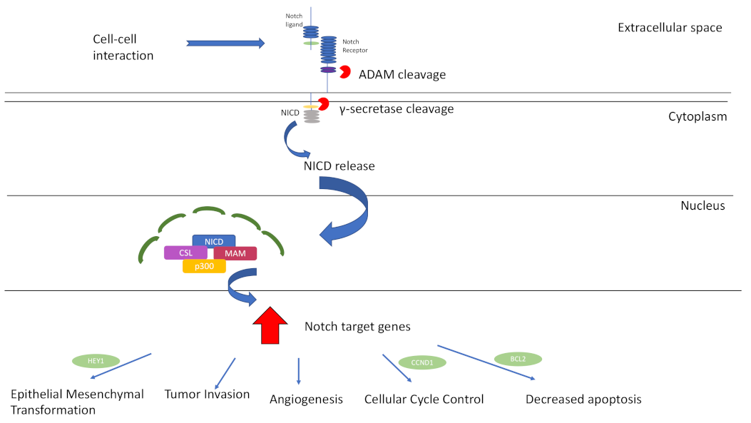
The Notch intracellular-signalling pathway is a highly conserved pathway that plays an integral role in embryogenesis. It is also a key regulator of several cellular processes in adults, such as proliferation, differentiation and survival [64]. The Notch-signalling pathway is described in Figure 4. Dysregulated Notch-signalling pathway has been implicated in cancer pathogenesis for decades, since its first association with human T-cell acute lymphoblastic leukaemia (ALL) was noted in 1991 [65]. Aberrant Notch expression is associated with many solid carcinomas, including breast, prostate, colorectal, lung and central nervous system cancer [66]. In terms of its tumorigenic properties, Notch upregulation promotes epithelial-to-mesenchymal transition (EMT), angiogenesis, tumour invasion and cellular survival [67]. Depending on the context, Notch has tumour-suppressive genetic (TSG) properties [43]. Notch behaves as a TSG in oral squamous cell carcinoma [68].
In recent years, several bodies of work have aimed to uncover the association with aberrant Notch signalling and ACC disease pathogenesis. Notch pathway mutations have been shown to be present in between 11 and 29% of ACC patients [12,72]. Aberrant Notch signalling promotes ACC proliferation and plays an important role in tumour metastasis via neoangiogenic induction, promoting tumour growth and survival [73]. Notch1, Notch2, Notch3 and Notch4 were all upregulated in metastatic salivary gland ACC cell lines. Furthermore, an important role for Notch4 was identified in the metastasis of salivary gland ACC, and the knockdown of Notch4 using small-interfering RNA (siRNA) inhibited metastatic invasion of ACC cells [74]. Ho et al. analysed 1045 ACC patients and found that Notch was significantly overexpressed in R/M tumours when compared to primary tumours [43]. Another study by Su et al. demonstrated that Notch1 was overexpressed in salivary gland ACC R/M tissue samples, and this overexpression was associated with disease R/M [75]. They showed that Notch1 overexpression drove cellular processes, such as tumour migration, invasion, proliferation and cell growth. Inhibition of Notch1 decreased tumorigenicity through promoting programmed cell death. These results further demonstrate that Notch1 plays an integral role in ACC metastasis [75]. Notch1 activation promotes upregulation of the antiapoptotic and cell-cycle genes BCL-2 ad CCND1, which is a potential mechanism by which Notch1 drives ACC metastasis [73].
Ferrarotto et al. genotyped 102 ACC tissue samples and demonstrated that Notch1 mutations were associated with an aggressive subgroup and conferred a poor prognosis [76]. Notch1 mutations were associated with bone and liver metastasis as well as a significantly shorter overall survival when compared to the Notch1 wildtype cohort. This study also suggests that further research investigating Notch1 inhibitors as potential therapeutic targets in the nonsurgical management of ACC is warranted [76]. Sajed et al. performed whole-exome sequencing on 194 cases of confirmed ACC of the salivary gland and demonstrated that Notch1 gain-of-function mutations correlated with a solid disease histological pattern, as well as being associated with a poorer clinical outcome [77]. Rettig et al. analysed whole-genome sequencing results from 25 ACC patients and found that Notch1 was the most commonly mutated gene, present in 12% of tumours [78].
There is a growing body of evidence that Notch1 inhibition may be a promising treatment option for ACC. A phase I clinical trial found that 17% of patients demonstrated at least partial disease stabilization when treated with the Notch1 inhibitor, brontictuzumab [79]. Preliminary results involving the upstream Notch1 inhibitor CB-103 showed that the median progression-free survival was almost 22 weeks, and the disease control rate was 79% after 8 weeks and 58% after 20 weeks. A total of 16% of patients with Notch mutations had stable disease confirmed by radiology after six months [80]. A study of AL101 in patients with ACC (ACCURACY) is a phase II multicentre trial analysing the effect of Notch inhibitor AL101 in R/M ACC patients harbouring Notch1-, Notch2-, Notch3- or Notch4-activating mutations. AL101 is a selective gamma-secretase inhibitor, and preliminary trial results indicate a disease control rate of 68%, with 15% of patients demonstrating partial responses [81].

Notch Signalling and Lacrimal Gland ACC

Notch signalling is essential for the correct development of the lacrimal gland [82]. Notch has recently been identified as a player in the pathogenesis of lacrimal gland ACC, although there are limited studies in the literature that fully establish its role. Sant et al. used whole-exome sequencing to screen for and identify mutations in lacrimal gland ACC. Notch1 and Notch2 mutations were associated with functionally severe disease. These mutations were present in 31% of patient samples analysed. This study identifies the Notch signalling pathway as a potential therapeutic target for lacrimal gland ACC [83]. Anjum et al. studied the expression of Notch1 receptor for the first time in the lacrimal gland, and activated Notch 1 (NICD) in 23 cases of lacrimal gland ACC. They found that Notch1 receptor was overexpressed in 65% of patients and NICD was overexpressed in 39% of cases. Notch1 overexpression conferred a poorer prognosis and was correlated with significantly reduced disease-free survival rates [9]. This study is the first to identify a key role for Notch1 in lacrimal gland ACC pathogenesis, and its overexpression is associated with an aggressive disease phenotype. Notch1 is overexpressed in other types of cancer, including papillary thyroid carcinoma [84], colorectal cancer [85], lung adenocarcinoma [86] and breast cancer, and its upregulation is associated with aggressive disease phenotypes, disease progression [66] and poor clinical prognoses.
Overall, the collective deregulation of Notch signalling pathway genes consolidates it as a central mediator of ACC pathogenesis.

4. DNA Damage Repair Gene (DDRG) Mutations

DNA damage repair genes (DDRGs) are integral for the proper maintenance of genomic stability [87,88]. There are two primary DDR pathways—base excision repair and nucleotide excision repair [89]. Collectively, DDR response pathways comprise a complex network of proteins that identify DNA damage, signal its presence and promote its repair in a substrate-dependent manner [90]. Approximately 450 genes code for DDR proteins [91]. DDRGs are important cell-cycle regulators, as well as critical initiators of apoptosis in situations when DNA is damaged beyond repair.
Dysregulation and aberrant expression of DDRGs have been implicated with the development of a plethora of disease pathologies, including neurodegenerative conditions, ageing, immune deficiencies and cancer pathogenesis [89]. Defective DDRGs lead to a blockade of critical metabolic processes, such as transcription and replication, or mutations resulting in cellular senescence and apoptosis [90,92]. DDRG mutations promote carcinogenesis and accelerate cancer progression. DDR pathways have also been studied as potential targeted therapeutic options in cancer treatment [91,93].
ACC occurs due to somatic aberrations in the human genome. Genetic alterations are defined by high-level amplifications, somatic mutations, structural variants and homozygous deletions. These alterations take place at a cellular level in somatic tissues post-fertilisation and do not involve the germline; rather, these mutations spontaneously occur throughout a patient’s lifetime, commonly as a result of mistakes in DDR mechanisms [90]. Due to the low mutational burden observed in common tumour suppressor genes and oncogenes, a predisposition is present whereby alterations in specific transcriptional regulatory genes are upregulated by alterations in chromatin structure, which are the driving force of the neoplastic process in ACC [94,95].
Although there is a lack of data in general exploring the role of DDR pathways in salivary gland malignancies [96], DDR genes have been recently implicated in ACC pathogenesis. Ho et al. demonstrated that 27% of ACC patients demonstrated altered expression of DDR/checkpoint signalling genes, including TP53, UHRF1, ATM and BRCA1 [12]. Andersson et al. were the first group to note the overexpression of ATR in primary ACC cell lines. ATR is a DNA-damage sensor kinase that is activated by replication stress and acts downstream of MYB [18]. On a cellular level, ATR overexpression exhibits oncogenic properties and is critical for tumour survival with aberrant cell-cycle check points or DNA repair mechanisms [97]. Lecona et al. demonstrated that ATR overactivation resulted in the activation of DDR genes, cell-cycle genes and DNA replication genes. The net effect is genomic stability, which in turn promotes cancer pathogenesis. Furthermore, treatment with an ATR kinase inhibitor (VX-970) induced programmed cell death in ACC cells that are MYB-positive and also significantly decreased cellular proliferation rates and inhibited ACC tumour growth, with one mouse demonstrating tumour regression. This study is the first to identify ATR as an attractive novel downstream therapeutic target of MYB [98]. There are several clinical trials ongoing targeting the development of ATR inhibitors for several cancer types [98].
Felix et al. examined the mutational profile of DDR genes in salivary gland ACC pathogenesis and noted high expression levels of a nucleotide excision repair gene XPF in minor salivary gland ACC samples [96]. Furthermore, the APE1 protein that acts as a redox transcription factor coactivator and is involved in the base excision repair pathway was expressed in the nucleus/cytoplasm of 50% of ACC cases. Cystoplasmic/nuclear-cytoplasmic APE1 expression is associated with increased cellular proliferation and metabolism [96,99].
ARID1A is one of the most frequently mutated genes in cancers. It is involved in transcription inactivation and repression, similar to SMARCA2, a TSG found mutated in 5% of ACC patients [39]. Alterations in the gene in conjunction with other ACC driver mutations lead to ACC proliferation due to its inactivating effect resulting in a TSG function. Inactivation of ARID1A can lead to synthetic lethality via inactivation of the SWI/SNF complex by deletions or knockdowns compromising double-stranded DNA repair. This increases sensitivity to DNA-damaging agents [100]. Loss of ARID1A impairs the G2/M DNA damage checkpoint.
Taken together, disruptions in the DDR pathway increase mutagenesis and genomic instability thereby promoting neoplastic progression [92].

5. Epigenetic Modifications in ACC Pathogenesis

The field of epigenetics refers to the heritable modification of gene expression without alteration of the DNA sequence itself [101]. Epigenetic modifications have been widely studied in tumorigenesis, and it is known that malignant cells exploit epigenetic mechanisms in order to control invasion, therapy resistance and growth [102]. Epigenetic alterations involve chromatin modifications in response to hormonal and/or environmental stimuli, as well as histone acetylation affecting chromatin stability and noncoding RNA regulations [103]. Targeting epigenetics is a promising therapeutic strategy for cancer treatment, either as a monotherapy or in conjunction with adjuvant treatment [103].
In terms of ACC pathogenesis, epigenetic modifications remain poorly understood, but have shown to upregulate tumorigenic pathways associated with poor prognosis, such as MYB translocations with NFIB. Using exome sequencing of 24 patients with confirmed ACC, Frierson et al. identified chromatin remodelling mutations in 50% of ACC patients, including ARID1A, CREBBP, EP300 and KDM6A. Aberrant expression of these chromatin remodelling genes may imply that ACC pathogenesis is driven by histone modifications, resulting in transcriptional reprogramming [94]. Mutations in the epigenetic modifier genes CREPPB, KANSL1 are involved in histone acetyltransferase activity, KDM6A involved in histone demethylase activity, BCOR involved in histone deacetylase activity and ARID1B in the SWJ/SNF chromatin remodelling complexes are found in approximately 50% of ACC. Recurrent/metastatic ACC are enriched for BCOR and KDM6A mutations [104].
Using whole-genome sequencing analyses of 25 fresh-frozen salivary gland ACC samples, Rettig et al. proposed a role for epigenetic regulation in ACC pathogenesis. Their results noted alterations in the chromatin-remodelling genes SMARCA2, KDM6A and CREBBP [78]. Ho et al. also demonstrated that 7% of ACC patients possessed a CREBBP mutation, a histone acetyltransferase enabling transcription across critical signalling pathways, had been mutated in 7% of patients in previous ACC studies [12]. TERT-promoter genes have been described in ACC tumours without MYB/MYBL1 fusions and without Notch mutations, possibly representing an alternative method of tumourigenesis [43]. Further studies are necessary to fully elucidate the role of chromatin remodelling and epigenetic modifications in ACC pathogenesis. They could be targeted by epigenetic drug therapies.

6. Future Directions and Conclusions

ACC remains both a diagnostic and treatment challenge for clinicians, and the molecular mechanisms that underscore disease pathogenesis are only beginning to emerge. This review aimed to explore the molecular landscape of ACC pathogenesis, paying particular emphasis to MYB/NFIB gene fusion and its potential as a diagnostic biomarker, as well as reporting on the role of aberrant Notch signalling as prognostic markers, mutated DNA damage repair genes and epigenetic modifications as drivers of disease pathogenesis. Whilst clinical trials aimed at inhibiting Notch have shown initial promise, it is of paramount importance that further large-scale studies aimed at targeting the underlying cellular mechanisms and biological processes that govern ACC are conducted. The development of targeted therapeutic options to ameliorate disease will pave the way for personalised ACC therapy, ultimately improving overall patient survival rates and reducing morbidity and mortality associated with the disease.

Author Contributions

Conceptualization, S.K. and K.K.; writing S.K.P., S.K and K.K.; supervision, S.K. All authors have read and agreed to the published version of the manuscript.

Funding

This research received no external funding.

Institutional Review Board Statement

Not applicable.

Informed Consent Statement

Not applicable.

Data Availability Statement

No new data were created or analyzed in this study. Data sharing is not applicable to this article..

Conflicts of Interest

The authors declare no conflict of interest.

References

  1. Coca-Pelaz, A.; Rodrigo, J.P.; Bradley, P.J.; Poorten, V.V.; Triantafyllou, A.; Hunt, J.L.; Strojan, P.; Rinaldo, A.; Haigentz, M.; Takes, R.P.; et al. Adenoid cystic carcinoma of the head and neck–An update. Oral Oncol. 2015, 51, 652–661. [Google Scholar] [CrossRef]
  2. Thierauf, J.; Ramamurthy, N.; Jo, V.Y.; Robinson, H.; Frazier, R.P.; Gonzalez, J.; Pacula, M.; Meneses, E.D.; Nose, V.; Nardi, V.; et al. Clinically Integrated Molecular Diagnostics in Adenoid Cystic Carcinoma. Oncologist 2019, 24, 1356–1367. [Google Scholar] [CrossRef]
  3. Chummun, S.; McLean, N.; Kelly, C.; Dawes, P.; Fellows, S.; Meikle, D.; Soames, J. Adenoid cystic carcinoma of the head and neck. Br. J. Plast. Surg. 2001, 54, 476–480. [Google Scholar] [CrossRef]
  4. Belulescu, I.C.; Margaritescu, C.; Dumitrescu, C.I.; DĂguci, L.; Munteanu, C.; Margaritescu, O.C. Adenoid Cystic Carcinoma of Salivary Gland: A Ten-Year Single Institute Experience. Curr. Health Sci. J. 2020, 46, 56–65. [Google Scholar] [CrossRef] [PubMed]
  5. Li, N.; Xu, L.; Zhao, H.; El-Naggar, A.K.; Sturgis, E.M. A comparison of the demographics, clinical features, and survival of patients with adenoid cystic carcinoma of major and minor salivary glands versus less common sites within the Surveillance, Epidemiology, and End Results registry. Cancer 2012, 118, 3945–3953. [Google Scholar] [CrossRef]
  6. von Holstein, S.L.; Therkildsen, M.H.; Prause, J.U.; Stenman, G.; Siersma, V.D.; Heegaard, S. Lacrimal gland lesions in Denmark between 1974 and 2007. Acta Ophthalmol. 2013, 91, 349–354. [Google Scholar] [CrossRef] [PubMed]
  7. Grossniklaus, H.E.; Eberhart, C.G.; Kivelä, T.T. WHO Classification of Tumours of the Eye; WHO: Geneva, Switzerland, 2018. [Google Scholar]
  8. Bernardini, F.P.; Devoto, M.H.; Croxatto, J.O. Epithelial tumors of the lacrimal gland: An update. Curr. Opin. Ophthalmol. 2008, 19, 409–413. [Google Scholar] [CrossRef] [PubMed]
  9. Anjum, S.; Sen, S.; Pushker, N.; Bajaj, M.S.; Kashyap, S.; Bakhshi, S.; Chosdol, K.; Meel, R.; Sharma, M.C. Prognostic impact of Notch1 receptor and clinicopathological High-Risk Predictors in lacrimal gland adenoid cystic carcinoma. Acta Ophthalmol. 2021, 99, e1467–e1473. [Google Scholar] [CrossRef]
  10. Lesueur, P.; Rapeaud, E.; De Marzi, L.; Goudjil, F.; Levy, C.; Galatoire, O.; Jacomet, P.V.; Dendale, R.; Calugaru, V. Adenoid Cystic Carcinoma of the Lacrimal Gland: High Dose Adjuvant Proton Therapy to Improve Patients Outcomes. Front. Oncol. 2020, 10, 135. [Google Scholar] [CrossRef] [PubMed]
  11. Xu, B.; Drill, E.; Ho, A.; Ho, A.; Dunn, L.; Prieto-Granada, C.N.; Chan, T.; Ganly, I.; Ghossein, R.; Katabi, N. Predictors of Outcome in Adenoid Cystic Carcinoma of Salivary Glands: A Clinicopathologic Study with Correlation between MYB Fusion and Protein Expression. Am. J. Surg. Pathol. 2017, 41, 1422–1432. [Google Scholar] [CrossRef]
  12. Ho, A.S.; Kannan, K.; Roy, D.M.; Morris, L.G.T.; Ganly, I.; Katabi, N.; Ramaswami, D.; Walsh, L.A.; Eng, S.; Huse, J.T.; et al. The mutational landscape of adenoid cystic carcinoma. Nat. Genet. 2013, 45, 791–798. [Google Scholar] [CrossRef]
  13. Williams, M.D.; Al-Zubidi, N.; Debnam, J.M.; Shinder, R.; DeMonte, F.; Esmaeli, B. Bone Invasion by Adenoid Cystic Carcinoma of the Lacrimal Gland: Preoperative Imaging Assessment and Surgical Considerations. Ophthalmic Plast. Reconstr. Surg. 2010, 26, 403–408. [Google Scholar] [CrossRef] [PubMed]
  14. Gamel, J.W.; Font, R.L. Adenoid cystic carcinoma of the lacrimal gland: The clinical significance of a basaloid histologic pattern. Hum. Pathol. 1982, 13, 219–225. [Google Scholar] [CrossRef] [PubMed]
  15. Font, R.L.; Smith, S.L.; Bryan, R.G. Malignant Epithelial Tumors of the Lacrimal Gland: A Clinicopathologic Study of 21 Cases. Arch. Ophthalmol. 1998, 116, 613–616. [Google Scholar] [CrossRef] [PubMed]
  16. Emerick, C.; Mariano, F.V.; Vargas, P.A.; Nör, J.E.; Squarize, C.H.; Castilho, R.M. Adenoid Cystic Carcinoma from the salivary and lacrimal glands and the breast: Different clinical outcomes to the same tumor. Crit. Rev. Oncol. Hematol. 2022, 179, 103792. [Google Scholar] [CrossRef]
  17. Esmaeli, B.; Ahmadi, M.A.; Youssef, A.; Diba, R.; Amato, M.; Myers, J.; Kies, M.; El-Naggar, A. Outcomes in Patients with Adenoid Cystic Carcinoma of the Lacrimal Gland. Ophthalmic Plast. Reconstr. Surg. 2004, 20, 156. [Google Scholar] [CrossRef]
  18. Andersson, M.K.; Mangiapane, G.; Nevado, P.T.; Tsakaneli, A.; Carlsson, T.; Corda, G.; Nieddu, V.; Abrahamian, C.; Chayka, O.; Rai, L.; et al. ATR is a MYB regulated gene and potential therapeutic target in adenoid cystic carcinoma. Oncogenesis 2020, 9, 5. [Google Scholar] [CrossRef] [PubMed]
  19. Barrett, A.W.; Speight, P.M. Perineural invasion in adenoid cystic carcinoma of the salivary glands: A valid prognostic indicator? Oral Oncol. 2009, 45, 936–940. [Google Scholar] [CrossRef]
  20. Spiro, R.H. Distant metastasis in adenoid cystic carcinoma of salivary origin. Am. J. Surg. 1997, 174, 495–498. [Google Scholar] [CrossRef]
  21. Chen, T.Y.; Keeney, M.G.; Chintakuntlawar, A.V.; Knutson, D.L.; Kloft-Nelson, S.; Greipp, P.T.; Garrity, J.A.; Salomao, D.R.; Garcia, J.J. Adenoid cystic carcinoma of the lacrimal gland is frequently characterized by MYB rearrangement. Eye 2017, 31, 720–725. [Google Scholar] [CrossRef]
  22. Singh, F.M.; Mak, S.Y.; Bonington, S.C. Patterns of spread of head and neck adenoid cystic carcinoma. Clin. Radiol. 2015, 70, 644–653. [Google Scholar] [CrossRef] [PubMed]
  23. Laurie, S.A.; Ho, A.L.; Fury, M.G.; Sherman, E.; Pfister, D.G. Systemic therapy in the management of metastatic or locally recurrent adenoid cystic carcinoma of the salivary glands: A systematic review. Lancet Oncol. 2011, 12, 815–824. [Google Scholar] [CrossRef] [PubMed]
  24. Rodriguez-russo, C.A.; Junn, J.C.; Yom, S.S.; Bakst, R.L. Radiation Therapy for Adenoid Cystic Carcinoma of the Head and Neck. Cancers 2021, 13, 6335. [Google Scholar] [CrossRef] [PubMed]
  25. Maniar, A.; Saqi, A.; Troob, S.H.; Belinsky, I.; Charles, N.C.; Gobin, Y.P.; Marr, B.P. Targeted Neoadjuvant Intra-arterial Chemotherapy in Lacrimal Gland Adenoid Cystic Carcinoma: A Histological Correlation Using Apoptotic Tumor Markers. Ophthalmic Plast. Reconstr. Surg. 2022, 38, e28–e33. [Google Scholar] [CrossRef]
  26. Du, F.; Zhou, C.-X.; Gao, Y. Myoepithelial differentiation in cribriform, tubular and solid pattern of adenoid cystic carcinoma: A potential involvement in histological grading and prognosis. Ann. Diagn. Pathol. 2016, 22, 1092–9134. [Google Scholar] [CrossRef]
  27. Szanto, P.A.; Luna, M.A.; Tortoledo, M.E.; White, R.A. Histologic grading of adenoid cystic carcinoma of the salivary glands. Cancer 1984, 54, 1062–1069. [Google Scholar] [CrossRef]
  28. Batsakis, J.G.; Luna, M.A.; El-Naggar, A. Histopathologic grading of salivary gland neoplasms: III. Adenoid cystic carcinomas. Ann. Otol. Rhinol. Laryngol. 1990, 99, 1007–1009. [Google Scholar] [CrossRef]
  29. Perzin, K.H.; Gullane, P.; Clairmont, A.C. Adenoid cystic carcinomas arising in salivary glands. A correlation of histologic features and clinical course. Cancer 1978, 42, 265–282. [Google Scholar] [CrossRef]
  30. Osborn, D.A. Morphology and the natural history of cribriform adenocarcinoma (adenoid cystic carcinoma). J. Clin. Pathol. 1977, 30, 195–205. [Google Scholar] [CrossRef]
  31. van Weert, S.; van der Waal, I.; Witte, B.I.; René Leemans, C.; Bloemena, E. Histopathological grading of adenoid cystic carcinoma of the head and neck: Analysis of currently used grading systems and proposal for a simplified grading scheme. Oral Oncol. 2015, 51, 71–76. [Google Scholar] [CrossRef]
  32. Ouyang, D.; Liang, L.; Zheng, G.; Ke, Z.; Weng, D.; Yang, W.; Su, Y.; Liao, G. Risk factors and prognosis for salivary gland adenoid cystic carcinoma in southern China: A 25-year retrospective study. Medicine 2017, 96, e5964. [Google Scholar] [CrossRef] [PubMed]
  33. Andersson, M.K.; Afshari, M.K.; Andrén, Y.; Wick, M.J.; Stenman, G. Targeting the Oncogenic Transcriptional Regulator MYB in Adenoid Cystic Carcinoma by Inhibition of IGF1R/AKT Signaling. JNCI J. Natl. Cancer Inst. 2017, 109, djx017. [Google Scholar] [CrossRef] [PubMed]
  34. Stephens, P.J.; Davies, H.R.; Mitani, Y.; Van Loo, P.; Shlien, A.; Tarpey, P.S.; Papaemmanuil, E.; Cheverton, A.; Bignell, G.R.; Butler, A.P.; et al. Whole exome sequencing of adenoid cystic carcinoma. J. Clin. Investig. 2013, 123, 2965–2968. [Google Scholar] [CrossRef]
  35. Nambiar, M.; Kari, V.; Raghavan, S.C. Chromosomal translocations in cancer. Biochim. Biophys. Acta (BBA) Rev. Cancer 2008, 1786, 139–152. [Google Scholar] [CrossRef]
  36. Wagner, V.P.; Bingle, C.D.; Bingle, L. MYB-NFIB fusion transcript in adenoid cystic carcinoma: Current state of knowledge and future directions. Crit. Rev. Oncol. Hematol. 2022, 176, 103745. [Google Scholar] [CrossRef]
  37. Nowell, P.C. A minute chromosome in human chronic granulocytic leukemia. Science 1960, 132, 1497. [Google Scholar]
  38. Drier, Y.; Cotton, M.J.; Williamson, K.E.; Gillespie, S.M.; Ryan, R.J.H.; Kluk, M.J.; Carey, C.D.; Rodig, S.J.; Sholl, L.M.; Afrogheh, A.H.; et al. An oncogenic MYB feedback loop drives alternate cell fates in adenoid cystic carcinoma. Nat. Genet. 2016, 48, 265–272. [Google Scholar] [CrossRef] [PubMed]
  39. Moon, A.; Cohen, C.; Siddiqui, M.T. MYB expression: Potential role in separating adenoid cystic carcinoma (ACC) from pleomorphic adenoma (PA). Diagn. Cytopathol. 2016, 44, 799–804. [Google Scholar] [CrossRef]
  40. Brill, L.B., II; Kanner, W.A.; Fehr, A.; Andrén, Y.; Moskaluk, C.A.; Löning, T.; Stenman, G.; Frierson, H.F. Analysis of MYB expression and MYB-NFIB gene fusions in adenoid cystic carcinoma and other salivary neoplasms. Mod. Pathol. 2011, 24, 1169–1176. [Google Scholar] [CrossRef]
  41. Claringbould, A.; Zaugg, J.B. Enhancers in disease: Molecular basis and emerging treatment strategies. Trends Mol. Med. 2021, 27, 1060–1073. [Google Scholar] [CrossRef]
  42. Botten, G.A.; Zhang, Y.; Dudnyk, K.; Liu, X.; Sanders, J.T.; Imanci, A.; Droin, N.; Cao, H.; Kaphle, P.; Dickerson, K.E.; et al. Structural Variation Cooperates with Permissive Chromatin to Control Enhancer Hijacking-Mediated Oncogenic Transcription. Blood 2022, 140 (Suppl. 1), 1007–1008. [Google Scholar] [CrossRef]
  43. Ho, A.S.; Ochoa, A.; Jayakumaran, G.; Zehir, A.; Mayor, C.V.; Tepe, J.; Makarov, V.; Dalin, M.G.; He, J.; Bailey, M.; et al. Genetic hallmarks of recurrent/metastatic adenoid cystic carcinoma. J. Clin. Investig. 2019, 129, 4276–4289. [Google Scholar] [CrossRef] [PubMed]
  44. Bell, D.; Sniegowski, M.C.; Wani, K.; Prieto, V.; Esmaeli, B. Mutational landscape of lacrimal gland carcinomas and implications for treatment. Head Neck 2016, 38, E724–E729. [Google Scholar] [CrossRef]
  45. Fujii, K.; Murase, T.; Beppu, S.; Saida, K.; Takino, H.; Masaki, A.; Ijichi, K.; Kusafuka, K.; Iida, Y.; Onitsuka, T.; et al. MYB, MYBL1, MYBL2 and NFIB gene alterations and MYC overexpression in salivary gland adenoid cystic carcinoma. Histopathology 2017, 71, 823–834. [Google Scholar] [CrossRef]
  46. Mitani, Y.; Li, J.; Rao, P.H.; Zhao, Y.-J.; Bell, D.; Lippman, S.M.; Weber, R.S.; Caulin, C.; El-Naggar, A.K. Comprehensive Analysis of the MYB-NFIB Gene Fusion in Salivary Adenoid Cystic Carcinoma: Incidence, Variability, and Clinicopathologic Significance. Clin. Cancer Res. 2010, 16, 4722–4731. [Google Scholar] [CrossRef] [PubMed]
  47. Persson, M.; Andrén, Y.; Mark, J.; Horlings, H.M.; Persson, F.; Stenman, G. Recurrent fusion of MYB and NFIB transcription factor genes in carcinomas of the breast and head and neck. Proc. Natl. Acad. Sci. USA 2009, 106, 18740–18744. [Google Scholar] [CrossRef]
  48. Lin, Q.-Q.; Sun, J.-L.; Wang, F.; Zhang, H.-Z.; Zhou, G.; Xi, Q. Current understanding of adenoid cystic carcinoma in the gene expression and targeted therapy. Holist. Integr. Oncol. 2023, 2, 7. [Google Scholar] [CrossRef]
  49. Brayer, K.J.; Frerich, C.A.; Kang, H.; Ness, S.A. Recurrent fusions in MYB and MYBL1 define a common, transcription factor–driven oncogenic pathway in salivary gland adenoid cystic carcinoma. Cancer Discov. 2016, 6, 176–187. [Google Scholar] [CrossRef]
  50. Andreasen, S.; Tan, Q.; Agander, T.K.; Steiner, P.; Bjørndal, K.; Høgdall, E.; Larsen, S.R.; Erentaite, D.; Olsen, C.H.; Ulhøi, B.P.; et al. Adenoid cystic carcinomas of the salivary gland, lacrimal gland, and breast are morphologically and genetically similar but have distinct microRNA expression profiles. Mod. Pathol. 2018, 31, 1211–1225. [Google Scholar] [CrossRef]
  51. Shakoori, A.R. Fluorescence in situ hybridization (FISH) and its applications. In Chromosome Structure and Aberrations; Bhat, T.A., Wani, A.A., Eds.; Springer: New Delhi, India, 2017; pp. 343–367. [Google Scholar] [CrossRef]
  52. Heyer, E.E.; Deveson, I.W.; Wooi, D.; Selinger, C.I.; Lyons, R.J.; Hayes, V.M.; O’toole, S.A.; Ballinger, M.L.; Gill, D.; Thomas, D.M.; et al. Diagnosis of fusion genes using targeted RNA sequencing. Nat. Commun. 2019, 10, 1388. [Google Scholar] [CrossRef]
  53. Chen, W.; Kalscheuer, V.; Tzschach, A.; Menzel, C.; Ullmann, R.; Schulz, M.H.; Erdogan, F.; Li, N.; Kijas, Z.; Arkesteijn, G.; et al. Mapping translocation breakpoints by next-generation sequencing. Genome Res. 2008, 18, 1143–1149. [Google Scholar] [CrossRef] [PubMed]
  54. Kumar, S.; Razzaq, S.K.; Vo, A.D.; Gautam, M.; Li, H. Identifying fusion transcripts using next generation sequencing. WIREs RNA 2016, 7, 811–823. [Google Scholar] [CrossRef] [PubMed]
  55. Abel, H.J.; Al-Kateb, H.; Cottrell, C.E.; Bredemeyer, A.J.; Pritchard, C.C.; Grossmann, A.H.; Wallander, M.L.; Pfeifer, J.D.; Lockwood, C.M.; Duncavage, E.J. Detection of gene rearrangements in targeted clinical next-generation sequencing. J. Mol. Diagn. 2014, 16, 405–417. [Google Scholar] [CrossRef] [PubMed]
  56. McIntyre, J.B.; Ko, J.J.; Siever, J.; Chan, A.M.Y.; Simpson, R.H.W.; Hao, D.; Lau, H.Y. MYB-NFIB gene fusions identified in archival adenoid cystic carcinoma tissue employing NanoString analysis: An exploratory study. Diagn. Pathol. 2019, 14, 78. [Google Scholar] [CrossRef]
  57. Humtsoe, J.O.; Kim, H.-S.; Jones, L.; Cevallos, J.; Boileau, P.; Kuo, F.; Morris, L.G.T.; Ha, P. Development and Characterization of MYB-NFIB Fusion Expression in Adenoid Cystic Carcinoma. Cancers 2022, 14, 2263. [Google Scholar] [CrossRef]
  58. Pham, T.; Pereira, L.; Roth, S.; Galletta, L.; Link, E.; Akhurst, T.; Solomon, B.; Michael, M.; Darcy, P.; Sampurno, S.; et al. First-in-human phase I clinical trial of a combined immune modulatory approach using TetMYB vaccine and Anti-PD-1 antibody in patients with advanced solid cancer including colorectal or adenoid cystic carcinoma: The MYPHISMO study protocol (NCT03287427). Contemp. Clin. Trials Commun. 2019, 16, 100409. [Google Scholar] [CrossRef]
  59. Ferrarotto, R.; Mitani, Y.; McGrail, D.J.; Li, K.; Karpinets, T.V.; Bell, D.; Frank, S.J.; Song, X.; Kupferman, M.E.; Liu, B.; et al. Proteogenomic Analysis of Salivary Adenoid Cystic Carcinomas Defines Molecular Subtypes and Identifies Therapeutic Targets. Clin. Cancer Res. 2023, 27, 852–864. [Google Scholar] [CrossRef]
  60. Brayer, K.J.; Kang, H.; El-Naggar, A.K.; Andreasen, S.; Homøe, P.; Kiss, K.; Mikkelsen, L.; Heegaard, S.; Pelaez, D.; Moeyersoms, A.; et al. Dominant Gene Expression Profiles Define Adenoid Cystic Carcinoma (ACC) from Different Tissues: Validation of a Gene Signature Classifier for Poor Survival in Salivary Gland ACC. Cancers 2023, 15, 1390. [Google Scholar] [CrossRef]
  61. Frerich, C.A.; Brayer, K.J.; Painter, B.M.; Kang, H.; Mitani, Y.; El-Naggar, A.K.; Ness, S.A. Transcriptomes define distinct subgroups of salivary gland adenoid cystic carcinoma with different driver mutations and outcomes. Oncotarget 2017, 9, 7341–7358. [Google Scholar] [CrossRef]
  62. von Holstein, S.L.; Fehr, A.; Persson, M.; Therkildsen, M.H.; Prause, J.U.; Heegaard, S.; Stenman, G. Adenoid Cystic Carcinoma of the Lacrimal Gland: MYB Gene Activation, Genomic Imbalances, and Clinical Characteristics. Ophthalmology 2013, 120, 2130–2138. [Google Scholar] [CrossRef]
  63. Kim, J.; Geyer, F.C.; Martelotto, L.G.; Ng, C.K.Y.; Lim, R.S.; Selenica, P.; Li, A.; Pareja, F.; Fusco, N.; Edelweiss, M.; et al. MYBL1 rearrangements and MYB amplification in breast adenoid cystic carcinomas lacking the MYBNFIB fusion gene. J. Pathol. 2018, 244, 143–150. [Google Scholar] [CrossRef] [PubMed]
  64. Ranganathan, P.; Weaver, K.L.; Capobianco, A.J. Notch signalling in solid tumours: A little bit of everything but not all the time. Nat. Rev. Cancer. 2011, 11, 338–351. [Google Scholar] [CrossRef] [PubMed]
  65. Zhou, B.; Lin, W.; Long, Y.; Yang, Y.; Zhang, H.; Wu, K.; Chu, Q. Notch signaling pathway: Architecture, disease, and therapeutics. Signal Transduct. Target. Ther. 2022, 7, 95. [Google Scholar] [CrossRef] [PubMed]
  66. Yuan, X.; Zhang, M.; Wu, H.; Xu, H.; Han, N.; Chu, Q.; Yu, S.; Chen, Y.; Wu, K. Expression of Notch1 Correlates with Breast Cancer Progression and Prognosis. PLoS ONE 2015, 10, e0131689. [Google Scholar] [CrossRef]
  67. Misiorek, J.O.; Przybyszewska-Podstawka, A.; Kałafut, J.; Paziewska, B.; Rolle, K.; Rivero-Müller, A.; Nees, M. Context Matters: NOTCH Signatures and Pathway in Cancer Progression and Metastasis. Cells 2021, 10, 94. [Google Scholar] [CrossRef]
  68. Pickering, C.R.; Zhang, J.; Yoo, S.Y.; Bengtsson, L.; Moorthy, S.; Neskey, D.M.; Zhao, M.; Alves, M.V.O.; Chang, K.; Drummond, J.; et al. Integrative Genomic Characterization of Oral Squamous Cell Carcinoma Identifies Frequent Somatic Drivers. Cancer Discov. 2013, 3, 770–781. [Google Scholar] [CrossRef]
  69. Gallenstein, N.; Tichy, L.; Weigand, M.A.; Schenz, J. Notch Signaling in Acute Inflammation and Sepsis. Int. J. Mol. Sci. 2023, 24, 3458. [Google Scholar] [CrossRef]
  70. O’Brien, K.A.; Murray, A.J.; Simonson, T.S. Notch Signaling and Cross-Talk in Hypoxia: A Candidate Pathway for High-Altitude Adaptation. Life 2022, 12, 437. [Google Scholar] [CrossRef]
  71. Takebe, N.; Nguyen, D.; Yang, S.X. Targeting Notch signaling pathway in cancer: Clinical development advances and challenges. Pharmacol. Ther. 2014, 141, 140–149. [Google Scholar] [CrossRef]
  72. Ross, J.S.; Wang, K.; Rand, J.V.; Sheehan, C.E.; Jennings, T.A.; Al-Rohil, R.N.; Otto, G.A.; Curran, J.C.; Palmer, G.; Downing, S.R.; et al. Comprehensive Genomic Profiling of Relapsed and Metastatic Adenoid Cystic Carcinomas by Next-generation Sequencing Reveals Potential New Routes to Targeted Therapies. Am. J. Surg. Pathol. 2014, 38, 235–238. [Google Scholar] [CrossRef]
  73. Miller, L.E.; Au, V.; Mokhtari, T.E.; Goss, D.; Faden, D.L.; Varvares, M.A. A Contemporary Review of Molecular Therapeutic Targets for Adenoid Cystic Carcinoma. Cancers 2022, 14, 992. [Google Scholar] [CrossRef]
  74. Ding, L.-C.; She, L.; Zheng, D.-L.; Huang, Q.-L.; Wang, J.-F.; Zheng, F.-F.; Lu, Y.-G. Notch-4 contributes to the metastasis of salivary adenoid cystic carcinoma. Oncol. Rep. 2010, 24, 363–368. [Google Scholar] [CrossRef]
  75. Su, B.H.; Qu, J.; Song, M.; Huang, X.-Y.; Hu, X.-M.; Xie, J.; Zhao, Y.; Ding, L.-C.; She, L.; Chen, J.; et al. NOTCH1 signaling contributes to cell growth, anti-apoptosis and metastasis in salivary adenoid cystic carcinoma. Oncotarget 2014, 5, 6885–6895. [Google Scholar] [CrossRef]
  76. Ferrarotto, R.; Mitani, Y.; Diao, L.; Guijarro, I.; Wang, J.; Zweidler-McKay, P.; Bell, D.; Jr, W.N.W.; Glisson, B.S.; Wick, M.J.; et al. Activating NOTCH1 mutations define a distinct subgroup of patients with adenoid cystic carcinoma who have poor prognosis, propensity to bone and liver metastasis, and potential responsiveness to Notch1 inhibitors. J. Clin. Oncol. 2017, 35, 352–360. [Google Scholar] [CrossRef]
  77. Sajed, D.P.; Faquin, W.C.; Carey, C.; Severson, E.A.; Afrogheh, A.H.; Johnson, C.A.; Blacklow, S.C.; Chau, N.G.; Lin, D.T.; Krane, J.F.; et al. Diffuse Staining for Activated NOTCH1 Correlates With NOTCH1 Mutation Status and Is Associated With Worse Outcome in Adenoid Cystic Carcinoma. Am. J. Surg. Pathol. 2017, 41, 1473–1482. [Google Scholar] [CrossRef]
  78. Rettig, E.M.; Talbot, C.C., Jr.; Sausen, M.; Jones, S.; Bishop, J.A.; Wood, L.D.; Tokheim, C.; Niknafs, N.; Karchin, R.; Fertig, E.J.; et al. Whole-Genome Sequencing of Salivary Gland Adenoid Cystic Carcinoma. Cancer Prev. Res. 2016, 9, 265–274. [Google Scholar] [CrossRef]
  79. Ferrarotto, R.; Eckhardt, G.; Patnaik, A.; LoRusso, P.; Faoro, L.; Heymach, J.; Kapoun, A.; Xu, L.; Munster, P. A phase I dose-escalation and dose-expansion study of brontictuzumab in subjects with selected solid tumors. Ann. Oncol. 2018, 29, 1561–1568. [Google Scholar] [CrossRef] [PubMed]
  80. Lopez Miranda, E.; Stathis, A.; Hess, D.; Racca, F.; Quon, D.; Rodon, J.; Gadea, O.S.S.; Garcia, J.M.P.; Nuciforo, P.; Vivancos, A.; et al. Phase 1 study of CB-103, a novel first-in-class inhibitor of the CSL-NICD gene transcription factor complex in human cancers. J. Clin. Oncol. 2021, 39 (Suppl. 15). [Google Scholar] [CrossRef]
  81. Ferrarotto, R.; Metcalf, R.; Rodriguez, C.P.; Muzaffar, J.; Even, C.; Perez, C.A.; Van Herpen, C.M.L.; Oliva, M.; Xia, B.; Bowles, D.W.; et al. Results of ACCURACY: A phase 2 trial of AL101, a selective gamma secretase inhibitor, in subjects with recurrent/metastatic (R/M) adenoid cystic carcinoma (ACC) harboring Notch activating mutations (Notchmut). J. Clin. Oncol. 2022, 40 (Suppl. 16), 6046. [Google Scholar]
  82. Ivanov, D. Notch Signaling-Induced Oscillatory Gene Expression May Drive Neurogenesis in the Developing Retina. Front. Mol. Neurosci. 2019, 12, 226. [Google Scholar] [CrossRef]
  83. Sant, D.W.; Tao, W.; Field, M.G.; Pelaez, D.; Jin, K.; Capobianco, A.; Dubovy, S.R.; Tse, D.T.; Wang, G. Whole exome sequencing of lacrimal gland adenoid cystic carcinoma. Investig. Ophthalmol. Vis. Sci. 2017, 58, BIO240–BIO246. [Google Scholar] [CrossRef] [PubMed]
  84. Piana, S.; Zanetti, E.; Bisagni, A.; Ciarrocchi, A.; Giordano, D.; Torricelli, F.; Rossi, T.; Ragazzi, M. Expression of NOTCH1 in thyroid cancer is mostly restricted to papillary carcinoma. Endocr. Connect. 2019, 8, 1089–1096. [Google Scholar] [CrossRef] [PubMed]
  85. Huang, R.; Tang, Q.; You, Q.; Liu, Z.; Wang, G.; Chen, Y.; Sun, Y.; Muhammad, S.; Wang, X. Disparity Expression of Notch1 in Benign and Malignant Colorectal Diseases. PLoS ONE 2013, 8, e81005. [Google Scholar] [CrossRef]
  86. Huang, J.; Song, H.; Liu, B.; Yu, B.; Wang, R.; Chen, L. Expression of Notch-1 and its clinical significance in different histological subtypes of human lung adenocarcinoma. J. Exp. Clin. Cancer Res. 2013, 32, 84. [Google Scholar] [CrossRef] [PubMed]
  87. Broustas, C.G.; Lieberman, H.B. DNA Damage Response Genes and the Development of Cancer Metastasis. Radiat. Res. 2014, 181, 111–130. [Google Scholar] [CrossRef]
  88. Peng, L.; Liang, J.; Wang, Q.; Chen, G. A DNA Damage Repair Gene Signature Associated With Immunotherapy Response and Clinical Prognosis in Clear Cell Renal Cell Carcinoma. Front. Genet. 2022, 13, 798846. [Google Scholar] [CrossRef]
  89. Curtin, N.J. DNA repair dysregulation from cancer driver to therapeutic target. Nat. Rev. Cancer. 2012, 12, 801–817. [Google Scholar] [CrossRef]
  90. Chatterjee, N.; Walker, G.C. Mechanisms of DNA damage, repair, and mutagenesis. Environ. Mol. Mutagen. 2017, 58, 235–263. [Google Scholar] [CrossRef]
  91. Gourley, C.; Balmaña, J.; Ledermann, J.A.; Serra, V.; Dent, R.; Loibl, S.; Pujade-Lauraine, E.; Boulton, S.J. Moving from poly (ADP-ribose) polymerase inhibition to targeting DNA repair and DNA damage response in cancer therapy. J. Clin. Oncol. 2019, 37, 2257–2269. [Google Scholar] [CrossRef]
  92. Knijnenburg, T.A.; Wang, L.; Zimmermann, M.T.; Chambwe, N.; Gao, G.F.; Cherniack, A.D.; Fan, H.; Shen, H.; Way, G.P.; Greene, C.S.; et al. Genomic and Molecular Landscape of DNA Damage Repair Deficiency across The Cancer Genome Atlas. Cell Rep. 2018, 23, 239–254.e6. [Google Scholar] [CrossRef]
  93. O’Connor, M.J. Targeting the DNA Damage Response in Cancer. Mol. Cell 2015, 60, 547–560. [Google Scholar] [CrossRef]
  94. Frierson, H.F., Jr.; Moskaluk, C.A. Mutation signature of adenoid cystic carcinoma: Evidence for transcriptional and epigenetic reprogramming. J. Clin. Investig. 2013, 123, 2783–2785. [Google Scholar] [CrossRef]
  95. Moskaluk, C.A. Adenoid Cystic Carcinoma: Clinical and Molecular Features. Head Neck Pathol. 2013, 7, 17–22. [Google Scholar] [CrossRef] [PubMed]
  96. Felix, F.A.; da Silva, L.P.; de Sousa Lopes, M.L.D.; Sobral, A.P.V.; de Almeida Freitas, R.; de Souza, L.B.; Barboza, C.A.G. DNA base excision repair and nucleotide excision repair proteins in malignant salivary gland tumors. Arch. Oral Biol. 2021, 121, 104987. [Google Scholar] [CrossRef] [PubMed]
  97. Gaillard, H.; García-Muse, T.; Aguilera, A. Replication stress and cancer. Nat. Rev. Cancer. 2015, 15, 276–289. [Google Scholar] [CrossRef]
  98. Lecona, E.; Fernandez-Capetillo, O. Targeting ATR in cancer. Nat. Rev. Cancer 2018, 18, 586–595. [Google Scholar] [CrossRef] [PubMed]
  99. Laev, S.S.; Salakhutdinov, N.F.; Lavrik, O.I. Inhibitors of nuclease and redox activity of apurinic/apyrimidinic endonuclease 1/redox effector factor 1 (APE1/Ref-1). Bioorg. Med. Chem. 2017, 25, 2531–2544. [Google Scholar] [CrossRef]
  100. Mandal, J.; Mandal, P.; Wang, T.-L.; Shih, I.-M. Treating ARID1A mutated cancers by harnessing synthetic lethality and DNA damage response. J. Biomed. Sci. 2022, 29, 71. [Google Scholar] [CrossRef]
  101. Bird, A. Perceptions of epigenetics. Nature 2007, 447, 396. [Google Scholar] [CrossRef]
  102. Jones, P.A.; Baylin, S.B. The fundamental role of epigenetic events in cancer. Nat. Rev. Genet. 2002, 3, 415–428. [Google Scholar] [CrossRef]
  103. Lu, Y.; Chan, Y.-T.; Tan, H.-Y.; Li, S.; Wang, N.; Feng, Y. Epigenetic regulation in human cancer: The potential role of epi-drug in cancer therapy. Mol. Cancer 2020, 19, 79. [Google Scholar] [CrossRef]
  104. Urnov, F.D.; Wolffe, A.P. Chromatin remodeling and transcriptional activation: The cast (in order of appearance). Oncogene 2001, 20, 2991–3006. [Google Scholar] [CrossRef]
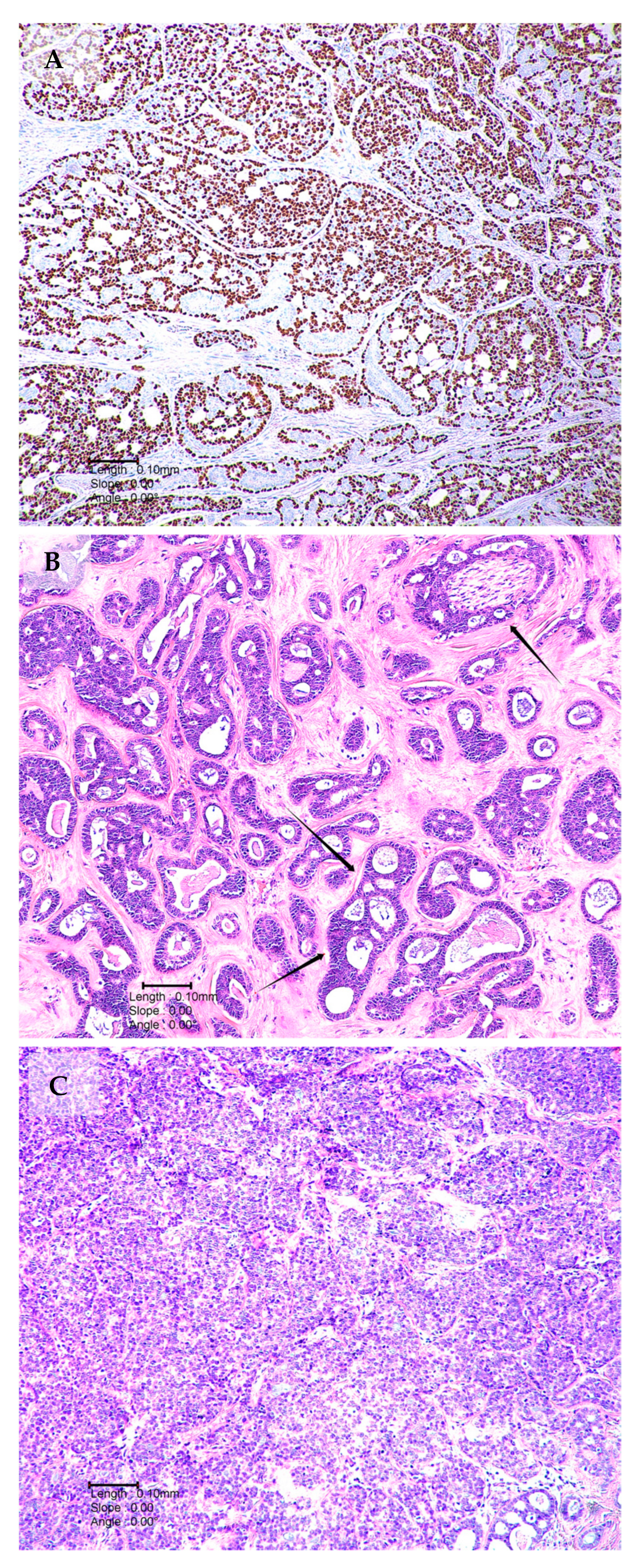
Figure 1. Histological patterns of ACC (A) p63 immunological staining demonstrating myoepithelial cells in a tumour composed of tubular and cribriform growth patterns. (B) Tumour with both tubular and cribriform areas. The single arrows demonstrate perineural invasion. Double arrows illustrate cribriform area. (C) Solid ACC growth pattern characterised the presence of pleomorphic cells, nests and sheets.
Ijms 24 13755 g001
Figure 2. Diagrammatic representation of the MYB/NFIB and MYBL1/NFIB gene fusions. MYB is found at chromosomal band 6q23; MYBL1 is found at chromosome 8q23, and NFIB is located at chromosomal band 9p22-23. During MYB/NFIB or MYBL1/NFIB fusion, a large part of MYB or MYBL1 is fused to a small part of NFIB. The fusion results in significant upregulation of the MYB fusion protein, which in turn drives several oncogenic events, including increased cellular proliferation, cellular differentiation, angiogenesis, growth factor upregulation, cellular adhesion and downstream activation of oncogenic genes [36,48].
Ijms 24 13755 g002
Figure 3. Microscopic image (magnification 1000×) of FISH analysis displaying negativity for rearrangements in MYBL1 (A) and positivity for rearrangements in MYB (B) and NFIB (C). FFPE samples were cut using microtomy and stained following H&E staining protocols. The tumour location was marked off on the H&E-stained slide of each sample. For the detection of rearrangements in MYB, the ZytoLight SPEC MYB Dual Color Break Apart Probe (ZytoVision GmbH) was used. Custom-designed SureFISH NFIB and MYBL1 break-apart probes were used for the detection of rearrangements of NFIB and MYBL1, respectively. The human genome (hg) build 19 was utilised for the chromosomal locations of the custom NFIB break-apart probe oligos, chr9:13740671-14140560 and chr9:14340306-14740560, and the MYBL1 break-apart probe, chr8:67076230-67474559 and chr8:67526335-68426199.
Ijms 24 13755 g003
Figure 4. The Notch-signalling pathway and downstream cellular sequelae in ACC. Notch receptors are bound to Notch ligands at epidermal growth factor repeats, resulting in a conformational change. Notch undergoes two cleavages: firstly by ADAM metalloproteases and again by γ secretase. Notch intracellular domain (NICD) is then released from the cytoplasm and travels to the nucleus where it interacts with various transcriptional coactivators, including DNA-binding protein CFB1/Suppressor of Hairless/LAG1, mastermind (MAM) and p300. These interactions result in upregulation of target genes, having downstream cellular consequences, including epithelial–mesenchymal transformation (EMT) (via HEY1), tumour invasion, angiogenesis, regulation of cell-cycle control (via CCND1) and apoptosis (via BCL2) [48,69,70,71].
Ijms 24 13755 g004
Disclaimer/Publisher’s Note: The statements, opinions and data contained in all publications are solely those of the individual author(s) and contributor(s) and not of MDPI and/or the editor(s). MDPI and/or the editor(s) disclaim responsibility for any injury to people or property resulting from any ideas, methods, instructions or products referred to in the content.