Article

ATRX Loss in the Development and Prognosis of Conjunctival Melanoma

by
Jolique A. van Ipenburg
1,†,
Quincy C. C. van den Bosch
2,†,
Dion Paridaens
2,3,
Hendrikus J. Dubbink
4,
Emine Kiliç
2,
Nicole Naus
2 and
Robert M. Verdijk
3,4,5,* on behalf of the Rotterdam Ocular Melanoma Study Group
1
Department of Pathology, Radboud University Medical Center, Geert Grooteplein Zuid 10, 6525 GA Nijmegen, The Netherlands
2
Department of Ophthalmology, Erasmus University Medical Center, Doctor Molewaterplein 40, 3015 GD Rotterdam, The Netherlands
3
Department of Ocular Oncology, The Rotterdam Eye Hospital, Schiedamse Vest 180, 3011 BH Rotterdam, The Netherlands
4
Department of Pathology, Section Ophthalmic Pathology, Erasmus University Medical Center, Doctor Molewaterplein 40, 3015 GD Rotterdam, The Netherlands
5
Department of Pathology, Leiden University Medical Center, Albinusdreef 2, 2333 ZA Leiden, The Netherlands
*
Author to whom correspondence should be addressed.
These authors contributed equally to this work.
Int. J. Mol. Sci. 2023, 24(16), 12988; https://doi.org/10.3390/ijms241612988
Submission received: 13 July 2023 / Revised: 8 August 2023 / Accepted: 18 August 2023 / Published: 20 August 2023
(This article belongs to the Special Issue Advances in Molecular Understanding of Ocular Adnexal Disease)

Abstract

:
Metastatic disease is linked to TERT promoter mutations in conjunctival melanomas (CM). Both TERT promoter and ATRX mutations are associated with faulty telomere maintenance. This study aimed to determine the prognostic value of ATRX loss in conjunctival melanocytic lesions. Eighty-six conjunctival melanocytic lesions from the Rotterdam Ocular Melanoma Study group were collected. ATRX status and TERT promoter status were determined using immunohistochemical staining and molecular diagnostics, respectively. None of the nevi (n = 16) and primary acquired melanosis (PAM) without atypia (n = 6) showed ATRX loss. ATRX loss was found in 2/5 PAM with atypia without CM and in 8/59 CM. No cases with a TERT promoter mutation (n = 26) showed ATRX loss. Eight/eleven metastatic CM harbored a TERT promoter mutation, two other metastatic CM showed ATRX loss and one metastatic case showed no TERT promoter/ATRX alterations. In conclusion ATRX loss and TERT promoter mutations are only found in (pre)malignant conjunctival melanocytic lesions, with most metastatic cases harboring one of these alterations, suggesting that both alterations are associated with adverse behavior. Similar to TERT promoter mutations, ATRX loss may be used as a diagnostic tool in determining whether a conjunctival melanocytic lesion is prone to having an adverse course.

1. Introduction

Conjunctival melanomas (CM) derive from melanocytes in the conjunctiva, a transparent mucosal membrane that covers the eye and the inside of the eyelids. CM has a crude incidence of 0.39 and 0.46 per million in US and European countries, respectively. Although this disease is rare, the incidence is increasing and increases with age [1,2,3]. Recurrences of CM are common with a low survival rate for disseminated melanomas [4]. The 5- and 10-year mortality rate for CM patients is reported up to 26% and 59%, respectively [5]. This emphasizes the need for insight into factors that predict the lesion’s behavior, so that lesions with a suspected adverse course can be treated is a timely manner.
Recent developments in ocular oncology have led to a better molecular understanding of CM, which enabled improved prognostication and led to the introduction of novel therapies. CM is a mucosal melanoma with much resemblance to melanoma of the skin [6]. The majority derives from primary acquired melanosis with atypia (PAM+), but infrequently CM develops from a pre-existing nevus or de novo [7]. Similar to cutaneous melanoma, CM are often classified according to their driver mutational status, resulting in groups of BRAF-mutated, NRAS-mutated, NF1-mutated, and triple-wild-type (triple-WT) melanoma [8]. The triple-WT cohort appears to be heterogeneous and may alternatively carry CTNNB1, c-Kit, or SF3B1 mutations [9]. As no relation has been reported with recurrences, metastasis, or survival, the presence of a BRAF, NRAS, or NF1 mutation is of limited use for prognosis in CM [6,10]. We have reported that the TERT promoter mutation, which is present in 43% of all CM cases, is associated with reduced metastasis-free survival [11]. A TERT promoter mutation may be seen in all above-mentioned mutational groups [7,8].
Enhanced telomere maintenance is a key event in cellular immortality in order to escape cellular crisis, potentially occurring through activation of telomerase (TERT) or through alternative lengthening of telomeres (ALT), most commonly resulting from ATRX inactivation [12,13,14].
Because of the implications in cancer and therapy there is growing interest in ATRX, which is reported as a frequently mutated gene in cancer. ATRX has several domains, binding to heterochromatin with its ADD domain. ATRX forms a complex with DAXX in order to deposit histone variant H3.3 at repetitive regions, which is hypothesized to maintain chromatin structural stability. Maintenance of the repetitive regions in a heterochromatic state is essential in order to avoid aberrant transcription at telomeres. In case of ATRX loss there is decreased H3.3 deposition at telomeric ends, causing reduced chromatin compaction and increased transcription of long noncoding RNA. This leads to genome instability because of telomere dysfunction, telomeric DNA damage, and excessive telomere recombination, with the forming of secondary structures [14]. TERT promoter mutations and loss of function ATRX mutations are usually mutually exclusive, as has been observed in gliomas [15]. Approximately 25% of CM have recently been reported to exhibit inactivating mutations in ATRX [16]. This finding has prompted us to investigate the role of ATRX within the pathogenesis of conjunctival melanocytic lesions and to elucidate the prognostic value of ATRX loss in CM in addition to the known adverse effect of TERT promoter mutations.

2. Results

To determine whether ATRX loss plays a role in the development of CM, we compared the ATRX status in conjunctival melanocytic nevi with the ATRX status in CM. As most CM derive from PAM+, we compared primary acquired melanosis without atypia (PAM−) with PAM+ to determine whether ATRX loss is likely to be an early event in melanoma genesis. Subsequently, in order to determine whether ATRX loss has prognostic value in CM we evaluated ATRX status in CM cases with recurrent disease versus CM cases without recurrent disease as well as CM cases with metastatic disease versus CM cases without the development of metastatic disease. Moreover, because of the aforementioned prognostic relevance of TERT promoter mutations, we analyzed whether there might be patterns similar to TERT promoter mutations analyzing the ATRX status. In case of ATRX loss none of the atypical melanocytes showed ATRX expression in contrast to cells in the surrounding tissue, including lymphocytes and vessels, which served as an internal control in addition to the external control tissue. None of the cases showed heterogeneous ATRX expression in the (pre)malignant melanocytic lesions. In case of CM in the context of PAM + both melanocytic components showed a similar ATRX staining pattern.

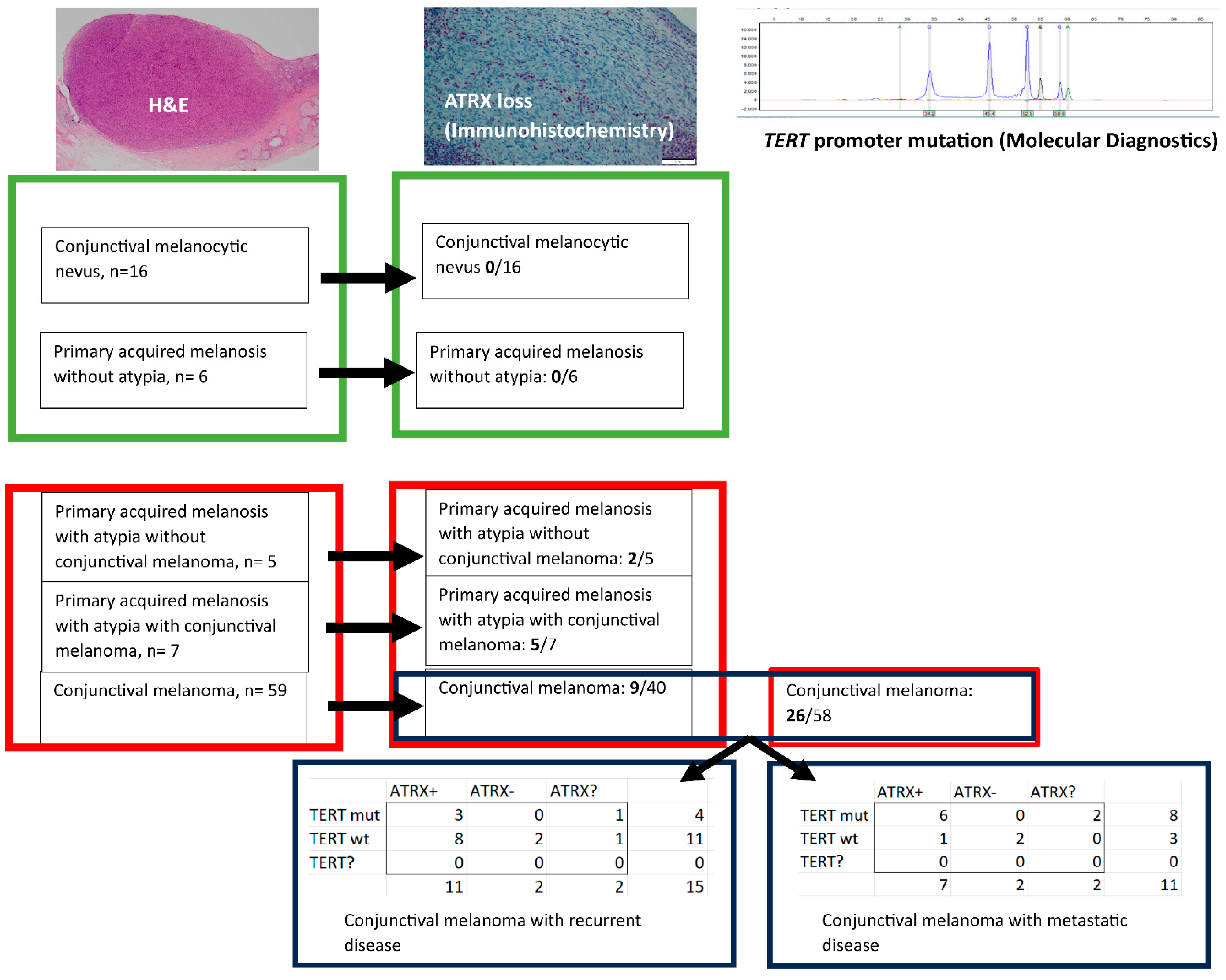
2.1. ATRX Status in Conjunctival Melanocytic Nevi versus CM

ATRX status was determined in 16 nevi, with none of the cases (0%) showing ATRX loss. ATRX status was determined in 40 CM cases, with 9 cases (23%) showing ATRX loss (Figure 1).
The flow chart in Figure 1 concerns the analysis of the different conjunctival melanocytic lesions, depicting the results from the benign lesions (green boxes) versus the (pre)malignant lesions (red boxes), with ATRX loss only found in the (pre)malignant conjunctival melanocytic lesions. Fifteen patients with conjunctival melanoma developed recurrent disease and eleven patients with conjunctival melanoma developed metastatic disease, with most of the cases with metastatic disease harboring either a TERT promoter mutation or ATRX loss.
The diagnosis (conjunctival melanocytic nevus, primary acquired melanosis either with or without atypia and conjunctival melanoma) was made based on hematoxylin and eosin staining (H&E). ATRX status was (ATRX+ = no ATRX loss, ATRX- = ATRX loss, ATRX? = ATRX expression unknown) based on immunohistochemistry and TERT promoter status (TERT promoter mutation = TERT mut, no TERT promoter mutation = TERT wt, TERT? = TERT promoter status unknown) based on SNaPshot analysis and/or next generation sequencing.

2.2. ATRX Status in PAM− versus PAM+

ATRX status was determined in six cases of PAM− and five cases of PAM+ without an invasive component and seven cases of PAM+ with an invasive component. None of the cases (0%) of PAM− revealed ATRX loss. However, ATRX loss was found in two cases (40%) of PAM+ without an invasive component and five cases (71%) of PAM+ with invasive component (Figure 1).

2.3. ATRX Status and CM with Recurrent Disease

Twelve CM cases with known ATRX status developed recurrent disease, including two cases with ATRX loss. One case with recurrent disease had an unknown ATRX status (Figure 1).

2.4. ATRX Status and CM with Metastatic Disease

Nine CM cases with known ATRX status developed metastases, including two cases (22%) with ATRX loss. Two cases with metastatic disease had an unknown ATRX status (Figure 1).

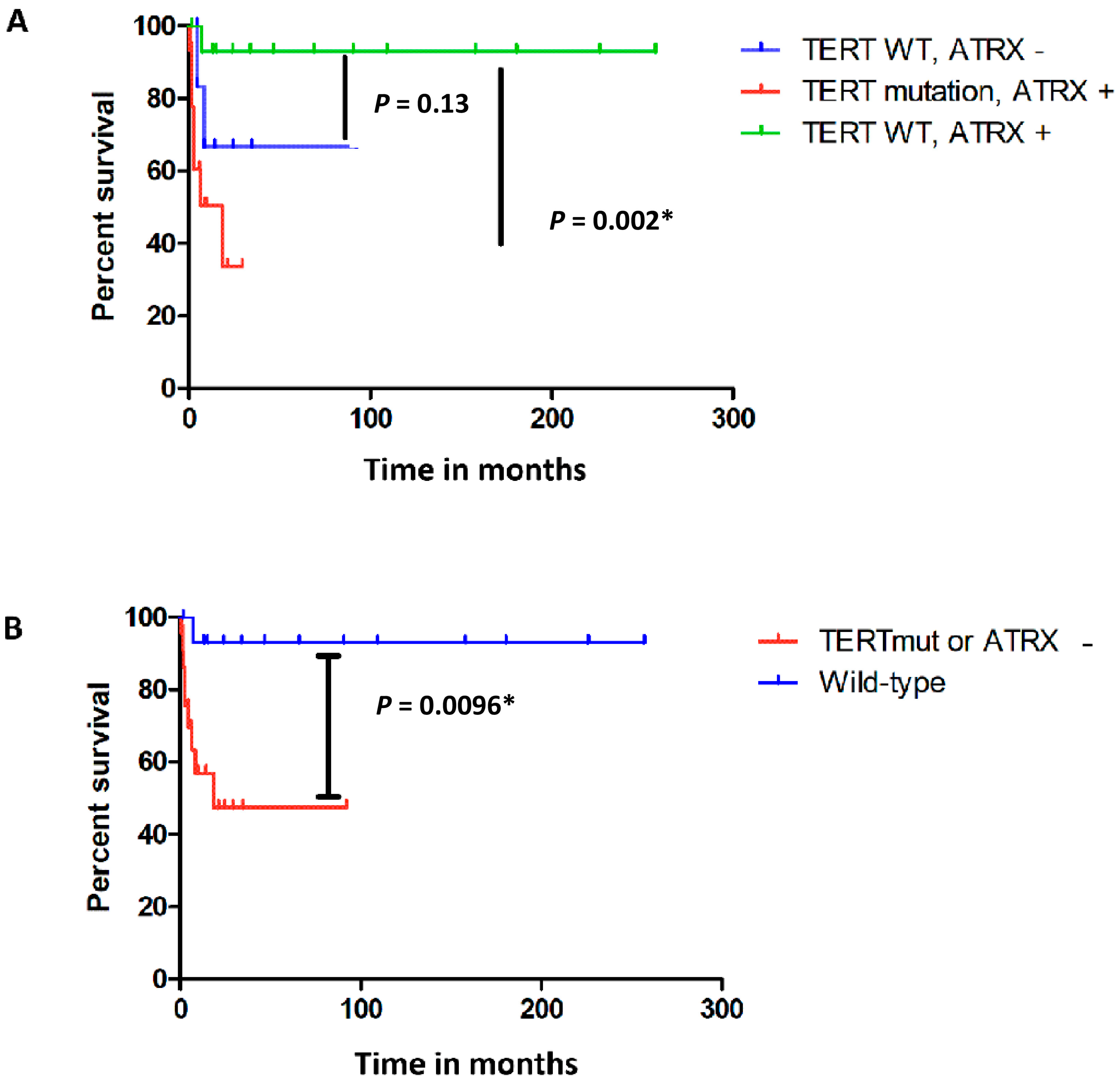
2.5. TERT Promoter Mutation and ATRX Loss in CM

TERT promoter status could be determined in 58 CM cases. A TERT promoter mutation was present in 26 cases (45%), with none of these cases (0%) showing ATRX loss. ATRX loss was found in nine cases without a TERT promoter mutation (23%). A TERT promoter mutation was found in eight of eleven cases with metastatic disease, with no ATRX loss in six cases and unknown ATRX status in two cases. Two cases with metastatic disease without a TERT promoter mutation did show ATRX loss. Only one CM case with metastatic disease revealed neither a TERT promoter mutation nor ATRX loss (Figure 1).
ATRX loss could not independently be observed to be significantly associated with a shorter metastatic free survival (p = 0.13), while the presence of a TERT promoter mutation was significantly correlated with a shorter metastatic-free survival (p = 0.002). However, when comparing all the cases with presumed enhanced telomere lengthening changes as a result of TERT or ATRX aberrations versus cases without these aberrations, we found that the presumed telomere affected group was associated with a significantly shorter metastasis-free survival (p = 0.01) (Figure 2). Due to the low number of cases, it was not possible to adjust for clinicohistopathological features, which we previously reported to be associated with a shorter metastasis-free survival [11]. Fifteen CM cases developed recurrent disease, including four cases with a TERT promoter mutation. Of the eleven cases without a TERT promoter mutation two cases showed ATRX loss. No significant correlation with recurrent disease was observed (p > 0.05).

3. Discussion

In non-neoplastic tissue the repetitive DNA sequences at the terminal ends of the chromosome progressively shorten during each cell division. In cases of uncontrolled proliferation several telomere maintenance mechanisms (TMM) are described. Eighty to ninety percent of all tumors reactivate telomerase, an enzyme consisting of multiple proteins, adding TTAGGG at the terminal end of the chromosome when the telomeres are getting too short. In contrast, in ten to twenty percent of all tumors telomere elongation is achieved via ALT. This second type of TMM does not rely on a single enzyme and results from newfound complementary sequences, which serve as elongation templates for the invading strand, including the homologous chromosome, an unrelated chromosome or extrachromosomal telomeric repeat [17]. ALT activation is described in sarcoma, astrocytoma, neuroblastoma, and carcinoma [18] cases as well as CM [16] and is related to functional loss of ATRX and/or histone H3.3 chaperone DAXX. In mucosal-melanoma-inactivating ATRX mutations can be revealed using immunohistochemistry [19]. In the current study ATRX loss was only found in (pre)malignant lesions and not in benign lesions. This finding of ATRX loss in PAM+ as well as in CM is congruent with findings in other studies, that suggested that TERT promoter mutations [20] and ATRX loss may be early events in CM progression [16]. Moreover, the presence of a TERT promoter mutation was evaluated, with the usually mutually exclusive presence of TERT promoter mutations and ATRX mutations described in gliomas [21] as well as cutaneous melanomas [22]. This suggests that the functional effects of the different telomere maintenance mechanisms are similar. Consistent with these studies and other mucosal melanoma studies [13,16,19], our current study indicates that ATRX loss and TERT promoter mutations are also mutually exclusive in CM. If enhancement of telomere maintenance is associated to an adverse clinical course for conjunctival melanocytic lesions, CM associated with metastasis but without detected TERT or ATRX alterations may harbor alterations in other components of the machinery involved in telomere maintenance, including POT1 [21]. Another less frequently occurring explanation may be that in cases without a TERT mutation and ATRX loss, a loss of function ATRX mutation is missed due to the limitations of the detection method [18].
Conjunctival melanoma is a mucosal melanoma and, based on the location, it is also an ocular melanoma. The genetic make-up partly overlaps with other ocular melanomas and mucosal melanomas originating from other sites. In addition, there are many similarities with the genetic make-up of cutaneous melanomas. Depending on the location, cutaneous melanomas are frequently exposed to ultraviolet radiation (UV) as are most CM. Melanomas from both sites frequently harbor the UV-signature (C > T nucleotide transition) [20]. Because of this signature it is likely that UV exposure (partly) plays a role in pathogenesis in at least a subset of CM. Unlike TERT promoter mutations, the relationship between UV exposure and ALT is not as well comprehended. However, there is a suggested connection between UV exposure and telomere changes, supported by the discovery of shorter telomeres in sun-exposed skin compared to nonexposed skin. It has been hypothesized that nucleotide excision repair restores telomeres by removing DNA photoproducts that are formed at telomeres after UV exposure. Lower levels of photoproducts at telomeres are formed due to shelterin protection [23]. A mutated shelterin complex, e.g., by POT1 mutations, might activate a noncanonical mechanism of ALT due to shelterin dysfunction [24]. If there is a relation between an ATRX mutation and UV exposure, maybe by interaction with the noncanonical mechanism of ALT, this still remains to be elucidated. A subset of CM reveals ATRX loss. ATRX mutations are also described in other mucosal melanomas, including melanomas from gynecologic sites [19], the anorectal region, and the oral region [13,25]. Similar to TERT promoter mutations [26], ATRX loss is very rare in uveal melanoma [27], which is another ocular melanoma [20]. The finding of ATRX mutations in melanoma originating from locations without sun exposure and the association with non-sun-exposed conjunctiva [16] suggest that a (direct) relation between UV and ATRX is less likely.
In the current study the presence of a TERT promoter mutation was strongly associated with a shorter metastasis-free survival, as was also described in other studies [11,20]. In publicly available datasets ATRX aberrations (including deep deletions or mutations) are reported in melanoma in general, with data suggesting a more favorable course for ATRX-unaltered cancer (according to cBioPortal database and Human Protein Atlas, accessed on 1 August 2023), although the data for ATRX-altered cases are limited, with a lack of specific information concerning the melanoma arising from different locations, especially mucosal melanoma. However, this information supports our hypothesis and certainly warrants further exploration. In an earlier study, the presence of an ATRX mutation has been suggested to be associated with a beneficial clinical course compared to ATRX wildtype CM, although a significant association with overall survival was not found [16]. The limitations of this study, as acknowledged by the authors, include a restricted median follow-up period, which may introduce biases to the reported findings [16]. Due to the counterintuitive nature of the findings in the previous study, given the presumed underlying mechanism, we performed the current study. We found a similar pattern for ATRX loss and TERT promoter mutations. Our hypothesis was supported by the finding of a significantly shorter metastasis-free survival in the presumed telomere affected group versus the group without TERT promoter or ATRX aberrations. The lack of a significant direct correlation between ATRX loss and metastasis-free survival is likely attributed to the lower occurrence of ATRX loss (23%) compared to TERT promoter mutations (40%) and the subsequent inclusion of a smaller number of cases with ATRX loss in the analysis. This supposition is further supported by the finding that the majority of CM with metastatic disease revealed either ATRX loss or a TERT promoter mutation.

4. Materials and Methods

4.1. Material Selection

We collected 16 conjunctival melanocytic nevi, 6 PAM without atypia (PAM−), 5 PAM+ without an associated CM, 7 PAM+ with an associated CM, and 59 CM, diagnosed between 1987 and 2016 at the Erasmus MC University Medical Center (Rotterdam, The Netherlands) and The Rotterdam Eye Hospital (Rotterdam, The Netherlands). Clinicopathological characteristics of the CM cases are described in Table 1, with a median follow-up time of 35 months (range 0–257 months). Eleven CM (19%) showed metastatic disease (median 15 months (range 2–49 months)) and 13 CM showed recurrent disease (median 23 months (range 5–157 months)) (Table 1).
Ethics Committee approval was obtained by the Medical Ethics Committee, Erasmus University Medical Center, Rotterdam, The Netherlands (4 October 2018), and was registered with reference 67865. The study was performed according to the tenets of the Declaration of Helsinki. Data regarding gender, age at the time of diagnosis, location, tumor thickness, the origin of the lesion, and information on the development of recurrences and metastasis were collected from the patient records and information was obtained from the pathology reports and the nationwide pathology network and registry system (Pathologisch-Anatomisch Landelijk Geautomatiseerd Archief).
Recurrence was defined as described earlier [11] with recurrence-free survival and metastasis-free survival defined as the time from the primary treatment to the date of recurrent disease and metastatic disease, respectively, or last date of follow-up.
TERT promoter status was determined in cases of CM using a sufficient DNA quantity and quality, to comply with the manufacturer’s instructions. TERT promoter mutation status was determined using SNaPshot analysis and/or next generation sequencing, as described earlier [20,26]. We did not find any discrepancies in TERT promoter status in cases with sufficient DNA quantity and quality to validate TERT promoter status using both techniques.
ATRX expression was determined using immunohistochemistry (IHC), with staining of the samples using an automated IHC staining system (Ventana Benchmark ULTRA, Ventana Medical System Inc., Tucson, AZ, USA) in an ISO 15189:2012 certified laboratory. The tissue sections were incubated for 32 min at 37 °C with a rabbit–anti-human ATRX polyclonal IgG antibody (Atlas Antibodies, Bromma, Sweden, Product number HPA001906, dilution 1:150). Human tonsil tissue was used as control tissue in addition to internal control tissue. The hematoxylin and eosin (H&E) staining and IHC staining was evaluated by an ophthalmic pathologist (RVE), with inactivation of ATRX reflected as no expression of ATRX (Figure 3), hereafter referred to as ATRX loss. For 19 cases it was not possible to reliably assess ATRX status because of ambiguous staining or there was no material available for further analysis.

4.2. Statistical Analysis

Statistical Package for Social Science (IBM SPSS Statistics, version 25) was used for data analysis, with differences between categorical data calculated using either the Pearson’s χ2 test or the Fisher’s exact test. Kaplan Meier survival curves were made using log-rank statistics. p < 0.05 is considered statistically significant.

5. Conclusions

Conjunctival melanocytic lesions may be difficult to characterize adequately based solely on clinicopathological characteristics. One of the conclusions drawn from this study is that ATRX loss can be helpful in distinguishing between benign and (pre)malignant conjunctival melanocytic lesions. A correlation was observed between the presence of a TERT promoter mutation and decreased metastasis-free survival. The findings related to CM with ATRX loss indicate a similar trend. Since most CMs with metastatic disease show either ATRX loss or a TERT promoter mutation, both alterations are suggestive of an association with adverse clinical behavior. Our study shows that the presence of a TERT promoter mutation in CM is strongly associated with a shorter metastasis-free survival, and is indicative that this may also hold for CM with ATRX loss. The major implications of these findings warrant further research using larger multicenter cohort studies. For this reason, an international collaborative study on the prognostics of TERT promoter mutation and ATRX loss has been initiated by the European section of the Ophthalmic Oncology Group, an independent scientific group devoted to clinical ophthalmic oncology and related basic science research.

Author Contributions

Conceptualization, E.K. and R.M.V.; Data curation, J.A.v.I., Q.C.C.v.d.B. and R.M.V.; Formal analysis, J.A.v.I. and Q.C.C.v.d.B.; Funding acquisition, E.K. and R.M.V.; Investigation, J.A.v.I., Q.C.C.v.d.B. and R.M.V.; Methodology, J.A.v.I., Q.C.C.v.d.B., H.J.D. and R.M.V.; Project administration, E.K. and R.M.V.; Resources, D.P., H.J.D., E.K. and N.N.; Software, Q.C.C.v.d.B.; Supervision, R.M.V.; Validation, J.A.v.I., Q.C.C.v.d.B. and R.M.V.; Visualization, J.A.v.I. and Q.C.C.v.d.B.; Writing—original draft, J.A.v.I. and Q.C.C.v.d.B.; Writing—review and editing, D.P., H.J.D., E.K., N.N. and R.M.V. All authors have read and agreed to the published version of the manuscript.

Funding

Stichting Nederlands Oogheelkundig Onderzoek (SNOO), grant No. 2013–2021; Stichting Wetenschappelijk Oogheelkundig Onderzoek (SWO), grant No. 2016–2017.

Institutional Review Board Statement

The study was conducted according to the guidelines of the Declaration of Helsinki and approved by the Medical Ethics Committee, Erasmus University Medical Center, Rotterdam, The Netherlands (reference 67865, 4 October 2018).

Informed Consent Statement

Patient consent was waived due to the Code of Conduct for Responsible Use of Human Tissue and Medical Research that applies to medical research in The Netherlands.

Data Availability Statement

Data can be provided upon request.

Acknowledgments

The authors wish to acknowledge A. Fellah for his help with ATRX immunohistochemistry.

Conflicts of Interest

The authors declare no conflict of interest.

References

  1. Kaštelan, S.; Antunica, A.G.; Orešković, L.B.; Rabatić, J.S.; Kasun, B.; Bakija, I. Conjunctival Melanoma—Epidemiological Trends and Features. Pathol. Oncol. Res. 2018, 24, 787–796. [Google Scholar] [CrossRef] [PubMed]
  2. Weppelmann, T.A.; Zimmerman, K.T.; Rashidi, V. Trends in Incidence of Conjunctival Melanoma in the US. JAMA Netw. Open 2022, 5, e2237229. [Google Scholar] [CrossRef] [PubMed]
  3. Virgili, G.; Parravano, M.; Gatta, G.; Capocaccia, R.; Mazzini, C.; Mallone, S.; Botta, L.; RARECAREnet Working Group. Incidence and Survival of Patients With Conjunctival Melanoma in Europe. JAMA Ophthalmol. 2020, 138, 601–608. [Google Scholar] [CrossRef] [PubMed]
  4. Anastassiou, G.; Heiligenhaus, A.; Bechrakis, N.; Bader, E.; Bornfeld, N.; Steuhl, K.-P. Prognostic value of clinical and histopathological parameters in conjunctival melanomas: A retrospective study. Br. J. Ophthalmol. 2002, 86, 163–167. [Google Scholar] [CrossRef]
  5. Zhou, C.; Wang, Y.; Jia, R.; Fan, X. Conjunctival Melanoma in Chinese Patients: Local Recurrence, Metastasis, Mortality, and Comparisons With Caucasian Patients. Investig. Opthalmol. Vis. Sci. 2017, 58, 5452–5459. [Google Scholar] [CrossRef] [PubMed]
  6. Mikkelsen, L.H.; Maag, E.; Andersen, M.K.; Kruhøffer, M.; Larsen, A.-C.; Melchior, L.C.; Toft, P.B.; von Buchwald, C.; Wadt, K.; Heegaard, S. The molecular profile of mucosal melanoma. Melanoma Res. 2020, 30, 533–542. [Google Scholar] [CrossRef]
  7. Gkiala, A.; Palioura, S. Conjunctival Melanoma: Update on Genetics, Epigenetics and Targeted Molecular and Immune-Based Therapies. Clin. Ophthalmol. 2020, 14, 3137–3152. [Google Scholar] [CrossRef] [PubMed]
  8. The Cancer Genome Atlas Network. Genomic Classification of Cutaneous Melanoma. Cell 2015, 161, 1681–1696. [Google Scholar] [CrossRef]
  9. Pipek, O.; Vizkeleti, L.; Doma, V.; Alpár, D.; Bödör, C.; Kárpáti, S.; Timar, J. The Driverless Triple-Wild-Type (BRAF, RAS, KIT) Cutaneous Melanoma: Whole Genome Sequencing Discoveries. Cancers 2023, 15, 1712. [Google Scholar] [CrossRef]
  10. Griewank, K.G.; Westekemper, H.; Murali, R.; Mach, M.; Schilling, B.; Wiesner, T.; Schimming, T.; Livingstone, E.; Sucker, A.; Grabellus, F.; et al. Conjunctival melanomas harbor BRAF and NRAS mutations and copy number changes similar to cutaneous and mucosal melanomas. Clin. Cancer Res. 2013, 19, 3143–3152. [Google Scholar] [CrossRef]
  11. Van Ipenburg, J.A.; Naus, N.C.; Dubbink, H.J.; Van Ginderdeuren, R.; Missotten, G.S.; Paridaens, D.; Verdijk, R.M. Prognostic value of TERT promoter mutations in conjunctival melanomas in addition to clinicopathological features. Br. J. Ophthalmol. 2021, 105, 1454–1461. [Google Scholar] [CrossRef] [PubMed]
  12. Barthel, F.P.; Wei, W.; Tang, M.; Martinez-Ledesma, E.; Hu, X.; Amin, S.B.; Akdemir, K.C.; Seth, S.; Song, X.; Wang, Q.; et al. Systematic analysis of telomere length and somatic alterations in 31 cancer types. Nat. Genet. 2017, 49, 349–357. [Google Scholar] [CrossRef]
  13. Newell, F.; Kong, Y.; Wilmott, J.S.; Johansson, P.A.; Ferguson, P.M.; Cui, C.; Li, Z.; Kazakoff, S.H.; Burke, H.; Dodds, T.J.; et al. Whole-genome landscape of mucosal melanoma reveals diverse drivers and therapeutic targets. Nat. Commun. 2019, 10, 3163. [Google Scholar] [CrossRef] [PubMed]
  14. Aguilera, P.; López-Contreras, A.J. ATRX, a guardian of chromatin. Trends Genet. 2023, 39, 505–519. [Google Scholar] [CrossRef] [PubMed]
  15. Koelsche, C.; Sahm, F.; Capper, D.; Reuss, D.; Sturm, D.; Jones, D.T.W.; Kool, M.; Northcott, P.A.; Wiestler, B.; Böhmer, K.; et al. Distribution of TERT promoter mutations in pediatric and adult tumors of the nervous system. Acta Neuropathol. 2013, 126, 907–915. [Google Scholar] [CrossRef] [PubMed]
  16. Lally, S.E.; Milman, T.; Orloff, M.; Dalvin, L.A.; Eberhart, C.G.; Heaphy, C.M.; Rodriguez, F.J.; Lin, C.-C.; Dockery, P.W.; Shields, J.A.; et al. Mutational Landscape and Outcomes of Conjunctival Melanoma in 101 Patients. Ophthalmology 2022, 129, 679–693. [Google Scholar] [CrossRef]
  17. MacKenzie, D.; Watters, A.K.; To, J.T.; Young, M.W.; Muratori, J.; Wilkoff, M.H.; Abraham, R.G.; Plummer, M.M.; Zhang, D. ALT Positivity in Human Cancers: Prevalence and Clinical Insights. Cancers 2021, 13, 2384. [Google Scholar] [CrossRef]
  18. Chami, R.; Marrano, P.M.; Teerapakpinyo, C.; Arnoldo, A.; Shago, M.; Shuangshoti, S.; Thorner, P.S. Immunohistochemistry for ATRX Can Miss ATRX Mutations. Am. J. Surg. Pathol. 2019, 43, 1203–1211. [Google Scholar] [CrossRef]
  19. Yuan, G.; Song, J.; Li, N.; Song, Q.; Li, Y.; Du, Y.; Wang, X.; Jiao, Y.; Wu, L. Telomere Maintenance Associated Mutations in the Genetic Landscape of Gynecological Mucosal Melanoma. Front. Oncol. 2020, 10, 1707. [Google Scholar] [CrossRef]
  20. Van Poppelen, N.M.; van Ipenburg, J.A.; van den Bosch, Q.; Vaarwater, J.; Brands, T.; Eussen, B.; Magielsen, F.; Dubbink, H.J.; Paridaens, D.; Brosens, E.; et al. Molecular Genetics of Conjunctival Melanoma and Prognostic Value of TERT Promoter Mutation Analysis. Int. J. Mol. Sci. 2021, 22, 5784. [Google Scholar] [CrossRef]
  21. Eckel-Passow, J.E.; Lachance, D.H.; Molinaro, A.M.; Walsh, K.M.; Decker, P.A.; Sicotte, H.; Pekmezci, M.; Rice, T.W.; Kosel, M.L.; Smirnov, I.V.; et al. Glioma Groups Based on 1p/19q, IDH, and TERTPromoter Mutations in Tumors. N. Engl. J. Med. 2015, 372, 2499–2508. [Google Scholar] [CrossRef] [PubMed]
  22. Hayward, N.K.; Wilmott, J.S.; Waddell, N.; Johansson, P.A.; Field, M.A.; Nones, K.; Patch, A.-M.; Kakavand, H.; Alexandrov, L.B.; Burke, H.; et al. Whole-genome landscapes of major melanoma subtypes. Nature 2017, 545, 175–180. [Google Scholar] [CrossRef] [PubMed]
  23. Parikh, D.; Fouquerel, E.; Murphy, C.T.; Wang, H.; Opresko, P.L. Telomeres are partly shielded from ultraviolet-induced damage and proficient for nucleotide excision repair of photoproducts. Nat. Commun. 2015, 6, 8214. [Google Scholar] [CrossRef]
  24. Shen, E.; Xiu, J.; Lopez, G.Y.; Bentley, R.; Jalali, A.; Heimberger, A.B.; Bainbridge, M.N.; Bondy, M.L.; Walsh, K.M. POT1 mutation spectrum in tumour types commonly diagnosed among POT1-associated hereditary cancer syndrome families. J. Med. Genet. 2020, 57, 664–670. [Google Scholar] [CrossRef]
  25. Beaudoux, O.; Oudart, J.-B.; Riffaud, L.; Visseaux, L.; Marchal, A.; Lebre, A.-S.; Grange, F. Mutational Characteristics of Primary Mucosal Melanoma: A Systematic Review. Mol. Diagn. Ther. 2022, 26, 189–202. [Google Scholar] [CrossRef]
  26. Koopmans, A.E.; Ober, K.; Dubbink, H.J.; Paridaens, D.; Naus, N.C.; Belunek, S.; Krist, B.; Post, E.; Zwarthoff, E.C.; De Klein, A.; et al. Prevalence and implications of TERT promoter mutation in uveal and conjunctival melanoma and in benign and premalignant conjunctival melanocytic lesions. Investig. Opthalmol. Vis. Sci. 2014, 55, 6024–6030. [Google Scholar] [CrossRef] [PubMed]
  27. Harford, E.; Palmer, J.M.; Glasson, W.J.; Warrier, S.K.; Whitehead, K.J.; Brooks, K.M.; Johansson, P.A.; Hayward, N.K.; McGrath, L.A. Choroidal melanoma with synchronous Fuchs’ adenoma and novel ATRX mutation. Int. J. Retin. Vitr. 2022, 8, 24. [Google Scholar] [CrossRef] [PubMed]
Figure 1. TERT promoter and ATRX status in conjunctival melanocytic lesions. These images are meant to highlight the methods that are used to obtain the data (H&E staining and immunohistochemistry instead of other (diagnostic) methods; Microscope's magnification or scale bar is not of relevance for understanding this figure. Exact data concerning these images can be provided upon request.
Ijms 24 12988 g001
Figure 2. Kaplan Meier ATRX and TERT aberrations in conjunctival melanoma cases with metastatic disease. The Kaplan Meier survival estimate for the time to metastasis of presumed telomere affected conjunctival melanoma (CM) (cases with a TERT promoter mutation (“TERTmut”) and cases with ATRX loss (“ATRX−”) shown separately) shows that CM cases harboring a TERT promoter mutation (depicted in red) have a significantly shorter metastasis-free survival compared to CM cases without a TERT promoter mutation or ATRX loss, the presumed telomere unaffected CM (depicted in green). This pattern for CM with ATRX loss is similar to CM cases with TERT promoter mutations, suggesting that ATRX loss is also associated with a shorter metastasis-free survival (A). Although this finding concerning the cases with ATRX loss is not proved to be significant (p-value 0.13) (A), the hypothesis is supported by the Kaplan Meier survival estimate in (B), showing that the presumed telomere-affected CM (depicted in red) have a significant shorter metastasis-free survival compared to the CM without TERT promoter and ATRX aberrations (depicted in blue). * = statistically significant.
Ijms 24 12988 g002
Figure 3. Determination of ATRX inactivation using immunohistochemical staining as determined in (pre) malignant conjunctival melanocytic lesions. H&E staining within the box in (A) primary acquired melanosis with atypia (PAM+), magnification 200× (scale bar 1 mm), with the PAM+ highlighted in (B), shows ATRX loss in the atypical melanocytes in (C), with the atypical melanocytes depicted with an arrow. ATRX status was evaluated using immunohistochemistry, with the red stained cells representing pre-existing tissue, including normal epithelium and fibroblasts, serving as an internal control. H&E staining within the box in (D) conjunctival melanoma, magnification 200× (scale bar 1 mm), with the stromal part with atypical melanocytes (conjunctival melanoma) highlighted in (E), shows ATRX loss concerning the atypical melanocytes in (F), with the atypical melanocytes depicted with an arrow.
Ijms 24 12988 g003
Table 1. Clinicohistopathological characteristics of the conjunctival melanomas.
Age at Time of Diagnosis (y)GenderRecurrent Disease, Months after DiagnosisMetastasis,
Months after Diagnosis
Breslow (mm)UlcerationPresence of Mitotic Figure (s)Presence of Epithelioid CellspT StatusOriginDiameter (mm)Bulbar InvolvementFollow-Up (Months after Diagnosis)
143M2047UKYesYesMixedpT2bUK7UK68
262MNoNoUKYesYesMixedUKUK17UK4
344MNo381.7YesYesMixedpT1aNevus21 *UK38
465F55No3.46YesYesMixedpT2bPAM+7 *UK257
560FNoNo0.4NoYesMixedUKDe novo6 *UK1
668FNo87YesYesMixedUKPAM+18UK54
784MNoNo2.6 *YesYesMixedpT1bDe novo6 *UK1
865MNoNo3YesYesSpindle cellspT1bPAM+6UK6
964MNoNo1.55 *NoYesMixedpT1aNevus6UK29
1051M890.96 *NoYesSpindle cellsUKDe novo12 *UK22
1149MNo492.3 *YesYesMixedUKPAM+3UK86
1284M23No2.3 *UKUKEpithelioidUKPAM+UKUK35
1373F157No3UKYesSpindle cellsUKPAM+UKUK158
1453MNo143.69YesYesEpithelioidUKNevus7UK36
1564M81No1.1NoYesSpindle cellspT1aPAM+UKUK109
1649FNoNo0.3NoNoMixedpT1aPAM+3UK166
1779FNoNoUKUKUKUKUKPAM+UKUK21
1866MNoNo2YesNoMixedpT1aDe novo4UK9
1983F87No5.1NoYesMixedpT2bPAM+6 *UK90
2046FNo27 *YesYesMixedUKPAM+11UK69
2165M11151.9 *YesYesMixedUKDe novo4.2UK58
2263MNoNoUKUKUKUKUKPAM+5UK24
2340MNoNo0.3NoNoSpindle cellspT1aPAM+1.3Yes3
2455FNoNo0.35NoNoSpindle cellspT1aPAM+10Yes187
2575F5No0.37NoNoSpindle cellspT1aPAM+2Yes226
2671FNoNo0.5NoNoSpindle cellsUKPAM+UKUK1
2785MNoNo0.5YesYesSpindle cellspT1aDe novoUKYes0
2854MNoNo0.5NoNoUKpT1aPAM+UKYes142
2972MNoNo0.6NoNoMixedpT1aPAM+9 *Yes92
3056FNoNo0.62NoNoMixedUKNevus4.5 *UK185
3168MNoNo0.7YesNoSpindle cellspT1aPAM+UKYes112
3243MNoNo0.78NoNoSpindle cellspT2aDe novo2.5No69
3352MNoNo0.9NoNoEpithelioidUKPAM+UKUK15
3484MNoNo0.9UKUKMixedpT1aPAM+UKYes24
3573MNoNo1.3YesYesMixedpT1aDe novoUKYes3
3667FNoNo1YesYesEpithelioidpT1aPAM+6 *Yes2
3716FNo181.04YesYesMixedpT1aNevus0.5Yes180
3857MNoNo1.05NoYesEpithelioidpT2aUKUKNo2
3963FNoNo1.1NoNoUKpT1aNevus3Yes54
4056MNoNo1.3NoYesMixedpT1aPAM+4.5 *Yes0
4176FNoNo2.3YesYesMixedpT1bPAM+17 *Yes2
4251FNoNo3YesYesSpindle cellspT2bDe novo10No95
4389MNoNo7.7 *YesYesMixedpT3aPAM+14No13
4441MNoNo1 *NoYesSpindle cellsUKNevus6Yes14
4569F8No1.1 *NoNoMixedUKPAM+UKYes34
4666MNoNo1.1 *NoYesSpindle cellsUKPAM+4 *yes81
4770FNoNo1.2 *NoNoMixedUKUK4Yes13
4871MNoNo2.7 *YesYesMixedpT1bDe novo6Yes179
4957MNoNo2.5 *YesUKMixedpT1bPAM+6Yes15
5079FNoNo2.3UKUKUKUKPAM+0.7Yes0
5173MNoNo6.2UKUKUKUKPAM+1.2No22
5283FNoNo3UKUKUKUKNevus1Yes0
5380F29No0.2UKUKUKUKPAM+0.8Yes73
5474FNo45UKUKUKUKUKPAM+UKYes49
5537FNo74UKUKUKUKUKUKNo (though the caruncle is involved)66
5656M7No3.5UKUKUKUKPAM+UKYes47
5758M42No3UKUKUKUKPAM+0.6Yes174
5860MNoNo1.1UKUKUKUKUK0.6No5
5955MNoNo2.5UKUKUKUKUK0.7UK4
Clinical and histopathological characteristics of the cases with conjunctival melanoma. PAM+: primary acquired melanosis with atypia; UK: unknown; * these values are the minimum values as in these cases it was not possible to determine the exact value since the lesion was not completely excised and/or not all slides were available for review. Mixed: presence of both spindle cells and epithelioid cells.
Disclaimer/Publisher’s Note: The statements, opinions and data contained in all publications are solely those of the individual author(s) and contributor(s) and not of MDPI and/or the editor(s). MDPI and/or the editor(s) disclaim responsibility for any injury to people or property resulting from any ideas, methods, instructions or products referred to in the content.

