Article

Potential Association of Cytochrome P450 Copy Number Alteration in Tumour with Chemotherapy Resistance in Lung Adenocarcinoma Patients

1
Institute of Enzymology, Research Centre for Natural Sciences, Magyar Tudósok 2, H-1117 Budapest, Hungary
2
Doctoral School of Pharmaceutical Sciences, Semmelweis University, Üllői 26, H-1085 Budapest, Hungary
3
Department of Pathology, National Korányi Institute of Pulmonology, Pihenő 1, H-1121 Budapest, Hungary
4
1st Department of Pulmonology, National Korányi Institute of Pulmonology, Pihenő 1, H-1121 Budapest, Hungary
*
Author to whom correspondence should be addressed.
Int. J. Mol. Sci. 2023, 24(17), 13380; https://doi.org/10.3390/ijms241713380
Submission received: 1 August 2023 / Revised: 25 August 2023 / Accepted: 26 August 2023 / Published: 29 August 2023
(This article belongs to the Special Issue Genetic Variations in Human Diseases)

Abstract

:
Resistance to anticancer agents is a major obstacle to efficacious tumour therapy and responsible for high cancer-related mortality rates. Some resistance mechanisms are associated with pharmacokinetic variability in anticancer drug exposure due to genetic polymorphisms of drug-metabolizing cytochrome P450 (CYP) enzymes, whereas variations in tumoural metabolism as a consequence of CYP copy number alterations are assumed to contribute to the selection of resistant cells. A high-throughput quantitative polymerase chain reaction (qPCR)-based method was developed for detection of CYP copy number alterations in tumours, and a scoring system improved the identification of inappropriate reference genes that underwent deletion/multiplication in tumours. The copy numbers of both the target (CYP2C8, CYP3A4) and the reference genes (ALB, B2M, BCKDHA, F5, CD36, MPO, TBP, RPPH1) established in primary lung adenocarcinoma by the qPCR-based method were congruent with those determined by next-generation sequencing (for 10 genes, slope = 0.9498, r2 = 0.72). In treatment naïve adenocarcinoma samples, the copy number multiplication of paclitaxel-metabolizing CYP2C8 and/or CYP3A4 was more prevalent in non-responder patients with progressive disease/exit than in responders with complete remission. The high-throughput qPCR-based method can become an alternative approach to next-generation sequencing in routine clinical practice, and identification of altered CYP copy numbers may provide a promising biomarker for therapy-resistant tumours.

Graphical Abstract

1. Introduction

Cancer is one of the leading causes of death, and despite the huge efforts of health-care systems and extensive research into novel diagnostic strategies and chemotherapeutic agents, cancer incidence and mortality are still growing [1]. Resistance to anticancer drugs represents a major obstacle to efficacious cancer therapies, and is considered to be responsible for 90% of cancer-related mortality [2]. The drug-dependent resistance mechanisms are often associated with ADME systems (drug absorption, distribution, metabolism, excretion) denoting a significant issue in clinical practice [3]. Genetic polymorphisms of drug-metabolizing enzymes, uptake and efflux transporters resulting in altered function are crucial in interindividual variations in drug exposure [4,5]. Most anticancer agents are enzymatically transformed into inactive, active and/or toxic metabolites; therefore, the function of drug-metabolizing enzymes (e.g., cytochrome P450 enzymes, CYPs) is of high importance in drug resistance [6,7]. One must also note the relevance of the germline polymorphisms of drug-metabolizing CYP enzymes to the therapeutic outcome of anticancer drugs, as an example of innate resistance. Prodrug type agents (e.g., tamoxifen, cyclophosphamide), requiring functional CYPs for the formation of active metabolites, have been demonstrated to lose their effectiveness in patients with loss-of-function CYP variants [8]. Besides the pharmacokinetic variability in the patients’ systemic circulation of a drug due to pharmacogenetic polymorphisms, the intratumoural drug disposition can also contribute to resistance to anticancer agents [9,10]. The complex nature of pharmacokinetic drug resistance is notably associated with (1) high variability of extratumoural and intratumoural drug concentrations, (2) synergistic interplay between the key systems involved in tumour-specific elimination (drug-metabolizing enzymes, transporters), and (3) strong genomic instability in cancer cells leading to a highly variable activity of these enzymes/proteins and even to resistant cell clones.
Increased efflux transport of anticancer agents mediated by ABC transporters (ATP-binding cassette transporters) has been demonstrated to decrease intratumoural drug accumulation and has been well established to be a critical reason for drug resistance in many tumours [11]. However, only a few studies have focused on the biotransformation of these drugs in tumour cells as one of the resistance mechanisms [12,13]. The anticancer drug-metabolizing capacity of tumour cells is a potential either for local activation of prodrugs to an efficient metabolite or for inactivation of anticancer agents in the tumour leading to resistance to the therapy. The expression and activity of drug-metabolizing enzymes in tumours has been assumed to influence the success of drug therapy and may be a prognostic marker for treatment response [14]. Copy number alterations of these enzymes and consequent increase (or decrease) in enzyme activities may be manifested in tumours due to genomic instability and alterations in chromatin structure. The most commonly used methods for the identification of gene copy numbers in cancer cells are next-generation sequencing and single-nucleotide polymorphism (SNP) arrays. Next-generation sequencing can detect large-scale and small-scale genomic changes from the high-throughput whole genome and whole exome sequencing data of cancer cells. Relatively high costs, huge space requirement for data storage, complex data analysis using specific bioinformatic tool(s) by a trained bioinformatician, and the inaccessibility of the generated data to the public contribute to the disadvantages of sequencing methods [15,16]. From SNP arrays, diverse information can be extracted, e.g., they can detect small-scale copy number gains and losses. The process of evaluation by trained personnel requires several platform-specific tools; thus, interpretation of the results may take a long time [17]. Alternatively, quantitative polymerase chain reaction (qPCR)-based methods can be applied for the identification of copy number alterations. These methods require relatively small amounts of sample DNA and are simple, open-format tools that allow the application of researcher-designed assays. Furthermore, simple evaluation of the results facilitated their widespread use [18]. In normal tissues, the PCR-based approaches generally assess the copy number of target genes by quantification relative to reference diploid genes (intrasample normalization), whereas in tumour tissues, the copy numbers are compared to those in normal tissues (intersample normalization). In the present study, the copy number alterations of two drug-metabolizing CYP genes, CYP2C8 and CYP3A4, were determined in lung adenocarcinoma samples using normal lung tissues or blood samples for normalization.
Lung cancer is the second most common cancer in incidence, estimated to be 22.3% with 19.6% mortality in both sexes in Europe [1]. The most common subtype is adenocarcinoma with a 5-year survival rate of 15% primarily due to resistance to anti-tumour treatments [19]. Chemotherapeutic treatment typically includes taxanes, such as paclitaxel, often combined with platinum-based compounds. Paclitaxel is an antiproliferative agent that efficiently stabilizes microtubules inhibiting spindle function and the progression of mitosis, eventually triggering apoptosis [20]. CYP-mediated metabolism of paclitaxel includes CYP2C8 as the major catalyst forming the 6α-hydroxy-paclitaxel metabolite, while CYP3A4 is responsible for the minor 3′p-hydroxylation pathway [21]. These metabolites have no or a negligible effect on microtubule stabilization, which means that biotransformation results in inactivation of paclitaxel [22]. The genetic variability of CYP2C8 and CYP3A enzymes in patients may lead to variations in the exposure to paclitaxel and in patients’ response to the therapy. In vitro studies have revealed that SNPs in CYP2C8*3 (2130G>A, rs11572080 and 30411A>G, rs10509681) and in CYP2C8*4 (11041C>G, rs1058930) result in decreased enzyme activities [23,24], whereas CYP3A4 and CYP3A5 polymorphisms (CYP3A4*1B: −392A>G, rs2740574; CYP3A4*22: 15389C>T, rs35599367; CYP3A5*3: 6986A>G, rs776746) may also contribute to hepatic CYP3A activity and may have an impact on paclitaxel clearance [9]. Several studies have investigated the effects of patients’ CYP2C8, CYP3A4 and CYP3A5 genotypes on paclitaxel pharmacokinetics [25,26,27,28]; however, the direct involvement of CYP enzymes in therapy resistance has not been shown.
According to the hypothesis of the present study, altered copy numbers of paclitaxel-metabolizing CYP enzymes as an intrinsic genetic profile of tumour subclones may potentiate them for an increase in CYP expression and metabolic activity that may contribute to the emergence of clones resistant to paclitaxel therapy. These resistant cells are often present in the primary, non-treated tumour, leading to disease relapse and an unfavourable therapeutic outcome. The major aim of the present study was to develop a high-throughput TaqMan-based qPCR method for copy number analysis which is easily accessible and may provide an alternative to sequencing methods in everyday clinical practice. This approach may also facilitate better understanding of the therapy-resistance mechanisms emerging locally in the tumour.

2. Results

Alterations in copy numbers of paclitaxel-metabolizing CYPs (CYP2C8, CYP3A4) were aimed to be determined in primary lung adenocarcinoma samples prior to therapy. High-throughput qPCR methods were developed for the identification of copy numbers of the target (CYP) and the reference genes for which the primary lung adenocarcinoma, lung and liver metastasis samples of the OK18 patient were applied. Whole genome sequencing was used to confirm the copy numbers in the primary adenocarcinoma of the OK18 determined by qPCR-based methods. We attempted an investigation into the association between the copy number alterations of paclitaxel-metabolizing CYPs in the primary lung adenocarcinoma samples and therapeutic outcome in 17 patients under paclitaxel therapy. OK18 patient was excluded from the association analysis because his therapeutic regimen did not contain paclitaxel.

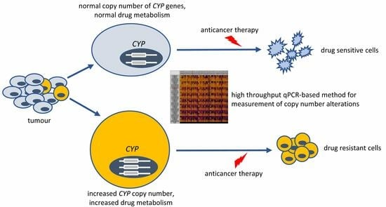
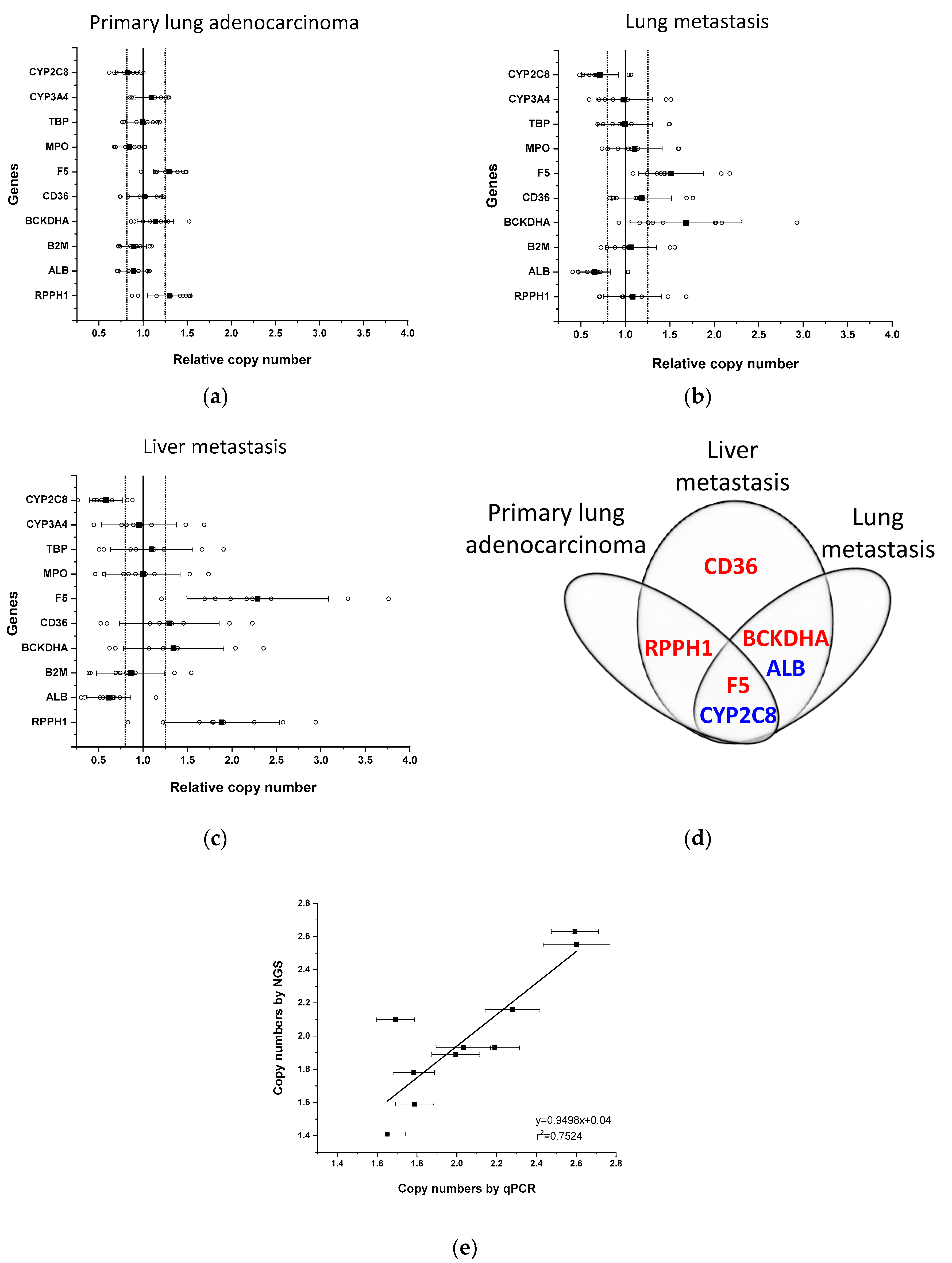
2.1. Copy Number Alterations in Tumour Determined by qPCR-Based Method

To improve the accuracy and reliability of the qPCR approach, we selected eight reference genes that are located on different chromosomes (chr1, chr4, chr6, chr7, chr14, chr15, chr17, chr19) and on various chromosomal regions (close to the centromere: ALB, CD36, RPPH1; close to the telomere: TBP; in between: F5, B2M, MPO, BCKDHA), and that are known to have two copies in a diploid genome (Supplementary Figure S1). In tumour tissues, the copy number of each gene was calculated by the comparative ∆∆Ct method obtained from the tumour and the control tissue (blood or normal lung tissue) pairs of each patient, and the relative copy number of each gene was determined in comparison to the other nine genes in each sample (Figure 1a–c). The control tissues were expected to have a normal (diploid) copy number for both the reference and the target CYP genes; therefore, the relative copy numbers obtained by intrasample normalization were set to 1. In tumour samples, the acceptable range of the relative copy numbers where the copy numbers in the tumour and the reference control tissue were equal (intersample normalization), was within 0.8 and 1.25 (with a 90% confidence interval and α = 0.1). The outlier gene pairs with the relative copy number lower than 0.8 and higher than 1.25 were considered to indicate inequality, obtaining scores of −1 and +1, respectively. It should be mentioned that the relative copy numbers did not show discrete values in tumour samples, and the continuous values may be explained by intratumoural heterogeneity. When we compared the copy numbers of each gene to each other in tumour samples, the sum of copy number alteration scores assigned gene deletion (score ≤ −5) and multiplication (score ≥ 5) (Table 1).
For the development of qPCR-based copy number analysis, the primary lung adenocarcinoma as well as lung and liver metastases of a non-smoker patient (OK18) were analyzed (Figure 1, Supplementary Table S1). Relative copy number calculation indicated an increase in two genes, F5 and RPPH1, and a decrease in CYP2C8 in the primary tumour sample (the mean of the relative copy number for F5: 1.297, for RPPH1: 1.301 and for CYP2C8: 0.80), whereas in lung and liver metastases, both an increase and a decrease in relative copy numbers of several genes were detected (lung metastasis sample: F5: 1.514, BCKDHA: 1.681, ALB: 0.653, CYP2C8: 0.713; liver metastasis: F5: 2.289, RPPH1: 1.882, BCKDHA: 1.343, CD36: 1.295, ALB: 0.616, CYP2C8: 0.583) (Figure 1a–c). Furthermore, some alterations in copy numbers in liver metastasis were also observed in the primary lung tumour (multiplication of F5 and RPPH1; deletion of CYP2C8) and in the lung metastasis (multiplication of F5 and BCKDHA; deletion of ALB and CYP2C8) (Figure 1d). The sum of the scores for these genes was higher than 5 or lower than −5 (Table 1) except for BCKDHA and CD36 in liver metastasis; therefore, in the particular tumour sample, these genes were considered to be inappropriate for normalization of the number of gene copies. For absolute quantification of gene copies in tumour samples, (1) the genes were applied as reference genes that were assessed to have the same copy number in the particular tumour as in the normal tissue (blood or normal lung tissue) with the sum of the scores within the range of 5 and −5, and (2) the average of the relative copy numbers was multiplied by 2 because of the diploidy of the control (normal) tissue. For the paclitaxel-metabolizing CYPs, deletion of CYP2C8, but not of CYP3A4 was detected in primary lung tumour and in lung and liver metastases (the average copy number of CYP2C8 in primary lung adenocarcinoma: 1.60, in lung metastasis: 1.47, in liver metastasis: 1.08). It also means that in a primary lung tumour, 40% of the cells and in lung metastasis, approximately half of the tumour cells displayed CYP2C8 deletion, whereas in liver metastasis, on average one copy of CYP2C8 was deleted from all the cells.
The copy number results assessed by the qPCR-based method were compared to those obtained by whole genome sequencing of the primary lung tumour of OK18 patient. We used ASCAT [29] to determine potential somatic copy number changes in the primary lung adenocarcinoma sample (Supplementary Figure S2). This tool uses allele-specific piecewise constant fitting to model the copy number across the whole genome. The copy number profile of the sample was largely diploid and showed only low levels of heterogeneity. For each target and reference gene, the unrounded sums of the predicted copy numbers of the two alleles were taken as the local absolute ploidy level. The copy number alterations of the genes F5, RPPH1 and CYP2C8 identified by the qPCR-based method in the primary tumour sample were confirmed by next-generation sequencing, and the absolute copy numbers of both the target and the reference genes established by sequencing and qPCR-based methods were congruent with each other (for 10 genes, slope: 0.9498, r2 = 0.7524) (Figure 1e).

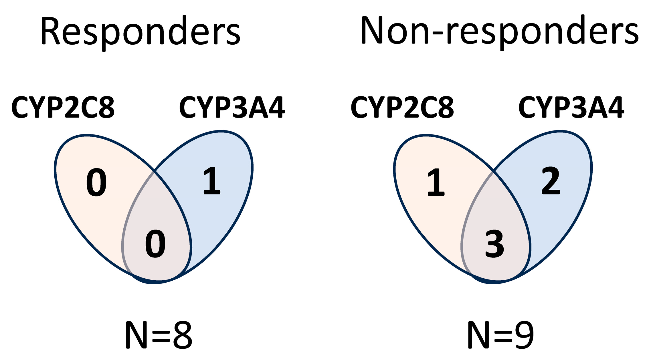
2.2. CYP Haplotype Distribution and Copy Number Alterations in Lung Adenocarcinoma Samples

For the estimation of patients’ paclitaxel-metabolizing capacity, CYP2C8, CYP3A4 and CYP3A5 alleles (CYP2C8*3, CYP2C8*4, CYP3A4*1B, CYP3A4*22, CYP3A5*3) most common in Caucasian populations were identified in lung adenocarcinoma patients on paclitaxel therapy (N = 17) (Table 2). Those who did not carry any of the CYP2C8, CYP3A4 or CYP3A5 polymorphisms were considered to have the CYP2C8*1, CYP3A4*1 or CYP3A5*1 wild-type alleles.
Most of the patients (N = 14) carried the CYP2C8*1/*1 genotype, possessing the potential for having the functional CYP2C8 enzyme, whereas three patients were heterozygous for CYP2C8*3, predicting reduced CYP2C8 activity. The loss-of-function CYP3A5*3 allele was identified in all patients, generally having the homozygous CYP3A5*3/*3 genotype, and only three patients carried the functional CYP3A5*1 allele (CYP3A5*1/*3). The SNP in the CYP3A4*1B (rs2740574) allele has been reported to be in genetic linkage with CYP3A5*1 [30]; therefore, it was not surprising that two of the CYP3A5*1 carriers also carried the CYP3A4*1B allele. The relative frequencies of CYP2C8*3, CYP3A5*3 and CYP3A4*1B were similar to the prevalence reported in Caucasian populations (Table 2) [5,31]; however, no patient with CYP2C8*4 or with CYP3A4*22 was identified.
The multiplication of the CYP2C8*3 allele is not expected to increase paclitaxel-metabolizing activity of the tumour cells; therefore, the multiplication of the functional wild-type allele is reasonable to consider in a tumour. We have designed the sequences of primers and probes for copy number measurements that were able to distinguish the wild-type and polymorphic CYP2C8*3 alleles. Only those tumour samples were considered to carry multiplication of CYP2C8 in which the CYP2C8*1 copy number was increased. The CYP3A4*1B allele produces functional CYP3A4 enzymes, and multiplication of CYP3A4*1B may lead to an increase in CYP3A4 function similarly to CYP3A4*1. Of the 17 lung adenocarcinoma samples, 7 displayed an increase in CYP2C8 and/or CYP3A4 genes (multiplication of CYP2C8 in 1 sample, of CYP3A4 in 3, and of both CYP2C8 and CYP3A4 in 3 samples), predicting elevated paclitaxel-metabolizing capacity of these tumours (OK18 was excluded because he was under non-paclitaxel therapy). A significant association was observed between the multiplication of paclitaxel-metabolizing CYPs and the outcome of paclitaxel therapy (responders: complete remission; non-responders: progressive disease/exit). In the primary tumour samples from non-responder patients (N = 9), copy number alterations in CYP2C8 and/or CYP3A4 were detected more frequently than in the samples from responders (N = 8) (non-responders 6/9 vs. responders 1/8, p = 0.0498, Fisher’s exact test) (Figure 2). However, for reliable association analysis, an increase in the size of the patient groups would be required.

3. Discussion

Resistance to anticancer agents is often associated with patients’ drug-metabolizing capacity, and germline polymorphisms of drug-metabolizing enzymes have been demonstrated to contribute to chemoresistance [3,8]. An increased inactivation rate of anticancer drugs (e.g., vincristine, erlotinib) is attributed to normal and ultra-rapid metabolizer phenotypes related to wild-type and gain-of-function CYP alleles, whereas poor metabolism linked to loss-of-function allele(s) clearly results in the low bioactivation rate of prodrugs, such as tamoxifen and cyclophosphamide. Significant interpatient variability in paclitaxel pharmacokinetics has also been assumed to be the source of substantial variations in treatment responses as well as in adverse effects [32,33]. Although in vitro studies have demonstrated the role of CYP2C8 and CYP3A enzymes in paclitaxel metabolism [21,23], the impact of CYP genetic polymorphisms (CYP2C8, CYP3A4 and CYP3A5) on paclitaxel clearance appears to be controversial in patients. A lower paclitaxel clearance has been demonstrated in CYP2C8*3 carrier patients with ovarian cancer than in non-carriers [34], and in contrast, increased paclitaxel-metabolizing activity was attributed to CYP2C8*3 allele in breast cancer patients [35]. Furthermore, Hennigsson et al. and Marsh et al. have suggested that the substantial interindividual variability in paclitaxel pharmacokinetics can be explained by variant alleles of neither CYP2C8 nor of CYP3A4 and CYP3A5 [25,27]. The limited patient group size of the present study did not allow us to evaluate the impact of CYP polymorphisms on patients’ response to paclitaxel therapy; however, it should be mentioned that the CYP2C8*3 allele was identified both in responders and in non-responder groups (1 and 2, respectively), whereas all the three heterozygous patients with the CYP3A5*1/*3 genotype were non-responders.
In addition to interpatient variability in the pharmacokinetics of anticancer agents, the biotransformation activity of cancer cells has also been raised as one of the relevant resistance mechanisms [7]. Altered expression (protein and mRNA) and activities of drug-metabolizing CYPs have been demonstrated in several tumour types compared to the normal non-tumourous tissues [9,36]; furthermore, as a consequence of altered drug metabolism, drug-tolerant cell clones have been assumed to be developed that can contribute to drug resistance [37,38]. Genomic instability, one of the main characteristics of tumour cells, is often manifested in structural variations, including copy number alterations (multiplication or deletion) of genes, e.g., drug-metabolizing CYP genes. Transcriptional changes owing to CYP copy number alterations can influence the ability of tumour cells to metabolize anticancer drugs, fostering the development of drug-tolerant cells within the tumour and progression of the disease. Thus, the multiplication or deletion of CYP genes may become a potential biomarker for tumours insensitive to a particular anticancer agent due to altered drug metabolism. There are various methods available for the detection of copy number alterations, using micro-array or next-generation sequencing techniques [39]; however, the development of simple and reliable alternative methods for routine clinical practice is still a requirement. A sensitive qPCR-based method with an optimal normalization procedure was presented here for the assessment of the copy numbers of paclitaxel-metabolizing CYP2C8 and CYP3A4 in lung adenocarcinoma samples. The identification of copy numbers of the target genes in a tumour is challenging because of the possibility of copy number alterations in the reference genes and because of the heterogeneity within the tumour [40]. The reference genes in normal diploid human cells are in two copies, whereas multiplication or deletion of the target genes may occur. Inherited (germline) copy number variations have been reported for CYP2D6 and CYP2C19; however, such variations have not been described for other drug-metabolizing CYPs in normal tissues [41]. Because in cancer cells, structural changes can occur in nearly any gene due to genomic instability, multiple reference genes located on different chromosomes were applied. As a subsequent step, the copy numbers of the eight reference genes and the two target CYP genes in the tumour samples were compared to each other and to those in normal tissues (blood or normal lung tissue), and the relative copy numbers were calculated. The scoring system described in the present study simplified the identification of those genes that underwent copy number alterations in the tumour. Finally, only those genes were selected as reference genes that were not implicated in the disease.
Tumour heterogeneity is an important issue that complicates the analysis of copy number alterations. Due to the intratumoural heterogeneity, genetically diverse tumour-cell subpopulations may exist within a single tumour site as well as across various disease sites in a single patient [42]. As an unsurprising consequence, (1) the relative copy numbers of the genes in tumour samples displayed continuous values, (2) and the copy number profile of the primary lung tumour was observed to be distinct from those in lung or in liver metastases (in the OK18 patient). The intertumoural heterogeneity in patients with the same cancer type may also result in variations in copy number profiles. The qPCR-based copy number analysis, however, identified an increase in F5 (chr 1q24.2) in all primary adenocarcinoma samples of the present study (N = 18); therefore, it was considered to be inappropriate as a reference gene. The arm-level copy number gain of chr 1q has been demonstrated in several cancer types, including lung adenocarcinoma, which explained the multiplication of the F5 gene in the samples of the present study [43,44].
Involvement of uptake and efflux transporters in resistance mechanisms is well described in cancer cells [11]; however, drug-metabolizing CYP activities in tumours are rarely linked to the selection of drug-tolerant cell clones. In lung adenocarcinoma samples from patients, CYP3A4 mRNA and protein expression has been demonstrated to significantly exceed the expression in normal lung tissues [45]. Furthermore, CYP2C8 expression was also detected in many human lung cancer cell lines [36,46]. Resistance to paclitaxel has been assumed to be partly attributed to overexpression of paclitaxel-metabolizing CYP enzymes. Hofman et al. investigated the impact of CYP3A4 and CYP2C8 overexpression on the taxane (docetaxel, paclitaxel) sensitivity of HepG2 cells, and concluded that CYP3A4 was of paramount importance for docetaxel insensitivity, whereas overexpression of the CYP2C8 enzyme was found to be associated with increased viability of the overexpressed cells under paclitaxel exposure [12]. In addition, paclitaxel has been revealed to efficiently activate the human pregnane X receptor (PXR) and to induce CYP3A4 expression and activity [38]. Furthermore, the paclitaxel resistance of A549 cells (human lung adenocarcinoma cell line) has been reported to be associated with the PXR-mediated transcriptional induction of CYP2C8 enzyme and Pgp transporter protein (ABCB1 transporter) [46]. Increased expression of CYP2C8 and/or CYP3A4 and the high paclitaxel-metabolizing activity of tumour cells are also assumed to be developed as a consequence of the multiplication of CYP copy numbers. The present qPCR-based method successfully established CYP copy number alterations in lung adenocarcinoma samples, and the present study was the first that reasonably assumed an association between CYP multiplication in tumours and the disease outcome of paclitaxel-treated patients; namely, multiplication of CYP2C8 and/or CYP3A4 copy numbers was observed more frequently in non-responder patients (progression of disease/exit group) than in responders with complete remission of the disease. To establish a resistance mechanism related to CYP copy number alteration in the tumour would require a group size larger than the 17 patients presented here, which was the limitation of this study. However, it should be emphasized that the primary aim of the study was to develop a simple method for routine clinical testing of copy number alterations which may result in an increase in the expression of paclitaxel-metabolizing CYPs, and may contribute to the selection of resistant cells in the tumour.
A further question was raised that the copy number alteration may extend beyond the CYP2C8 gene, and the alterations in tumour suppressor genes or oncogenes surrounding the CYP2C8 may also contribute to disease evolution. The chromosomal location of the CYP2C8 gene (chr 10q23.33) is close to tumour suppressor genes, such as PTEN (phosphatase and tensin homolog, chr 10q23.31), being involved in cell cycle regulation and CPEB3 (cytoplasmic polyadenylation element binding protein 3, chr 10q23.32), the regulator of EGFR transcription [47]. The copy number alterations that can have an impact on tumour development and progression often comprise deletion rather than multiplication of tumour suppressor genes [48]. The extended multiplication of the chr 10q23.3 region may be expected to manifest as a clinically relevant increase in CYP2C8 expression, and the negative outcome of the disease (progression or exit) in the present study was assumed to be associated with CYP2C8 multiplication and the increased paclitaxel-metabolizing capacity of tumour cells rather than the multiplication of tumour suppressor genes presumed on the basis of genomic location in CYP2C8 surroundings.
In conclusion, drug-resistance mechanisms related to the metabolism of anticancer agents are assumed to be associated with the drug-metabolizing capacity of the patients and/or the tumour. Therefore, it is of particular importance to identify genetic polymorphisms of drug-metabolizing CYPs and to establish an association between loss-of-function or gain-of-function CYP alleles and therapeutic outcomes. In addition, the copy number alterations of CYPs in the tumour may also contribute to the selection of resistant tumour cells. A high-throughput qPCR-based method was successfully developed for the detection of copy number changes in the tumour, and the scoring system, which was first applied, improved the identification of those genes that underwent deletion or multiplication and consequently were not appropriate to become a reference. The present method appears to be an alternative to next-generation sequencing in routine clinical practice. In the treatment naïve lung adenocarcinoma samples, the multiplication of paclitaxel-metabolizing CYP2C8 and/or CYP3A4 was detected, and the CYP copy number increase was more prevalent in non-responder patients than in responders to paclitaxel therapy. Although further research is required to confirm the findings of the present study and to validate the clinical utility of this approach, the implication of altered CYP copy numbers may provide an appropriate biomarker of therapy resistance and prognosis for clinical outcomes.

4. Materials and Methods

4.1. Patients

Patients (N = 18) treated with lung adenocarcinoma at the National Korányi Institute of Pulmonology (Budapest, Hungary) were enrolled in the present study. Written informed consent was obtained from all participants to perform genetic analysis of the tumour and blood or non-tumourous lung samples. The patients belonged to the Caucasian population, and their demographic and clinical data were recorded (Table 3). The patients’ response to the paclitaxel-containing therapy (responders: complete or partial remission; non-responders: progressive disease/exit) was defined according to the Response Evaluation Criteria in Solid Tumors (RECIST 1.1) [49].

4.2. CYP Genotyping

The patients who were on paclitaxel therapy (N = 17) were genotyped for CYP2C8, CYP3A4 and CYP3A5 (for CYP2C8*3, CYP2C8*4, CYP3A4*1B, CYP3A4*22 and CYP3A5*3). The patients’ blood samples were used to identify the SNPs in CYP2C8 [2130G>A (rs11572080), 30411A>G (rs10509681), 11041C>G (rs1058930)], CYP3A4 [−392A>G (rs2740574), 15389C>T (rs35599367)] and CYP3A5 [6986A>G (rs776746)]. Genomic DNA was extracted from blood samples using a Quick-DNA™ Universal Kit (Zymo Research, Irvine, CA, USA) according to the manufacturer’s instructions for biological fluids and cells. Hydrolysis SNP analyses for CYP2C8*3 (rs11572080 and rs10509681), CYP2C8*4 (rs1058930), CYP3A4*1B (rs2740574), CYP3A4*22 (rs35599367) and CYP3A5*3 (rs776746) were performed by PCR with self-designed TaqMan assays (primer and probe sequences are in Supplementary Table S2). Real-time PCR was carried out with 20 ng of genomic DNA by using a Luminaris Color Probe qPCR Master Mix (Thermo Fisher Scientific, Waltham, MA, USA). Allelic discrimination was based on two TaqMan probes, specific to the wild-type and the variant alleles labelled with FAM and HEX fluorescent tags, respectively.

4.3. Copy Number Analyses Using High-Throughput qPCR

Patients’ blood or non-tumourous lung tissue and tumour samples were used to determine the relative copy numbers of CYP2C8 and CYP3A4. Genomic DNA was extracted from non-tumourous lung tissues and tumour samples using the Quick-DNA™ Universal Kit (Zymo Research, Irvine, CA) according to the manufacturer’s instructions for solid tissues, whereas genomic DNA isolated from peripheral blood for CYP genotyping was also applied for copy number analyses. To establish CYP copy number alterations, the relative copy number approach was followed using the qPCR method. Briefly, the copy number assays applied a TaqMan method in a duplex PCR using a pair of primers for each gene and a VIC-labelled probe for RPPH1 and a FAM-labelled probe for any of the target CYPs (CYP2C8, CYP3A4) or seven of the reference genes (ALB, B2M, BCKDHA, F5, CD36, MPO, TBP). Primers and probes were self-designed based on the reference sequences in the National Center for Biotechnology Information reference assembly, except for RPPH1 for which the assay was commercially available (TaqMan® Copy Number Reference Assay, Cat. No: 4403326, Thermo Fisher Scientific) (Table 4). In blood samples or non-tumourous lung tissues for intrasample normalization, the qPCR method compared threshold cycles (Ct) of the target CYP genes and the reference genes that were known to be non-variable in the copy content (stable two copies in a diploid genome), to generate ΔCt values. The copy number assessment was based on the target sequences in CYP2C8 or CYP3A4 normalized to the sequences in eight reference genes (ALB, B2M, BCKDHA, F5, CD36, MPO, TBP and RPPH1) (Table 4). However, in the tumour an alteration in copy numbers of the reference genes may also occur; therefore, intersample normalization was required. Namely, the ∆∆Ct method comparing the copy numbers in the tumour to blood or non-tumourous lung tissue as the reference control tissues was applied for the assessment of relative copy numbers of both the target and the reference genes (for calculation of relative copy numbers in tumour samples, see Section 4.5 Data analysis).
A Biomark HDTM (Fluidigm, San Francisco, CA, USA) high-throughput microfluidics qPCR system with Flex SixTM integrated fluidics circuit chips was employed for copy number analyses. Specific target amplification as an initial step was performed according to the manufacturer’s instructions (TaqMan PreAmp Master Mix, Fluidigm). Thermal protocol for specific target amplification included an initial activation at 95 °C for 10 min followed by 14 cycles of denaturation at 95 °C for 15 s and annealing/extension at 60 °C for 4 min. The preamplification products were diluted to 1:5 with dilution buffer (Suspension Buffer, Fluidigm) and used as template samples in the qPCR. Assay mixtures for duplex reactions were prepared using Luminaris Probe qPCR Master Mix with ROX (Thermo Fisher Scientific, Waltham, MA, USA), primers and probes for RPPH1 (labeled with VIC) and either CYP2C8, CYP3A4, ALB, B2M, BCKHDA, CD36, F5, MPO or TBP (labeled with FAM). Each assay and sample was transferred into the inlets of the primed chip and loaded with an integrated fluidics circuit controller (Juno, Fluidigm). After loading, the PCR was carried out as follows: 50 °C for 2 min, 95 °C for 10 min and 40 PCR cycles at 95 °C for 15 s and at 60 °C for 1 min.

4.4. Whole Genome Sequencing

For validation of the copy numbers obtained by the high-throughput qPCR method, whole genome sequencing was performed in the primary lung tumour sample and in the peripheral blood (corresponding normal tissue) of a lung adenocarcinoma patient (OK18) (alignment files: accession ID EGAS00001003416), and genome-wide ploidy levels were determined using ASCAT [29]. This tool uses allele frequency and coverage information for a large set of SNPs across the whole genome. The SNPs were taken from a list of common variants from the 1000 Genomes Project. Briefly, we calculated coverage and variant allele frequency values using alleleCount (https://github.com/cancerit/alleleCount, access on 28 July 2023) on a custom mutation list containing common human SNPs spaced evenly at minimal distances of 10kb. Genomic positions inside annotated centromeres in the GRCh38 reference or ENCODE blacklisted regions [50] were omitted, and coverages were corrected by local GC contents and replication timing values, which were obtained using the dedicated helper scripts bundled with ASCAT. Finally, we ran the ASCAT pipeline to infer regional ploidy levels by considering the average coverage levels and allele frequency distributions in the predicted regions with changed copy numbers. To account for tumour heterogeneity, we used the unrounded copy number predictions.

4.5. Data Analysis

In normal tissues (blood and non-tumourous lung tissues), the relative copy numbers of the genes were defined as 1 because of the two copies of both the target and the reference genes in a diploid genome. In tumour samples, the copy numbers of both the target CYP genes (N = 2) and the reference genes (N = 8) were evaluated relative to those in blood or non-tumourous lung samples by the ∆∆Ct method (intersample normalization). Briefly, in tumour samples, the relative copy numbers of any two of the genes were considered to be equal with those in normal tissues if the 90% confidence intervals for the ratio of gene 1 and gene 2 in the tumour lay within the 0.80–1.25 acceptance range of copy number relative to normal tissue. The relative copy number of each gene pair was calculated and was scored as 0 if it was within the 0.8–1.25 range, −1 if it was <0.8 and +1 if it was >1.25. Alteration in the copy number of a gene in the tumour was established if the sum of the scores was ≤5 (deletion) or ≥5 (multiplication). For the calculation of the absolute copy number of a gene in the tumour, (1) these genes were applied as reference genes that were proved to be in two copies, (2) and the relative copy numbers were multiplied by 2. Association between alterations in CYP copy numbers in lung adenocarcinoma samples and therapeutic outcome was evaluated by Fisher’s exact test. A p value < 0.05 was considered to be a statistically significant difference.

Supplementary Materials

The supporting information can be downloaded at: https://www.mdpi.com/article/10.3390/ijms241713380/s1.

Author Contributions

Study conception and design: K.M. (Katalin Monostory); patient recruiting and diagnosis: J.M.; histopathology: T.H.; sample preparation, CYP testing, data acquisition: E.I., K.M. (Katalin Mangó), F.F. and Á.F.K.; next-generation sequencing: Á.P. and D.S.; data analysis and interpretation: E.I. and K.M. (Katalin Monostory); manuscript drafting: E.I. and K.M. (Katalin Monostory). All authors have read and agreed to the published version of the manuscript.

Funding

This work was supported by the grants of 2018-1.2.1-NKP-2018-00005 and TKP2021-EGA-31 (National Research, the Development and Innovation Fund of Hungary), K129065 (the National Research, Development and Innovation Office of Hungary), VEKOP-2.3.3-15-2017-00014 (Territorial Development Operational Programs of the Prime Minister’s Office in Hungary) and ELIXIR-HU-2019-HCNV (ELIXIR).

Institutional Review Board Statement

The study was approved by the Hungarian Committee of Science and Ethics, the Medical Research Council (protocol code ad.510/2013 52614-4/2013/EKU 04.11.2013). It was performed under the regulations of Act CLIV of 1997 on Health and decree 23/2002 of the Minister of Health of Hungary, as well as in accordance with the Declaration of Helsinki.

Informed Consent Statement

Written informed consent was obtained from all participants to perform genetic analysis of the tumour and blood or non-tumourous lung samples.

Data Availability Statement

The CYP SNP data have been deposited in the European Variation Archive (EVA) at EMBL-EBI under accession number PRJEB64027 (https://www.ebi.ac.uk/eva/?eva-study=PRJEB64027, access on 28 July 2023), and the alignment file of the primary lung tumour and peripheral blood samples of OK18 patient are accessible in the European Genome–phenome Archive under study ID EGAS00001003416 (https://onlinelibrary.wiley.com/doi/epdf/10.1002/ijc.32159; access on 28 July 2023).

Conflicts of Interest

The authors declare no conflict of interest.

References

  1. Sung, H.; Ferlay, J.; Siegel, R.L.; Laversanne, M.; Soerjomataram, I.; Jemal, A.; Bray, F. Global cancer statistics 2020: GLOBOCAN estimates of incidence and mortality worldwide for 36 cancers in 185 countries. CA Cancer J. Clin. 2021, 71, 209–249. [Google Scholar] [CrossRef] [PubMed]
  2. Wang, X.; Zhang, H.; Chen, X. Drug resistance and combating drug resistance in cancer. Cancer Drug Resist. 2019, 2, 141–160. [Google Scholar] [CrossRef]
  3. Kaur, G.; Gupta, S.K.; Singh, P.; Ali, V.; Kumar, V.; Verma, M. Drug-metabolizing enzymes: Role in drug resistance in cancer. Clin. Transl. Oncol. 2020, 22, 1667–1680. [Google Scholar] [CrossRef] [PubMed]
  4. Bruckmueller, H.; Cascorbi, I. ABCB1, ABCG2, ABCC1, ABCC2, and ABCC3 drug transporter polymorphisms and their impact on drug bioavailability: What is our current understanding? Expert Opin. Drug Metab. Toxicol. 2021, 17, 369–396. [Google Scholar] [CrossRef] [PubMed]
  5. Zanger, U.M.; Schwab, M. Cytochrome P450 enzymes in drug metabolism: Regulation of gene expression, enzyme activities, and impact of genetic variation. Pharmacol. Ther. 2013, 138, 103–141. [Google Scholar] [CrossRef] [PubMed]
  6. Mittal, B.; Tulsyan, S.; Kumar, S.; Mittal, R.D.; Agarwal, G. Cytochrome P450 in cancer susceptibility and treatment. In Advances in Clinical Chemistry; Makowski, G.S., Ed.; Elsevier: Amsterdam, The Netherlands, 2015; pp. 77–139. [Google Scholar] [CrossRef]
  7. Rodriguez-Antona, C.; Ingelman-Sundberg, M. Cytochrome P450 pharmacogenetics and cancer. Oncogene 2006, 25, 1679–1691. [Google Scholar] [CrossRef] [PubMed]
  8. Rodriguez-Antona, C.; Gomez, A.; Karlgren, M.; Sim, S.C.; Ingelman-Sundberg, M. Molecular genetics and epigenetics of the cytochrome P450 gene family and its relevance for cancer risk and treatment. Hum. Genet. 2010, 127, 1–17. [Google Scholar] [CrossRef]
  9. van Eijk, M.; Boosman, R.J.; Schinkel, A.H.; Huitema, A.D.R.; Beijnen, J.H. Cytochrome P450 3A4, 3A5, and 2C8 expression in breast, prostate, lung, endometrial, and ovarian tumors: Relevance for resistance to taxanes. Cancer Chemother. Pharmacol. 2019, 84, 487–499. [Google Scholar] [CrossRef]
  10. Rochat, B. Role of cytochrome P450 activity in the fate of anticancer agents and in drug resistance. Clin. Pharmacokinet. 2005, 44, 349–366. [Google Scholar] [CrossRef]
  11. Turner, A.P.; Alam, C.; Bendayan, R. Efflux transporters in cancer resistance: Molecular and functional characterization of P-glycoprotein. In Cancer Sensitizing Agents for Chemotherapy; Sosnik, A., Bendayan, R., Eds.; Academic Press: Cambridge, MA, USA, 2020; pp. 1–30. [Google Scholar] [CrossRef]
  12. Hofman, J.; Vagiannis, D.; Chen, S.; Guo, L. Roles of CYP3A4, CYP3A5 and CYP2C8 drug-metabolizing enzymes in cellular cytostatic resistance. Chem. Biol. Interact. 2021, 340, 109448. [Google Scholar] [CrossRef]
  13. Lavrov, A.V.; Ustaeva, O.A.; Adilgereeva, E.P.; Smirnikhina, S.A.; Chelysheva, E.Y.; Shukhov, O.A.; Shatokhin, Y.V.; Mordanov, S.V.; Turkina, A.G.; Kutsev, S.I. Copy number variation analysis in cytochromes and glutathione S-transferases may predict efficacy of tyrosine kinase inhibitors in chronic myeloid leukemia. PLoS ONE 2017, 12, e0182901. [Google Scholar] [CrossRef] [PubMed]
  14. Sneha, S.; Baker, S.C.; Green, A.; Storr, S.; Aiyappa, R.; Martin, S.; Pors, K. Intratumoural cytochrome P450 expression in breast cancer: Impact on standard of care treatment and new efforts to develop tumour-selective therapies. Biomedicines 2021, 9, 290. [Google Scholar] [CrossRef] [PubMed]
  15. Coutelier, M.; Holtgrewe, M.; Jäger, M.; Flöttman, R.; Mensah, M.A.; Spielmann, M.; Krawitz, P.; Horn, D.; Beule, D.; Mundlos, S. Combining callers improves the detection of copy number variants from whole-genome sequencing. Eur. J. Hum. Genet. 2022, 30, 178–186. [Google Scholar] [CrossRef]
  16. Luo, F. A systematic evaluation of copy number alterations detection methods on real SNP array and deep sequencing data. BMC Bioinform. 2019, 20, 692. [Google Scholar] [CrossRef] [PubMed]
  17. Balagué-Dobón, L.; Cáceres, A.; González, J.R. Fully exploiting SNP arrays: A systematic review on the tools to extract underlying genomic structure. Brief. Bioinform. 2022, 23, bbac043. [Google Scholar] [CrossRef] [PubMed]
  18. Ma, L.; Chung, W.K. Quantitative analysis of copy number variants based on real-time LightCycler PCR. Curr. Protoc. Hum. Genet. 2014, 80, 7.21.1–7.21.8. [Google Scholar] [CrossRef]
  19. Seguin, L.; Durandy, M.; Feral, C.C. Lung adenocarcinoma tumor origin: A guide for personalized medicine. Cancers. 2022, 14, 1759. [Google Scholar] [CrossRef]
  20. Barbuti, A.; Chen, Z.-S. Paclitaxel through the ages of anticancer therapy: Exploring its role in chemoresistance and radiation therapy. Cancers 2015, 7, 2360–2371. [Google Scholar] [CrossRef]
  21. Cresteil, T.; Monsarrat, B.; Dubois, J.; Sonnier, M.; Alvinerie, P.; Gueritte, F. Regioselective metabolism of taxoids by human CYP3A4 and 2C8: Structure-activity relationship. Drug Metab. Dispos. 2002, 30, 438–445. [Google Scholar] [CrossRef]
  22. Sparreboom, A.; Huizing, M.T.; Boesen, J.J.B.; Nooijen, W.J.; van Tellingen, O.; Beijnen, J.H. Isolation, purification, and biological activity of mono- and dihydroxylated paclitaxel metabolites from human feces. Cancer Chemother. Pharmacol. 1995, 36, 299–304. [Google Scholar] [CrossRef]
  23. Dai, D.; Zeldin, D.C.; Blaisdell, J.A.; Chanas, B.; Coulter, S.J.; Ghanayem, B.I.; Goldstein, J.A. Polymorphisms in human CYP2C8 decrease metabolism of the anticancer drug paclitaxel and arachidonic acid. Pharmacogenetics 2001, 11, 597–607. [Google Scholar] [CrossRef] [PubMed]
  24. Gao, Y.; Liu, D.; Wang, H.; Zhu, J.; Chen, C. Functional characterization of five CYP2C8 variants and prediction of CYP2C8 genotype-dependent effects on in vitro and in vivo drug–drug interactions. Xenobiotica 2010, 40, 467–475. [Google Scholar] [CrossRef]
  25. Henningsson, A.; Marsh, S.; Loos, W.J.; Karlsson, M.O.; Garsa, A.; Mross, K.; Mielke, S.; Viganò, L.; Locatelli, A.; Verweij, J.; et al. Association of CYP2C8, CYP3A4, CYP3A5, and ABCB1 polymorphisms with the pharmacokinetics of paclitaxel. Clin. Cancer Res. 2005, 11, 8097–8104. [Google Scholar] [CrossRef]
  26. Leskelä, S.; Jara, C.; Leandro-García, L.J.; Martínez, A.; García-Donas, J.; Hernando, S.; Hurtado, A.; Vicario, J.C.C.; Montero-Conde, C.; Landa, I.; et al. Polymorphisms in cytochromes P450 2C8 and 3A5 are associated with paclitaxel neurotoxicity. Pharmacogenomics J. 2011, 11, 121–129. [Google Scholar] [CrossRef] [PubMed]
  27. Marsh, S.; Somlo, G.; Li, X.; Frankel, P.; King, C.R.; Shannon, W.D.; McLeod, H.L.; Synold, T.W. Pharmacogenetic analysis of paclitaxel transport and metabolism genes in breast cancer. Pharmacogenomics J. 2007, 7, 362–365. [Google Scholar] [CrossRef] [PubMed]
  28. Gréen, H.; Söderkvist, P.; Rosenberg, P.; Mirghani, R.A.; Rymark, P.; Lundqvist, E.Å.; Peterson, C. Pharmacogenetic studies of paclitaxel in the treatment of qvarian cancer. Basic Clin. Pharmacol. Toxicol. 2009, 104, 130–137. [Google Scholar] [CrossRef]
  29. Van Loo, P.; Nordgard, S.H.; Lingjærde, O.C.; Russnes, H.G.; Rye, I.H.; Sun, W.; Weigman, V.J.; Marynen, P.; Zetterberg, A.; Naume, B.; et al. Allele-specific copy number analysis of tumors. Proc. Natl. Acad. Sci. USA 2010, 107, 16910–16915. [Google Scholar] [CrossRef]
  30. Floyd, M.D.; Gervasini, G.; Masica, A.L.; Mayo, G.; George, A.L.; Bhat, K.; Kim, R.B.; Wilkinson, G.R. Genotype-phenotype associations for common CYP3A4 and CYP3A5 variants in the basal and induced metabolism of midazolam in European- and African-American men and women. Pharmacogenetics 2003, 13, 595–606. [Google Scholar] [CrossRef]
  31. Zhou, Y.; Ingelman-Sundberg, M.; Lauschke, V.M.V. Worldwide distribution of cytochrome P450 alleles: A meta-analysis of population-scale sequencing projects. Clin. Pharmacol. Ther. 2017, 102, 688–700. [Google Scholar] [CrossRef]
  32. Joerger, M.; Huitema, A.D.R.; Richel, D.J.; Dittrich, C.; Pavlidis, N.; Briasoulis, E.; Vermorken, J.B.; Strocchi, E.; Martoni, A.; Sorio, R.; et al. Population pharmacokinetics and pharmacodynamics of paclitaxel and carboplatin in ovarian cancer patients: A study by the European Organization for Research and Treatment of Cancer-Pharmacology and Molecular Mechanisms Group and New Drug Development Group. Clin. Cancer Res. 2007, 13, 6410–6418. [Google Scholar] [CrossRef]
  33. Mielke, S.; Sparreboom, A.; Behringer, D.; Mross, K. Paclitaxel pharmacokinetics and response to chemotherapy in patients with advanced cancer treated with a weekly regimen. Anticancer Res. 2005, 25, 4423–4427. [Google Scholar] [PubMed]
  34. Bergmann, T.K.; Brasch-Andersen, C.; Gréen, H.; Mirza, M.; Pedersen, R.S.; Nielsen, F.; Skougaard, K.; Wihl, J.; Keldsen, N.; Damkier, P.; et al. Impact of CYP2C8*3 on paclitaxel clearance: A population pharmacokinetic and pharmacogenomic study in 93 patients with ovarian cancer. Pharmacogenomics J. 2011, 11, 113–120. [Google Scholar] [CrossRef]
  35. Marcath, L.A.; Kidwell, K.M.; Robinson, A.C.; Vangipuram, K.; Burness, M.L.; Griggs, J.J.; Van Poznak, C.; Schott, A.F.; Hayes, D.F.; Henry, N.L.; et al. Patients carrying CYP2C8*3 have shorter systemic paclitaxel exposure. Pharmacogenomics 2019, 20, 95–104. [Google Scholar] [CrossRef]
  36. Yu, L.J.; Matias, J.; Scudiero, D.A.; Hite, K.M.; Monks, A.; Sausville, E.A.; Waxman, D.J. P450 enzyme expression patterns in the NCI human tumor cell line panel. Drug Metab. Dispos. 2001, 29, 304–312. [Google Scholar]
  37. De Conti, G.; Dias, M.H.; Bernards, R. Fighting drug resistance through the targeting of drug-tolerant persister cells. Cancers 2021, 13, 1118. [Google Scholar] [CrossRef]
  38. Nallani, S.C.; Goodwin, B.; Buckley, A.R.; Buckley, D.J.; Desai, P.B. Differences in the induction of cytochrome P450 3A4 by taxane anticancer drugs, docetaxel and paclitaxel, assessed employing primary human hepatocytes. Cancer Chemother. Pharmacol. 2004, 54, 219–229. [Google Scholar] [CrossRef]
  39. Riggs, E.R.; Andersen, E.F.; Cherry, A.M.; Kantarci, S.; Kearney, H.; Patel, A.; Raca, G.; Ritter, D.I.; South, S.T.; Thorland, E.C.; et al. Technical standards for the interpretation and reporting of constitutional copy-number variants: A joint consensus recommendation of the American College of Medical Genetics and Genomics (ACMG) and the Clinical Genome Resource (ClinGen). Genet. Med. 2020, 22, 245–257. [Google Scholar] [CrossRef] [PubMed]
  40. Lim, Z.-F.; Ma, P.C. Emerging insights of tumor heterogeneity and drug resistance mechanisms in lung cancer targeted therapy. J. Hematol. Oncol. 2019, 12, 134. [Google Scholar] [CrossRef]
  41. Manikandan, P.; Nagini, S. Cytochrome P450 structure, function and clinical significance: A review. Curr. Drug Targets 2018, 19, 38–54. [Google Scholar] [CrossRef] [PubMed]
  42. Dagogo-Jack, I.; Shaw, A.T. Tumour heterogeneity and resistance to cancer therapies. Nat. Rev. Clin. Oncol. 2018, 15, 81–94. [Google Scholar] [CrossRef]
  43. Beroukhim, R.; Mermel, C.H.; Porter, D.; Wei, G.; Raychaudhuri, S.; Donovan, J.; Barretina, J.; Boehm, J.S.; Dobson, J.; Urashima, M.; et al. The landscape of somatic copy-number alteration across human cancers. Nature 2010, 463, 899–905. [Google Scholar] [CrossRef]
  44. Han, X.; Tan, Q.; Yang, S.; Li, J.; Xu, J.; Hao, X.; Hu, X.; Xing, P.; Liu, Y.; Lin, L.; et al. Comprehensive profiling of gene copy number alterations predicts patient prognosis in resected stages I–III lung adenocarcinoma. Front. Oncol. 2019, 9, 556. [Google Scholar] [CrossRef] [PubMed]
  45. Qixing, M.; Juqing, X.; Yajing, W.; Gaochao, D.; Wenjie, X.; Run, S.; Anpeng, W.; Lin, X.; Feng, J.; Jun, W. The expression levels of CYP3A4 and CYP3A5 serve as potential prognostic biomarkers in lung adenocarcinoma. Tumor Biol. 2017, 39, 101042831769834. [Google Scholar] [CrossRef] [PubMed]
  46. Chen, Y.; Huang, W.; Chen, F.; Hu, G.; Li, F.; Li, J.; Xuan, A. Pregnane X receptors regulate CYP2C8 and P-glycoprotein to impact on the resistance of NSCLC cells to Taxol. Cancer Med. 2016, 5, 3564–3571. [Google Scholar] [CrossRef]
  47. Liu, F.; Zhang, G.; Lv, S.; Wen, X.; Liu, P. miRNA-301b-3p accelerates migration and invasion of high-grade ovarian serous tumor via targeting CPEB3/EGFR axis. J. Cell. Biochem. 2019, 120, 12618–12627. [Google Scholar] [CrossRef] [PubMed]
  48. Wang, L.-H.; Wu, C.-F.; Rajasekaran, N.; Shin, Y.K. Loss of tumor suppressor gene function in human cancer: An overview. Cell. Physiol. Biochem. 2018, 51, 2647–2693. [Google Scholar] [CrossRef]
  49. Schwartz, L.H.; Litière, S.; de Vries, E.; Ford, R.; Gwyther, S.; Mandrekar, S.; Shankar, L.; Bogaerts, J.; Chen, A.; Dancey, J.; et al. RECIST 1.1—Update and clarification: From the RECIST committee. Eur. J. Cancer 2016, 62, 132–137. [Google Scholar] [CrossRef]
  50. Amemiya, H.M.; Kundaje, A.; Boyle, A.P. The ENCODE blacklist: Identification of problematic regions of the genome. Sci. Rep. 2019, 9, 9354. [Google Scholar] [CrossRef]
Figure 1. Copy numbers of genes in tumour samples of patient OK18. Relative copy numbers for each gene to those of the other nine genes (open circles) were assessed in the primary lung tumour (left inferior lobe, S6) (a) and in lung (right inferior lobe) (b) and liver metastases (right lobe) (c) by comparing to the normal lung tissue. Black squares represent the mean and whiskers are for standard deviation of relative copy numbers. Bold vertical line represents the normal relative copy number of the genes (set to 1). Dotted lines represent the acceptable range of normal copy numbers (0.8–1.25) in a sample. (d) Copy number alterations in genes in primary lung adenocarcinoma, liver and lung metastases (multiplication in red; deletion in blue). (e) Copy numbers of genes in primary lung adenocarcinoma determined by qPCR-based method and by next-generation sequencing. Whiskers represent the SD in copy numbers determined by qPCR-based method.
Ijms 24 13380 g001
Figure 2. Copy number alterations in CYP2C8 and/or CYP3A4 in primary lung adenocarcinoma samples in responder and non-responder patients under paclitaxel therapy.
Ijms 24 13380 g002
Table 1. The sum of the relative copy number scores for the genes in primary lung tumour and in lung and liver metastases of patient OK18.
GenePrimary Lung
Adenocarcinoma
Lung MetastasisLiver Metastasis
RPPH16 *07
ALB−3−8−8
B2M−30−2
BCKDHA373
CD36−223
F5678
MPO−41−1
TBP−2−10
CYP3A43−1−1
CYP2C8−5−7−7
* Alterations (multiplication or deletion) in copy numbers are indicated in bold.
Table 2. CYP allele and genotype frequencies in lung adenocarcinoma patients (N = 17) on paclitaxel therapy and in Caucasian population.
CYP Allele/GenotypeNFrequency (%)
Lung Adenocarcinoma PatientsCaucasian Population *
CYP allele
CYP2C8
*13191.279–90.5
*338.86.5–14
*4003–7
CYP3A4
*13294.183–95.5
*1B25.92–9
*22002.5–8
CYP3A5
*138.83–12
*33191.288–97
CYP genotype
CYP2C8
*1/*11482.381.8–85
*1/*3317.711.9–18.1
CYP3A4
*1/*11588.290–90.9
*1/*1B211.87.7–9
CYP3A5
*1/*1003.4
*1/*3211.811.6–19.71
*3/*31588.276.7–87.4
* CYP genotype frequencies in Caucasian population according to Gréen et al., Henningsson et al., Marsh et al. and Zanger et al. [5,25,27,28].
Table 3. Demographic and clinical data of lung adenocarcinoma patients.
Complete
Remission
Progressive Disease/Exit
Number of patients 810 b
Gender (male/female) 2/64/6
Age (year) a 64 (49; 74)63.5 (22; 72)
Smokingactive64
quit after 1 year02
never23
NA c-1
Primary tumour d1 (1a–1b)-2
2 (2a–2b)77
3 (3a–3b)1-
Lymph nodes d035
12-
223
x11
Metastasis d022
x67
Surgical interventionright superior lobectomy53
left superior lobectomy-2
right middle lobectomy1-
right inferior lobectomy21
left inferior lobectomy-1
atypical resection in right superior lobe-1
atypical resection in left inferior lobe-1
autopsy: left inferior lobe-1 e
Therapypaclitaxel+carboplatin89
gefitinib, cisplatin, afatinib-1 e
adjuvant radiotherapy14
a median (min; max); b one patient without TNM classification; c data not available; d TNM classification; e OK18 patient.
Table 4. List of genes and sequences of primers and probes for qPCR-based copy number measurement.
GeneGene NameChromosome LocationForward PrimerReverse PrimerProbe
RPPH1Ribonuclease P RNA component H114q11.2TaqMan® Copy Number Reference Assay, Thermo Fisher Scientific
Catalog number: 4403326
ALBalbumin4q13.3TCT TCT GTC AAC CCC ACAATC TCG ACG AAA CAC ACCGGC ACA ATG AAG TGG GTA ACC
B2Mbeta-2-microglobulin15q21.1GAA TGA GTC CCA TCC CAT CTCAG GGA AAC TAC TGG TTC AGACAG TAT CTC AGC AGG TGC CA
BCKDHAbranched-chain alpha-keto acid dehydrogenase19q1.2GTC GGC AGG TTA CCA CTAGAA GGG CTG GGA CAT ACATAT CTG GGT GCC ATC TCC TC
CD36cluster of differentiation 367q21.11TGA AGC TGA AAT TGA AG TGG AGAAC AGG TCT CAG TAC AGC ATAGT CAG TTG TCC TCG TCG AAA TC
F5coagulation factor V1q24.2TGA TCG AGG ATT TCA ACT CGTCC CAT GAC AGA ACT CCTAAG GTA AGA ACA CCC CCA CC
MPOmyelo-peroxidase17p22.1ACA CTA GGG TCA GTC CTT GCAA GCC TGG GTC ATT ATG ACCAT CTG GAG GAT TCC TGG AAC A
TBPTATA box binding protein6q27CCT GCT CTG TTT TCA GAT GGATA GCT TTG CTT CCC TTT CCAGT GGC ACT AAC GGT AAT TGT GT
CYP2C8cytochrome P450 2C810q23.33CGT TTC TCC CTC ACA ACC TTG CACT GTT AAG GTC AAT GAC GCA GACCT CAA TGC TCC TCT TCC CCA TCC CA
CYP3A4cytochrome P450 3A47q22.1CAG AGG TAG GTC TAA TTC AGT TCAAGA TCA CCT TCT ATC ACA CTC CATC ACA CCC AGC GTA GGG C
Disclaimer/Publisher’s Note: The statements, opinions and data contained in all publications are solely those of the individual author(s) and contributor(s) and not of MDPI and/or the editor(s). MDPI and/or the editor(s) disclaim responsibility for any injury to people or property resulting from any ideas, methods, instructions or products referred to in the content.

