Article

Evolutionary Origin of Human PALB2 Germline Pathogenic Variants

MoE Frontiers Science Center for Precision Oncology, Cancer Center and Institute of Translational Medicine, Faculty of Health Sciences, University of Macau, Macao
*
Author to whom correspondence should be addressed.
Int. J. Mol. Sci. 2023, 24(14), 11343; https://doi.org/10.3390/ijms241411343
Submission received: 19 June 2023 / Revised: 9 July 2023 / Accepted: 10 July 2023 / Published: 12 July 2023
(This article belongs to the Special Issue Molecular Advances in Cancer Genetics 3.0)

Abstract

:
PALB2 (Partner and localizer of BRCA2) is crucial for repairing DNA double-stranded breaks (DSBs) through homologous recombination (HR). Germline pathogenic variation in PALB2 disrupts DNA damage repair and increases the risk of Fanconi Anemia, breast cancer, and ovarian cancer. Determination of the evolutionary origin of human PALB2 variants will promote a deeper understanding of the biological basis of PALB2 germline variation and its roles in human diseases. We tested the evolution origin for 1444 human PALB2 germline variants, including 484 pathogenic and 960 benign variants. We performed a phylogenic analysis by tracing the variants in 100 vertebrates. However, we found no evidence to show that cross-species conservation was the origin of PALB2 germline pathogenic variants, but it is indeed a rich source for PALB2 germline benign variants. We performed a paleoanthropological analysis by tracing the variants in over 5000 ancient humans. We identified 50 pathogenic in 71 ancient humans dated from 32,895 to 689 before the present, of which 90.1% were dated within the recent 10,000 years. PALB2 benign variants were also highly shared with ancient humans. Data from our study reveal that human PALB2 pathogenic variants mostly arose in recent human history.

1. Introduction

PALB2 is a tumor suppressor gene. It plays a key role in the repair of DNA double-stranded breaks (DSBs) through the DNA homologous recombination (HR) pathway [1]. PALB2 functions as a central connecting hub to facilitate the formation of the BRCA1-PALB2-BRCA2 complex. The complex is essential for recruiting RAD51 for DNA strand exchange to repair at the sites of DSBs and also helps maintain the stability of stalled DNA replication forks [2]. Extensive studies showed that the carriers of monoallelic PALB2 pathogenic variants (PVs) are predisposed to develop multiple types of cancer, including breast, pancreatic, and ovarian cancers [3,4,5]; the carriers of biallelic PALB2 PVs can cause the subtype N of Fanconi anemia (FA-N) [6]. Furthermore, PALB2 PVs contribute to BRCAness, the phenotype caused by the damaged homologous recombination (HR) function due to the defects in HR-involved non-BRCA1/2 genes [7,8]. BRCAness is the specific marker for cancer diagnosis, prevention, and treatment. The PARP inhibitors (PARPi) successfully used in the treatment of BRCA-deficient cancers through synthetic lethality can also be applied to treat the cancer types with PALB2 pathogenic variation [9,10].
Knowledge for the origin of human PALB2 germline PVs is important to understand the biological basis of PALB2 germline PVs in cancer. However, little is known about the origin of human PALB2 PVs although it is known that ChAM, a functional domain of PALB2, exhibits high evolutionary conservation [11], and human PALB2 is under negative selection [12]. Our recent study revealed that the PVs in human BRCA1 and BRCA2 (BRCA) were not inherited through cross-species conservation but originated in recent human history [13]. These findings raise the question if the same patterns in human BRCA also exist in human PALB2 germline PVs, considering the BRCAness features of human PALB2 germline PVs.
In this study, we performed a phylogenetic analysis by searching human PALB2 PVs in 100 non-human vertebrates. We did not find evidence to support evolutionary conservation as the source for human PALB2 PVs. We further performed a paleoanthropological analysis by searching for human PALB2 PVs in ancient humans. The results showed that human PALB2 PVs mostly arose in the past 10,000 years. Therefore, our study demonstrated that both human PALB2 PVs and human BRCA PVs arose in recent human history.

2. Results

2.1. Cross Species Conservation Analysis

From the Clinvar database, we identified a total of 1444 human PALB2 germline variants consisting of 484 pathogenic variants (PVs) and 960 benign variants (BVs) for the study (Table S1). We downloaded from the UCSC genome browser the corresponding coordinates and bases of the 100 non-human vertebrates in 8 clades of Primates, Euarchontoglires, Laurasiatheria, Afrotheria, Mammals, Aves, Sarcopterygii and Fishs (Table S2). We searched the human PALB2 germline variants in the 100 vertebrates to determine if there could be shared PALB2 variants between humans and non-human species.
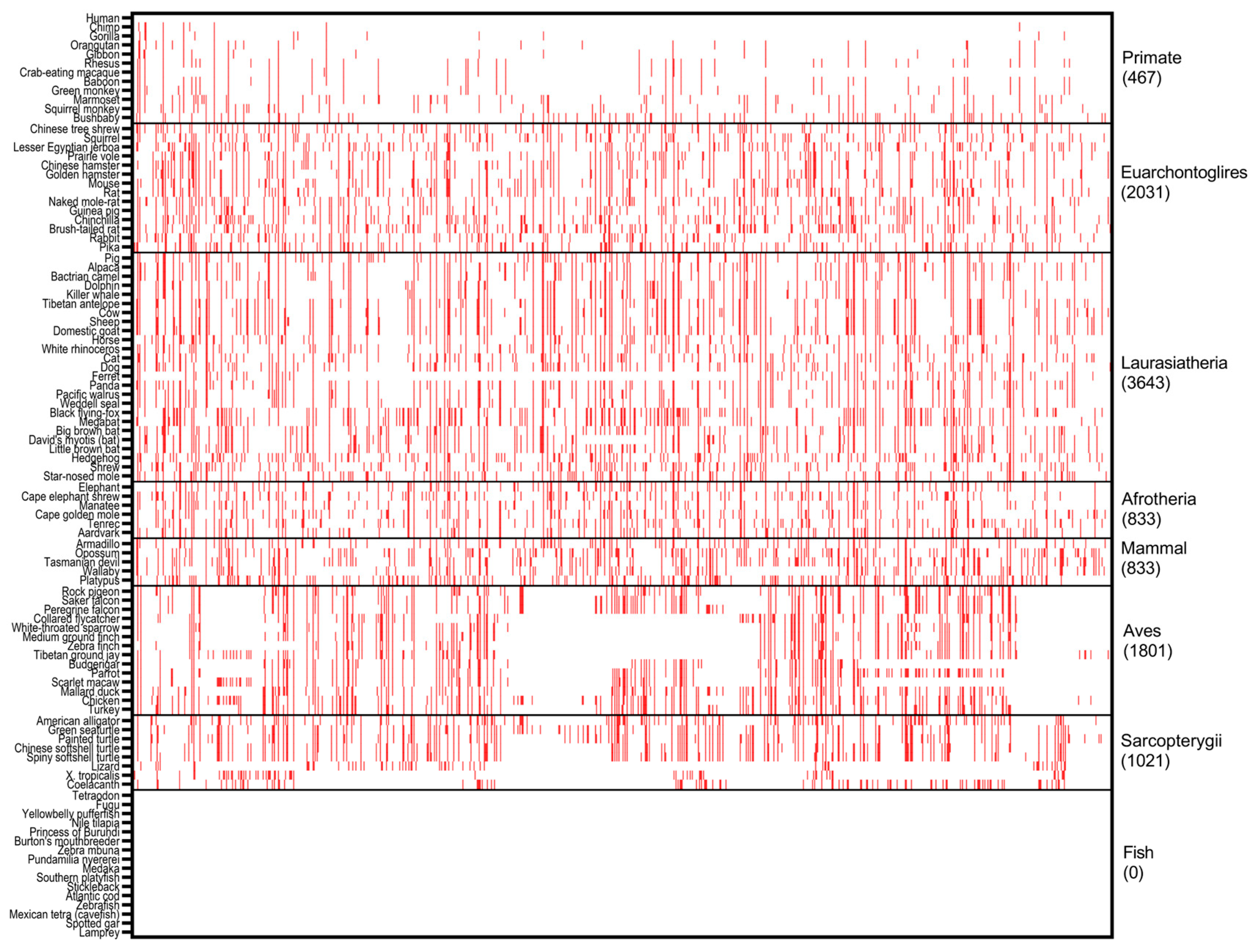
For PALB2 PVs, we identified 81 (16.7%) human variants shared with 72 non-human species (Figure 1, Table S2A). Of the species sharing with human PALB2 PVs, there were 21 species in Laurasiatheria clade of Mammalian class shared human c.2336C>G (p.Ser779Ter); chicken, mallard duck, and turkey of Aves clade had the sharing number of 15, 11 and 11, respectively. Species in Primates clade are close relatives of humans. However, only six human PVs were shared with the species in Primates, including three PVs of c.595del (pLeu199PhefsTer2), c.1451T>A (p.Leu484Ter) and c.1675C>T (p.Gln559Ter) shared with bushbaby, the most distal species in Primates to humans diverged 74 million years ago [14], and one PV of c.1018del (p.Glu347AsnfsTer9) shared between humans, rhesus, baboon and green monkey. Human PVs did not share with chimpanzees, gorillas, or orangutans. As the frequently used animal models in biomedical research, rat and mouse had only one and four PALB2 PVs shared with humans. Zebrafish, another important model, did not share any PV with humans. Of the human PVs shared with multiple species, c.751C>T (p.Gln251Ter) was shared with 27 species, from prairie voles in Euarchontoglires to spiny softshell turtles in Sarcopterygii. We searched the sharing of two PALB2 founder mutations of c.2323C>T (p.Gln775Ter) in the French Canadian population [15] and c.1592delT (p.Leu531Cysfs) in the Finnish population [16]. We found no evidence for their presence among the 100 species. As the majority of human PALB2 germline PVs were not present in non-human species, and the small number of PVs shared between humans and other species did not follow the order of evolutionary tree, cross-species conservation was unlikely the source of human PALB2 germline PVs.
As a control, we performed the same analysis for human PALB2 germline BVs. We identified 884 (90.5%) human variants shared with 83 species distributed in all clades except Fish (Figure 2, Table S2B). Platypus (194) and opossum (184) in Mammals and black flying fox (190) in Laurasiatheria had the highest sharing numbers. The most shared variant, c.3276C>G (p.Leu1092=), was distributed among 72 species from orangutan in Primates to coelacanth in Sarcopterygii. The sharing rate of PALB2 BVs of Primates is higher than PALB2 PVs, although it is still lower than other clades except Fish. For instance, c.2586+58C>T was shared by all the species within Primates, and Bushbaby in Primates had the highest sharing number of 86 BVs.
The sharing patterns between PALB2 PVs and BVs in 100 species were significantly different (p < 0.05) (Figure 3). The results indicated that evolutionary conservation was unlikely to contribute to the human PALB2 germline PVs but a rich source for human PALB2 germline BVs.

2.2. Ancient Human Genomic Analysis

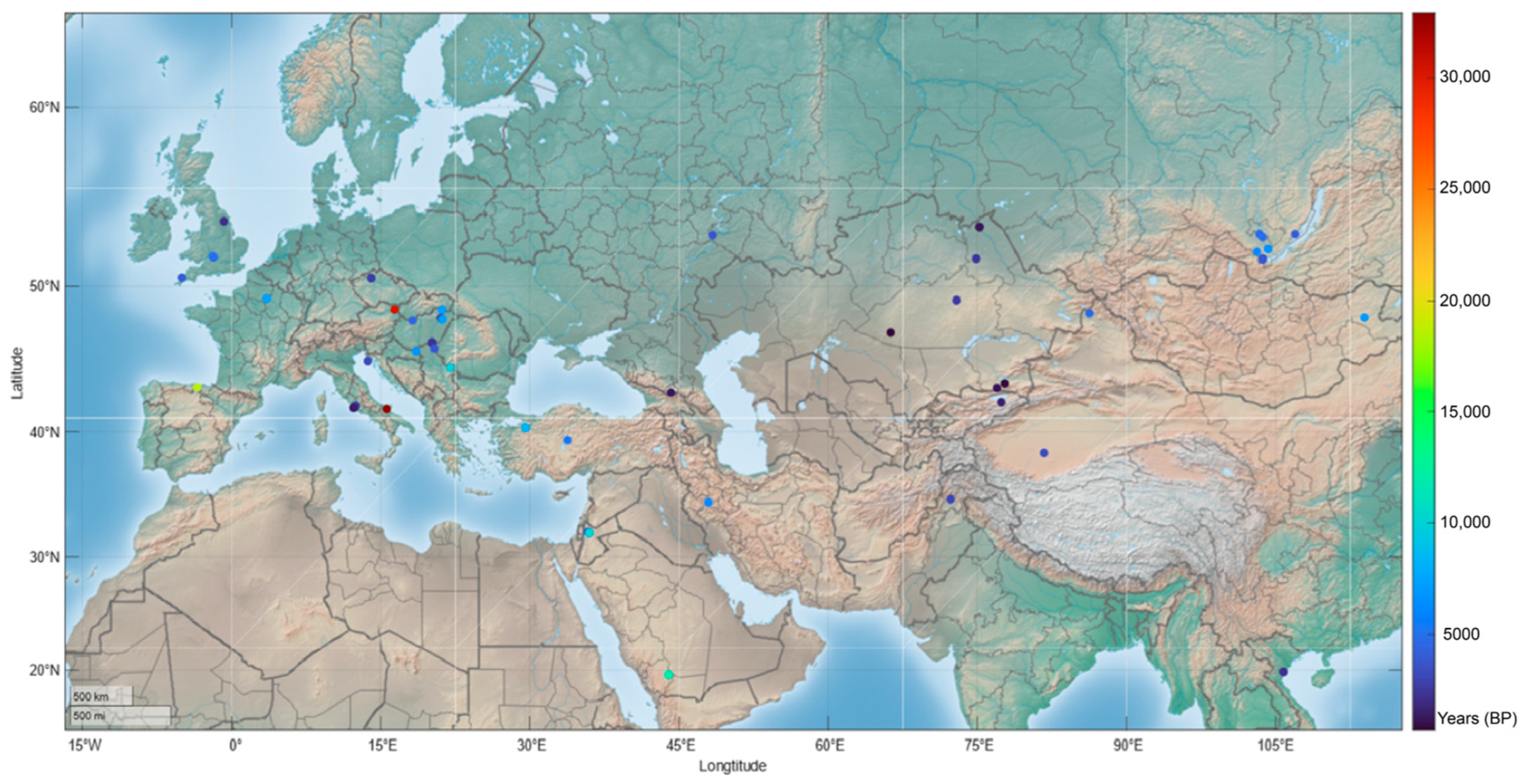
As cross-species conservation is unlikely the source for human PALB2 PVs, we then performed a paleoanthropological analysis to identify whether human PALB2 PVs could be originated from human itself. We searched human PALB2 PVs in the genomes of 5031 ancient human samples dated between 48,426 and 270 BP. We identified 50 human PALB2 PVs in 71 ancient human carriers dated from 32,895 to 689 BP, of which 64 (90.1%) were dated within the recent 10,000 years before the present (BP) (Table 1, Figure 4). Stopgain variants constituted 64% (32/50) of the PVs identified in ancient humans, and 18 (36%) PVs were located at the WD40 domain in the C-terminus of PALB2 (Table 1, Figure 5). The oldest carrier sharing PALB2 PV c.1837C>T (p.Gln613Ter) was an Italian dated about 32,985 BP, while the youngest carrier sharing PALB2 PV c.49-1G>A was in Tian Shan, China, dated 689 BP. Of the 50 identified PALB2 PVs, 16 (32%) were present in multiple ancient individuals. For instance, c.3256C>T (p.Arg1086Ter) was shared by four ancient individuals dated in 9860, 4475, 2175, and 1450 BP, accordingly; three PALB2 PVs were shared by three ancient individuals, and seven PALB2 PVs were shared by two individuals (Table 1). We did not identify the presence of the two reported PALB2 founder mutations (c.2323C>T and c.1592delT) in ancient humans.
PALB2 BVs were highly shared within ancient human populations. Of the 960 PALB2 BVs, 234 (24.4%) were identified in 2141 ancient humans, of which 143 (61.1%) were synonymous SNV and 78 (33.3%) were intron variants, 107 (45.7%) were present in multiple individuals and 16 (6.8%) were shared with more than 10 individuals (Table S3). c.2996+264T>C had the highest sharing numbers of 553 carriers, and c.3114-51T>A were present in 370 carriers. A nonsynonymous SNV c.1676A>G (p.Gln559Arg) was shared in 62 individuals. The results indicated that human PALB2 PVs arose during the recent human evolutionary process, whereas human PALB2 BVs originated from both cross-species conservation and human evolutionary process.

3. Discussion

Our study investigated the evolutionary origin of human PALB2 germline variation. Our comprehensive phylogenetical and paleoanthropological analyses demonstrate that PALB2 germline PVs in modern humans were not originated through cross-species conservation but rather arose in the evolutionary process of human itself, likely within the past 10,000 years, if not earlier. We are confident for the 1st part of the conclusion that PALB2 germline PVs in modern humans were not originated through cross-species conservation as the majority of human PALB2 germline PVs were not present in non-human species, and the small number of PVs shared between human and other species did not follow the order of the evolutionary tree; we are also rather confident in the 2nd part of the conclusion that PALB2 germline PVs in modern humans arose in human evolutionary process. However, whether PALB2 germline PVs in modern humans fully originated in the past 10,000 years could be debatable by the fact that 4518 (89.8%) ancient humans available for the study were dated within the last 10,000 years, as it could bias the arising time limited within the 10,000 years. The possibility that the PVs could arise earlier cannot be ruled out unless they are absent in more ancient humans older than 10,000 years when available, which are currently not available.
By conducting phylogenetical analysis in 100 non-human vertebrate species, we found no evidence to support cross-species conservation as the source of PALB2 PVs. In the species of Primates, only bushbaby, the most distant to human, shared three human PVs (c.595del, c.1451T>A and c.1675C>T). Rhesus, baboon, and green monkeys had only one PV c.1018del (p.Glu347AsnfsTer9) shared with humans. In contrast, the species in Aves and Sarcopterygii clades shared many human PVs. The sharing pattern may occur coincidentally rather than cross-species conservation, as it did not follow the evolutionary tree among the species. A proposed theory called “compensatory changes” proposed that the sharing of PVs between humans and distant species can be explained by other compensatory variants occurring at other coding or regulatory sites, neutralizing the deleterious effect of the mutations [36]. A deleterious variant in humans may be tolerated or have different impacts on other species. This may partially explain why the c.2336C>G (p.Ser779Ter) variant was only shared with 21 out of 25 species in the Laurasiatheria clade. Of the human PVs shared with multiple species, c.751C>T (p.Gln251Ter) was shared with 27 species, from prairie voles in Euarchontoglires to spiny softshell turtles in Sarcopterygii. The presence of human c.751C>T p.(Gln251Ter) in 27 species is likely by chance. Its presence only in 2/524 modern human families likely reflects its true prevalence in modern humans [5]. One of the possibilities exists that the C>T changes may also be de novo variants rather than originating from ancient humans. This is particularly true for the C>T variation at CpG islands, which is highly mutable due to high-degree spontaneous deamination [37,38]. In contrast, although the sharing rates of BVs in Primates were lower compared to other species, they followed the evolutionary relationship. Therefore, cross-species conservation can be a potential source for human PALB2 BVs.
It is interesting that Fish clades did not share any PVs or BVs with humans. The evolutionary divergence between fish and human occurred approximately 450 million years ago [39]. Take zebrafish as an example: zebrafish PALB2 consists of only 459 amino acids, while human PALB2 has 1186 amino acids. Possible reasons for the absence of shared variants could be that the chromosome location of the zebrafish Palb2 (chr1) is not the same as the human PALB2 (chr16) and the vast differences in protein may prevent proper PALB2 alignment between fish and humans.
Of the 5031 ancient humans, 4518 (89.8%) were dated within 10,000 years. In the paleoanthropological analysis, we identified 71 carriers of 50 different PALB2 PVs among ancient. Remarkably, 64 (90.1%) of these ancient carriers were dated within the past 10,000 years. This could be directly attributed to the significant human population expansion after the last glacial period around 10,000 years ago [40], the subsequent agricultural revolution [41], and the better preservation of ancient DNA for sequencing.
The most common PALB2 PV published is c.3113G>A p.(Trp1038Ter) (61/524 families), yet this did not appear in ancient humans, whereas the most frequent c.3256C>T p.(Arg1086Ter) in ancient humans was only present in 10/524 families [5]. This is an interesting feature in reflecting the dynamic prevalence of certain PALB2 PVs along the human evolutionary process. For the PVs with severe deleterious impact, they can be suppressed by negative selection to prevent their spread in human population; for the PVs with both deleterious and “favorable” impacts on different developmental stages, they could be selected by positive selection. An example is the PVs in human BRCA1. While its classical function is DNA damage repair through HR and NHEJ pathways, human BRCA1 gains multiple new functions, including regulation of immunity against viral infection [42], regulation of gene expression [43], regulation of neural development [44], and reproduction enhancement [45]. As such, human BRCA1 is under strong positive selection [42] that leads to a high prevalence of 0.2–0.5% for BRCA1 PVs, or one carrier in a few hundred individuals, in modern humans [46,47,48,49]. It is also important to note that most of the PVs arisen within a few thousand years ago were too young to be fixed in the genome, which can also lead to their unstable prevalence in either ancient or modern human populations. A possibility for c.3113G>A p.(Trp1038Ter) thought to be originated from the UK absent in ancient humans could be due to the small size of ancient samples and its lower prevalence that may not allow its detection, although 555 ancient humans from the UK were included in our study. It is also possible that c.3113G>A was young but has been rapidly spreading in the modern UK population.
Other factors contributing to the inconsistent prevalent rates of PALB2 PVs in ancient and modern humans could be the different PV spectra used in the comparison. The study [50] included only the protein-truncating variants (PTVs) but other types of PVs were included in our study, a small sample size of ancient humans causing a punctured but not linear prevalence of certain PVs in ancient humans, poor quality and low coverage of DNA sequences of certain ancient humans, contributing to artifacts such as the false C>T variant caused by deamination, the time difference between ancient and modern human populations that the ancient humans had a wide distribution of over 30,000 years, whereas the PV-delivered modern humans had a narrow time window of few decades. Our results raise an interesting question whether the pathogenic variants in other DNA damage-repair genes would also follow a similar path as did for human PALB2 PVs [51].

4. Methods and Materials

4.1. Source of Human PALB2 Variants

The source of human PALB2 variants was obtained from the ClinVar database (https://www.ncbi.nlm.nih.gov/clinvar/, accessed on 25 January 2023). The annotation of the PALB2 variants utilized the following reference sequences: cDNA: NM_024675.4, protein: NP_078951.2, and genome (hg38): NC_000016.10. Only the single nucleotide variants (SNV) and indels involving a single base pair (bp) were included in the study. The study only included the pathogenic variants (PVs) and benign variants (BVs), as defined by existing pathogenicity classifications. PVs consisted of variants categorized as “Pathogenic” and “Likely pathogenic”, while BVs included variants classified as “Benign” and “Likely benign”. Variants with uncertain significance (VUS) and conflicting interpretations were excluded from the study.

4.2. Cross-Species Genomic Analysis

The PALB2 sequences of 100 vertebrate species were obtained through a search on the UCSC genome browser (https://genome.ucsc.edu/, accessed on 15 March 2023). The detailed process of the phylogenetic analysis was described in our previous study [13]. In brief, the sequence alignment among these 100 species was performed following the steps outlined in Multiz Alignments within the UCSC genome browser [52]. The phyloP and PhyloFit programs were used to measure cross-species evolutionary conservation and construct a tree model based on the evolutionary relationships of the 100 vertebrate species [53,54]. The bases corresponding to human PALB2 variants in the hg38 reference genome and other species were obtained using the GetBase tool [55]. LASTZ and Multiz were employed to align the sequences of the human genome and the non-human PALB2 genome [56]. For indel alignments, any matched indel across all species was manually examined to ensure the accuracy of the alignment.

4.3. Ancient Human Genomic Analysis

Through searching PubMed and Allen Ancient DNA Resource (version 54.1) [57], we collected the genomic sequences of 5231 ancient human samples information and genome sequences dated from 48,426 to 270 BP. The ancient human genome data were downloaded, and the human genome reference sequences containing PALB2 (chr16:23,603,165–23,641,310 in hg38 and chr16:23,614,486–23,652,631 in hg19) were used for mapping analysis. BWA algorithm was employed to map PALB2 sequences to human genome sequences [58]. The resulting SAM files were converted to BAM files and sorted using SAMtools [59]. Duplicates were removed using MarkDuplicates, and the BAM files were further processed with the GATK toolkit using default settings for quality control, filtration, and variant calling [60,61]. The called ancient PALB2 variants were annotated by ANNOVAR using default parameters to differentiate between known and novel variants and provide information about the variants [62]. The ancient human PALB2 variants were then compared with those from the ClinVar database, which represents the PALB2 variants in modern humans, to find the shared variants between modern and ancient humans. The estimated age and location of the ancient human fossils, which carried PALB2 variants, were extracted from the original publications. For the PALB2 PVs, the distribution and dated time for each variant were generated using MATLAB (The Math Works, Inc., Natick, MA, USA).

4.4. Statistical Analysis

Mann–Whitney U test was performed to compare the differences in the sharing rate of PALB2 between PVs and BVs groups. The statistical analysis was performed using SPSS (version 26.0, IBM, New York, NY, USA). A p-value below 0.05 was considered statistically significant.

Supplementary Materials

The following supporting information can be downloaded at: https://www.mdpi.com/article/10.3390/ijms241411343/s1.

Author Contributions

Conceptualization: S.M.W.; Methodology, J.S.C. and J.L.; Software, J.L.; Formal Analysis, J.S.C.; Data Curation, J.S.C.; Writing—Original Draft Preparation, J.S.C.; Writing—Review and Editing, S.M.W., J.S.C., and J.L.; Visualization, J.S.C.; Supervision, S.M.W.; Funding Acquisition, S.M.W. All authors have read and agreed to the published version of the manuscript.

Funding

This work was supported by grants from the Macau Science and Technology Development Fund (085/2017/A2, 0077/2019/AMJ, 0032/2022/A1), the University of Macau (SRG2017-00097-FHS, MYRG2019-00018-FHS, 2020-00094-FHS), the Faculty of Health Sciences, University of Macau (FHSIG/SW/0007/2020P, MOE Frontiers Science Center for Precision Oncology pilot grant, and a startup fund) to S.M.W.

Institutional Review Board Statement

Not applicable.

Informed Consent Statement

Not applicable.

Data Availability Statement

The data presented in this study are available in online Supplementary Materials.

Acknowledgments

We are thankful to the Information and Communication Technology Office, University of Macau, for providing the High-Performance Computing Cluster resource and facilities for the study.

Conflicts of Interest

The authors declare no conflict of interest.

References

  1. Xia, B.; Sheng, Q.; Nakanishi, K.; Ohashi, A.; Wu, J.; Christ, N.; Liu, X.; Jasin, M.; Couch, F.J.; Livingston, D.M. Control of BRCA2 cellular and clinical functions by a nuclear partner, PALB2. Mol. Cell 2006, 22, 719–729. [Google Scholar] [CrossRef]
  2. Buisson, R.; Dion-Côté, A.M.; Coulombe, Y.; Launay, H.; Cai, H.; Stasiak, A.Z.; Stasiak, A.; Xia, B.; Masson, J.Y. Cooperation of breast cancer proteins PALB2 and piccolo BRCA2 in stimulating homologous recombination. Nat. Struct. Mol. Biol. 2010, 17, 1247–1254. [Google Scholar] [CrossRef] [Green Version]
  3. Rahman, N.; Seal, S.; Thompson, D.; Kelly, P.; Renwick, A.; Elliott, A.; Reid, S.; Spanova, K.; Barfoot, R.; Chagtai, T.; et al. PALB2, which encodes a BRCA2-interacting protein, is a breast cancer susceptibility gene. Nat. Genet. 2007, 39, 165–167. [Google Scholar] [CrossRef] [Green Version]
  4. Ohmoto, A.; Yachida, S.; Morizane, C. Genomic Features and Clinical Management of Patients with Hereditary Pancreatic Cancer Syndromes and Familial Pancreatic Cancer. Int. J. Mol. Sci. 2019, 20, 561. [Google Scholar] [CrossRef] [Green Version]
  5. Yang, X.; Leslie, G.; Doroszuk, A.; Schneider, S.; Allen, J.; Decker, B.; Dunning, A.M.; Redman, J.; Scarth, J.; Plaskocinska, I.; et al. Cancer Risks Associated with Germline PALB2 Pathogenic Variants: An International Study of 524 Families. J. Clin. Oncol. 2020, 38, 674–685. [Google Scholar] [CrossRef]
  6. Xia, B.; Dorsman, J.C.; Ameziane, N.; de Vries, Y.; Rooimans, M.A.; Sheng, Q.; Pals, G.; Errami, A.; Gluckman, E.; Llera, J.; et al. Fanconi anemia is associated with a defect in the BRCA2 partner PALB2. Nat. Genet. 2007, 39, 159–161. [Google Scholar] [CrossRef]
  7. Guo, M.; Wang, S.M. The BRCAness Landscape of Cancer. Cells 2022, 11, 3877. [Google Scholar] [CrossRef] [PubMed]
  8. Byrum, A.K.; Vindigni, A.; Mosammaparast, N. Defining and Modulating ‘BRCAness’. Trends Cell Biol. 2019, 29, 740–751. [Google Scholar] [CrossRef] [PubMed]
  9. Cortesi, L.; Rugo, H.S.; Jackisch, C. An Overview of PARP Inhibitors for the Treatment of Breast Cancer. Target Oncol. 2021, 16, 255–282. [Google Scholar] [CrossRef] [PubMed]
  10. Tutt, A.; Tovey, H.; Cheang, M.C.U.; Kernaghan, S.; Kilburn, L.; Gazinska, P.; Owen, J.; Abraham, J.; Barrett, S.; Barrett-Lee, P.; et al. Carboplatin in BRCA1/2-mutated and triple-negative breast cancer BRCAness subgroups: The TNT Trial. Nat. Med. 2018, 24, 628–637. [Google Scholar] [CrossRef] [PubMed] [Green Version]
  11. Bleuyard, J.Y.; Buisson, R.; Masson, J.Y.; Esashi, F. ChAM, a novel motif that mediates PALB2 intrinsic chromatin binding and facilitates DNA repair. EMBO Rep. 2012, 13, 135–141. [Google Scholar] [CrossRef] [PubMed] [Green Version]
  12. Zhao, B.; Li, J.; Wang, S.M. Pathogenic variants in human DNA damage repair genes mostly arose in recent human history. Nat. Commun. 2023. submitted. [Google Scholar]
  13. Li, J.; Zhao, B.; Huang, T.; Qin, Z.; Wang, S.M. Human BRCA pathogenic variants were originated during recent human history. Life Sci. Alliance 2022, 5, e202101263. [Google Scholar] [CrossRef] [PubMed]
  14. Kumar, S.; Suleski, M.; Craig, J.M.; Kasprowicz, A.E.; Sanderford, M.; Li, M.; Stecher, G.; Hedges, S.B. TimeTree 5: An Expanded Resource for Species Divergence Times. Mol. Biol. Evol. 2022, 39, msac174. [Google Scholar] [CrossRef]
  15. Foulkes, W.D.; Ghadirian, P.; Akbari, M.R.; Hamel, N.; Giroux, S.; Sabbaghian, N.; Darnel, A.; Royer, R.; Poll, A.; Fafard, E.; et al. Identification of a novel truncating PALB2 mutation and analysis of its contribution to early-onset breast cancer in French-Canadian women. Breast Cancer Res. 2007, 9, R83. [Google Scholar] [CrossRef] [PubMed]
  16. Erkko, H.; Xia, B.; Nikkilä, J.; Schleutker, J.; Syrjäkoski, K.; Mannermaa, A.; Kallioniemi, A.; Pylkäs, K.; Karppinen, S.M.; Rapakko, K.; et al. A recurrent mutation in PALB2 in Finnish cancer families. Nature 2007, 446, 316–319. [Google Scholar] [CrossRef]
  17. Patterson, N.; Isakov, M.; Booth, T.; Büster, L.; Fischer, C.E.; Olalde, I.; Ringbauer, H.; Akbari, A.; Cheronet, O.; Bleasdale, M.; et al. Large-scale migration into Britain during the Middle to Late Bronze Age. Nature 2022, 601, 588–594. [Google Scholar] [CrossRef]
  18. Mathieson, I.; Alpaslan-Roodenberg, S.; Posth, C.; Szécsényi-Nagy, A.; Rohland, N.; Mallick, S.; Olalde, I.; Broomandkhoshbacht, N.; Candilio, F.; Cheronet, O.; et al. The genomic history of southeastern Europe. Nature 2018, 555, 197–203. [Google Scholar] [CrossRef] [Green Version]
  19. Antonio, M.L.; Gao, Z.; Moots, H.M.; Lucci, M.; Candilio, F.; Sawyer, S.; Oberreiter, V.; Calderon, D.; Devitofranceschi, K.; Aikens, R.C.; et al. Ancient Rome: A genetic crossroads of Europe and the Mediterranean. Science 2019, 366, 708–714. [Google Scholar] [CrossRef]
  20. Agranat-Tamir, L.; Waldman, S.; Martin, M.A.S.; Gokhman, D.; Mishol, N.; Eshel, T.; Cheronet, O.; Rohland, N.; Mallick, S.; Adamski, N.; et al. The Genomic History of the Bronze Age Southern Levant. Cell 2020, 181, 1146–1157.e1111. [Google Scholar] [CrossRef]
  21. Wang, C.C.; Yeh, H.Y.; Popov, A.N.; Zhang, H.Q.; Matsumura, H.; Sirak, K.; Cheronet, O.; Kovalev, A.; Rohland, N.; Kim, A.M.; et al. Genomic insights into the formation of human populations in East Asia. Nature 2021, 591, 413–419. [Google Scholar] [CrossRef]
  22. Lazaridis, I.; Nadel, D.; Rollefson, G.; Merrett, D.C.; Rohland, N.; Mallick, S.; Fernandes, D.; Novak, M.; Gamarra, B.; Sirak, K.; et al. Genomic insights into the origin of farming in the ancient Near East. Nature 2016, 536, 419–424. [Google Scholar] [CrossRef] [Green Version]
  23. Damgaard, P.B.; Marchi, N.; Rasmussen, S.; Peyrot, M.; Renaud, G.; Korneliussen, T.; Moreno-Mayar, J.V.; Pedersen, M.W.; Goldberg, A.; Usmanova, E.; et al. 137 ancient human genomes from across the Eurasian steppes. Nature 2018, 557, 369–374. [Google Scholar] [CrossRef]
  24. de Barros Damgaard, P.; Martiniano, R.; Kamm, J.; Moreno-Mayar, J.V.; Kroonen, G.; Peyrot, M.; Barjamovic, G.; Rasmussen, S.; Zacho, C.; Baimukhanov, N.; et al. The first horse herders and the impact of early Bronze Age steppe expansions into Asia. Science 2018, 360, eaar7711. [Google Scholar] [CrossRef] [Green Version]
  25. McColl, H.; Racimo, F.; Vinner, L.; Demeter, F.; Gakuhari, T.; Moreno-Mayar, J.V.; van Driem, G.; Gram Wilken, U.; Seguin-Orlando, A.; de la Fuente Castro, C.; et al. The prehistoric peopling of Southeast Asia. Science 2018, 361, 88–92. [Google Scholar] [CrossRef] [PubMed] [Green Version]
  26. Sikora, M.; Pitulko, V.V.; Sousa, V.C.; Allentoft, M.E.; Vinner, L.; Rasmussen, S.; Margaryan, A.; de Barros Damgaard, P.; de la Fuente, C.; Renaud, G.; et al. The population history of northeastern Siberia since the Pleistocene. Nature 2019, 570, 182–188. [Google Scholar] [CrossRef] [Green Version]
  27. Fu, Q.; Posth, C.; Hajdinjak, M.; Petr, M.; Mallick, S.; Fernandes, D.; Furtwängler, A.; Haak, W.; Meyer, M.; Mittnik, A.; et al. The genetic history of Ice Age Europe. Nature 2016, 534, 200–205. [Google Scholar] [CrossRef] [Green Version]
  28. Zhang, F.; Ning, C.; Scott, A.; Fu, Q.; Bjørn, R.; Li, W.; Wei, D.; Wang, W.; Fan, L.; Abuduresule, I.; et al. The genomic origins of the Bronze Age Tarim Basin mummies. Nature 2021, 599, 256–261. [Google Scholar] [CrossRef] [PubMed]
  29. Narasimhan, V.M.; Patterson, N.; Moorjani, P.; Rohland, N.; Bernardos, R.; Mallick, S.; Lazaridis, I.; Nakatsuka, N.; Olalde, I.; Lipson, M.; et al. The formation of human populations in South and Central Asia. Science 2019, 365, eaat7487. [Google Scholar] [CrossRef] [PubMed]
  30. Mathieson, I.; Lazaridis, I.; Rohland, N.; Mallick, S.; Patterson, N.; Roodenberg, S.A.; Harney, E.; Stewardson, K.; Fernandes, D.; Novak, M.; et al. Eight thousand years of natural selection in Europe. bioRxiv 2015. [Google Scholar] [CrossRef]
  31. Fowler, C.; Olalde, I.; Cummings, V.; Armit, I.; Büster, L.; Cuthbert, S.; Rohland, N.; Cheronet, O.; Pinhasi, R.; Reich, D. A high-resolution picture of kinship practices in an Early Neolithic tomb. Nature 2022, 601, 584–587. [Google Scholar] [CrossRef] [PubMed]
  32. Moreno-Mayar, J.V.; Vinner, L.; de Barros Damgaard, P.; de la Fuente, C.; Chan, J.; Spence, J.P.; Allentoft, M.E.; Vimala, T.; Racimo, F.; Pinotti, T.; et al. Early human dispersals within the Americas. Science 2018, 362, eaav2621. [Google Scholar] [CrossRef] [PubMed] [Green Version]
  33. Flegontov, P.; Altınışık, N.E.; Changmai, P.; Rohland, N.; Mallick, S.; Adamski, N.; Bolnick, D.A.; Broomandkhoshbacht, N.; Candilio, F.; Culleton, B.J.; et al. Palaeo-Eskimo genetic ancestry and the peopling of Chukotka and North America. Nature 2019, 570, 236–240. [Google Scholar] [CrossRef] [PubMed]
  34. Harney, É.; Cheronet, O.; Fernandes, D.M.; Sirak, K.; Mah, M.; Bernardos, R.; Adamski, N.; Broomandkhoshbacht, N.; Callan, K.; Lawson, A.M.; et al. A minimally destructive protocol for DNA extraction from ancient teeth. Genome Res. 2021, 31, 472–483. [Google Scholar] [CrossRef]
  35. Posth, C.; Nakatsuka, N.; Lazaridis, I.; Skoglund, P.; Mallick, S.; Lamnidis, T.C.; Rohland, N.; Nägele, K.; Adamski, N.; Bertolini, E.; et al. Reconstructing the Deep Population History of Central and South America. Cell 2018, 175, 1185–1197.e1122. [Google Scholar] [CrossRef] [Green Version]
  36. Gao, L.; Zhang, J. Why are some human disease-associated mutations fixed in mice? Trends Genet. 2003, 19, 678–681. [Google Scholar] [CrossRef]
  37. Reich, D.; Patterson, N.; Kircher, M.; Delfin, F.; Nandineni, M.R.; Pugach, I.; Ko, A.M.; Ko, Y.C.; Jinam, T.A.; Phipps, M.E.; et al. Denisova admixture and the first modern human dispersals into Southeast Asia and Oceania. Am. J. Hum. Genet. 2011, 89, 516–528. [Google Scholar] [CrossRef] [Green Version]
  38. Evans, D.G.; Burghel, G.J.; Smith, M.J. Differential rates of germline heterozygote and mosaic variants in NF2 may show varying propensity for meiotic or mitotic mutation. J. Med. Genet. 2023. [Google Scholar] [CrossRef] [PubMed]
  39. Hedges, S.B. The origin and evolution of model organisms. Nat. Rev. Genet. 2002, 3, 838–849. [Google Scholar] [CrossRef]
  40. Henn, B.M.; Cavalli-Sforza, L.L.; Feldman, M.W. The great human expansion. Proc. Natl. Acad. Sci. USA 2012, 109, 17758–17764. [Google Scholar] [CrossRef]
  41. Gignoux, C.R.; Henn, B.M.; Mountain, J.L. Rapid, global demographic expansions after the origins of agriculture. Proc. Natl. Acad. Sci. USA 2011, 108, 6044–6049. [Google Scholar] [CrossRef] [PubMed]
  42. Lou, D.I.; McBee, R.M.; Le, U.Q.; Stone, A.C.; Wilkerson, G.K.; Demogines, A.M.; Sawyer, S.L. Rapid evolution of BRCA1 and BRCA2 in humans and other primates. BMC Evol. Biol. 2014, 14, 155. [Google Scholar] [CrossRef] [PubMed] [Green Version]
  43. Rosen, E.M.; Fan, S.; Ma, Y. BRCA1 regulation of transcription. Cancer Lett. 2006, 236, 175–185. [Google Scholar] [CrossRef] [PubMed]
  44. Pao, G.M.; Zhu, Q.; Perez-Garcia, C.G.; Chou, S.J.; Suh, H.; Gage, F.H.; O’Leary, D.D.; Verma, I.M. Role of BRCA1 in brain development. Proc. Natl. Acad. Sci. USA 2014, 111, E1240–E1248. [Google Scholar] [CrossRef] [PubMed]
  45. Smith, K.R.; Hanson, H.A.; Hollingshaus, M.S. BRCA1 and BRCA2 mutations and female fertility. Curr. Opin. Obs. Gynecol. 2013, 25, 207–213. [Google Scholar] [CrossRef] [Green Version]
  46. Maxwell, K.N.; Domchek, S.M.; Nathanson, K.L.; Robson, M.E. Population Frequency of Germline BRCA1/2 Mutations. J. Clin. Oncol. 2016, 34, 4183–4185. [Google Scholar] [CrossRef]
  47. Dong, H.; Chandratre, K.; Qin, Y.; Zhang, J.; Tian, X.; Rong, C.; Wang, N.; Guo, M.; Zhao, G.; Wang, S.M. Prevalence of BRCA1/BRCA2 pathogenic variation in Chinese Han population. J. Med. Genet. 2021, 58, 565–569. [Google Scholar] [CrossRef]
  48. Qin, Z.; Kuok, C.N.; Dong, H.; Jiang, L.; Zhang, L.; Guo, M.; Leong, H.K.; Wang, L.; Meng, G.; Wang, S.M. Can population BRCA screening be applied in non-Ashkenazi Jewish populations? Experience in Macau population. J. Med. Genet. 2021, 58, 587–591. [Google Scholar] [CrossRef]
  49. Chian, J.; Sinha, S.; Qin, Z.; Wang, S.M. BRCA1 and BRCA2 Variation in Taiwanese General Population and the Cancer Cohort. Front. Mol. Biosci. 2021, 8, 685174. [Google Scholar] [CrossRef]
  50. Dorling, L.; Carvalho, S.; Allen, J.; González-Neira, A.; Luccarini, C.; Wahlström, C.; Pooley, K.A.; Parsons, M.T.; Fortuno, C.; Wang, Q.; et al. Breast Cancer Risk Genes—Association Analysis in More than 113,000 Women. N. Engl. J. Med. 2021, 384, 428–439. [Google Scholar] [CrossRef]
  51. Fu, W.; O’Connor, T.D.; Jun, G.; Kang, H.M.; Abecasis, G.; Leal, S.M.; Gabriel, S.; Rieder, M.J.; Altshuler, D.; Shendure, J.; et al. Analysis of 6,515 exomes reveals the recent origin of most human protein-coding variants. Nature 2013, 493, 216–220. [Google Scholar] [CrossRef] [PubMed] [Green Version]
  52. Blanchette, M.; Kent, W.J.; Riemer, C.; Elnitski, L.; Smit, A.F.; Roskin, K.M.; Baertsch, R.; Rosenbloom, K.; Clawson, H.; Green, E.D.; et al. Aligning multiple genomic sequences with the threaded blockset aligner. Genome Res. 2004, 14, 708–715. [Google Scholar] [CrossRef] [PubMed] [Green Version]
  53. Hubisz, M.J.; Pollard, K.S.; Siepel, A. PHAST and RPHAST: Phylogenetic analysis with space/time models. Brief. Bioinform. 2011, 12, 41–51. [Google Scholar] [CrossRef] [PubMed] [Green Version]
  54. Ramani, R.; Krumholz, K.; Huang, Y.F.; Siepel, A. PhastWeb: A web interface for evolutionary conservation scoring of multiple sequence alignments using phastCons and phyloP. Bioinformatics 2019, 35, 2320–2322. [Google Scholar] [CrossRef]
  55. GetBase. Available online: https://github.com/Skylette14/GetBase (accessed on 4 February 2023).
  56. Harris, R.S. Improved Pairwise Alignment of Genomic DNA. Ph.D. Thesis, The Pennsylvania State University, University Park, PA, USA, 2007. [Google Scholar]
  57. Mallick, S.; Reich, D. The Allen Ancient DNA Resource (AADR): A curated compendium of ancient human resources. Harv. Dataverse 2023, V8. [Google Scholar] [CrossRef]
  58. Li, H.; Durbin, R. Fast and accurate short read alignment with Burrows-Wheeler transform. Bioinformatics 2009, 25, 1754–1760. [Google Scholar] [CrossRef] [Green Version]
  59. Danecek, P.; Bonfield, J.K.; Liddle, J.; Marshall, J.; Ohan, V.; Pollard, M.O.; Whitwham, A.; Keane, T.; McCarthy, S.A.; Davies, R.M.; et al. Twelve years of SAMtools and BCFtools. Gigascience 2021, 10, giab008. [Google Scholar] [CrossRef]
  60. McKenna, A.; Hanna, M.; Banks, E.; Sivachenko, A.; Cibulskis, K.; Kernytsky, A.; Garimella, K.; Altshuler, D.; Gabriel, S.; Daly, M.; et al. The Genome Analysis Toolkit: A MapReduce framework for analyzing next-generation DNA sequencing data. Genome Res. 2010, 20, 1297–1303. [Google Scholar] [CrossRef] [Green Version]
  61. Van der Auwera, G.A.; Carneiro, M.O.; Hartl, C.; Poplin, R.; Del Angel, G.; Levy-Moonshine, A.; Jordan, T.; Shakir, K.; Roazen, D.; Thibault, J.; et al. From FastQ data to high confidence variant calls: The Genome Analysis Toolkit best practices pipeline. Curr. Protoc. Bioinform. 2013, 43, 11.10.11–11.10.33. [Google Scholar] [CrossRef] [Green Version]
  62. Wang, K.; Li, M.; Hakonarson, H. ANNOVAR: Functional annotation of genetic variants from high-throughput sequencing data. Nucleic Acids Res. 2010, 38, e164. [Google Scholar] [CrossRef]
Figure 1. Cross-species analysis of PALB2 germline PVs. Human PALB2 PVs were searched in 100 non-human vertebrates. The results showed that cross-species conservation was unlikely the source of human PALB2 germline PVs. The red cell represents the PVs shared between humans and other species. X axis: human PALB2 PVs in Clinvar database; Y axis: 100 species from humans (top) in Primates to lamprey (bottom) in Fish. See Table S2A for details.
Ijms 24 11343 g001
Figure 2. Cross-species analysis of PALB2 germline BVs. The distribution of human PALB2 BVs in 100 vertebrates is shown above. The red cell represents the BVs shared between humans and other species. X axis: human PALB2 BVs in Clinvar database; Y axis: 100 species from human in Primates to lamprey in Fish. See Table S2B for details.
Ijms 24 11343 g002
Figure 3. The sharing rates of human PALB2 PVs and BVs with other non-human species. The sharing rates of each species were calculated by the number of shared variants divided by the total number of PVs or BVs. X axis: 100 species in the eight clades (Primates, Euarchontoglires, Laurasiatheria, Afrotheria, Mammals, Aves, Sarcopterygii, and Fish); Y axis: the sharing rate. See Table S2A,B for details.
Ijms 24 11343 g003
Figure 4. Geographic distribution of PALB2 PVs identified in ancient humans. A total of 50 PVs was identified in 71 ancient humans dated from 32,895 to 689 BP; 64 (90.1%) PV carriers were mostly in Eurasia dated within the last 10,000 BP. Color gradient bar on right side shows the dated age of fossils. See Table 1 for details.
Ijms 24 11343 g004
Figure 5. Distribution and mutation types of the 50 PALB2 PVs identified in ancient humans. Each line represents the location of PV, and the frequencies of variants were shown in the numbers inside the circle if the PV had more than one carrier. See Table 1 for details.
Ijms 24 11343 g005
Table 1. List of PALB2 PVs identified in ancient humans.
Carrier NumbercDNAProteinMutation TypeDomainArisen Time (BP) *References
4c.3256C>Tp.Arg1086TerstopgainWD409860[17,18,19]
3c.2257C>Tp.Arg753Terstopgain-3325[17,19,20]
3c.2509G>Ap.Glu837Lysnonsynonymous SNV-7050[19,21,22]
3c.2834+1G>A-splice siteWD406319[23,24,25]
2c.212-1G>A-splice siteETGE2275[25,26]
2c.211+1G>A-splice siteETGE10,050[27]
2c.1042C>Tp.Gln348Terstopgain-6319[24,28]
2c.1117G>Tp.Glu373Terstopgain-3157[19,29]
2c.1240C>Tp.Arg414TerstopgainChAM3740[23,30]
2c.1837C>Tp.Gln613Terstopgain-32,895[26,27]
2c.2218C>Tp.Gln740Terstopgain-8315[23,30]
2c.2512C>Tp.Gln838Terstopgain-3871[23,24]
2c.2515-1G>A-splice site-4106[17,24]
2c.2630G>Ap.Trp877TerstopgainWD405677[17,31]
2c.2693G>Ap.Trp898TerstopgainWD40>10,000[24,32]
2c.3523C>Tp.Gln1175TerstopgainWD406713[24]
1c.3G>Ap.Met1Ilestartloss-31,630[26]
1c.48+1G>C-splice sitecoiled-coil30,260[27]
1c.49-1G>A-splice sitecoiled-coil689[23]
1c.103delCp.Leu35PhefsTer18frameshift deletioncoiled-coil2758[23]
1c.173dupTp.Leu58PhefsTer16frameshift insertion-2554[23]
1c.178C>Tp.Gln60Terstopgain-6750[22]
1c.196C>Tp.Gln66Terstopgain-4158[24]
1c.428delTp.Leu143ArgfsTer34frameshift deletion-7178[30]
1c.451C>Tp.Gln151Terstopgain-8790[22]
1c.535C>Tp.Gln179Terstopgain-4885[32]
1c.940C>Tp.Gln314Terstopgain-1613[23]
1c.1027C>Tp.Gln343Terstopgain-2255[25]
1c.1174G>Tp.Glu392Terstopgain-4161[23]
1c.1257C>Ap.Cys419TerstopgainChAM3900[17]
1c.1258C>Tp.Gln420TerstopgainChAM725[33]
1c.1378C>Tp.Gln460Terstopgain-3440[28]
1c.1592delTp.Leu531CysfsTer30frameshift deletion-700[23]
1c.1969G>Tp.Glu657Terstopgain-2500[34]
1c.2066C>Tp.Ser689Leunonsynonymous SNV-6850[18]
1c.2386G>Tp.Gly796Terstopgain-1506[19]
1c.2389C>Tp.Gln797Terstopgain-4862[26]
1c.2594C>Tp.Ser865Leunonsynonymous SNVWD404940[31]
1c.2674G>Tp.Glu892TerstopgainWD408570[35]
1c.2674G>Ap.Glu892Lysnonsynonymous SNVWD403740[30]
1c.2694G>Ap.Trp898TerstopgainWD40>10,000[32]
1c.2761C>Tp.Gln921TerstopgainWD402478[23]
1c.2950dupCp.Leu984ProfsTer3frameshift insertionWD407221[30]
1c.2962C>Tp.Gln988TerstopgainWD401300[23]
1c.3058C>Tp.Gln1020TerstopgainWD401246[23]
1c.3114-1G>A-splice siteWD402279[23]
1c.3113+1G>A-splice siteWD401646[26]
1c.3420G>Ap.Trp1140TerstopgainWD401246[23]
1c.3469C>Tp.Gln1157TerstopgainWD408315[30]
1c.3492G>Ap.Trp1164TerstopgainWD4018,720[27]
* Dated time of the oldest carriers (BP, before present).
Disclaimer/Publisher’s Note: The statements, opinions and data contained in all publications are solely those of the individual author(s) and contributor(s) and not of MDPI and/or the editor(s). MDPI and/or the editor(s) disclaim responsibility for any injury to people or property resulting from any ideas, methods, instructions or products referred to in the content.