Article

Lactobacilli Downregulate Transcription Factors in Helicobacter pylori That Affect Motility, Acid Tolerance and Antimicrobial Peptide Survival

Department of Molecular Biosciences, The Wenner-Gren Institute, Stockholm University, 10691 Stockholm, Sweden
*
Author to whom correspondence should be addressed.
Int. J. Mol. Sci. 2022, 23(24), 15451; https://doi.org/10.3390/ijms232415451
Submission received: 11 October 2022 / Revised: 22 November 2022 / Accepted: 24 November 2022 / Published: 7 December 2022

Abstract

:
Helicobacter pylori infection triggers inflammation that may lead to gastritis, stomach ulcers and cancer. Probiotic bacteria, such as Lactobacillus, have been of interest as treatment options, however, little is known about the molecular mechanisms of Lactobacillus-mediated inhibition of H. pylori pathogenesis. In this work, we investigated the effect of Lactobacillus culture supernatants, so-called conditioned medium (CM), from two gastric isolates, L. gasseri and L. oris, on the expression of transcriptional regulators in H. pylori. Among the four known two-component systems (TCSs), i.e., ArsRS, FlgRS, CheAY and CrdRS, the flagellar regulator gene flgR and the acid resistance associated arsS gene were down-regulated by L. gasseri CM, whereas expression of the other TCS-genes remained unaffected. L. gasseri CM also reduced the motility of H. pylori, which is in line with reduced flgR expression. Furthermore, among six transcription factors of H. pylori only the ferric uptake regulator gene fur was regulated by L. gasseri CM. Deletion of fur further led to dramatically increased sensitivity to the antimicrobial peptide LL-37. Taken together, the results highlight that released/secreted factors of some lactobacilli, but not all, downregulate transcriptional regulators involved in motility, acid tolerance and LL-37 sensitivity of H. pylori.

1. Introduction

Helicobacter pylori is a Gram-negative bacterium that colonizes the stomach of approximately half of the human population worldwide. Infection by H. pylori can trigger the development of gastroduodenal ulcers, mucosa-associated lymphoid tissue lymphoma, and gastric carcinoma [1]. H. pylori is able to persistently colonize the hostile environment of the human stomach by expressing virulence factors, such as flagella, cytotoxins and adhesins [2]. However, H. pylori possesses a small genome of approximately 1.6 Mb and encodes only a few known transcriptional regulators, which control the expression of genes involved in bacterial metabolism and pathogenicity [3]. Among 14 bona fide transcription factors, there are four two-component systems (ArsRS for acid acclimation, FlgRS and CheAY for flagellar motility, and CrdRS for copper resistance); two transcriptional regulators HspR and HrcA involved in the stress response; two transcriptional regulators Fur and NikR involved in metal ion homeostasis; and two orphan response regulators HP1043 and HP1021 with unknown or stress-related functions [4].
At the site of colonization, the bacteria face the innate immune system. Antimicrobial peptides (AMPs) are produced in response to bacterial infection and can be found in considerable amounts at various locations in the human body [5]. LL-37 is a human cathelicidin that is secreted from epithelial cells and released from neutrophils [6]. Human beta defensin-2 (hBD2) is a defensin that is produced from epithelial cells and at the lesional skin [7,8]. H. pylori is sensitive to both of these AMPs in vitro and can also upregulate hBD2 and LL-37 in gastric cells [9,10]. There is evidence that H. pylori can evade the killing effects of AMPs in the gastric tract [11].
Lactobacillus is a commensal group of bacteria that colonizes the human stomach and has the ability to exclude bacterial pathogens and prevent infections by direct and indirect mechanisms known as colonization resistance [12]. Some of these lactobacilli are used as an alternative or adjunct therapy to antibiotics for the eradication of H. pylori, even though the underlying mechanisms of the antagonistic effects are still incompletely understood [13]. Previous studies have suggested that lactobacilli can interfere with H. pylori pathogenesis by affecting virulence gene expression. For example, the bacteriocin reuterin from a L. reuteri strain has been reported to inhibit the transcription levels of vacA and flaA in H. pylori [14]. It has also been reported that certain lactobacilli strains can influence the transcription levels of the adhesin gene sabA of H. pylori [15].
In this work, we investigated the effect of lactobacilli supernatants, so-called conditioned medium (CM), on the four two-component systems of H. pylori and found that L. gasseri downregulated the flgR regulator of motility and the acid resistance gene arsS, whereas L. oris did not affect any of the regulator genes tested. Motility was impaired by incubation in L. gasseri supernatant, confirming an effect on the corresponding phenotype. Among the six additional transcriptional regulators tested, only fur, which is necessary for colonization and ferric regulation, was downregulated by L. gasseri supernatant. Deletion of fur led to increased sensitivity to LL-37. Taken together, the data show that certain lactobacilli strains can affect the motility, acid susceptibility and antimicrobial peptide resistance of H. pylori by downregulating key regulatory genes.

2. Results

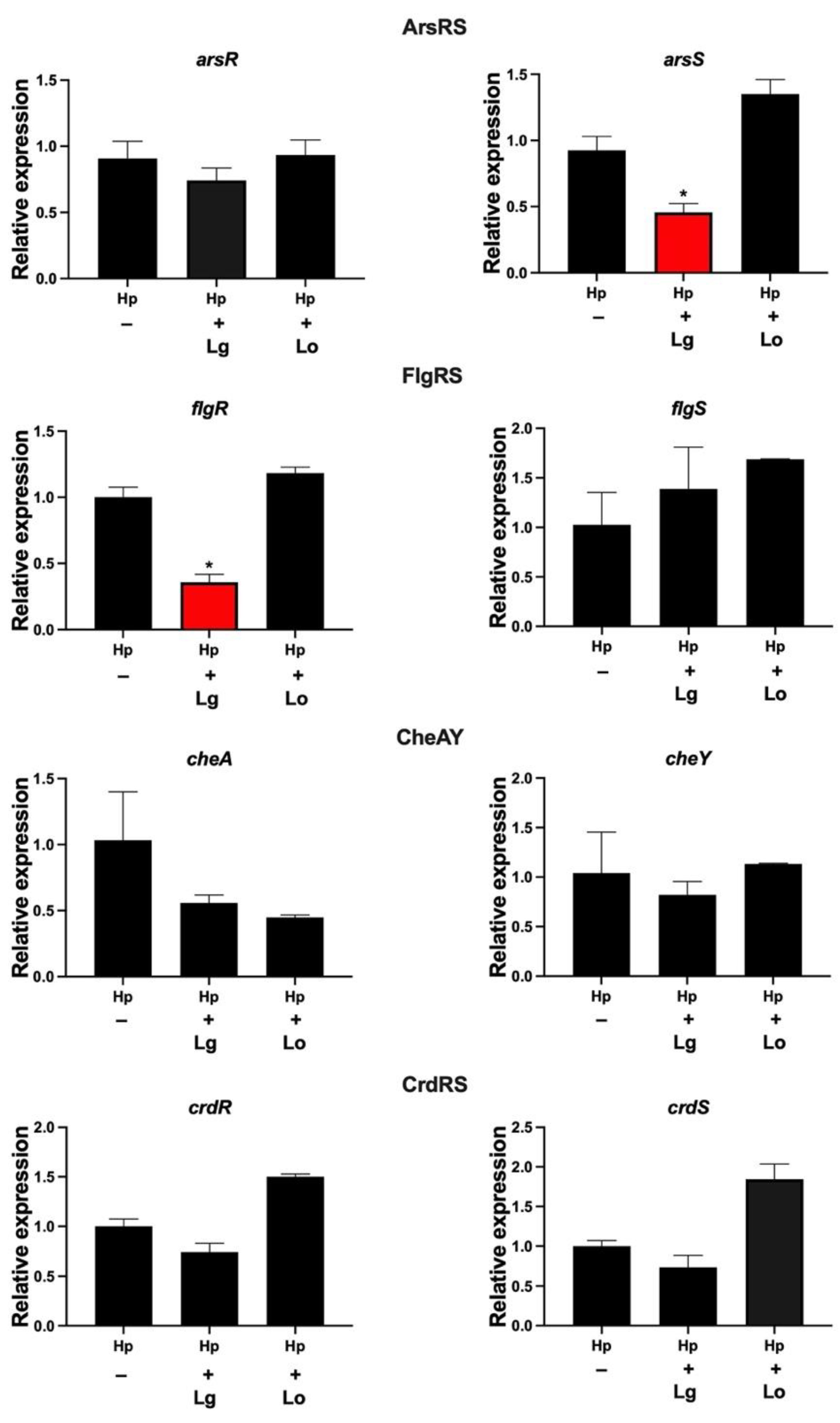
2.1. Lactobacilli Affect the Gene Expression of flgR and arsS in H. pylori

Two-component systems (TCSs) are important for the regulation of various bacterial factors. To assess the effect of lactobacilli on the expression of TCSs in H. pylori, we used supernatants, so-called conditioned medium (CM), from lactobacilli. H. pylori incubated with CM for 2 h was examined by qPCR for the expression of the genes of the four known TCSs, i.e., ArsRS, FlgRS, CheAY and CrdRS. Most genes were unaffected by incubation in CM, but two genes were significantly downregulated by lactobacilli. H. pylori incubated in L. gasseri CM showed a significant reduction in the expression of flgR, which is involved in flagella regulation, and in the expression of arsS, which is involved in acid adaptation (Figure 1). There were no significant changes in the expression of these genes when H. pylori was coincubated with L. oris CM. Thus, certain lactobacilli can affect the expression of genes encoding H. pylori TCSs. In summary, a soluble or secreted component(s) of L. gasseri, but not L. oris, both of which were isolated from gastric biopsies, downregulated the response regulator gene flgR, which is involved in motility, and the sensory histidine kinase gene arsS, which is involved in acid resistance.

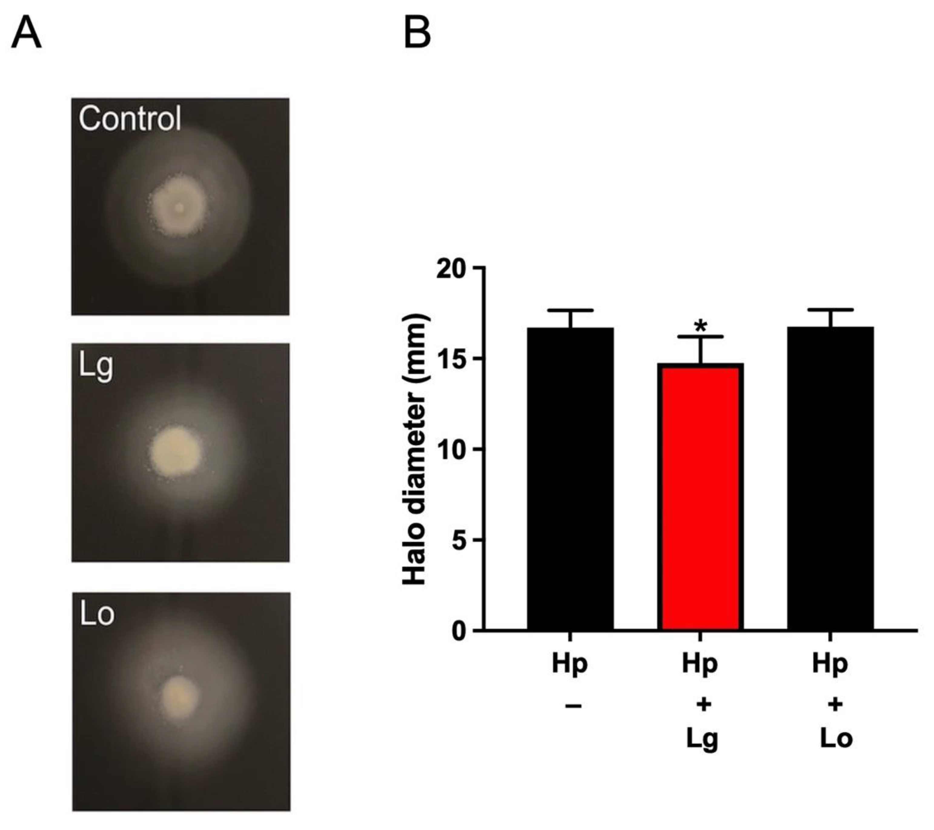
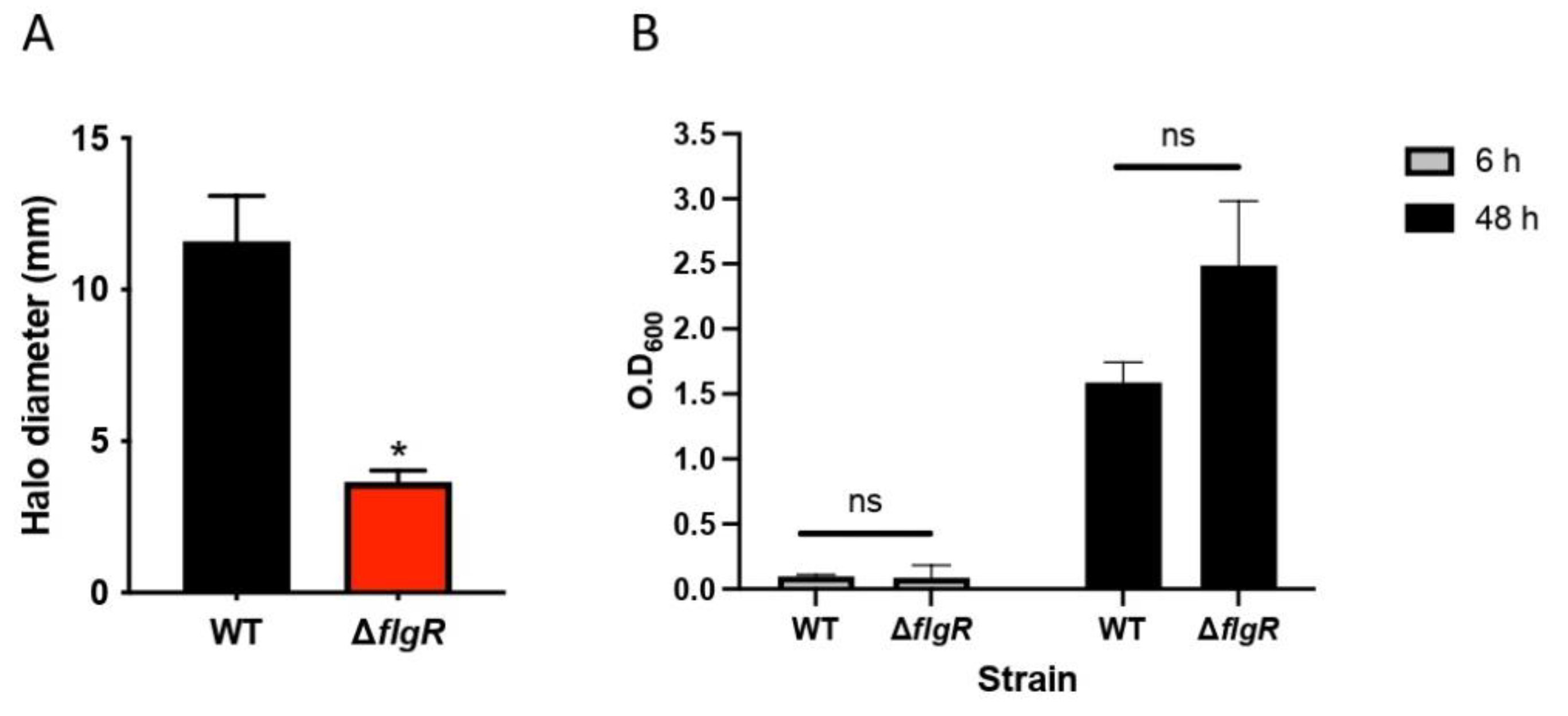
2.2. Lactobacilli Affect H. pylori Motility in a Strain-Specific Manner

Motility is an important virulence factor of H. pylori. It is well known that the two-component system FlgRS is involved in motility and flagellar gene regulation in H. pylori [16]. Since flgR was downregulated by L. gasseri, we next assessed whether lactobacilli strains might affect H. pylori motility. To examine the effect of lactobacilli CM on the motility of H. pylori, we inoculated the H. pylori wild-type strain in motility agar supplemented with CM of lactobacilli. As shown in Figure 2, the motility halo diameter of H. pylori in motility agar supplemented with CM of L. gasseri was slightly, but significantly, decreased compared with that of the control. However, the motility halo diameter in motility agar supplemented with CM of L. oris remained unaffected. To confirm the function of flgR in the motility of the H. pylori strain, we assessed the motility of a deletion mutant ∆flgR strain and the wild-type strain. As expected, the H. pylori wild-type strain was fully motile in motility agar, while the ΔflgR mutant strain showed impaired motility (Figure 3A). The ΔflgR mutant strain showed a colony phenotype and growth rate similar to those of the wild-type strain (Figure 3B). These results suggest that L. gasseri, but not L. oris, can inhibit the motile capacity of H. pylori, possibly by repressing the expression of genes associated with motility functions, such as flgR.

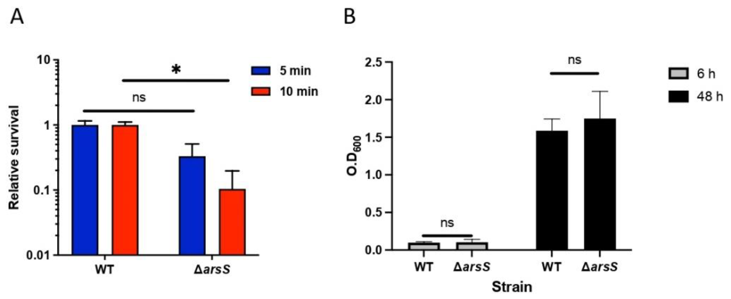
2.3. The ArsRS System Influences the Acid Sensitivity of H. pylori at pH 2

The ability to survive in an acidic environment is an important survival factor of H. pylori. The ArsRS system is involved in acid adaptation [17]. Since arsS was downregulated by L. gasseri, we assessed the sensitivity of H. pylori to environmental stress by incubating H. pylori cells at pH 2 for 5 or 10 min. For this experiment, we constructed an H. pyloriarsS mutant strain, which showed a colony phenotype and growth similar to those of the wild-type strain (Figure 4A), to confirm the function of arsS in acid adaptation [18]. As expected, the ΔarsS mutant strain was significantly more sensitive to pH 2 than the H. pylori wild-type strain (Figure 4B), which is in line with data reported for other strains that show the dependence of H. pylori on the ArsRS system to express proteins required for acid acclimation, for example in strains 26695 and 43504 ArsRS regulates acid-induced expression of α-carbonic anhydrase [19].

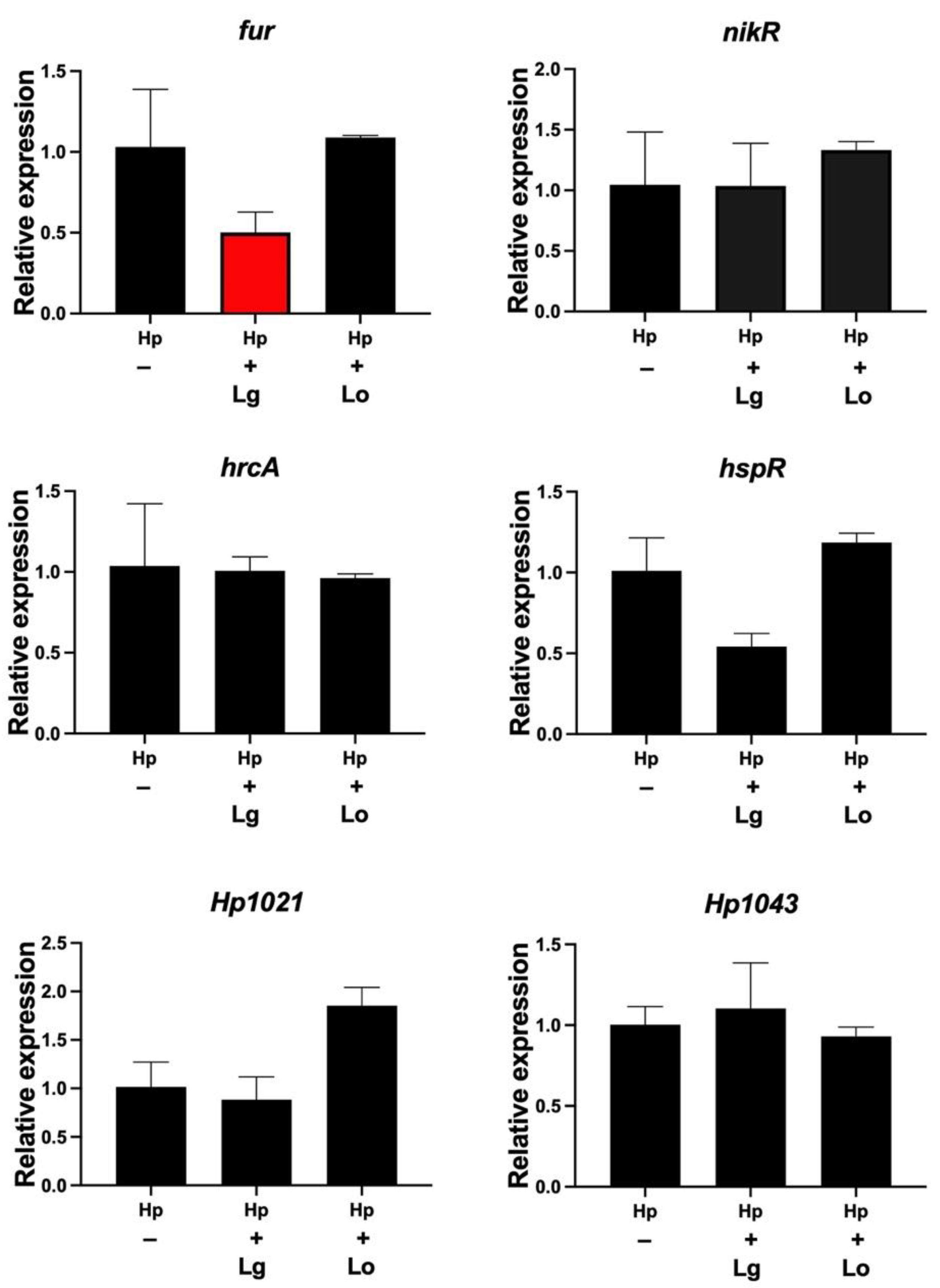
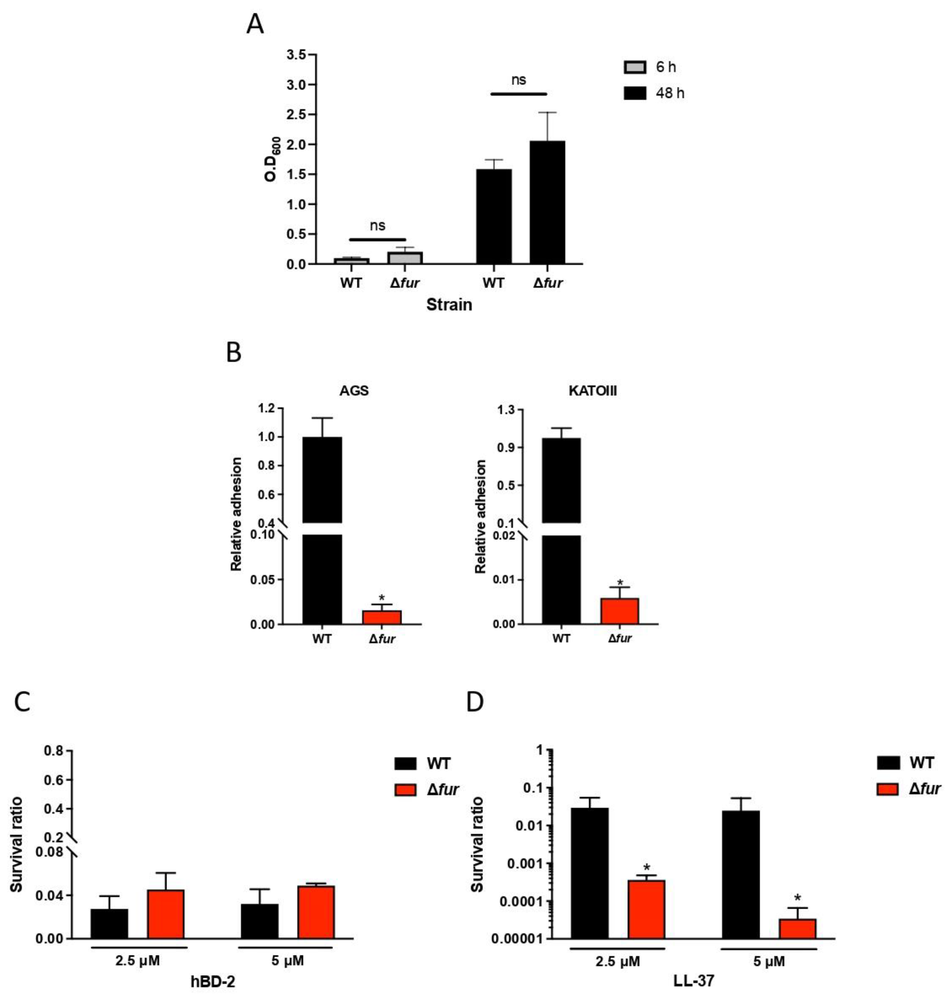
2.4. Lactobacillus Gasseri Downregulates the Ferric Uptake Regulator

We next investigated the expression of six transcription factors, i.e., fur, nikR, hspR, hrcA, Hp1021 and Hp1043, by qPCR after incubation of H. pylori in lactobacilli conditioned medium (CM). H. pylori incubated for 2 h in L. gasseri CM showed a significant reduction in fur, which is involved in ferric uptake and colonization. There was no significant change in the expression of fur when H. pylori was coincubated with L. oris CM. The expression levels of genes for nickel homeostasis (nikR), heat shock (hspR, hrcA) and orphan regulators (Hp1021, Hp1043) did not significantly change in response to CM from any of the Lactobacillus strains (Figure 5). These data demonstrate that L. gasseri, but not L. oris, reduced the gene expression of the transcriptional global regulator fur.

2.5. Fur Is Involved in Antimicrobial Peptide LL-37 Resistance in H. pylori

Fur has been extensively studied by gene expression analysis and has been linked to reduced colonization [20]. Epithelial cells are known to secrete antimicrobial peptides, such as hBD-2 and LL-37, in response to H. pylori infection [9,21]. Since fur is important for colonization, we next investigated whether fur might influence the ability of bacteria to survive antimicrobial peptides (AMPs). To test this hypothesis, we assessed the survival of the wild-type and Δfur strains in the presence of two AMPs, LL-37 and hBD-2. A Δfur mutant was constructed in strain 67:21 and showed a colony phenotype and growth similar to those of the wild-type strain (Figure 6A), but it lost the ability to attach to host cells (Figure 6B), as previously shown for strain 7.13 [22]. The hBD-2 peptide showed a strong bactericidal effect against H. pylori, with no difference between the wild-type and ∆fur mutant strains and no significant difference between 2.5 µM and 5 µM hBD-2 (Figure 6C). The bactericidal effect of LL-37 was strong against wild-type H. pylori; however, the ∆fur mutant was extremely sensitive to LL-37, with 100-fold and 1000-fold reductions in viability after incubation with 2.5 and 5 μM LL-37, respectively, compared to wild-type H. pylori (Figure 6D). These results suggest that the expression of Fur plays an important role in H. pylori resistance to antimicrobial peptide LL-37.

3. Discussion

In this study, we explored the gene expression of two-component systems and transcription factors of H. pylori after incubation with lactobacilli. The expression levels of the motility-related gene flgR and the acid resistance-associated gene arsS were reduced after treatment with conditioned medium (CM) from L. gasseri but not after treatment with CM from L. oris. Among the tested transcription factors, only the ferric uptake-associated transcription factor fur was regulated by lactobacilli, and it was found to dramatically affect the resistance of H. pylori to the antimicrobial peptide LL-37.
It is well known that the two-component system FlgRS is involved in motility and flagellar gene regulation in H. pylori [16]. Numerous factors can influence the establishment of H. pylori colonization either directly or indirectly. One of these factors is the ability of H. pylori to be fully motile. Chemotaxis and helical rod shapes promote flagellar motility away from the acidic lumen to the preferred niche of H. pylori, which is on and close to gastric epithelial cells [23]. Incubation of H. pylori in L. gasseri CM downregulated the expression of the motility-related regulator flgR. Furthermore, when H. pylori was incubated with L. gasseri CM, motility was slightly but significantly reduced. It is tempting to speculate that the downregulation of flgR expression strongly contributes to the observed reduction in motility; however, we can not exclude the possibility that the regulation of other factors not tested also influenced motility. In other pathogens, such as Escherichia coli, Pseudomonas aeruginosa, Clostridium difficile, and Yersinia enterocolitica, flagellin and/or the distally located flagellar cap protein have been reported to function as adhesins [24,25]. However, there is no evidence of specific attachment of H. pylori flagella to epithelial cells, although it has been speculated that flagella have a role in cell adhesion [26]. During initial infection of the stomach lumen, urease-dependent ammonia production locally raises the pH, which promotes bacterial survival. We found that L. gasseri CM decreased the expression of arsS, which affects the expression of numerous genes, including urease genes [17]. It is reasonable to assume that the influence of secreted or released factors from L. gasseri on H. pylori regulatory genes modifies the virulence properties of the pathogen.
Among the tested transcriptional regulators, only the ferric uptake regulator fur was regulated by L. gasseri. Fur is a global regulator known to interact with the apo- and/or the holo-regulator on more than 200 identified target gene loci involved in metal homeostasis, acidic response, and virulence [27]. Fur has also been shown to regulate the transcription of several genes that may be important for cell adhesion [27], and Fur seems to directly or indirectly regulate other unknown factors that are essential for H. pylori adhesion to gastric epithelial cells. Analysis of the adhesion properties of the wild-type and Δfur mutant indicated that Fur plays a critical role in H. pylori attachment to host epithelial cells. Even though many Fur-regulated genes have been shown to play a role in colonization [20], none of them have been previously shown to be directly involved in host cell attachment. The exact mechanism of the adherence deficiency of the H. pylori 67:21 Δfur mutant needs to be further determined, with emphasis on the downstream adhesion-related genes that are positively regulated by Fur. Furthermore, the reduced adhesion ability and the high sensitivity to antimicrobial peptide LL-37 of the Δfur mutant may be a reason for its deficiency in colonization of the mouse stomach [20]. The physiological concentration of LL-37 ranges between 0.03 μg/mL in human plasma and 8 μg/mL in bronchoalveolar lavage fluid, and it can reach up to 7 mg/mL in psoriatic lesions of inflammatory conditions [7,8,28]. Surface gastric epithelial cells and epithelial cells in the fundic glands of patients with gastritis can constitutively produce LL-37, and its production is upregulated by H. pylori infection [9].
Helicobacter pylori is the main risk factor for developing stomach cancer. Today, treatment with antibiotics that cure a majority of infections is recommended [29]. However, the use of antibiotics against H. pylori infection causes side effects and in particular leads to increased resistance of H. pylori as well as other pathogens to antibiotics. Indeed, the antimicrobial eradication rate of H. pylori has been declining globally because of antibiotic resistance [30,31]. A promising alternative or adjunct therapy is probiotic administration, which is generally prescribed as a supplement to standard therapy to reduce the adverse effects caused by antibiotics [32,33]. In particular, probiotics, such as lactobacilli, can combat H. pylori through multiple strategies; however, the molecular mechanisms underlying these antagonistic effects are still largely unknown [13,34,35]. In this study, since we see that the effect of lactobacilli on H. pylori is through a secreted component that does not require the two bacteria to be in contact with each other, it is tempting to speculate that the active molecule or molecules could be directly affecting virulence factors, as seen in the effect of L. rhamnosus on the Shiga toxin 2 mRNA expression in E. coli, without affecting viability [36]. Furthermore, motility of S. typhimurium is impaired by the secretion of a small, heat resistance compound(s) by L. acidophilus that works by depolarizing their flagellar motor, which is also a possible mechanism by which motility is impaired in the presence of L. gasseri in this study [34].
In summary, in this work, we demonstrated that lactobacilli supernatants can reduce the expression of two component systems affecting flagellar motility and pH sensitivity. Furthermore, the expression of fur, a global regulator controlling ferric uptake and colonization, was reduced by lactobacilli and was found to be important for resistance to the antimicrobial peptide LL-37, which is present at mucosal surfaces. In summary, we conclude that certain lactobacilli alter the expression of transcriptional regulators of H. pylori that may affect motility, LL-37 resistance and acid acclimatization.

4. Materials and Methods

4.1. Bacterial Strains, Media and Growth Conditions

The bacterial strains used in this study are listed in Table 1. The H. pylori strain 67:21 [37] was grown on Columbia blood agar plates (Acumedia, San Bernardino, CA, USA) supplemented with 8% defibrinated horse blood and 8% inactivated horse serum (Håtunalab, Stockholm, Sweden) (CBA) or in Brucella broth supplemented with 10% heat-inactivated fetal bovine serum (FBS) (BB10) at 37 °C under microaerophilic conditions, i.e., in an incubator with 5% O2, 10% CO2 and 85% N2. Lactobacilli were grown on Rogosa agar plates and cultured overnight in MRS broth (Oxoid, Göteborg, Sweden) at 37 °C and 5% CO2 in a humidified environment. Escherichia coli strains were routinely grown in LB medium at 37 °C. For the selection of H. pylori mutants, 5 μg/mL chloramphenicol was used. For the selection of plasmid-bearing E. coli, 10 μg/mL chloramphenicol was used.

4.2. Preparation of Conditioned Medium

Conditioned medium (CM) from lactobacilli was prepared by incubating lactobacilli in RPMI 1640 at 2 × 10⁷ CFU/mL at 37 °C and 5% CO2 for 2 h. The suspension was filtered through a 0.2-μM sterile filter to remove the bacterial cells [15].

4.3. qPCR Analysis

Incubation of H. pylori 67:21 wild-type and mutants in lactobacilli CM was performed for 2 h, based on changes in gene expression in this strain of Heliocbacter pylori 67:21 at a 2 h time point that have been previously studied [15]. These samples were then resuspended in lysis buffer (30 mM Tris-HCl, 1 mM EDTA, 30 mg/mL lysozyme, and proteinase K; pH 8.0) and incubated for 20 min at room temperature, with 10 s of vortexing and 2 min rest cycles. RNA was isolated using an RNeasy kit (Qiagen, Stockholm, Sweden) according to the manufacturer’s instructions. SuperScript VILO Mastermix (Thermo Fisher Scientific, Uppsala, Sweden) was used to synthesize the cDNA. Quantitative PCR (qPCR) was performed using a LightCycler 480 (Roche, Solna, Sweden) and a SYBR Green I Master kit (Roche, Sweden). The primers used are listed in Table 2. The PCR program was as follows: initial denaturation at 95 °C for 10 min followed by amplification for 40 cycles with denaturation at 95 °C for 10 s, annealing at 54 °C for 20 s, and extension at 72 °C for 20 s. The expression was analyzed by the 2(-ΔΔCt) method and was normalized to that of the housekeeping gene gyrB encoding DNA gyrase B. The expression levels were given as the fold change relative to the control samples.

4.4. Construction of H. pylori Deletion Mutants

The donor DNA constructs were achieved by fusing the upstream DNA of the target gene (amplified from the genome of H. pylori 67:21 by the up primer pair), the chloramphenicol resistance cassette (amplified from the plasmid pACYC184 by the Cmr primer pair), and the downstream DNA of the target gene (amplified by the down primer pair) by overlap PCR. The fusion products were inserted into the pJET1.2/blunt cloning vector (Thermo Fisher Scientific, Sweden), and the ligation mixtures were used to transform E. coli DH5α (Invitrogen). High-fidelity Phusion DNA polymerase (Thermo Fisher Scientific) was used for all PCRs. The H. pylori 67:21 mutants were created by electroporation of the H. pylori 67:21 wild-type with these plasmids and selection for chloramphenicol resistance as described previously [38]. The desired mutants with target genes replaced by an antibiotic resistance cassette were confirmed by PCR and sequencing (Eurofins, Hamburg, Germany).

4.5. Motility Assay

Soft-agar assays were performed using Brucella broth, 2.5% (v/v) FBS, and 0.35% Bacto agar as previously described [39]. To test the effect of lactobacilli on the motility of H. pylori 67:21, 5 mL CM from lactobacilli was mixed with 15 mL soft agar as described above. H. pylori 67:21 strains were stab inoculated with a pipette tip into soft agar plates and incubated for 5 days under microaerophilic conditions, and then the growth halo diameters were measured. Experiments using soft agar with 5 ml added CM compared to soft agar without added CM, resulted as expected in slightly larger halo diameters, since agar with CM had been diluted to give a lower agar concentration. These experiments were never compared to each other.

4.6. Antimicrobial Peptide Susceptibility

To determine the sensitivity of the H. pylori 67:21 wild-type and Δfur mutant strains to synthetic antimicrobial peptide human beta defensin 2 (hBD-2) and LL-37 (Innovagen), bacterial strains were suspended in RPMI 1640 at ~2 × 10⁷ CFU/mL, and the antimicrobial peptides were added at final concentrations of 2.5 and 5 mM, respectively. After incubation for 2 h at 37 °C, the bacterial suspensions were plated on CBA and incubated at 37 °C microaerobically for 5 days, after which CFUs were counted. Bacterial survival is expressed relative to the untreated control.

4.7. Acid Survival Assay

H. pylori strains were grown in BB10 for 24 h, and the cells were suspended in buffer (20 mM Tris-HCl and 150 mM NaCl) at pH 7.0 or pH 2.0 at a concentration of ~2 × 10⁷ CFU/mL. The cell suspensions were incubated at 37 °C under microaerobic conditions for 5 and 10 min. The samples were serially diluted and spread onto CBA plates, and the CFUs were counted after 4 days of incubation under microaerobic conditions. The percentage of cell survival at pH 2.0 relative to that at pH 7.0 was calculated. The data are presented as relative survival to the wild-type.

4.8. Adhesion Assay

H. pylori wildtype or Δfur mutant strains were suspended to homogeneity in RPMI 1640 to an optical density of 0.7, i.e., 108 CFU/ml. AGS cells seeded in 24-well plates the day before were infected with the bacterial suspensions at an MOI of 100. After 2 h of incubation, the cells were washed three times with phosphate-buffered saline (PBS) to remove any unbound bacteria. Following the final wash, the cells were lysed with 1% saponin in RPMI 1640 for 5 min. The number of adhered CFU was determined by serial dilution and spreading the lysate on Columbia blood agar plates.

4.9. Statistical Analysis

GraphPad Prism 7 was used for all statistical analyses. Significant differences were determined by two-tailed and unpaired Student’s t tests or one-way analysis of variance (ANOVA) followed by Dunnett’s multiple-comparisons test. Differences with p values less than 0.05 were considered significant. Error bars represent the standard deviation.

Author Contributions

F.Z., H.G.G., T.S. and A.-B.J. conceived the study. F.Z., H.G.G., T.S. and A.-B.J. designed the experiments. F.Z., T.S. and H.G.G. performed all the experiments. F.Z., H.G.G., T.S. and A.-B.J. wrote the manuscript. All authors have read and agreed to the published version of the manuscript.

Funding

This work was funded by the Swedish Research Council (2016-10279 and 2019-01355) to ABJ), the Swedish Cancer Society (CAN2017/362 and 20 0795 PfF to A.B.J.), and Sven och Lilly Lawskis fond för naturvetenskaplig forskning (N2018-0030 and P2019-0001 to F.Z.). The funders played no role in the study design, data collection or interpretation, or the decision to submit the work for publication.

Institutional Review Board Statement

Not applicable.

Informed Consent Statement

Not applicable.

Data Availability Statement

The datasets used and/or analyzed during the current study are available from the corresponding author upon reasonable request.

Conflicts of Interest

The authors declare no competing interest.

References

  1. Yamaoka, Y. Mechanisms of disease: Helicobacter pylori virulence factors. Nat. Rev. Gastroenterol. Hepatol. 2010, 7, 629. [Google Scholar] [CrossRef] [PubMed] [Green Version]
  2. Salama, N.R.; Hartung, M.L.; Müller, A. Life in the human stomach: Persistence strategies of the bacterial pathogen Helicobacter pylori. Nat. Rev. Microbiol. 2013, 11, 385–399. [Google Scholar] [CrossRef] [PubMed]
  3. Abadi, A.T.B. Strategies used by helicobacter pylori to establish persistent infection. World J. Gastroenterol. 2017, 23, 2870. [Google Scholar] [CrossRef] [PubMed]
  4. Danielli, A.; Amore, G.; Scarlato, V. Built shallow to maintain homeostasis and persistent infection: Insight into the transcriptional regulatory network of the gastric human pathogen Helicobacter pylori. PLoS Pathog. 2010, 6, e1000938. [Google Scholar] [CrossRef]
  5. Dale, B.A.; Fredericks, L.P. Antimicrobial peptides in the oral environment: Expression and function in health and disease. Curr. Issues Mol. Biol. 2005, 7, 119–133. [Google Scholar] [PubMed]
  6. Kosciuczuk, E.M.; Lisowski, P.; Jarczak, J.; Strzalkowska, N.; Jozwik, A.; Horbanczuk, J.; Krzyzewski, J.; Zwierzchowski, L.; Bagnicka, E. Cathelicidins: Family of antimicrobial peptides. A review. Mol. Biol. Rep. 2012, 39, 10957–10970. [Google Scholar] [CrossRef] [Green Version]
  7. Ong, P.Y.; Ohtake, T.; Brandt, C.; Strickland, I.; Boguniewicz, M.; Ganz, T.; Gallo, R.L.; Leung, D.Y. Endogenous antimicrobial peptides and skin infections in atopic dermatitis. N. Engl. J. Med. 2002, 347, 1151–1160. [Google Scholar] [CrossRef] [Green Version]
  8. Schaller-Bals, S.; Schulze, A.; Bals, R. Increased levels of antimicrobial peptides in tracheal aspirates of newborn infants during infection. Am. J. Respir. Crit. Care Med. 2002, 165, 992–995. [Google Scholar] [CrossRef]
  9. Hase, K.; Murakami, M.; Iimura, M.; Cole, S.P.; Horibe, Y.; Ohtake, T.; Obonyo, M.; Gallo, R.L.; Eckmann, L.; Kagnoff, M.F. Expression of LL-37 by human gastric epithelial cells as a potential host defense mechanism against Helicobacter pylori. Gastroenterology 2003, 125, 1613–1625. [Google Scholar] [CrossRef]
  10. Hamanaka, Y.; Nakashima, M.; Wada, A.; Ito, M.; Kurazono, H.; Hojo, H.; Nakahara, Y.; Kohno, S.; Hirayama, T.; Sekine, I. Expression of human beta-defensin 2 (hBD-2) in Helicobacter pylori induced gastritis: Antibacterial effect of hBD-2 against Helicobacter pylori. Gut 2001, 49, 481–487. [Google Scholar] [CrossRef]
  11. Nuding, S.; Gersemann, M.; Hosaka, Y.; Konietzny, S.; Schaefer, C.; Beisner, J.; Schroeder, B.O.; Ostaff, M.J.; Saigenji, K.; Ott, G.; et al. Gastric antimicrobial peptides fail to eradicate Helicobacter pylori infection due to selective induction and resistance. PLoS ONE 2013, 8, e73867. [Google Scholar] [CrossRef] [PubMed] [Green Version]
  12. Buffie, C.G.; Pamer, E.G. Microbiota-mediated colonization resistance against intestinal pathogens. Nat. Rev. Immunol. 2013, 13, 790. [Google Scholar] [CrossRef] [PubMed] [Green Version]
  13. Qureshi, N.; Li, P.; Gu, Q. Probiotic therapy in Helicobacter pylori infection: A potential strategy against a serious pathogen? Appl. Microbiol. Biotechnol. 2019, 103, 1573–1588. [Google Scholar] [CrossRef] [PubMed]
  14. Urrutia-Baca, V.H.; Escamilla-García, E.; de la Garza-Ramos, M.A.; Tamez-Guerra, P.; Gomez-Flores, R.; Urbina-Ríos, C.S. In vitro antimicrobial activity and downregulation of virulence gene expression on Helicobacter pylori by reuterin. Probiotics Antimicrob. Proteins 2018, 10, 168–175. [Google Scholar] [CrossRef] [PubMed]
  15. de Klerk, N.; Maudsdotter, L.; Gebreegziabher, H.; Saroj, S.D.; Eriksson, B.; Eriksson, O.S.; Roos, S.; Linden, S.; Sjolinder, H.; Jonsson, A.B. Lactobacilli Reduce Helicobacter pylori Attachment to Host Gastric Epithelial Cells by Inhibiting Adhesion Gene Expression. Infect. Immun. 2016, 84, 1526–1535. [Google Scholar] [CrossRef] [Green Version]
  16. Danielli, A.; Scarlato, V. Regulatory circuits in Helicobacter pylori: Network motifs and regulators involved in metal-dependent responses. FEMS Microbiol. Rev. 2010, 34, 738–752. [Google Scholar] [CrossRef] [Green Version]
  17. Pflock, M.; Dietz, P.; Schar, J.; Beier, D. Genetic evidence for histidine kinase HP165 being an acid sensor of Helicobacter pylori. FEMS Microbiol. Lett. 2004, 234, 51–61. [Google Scholar] [CrossRef]
  18. Bury-Mone, S.; Thiberge, J.M.; Contreras, M.; Maitournam, A.; Labigne, A.; De Reuse, H. Responsiveness to acidity via metal ion regulators mediates virulence in the gastric pathogen Helicobacter pylori. Mol. Microbiol. 2004, 53, 623–638. [Google Scholar] [CrossRef]
  19. Wen, Y.; Feng, J.; Scott, D.R.; Marcus, E.A.; Sachs, G. The HP0165-HP0166 two-component system (ArsRS) regulates acid-induced expression of HP1186 alpha-carbonic anhydrase in Helicobacter pylori by activating the pH-dependent promoter. J. Bacteriol. 2007, 189, 2426–2434. [Google Scholar] [CrossRef] [Green Version]
  20. Pich, O.Q.; Merrell, D.S. The ferric uptake regulator of Helicobacter pylori: A critical player in the battle for iron and colonization of the stomach. Future Microbiol. 2013, 8, 725–738. [Google Scholar] [CrossRef]
  21. Grubman, A.; Kaparakis, M.; Viala, J.; Allison, C.; Badea, L.; Karrar, A.; Boneca, I.G.; Le Bourhis, L.; Reeve, S.; Smith, I.A. The innate immune molecule, NOD1, regulates direct killing of Helicobacter pylori by antimicrobial peptides. Cell. Microbiol. 2010, 12, 626–639. [Google Scholar] [CrossRef] [PubMed]
  22. Miles, S.; Piazuelo, M.B.; Semino-Mora, C.; Washington, M.K.; Dubois, A.; Peek, R.M., Jr.; Correa, P.; Merrell, D.S. Detailed in vivo analysis of the role of Helicobacter pylori Fur in colonization and disease. Infect. Immun. 2010, 78, 3073–3082. [Google Scholar] [CrossRef] [PubMed] [Green Version]
  23. Ottemann, K.M.; Lowenthal, A.C. Helicobacter pylori uses motility for initial colonization and to attain robust infection. Infect. Immun. 2002, 70, 1984–1990. [Google Scholar] [CrossRef] [PubMed] [Green Version]
  24. Iriarte, M.; Stainier, I.; Mikulskis, A.V.; Cornelis, G.R. The fliA gene encoding sigma 28 in Yersinia enterocolitica. J. Bacteriol. 1995, 177, 2299–2304. [Google Scholar] [CrossRef] [PubMed] [Green Version]
  25. Haiko, J.; Westerlund-Wikström, B. The role of the bacterial flagellum in adhesion and virulence. Biology 2013, 2, 1242–1267. [Google Scholar] [CrossRef] [Green Version]
  26. Kao, C.-Y.; Sheu, B.-S.; Wu, J.-J. Helicobacter pylori infection: An overview of bacterial virulence factors and pathogenesis. Biomed. J. 2016, 39, 14–23. [Google Scholar] [CrossRef] [Green Version]
  27. Danielli, A.; Roncarati, D.; Delany, I.; Chiarini, V.; Rappuoli, R.; Scarlato, V. In vivo dissection of the Helicobacter pylori Fur regulatory circuit by genome-wide location analysis. J. Bacteriol. 2006, 188, 4654–4662. [Google Scholar] [CrossRef] [Green Version]
  28. Jeng, L.; Yamshchikov, A.V.; Judd, S.E.; Blumberg, H.M.; Martin, G.S.; Ziegler, T.R.; Tangpricha, V. Alterations in vitamin D status and anti-microbial peptide levels in patients in the intensive care unit with sepsis. J. Transl. Med. 2009, 7, 28. [Google Scholar] [CrossRef] [Green Version]
  29. Graham, D.Y.; Fischbach, L. Helicobacter pylori treatment in the era of increasing antibiotic resistance. Gut 2010, 59, 1143–1153. [Google Scholar] [CrossRef]
  30. Savoldi, A.; Carrara, E.; Graham, D.Y.; Conti, M.; Tacconelli, E. Prevalence of antibiotic resistance in Helicobacter pylori: A systematic review and meta-analysis in World Health Organization regions. Gastroenterology 2018, 155, 1372–1382.e17. [Google Scholar] [CrossRef]
  31. Tacconelli, E.; Carrara, E.; Savoldi, A.; Harbarth, S.; Mendelson, M.; Monnet, D.L.; Pulcini, C.; Kahlmeter, G.; Kluytmans, J.; Carmeli, Y. Discovery, research, and development of new antibiotics: The WHO priority list of antibiotic-resistant bacteria and tuberculosis. Lancet Infect. Dis. 2018, 18, 318–327. [Google Scholar] [CrossRef] [PubMed]
  32. Lu, C.; Sang, J.; He, H.; Wan, X.; Lin, Y.; Li, L.; Li, Y.; Yu, C. Probiotic supplementation does not improve eradication rate of Helicobacter pylori infection compared to placebo based on standard therapy: A meta-analysis. Sci. Rep. 2016, 6, 23522. [Google Scholar] [CrossRef] [PubMed] [Green Version]
  33. Losurdo, G.; Cubisino, R.; Barone, M.; Principi, M.; Leandro, G.; Ierardi, E.; Di Leo, A. Probiotic monotherapy and Helicobacter pylori eradication: A systematic review with pooled-data analysis. World J. Gastroenterol. 2018, 24, 139. [Google Scholar] [CrossRef] [PubMed]
  34. Liévin-Le Moal, V.; Servin, A.L. Anti-infective activities of lactobacillus strains in the human intestinal microbiota: From probiotics to gastrointestinal anti-infectious biotherapeutic agents. Clin. Microbiol. Rev. 2014, 27, 167–199. [Google Scholar] [CrossRef] [Green Version]
  35. Lebeer, S.; Bron, P.A.; Marco, M.L.; Van Pijkeren, J.-P.; Motherway, M.O.C.; Hill, C.; Pot, B.; Roos, S.; Klaenhammer, T. Identification of probiotic effector molecules: Present state and future perspectives. Curr. Opin. Biotechnol. 2018, 49, 217–223. [Google Scholar] [CrossRef] [Green Version]
  36. Carey, C.M.; Kostrzynska, M.; Ojha, S.; Thompson, S. The effect of probiotics and organic acids on Shiga-toxin 2 gene expression in enterohemorrhagic Escherichia coli O157:H7. J. Microbiol. Methods 2008, 73, 125–132. [Google Scholar] [CrossRef]
  37. Bjorkholm, B.; Lundin, A.; Sillén, A.; Guillemin, K.; Salama, N.; Rubio, C.; Gordon, J.I.; Falk, P.; Engstrand, L. Comparison of genetic divergence and fitness between two subclones of Helicobacter pylori. Infect. Immun. 2001, 69, 7832–7838. [Google Scholar] [CrossRef] [Green Version]
  38. Ge, Z.; Taylor, D.E. H. pylori DNA Transformation by Natural Competence and Electroporation. Methods Mol. Med. 1997, 8, 145–152. [Google Scholar]
  39. Rotcheewaphan, S.; Belisle, J.T.; Webb, K.J.; Kim, H.J.; Spencer, J.S.; Borlee, B.R. Diguanylate cyclase activity of the Mycobacterium leprae T cell antigen ML1419c. Microbiology 2016, 162, 1651–1661. [Google Scholar] [CrossRef]
Figure 1. L. gasseri CM affects the gene expression of arsS and flgA. Gene expression of H. pylori (Hp) two-component systems analyzed by qPCR after incubation for 2 h in CM from L. gasseri or L. oris (Lo). Target mRNA levels were normalized to those of the housekeeping gene gyrB. Gene expression levels in the control were set to 1. Data are presented as the means and standard deviation of duplicate samples and are average of three independent experiments. * p < 0.05.
Ijms 23 15451 g001
Figure 2. L. gasseri CM reduces H. pylori motility. The effect of lactobacilli CM on the motility of H. pylori. (A) Bacteria were stab inoculated with a pipette tip in Brucella motility agar and incubated at 37 °C under microaerobic conditions for 5 days. A representative agar plate is shown. (B) The halo diameter was measured in three independent experiments. Data are presented the means and standard deviation of duplicate samples and are average of three independent experiments. * p < 0.05.
Ijms 23 15451 g002
Figure 3. The motility phenotypes of wild-type H. pylori and the ∆flgR mutant. (A) The motility of H. pylori and mutant strains in semisolid Brucella motility agar. Bacteria were inoculated into Brucella motility agar and incubated at 37 °C under microaerobic conditions for 5 days. The growth halo diameters were measured and are presented as the mean ± SD from measurements of five independent experiments, * p < 0.05, ns: non-significant. (B) Growth of H. pylori and the ∆flgR mutant in medium BB10 over time was measured by OD600nm at 6 h and 48 h of incubation. Data are represented as the mean ± standard deviation from three independent experiments.
Ijms 23 15451 g003
Figure 4. The effect of pH on the survival of H. pylori wild-type and ∆arsS mutant. (A) Bacteria were incubated in pH 2.0 for 5 min or 10 min. Viable counts were determined by plating. Survival is expressed relative to the untreated control. Data are presented as the means and standard deviation of duplicate samples and are representative of three independent experiments. * p < 0.05, ns: non-significant. (B) Growth of H. pylori and the ∆arsS mutant in medium BB10 over time was measured by OD600nm at 6 h and 48 h of incubation. Data are represented as the mean ± standard deviation from three independent experiments.
Ijms 23 15451 g004
Figure 5. L. gasseri CM affects fur gene expression. Gene expression of H. pylori (Hp) transcription factors analyzed by qPCR after incubation for 2 h in CM from L. gasseri or L. oris (Lo). Target mRNA levels were normalized to those of the housekeeping gene gyrB. Gene expression levels in the control were set to 1. Data are presented as the means and standard deviation of duplicate samples and are average of three independent experiments.
Ijms 23 15451 g005
Figure 6. Survival of H. pylori and ∆fur in the presence of antimicrobial peptides. (A) Growth of H. pylori WT and the ∆fur mutant in medium BB10 over time was measured by OD600nm at 6 h and 48 h of incubation. Data are represented as the mean ± standard deviation from three independent experiments. (B) Attachment of H. pylori wild-type and the ∆fur mutant to human epithelial gastric cells. Attachment to the gastric epithelial cell lines AGS (left panel) and KATO-III (right panel) of H. pylori at an MOI of 100 for 2 h. Unbound bacteria were removed by washing, and the adherent H. pylori were determined by serial dilution and plating. Data represent the relative adhesion ratio to the wild-type from three independent experiments with duplicate samples. The error bars represent the standard error of the mean. * p < 0.05, ns: non-significant. (C) The H. pylori wild-type and ∆fur mutant strains were incubated with hBD2 (2.5 or 5 μM) or (D) LL-37 (2.5 or 5 μM) for 2 h at 37 °C. Viable counts were determined by serial dilution and plating. The survival ratio is expressed relative to the untreated control. Data are presented as the means ± SD from three independent experiments. * p < 0.05.
Ijms 23 15451 g006
Table 1. Bacterial strains and plasmids used in this study.
Strain or PlasmidCharacteristics aSource or Reference
Strains
E. coli DH5α Invitrogen
H. pylori 67:21Isolated from a patient with gastric ulcer[37]
H. pylori 67:21ΔarsRCmr; H. pylori 67:21 arsR mutantThis study
H. pylori 67:21ΔflgRCmr; H. pylori 67:21 flgR mutantThis study
H. pylori 67:21ΔfurCmr; H. pylori 67:21 fur mutantThis study
L. gasseri Kx 110 A1Isolated from human gastric biopsy specimen[15]
L. oris Kx 112 A1Isolated from human gastric biopsy specimen[15]
Plasmids
pACYC184Cmr, Tetr; E. coli cloning vectorATCC
pJET1.2./bluntAmpr; E. coli cloning vectorThermoFisher Scientific
pJET-arsR-cmrpJET1.2./blunt derivative containing arsR-Cmr fusion PCR productThis study
pJET-flgR-cmrpJET1.2./blunt derivative containing flgR-Cmr fusion PCR productThis study
pJET-fur-cmrpJET1.2./blunt derivative containing fur-Cmr fusion PCR productThis study
a Tetr, tetracycline resistant; Cmr, chloramphenicol resistant; Ampr, ampicillin resistant.
Table 2. Primers used in this study.
PrimersSequence (5′-3′) aPurposeSource
arsS_up_FGGCATTAGTGCGGCTAACACACAAAATCloningThis study
arsS_up_RACTGATTTAGTGTATGATGGAACCCCTTAACTCCTTATTAGAATCloningThis study
arsS_down_FATAATAAGCGGATGAATGGCAGAAAAACAAAAAGAGAGAACATGCloningThis study
arsS_down_RTTAGTGGAATAACTCATGATGGGCGTGTCloningThis study
arsS_Cmr_FGGAGTTAAGGGGTTCCATCATACACTAAATCAGTAAGTCloningThis study
arsS_Cmr_RCTTTTTGTTTTTCTGCCATTCATCCGCTTATTATCACTCloningThis study
flgR_up_FTAGAAGATCAAGAATTTTTAATTTCGTCloningThis study
flgR_up_RTTAGTGTATGATGGTCTTCTTCCTTTCTAAAAATATCTCloningThis study
flgR_down_FATAAGCGGATGAATGGCAATAAAGGCACGATCTTTTTAGATCloningThis study
flgR_down_RCCACGACGCCTAAAAGCTCTCGCCloningThis study
flgR_Cmr_FAAAGGAAGAAGACCATCATACACTAAATCAGTAAGTCloningThis study
flgR_Cmr_RCTTTATTGCCATTCATCCGCTTATTATCACTTATCloningThis study
fur_up_FCTACCCTGAAGCGCGCATCATCloningThis study
fur_up_RAGTGTATGATGGCCTTATCCGTAAAATGATTTTTATAACTCloningThis study
fur_down_FATGAATGGCAGAGTGAATGTTAAAAGATTTTAAAAAAGCloningThis study
fur_down_RGAAAAGCTCTTTTGTGGAGTTTTTTGCloningThis study
fur_Cmr_FATTTTACGGATAAGGCCATCATACACTAAATCAGTAAGTTGCloningThis study
fur_Cmr_RATCTTTTAACATTCACTCTGCCATTCATCCGCTTATTATCloningThis study
Publisher’s Note: MDPI stays neutral with regard to jurisdictional claims in published maps and institutional affiliations.

