Article

Nucleosome Assembly Protein 1, Nap1, Is Required for the Growth, Development, and Pathogenicity of Magnaporthe oryzae

1
State Key Laboratory for Managing Biotic and Chemical Threats to the Quality and Safety of Agro-Products, College of Life Sciences, Zhejiang University, Hangzhou 310058, China
2
Institute of Biotechnology, Zhejiang University, Hangzhou 310058, China
3
State Key Laboratory for Managing Biotic and Chemical Threats to the Quality and Safety of Agro-Products, Institute of Plant Protection and Microbiology, Zhejiang Academy of Agricultural Sciences, Hangzhou 310021, China
*
Author to whom correspondence should be addressed.
Int. J. Mol. Sci. 2022, 23(14), 7662; https://doi.org/10.3390/ijms23147662
Submission received: 25 May 2022 / Revised: 5 July 2022 / Accepted: 8 July 2022 / Published: 11 July 2022
(This article belongs to the Special Issue Advances of Plants-Pathogen Interaction)

Abstract

:
Magnaporthe oryzae is the causal agent of rice blast, leading to significant reductions in rice and wheat productivity. Nap1 is a conserved protein in eukaryotes involved in diverse physiological processes, such as nucleosome assembly, histone shuttling between the nucleus and cytoplasm, transcriptional regulation, and the cell cycle. Here, we identified Nap1 and characterized its roles in fungal development and virulence in M. oryzae. MoNap1 is involved in aerial hyphal and conidiophore differentiation, sporulation, appressorium formation, plant penetration, and virulence. ΔMonap1 generated a small, elongated, and malformed appressorium with an abnormally organized septin ring on hydrophobic surfaces. ΔMonap1 was more sensitive to cell wall integrity stresses but more resistant to microtubule stresses. MoNap1 interacted with histones H2A and H2B and the B-type cyclin (Cyc1). Moreover, a nuclear export signal (NES) domain is necessary for Nap1’s roles in the regulation of the growth and pathogenicity of M. oryzae. In summary, NAP1 is essential for the growth, appressorium formation, and pathogenicity of M. oryzae.

1. Introduction

Magnaporthe oryzae (syn. Pyricularia oryzae)-induced rice blast is a highly destructive rice disease capable of causing massive yield reductions in rice or wheat crops worldwide [1]. At the beginning of infection, a three-celled conidium adheres to the rice surface, germinates under suitable conditions (appropriate temperature and humidity), and forms a germ tube, which subsequently differentiates into a melanized, dome-shaped appressorium stimulated by external hydrophobic signals and hardness [2,3]. During appressorium maturation, the glycogen granules and lipid droplets in conidia are gradually degraded and translocated to appressoria, resulting in the accumulation of a high concentration of glycerol, which generates a turgor pressure of up to 8 MPa [4]. The immense turgor provokes great mechanical pressure in an appressorium and promotes its further development into a penetration peg that quickly develops into invasive hyphae in plant cells. After approximately 5–7 days, the typical necrotic spots of rice blast appear on plant surfaces. At this time, a large number of secondary conidia are released from conidial stalks, and start the next round of the infection cycle [5].
Nap1, nucleosome assembly protein 1, was first identified in HeLa cell extracts due to its function in stimulating nucleosome assembly [6]. Nap1 is conserved in eukaryotes and plays diverse biological roles, such as in yeast, Xenopus, Drosophila [7], Arabidopsis thaliana [8], and soybean [9]. In Drosophila melanogaster, the deletion of NAP1 greatly reduces viability [10]. In mice, Nap1 belongs to a multigene family, and the knockout of the neuron-specific NAP1-homolog-2 gene is embryo lethal [11]. In plants, Nap1 also belongs to a multigene family, and different Nap1 members in tobacco (Nicotiana tabacum) and rice (Oryza sativa) have different subcellular localizations and appear to perform specific functions [12].
In the nucleosome assembly, Nap1 directly binds core histones and transfers them to naked DNA [13]. In histones, Nap1 has higher affinity levels for binding H2A and H2B than H3 and H4 in vitro [14]. In Saccharomyces cerevisiae, the deletion of NAP1 led to defects in nucleosome assembly and altered the expression of approximately 10% of nuclear genes [15]. In addition to the nucleosome assembly, Nap1 is involved in the cell cycle [16], histone shuttling between the nucleus and cytoplasm [17], microtubule dynamics and septin assembly [18]. In S. cerevisiae, Nap1 was found to bind B-type cyclin 2 (Clb2) and participate in mitosis by regulating Clb2/p34CDC28 kinase activity [19]. The protein kinase Gin4 is specifically activated during mitosis, and the phosphorylation and activation of Gin4 during mitosis depend on Clb2 and Nap1 [16]. In animals and yeasts, the dynamic shuttling of Nap1 and Nap1-like proteins between the cytoplasm and nucleus is important for the cell cycle [20]. In Drosophila embryonic cells, Nap1 is localized in the nucleus during the S phase and significantly in the cytoplasm during the G2 phase [21]. Similarly, human Nap1L4 appears in the nucleus only during the S phase [22]. Nap1 contains a series of evolutionarily conserved structural domains and motifs, including a nuclear localization signal (NLS) [21], a nuclear export signal (NES) [7], and an acidic segment at the C-terminus that is functionally dispensable for promoting nucleosome assembly [23]. When a nuclear export sequence (NES) is deleted, yeast Nap1 appears to localize in the nucleus [20]. A crystal structure analysis revealed that the NES sequence is covered by a domain that harbors several casein kinase 2 (CK2) phosphorylation sites. The cell cycle-dependent location of Nap1 proteins seems to depend on its phosphorylation state, which is controlled by CK2 [24]. Moreover, Nap1 may have a direct effect on microtubule dynamics. In particular, NAP1-deficient yeast cells are resistant to benomyl, a drug that destabilizes microtubules [19]. These data suggest that Nap1 is certainly a multifunctional protein that performs functions in both the nucleus and the cytoplasm.
The function of Nap1 in M. oryzae and other pathogenic fungi is not well understood. In this study, we identified the biological functions of Nap1 in M. oryzae through gene knockout and phenotypic analyses. We confirmed that MoNap1 interacted with histones H2A and H2B and a B-type cyclin (Cyc1). However, MoNap1 was primarily localized in the cytoplasm, and the NES domain of MoNap1 was dispensable for localization but necessary for its roles in growth and virulence. Overall, we found that NAP1 played indispensable roles in regulating the growth, sporulation, appressorium formation, and pathogenicity of M. oryzae.

2. Results

2.1. Identification and Targeted Gene Deletion of NAP1 in M. oryzae

Nucleosome assembly protein 1 in M. oryzae, Nap1 (MGG_06924/XP_003709662), was identified by searching for proteins homologous to S. cerevisiae Nap1 in the rice blast genome. MoNap1 contains a 404-amino acid polypeptide that shares 45.99% sequence identity with S. cerevisiae Nap1 [7]. A phylogenetic tree was constructed based on the amino acid sequences of nucleosome assembly proteins in several species, including S. cerevisiae [25], D. melanogaster [24], Homo sapiens [26], Candida albicans [18], Schizosaccharomyces pombe [27], Aspergillus clavatus, Aspergillus flavus, Aspergillus melleus, Neosartorya fischeri, Neosartorya fumigate, and Fusarium oxysporum. The alignment tree showed that Nap1 of M. oryzae was more homologous to Nap1 of the filamentous fungus F. oxysporum than to those of S. cerevisiae, C. albicans, and H. sapiens (Figure 1).
Using the gene replacement strategy, we knocked out NAP1 in the wild-type M. oryzae strain 70-15 (Figure S1A). We verified that the insertion copy number of HPH into ΔMonap1 mutant genome was single by qPCR (Table S1). Based on ΔMonap1, we characterized the role of NAP1 in the growth and pathogenicity of M. oryzae. To determine whether the mutant phenotype is caused by the deletion of MoNAP1, we built a complementation strain of ΔMonap1 (Monap1c) by transforming the full-length genomic copy of MoNAP1 into ΔMonap1. We also complemented ΔMonap1 with the NAP1 gene of S. cerevisiae (Ynap1c) (Figure S1B–F). In the following section, we describe the biological function of MoNap1 in detail.

2.2. NAP1 Is Involved in Sporulation and Appressorium Formation in M. oryzae

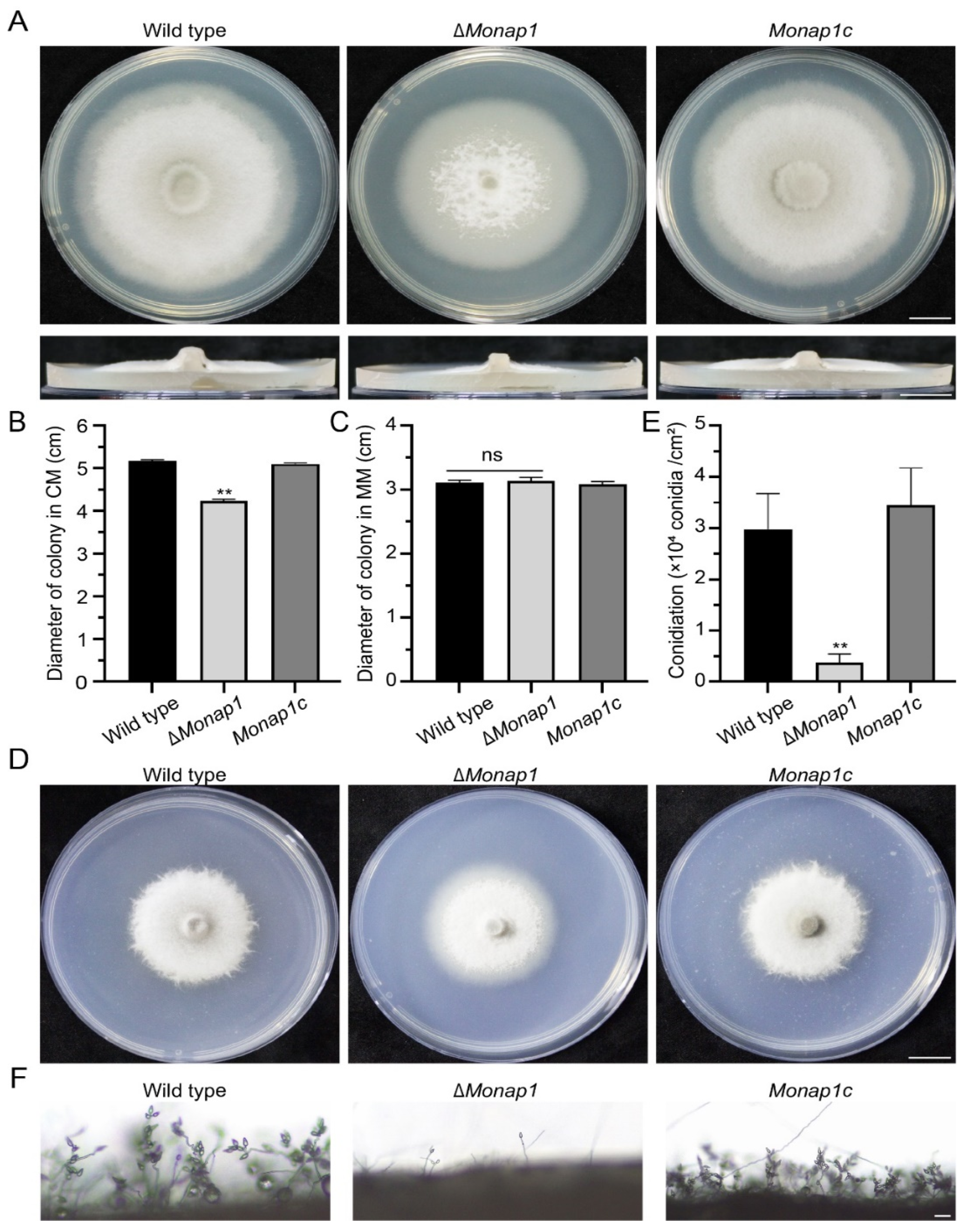
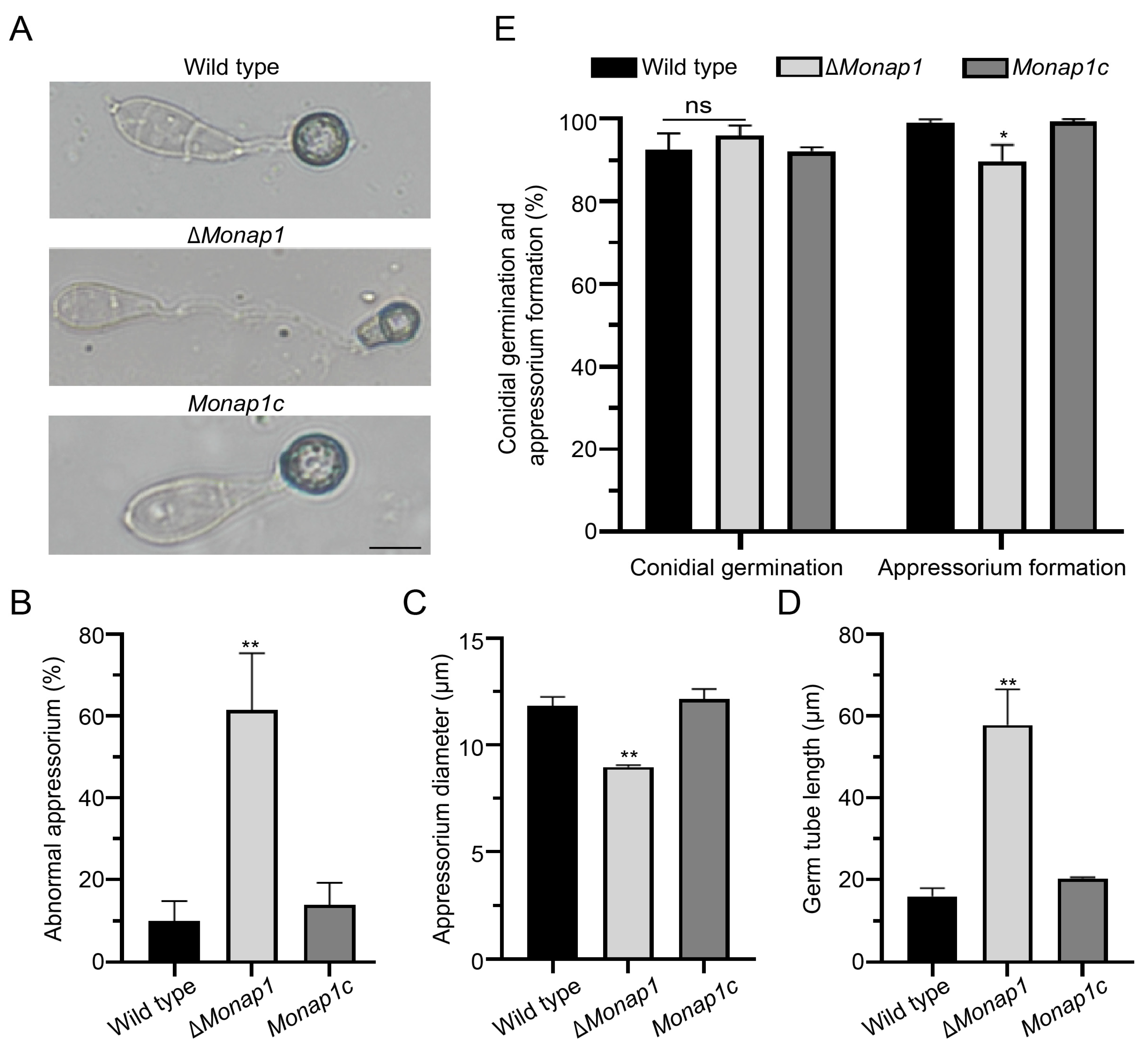
The ΔMonap1 mutant grew more slowly than the wild type 70-15 in CM, exhibiting reductions in mycelial growth of approximately 20%. The colony diameter of ΔMonap1 was 4.24 ± 0.04 cm, while that of the wild type was 5.18 ± 0.02 cm at 9 dpi (days post inoculation) (Figure 2A,B). In addition, the colonies of the mutant appeared thinner and whiter than those of the wild type (Figure 2A). ΔMonap1 is more susceptible to senescence, which can be observed by the collapse and autolysis of the aerial hyphae at the center of the colony (Figure 2A), indicating that MoNap1 may play a role in the aging process. However, the growth of ΔMonap1 in MM was comparable to that of the wild type 70-15 (Figure 2C,D). ΔMonap1 produced significantly fewer spores, approximately 1/8 of the wild type (Figure 2E). In ΔMonap1, the mRNA levels of two transcription factor genes FLBC and CON7, which are involved in the regulation of sporulation, were significantly downregulated (Figure S2). In the wild type, many conidiophores bearing spores in a typical sympodial mode were observed, while ΔMonap1 produced fewer conidiophores and fewer spores in each conidiophore (Figure 2F). The appressorial morphology of ΔMonap1 was distorted. The appressoria of the wild type were usually circular or dome-shaped, while approximately 61.5 ± 11.4% of the appressoria in ΔMonap1 were abnormal (Figure 3A,B) and smaller than those in the wild type (the diameter of appressoria in the wild type was 11.86 ± 0.34 μm, whereas that in the ΔMonap1 mutant was 8.98 ± 0.07 μm) (Figure 3C). The germ tubes of the ΔMonap1 mutants were significantly longer than those of the wild type when germinated on hydrophobic plastic coverslips (Figure 3A,D). The germ tube of the wild type was 16.04 ± 1.55 μm in length, whereas that of ΔMonap1 was 57.89 ± 7.07 μm (Figure 3D). Spore germination was not affected by the MoNAP1 deletion at 4 hpi (hours post inoculation); however, the appressorial formation rate in ΔMonap1 was lower than that in the wild type at 24 hpi (89.8 ± 3.2% in ΔMonap1 compared to 99.0 ± 0.7% in the wild type) (Figure 3E). In addition, turgor pressure of appressoria at 24 hpi was not significantly different between ΔMonap1 mutants and wild-type (Figure S3).
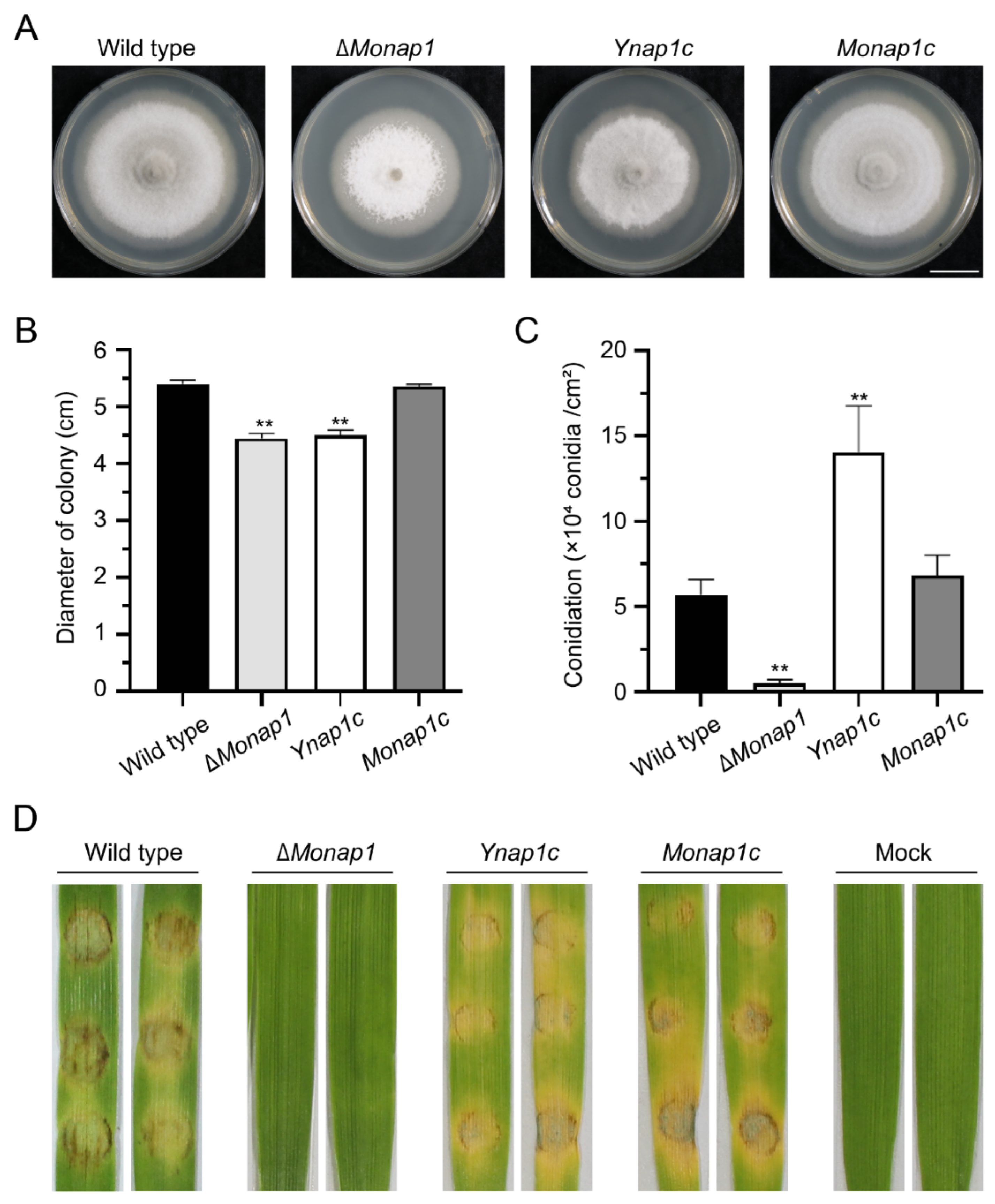
The complementation strain Monap1c recovered the mutant’s defects in mycelial growth, sporulation, conidiophore formation, and conidium differentiation, indicating that the defects in growth and conidiation of ΔMonap1 were caused by the deletion of NAP1. We also complemented ΔMonap1 with the NAP1 gene of S. cerevisiae and found that NAP1 in S. cerevisiae can restore the sporulation and virulence of the ΔMonap1 mutant but not the growth of ΔMonap1 (Figure 4A–D).

2.3. NAP1 Is Required for Virulence in M. oryzae

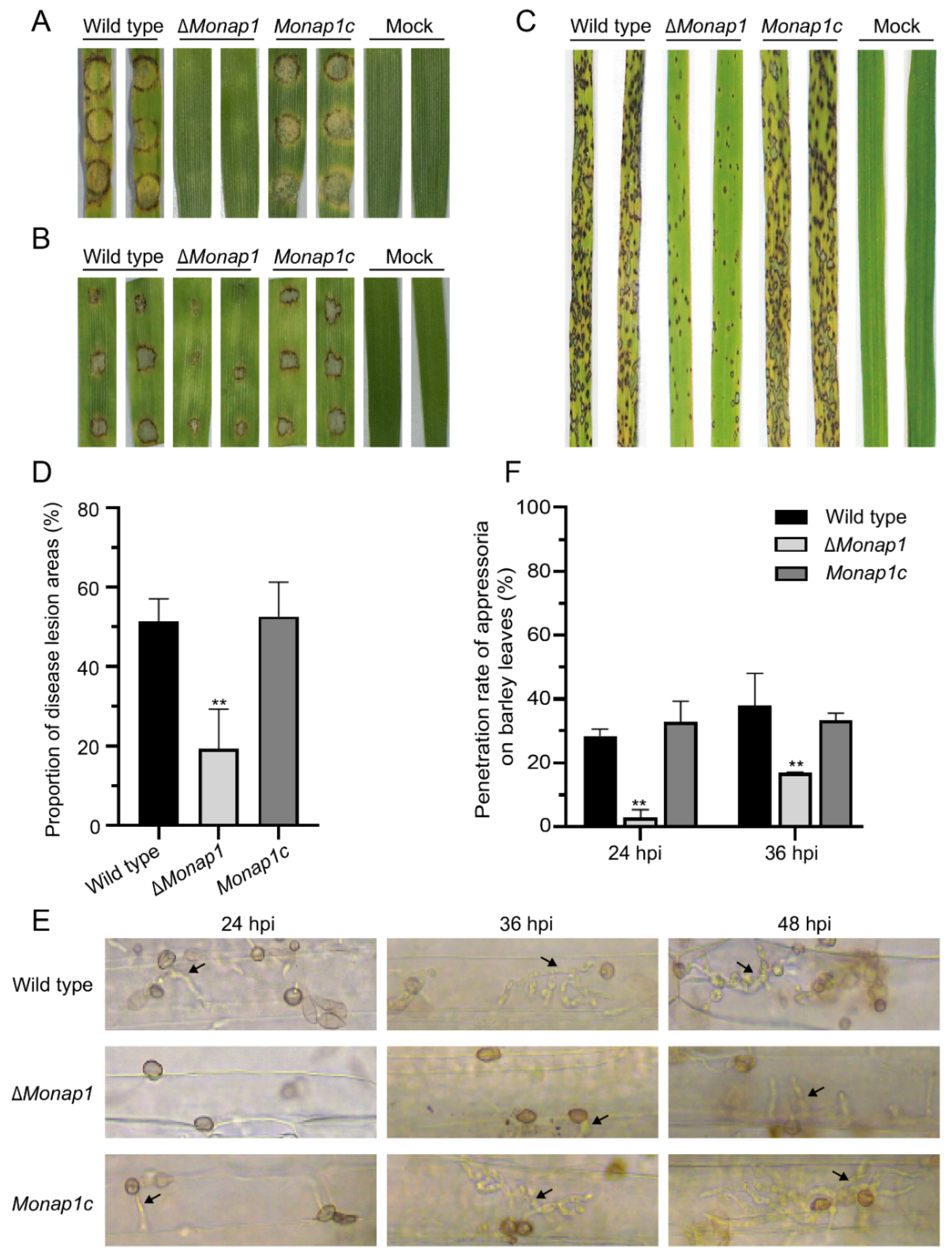
The ΔMonap1 mutant displayed reduced virulence on barley leaves and rice seedlings. We evaluated the fungal pathogenicity using three inoculation methods. At 4 dpi, the mycelial plugs of the wild-type 70-15 and the complementation strain Monap1c caused yellow and brown blast lesions and rotten plant tissues, while the mycelial plugs of ∆Monap1 did not (Figure 5A). When inoculated on detached barley leaves with 20 μL spore suspensions (5 × 10⁴ conidia mL−1) for 4 days, the disease lesions caused by ∆Monap1 were apparently weaker than those caused by the wild type and the complementation strain (Figure 5B). When sprayed on 2-week-old rice seedlings (CO-39) with conidial suspensions of 5 × 10⁴ conidia mL−1, the ∆Monap1 mutant caused small and restricted lesions in contrast to the typical spindle-like, gray centered, and merged blast lesions caused by the wild type and complementation strain at 7 dpi (Figure 5C). The lesion areas caused by ∆Monap1 were significantly lower than those caused by the wild type and Monap1c (those caused by ∆Monap1 were 19 ± 8.1%, while those caused by the wild type and Monap1c were 52 ± 4.6% and 53 ± 7.0%, respectively) (Figure 5D). Furthermore, we measured the penetration rate of the wild type, ∆Monap1, and Monap1c at three time points (24 hpi, 36 hpi, and 48 hpi) (Figure 5E). At 24 hpi, 28.4 ± 1.7% and 32.9 ± 5.3% of appressoria penetrated into barley cuticle cells and formed invasive hyphae (IH) structures with the wild type and complementation strain Monap1c. However, only 3.1 ± 1.8% of ∆Monap1 appressoria formed IH structures. At 36 hpi and 48 hpi, the appressorial penetration rate of ∆Monap1 was still lower than that of the wild type and the complementation strain Monap1c (Figure 5F). Moreover, the complemented ΔMonap1 with the NAP1 gene of S. cerevisiae can restore the virulence of the ΔMonap1 mutant (Figure 4D).

2.4. ∆Monap1 Is Sensitive to Cell Wall Integrity Stresses and Resistant to Microtubule Stresses

The integrity of the cell wall, which is the first barrier contacting the external environment, plays a crucial role in the homeostasis of M. oryzae [28]. To assess the role of MoNAP1 in cell wall integrity stress, the growth of the wild-type 70-15, ∆Monap1 and Monap1c strains was measured in CM with three cell wall integrity stresses, including 75 μg mL−1 calcofluor white (CFW), 600 μg mL−1 Congo red (CR), and 0.004% sodium dodecyl sulfate (SDS) (Figure 6A). The relative growth rate of mycelial growth of ∆Monap1 was significantly decreased compared to that of the wild type on CFW and CR (Figure 6B). In contrast, in the presence of 0.004% SDS, ∆Monap1 showed an increased relative growth rate compared to that of the wild type. In addition, ∆Monap1 was more resistant to bleomycin (BLM) than the wild type (Figure 6A,B).
In S. cerevisiae, the ∆nap1 mutant showed increased resistance to benomyl, a microtubule destabilizing drug that causes mitotic arrest at high concentrations, suggesting that Nap1 plays a role in regulating microtubule dynamics [19]. Similar to yeast, the ∆Monap1 mutant grew faster than the wild type in plates containing 15 μg mL−1 benomyl in M. oryzae (Figure 6A,B). In addition to benomyl, ∆Monap1 was more resistant to high temperature. ∆Monap1 exhibited a lower growth defect than the wild type at 32 °C (Figure 6A,B). When wild-type conidia are treated with HU, a DNA replication inhibitor, appressorium formation is blocked [29]. We inoculated wild-type 70-15, ∆Monap1 and Monap1c in CM containing 2 mM HU and found that ∆Monap1 had a higher relative growth rate than the wild type and Monap1c (Figure 6A,B).

2.5. Nap1 Is Necessary for Proper Septin Ring Organization in M. oryzae

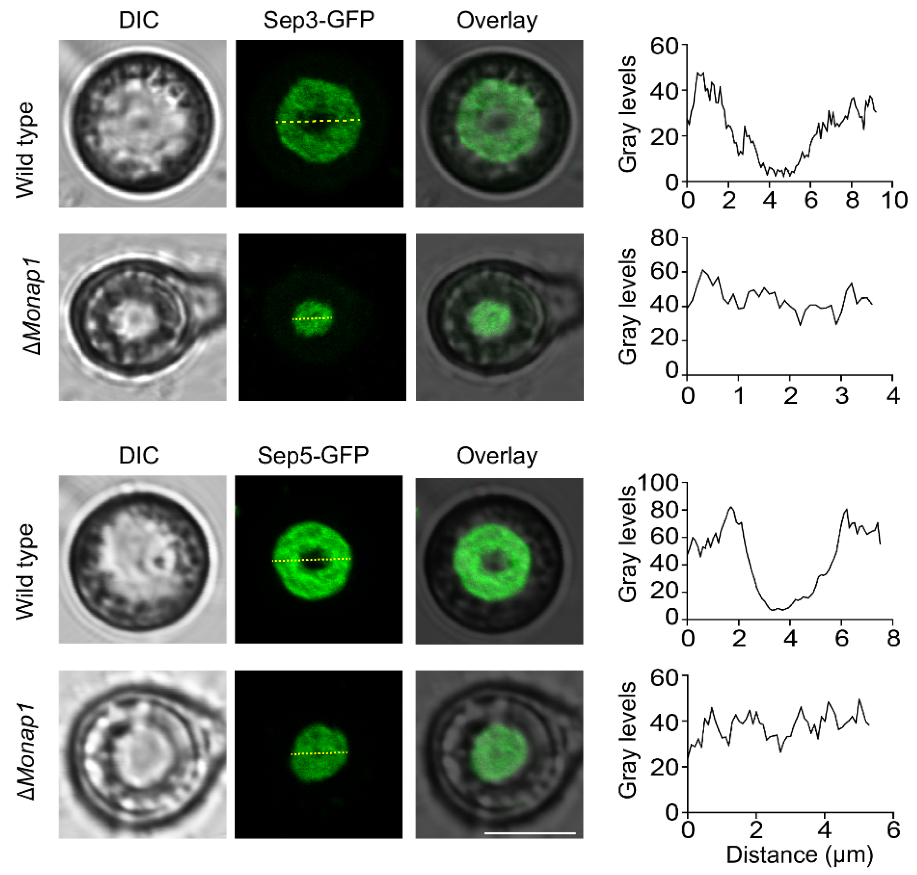
A critical requirement for appressorium morphology and function in M. oryzae is the recruitment and organization of septin-dependent cytoskeletal components [30]. In C. albicans, Nap1 plays a role in septin ring organization, and the deletion of NAP1 affects the stability of the septin ring [18]. The abnormal appressorial morphology of ∆Monap1 prompted us to observe the localization of the septin ring in the appressorium. We visualized the septin ring by expressing Sep3-GFP and Sep5-GFP fusion proteins in the wild type and mutants. In the wild type, Sep3-GFP and Sep5-GFP showed a ring structure around the appressorium pore, while in the ∆Monap1 mutant, Sep3-GFP and Sep5-GFP were evenly distributed and appeared similar to a round pie at the bottom of the appressorium at 24 hpi (Figure 7).

2.6. Nap1 Binds Histones H2A and H2B and G2/Mitotic-Specific B-Type Cyclin Cyc1 in M. oryzae

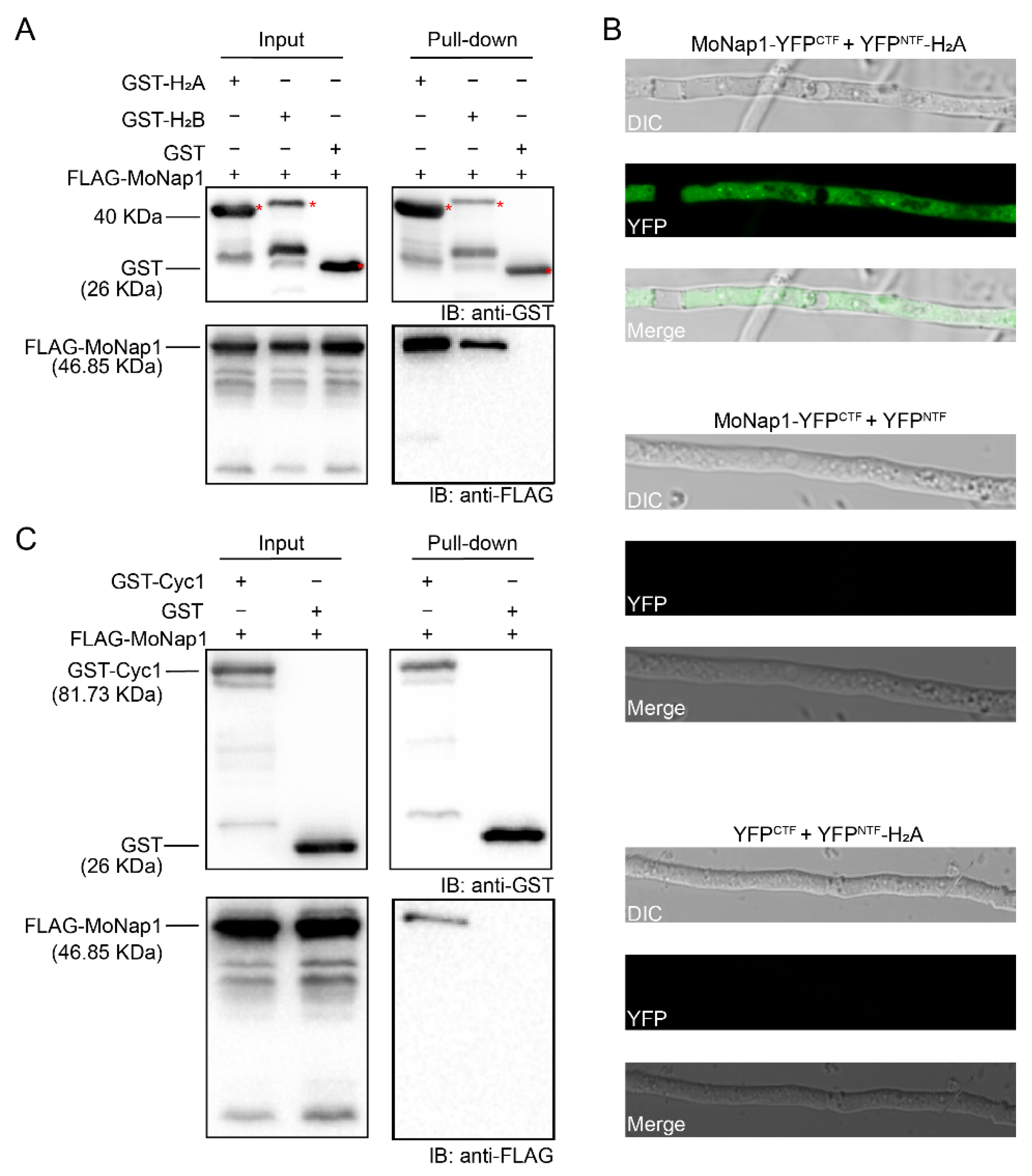
As a nucleosome assembly protein, Nap1 can tightly bind histones H2A and H2B in yeast [31], Drosophila [21], and tobacco [32]. To characterize its biochemical properties in M. oryzae, we detected whether MoNap1 bound core histones. Using an in vitro GST pull-down assay, we identified the interaction between MoNap1 and histones H2A and H2B. The recombinant proteins GST-H2A and GST-H2B pulled down FLAG-MoNap1, but GST did not (Figure 8A). Furthermore, we detected in vivo protein interactions between MoNap1 and H2A using a bimolecular fluorescence complementation (BiFC) assay. MoNap1 was fused to the C-terminal fragment of YFP (YFPCTF), and H2A was fused to the N-terminal fragment of YFP (YFPNTF). The two vectors expressing MoNap1-YFPCTF and YFPNTF-H2A were cotransformed into the wild type; as negative controls, MoNap1-YFPCTF and YFPNTF as well as YFPCTF and YFPNTF-H2A were cotransformed into the wild type. Only transformants coexpressing MoNap1-YFPCTF and YFPNTF-H2A showed YFP signals in the cytoplasm, but the transformants coexpressing two pairs of control vectors did not (Figure 8B).
In budding yeast, Nap1 acts with Clb2 to perform mitotic functions and suppress polar bud growth [19]. In M. oryzae, a B-type cyclin gene, CYC1, is the homolog of CLB2 [33]. The different sensitivities of ∆MoNap1 to benomyl and temperature prompted us to investigate the relationship between Nap1 and the Cyc1 protein. In the pull-down experiments, MoNap1 was pulled down by Cyc1 (Figure 8C). Thus, MoNap1 physically interacted with Cyc1 in M. oryzae.

2.7. The NES of MoNap1 Is Dispensable for Localization and Binding the Histone Core Proteins H2A and H2B but Required for Growth and Pathogenicity

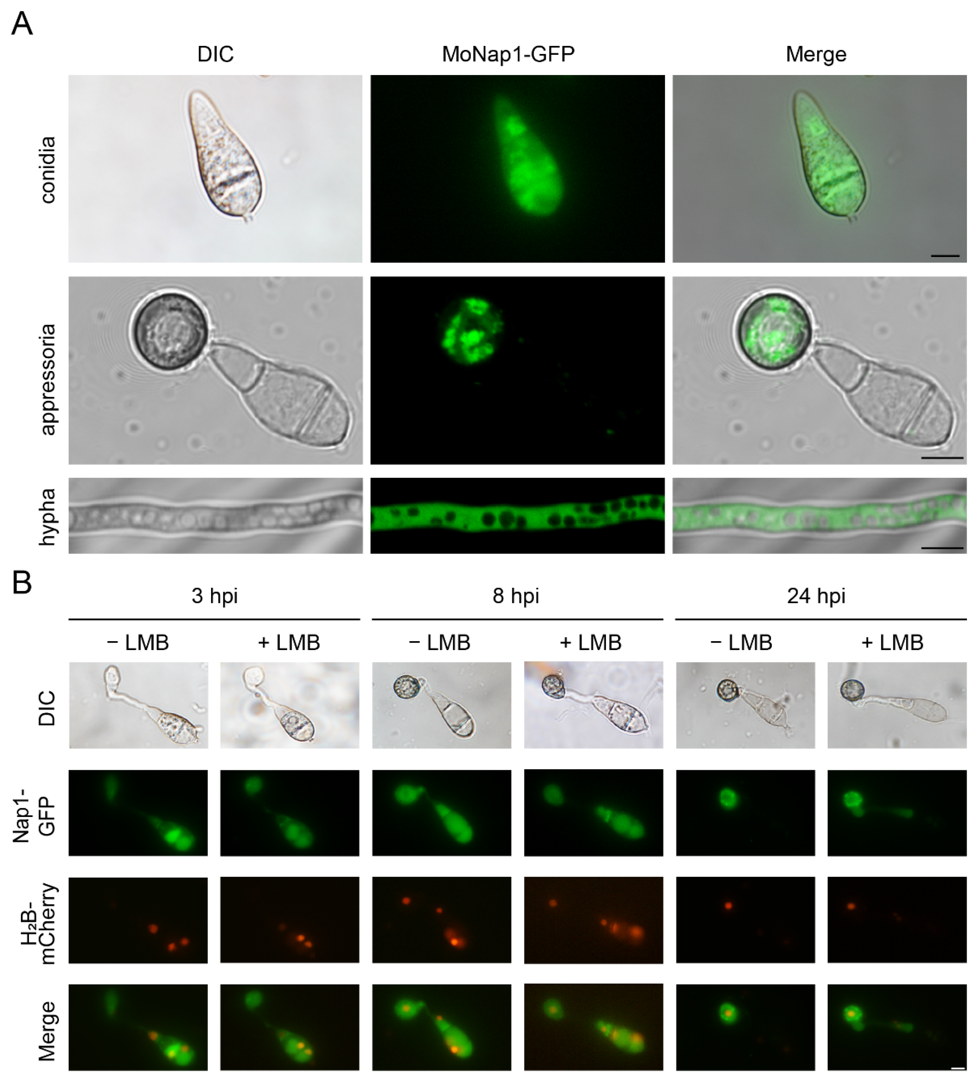
In both yeast and Drosophila, Nap1 has been reported to be a nucleocytoplasmic shuttling protein. However, in M. oryzae, MoNap1 mainly localized in the cytoplasm of conidial, appressorial, and hyphal cells (Figure 9A). Additionally, the treatment with leptomycin B (LMB, a specific nuclear export inhibitor) [34] did not result in the obviously increased accumulation of Nap1-GFP in the nucleus (Figure 9B).
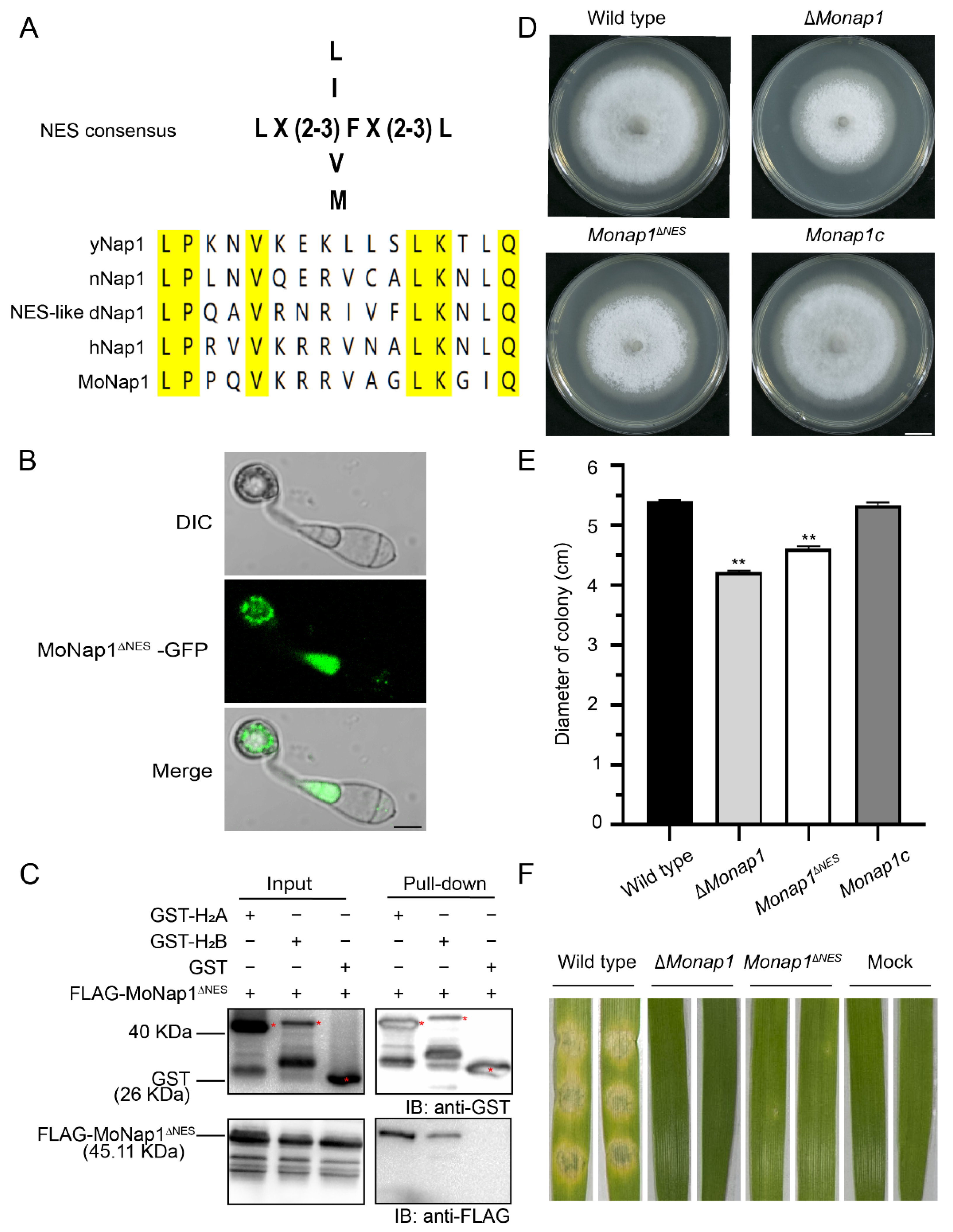
In yeast, Nap1 shuttles via a leucine-rich NES [20], and this short peptide sequence is conserved in many species, including nematode Nap1 (nNap1), Drosophila Nap1 (dNap1), and human Nap1 (hNap1). MoNap1 contains a 16-amino acid sequence in the N-terminal region that is similar to the NES of the yeast Nap1 protein (Figure 10A). To investigate whether these sequences function as active NES signals, we constructed mutated Nap1∆NES in which Nap1 lacked the corresponding NES region. However, the deletion of the NES sequence in MoNap1-GFP (MoNap1∆NES-GFP) did not make Nap1 preferentially localize in the nucleus (Figure 10B).
We examined the possible relationship between histone binding and the NES sequence of Nap1. Therefore, we performed GST pull-down assays with FLAG-MoNap1∆NES and core histones. We found that MoNap1∆NES bound core histones similar to MoNap1 (Figure 10C). Therefore, the NES region is not involved in H2A/H2B binding.
Nevertheless, we found that Monap1NES exhibited a phenotype similar to that of ∆Monap1, including reduced growth and virulence (Figure 10D–F).

3. Discussion

Nap1 is a well-conserved protein in eukaryotes, such as mammals, Drosophila, and yeast; however, its roles in filamentous fungi are not well understood. M. oryzae, an important plant pathogenic fungus, is often used as a model organism in investigating pathogenic fungal-host molecular interactions. In this study, we characterized the biological roles of Nap1 in M. oryzae by knocking out NAP1 and found that Nap1 plays diversified roles in aerial hyphal development, conidiophore and spore differentiation, appressorium formation, and virulence. NAP1 in S. cerevisiae can eliminate the defects in the sporulation and virulence of the ΔMonap1 mutant, suggesting that Nap1s in M. oryzae and S. cerevisiae are homologous in both biological function and protein sequence.
In M. oryzae, ∆Monap1 displayed reduced spore production, abnormal appressoria, and decreased infection growth and virulence. The reduced conidiation in ΔMonap1 is due to its fewer conidiophores and fewer spore numbers on each conidiophore. In M. oryzae, several transcription factor genes (COS1, CONX2, MSTU1, GTA1, GCC1, FLBC, HOX2, and CON7) [35,36,37] and autophagy genes, such as ATG5 [38] and TEA4 [39], are involved in the regulation of sporulation. The mRNA levels of two genes, FLBC and CON7, were significantly downregulated, suggesting a possible pathway regulating conidiation via FLBC and CON7. In addition, melanin promotes sporulation in the wild type 70-15 [40]. The lower melanin contents in the aerial hyphae of ΔMonap1 are also responsible for its reduced spore production. M. oryzae penetrates the rice cell wall by a melanized, dome-shaped appressorium with a large turgor. In ∆Monap1, the appressorium morphology is abnormal and smaller than that in the wild type. Normal appressorium function depends on the microtubule arrangement and F-actin polymerization organized by septin [41]. The septin ring determines the polarity of the appressorium, which is required for plant penetration [42]. Septin GTPases can regulate the organization and functions of microtubule [43]. Moreover, in mammals, the stability of septin disks is dependent on intact microtubules [44]. In C. albicans, Nap1 is associated with the assembly of septin rings, and NAP1 deletion leads to changes in septin dynamics [18]. In tobacco, Nap1 colocalizes with the mitotic spindle and the phragmoplast [12] and interacts with tubulins [32]. Benomyl, a microtubule inhibitor, destabilized microtubules in cells [45]. In M. oryzae, the deletion of MoNAP1 led to increased resistance to benomyl and an increased proportion of abnormal appressoria, suggesting its function in microtubule arrangement. Abnormal cytoskeleton and appressorium morphology cause an abnormal septin ring [30,46]. ∆Monap1 showed an abnormal distribution of Sep3 and Sep5 at the bottom of the appressorium at 24 hpi. This finding is consistent with the aberrant appressorium morphology of ∆Monap1. Thus, the reduced virulence of ∆Monap1 may be the result of an abnormal appressorium morphology, which is probably related to the altered microtubule dynamics.
A dramatic change in microtubule dynamics occurs when cells enter mitosis. Spindle orientation is a microtubule-dependent process during mitosis that determines the division plane. Mitosis is induced specifically by kinase complexes that contain B-type cyclins and a cyclin-dependent kinase [19]. In yeast, Nap1 interacts with the B-type cyclin Clb2, which is involved in the assembly of the mitotic spindle. Nap1 is required for the ability of Clb2 to suppress polarized bud growth. In M. oryzae, Nap1 interacts with the B-type cyclin Cyc1. The deletion of NAP1 alters the effect of benomyl on microtubule stability and alters microtubule stability during mitosis. In budding yeast, when the wild-type cells were cultured in media containing 11 μg mL−1 benomyl, the cells were unable to assemble a functional mitotic spindle. However, ∆nap1 cells do not exhibit a benomyl-induced mitotic delay [19]. This finding indicates that the deletion of NAP1 either increases microtubule stability or reduces the requirements for microtubule function during mitosis. In M. oryzae, when conidia are treated with HU, a DNA replication inhibitor, the cells are arrested at the S phase, and appressorium formation is blocked [29,33]. We found that ∆Monap1 also had an increased resistance to HU. Temperature is one of the key factors affecting division of cells, and high temperature inhibits the cell cycle in Chlamydomonas reinhardtii [47]. In A. nidulans, the NimA gene encodes a protein kinase necessary for mitosis [48]. An equivalent gene mutant MonimAE37G in M. oryzae grew normally at 26 °C but showed a reversible growth defect at 32 °C [49]. However, ∆Monap1 exhibited a higher relative growth rate at 32 °C. Therefore, it is possible that MoNap1 is involved in the regulation of mitosis in M. oryzae.
Chitin [(1-4)-β-linked N-acetylglucosamine] is a major structural cell wall component in ascomycetes, and most chitin is fully acetylated and associated with (1-3)-β-glucan and (1-6)-β-glucan [50,51]. In this study, ∆Monap1 was sensitive to two chitin-binding anionic dyes, CFW and CR, which inhibited the assembly of enzymes that connect chitin to (1-3)-β-glucan and (1-6)-β-glucan [52], indicating that the loss of MoNAP1 affects the CWI pathway. The CWI pathway controls the cellular remodeling process in response to environmental challenges in fungi [28,52]. Bleomycin (BLM) is a DNA-damaging agent that acts by not only inducing DNA strand breaks [53] but also destroying cell wall components through oxidation and preventing the formation of fungal septum and cytokinesis [54,55,56]. The nrp1-1 nrp2-1 mutant plants were significantly more sensitive to the bleomycin treatment and increased levels of DNA damage than the wild-type plants in Arabidopsis [57]. Conversely, our results showed that ∆Monap1 increased resistance to BLM. The difference in the response to BLM may be due to the differences in nucleus and cell wall components or the differences in action sites of BLM between Arabidopsis and M. oryzae.
The localization of Nap1 in the cell has been an interesting issue, and Nap1 exhibits nucleocytoplasmic shuttling in many species. The distinct subcellular localization of Nap1 in the cell reflects functional diversity and complexity. Nap1 is a nucleocytoplasmic shuttling protein in yeast that is primarily localized in the cytoplasm at the steady state but localized in the nucleus when a leucine-rich nuclear export sequence (NES) is mutated [20]. Nap1 binds histone H2A/H2B dimers in the cytoplasm and participates in the import of histones into the nucleus [58]. The analysis of Nap1 phosphorylation revealed that Nap1 is phosphorylated at 11 sites in vivo in S. cerevisiae [59]. In vitro, Nap1 is a substrate for the phosphorylation of CK2 at three serines. Normal S-phase progression requires the reversible phosphorylation of Nap1, and the phosphorylation of Nap1 by CK2 appears to facilitate its import into the nucleus [59]. However, we did not observe increased nuclear localization of MoNap1 after deleting a putative NES, which is consistent with the observation in C. albicans, where the deletion of the NES also did not result in NAP1 accumulation in the nucleus [18]. Interestingly, in the BiFC assay, we found that the fluorescent signal was also mainly concentrated in the cytoplasm, which may be related to the fact that NAP1 binds histones in the cytoplasm to form a complex and participates in histone import into the nucleus [60].
In summary, we characterized the roles of Nap1, nucleosome assembly protein 1, in M. oryzae by molecular genetic assays and found that MoNAP1 is required for fungal development and virulence probably through its involvement in microtubule dynamics.

4. Materials and Methods

4.1. Strains and Culture Conditions

Magnaporthe oryzae wild-type strain 70-15 (a strain artificially crossed in the laboratory) and the ΔMonap1 mutant, the complementary strain of ΔMonap1 (Monap1c), were cultured in complete medium (CM) or minimal medium (MM) at 25 °C under a 16 h light and 8 h dark cycle [61]. For the stress tests, the strains were cultured in CM supplemented with 0.004% sodium dodecyl sulfate (SDS), 75 μg mL−1 calcofluor white (CFW), 600 μg mL−1 Congo red (CR), 35 μg mL−1 bleomycin (BLM), 15 μg mL−1 benomyl, or 2 mM hydroxyurea (HU) at 25 °C in darkness for 9 d. For the temperature-sensitive stresses, the strains were cultured in CM at 32 °C under a 16 h light and 8 h dark cycle for 9 days.

4.2. Knockout of NAP1 in M. oryzae

An upstream fragment and a downstream fragment of NAP1 from M. oryzae genomic DNA were amplified with the primers Up-F/Up-R and Down-F/Down-R, respectively. The primers used in this study are listed in Supplementary Table S1. A hygromycin B resistance gene, HPH, was cloned using the primers HPH-F and HPH-R. Three DNA fragments were fused into a knockout cassette in HindIII- and XbaI-digested pKO3B [62] using a fusion enzyme (Vazyme, Nanjing, China). Then, the knockout cassette was transformed into the wild-type strain via Agrobacterium tumefaciens-mediated transformation (ATMT), and the null mutants were confirmed using a previously reported method [36,62]. First, the transformants were screened in selective CM containing 0.5 μM 5-fluoro-2′-deoxyuridine (F2dU) and 200 µg mL−1 hygromycin B. Second, the genomic DNA of the transformants was isolated using an improved CTAB method. Then, the null mutants were identified using double PCR in which MoNAP1 was amplified using the primer set S-F/S-R with β-TUBULIN (primers: Tbl-gF/Tbl-gR) as a positive control. The successful recombination of HPH into the deleted MoNAP1 site was confirmed by another PCR using the primer set L-F/HPH-CKR. Third, the insertion copy number of HPH in the mutant genome was determined by quantitative real-time PCR (qPCR) using β-TUBULIN as a control (primer sets qHPH-F/qHPH-R and qtub-F/qtub-R).
For the complementary strain Monap1c, a 3.68 kb native full NAP1 gene containing a promoter, coding sequence (CDS), and a terminator was cloned from M. oryzae wild-type genomic DNA by the primer set NAP1c-F/NAP1c-R and inserted into the EcoRI and XbaI-linearized vector pKD3, which contains a bialaphos resistance gene (BAR) [63]. The complemented DNA fragment was transformed into ∆Monap1 via ATMT, and the transformants were selected in selection medium containing 750 μg mL−1 glufosinate ammonium. The expression of MoNAP1 in the complementation strains was confirmed at the mRNA level by reverse transcription polymerase chain reaction (RT-PCR). To build a mutated MoNAP1 in which the NES sequence was deleted (MoNAP1NES), two segments (NAP11−430 bp and NAP1478−1549 bp) with primers (NAP1∆NES-F1/NAP1∆NES-R1 and NAP1∆NES-F2/NAP1∆NES-R2) were amplified in the genomic DNA of M. oryzae, fused to the BamHI-XbaI site of pKD3, and then transformed into ∆Monap1 via ATMT. To complement the NAP1 of S. cerevisiae into M. oryzae, a 2.35 kb NAP1 gene was cloned from S. cerevisiae genomic DNA using the primer set yNAP1-F/yNAP1-R and inserted into the BamHI and XbaI-linearized vector pKD5, which contains a sulfonylurea resistance gene (SUR) [63]. The resulting construct was transformed into ∆Monap1 by ATMT.

4.3. Phenotypic Characterization

The mutant phenotype was assayed according to previous reports [64,65]. To compare mycelial growth, a 5-mm agar block of 8-day-old wild type, ∆Monap1, and complementation strain Monap1c were inoculated in 7-cm plates containing 17.5 mL CM and cultured at 25 °C with a 16 h light and 8 h dark phase. The colony diameter was measured, and the colonies were photographed 9 days post-inoculation (dpi). Regarding sporulation, conidia were collected from 9-cm plates containing 30 mL CM at 12 dpi and counted with a counting chamber. Regarding conidiophore development, media containing vegetative hyphae were sliced into slender pieces and recultured under continuous light at 25 °C for 24 h [65]. To measure conidial germination and appressorium formation, 25 μL of spore suspension (1 × 10⁵ conidia mL−1) were dropped on plastic coverslips and incubated at 22 °C under dark conditions. Conidial germination and appressorium formation were observed at 4 hpi and 24 hpi, respectively.
For the mycelial virulence assays, 5-mm mycelial pellets were inoculated on excised leaves of 7-day-old barley (Hordeum vulgare) for 4 days. The virulence assays of rice seedlings (Oryza sativa cultivar CO39) were performed by spraying spore suspensions (5 × 10⁴ conidia mL−1) in 0.2% (w/v) gelatin on 14-day-old rice seedlings. The rice seedlings were cultured in a wet container under dark conditions at 22 °C for 48 h and then recultured under a 16 h light and 8 h dark cycle at 25 °C for 3 days. To assess the disease lesion severity (disease score), the area of a 5-cm-long leaf with the most serious disease lesions in each seedling was counted [65]. Regarding host penetration, 20 μL of spore suspensions (5 × 10⁴ conidia mL−1) of the wild-type, ∆Monap1, and Monap1c strains were dropped onto 7-day-old barley leaves and cultured at 25 °C. At 24 hpi, 36 hpi, and 48 hpi, the leaves were collected, decolored by methanol, fixed in alcoholic lactophenol, and observed under a microscope [64].

4.4. Observation of Fluorescence Fusion Proteins in M. oryzae

The fluorescence fusion protein Nap1-GFP was constructed by cloning a 1.5-kb NAP1 via PCR with a primer set (NAP1-GFP-F/NAP1-GFP-R) from the wild-type genomic DNA and fused with GFP in the BamHI and SmaI sites of pKD3-GFP [46]. To express Nap1∆NES -GFP, two segments (NAP11−430 bp and NAP1478−1549 bp) of the NAP1 coding sequence were amplified from the genome with primers (NAP1∆NES -GFP-F1/NAP1∆NES -GFP-R1 and NAP1∆NES -GFP-F2/NAP1∆NES -GFP-R2) and fused together with GFP in the pKD3 vector digested with XbaI. The fluorescent fusion protein expression vector was transformed into ∆Monap1 via ATMT. The fluorescence signals were observed under a laser scanning confocal microscope (FV3000). To observe the effects of LMB on the localization of Nap1, 20 nM LMB were added to the conidial suspension, and the fluorescence signals were observed at 3 hpi, 8 hpi, and 24 hpi. To localize Sep3 and Sep5 in the wild type and ∆Monap1 mutant, the vectors pKD5-Sep3-GFP and pKD5-Sep5-GFP into which SEP3 or SEP5 was inserted into the pKD5-GFP vector were transformed into the wild type and ∆Monap1, respectively [46].

4.5. Pull-Down Assays and Bimolecular Fluorescence Complementation (BiFC) Assay

For the pull-down assays, the cDNA fragments of MoNAP1 and MoNAP1∆NES were inserted into pET21 containing a 3 × FLAG tag, and the cDNA fragments of H2A, H2B, and CYC1 were inserted into pGEX-4T containing a GST tag. The expression vectors were transformed into Escherichia coli BL21 separately. FLAG-MoNap1 (46.85 kDa), FLAG-MoNap1∆NES (45.11 kDa), GST-H2A (41.27 kDa), GST-H2B (41.82 kDa), GST-Cyc1 (81.73 kDa), and GST (26 kDa) were expressed by induction using 0.2 M IPTG for 16 h and pulled down by GST beads C600913 (BBI, Shanghai, China). The proteins were separated by a Western blot assay and detected by an anti-GST antibody EM80701 (HUABIO, Hangzhou, China) or an anti-FLAG antibody M1403-2 (HUABIO, Hangzhou, China).
A BiFC analysis was used to observe the protein-protein interactions in vivo in M. oryzae. The plasmids used in the BiFC assay were constructed as previously described [66]. In brief, a TUBULIN promoter and a C-terminal fragment spanning approximately amino acids 155 to 238 of YFP (YFPCTF) were inserted into the vector pKD9-HPH to form the plasmid pKD9-YFPCTF-HPH. The plasmid pKD9-YFPCTF-HPH was digested with BamHI and SmaI and then ligated with the MoNAP1 fragment amplified with a primer pair (NAP1- YFPCTF-F/NAP1- YFPCTF-R), resulting in the final pKD9-MoNAP1-YFPCTF-HPH plasmid. Another N-terminal fragment spanning approximately amino acids 1 to 154 of YFP (YFPNTF) was inserted into the vector pKD5-SUR and a TUBULIN promoter to form the plasmid pKD5-YFPNTF-SUR. The plasmid pKD5-YFPNTF-SUR was digested with SalI and then ligated with the H2A fragment amplified with a primer pair (H2A-YFPNTF-F/H2A-YFPNTF-R), resulting in the final pKD5-YFPNTF-H2A-SUR plasmid. Next, the pKD9-MoNAP1-YFPCTF-HPH fusion vector was transformed into the 70-15 strain in combination with pKD5-YFPNTF-H2A-SUR. The transformants were screened in CM containing hygromycin B and sulfonylurea, and at least three independent transformants were examined under an LSM710nlo laser scanning confocal microscope (Zeiss, Jena, Germany).

Supplementary Materials

The following supporting information can be downloaded at: www.mdpi.com/article/10.3390/ijms23147662/s1.

Author Contributions

Conceptualization, J.L. and Q.W.; investigation, J.W., P.H., Z.H. and Y.L.; data curation, Q.W.; writing—original draft preparation, Q.W.; writing—review and editing, J.L. and Q.W.; visualization, Q.W., J.W., P.H., Z.H. and Y.L.; supervision, J.L. and X.L.; project administration, J.L. and X.L.; funding acquisition, J.L., X.L. and F.L. All authors have read and agreed to the published version of the manuscript.

Funding

This work was funded by the Key R&D projects of Zhejiang Province (grant no. 2021C02010) and the National Natural Science Foundation of China (grant nos. 31871908). This work was also supported by a grant Organism Interaction from Zhejiang Xianghu Laboratory to FCL.

Institutional Review Board Statement

Not applicable.

Informed Consent Statement

Not applicable.

Data Availability Statement

All data supporting the findings of the current study are available within figures and supporting information. All strains generated during this study are available from the corresponding author upon reasonable request.

Acknowledgments

We thank Xiaoxiao Feng at Agricultural Experiment Station, Zhejiang University for her help in experiment management.

Conflicts of Interest

The authors declare no conflict of interest.

References

  1. Talbot, N.J. On the Trail of a Cereal Killer: Exploring the Biology of Magnaporthe grisea. Annu. Rev. Microbiol. 2003, 57, 177–202. [Google Scholar] [CrossRef] [Green Version]
  2. Hamer, J.E.; Talbot, N.J. Infection-related development in the rice blast fungus Magnaporthe grisea. Curr. Opin. Microbiol. 1998, 1, 693–697. [Google Scholar] [CrossRef]
  3. Hamer, J.E.; Howard, R.J.; Chumley, F.G.; Valent, B. A Mechanism for Surface Attachment in Spores of a Plant Pathogenic Fungus. Science 1988, 239, 288–290. [Google Scholar] [CrossRef]
  4. De Jong, J.C.; McCormack, B.J.; Smirnoff, N.; Talbot, N.J. Glycerol generates turgor in rice blast. Nature 1997, 389, 244–245. [Google Scholar] [CrossRef]
  5. Gupta, L.; Vermani, M.; Ahluwalia, S.K.; Vijayaraghavan, P. Molecular virulence determinants of Magnaporthe oryzae: Disease pathogenesis and recent interventions for disease management in rice plant. Mycology 2021, 12, 174–187. [Google Scholar] [CrossRef]
  6. Ishimi, Y.; Kojima, M.; Yamada, M.-A.; Hanaoka, F. Binding mode of nucleosome-assembly protein (AP-I) and histones. Eur. J. Biochem. 1987, 162, 19–24. [Google Scholar] [CrossRef]
  7. Ishimi, Y.; Kikuchi, A. Identification and molecular cloning of yeast homolog of nucleosome assembly protein I which facilitates nucleosome assembly in vitro. J. Biol. Chem. 1991, 266, 7025–7029. [Google Scholar] [CrossRef]
  8. Mindrinos, M.; Katagiri, F.; Yu, G.-L.; Ausubel, F.M. The a-thaliana disease resistance gene RPS2 encodes a protein containing a nucleotide-binding site and leucine-rich repeats. Cell 1994, 78, 1089–1099. [Google Scholar] [CrossRef]
  9. Yoon, H.W.; Kim, M.C.; Lee, S.Y.; Hwang, I.; Bahk, J.D.; Hong, J.C.; Ishimi, Y.; Cho, M.J. Molecular cloning and functional characterization of a cDNA encoding nucleosome assembly protein 1 (NAP-1) from soybean. Mol. Gen. Genet. 1995, 249, 465–473. [Google Scholar] [CrossRef]
  10. Lankenau, S.; Barnickel, T.; Marhold, J.; Lyko, F.; Mechler, B.M.; Lankenau, D.-H. Knockout Targeting of the Drosophila Nap1 Gene and Examination of DNA Repair Tracts in the Recombination Products. Genetics 2003, 163, 611–623. [Google Scholar] [CrossRef]
  11. Rogner, U.C.; Spyropoulos, D.D.; Le Novère, N.; Changeux, J.-P.; Avner, P. Control of neurulation by the nucleosome assembly protein-1–like 2. Nat. Genet. 2000, 25, 431–435. [Google Scholar] [CrossRef]
  12. Dong, A.; Zhu, Y.; Yu, Y.; Cao, K.; Sun, C.; Shen, W.-H. Regulation of biosynthesis and intracellular localization of rice and tobacco homologues of nucleosome assembly protein 1. Planta 2003, 216, 561–570. [Google Scholar] [CrossRef]
  13. Ishimi, Y.; Hirosumi, J.; Sato, W.; Sugasawa, K.; Yokota, S.; Hanaoka, F.; Yamada, M.-A. Purification and initial characterization of a protein which facilitates assembly of nucleosome-like structure from mammalian cells. JBIC J. Biol. Inorg. Chem. 1984, 142, 431–439. [Google Scholar] [CrossRef]
  14. Andrews, A.J.; Downing, G.; Brown, K.; Park, Y.-J.; Luger, K. A Thermodynamic Model for Nap1-Histone Interactions. J. Biol. Chem. 2008, 283, 32412–32418. [Google Scholar] [CrossRef] [Green Version]
  15. Ohkuni, K.; Shirahige, K.; Kikuchi, A. Genome-wide expression analysis of NAP1 in Saccharomyces cerevisiae. Biochem. Biophys. Res. Commun. 2003, 306, 5–9. [Google Scholar] [CrossRef]
  16. Altman, R.; Kellogg, D. Control of Mitotic Events by Nap1 and the Gin4 Kinase. J. Cell Biol. 1997, 138, 119–130. [Google Scholar] [CrossRef]
  17. Asahara, H.; Tartare-Deckert, S.; Nakagawa, T.; Ikehara, T.; Hirose, F.; Hunter, T.; Ito, T.; Montminy, M. Dual Roles of p300 in Chromatin Assembly and Transcriptional Activation in Cooperation with Nucleosome Assembly Protein 1 In Vitro. Mol. Cell. Biol. 2002, 22, 2974–2983. [Google Scholar] [CrossRef] [Green Version]
  18. Huang, Z.-X.; Zhao, P.; Zeng, G.; Wang, Y.-M.; Sudbery, I.; Wang, Y. Phosphoregulation of Nap1 Plays a Role in Septin Ring Dynamics and Morphogenesis in Candida albicans. mBio 2014, 5, e00915-13. [Google Scholar] [CrossRef] [Green Version]
  19. Kellogg, D.R.; Murray, A.W. NAP1 acts with Clb2 to perform mitotic functions and to suppress polar bud growth in budding yeast. J. Cell Biol. 1995, 130, 675–685. [Google Scholar] [CrossRef]
  20. Miyaji-Yamaguchi, M.; Kato, K.; Nakano, R.; Akashi, T.; Kikuchi, A.; Nagata, K. Involvement of Nucleocytoplasmic Shuttling of Yeast Nap1 in Mitotic Progression. Mol. Cell. Biol. 2003, 23, 6672–6684. [Google Scholar] [CrossRef] [Green Version]
  21. Ito, T.; Bulger, M.; Kobayashi, R.; Kadonaga, J.T. Drosophila NAP-1 is a core histone chaperone that functions in ATP-facilitated assembly of regularly spaced nucleosomal arrays. Mol. Cell. Biol. 1996, 16, 3112–3124. [Google Scholar] [CrossRef] [Green Version]
  22. Rodrigueza, P.; Munroeb, D.; Prawittc, D.; Chu, L.L.; Bricb, E.; Kima, J.; Reid, L.H.; Daviese, C.; Nakagamaf, H.; Loebbertc, R.; et al. Functional Characterization of Human Nucleosome Assembly Protein-2 (NAP1L4) Suggests a Role as a Histone Chaperone. Genomics 1997, 44, 253–265. [Google Scholar] [CrossRef]
  23. Fujii-Nakata, T.; Ishimi, Y.; Okuda, A.; Kikuchi, A. Functional analysis of nucleosome assembly protein, NAP-1. The negatively charged COOH-terminal region is not necessary for the intrinsic assembly activity. J. Biol. Chem. 1992, 267, 20980–20986. [Google Scholar] [CrossRef]
  24. Li, M.; Strand, D.; Krehan, A.; Pyerin, W.; Heid, H.; Neumann, B.; Mechler, B.M. Casein kinase 2 binds and phosphorylates the nucleosome assembly protein-1 (NAP1) in Drosophila melanogaster. J. Mol. Biol. 1999, 293, 1067–1084. [Google Scholar] [CrossRef]
  25. McQuibban, G.A.; Commisso-Cappelli, C.N.; Lewis, P.N. Assembly, Remodeling, and Histone Binding Capabilities of Yeast Nucleosome Assembly Protein 1. J. Biol. Chem. 1998, 273, 6582–6590. [Google Scholar] [CrossRef] [Green Version]
  26. Lee, J.Y.; Lake, R.J.; Kirk, J.; Bohr, V.A.; Fan, H.-Y.; Hohng, S. NAP1L1 accelerates activation and decreases pausing to enhance nucleosome remodeling by CSB. Nucleic Acids Res. 2017, 45, 4696–4707. [Google Scholar] [CrossRef] [Green Version]
  27. Grande, M.; Lambea, E.; Fajardo, A.; López-Avilés, S.; Kellogg, D.; Aligue, R. Crosstalk between Nap1 protein and Cds1 checkpoint kinase to maintain chromatin integrity. Bba-Mol. Cell Res. 2008, 1783, 1595–1604. [Google Scholar] [CrossRef] [Green Version]
  28. Sun, L.; Qian, H.; Liu, M.; Wu, M.; Wei, Y.; Zhu, X.; Lu, J.; Lin, F.; Liu, X. Endosomal sorting complexes required for transport-0 (ESCRT -0) are essential for fungal development, pathogenicity, autophagy and ER -phagy in Magnaporthe oryzae. Environ. Microbiol. 2022, 24, 1076–1092. [Google Scholar] [CrossRef]
  29. Osés-Ruiz, M.; Sakulkoo, W.; Littlejohn, G.R.; Martin-Urdiroz, M.; Talbot, N.J. Two independent S-phase checkpoints regulate appressorium-mediated plant infection by the rice blast fungus Magnaporthe oryzae. Proc. Natl. Acad. Sci. USA 2017, 114, E237–E244. [Google Scholar] [CrossRef] [Green Version]
  30. Li, L.; Zhu, X.-M.; Su, Z.-Z.; Del Poeta, M.; Liu, X.-H.; Lin, F.-C. Insights of roles played by septins in pathogenic fungi. Virulence 2021, 12, 1550–1562. [Google Scholar] [CrossRef]
  31. Aguilar-Gurrieri, C.; Larabi, A.; Vinayachandran, V.; Patel, N.A.; Yen, K.; Reja, R.; Ebong, I.; Schoehn, G.; Robinson, C.V.; Pugh, B.F.; et al. Structural evidence for Nap1-dependent H2A–H2B deposition and nucleosome assembly. EMBO J. 2016, 35, 1465–1482. [Google Scholar] [CrossRef]
  32. Dong, A.; Liu, Z.; Zhu, Y.; Yu, F.; Li, Z.; Cao, K.; Shen, W.-H. Interacting Proteins and Differences in Nuclear Transport Reveal Specific Functions for the NAP1 Family Proteins in Plants. Plant Physiol. 2005, 138, 1446–1456. [Google Scholar] [CrossRef] [Green Version]
  33. Osés-Ruiz, M.; Talbot, N.J. Cell cycle-dependent regulation of plant infection by the rice blast fungus Magnaporthe oryzae. Commun. Integr. Biol. 2017, 10, e1372067. [Google Scholar] [CrossRef] [Green Version]
  34. Toyoshima, F.; Moriguchi, T.; Wada, A.; Fukuda, M.; Nishida, E. Nuclear export of cyclin B1 and its possible role in the DNA damage-induced G2 checkpoint. EMBO J. 1998, 17, 2728–2735. [Google Scholar] [CrossRef] [Green Version]
  35. Odenbach, D.; Breth, B.; Thines, E.; Weber, R.W.S.; Anke, H.; Foster, A.J. The transcription factor Con7p is a central regulator of infection-related morphogenesis in the rice blast fungus Magnaporthe grisea. Mol. Microbiol. 2007, 64, 293–307. [Google Scholar] [CrossRef]
  36. Lu, J.-P.; Cao, H.; Zhang, L.; Huang, P.; Lin, F. Systematic Analysis of Zn2Cys6 Transcription Factors Required for Development and Pathogenicity by High-Throughput Gene Knockout in the Rice Blast Fungus. PLOS Pathog. 2014, 10, e1004432. [Google Scholar] [CrossRef]
  37. Qu, Y.; Wang, J.; Zhu, X.; Dong, B.; Liu, X.; Lu, J.; Lin, F. The P5-type ATPase Spf1 is required for development and virulence of the rice blast fungus Pyricularia oryzae. Curr. Genet. 2020, 66, 385–395. [Google Scholar] [CrossRef]
  38. Lu, J.-P.; Liu, X.-H.; Feng, X.-X.; Min, H.; Lin, F.-C. An autophagy gene, MgATG5, is required for cell differentiation and pathogenesis in Magnaporthe oryzae. Curr. Genet. 2009, 55, 461–473. [Google Scholar] [CrossRef]
  39. Patkar, R.N.; Suresh, A.; Naqvi, N.I. MoTea4-Mediated Polarized Growth Is Essential for Proper Asexual Development and Pathogenesis in Magnaporthe oryzae. Eukaryot. Cell 2010, 9, 1029–1038. [Google Scholar] [CrossRef] [Green Version]
  40. Huang, P.; Cao, H.; Li, Y.; Zhu, S.; Wang, J.; Wang, Q.; Liu, X.; Lin, F.-C.; Lu, J. Melanin Promotes Spore Production in the Rice Blast Fungus Magnaporthe oryzae. Front. Microbiol. 2022, 13, 843838. [Google Scholar] [CrossRef]
  41. Dulal, N.; Rogers, A.M.; Proko, R.; Bieger, B.D.; Liyanage, R.; Krishnamurthi, V.R.; Wang, Y.; Egan, M.J. Turgor-dependent and coronin-mediated F-actin dynamics drive septin disc-to-ring remodeling in the blast fungus Magnaporthe oryzae. J. Cell Sci. 2021, 134, jcs251298. [Google Scholar] [CrossRef]
  42. Gupta, Y.K.; Dagdas, Y.F.; Martinez-Rocha, A.L.; Kershaw, M.J.; Littlejohn, G.R.; Ryder, L.S.; Sklenar, J.; Menke, F.; Talbot, N.J. Septin-Dependent Assembly of the Exocyst Is Essential for Plant Infection by Magnaporthe oryzae. Plant Cell 2015, 27, 3277–3289. [Google Scholar] [CrossRef] [Green Version]
  43. Spiliotis, E.T. Regulation of microtubule organization and functions by septin GTPases. Cytoskeleton 2010, 67, 339–345. [Google Scholar] [CrossRef]
  44. Sellin, M.E.; Holmfeldt, P.; Stenmark, S.; Gullberg, M. Microtubules support a disk-like septin arrangement at the plasma membrane of mammalian cells. Mol. Biol. Cell 2011, 22, 4588–4601. [Google Scholar] [CrossRef]
  45. Hoyt, M.; Totis, L.; Roberts, B.S. cerevisiae genes required for cell cycle arrest in response to loss of microtubule function. Cell 1991, 66, 507–517. [Google Scholar] [CrossRef]
  46. Qu, Y.; Cao, H.; Huang, P.; Wang, J.; Liu, X.; Lu, J.; Lin, F.-C. A kelch domain cell end protein, PoTea1, mediates cell polarization during appressorium morphogenesis in Pyricularia oryzae. Microbiol. Res. 2022, 259, 126999. [Google Scholar] [CrossRef]
  47. Zachleder, V.; Ivanov, I.; Vítová, M.; Bišová, K. Cell Cycle Arrest by Supraoptimal Temperature in the Alga Chlamydomonas reinhardtii. Cells 2019, 8, 1237. [Google Scholar] [CrossRef] [Green Version]
  48. Osmani, S.A.; Engle, D.B.; Doonan, J.; Morris, N. Spindle formation and chromatin condensation in cells blocked at interphase by mutation of a negative cell cycle control gene. Cell 1988, 52, 241–251. [Google Scholar] [CrossRef]
  49. Saunders, D.G.; Aves, S.J.; Talbot, N.J. Cell Cycle–Mediated Regulation of Plant Infection by the Rice Blast Fungus. Plant Cell 2010, 22, 497–507. [Google Scholar] [CrossRef] [Green Version]
  50. Wessels, J.G.H. Developmental regulation of fungal cell wall formation. Annu. Rev. Phytopathol. 1994, 32, 413–437. [Google Scholar] [CrossRef]
  51. Sun, D.; Cao, H.; Shi, Y.; Huang, P.; Dong, B.; Liu, X.; Lin, F.; Lu, J. The regulatory factor X protein MoRfx1 is required for development and pathogenicity in the rice blast fungus Magnaporthe oryzae. Mol. Plant Pathol. 2017, 18, 1075–1088. [Google Scholar] [CrossRef]
  52. Ram, A.F.J.; Klis, F.M. Identification of fungal cell wall mutants using susceptibility assays based on Calcofluor white and Congo red. Nat. Protoc. 2006, 1, 2253–2256. [Google Scholar] [CrossRef]
  53. Hecht, S.M. Bleomycin: New Perspectives on the Mechanism of Action. J. Nat. Prod. 2000, 63, 158–168. [Google Scholar] [CrossRef]
  54. Beaudouin, R.; Lim, S.T.; Steide, J.A.; Powell, M.; McKoy, J.; Pramanik, A.J.; Johnson, E.; Moore, C.W.; Lipke, P.N. Bleomycin affects cell wall anchorage of mannoproteins in Saccharomyces cerevisiae. Antimicrob. Agents Chemother. 1993, 37, 1264–1269. [Google Scholar] [CrossRef] [Green Version]
  55. Lim, S.T.; Jue, C.K.; Moore, C.W.; Lipke, P. Oxidative cell wall damage mediated by bleomycin-Fe(II) in Saccharomyces cerevisiae. J. Bacteriol. 1995, 177, 3534–3539. [Google Scholar] [CrossRef] [Green Version]
  56. Moore, C.W.; McKoy, J.; Del Valle, R.; Armstrong, D.; Bernard, E.M.; Katz, N.; Gordon, R.E. Fungal Cell Wall Septation and Cytokinesis Are Inhibited by Bleomycins. Antimicrob. Agents Chemother. 2003, 47, 3281–3289. [Google Scholar] [CrossRef] [Green Version]
  57. Zhu, Y.; Dong, A.; Meyer, D.; Pichon, O.; Renou, J.-P.; Cao, K.; Shen, W.-H. Arabidopsis NRP1 and NRP2 Encode Histone Chaperones and Are Required for Maintaining Postembryonic Root Growth. Plant Cell 2006, 18, 2879–2892. [Google Scholar] [CrossRef] [Green Version]
  58. Mosammaparast, N.; Ewart, C.S.; Pemberton, L.F. A role for nucleosome assembly protein 1 in the nuclear transport of histones H2A and H2B. EMBO J. 2002, 21, 6527–6538. [Google Scholar] [CrossRef] [Green Version]
  59. Calvert, M.E.K.; Keck, K.M.; Ptak, C.; Shabanowitz, J.; Hunt, D.F.; Pemberton, L.F. Phosphorylation by Casein Kinase 2 Regulates Nap1 Localization and Function. Mol. Cell. Biol. 2008, 28, 1313–1325. [Google Scholar] [CrossRef] [Green Version]
  60. Mosammaparast, N.; Jackson, K.R.; Guo, Y.; Brame, C.J.; Shabanowitz, J.; Hunt, D.F.; Pemberton, L.F. Nuclear Import of Histone H2a and H2b Is Mediated by a Network of Karyopherins. J. Cell Biol. 2001, 153, 251–262. [Google Scholar] [CrossRef]
  61. Talbot, N.J.; Ebbole, D.J.; Hamer, J.E. Identification and characterization of MPG1, a gene involved in pathogenicity from the rice blast fungus Magnaporthe grisea. Plant Cell 1993, 5, 1575–1590. [Google Scholar] [CrossRef] [PubMed] [Green Version]
  62. Yan, Y.; Wang, H.; Zhu, S.; Wang, J.; Liu, X.; Lin, F.; Lu, J. The Methylcitrate Cycle Is Required for Development and Virulence in the Rice Blast Fungus Pyricularia oryzae. Mol. Plant-Microbe Interact. 2019, 32, 1148–1161. [Google Scholar] [CrossRef] [PubMed]
  63. Zhu, S.; Yan, Y.; Qu, Y.; Wang, J.; Feng, X.; Liu, X.; Lin, F.; Lu, J. Role refinement of melanin synthesis genes by gene knockout reveals their functional diversity in Pyricularia oryzae strains. Microbiol. Res. 2020, 242, 126620. [Google Scholar] [CrossRef] [PubMed]
  64. Lu, J.-P.; Feng, X.-X.; Liu, X.-H.; Lu, Q.; Wang, H.-K.; Lin, F.-C. Mnh6, a nonhistone protein, is required for fungal development and pathogenicity of Magnaporthe grisea. Fungal Genet. Biol. 2007, 44, 819–829. [Google Scholar] [CrossRef]
  65. Cao, H.; Huang, P.; Zhang, L.; Shi, Y.; Sun, D.; Yan, Y.; Liu, X.; Dong, B.; Chen, G.; Snyder, J.H.; et al. Characterization of 47 Cys2 -His2 zinc finger proteins required for the development and pathogenicity of the rice blast fungus Magnaporthe oryzae. New Phytol. 2016, 211, 1035–1051. [Google Scholar] [CrossRef] [PubMed] [Green Version]
  66. Liu, T.-B.; Liu, X.-H.; Lu, J.-P.; Zhang, L.; Min, H.; Lin, F.-C. The cysteine protease MoAtg4 interacts with MoAtg8 and is required for differentiation and pathogenesis in Magnaporthe oryzae. Autophagy 2010, 6, 74–85. [Google Scholar] [CrossRef] [Green Version]
Figure 1. Nap1s are conserved in eukaryotes. Alignment tree of MoNap1 and Nap1 in S. cerevisiae (NP_012974.1), Drosophila melanogaster (NP_477128.1), Homo sapiens (XP_016874829.1, NP_068798.1), Candida albicans (XP_718658.1), Schizosaccharomyces pombe (NP_587838.1), Aspergillus melleus (XP_045950078.1), Aspergillus clavatus (XP_001273890.1), Aspergillus fischeri (XP_001266074.1), Aspergillus flavus (QRD88693.1), Aspergillus fumigatus (XP_754077.1), and Fusarium oxysporum (XP_031048533.1).
Ijms 23 07662 g001
Figure 2. NAP1 is required for the growth and conidiation of M. royzae. (A) Colonies of the wild-type, ΔMonap1, and MoNAP1 complemented strains of ΔMonap1 (Monap1c). Bar, 1 cm. (B) Mycelial growth (cm) of the wild-type, ΔMonap1 and Monap1c colonies in CM. n = 5 independent biological replicates. Error bars represent the standard deviations. The data were analyzed by GraphPad Prism 8.0 and significant differences compared with the wild type were estimated by multiple t tests: ** p < 0.01. (C) Mycelial growth (cm) of the wild type, ΔMonap1 and Monap1c colonies in MM. n = 5 independent biological replicates. Error bars represent the standard deviations. The data were analyzed by GraphPad Prism 8.0 and significant differences compared with the wild type were estimated by multiple t tests: ns, p > 0.05. (D) Colonies of the wild type, ΔMonap1, and Monap1c in MM. Bar, 1 cm. (E) Conidiation of the wild type, ΔMonap1 and Monap1c strains in CM. n = 5 independent biological replicates. Error bars represent the standard deviations. The data were analyzed by GraphPad Prism 8.0 and significant differences compared with the wild type were estimated by multiple t tests: ** p < 0.01. (F) Conidiophore development of M. oryzae strains. Bar, 50 μm.
Ijms 23 07662 g002
Figure 3. NAP1 is required for appressorium formation. (A) Appressorium formation in the wild-type, ΔMonap1 and Monap1c strains on hydrophobic surfaces at 24 hpi. Bar, 10 μm. (B) Abnormal appressoria rates (%) in the wild-type, ΔMonap1 and Monap1c strains. At least 150 spores were counted per replicate. n = 3 independent biological replicates. Error bars represent the standard deviations. The data were analyzed by GraphPad Prism 8.0 and significant differences compared with the wild type were estimated by multiple t tests: ** p < 0.01. (C) The appressorium diameter in the wild-type, ΔMonap1 and Monap1c strains at 24 hpi. At least 150 spores were counted per replicate. n = 3 independent biological replicates. Error bars represent the standard deviations. The data were analyzed by GraphPad Prism 8.0 and significant differences compared with the wild type were estimated by multiple t tests: ** p < 0.01. (D) Germ tube length (μm) in the wild-type, ΔMonap1 and Monap1c strains. Approximately 100 appressoria were photographed and measured using the software NIS-Elements D 3.2 in triplicate. At least 150 spores were counted per replicate. n = 3 independent biological replicates. Error bars represent the standard deviations. The data were analyzed by GraphPad Prism 8.0 and significant differences compared with the wild type were estimated by multiple t tests: ** p < 0.01. (E) Conidial germination rate (%) at 4 hpi and appressorium formation rate (%) at 24 hpi in the wild-type, ΔMonap1 and Monap1c strains. At least 150 spores were counted per replicate. n = 3 independent biological replicates. Error bars represent the standard deviations. The data were analyzed by GraphPad Prism 8.0 and significant differences compared with the wild type were estimated by multiple t tests: ns, p > 0.05, * p < 0.05.
Ijms 23 07662 g003
Figure 4. NAP1 in S. cerevisiae can eliminate defects in the sporulation and virulence of ΔMonap1. (A) Colonies of the wild type, ΔMonap1, NAP1 in S. cerevisiae complemented ΔMonap1 strain (Ynap1c) and Monap1c strains. Bar, 1 cm. (B) Mycelial growth (cm) in the wild type, ΔMonap1, Ynap1c, and Monap1c strains. n = 5 independent biological replicates. Error bars represent the standard deviations. The data were analyzed by GraphPad Prism 8.0 and significant differences compared with the wild type were estimated by multiple t tests: ** p < 0.01. (C) Conidiation in the wild type, ΔMonap1, Ynap1c, and Monap1c strains. At least 150 spores were counted per replicate. n = 3 independent biological replicates. Error bars represent the standard deviations. The data were analyzed by GraphPad Prism 8.0 and significant differences compared with the wild type were estimated by multiple t tests: ** p < 0.01. (D) Disease symptoms on leaf explants of barley inoculated with mycelial plugs from wild type, ∆Monap1, Ynap1c, and Monap1c. The pictures were photographed at 4 dpi.
Ijms 23 07662 g004
Figure 5. NAP1 is required for virulence. (A) Disease symptoms of leaf explants of barley inoculated with mycelial plugs from the wild-type, ∆Monap1 and Monap1c strains. The pictures were photographed at 4 dpi. (B) Disease symptoms of leaf explants of barley inoculated with spore suspensions (5 × 10⁴ conidia mL−1). (C) Two-week-old rice seedlings were inoculated by spraying 5 × 10⁴ conidia mL−1 conidial suspensions from the wild type, ∆Monap1 and the complemented strain. Lesion severity on rice leaves was evaluated at 7 dpi. (D) The proportion of disease lesion areas (%) caused by the wild type, ∆Monap1, and Monap1c strains on rice seedlings. The area occupied by disease spots per 5 cm of rice leaves was counted. At least 20 leaves were counted per replicate. n = 3 independent biological replicates. Error bars represent the standard deviations. The data were analyzed by GraphPad Prism 8.0 and significant differences compared with the wild type were estimated by multiple t tests: ** p < 0.01. (E) Invasive growth of M. oryzae. Barley leaf explants were inoculated with 20 μL of conidial suspension (5 × 10⁴ conidia mL−1) and cultured for 24 h, 36 h, and 48 h. The arrows indicate invasive hyphae. (F) Penetration rate (%) of the wild type, ∆Monap1 and Monap1c appressoria on barley leaves. At least 150 appressoria were counted per replicate. n = 3 independent biological replicates. Error bars represent the standard deviations. The data were analyzed by GraphPad Prism 8.0 and significant differences compared with the wild type were estimated by multiple t tests: ** p < 0.01.
Ijms 23 07662 g005
Figure 6. MoNAP1 is involved in the cell wall integrity (CWI) pathway and microtubule dynamics. (A) Mycelial colonies of the wild type, ∆Monap1 and Monap1c strains cultured in CM containing 75 μg mL−1 CFW, 600 μg mL−1 CR, 0.004% SDS, 35 μg mL−1 BLM, 15 μg mL−1 benomyl, and 2 mM HU in darkness at 25 °C for 9 days. For the temperature-sensitivity assay, the wild type, ∆Monap1 and Monap1c strains were cultured in CM at 25 °C or 32 °C under a 16 h light and 8 h dark cycle for 9 d. (B) Relative growth rate (%) of mycelial colonies in 75 μg mL−1 CFW, 600 μg mL−1 CR, 0.004% SDS and 35 μg mL−1 BLM, 15 μg mL−1 benomyl, 2 mM HU and 32 °C growing conditions. n = 5 independent biological replicates. Error bars represent the standard deviations. The data were analyzed by GraphPad Prism 8.0 and significant differences compared with the wild type were estimated by multiple t tests: ** p < 0.01.
Ijms 23 07662 g006
Figure 7.Monap1 exhibits impaired septin organization. Expression of Sep3-GFP (upper panel) and Sep5-GFP (lower panel) in appressoria of M. oryzae in the wild type and ∆Monap1 mutant at 24 hpi. The septin ring showed aberrant distribution and organization in ∆Monap1. The distribution of the fluorescence signal in a transverse section (indicated by the yellow line) was analyzed by ImageJ software. Bar, 5 μm.
Ijms 23 07662 g007
Figure 8. MoNap1 interacted with H2A, H2B, and Cyc1. (A) Pull-down results between FLAG-MoNap1 and GST-H2A or GST-H2B in E. coli BL21. FLAG-MoNap1 was detected in GST-H2A and GST-H2B eluents but not in GST eluents. Asterisks represent the bands of GST-H2A (41.27 kDa) or GST-H2B (42.82 kDa). (B) Visualization of the MoNap1-H2A interaction using a BiFC assay. YFP signals were observed in vegetative hyphae in the transformant harboring MoNap1-YFPCTF and YFPNTF-H2A. No detectable YFP signals were observed in the negative control transformants harboring MoNAP1-YFPCTF and YFPNTF or YFPCTF and YFPNTF-H2A. (C) Pull-down results between FLAG-MoNap1 and GST-Cyc1. FLAG-MoNap1 was detected in the GST-Cyc1 eluent but not in the GST eluent.
Ijms 23 07662 g008
Figure 9. Subcellular localization of MoNap1 in M. oryzae. (A) Fluorescence signals of MoNap1-GFP in conidial, appressorial and hyphal cells. Bar, 5 μm. (B) Fluorescence signals of MoNap1-GFP and H2B-mCherry under LMB treatment in conidia and appressoria. Bar, 5 μm.
Ijms 23 07662 g009
Figure 10. The NES region is not involved in H2A/H2B binding but is indispensable for growth and pathogenicity. (A) NES-like sequences of NAP1. The NES-like sequences of yeast Nap1 (yNap1), nematode Nap1 (nNap1), Drosophila Nap1 (dNap1), human Nap1 (hNap1) and MoNap1 are aligned. Yellow highlights indicate conserved amino acids. (B) Fluorescence signals of MoNap1∆NES-GFP in appressoria. Bar, 5 μm. (C) Pull-down results between FLAG-MoNap1∆NES and GST-H2A and GST-H2B in E. coli BL21. FLAG-MoNap1∆NES was detected in GST-H2A and GST-H2B eluents but not in GST eluents. Red asterisks represent the bands of GST-H2A (41.27 kDa), GST-H2B (41.82 kDa), and GST (26 kDa). (D) Colonies of the wild type and Monap1NES. Bar, 1 cm. (E) Mycelial growth (cm) of the wild type and Monap1NES colonies. n = 5 independent biological replicates. Error bars represent the standard deviations. The data were analyzed by GraphPad Prism 8.0 and significant differences compared with the wild type were estimated by multiple t tests: ** p < 0.01. (F) Disease symptoms of leaf explants of barley inoculated with mycelial plugs from the wild-type and Monap1∆NES strains. The pictures were photographed at 4 dpi.
Ijms 23 07662 g010
Publisher’s Note: MDPI stays neutral with regard to jurisdictional claims in published maps and institutional affiliations.