Abstract
The physiological response to a psychological stressor broadly impacts energy metabolism. Inversely, changes in energy availability affect the physiological response to the stressor in terms of hypothalamus, pituitary adrenal axis (HPA), and sympathetic nervous system activation. Glucocorticoids, the endpoint of the HPA axis, are critical checkpoints in endocrine control of energy homeostasis and have been linked to metabolic diseases including obesity, insulin resistance, and type 2 diabetes. Glucocorticoids, through the glucocorticoid receptor, activate transcription of genes associated with glucose and lipid regulatory pathways and thereby control both physiological and pathophysiological systemic energy homeostasis. Here, we summarize the current knowledge of glucocorticoid functions in energy metabolism and systemic metabolic dysfunction, particularly focusing on glucose and lipid metabolism. There are elements in the external environment that induce lifelong changes in the HPA axis stress response and glucocorticoid levels, and the most prominent are early life adversity, or exposure to traumatic stress. We hypothesise that when the HPA axis is so disturbed after early life adversity, it will fundamentally alter hepatic gluconeogenesis, inducing hyperglycaemia, and hence crystalise the significant lifelong risk of developing either the metabolic syndrome, or type 2 diabetes. This gives a “Jekyll and Hyde” role to gluconeogenesis, providing the necessary energy in situations of acute stress, but driving towards pathophysiological consequences when the HPA axis has been altered.
1. Introduction
The psychophysiological stress reaction is the manner in which the body reacts to an external stressor that requires a fight or flight response, disturbing physiological homeostasis. The stress reaction is primarily mediated by catecholamines and glucocorticoids. Initially, they maintain homeostasis and contribute to our overall survival. However, over the long-term, increased exposure to stress (allostatic load) has negative consequences [1]. Activation of stress reaction mobilises stored energy, induces immune cell trafficking, and biases the immune response as well as increasing heart rate and blood pressure, ensuring that oxygen and energy sources are available where needed.
Carbohydrate metabolism, in particular glucose homeostasis, is a key component of the metabolic reaction to an external stressor. Stress hormones such as the glucocorticoids play an important role in maintaining glucose homeostasis mediated by hepatocytes [2,3], where glycogenesis (storage of glucose in glycogen chains), glycogenolysis (glucose release from glycogen), gluconeogenesis (de novo glucose production), and glycolysis (ATP release as glucose is converted to pyruvate and ATP) are balanced to maintain plasma glucose levels with tightly controlled parameters [4,5]. Under normal physiological conditions, insulin, the only known glucose-lowering hormone, is principally counterbalanced by glucagon to control glucose homeostasis [5]. Plasma insulin, glucagon, and epinephrine levels are all intimately linked to blood glucose levels [6]. To maintain plasma glucose, insulin activates glucose consuming processes (glycolysis, glycogenesis) while glucagon and adrenaline increase glucose production (gluconeogenesis, glycogenolysis). During fasting, gluconeogenesis is triggered by glucagon via the cAMP/PKA/CREB/CRTC2 signalling pathway. This terminates at the peroxisome proliferator-activated receptor γ coactivator 1 α (PGC1α), which in turn coactivates transcription factors, including hepatocyte nuclear factor 4 α (HNF4α), forkhead box O1 (FOXO1), and GC receptor (GR), to activate hepatic gluconeogenesis [7]. When blood glucose levels rise after a meal, the rise in insulin levels inhibits gluconeogenesis by down-regulating the transcriptional mechanisms (FOXO1, PGC1α, and CRTC2) [7] as well as by activating glucose uptake by peripheral tissues.
The metabolic syndrome (MetS) is the umbrella term that includes impaired glucose metabolism, obesity, and hypertension, all of which increase the risk of type 2 diabetes (T2D) [8]. Over the last decade, MetS has been linked to chronic diseases by altered metabolic and pro-inflammatory pathways, and it has been demonstrated that it originates in early life, with early life socioeconomic position being the strongest lifelong driver of MetS [9]. Early life adversity (ELA) is a broad term that covers all negative experiences affecting an infant’s security or safety and inducing a large stress response. It ranges from growing up in a dysfunctional household, abuse or maltreatment to victimisation, bullying, or exposure to crime [10,11] and low socioeconomic status. Parental BMI, acting through a shared cultural environment and learned family eating patterns, influences adiposity, BMI, and lipid profiles in their children [9,12], passing through maternal education [13]. There is a strong epidemiological link between early life stress or adversity and T2D [14,15], as well as hypertension and dyslipidaemia [16], that was clear in meta-analyses over the last half-decade [17,18]. It is probable that the risk of long-term metabolic disturbances passes through “programming” during sensitive developmental windows during early life. Such critical windows of developmental plasticity permit the body to adapt to the environment in which it is developing, and are thought to be mediated by epigenetic changes, potentially in systems such as the HPA axis that are sensitive to the external environment [19].
Here, we review how the early life period programs the HPA axis, and how it interacts with metabolic pathways at baseline and under acute and chronic stress. We suggest that the link between exposure to chronic stress in early life and changes in the metabolic profile later in life with an increased risk of metabolic syndrome and type 2 diabetes may occur either through changes in gluconeogenesis in the liver, or through the manner in which HPA axis glucocorticoids regulate gluconeogenesis. “Classic” results have been generated over previous decades, however, only recently has detailed molecular evidence started to become available as to how glucocorticoids regulate gluconeogenesis [7], and to date, their exact role in the development of MetS after exposure to ELA has been rather under-explored.
3. Early Life Adversity
3.1. Early Life Adversity and Changes in the HPA Axis
In all societies studied so far, ELA is prevalent, with, for example 59% of the US population reporting at least one adverse event in the BRFSS (Behavioural Risk Factor Surveillance System) study [75]. ELA has broad long-term consequences on the neuroendocrine, immune, and metabolic systems (reviewed in [11]), as well as on neuroplasticity and neuronal morphology altering the overall cerebral maturation trajectory (reviewed in [76]). Although the literature is somewhat unclear and contradictory, the most useful classification proposed to date [76] has divided early life stressors into broad categories. “Mild” stress in the neo- and pre-natal period appears to induce HPA-axis hyperactivity, such as increasing the cortisol responses to a standardized stressor in pre-adolescent children [77,78]. Slightly higher “moderate” stress levels linked to more clear forms of early life adversity (e.g., frequent emotional maternal withdrawal, corporal punishment, or interparental aggression) increasing both the baseline cortisol level [79], and the HPA axis response to stress [80]. Severe early life stress or adversity, e.g., institutionalization, neglect, abuse, or deprivation, lowered basal cortisol levels [81,82] and blunted HPA axis reactivity [83,84]. Hypocortisolism and reduced HPA axis responsivity was initially proposed to be either due to a reduced pituitary response to hypothalamic CRF [85] or by the hypersensitivity of the final glucocorticoid target tissues and the HPA axis tissues in the negative feedback loop [86]. The latter would appear to be excluded, since in our EpiPath institutionalization/adoption “severe” early life stress cohort, peripheral glucocorticoid receptor signalling and functionality was essentially preserved and indistinguishable from non-exposed controls [87]. It should, however, be remembered that the literature has not come to a definitive conclusion as to the exact effects or classification of the different forms of ELA.
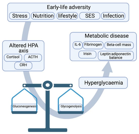
The situation may be somewhat more complicated and dependent on the timing of the adversity. Adversity in early childhood was associated with a decreased hippocampal volume, whilst prefrontal cortex volume was reduced if exposed during adolescence [88,89]. Psychopathologically, exposure to adversity or trauma before age 12 increased the lifelong risk of developing major depressive disorder, but when occurring between age 12 to 18, the risk of PTSD was increased [90]. It has been suggested that since the human hippocampus is not fully developed before age 2, the frontal cortex primarily matures between age 8–14 and the amygdala continues developing until early adulthood [91], and the hippocampus is most probably the brain area most affected by early life stress [76]. The sensitivity of the hippocampus to ELA is particularly important, as, outlined above, it plays a key inhibitory role in PVN activation of the HPA axis. Hence, ELA renders the HPA axis impaired, which in turn causes insidious changes to the stress response mechanism along with glucose metabolism, eventually contributing to MetS (Figure 3).

Figure 3.
Early life adversity dysregulates the HPA axis and its key effector molecules, which in turn disrupts glucose homeostatic balance, leading to hyperglycaemia and metabolic syndrome if left unchecked.
There is more to the HPA axis than reactivity to a laboratory stressor. ELA was initially reported to be associated with elements of the cortisol diurnal rhythm such as the cortisol awakening rise (CAR) [92], however, the most recent meta-analysis suggests that this is not the case [93], although the meta-analysis of the stress-induced changes was much stronger [94]. The weakness in the CAR meta-analysis was probably due to large heterogeneity between the techniques employed between the different studies.
3.2. Early Life Adversity, Diabetes and the Metabolic Syndrome
MetS has been shown to predict not only cardiovascular mortality, but also the progression to full type 2 diabetes (T2D) [95]. There is growing evidence that early life nutritional and psychosocial stress or disadvantage determine the trajectory and transition into metabolic dysfunction, MetS, and T2D later in life [9]. There are two principal components of the early life socioeconomic position that contribute to lifelong MetS and diabetic risk—Early life nutrition and early life stress. Reports from ourselves and others of early life adversity have shown effects on either the metabolic profile, obesity, or type 2 diabetes [96,97,98]. Socioeconomic status in early life has a similar effect [15], and is associated with T2D 50 years later [99]. ELA also predisposes individuals towards a chronic inflammatory phenotype [98,100,101,102]. Furthermore, cardiometabolic disease markers such as fibrinogen, C-reactive protein, and interleukin-6 were elevated [17,103], as were endothelial dysfunction markers such as ICAM-1, E-selectin [104], as well as clinical measures such as arterial stiffness [105] or poor blood pressure trajectories with age [106]. Recently, this was replicated by Chandan et al. (2020), confirming the role of ELA in determining “a significant proportion of the cardiometabolic and diabetic disease burden may be attributable to maltreatment” [107,108].
Although there are few mechanistic data available, these metabolic abnormalities may be due to changes in circulating adipokine levels. ELA has been directly associated with an increased leptin:adiponectin ratio [109]. Leptin, secreted by adipocytes, regulates energy balance through decreasing appetite and is associated with the metabolic syndrome [110,111], whilst adiponectin, has insulin-sensitizing effects. Low adiponectin levels are associated with type 2 diabetes and insulin resistance [111,112]. Furthermore, Irisin levels were increased by ELA [109]. Irisin, mediates glucose metabolism as well as exercise-related energy expenditure and is a peroxisome proliferator-activated receptor-γ coactivator 1-α (PGC-1α)-dependent myokine [109,113]. Glucocorticoids may also play a role, as, in utero exposure to maternal under nutritional affects beta-cell number and function lifelong in a manner dependent upon GR and GC since deletion of the GR in foetal pancreatic cell abrogated this effect [3]. These changes may, in part be due to the effects of ELA on the methylation of genes involved in obesity and metabolic pathways [114,115]. Furthermore, adverse early life conditions induce lifelong changes in gene transcription [116]. Low SES, for example, has been associated with inflammatory and diabetic genes such as TLR3 [117], NLRP12 [118], F8 [119], KLRG1 [120], CD1D [121], as well as the stress-associated genes OXTR [122], FKBP5 [123], and AVP [124], suggesting, as we have previously proposed, that a negative early life environment acts through inflammatory pathways that are also associated with T2D, targeting pathophysiological factors such as stress and inflammation, and participating in the aetiopathology of T2D [108]. Thus, it appears logical to conclude that ELA can effectively alter glucose homeostasis and that the aetiology of MetS and eventual T2D may have strong roots in ELA. Moreover, ELA would appear to be associated with more advanced or complicated diabetic-pathologies requiring more aggressive management [125].
3.3. Glucose Metabolism, Allostasis and Allostatic Load
Ever since Hans Selye described the “general adaptation syndrome” as our response to external stressors [126], there has been a paradox. The ANS and HPA axis protect in the short-term, but over the long-term they may accelerate disease as well as causing lasting damage. Allostatic Load (AL) is fundamentally a chain of causal events from the primary stress response of SNS and HPA axis activation with epinephrine and cortisol secretion, inflammation [127], leading to secondary markers of stress exposure including hyperglycaemia, hypertension, hyperlipidaemia, and central adiposity. The AL cascade, the overall sequence of responses, as well as their contribution to disease development, are not fully understood, and somewhat under-investigated, although markers of AL are associated with increased glycaemic measures in women [128]. Similarly, rat chronic stress models, a proxy for AL, consistently report high blood glucose levels up 6 months later [46]. In allostasis these rats also had dysregulated glucose metabolism pathways, in particular increased gluconeogenesis [46]. Furthermore, this stress induced hyperglycaemic state resulted in impaired glucose tolerance and reduced insulin sensitivity [46]. This may be due to metabolic memory, which causes the body to recall the influence of metabolic regulators for a much longer duration. This, in conjunction with persistent stress, can lead to maladapted allostasis and eventually AL. Prolonged AL can give rise to metabolic syndrome. There is also recent evidence that diet may act as a stressor. Increased glycaemic load (i.e., dietary sugar intake) is associated with increased markers of AL, particularly in women, suggesting that dietary carbohydrate intake may contribute towards dysregulation of the AL response [128]. It is well established that carbohydrate intake can stimulate the ANS [129], and hypothesised to cascade down increasing AL markers and blood glucose levels [128]. Thus, chronic stress/AL can adversely affect glucose metabolism and mount a faulty bodily adaptation that ultimately leads to MetS.
4. Gluconeogenesis at the Crossroads between Adversity and Metabolism
It is well established that energy metabolism and psychosocial stress are intimately intertwined. It would also appear that psychosocial adversity in early life sets the individual on a negative trajectory towards either MetS or T2D. The available literature suggests that ELA can effectively alter glucose homeostasis and participate in the aetiology of MetS and eventual T2D. It is possible that MetS and T2D may have very strong roots in the early life environment, with ELA as a strong driver of the eventual diabetic phenotype. This suggests that gluconeogenesis, originally named because of the intimate link between corticoid levels and glucose levels, may be at the heart of the mechanism. We suggest that this may be a double-edged sword. Gluconeogenesis is an integral part of energy homeostasis in response to an external stressor. However, it may show its true “Jekyll and Hyde” nature when the HPA axis is perturbed. Furthermore, the interaction between the HPA axis, glucocorticoids, and mechanisms of glucose homeostasis, such as gluconeogenesis or insulin resistance, may be the link between ELA and lifelong metabolic disturbances. Indeed, one consequence in the neonate of maternal separation is a rapid drop in blood glucose levels. This, together with increased ghrelin (“hunger hormone”) levels may actually trigger HPA axis activation, and glucose supplementation during maternal separation reverses the phenotype, confirming the link and providing a potential mechanism to counteract it [130].
Energy homeostasis during psychosocial stress is somewhat underexplored, however, a number of recent studies have started to investigate the full nature of the bi-directional regulation in more detail. There is a wealth of data on how glucose availability modulates the corticosteroid response to psychological and psychosocial stressors, however, the data is sparse or inexistent in the reverse direction. Glucose or levels of gluconeogenesis need to be determined after stress. Furthermore, they need to be recognised as genuine measures, e.g., the Trier Social Stress test, providing insight into how the stress axes interact with energy homeostasis. Elucidating these GC-glucose interactions during laboratory stressors is now essential. We need to initially understand the normal blood glucose response to an external stressor. This will subsequently permit investigation of glucose-cortisol coupling in situations such as exposure to ELA where the HPA axis has been significantly programmed, with lifelong changes in reactivity, setpoint, and secreted hormone levels. Recent work on the direct transcriptional control of gluconeogenesis [7], together with recent interest in stress-energy balance [131], open the field for more detailed investigation of the bi-directional regulation of these two essential physiological systems. Thus, it is extremely important to identify if the “Jekyll” or the “Hyde” of gluconeogenesis is at play, and how it balances energy homeostasis under stress, while avoiding gluconeogenesis driven T2D.
5. Conclusions
In light of the presented narrative, it is clear that there is a direct link between ELA, the HPA axis, glucose metabolism, MetS, and potentially T2D. It is now essential to understand how early life programming of the HPA axis, with lifelong changes in glucocorticoid secretory profiles, influences energy metabolism, and processes such as hepatic gluconeogenesis. To do this, and to provide the missing piece of the puzzle we need to consider glucose and energy homeostasis as genuine output measures in standardised laboratory psychological stress tests such as the TSST or the socially evaluated cold pressor test. We need to initially investigate the normal physiological interaction between the HPA axis (or the complete stress system), and energy homeostasis. We hypothesise that lifelong programming of the HPA axis after ELA will fundamentally alter hepatic gluconeogenesis, inducing hyperglycaemia, and the significant lifelong risk or MetS or T2D.
Early life developmental programming of the MetS or T2D risk dovetails nicely into the developmental origins of Health and Disease (DOHaD) model developed by David Barker. The DOHaD model has evolved over the last years into the current “three hit model” [132,133]. In the current concept the three “hits” are defined as (i) invariable genome that fixes genetic risk at conception, (ii) the early life environment during which many biological systems are modulated or adapted to the environment the individual is born into, and (iii) the later-life environment where a perturbation tips the balance between health and disease. The first and second hits produce a phenotype that remains latent or quiescent until the third hit crystallises the risk and initiates the health-disease transition. In our ELA-metabolic disease paradigm, we see the early life adversity during the SHRP as the second hit and the cluster of metabolic abnormalities as the final disease phenotype. Indeed, there is now evidence that ELA leaves a faint metabolic imprint almost immediately, although the full syndrome does not appear until much later in life, but usually becomes manifest no earlier than at adult age [134]. This mirrors the observation from pre-term infants, where increased baseline HPA axis activity, reduced stress reactivity, and components of the metabolic syndrome are seen in later life [135]. This may, however, be linked to the use of synthetic glucocorticoids such as dexamethasone in the perinatal period. However, Vargas et al. demonstrated that early life stress could concurrently increase HPA axis activity, and mild metabolic alterations, that were significantly increased with a subsequent environmental challenge akin to the third hit, although their model suggested that it was independent of GC [136].
One potential limitation to our postulate is that chronic stress has been shown to bias feeding choices in both animals and humans to addictive foods such as high sugar and high fat content foods, which can directly contribute to MetS over the years (reviewed in [137]). This could be mediated by alterations in the glucose metabolism, HPA axis related changes, etc. This limitation can be further dissected by studying laboratory models of chronic stress that are only fed standard chow diet to see if gluconeogenesis is indeed altered. It is also interesting to note that chronic stress is frequently accompanied by comorbidities like depression [138], which decrease sucrose preference in depressive animals and is often used as an index of anhedonia [139], providing a conflicting perspective with respect to chronic stress and diet.
Thus, we suggest a “Jekyll and Hyde” role to gluconeogenesis, providing the necessary energy in situations of acute stress, but driving towards pathophysiological consequences when the HPA axis has been altered. Furthermore, if our hypothesised link is correct, does this output have the predictive power to identify individuals at a higher risk of developing MetS at a later stage in life, before symptom onset?
Author Contributions
Conceptualization, S.V.S. and J.D.T.; literature review, S.V.S. and J.D.T.; writing and editing, S.V.S. and J.D.T. Both authors have read and agreed to the published version of the manuscript.
Funding
S.V.S. and J.D.T. were funded by Fonds National de Recherche Luxembourg (INTER/ANR/16/11568350 ‘MADAM’). The work of J.D.T. on the long term consequences of ELA was further funded by FNR-CORE C16/BM/11342695 ‘MetCOEPs’; C12/BM/3985792 ‘EpiPath’; and C19/SC/13650569, “ALAC”.
Institutional Review Board Statement
Not applicable.
Informed Consent Statement
Not applicable.
Data Availability Statement
Not applicable.
Acknowledgments
The authors would like to thank Sophie Mériaux, Pauline Guebels, Stephanie Schmitz, and Fanny Bonnemberger for their technical support in their work investigating the long-term effects of early life adversity over the years. All figures were created using BioRender.com by J.D.T.
Conflicts of Interest
The authors declare no conflict of interest.
References
- Sandi, C.; Haller, J. Stress and the social brain: Behavioural effects and neurobiological mechanisms. Nat. Rev. Neurosci. 2015, 16, 290–304. [Google Scholar] [CrossRef]
- Herzig, S. Liver: A target of late diabetic complications. Exp. Clin. Endocrinol. Diabetes 2012, 120, 202–204. [Google Scholar] [CrossRef]
- de Guia, R.M.; Rose, A.J.; Herzig, S. Glucocorticoid hormones and energy homeostasis. Horm. Mol. Biol. Clin. Investig. 2014, 19, 117–128. [Google Scholar] [CrossRef]
- Nuttall, F.Q.; Ngo, A.; Gannon, M.C. Regulation of hepatic glucose production and the role of gluconeogenesis in humans: Is the rate of gluconeogenesis constant? Diabetes Metab. Res. Rev. 2008, 24, 438–458. [Google Scholar] [CrossRef]
- Konig, M.; Bulik, S.; Holzhutter, H.G. Quantifying the contribution of the liver to glucose homeostasis: A detailed kinetic model of human hepatic glucose metabolism. PLoS Comput. Biol. 2012, 8, e1002577. [Google Scholar] [CrossRef]
- ter Horst, G.J.; Luiten, P.G. The projections of the dorsomedial hypothalamic nucleus in the rat. Brain Res. Bull. 1986, 16, 231–248. [Google Scholar] [CrossRef]
- Cui, A.; Fan, H.; Zhang, Y.; Niu, D.; Liu, S.; Liu, Q.; Ma, W.; Shen, Z.; Shen, L.; Liu, Y.; et al. Dexamethasone-induced Kruppel-like factor 9 expression promotes hepatic gluconeogenesis and hyperglycemia. J. Clin. Investig. 2019, 129, 2266–2278. [Google Scholar] [CrossRef]
- Grundy, S.M.; Brewer, H.B., Jr.; Cleeman, J.I.; Smith, S.C., Jr.; Lenfant, C.; National Heart, Lung and Blood Institiute; American Heart Association. Definition of metabolic syndrome: Report of the National Heart, Lung, and Blood Institute/American Heart Association conference on scientific issues related to definition. Circulation 2004, 109, 433–438. [Google Scholar] [CrossRef] [PubMed]
- Delpierre, C.; Fantin, R.; Barboza-Solis, C.; Lepage, B.; Darnaudéry, M.; Kelly-Irving, M. The early life nutritional environment and early life stress as potential pathways towards the metabolic syndrome in mid-life? A lifecourse analysis using the 1958 British Birth cohort. BMC Public Health 2016, 16, 815. [Google Scholar]
- Turner, J.D. Childhood adversity from conception onwards: Are our tools unnecessarily hindering us? J. Behav. Med. 2018, 41, 568–570. [Google Scholar] [CrossRef] [PubMed]
- Suglia, S.F.; Koenen, K.C.; Boynton-Jarrett, R.; Chan, P.S.; Clark, C.J.; Danese, A.; Faith, M.S.; Goldstein, B.I.; Hayman, L.L.; Isasi, C.R.; et al. Childhood and adolescent adversity and cardiometabolic outcomes: A scientific statement from the american heart association. Circulation 2018, 137, e15–e28. [Google Scholar] [CrossRef]
- Jaaskelainen, P.; Magnussen, C.G.; Pahkala, K.; Mikkila, V.; Kakohen, M.; Sabin, M.A.; Fogelholm, M.; Hutri-Kakohen, N.; Taittonen, L.; Telama, R.; et al. Childhood nutrition in predicting metabolic syndrome in adults: The cardiovascular risk in Young Finns Study. Diabetes Care 2012, 35, 1937–1943. [Google Scholar] [CrossRef] [PubMed]
- Huang, J.Y.; Gariepy, G.; Gavin, A.R.; Rowhani-Rahbar, A.; Siscovick, D.S.; Enquobahrie, D.A. Maternal education in early life and risk of metabolic syndrome in young adult american females and males: Disentangling life course processes through causal models. Epidemiology 2019, 30, S28–S36. [Google Scholar] [CrossRef]
- Afifi, T.O.; MacMillan, H.L.; Boyle, M.; Cheung, K.; Taillieu, T.; Turner, S.; Sareen, J. Child abuse and physical health in adulthood. Health Rep. 2016, 27, 10–18. [Google Scholar] [PubMed]
- Hostinar, C.E.; Ross, K.M.; Chan, E.; Miller, G.E. Early-life socioeconomic disadvantage and metabolic health disparities. Psychosom. Med. 2017, 79, 514–523. [Google Scholar] [CrossRef]
- Tomasdottir, M.O.; Sigurdsson, J.A.; Petursson, H.; Kirkengen, A.L.; Krokstad, S.; McEwen, B.; Hetlevik, I.; Getz, L. Self reported childhood difficulties, adult multimorbidity and allostatic load. A cross-sectional analysis of the Norwegian HUNT study. PLoS ONE 2015, 10, e0130591. [Google Scholar] [CrossRef]
- Danese, A.; Tan, M. Childhood maltreatment and obesity: Systematic review and meta-analysis. Mol. Psychiatry 2014, 19, 544–554. [Google Scholar] [CrossRef] [PubMed]
- Huang, H.; Yan, P.; Shan, Z.; Li, M.; Luo, C.; Gao, H.; Hao, L.; Liu, L. Adverse childhood experiences and risk of type 2 diabetes: A systematic review and meta-analysis. Metabolism 2015, 64, 1408–1418. [Google Scholar] [CrossRef] [PubMed]
- Zhang, S.; Rattanatray, L.; Morrison, J.L.; Nicholas, L.M.; Lie, S.; McMillen, I.C. Maternal obesity and the early origins of childhood obesity: Weighing up the benefits and costs of maternal weight loss in the periconceptional period for the offspring. Exp. Diabetes Res. 2011, 2011, 585749. [Google Scholar] [CrossRef]
- Chrousos, G.P.; Gold, P.W. The concepts of stress and stress system disorders. Overview of physical and behavioral homeostasis. JAMA 1992, 267, 1244–1252. [Google Scholar] [CrossRef]
- Tsigos, C.; Chrousos, G.P. Physiology of the hypothalamic-pituitary-adrenal axis in health and dysregulation in psychiatric and autoimmune disorders. Endocrinol. Metab. Clin. N. Am. 1994, 23, 451–466. [Google Scholar] [CrossRef]
- Rotenberg, S.; McGrath, J.J. Inter-relation between autonomic and HPA axis activity in children and adolescents. Biol. Psychol. 2016, 117, 16–25. [Google Scholar] [CrossRef] [PubMed]
- Jones, B.E.; Yang, T.Z. The efferent projections from the reticular formation and the locus coeruleus studied by anterograde and retrograde axonal transport in the rat. J. Comp. Neurol. 1985, 242, 56–92. [Google Scholar] [CrossRef] [PubMed]
- Lewis, D.I.; Coote, J.H. Excitation and inhibition of rat sympathetic preganglionic neurones by catecholamines. Brain Res. 1990, 530, 229–234. [Google Scholar] [CrossRef]
- Unnerstall, J.R.; Kopajtic, T.A.; Kuhar, M.J. Distribution of alpha 2 agonist binding sites in the rat and human central nervous system: Analysis of some functional, anatomic correlates of the pharmacologic effects of clonidine and related adrenergic agents. Brain Res. 1984, 319, 69–101. [Google Scholar] [CrossRef]
- Reiche, E.M.; Nunes, S.O.; Morimoto, H.K. Stress, depression, the immune system, and cancer. Lancet Oncol. 2004, 5, 617–625. [Google Scholar] [CrossRef]
- Ito, R.; Lee, A.C.H. The role of the hippocampus in approach-avoidance conflict decision-making: Evidence from rodent and human studies. Behav. Brain Res. 2016, 313, 345–357. [Google Scholar] [CrossRef]
- Fee, C.; Prevot, T.; Misquitta, K.; Banasr, M.; Sibille, E. Chronic stress-induced behaviors correlate with exacerbated acute stress-induced cingulate cortex and ventral hippocampus activation. Neuroscience 2020, 440, 113–129. [Google Scholar] [CrossRef]
- Jankord, R.; Herman, J.P. Limbic regulation of hypothalamo-pituitary-adrenocortical function during acute and chronic stress. Ann. N. Y. Acad. Sci. 2008, 1148, 64–73. [Google Scholar] [CrossRef] [PubMed]
- Nixon, M.; Mackenzie, S.D.; Taylor, A.I.; Homer, N.Z.M.; Livingstone, D.E.; Mouras, R.; Morgan, R.A.; Mole, D.J.; Stimson, R.H.; Reynolds, R.M. ABCC1 confers tissue-specific sensitivity to cortisol versus corticosterone: A rationale for safer glucocorticoid replacement therapy. Sci. Transl. Med. 2016, 8, 352ra109. [Google Scholar] [CrossRef] [PubMed]
- Wang, M. The role of glucocorticoid action in the pathophysiology of the metabolic syndrome. Nutr. Metab. (Lond.) 2005, 2, 3. [Google Scholar] [CrossRef]
- Reppert, S.M.; Weaver, D.R. Coordination of circadian timing in mammals. Nature 2002, 418, 935–941. [Google Scholar] [CrossRef] [PubMed]
- Ulrich-Lai, Y.M.; Herman, J.P. Neural regulation of endocrine and autonomic stress responses. Nat. Rev. Neurosci. 2009, 10, 397–409. [Google Scholar] [CrossRef]
- Antoni, F.A. Hypothalamic control of adrenocorticotropin secretion: Advances since the discovery of 41-residue corticotropin-releasing factor. Endocr. Rev. 1986, 7, 351–378. [Google Scholar] [CrossRef]
- Tsigos, C.; Chrousos, G.P. Hypothalamic-pituitary-adrenal axis, neuroendocrine factors and stress. J. Psychosom. Res. 2002, 53, 865–871. [Google Scholar] [CrossRef]
- Stavreva, D.A.; Wiench, M.; John, S.; Conway-Campbell, B.L.; McKenna, M.A.; Pooley, J.R.; Johnson, T.A.; Lightman, T.C.V.; Hager, G.L. Ultradian hormone stimulation induces glucocorticoid receptor-mediated pulses of gene transcription. Nat. Cell. Biol. 2009, 11, 1093–1102. [Google Scholar] [CrossRef] [PubMed]
- Trifonova, S.T.; Gantenbein, M.; Turner, J.D.; Muller, C.P. The use of saliva for assessment of cortisol pulsatile secretion by deconvolution analysis. Psychoneuroendocrinology 2013, 38, 1090–1101. [Google Scholar] [CrossRef]
- Lightman, S.L.; Wiles, C.C.; Atkinson, H.C.; Henley, D.E.; Russell, G.M.; Leendertz, J.A.; McKenna, M.A.; Spiga, F.; Wood, S.A.; Conway-Campbell, B.L. The significance of glucocorticoid pulsatility. Eur. J. Pharmacol. 2008, 583, 255–262. [Google Scholar] [CrossRef] [PubMed]
- Walker, J.J.; Terry, J.R.; Lightman, S.L. Origin of ultradian pulsatility in the hypothalamic-pituitary-adrenal axis. Proc. Biol. Sci. 2010, 277, 1627–1633. [Google Scholar] [CrossRef]
- Sapolsky, R.M.; Meaney, M.J. Maturation of the adrenocortical stress response: Neuroendocrine control mechanisms and the stress hyporesponsive period. Brain Res. 1986, 396, 64–76. [Google Scholar] [CrossRef]
- Romeo, R.D. The metamorphosis of adolescent hormonal stress reactivity: A focus on animal models. Front. Neuroendocrinol. 2018, 49, 43–51. [Google Scholar] [CrossRef]
- Kuo, T.; McQueen, A.; Chen, T.-C.; Wang, J.-C. Regulation of glucose homeostasis by glucocorticoids. Adv. Exp. Med. Biol. 2015, 872, 99–126. [Google Scholar]
- Thorens, B.; Mueckler, M. Glucose transporters in the 21st Century. Am. J. Physiol. Endocrinol. Metab. 2010, 298, E141–E145. [Google Scholar] [CrossRef]
- Huang, S.; Czech, M.P. The GLUT4 glucose transporter. Cell. Metab. 2007, 5, 237–252. [Google Scholar] [CrossRef] [PubMed]
- Gartner, K.; Buttner, D.; Dohler, K.; Friedel, R.; Lindena, J.; Trautschold, I. Stress response of rats to handling and experimental procedures. Lab. Anim. 1980, 14, 267–274. [Google Scholar] [CrossRef] [PubMed]
- Nirupama, R.; Devaki, M.; Yajurvedi, H.N. Chronic stress and carbohydrate metabolism: Persistent changes and slow return to normalcy in male albino rats. Stress 2012, 15, 262–271. [Google Scholar] [CrossRef]
- Wu, P.; Sato, J.; Zhao, Y.; Jaskiewicz, J.; Popov, K.M.; Harris, R.A. Starvation and diabetes increase the amount of pyruvate dehydrogenase kinase isoenzyme 4 in rat heart. Biochem. J. 1998, 329, 197–201. [Google Scholar] [CrossRef]
- Sato, T.; Yamamoto, H.; Sawada, N.; Nashiki, K.; Tsuji, M.; Muto, K.; Kume, H.; Sasiki, H.; Arai, H.; Nikawa, T.; et al. Restraint stress alters the duodenal expression of genes important for lipid metabolism in rat. Toxicology 2006, 227, 248–261. [Google Scholar] [CrossRef]
- Foley, P.; Kirschbaum, C. Human hypothalamus-pituitary-adrenal axis responses to acute psychosocial stress in laboratory settings. Neurosci. Biobehav. Rev. 2010, 35, 91–96. [Google Scholar] [CrossRef] [PubMed]
- Kirschbaum, C.; Bono, E.G.; Rohleder, N.; Gessner, C.; Pirke, K.M.; Salvador, A.; Hellhammer, D.H. Effects of fasting and glucose load on free cortisol responses to stress and nicotine. J. Clin. Endocrinol. Metab. 1997, 82, 1101–1105. [Google Scholar] [CrossRef] [PubMed]
- Gonzalez-Bono, E.; Rohleder, N.; Hellhammer, D.H.; Salvador, A.; Kirschbaum, C. Glucose but not protein or fat load amplifies the cortisol response to psychosocial stress. Horm. Behav. 2002, 41, 328–333. [Google Scholar] [CrossRef]
- Choi, S.; Horsley, C.; Aguilla, S.; Dallman, M.F. The hypothalamic ventromedial nuclei couple activity in the hypothalamo-pituitary-adrenal axis to the morning fed or fasted state. J. Neurosci. 1996, 16, 8170–8180. [Google Scholar] [CrossRef]
- Rosmond, R.; Holm, G.; Bjorntorp, P. Food-induced cortisol secretion in relation to anthropometric, metabolic and haemodynamic variables in men. Int. J. Obes. Relat. Metab. Disord. 2000, 24, 416–422. [Google Scholar] [CrossRef]
- Bergendahl, M.; Iranmanesh, A.; Evans, W.S.; Veldhuis, J.D. Short-term fasting selectively suppresses leptin pulse mass and 24-hour rhythmic leptin release in healthy midluteal phase women without disturbing leptin pulse frequency or its entropy control (pattern orderliness). J. Clin. Endocrinol. Metab. 2000, 85, 207–213. [Google Scholar]
- Sherwin, R.S.; Sacca, L. Effect of epinephrine on glucose metabolism in humans: Contribution of the liver. Am. J. Physiol. 1984, 247, E157–E165. [Google Scholar] [CrossRef]
- Burgess, S.C.; He, T.; Yan, Z.; Lindner, J.; Sherry, A.D.; Malloy, C.R.; Browning, J.D.; Magnuson, M.A. Cytosolic phosphoenolpyruvate carboxykinase does not solely control the rate of hepatic gluconeogenesis in the intact mouse liver. Cell. Metab. 2007, 5, 313–320. [Google Scholar] [CrossRef]
- Opherk, C.; Tronche, F.; Kellendonk, C.; Kohlmuller, D.; Schulze, A.; Schimd, W.; Schutz, G. Inactivation of the glucocorticoid receptor in hepatocytes leads to fasting hypoglycemia and ameliorates hyperglycemia in streptozotocin-induced diabetes mellitus. Mol. Endocrinol. 2004, 18, 1346–1353. [Google Scholar] [CrossRef] [PubMed]
- Barthel, A.; Schmoll, D. Novel concepts in insulin regulation of hepatic gluconeogenesis. Am. J. Physiol. Endocrinol. Metab. 2003, 285, E685–E692. [Google Scholar] [CrossRef] [PubMed]
- Petersen, M.C.; Shulman, G.I. Mechanisms of insulin action and insulin resistance. Physiol. Rev. 2018, 98, 2133–2223. [Google Scholar] [CrossRef] [PubMed]
- Liu, Y.; Nakagawa, Y.; Wang, Y.; Sakurai, R.; Tripathi, P.V.; Lutfy, K.; Friedman, T.C. Increased glucocorticoid receptor and 11{Betancur, #1833}-hydroxysteroid dehydrogenase type 1 expression in hepatocytes may contribute to the phenotype of type 2 diabetes in db/db mice. Diabetes 2005, 54, 32–40. [Google Scholar] [PubMed]
- Lee, D.; Le Lay, J.; Kaestner, K.H. The transcription factor CREB has no non-redundant functions in hepatic glucose metabolism in mice. Diabetologia 2014, 57, 1242–1248. [Google Scholar] [CrossRef] [PubMed]
- Shukla, R.; Basu, A.K.; Mandal, B.; Mukhopadhyay, P.; Maity, A.; Chakraborty, S.; Devrabhai, P.K. 11beta Hydroxysteroid dehydrogenase-1 activity in type 2 diabetes mellitus: A comparative study. BMC Endocr. Disord. 2019, 19, 15. [Google Scholar] [CrossRef]
- Granner, D.K. In pursuit of genes of glucose metabolism. J. Biol. Chem. 2015, 290, 22312–22324. [Google Scholar] [CrossRef] [PubMed]
- Schacke, H.; Docke, W.D.; Asadullah, K. Mechanisms involved in the side effects of glucocorticoids. Pharmacol. Ther. 2002, 96, 23–43. [Google Scholar] [CrossRef]
- Blondeau, B.; Sahly, I.; Massourides, E.; Singh-Estivalet, A.; Valtat, B.; Dorchene, D.; Jaisser, F.; Breant, B.; Tronche, F. Novel transgenic mice for inducible gene overexpression in pancreatic cells define glucocorticoid receptor-mediated regulations of beta cells. PLoS ONE 2012, 7, e30210. [Google Scholar] [CrossRef]
- Buren, J.; Lai, Y.C.; Lundgren, M.; Eriksson, J.W.; Jensen, J. Insulin action and signalling in fat and muscle from dexamethasone-treated rats. Arch. Biochem. Biophys. 2008, 474, 91–101. [Google Scholar] [CrossRef] [PubMed]
- Karnia, M.J.; Myslinska, D.; Dzik, K.P.; Flis, D.J.; Podlacha, M.; Kaczor, J.J. BST stimulation induces atrophy and changes in aerobic energy metabolism in rat skeletal muscles-the biphasic action of endogenous glucocorticoids. Int. J. Mol. Sci. 2020, 21, 2787. [Google Scholar] [CrossRef]
- Chiodini, I.; Adda, G.; Scillitani, A.; Coletti, F.; Morelli, V.; Di Lembo, S.; Epaminonda, P.; Masserini, B.; Beck-Peccoz, P.; Orsi, E.; et al. Cortisol secretion in patients with type 2 diabetes: Relationship with chronic complications. Diabetes Care 2007, 30, 83–88. [Google Scholar] [CrossRef] [PubMed]
- Nouwen, A.; Winkley, K.; Twisk, J.; Lloyd, C.E.; Peyrot, M.; Ismail, K.; Pouwer, F.; European Depression in Diabetes (EDID) Research Consortium. Type 2 diabetes mellitus as a risk factor for the onset of depression: A systematic review and meta-analysis. Diabetologia 2010, 53, 2480–2486. [Google Scholar] [CrossRef]
- Mosili, P.; Mchize, B.C.; Ngubane, P.; Sibiya, N.; Khathi, A. The dysregulation of the hypothalamic-pituitary-adrenal axis in diet-induced prediabetic male Sprague Dawley rats. Nutr. Metab. (Lond.) 2020, 17, 104. [Google Scholar] [CrossRef]
- Herman, J.P.; McKlveen, J.M.; Ghosal, S.; Kopp, B.; Wulsin, A.; Makinson, R.; Scheimann, J.; Myers, B. Regulation of the hypothalamic-pituitary-adrenocortical stress response. Compr. Physiol. 2016, 6, 603–621. [Google Scholar]
- Swierczynska, M.M.; Mateska, I.; Peitzsch, M.; Bornstein, S.R.; Chavakis, T.; Eisenhofer, G.; Lamounier-Zepter, V.; Eaton, S. Changes in morphology and function of adrenal cortex in mice fed a high-fat diet. Int. J. Obes. (Lond.) 2015, 39, 321–330. [Google Scholar] [CrossRef]
- Shu, H.J.; Isenberg, K.; Cormier, R.J.; Benz, A.; Zorumski, C.F. Expression of fructose sensitive glucose transporter in the brains of fructose-fed rats. Neuroscience 2006, 140, 889–895. [Google Scholar] [CrossRef] [PubMed]
- Harrell, C.S.; Burgado, J.; Kelly, S.D.; Johnson, Z.P.; Neigh, G.N. High-fructose diet during periadolescent development increases depressive-like behavior and remodels the hypothalamic transcriptome in male rats. Psychoneuroendocrinology 2015, 62, 252–264. [Google Scholar] [CrossRef] [PubMed]
- Centers for Disease, Control and Prevention (CDC). Adverse childhood experiences reported by adults—Five states, 2009. MMWR Morb. Mortal. Wkly. Rep. 2010, 59, 1609–1613. [Google Scholar]
- van Bodegom, M.; Homberg, J.R.; Henckens, M. Modulation of the hypothalamic-pituitary-adrenal axis by early life stress exposure. Front. Cell. Neurosci. 2017, 11, 87. [Google Scholar] [CrossRef]
- Gutteling, B.M.; de Weerth, C.; Buitelaar, J.K. Prenatal stress and children’s cortisol reaction to the first day of school. Psychoneuroendocrinology 2005, 30, 541–549. [Google Scholar] [CrossRef] [PubMed]
- O’Connor, T.G.; Ben-Shlomo, Y.; Heron, J.; Golding, J.; Adams, D.; Glover, V. Prenatal anxiety predicts individual differences in cortisol in pre-adolescent children. Biol. Psychiatry 2005, 58, 211–217. [Google Scholar] [CrossRef]
- Davies, P.T.; Sturge-Apple, M.L.; Cicchetti, D.; Manning, L.G.; Zale, E. Children’s patterns of emotional reactivity to conflict as explanatory mechanisms in links between interpartner aggression and child physiological functioning. J. Child Psychol. Psychiatry 2009, 50, 1384–1391. [Google Scholar] [CrossRef]
- Bugental, D.B.; Martorell, G.A.; Barraza, V. The hormonal costs of subtle forms of infant maltreatment. Horm. Behav. 2003, 43, 237–244. [Google Scholar] [CrossRef]
- Carlson, M.; Earls, F. Psychological and neuroendocrinological sequelae of early social deprivation in institutionalized children in Romania. Ann. N. Y. Acad. Sci. 1997, 807, 419–428. [Google Scholar] [CrossRef] [PubMed]
- Bernard, K.; Zwerling, J.; Dozier, M. Effects of early adversity on young children’s diurnal cortisol rhythms and externalizing behavior. Dev. Psychobiol. 2015, 57, 935–947. [Google Scholar] [CrossRef]
- Hengesch, X.; Elwenspoek, M.M.C.; Schaan, V.K.; Larra, M.F.; Finke, J.B.; Zhang, X.; Bachmann, P.; Turner, J.D.; Vogele, C.; Muller, C.P.; et al. Blunted endocrine response to a combined physical-cognitive stressor in adults with early life adversity. Child Abuse Negl. 2018, 85, 137–144. [Google Scholar] [CrossRef]
- Pesonen, A.K.; Raikkonen, K.; Feldt, K.; Heinnonen, K.; Osmond, C.; Philips, D.I.W.; Barker, D.J.P.; Eriksson, J.G.; Kajantie, E. Childhood separation experience predicts HPA axis hormonal responses in late adulthood: A natural experiment of World War II. Psychoneuroendocrinology 2010, 35, 758–767. [Google Scholar] [CrossRef] [PubMed]
- Fries, E.; Hesse, J.; Hellhammer, J.; Hellhammer, D.H. A new view on hypocortisolism. Psychoneuroendocrinology 2005, 30, 1010–1016. [Google Scholar] [CrossRef]
- Yehuda, R.; Yang, R.-K.; Buchsbaum, M.S.; Golier, J.A. Alterations in cortisol negative feedback inhibition as examined using the ACTH response to cortisol administration in PTSD. Psychoneuroendocrinology 2006, 31, 447–451. [Google Scholar] [CrossRef] [PubMed]
- Elwenspoek, M.M.C.; Hengesch, X.; Leenen, F.A.D.; Sias, K.; Fernandes, S.B.; Schaan, V.K.; Meriaux, S.B.; Schmitz, S.; Bonnemberger, F.; Schachinger, H.; et al. Glucocorticoid receptor signaling in leukocytes after early life adversity. Dev. Psychopathol. 2019, 32, 1–11. [Google Scholar] [CrossRef]
- Teicher, M.H.; Tomoda, A.; Andersen, S. Neurobiological consequences of early stress and childhood maltreatment: Are results from human and animal studies comparable? Ann. N. Y. Acad. Sci. 2006, 1071, 313–323. [Google Scholar] [CrossRef] [PubMed]
- Andersen, S.L.; Tomada, A.; Vincow, E.S.; Valente, E.; Polcari, A.; Teicher, M.H. Preliminary evidence for sensitive periods in the effect of childhood sexual abuse on regional brain development. J. Neuropsychiatry Clin. Neurosci. 2008, 20, 292–301. [Google Scholar] [CrossRef] [PubMed]
- Maercker, A.; Michael, T.; Fehm, L.; Becker, E.S.; Margraf, J. Age of traumatisation as a predictor of post-traumatic stress disorder or major depression in young women. Br. J. Psychiatry 2004, 184, 482–487. [Google Scholar] [CrossRef] [PubMed]
- Caballero, A.; Granberg, R.; Tseng, K.Y. Mechanisms contributing to prefrontal cortex maturation during adolescence. Neurosci. Biobehav. Rev. 2016, 70, 4–12. [Google Scholar] [CrossRef] [PubMed]
- Starr, L.R.; Dienes, K.; Stroud, C.B.; Shaw, Z.A.; Li, Y.I.; Mlawer, F.; Huang, M. Childhood adversity moderates the influence of proximal episodic stress on the cortisol awakening response and depressive symptoms in adolescents. Dev. Psychopathol. 2017, 29, 1877–1893. [Google Scholar] [CrossRef] [PubMed]
- Fogelman, N.; Canli, T. Early life stress and cortisol: A meta-analysis. Horm. Behav. 2018, 98, 63–76. [Google Scholar] [CrossRef] [PubMed]
- Bunea, I.M.; Szentagotai-Tatar, A.; Miu, A.C. Early-life adversity and cortisol response to social stress: A meta-analysis. Transl. Psychiatry 2017, 7, 1274. [Google Scholar] [CrossRef] [PubMed]
- Lorenzo, C.; Okoloise, M.; Williams, K.; Stern, M.P.; Haffner, S.M.; San Antonio Heart Study. The metabolic syndrome as predictor of type 2 diabetes: The San Antonio heart study. Diabetes Care 2003, 26, 3153–3159. [Google Scholar] [CrossRef]
- Rich-Edwards, J.W.; Spiegelman, D.; Lividoti Hibert, E.N.; Jun, H.-J.; Todd, T.J.; Kawachi, I.; Wright, R.J. Abuse in childhood and adolescence as a predictor of type 2 diabetes in adult women. Am. J. Prev. Med. 2010, 39, 529–536. [Google Scholar] [CrossRef]
- Boynton-Jarrett, R.; Rosenberg, L.; Palmer, J.R.; Boggs, D.A.; Wise, L.A. Child and adolescent abuse in relation to obesity in adulthood: The black women’s health study. Pediatrics 2012, 130, 245–253. [Google Scholar] [CrossRef]
- Elwenspoek, M.M.C.; Hengesch, X.; Leenen, F.A.D.; Schritz, A.; Sias, K.; Schaan, V.K.; Meriaux, S.B.; Schmitz, S.; Bonnemberger, F.; Schachinger, H.; et al. Proinflammatory T cell status associated with early life adversity. J. Immunol. 2017, 199, 4046–4055. [Google Scholar] [CrossRef]
- Horner, E.M.; Strombotne, K.; Huang, A.; Lapham, S. Investigating the early life determinants of type-II diabetes using a project talent-medicare linked data-set. SSM Popul. Health 2018, 4, 189–196. [Google Scholar] [CrossRef] [PubMed]
- Elwenspoek, M.M.C.; Kuehn, A.; Muller, C.P.; Turner, J.D. The effects of early life adversity on the immune system. Psychoneuroendocrinology 2017, 82, 140–154. [Google Scholar] [CrossRef] [PubMed]
- Elwenspoek, M.M.C.; Sias, K.; Hengesch, X.; Schaan, V.K.; Leenen, F.A.D.; Adam, P.; Meriaux, S.B.; Schmitz, S.; Bonnemberger, F.; Ewen, A.; et al. T cell immunosenescence after early life adversity: Association with Cytomegalovirus infection. Front. Immunol. 2017, 8, 1263. [Google Scholar] [CrossRef] [PubMed]
- Reid, B.M.; Coe, C.L.; Doyle, C.M.; Sheerar, D.; Slukvina, A.; Donzella, B.; Gunnar, M.R. Persistent skewing of the T-cell profile in adolescents adopted internationally from institutional care. Brain Behav. Immun. 2019, 77, 168–177. [Google Scholar] [CrossRef]
- Slopen, N.; Lewis, T.T.; Gruenewald, T.L.; Mujahid, M.S.; Ryff, C.D.; Albert, M.A.; Williams, D.R. Early life adversity and inflammation in African Americans and whites in the midlife in the United States survey. Psychosom. Med. 2010, 72, 694–701. [Google Scholar] [CrossRef]
- Hostinar, C.E.; Lachman, M.E.; Mroczek, D.K.; Seeman, T.E.; Miller, G.E. Additive contributions of childhood adversity and recent stressors to inflammation at midlife: Findings from the MIDUS study. Dev. Psychol. 2015, 51, 1630–1644. [Google Scholar] [CrossRef]
- Klassen, S.A.; Chirico, D.; O’Leary, D.D.; Cairney, J.; Wade, T.J. Linking systemic arterial stiffness among adolescents to adverse childhood experiences. Child Abuse Negl. 2016, 56, 1–10. [Google Scholar] [CrossRef]
- Su, S.; Wang, X.; Pollock, J.S.; Treiber, F.A.; Xu, X.; Snieder, H.; McCall, W.V.; Stefanek, M.; Harshfield, G.A. Adverse childhood experiences and blood pressure trajectories from childhood to young adulthood: The Georgia stress and Heart study. Circulation 2015, 131, 1674–1681. [Google Scholar] [CrossRef]
- Chandan, J.S.; Okoth, K.; Gokhale, K.M.; Bandyopadhyay, S.; Taylor, J.; Nirantharakumar, K. Increased cardiometabolic and mortality risk following childhood maltreatment in the United Kingdom. J. Am. Heart Assoc. 2020, 9, e015855. [Google Scholar] [CrossRef] [PubMed]
- Holuka, C.; Merz, M.P.; Fernandes, S.B.; Charalambous, E.G.; Seal, S.V.; Grova, N.; Turner, J.D. The COVID-19 pandemic: Does Our early life environment, life trajectory and socioeconomic status determine disease susceptibility and severity? Int. J. Mol. Sci. 2020, 21, 5094. [Google Scholar] [CrossRef]
- Joung, K.E.; Park, K.-H.; Zaichenko, L.; Sahin-Efe, A.; Thakkar, B.; Brinkotter, M.; Usher, N.; Warner, D.; David, C.R.; Crowell, J.A.; et al. Early life adversity is associated with elevated levels of circulating leptin, irisin, and decreased levels of adiponectin in midlife adults. J. Clin. Endocrinol. Metab. 2014, 99, E1055–E1060. [Google Scholar] [CrossRef] [PubMed]
- Mantzoros, C.S.; Magkos, F.; Brinkoetter, M.; Sienkiewicz, E.; Dardeno, T.A.; Kim, S.-Y.; Hamnvik, O.-P.R.; Koniaris, A. Leptin in human physiology and pathophysiology. Am. J. Physiol. Endocrinol. Metab. 2011, 301, E567–E584. [Google Scholar] [CrossRef]
- Yang, W.S.; Lee, W.J.; Funahashi, T.; Tanaka, S.; Matsuzawa, Y.; Chao, C.L.; Chen, C.L.; Tai, T.Y.; Chuang, L.M. Weight reduction increases plasma levels of an adipose-derived anti-inflammatory protein, adiponectin. J. Clin. Endocrinol. Metab. 2001, 86, 3815–3819. [Google Scholar] [CrossRef] [PubMed]
- Spranger, J.; Kroke, A.; Mohlig, M.; Bergmann, M.M.; Ristow, M.; Boeing, H.; Pfeiffer, A.F.H. Adiponectin and protection against type 2 diabetes mellitus. Lancet 2003, 361, 226–228. [Google Scholar] [CrossRef]
- Bostrom, P.; Wu, J.; Jedrychowski, M.P.; Korde, A.; Ye, L.; Lo, J.C.; Rasbach, K.A.; Bostrom, E.A.; Choi, J.H.; Long, J.Z.; et al. A PGC1-alpha-dependent myokine that drives brown-fat-like development of white fat and thermogenesis. Nature 2012, 481, 463–468. [Google Scholar] [CrossRef] [PubMed]
- Naumova, O.Y.; Rychkov, S.Y.; Kornilov, S.A.; Odintsova, V.V.; Anikina, V.O.; Solodunova, M.Y.; Arintcina, I.A.; Zhukova, M.A.; Ovchinnikova, I.V.; Burenkova, O.V.; et al. Effects of early social deprivation on epigenetic statuses and adaptive behavior of young children: A study based on a cohort of institutionalized infants and toddlers. PLoS ONE 2019, 14, e0214285. [Google Scholar] [CrossRef]
- Suderman, M.; Borghol, N.; Pappas, J.J.; Pereira, S.M.P.; Pembrey, M.; Hertzman, C.; Power, C.; Szyf, M. Childhood abuse is associated with methylation of multiple loci in adult DNA. BMC Med. Genom. 2014, 7, 13. [Google Scholar] [CrossRef]
- Needham, B.L.; Smith, J.A.; Zhao, W.; Wang, X.; Mukherjee, B.; Kardia, S.L.R.; Shively, C.A.; Seeman, T.E.; Liu, Y.; Roux, A.V.D. Life course socioeconomic status and DNA methylation in genes related to stress reactivity and inflammation: The multi-ethnic study of atherosclerosis. Epigenetics 2015, 10, 958–969. [Google Scholar] [CrossRef]
- Wu, L.H.; Huang, C.C.; Adhikarahunnathu, S.; Mateo, L.R.S.; Duffy, K.E.; Rafferty, P.; Bugelski, P.; Raymond, H.; Deutsch, H.; Picha, K.; et al. Loss of toll-like receptor 3 function improves glucose tolerance and reduces liver steatosis in obese mice. Metabolism 2012, 61, 1633–1645. [Google Scholar] [CrossRef]
- Truax, A.D.; Chen, L.; Tam, J.W.; Cheng, N.; Guo, H.; Koblansky, A.A.; Chou, W.-C.; Wilson, J.E.; Brickey, W.J.; Petrucelli, A.; et al. The inhibitory innate immune sensor NLRP12 maintains a threshold against obesity by regulating gut microbiota homeostasis. Cell. Host Microbe 2018, 24, 364–378. [Google Scholar] [CrossRef]
- Jackson, M.; Marks, L.; May, G.H.W.; Wilson, J.B. The genetic basis of disease. Essays Biochem. 2018, 62, 643–723. [Google Scholar] [CrossRef]
- Long, S.A.; Thorpe, J.; DeBerg, H.A.; Gersuk, V.; Eddy, J.; Harris, K.M.; Ehlers, M.; Herold, K.C.; Nepom, G.T.; Linsley, P.S. Partial exhaustion of CD8 T cells and clinical response to teplizumab in new-onset type 1 diabetes. Sci. Immunol. 2016, 1, 7793. [Google Scholar] [CrossRef]
- Zhang, H.; Xue, R.; Zhu, S.; Fu, S.; Chen, Z.; Zhou, R.; Tian, Z.; Bai, L. M2-specific reduction of CD1d switches NKT cell-mediated immune responses and triggers metaflammation in adipose tissue. Cell. Mol. Immunol. 2018, 15, 506–517. [Google Scholar] [CrossRef] [PubMed]
- Salonen, J.T.; Uimari, P.; Aalto, J.-M.; Pirskanen, M.; Kaikkonen, J.; Todorova, B.; Hypponen, J.; Korhonen, V.-P.; Asikainen, J.; Devine, C.; et al. Type 2 diabetes whole-genome association study in four populations: The DiaGen consortium. Am. J. Hum. Genet. 2007, 81, 338–345. [Google Scholar] [CrossRef]
- Sidibeh, C.O.; Pereira, M.J.; Abalo, X.M.; Boersma, G.J.; Skrtic, S.; Lundkvist, P.; Katsogiannos, P.; Hausch, F.; Cartillejo-Lopez, C.; Errikson, J.W. FKBP5 expression in human adipose tissue: Potential role in glucose and lipid metabolism, adipogenesis and type 2 diabetes. Endocrine 2018, 62, 116–128. [Google Scholar] [CrossRef]
- Carroll, H.A.; James, L.J. Hydration, arginine vasopressin, and glucoregulatory health in humans: A critical perspective. Nutrients 2019, 11, 1201. [Google Scholar] [CrossRef]
- Pisto, L.; Vaden, A.; Sillanmaki, L.; Mattila, K. Childhood adversities are associated with diabetes management in working age in Finland. Int. J. Family Med. 2014, 2014, 864572. [Google Scholar] [CrossRef]
- Selye, H. A syndrome produced by diverse nocuous agents. 1936. J. Neuropsychiatry Clin. Neurosci. 1998, 10, 230–231. [Google Scholar] [CrossRef] [PubMed]
- Dhabhar, F.S.; Meaney, M.J.; Sapolsky, R.M.; Spencer, R.L. Reflections on Bruce, S. McEwen’s contributions to stress neurobiology and so much more. Stress 2020, 23, 499–508. [Google Scholar] [CrossRef] [PubMed]
- Lopez-Cepero, A.; Rosal, M.C.; Frisard, C.; Person, S.; Ockene, I.; Tucker, K.L. Changes in glycemic load are positively associated with small changes in primary stress markers of allostatic load in Puerto Rican women. J. Nutr. 2020, 150, 554–559. [Google Scholar] [CrossRef] [PubMed]
- Young, J.B.; Landsberg, L. Stimulation of the sympathetic nervous system during sucrose feeding. Nature 1977, 269, 615–617. [Google Scholar] [CrossRef]
- Schmidt, M.V.; Levine, S.; Alam, S.; Sterlemann, V.; Ganea, K.; de Kloet, E.R.; Holsboer, F.; Muller, M.B. Metabolic signals modulate hypothalamic-pituitary-adrenal axis activation during maternal separation of the neonatal mouse. J. Neuroendocrinol. 2006, 18, 865–874. [Google Scholar] [CrossRef] [PubMed]
- von Dawans, B.; Zimmer, P.; Domes, G. Effects of glucose intake on stress reactivity in young, healthy men. Psychoneuroendocrinology 2020, 126, 105062. [Google Scholar] [CrossRef]
- Daskalakis, N.P.; Bagot, R.C.; Parker, K.J.; Vinkers, C.H.; de Kloet, E.R. The three-hit concept of vulnerability and resilience: Toward understanding adaptation to early-life adversity outcome. Psychoneuroendocrinology 2013, 38, 1858–1873. [Google Scholar] [CrossRef] [PubMed]
- Grova, N.; Schroeder, H.; Olivier, J.-L.; Turner, J.D. Epigenetic and neurological impairments associated with early life exposure to persistent organic pollutants. Int. J. Genom. 2019, 2019, 2085496. [Google Scholar] [CrossRef]
- de Jong, M.; Lafeber, H.N.; Cranendonk, A.; van Weissenbruch, M.M. Components of the metabolic syndrome in early childhood in very-low-birth-weight infants. Horm. Res. Paediatr. 2014, 81, 43–49. [Google Scholar] [CrossRef] [PubMed]
- Finken, M.J.; van der Voorn, B.; Heijboer, A.C.; de Waard, M.; van Goudoever, J.B.; Rotteveel, J. Glucocorticoid programming in very preterm birth. Horm. Res. Paediatr. 2016, 85, 221–231. [Google Scholar] [CrossRef]
- Vargas, J.; Junco, M.; Gomez, C.; Lajud, N. Early life stress increases metabolic risk, HPA axis reactivity, and depressive-like behavior when combined with postweaning social isolation in rats. PLoS ONE 2016, 11, e0162665. [Google Scholar] [CrossRef]
- Yau, Y.H.; Potenza, M.N. Stress and eating behaviors. Minerva Endocrinol. 2013, 38, 255–267. [Google Scholar] [PubMed]
- Cohen, J.I. Stress and mental health: A biobehavioral perspective. Issues Ment. Health Nurs. 2000, 21, 185–202. [Google Scholar] [CrossRef]
- Serchov, T.; van Calker, D.; Biber, K. Sucrose preference test to measure anhedonic behaviour in mice. Bio Protoc. 2016, 6, e1958. [Google Scholar] [CrossRef]
Publisher’s Note: MDPI stays neutral with regard to jurisdictional claims in published maps and institutional affiliations. |
© 2021 by the authors. Licensee MDPI, Basel, Switzerland. This article is an open access article distributed under the terms and conditions of the Creative Commons Attribution (CC BY) license (http://creativecommons.org/licenses/by/4.0/).

