Review

Therapeutic Potential of Jasmonic Acid and Its Derivatives

by
Iwona Jarocka-Karpowicz
* and
Agnieszka Markowska
Department of Analytical Chemistry, Medical University of Bialystok, 15-022 Bialystok, Poland
*
Author to whom correspondence should be addressed.
Int. J. Mol. Sci. 2021, 22(16), 8437; https://doi.org/10.3390/ijms22168437
Submission received: 9 July 2021 / Revised: 30 July 2021 / Accepted: 2 August 2021 / Published: 5 August 2021
(This article belongs to the Special Issue Natural Bioactive Compounds for Human Health)

Abstract

:
A modern method of therapeutic use of natural compounds that would protect the body are jasmonates. The main representatives of jasmonate compounds include jasmonic acid and its derivatives, mainly methyl jasmonate. Extracts from plants rich in jasmonic compounds show a broad spectrum of activity, i.e., anti-cancer, anti-inflammatory and cosmetic. Studies of the biological activity of jasmonic acid and its derivatives in mammals are based on their structural similarity to prostaglandins and the compounds can be used as natural therapeutics for inflammation. Jasmonates also constitute a potential group of anti-cancer drugs that can be used alone or in combination with other known chemotherapeutic agents. Moreover, due to their ability to stimulate exfoliation of the epidermis, remove discoloration, regulate the function of the sebaceous glands and reduce the visible signs of aging, they are considered for possible use in cosmetics and dermatology. The paper presents a review of literature data on the biological activity of jasmonates that may be helpful in treatment and prevention.

1. Jasmonate Compounds in Plants

Jasmonates are lipid derivatives (cyclic derivatives of unsaturated fatty acids) that belong to the group of plant growth regulators, which do not have a complex chemical structure [1]. The best known compounds belonging to the group are jasmonic acid (JA) and its methyl ester–methyl jasmonate (MJ) [2]. Jasmonic acid was first isolated from filtrates of the fungus Lasiodiplodia theobromae [3]. Its methyl derivative, however, was the first compound from the large group of jasmonates isolated from the essential oils of Jasminum grandiflorurm [4] and Rosmarinum officinalis [5].
The presence of jasmon compounds has been confirmed in almost all types of tissues of higher plants, i.e., flowering plants, bryophytes, and ferns. They are present, among others, in stems (combinations with amino acids), roots, tubers, leaves (combinations with amino acids; isoleucine or valine), flowers (conjugates with phenylalanine, tryptophan, and tyrosine), fruits (conjugates with isoleucine), and flower pollen [6]. Jasmonates are also components of essential oils and give fragrance to many flowers (e.g., jasmine) and fruits (e.g., apples). Jasmonic acid, as the basic representative of this group of compounds, is present in plants more often than its methyl ester, with the exception of Malus sylvestris fruits, where both compounds occur simultaneously. The presence of jasmonates has also been studied in lower plants (e.g., algae–Chorella) and fungi (Gibberella fujikuroi and Botryodiplodia theobromae). Unfortunately, previous studies have not revealed which plant parts are responsible for their synthesis [7].
Depending on the type, species, and age of the plant, the content of jasmonate compounds varies widely, ranging from 3 to 10 µg per 1 g of fresh weight [8,9]. More jasmonate compounds are present in the generative parts of the plant, i.e., the pericarp, fruit, and seeds, than in the vegetative parts, i.e., stems and leaves [8]. Biological and physicochemical factors as well as mechanical damage have a large influence on the increase in the amount of jasmonates [9,10]. Moreover, the amount of jasmonate compounds in the plant decreases with age (Figure 1) [8].

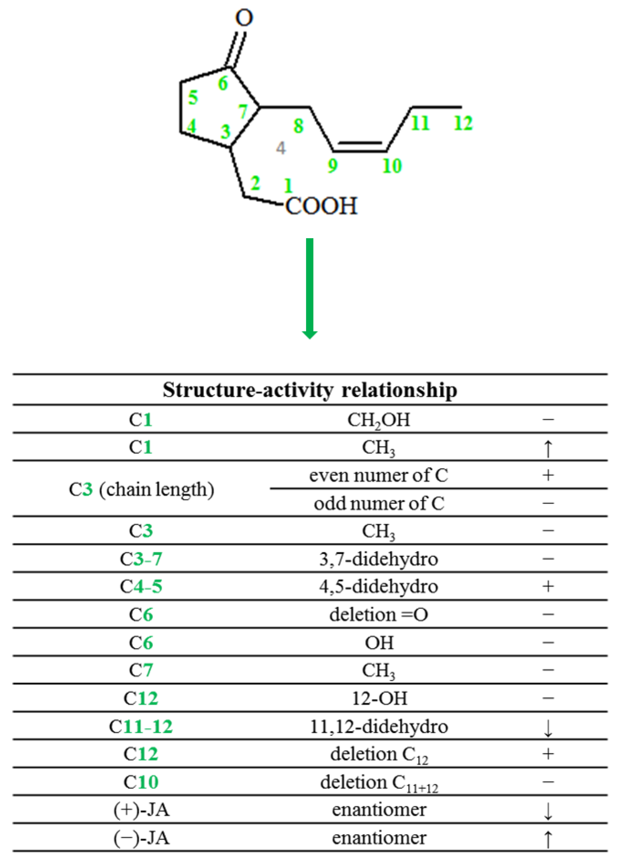
2. Chemical Structure of Jasmonate

The basic representative of the jasmonate group is jasmonic acid, i.e., 3-oxo-2-(pent-2′-enyl)cyclopentane acetic acid (IUPAC name), synthesized from linolenic acid found in the chloroplast membrane. Jasmonic acid (exhibiting optical activity) is composed of a cyclopentane ring to which three different substituents are attached, located at positions: C-3, C-6, and C-7 [11]. The following stereoisomers of jasmonic acid are known: trans-(-)-(3R,7R)-JA; (-)-JA; trans-(+)-(3S,7S)-JA; (+)-JA; cis-(-)-(3S,7R)-JA; (-)-epi-JA; cis-(+)-(3R,7S)-JA; (+)-epi-JA [12].
In plants, jasmonic acid exists in the following forms: (-)-JA and (+)-epi-JA. Due to the fact that cis stereoisomers are thermodynamically less stable, they epimerize at the C-7 atom to the stable trans form, which at the same time shows higher biological activity (Figure 2). The structure of compounds and jasmonic acid derivatives requires the presence of a cyclopentanone ring with a ketone group at C-6. The substituent that significantly affects the activity is the pentenyl side chain at the C-7 position, while hydroxylation at carbon 11 or 12 as well as reduction between carbon 11 and 12 decrease the biological activity of the compound. When a chain with an odd number of carbon atoms is present at the C-3 position, a reduction of the activity of the compound has also been observed. Additionally, the activity of the compound is maintained or increased as a result of methylation or conjugation with amino acids [9,13,14,15].
Due to their structure, jasmonates tend to inhibit, induce, and stimulate changes that take place in plants at the morphological, cellular, and molecular levels.

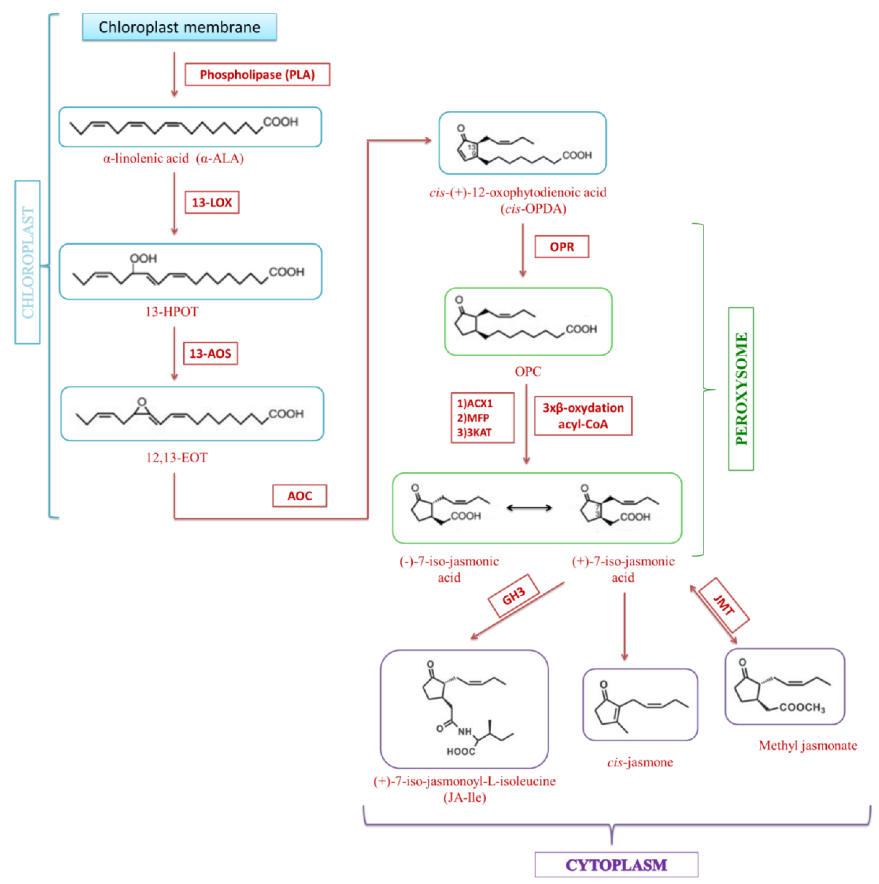
3. Jasmonate Biosynthesis

Jasmonate biosynthesis in plants is analogous to the biosynthesis of eicosanoids in animal cells, with the difference that eicosanoids are synthesized from arachidonic acid (C20:4), while jasmonates are derived from α-linolenic acid (C18:3) (α-LA) released from chloroplast membranes by lipases (DAD1, DGL, PLA2) [16].
The JA biosynthetic pathway takes place in three parts of the cell: first in chloroplasts, then partially in peroxisomes, and finally in the cytoplasm. The first stage is the oxidation of α-linolenic acid in a reaction catalyzed by lipoxygenases (13-LOX; 9-LOX), which play a key role in the regulation of the synthesis of jasmonate compounds [17]. As a result of the synthesis with the participation of 13-LOX located in the inner membrane of chloroplasts, lipid hydroperoxides of 13S-hydroperoxyoctadecatrienoic acid (13-HPOT) are formed. These compounds are then metabolized by allene oxide synthases (13-AOS), (a family of oxygen-independent CYP74A and NADPH enzymes) to unstable allene oxides (12,13-EOT) [8]. In the course of successive transformations, allene oxide cyclase (AOC) catalyzes the transformation of allen oxide into cis-(+)-12-oxophytodienoic acid, which is the end product of a part of the jasmonic acid biosynthesis pathway in chloroplasts [18]. However, the exact release mechanism of 12-oxo-OPDA is not yet known. Currently, only one protein has been identified as the expression product of the COMATOSE gene, which due to its ATP transporter’s (ABC) activity and localization in peroxisomes is responsible for the transport of JA to the subcellular compartment in question [19,20].
In the next step, reduction of the cyclopentenone ring takes place, catalyzed by peroxisomic oxophytodienoic acid reductase (OPR) [21]. The remaining steps in the synthesis of jasmonic acid are already taking place in peroxisomes. They include three successive β-oxidation reactions with the participation of the appropriate acyl-CoA ester [22]. As a result of these reactions, the side chain is shortened and transformed into the pentenyl form. In the case of some plants, such as radish or tomato, three β-oxidative enzymes are responsible for this process, i.e., acyl-CoA oxidase (ACX1), multifunctional protein (MFP), and L-3-ketoacyl-CoA thiolase (KAT) [5]. The end product of the transformation of linolenic acid is 12-carbon (+)-7-isojasmonic acid, which after transport to the cytoplasm can easily transform into (-)-7-isojasmonic acid or undergo further metabolic transformations, including formation reactions of active or inactive derivatives with sugars or amino acids (Figure 3) [17]. Jasmonic acid is then transported to the cytoplasm where it can be modified by enzymes such as JAR1, IAR3, or ILL6 [23]. The best-described protein responsible for the formation of JA adducts with amino acids is GH3 (Grechten Hagen 3), which most often binds JA to isoleucine, thus creating a biologically active conjugate [24,25].

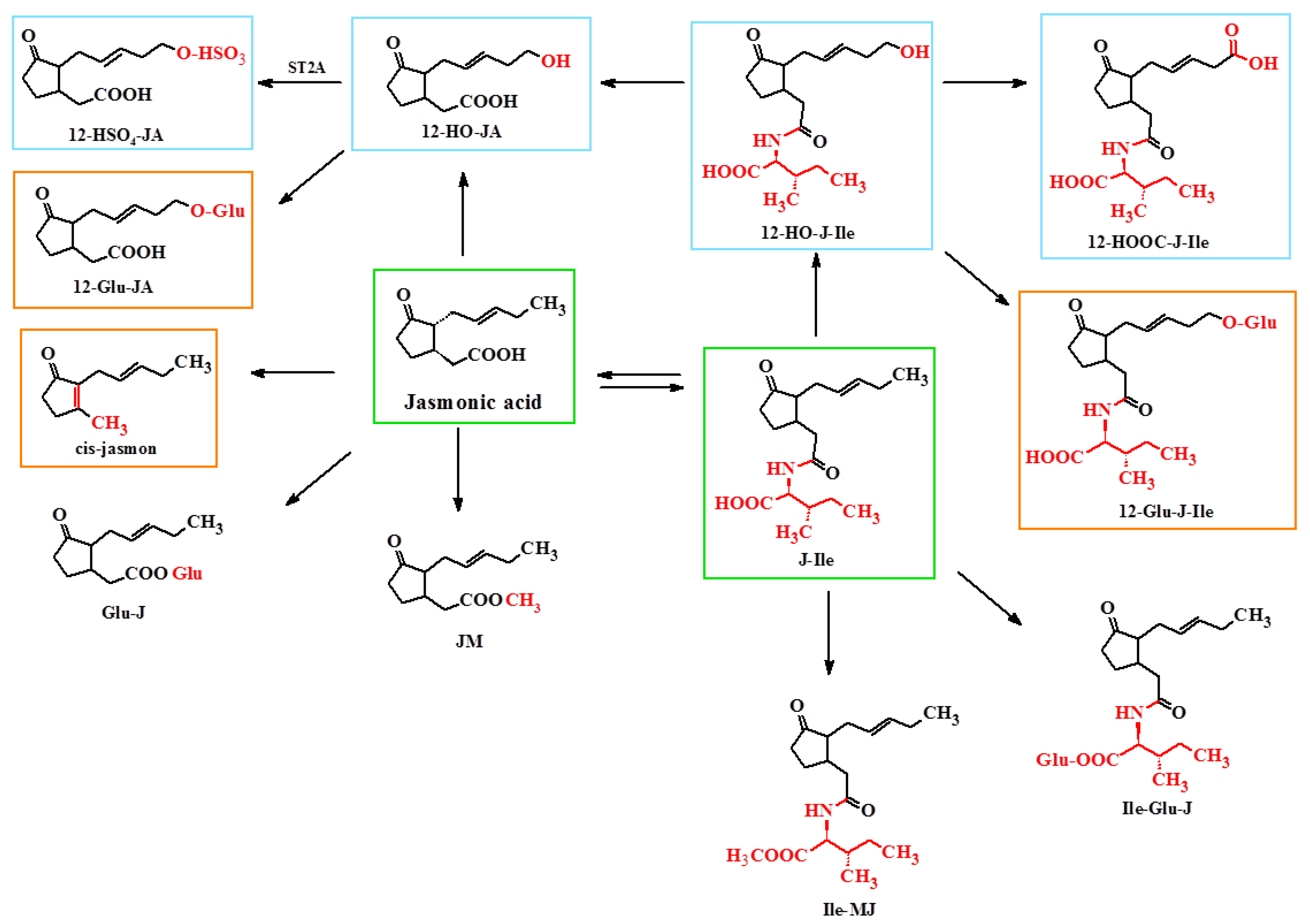
4. Metabolism of Jasmonate

Currently, twelve metabolic pathways are known that convert jasmonic acid into active, partially active, or inactive compounds (Figure 4) [17]. Most of the compounds are derivatives of jasmonic acid and isoleucine, while some reactions lead to the formation of compounds that are active in specific reactions in response to stress and stressful situations, such as, for example, a leaf movement.
Metabolites of jasmonic acid include various compounds obtained as a result of the following: conjugation with isoleucine to obtain jasmonylisoleucine (JA-Ile) [10,26,27]; methylation to methyl jasmonate (MJ) [10]; ester formation with glucose to 12-glucosyljasmonic acid (12-Glu-JA) [28]; decarboxylation to cis-jasmon [29]. As a result of hydroxylation at the C-12 position, 12-hydroxyasmonic acid is formed (12-OH-JA), present in Solanum tuberosum [30]. However, as a result of the reduction of the ketone group at the C-6 position of jasmonic acid leading to the formation of a hydroxyl group, cucurbic acid is formed. It can undergo glycosylation, methylation, and hydroxylation. It has been found in the seeds of Cucurbita pepo, in the leaves of Equisetum sp., and in the stems of Hordeum vulgare [31]. Both JA stereoisomers, apart from hydroxylation, can undergo glycosylation (conjugation mainly with glucose and gentiobiose) and combine with amino acids occurring in the S configuration [10,31]. This pertains to aromatic amino acids (conjugates with phenylalanine, tryptophan, and tyrosine have been found in flowers) and aliphatic amino acids (conjugates with isoleucine have been found in leaves and fruits; with leucine and valine found in Vicia faba leaves). Less common is the conjugation of jasmonic acid with tyramine—a biogenic amine formed by the decarboxylation of tyrosine. This conjugate has been found in the petunia pollen. Moreover, jasmonates accumulating in tuber-producing plants (mainly tuberic acid) may undergo sulfonation [32].

5. The Role of Jasmonates in Plant Growth

Jasmonic acid has been found to be an inhibitor of pollen germination in plants such as Camellia sinensis, Impatiens balsamina, and Lilium formosanum. Jasmonic acid acts quite differently on seeds that are in a state of dormancy. It stimulates the germination of embryos isolated from maple and plane seeds. Exogenous jasmonic acid inhibits seedling elongation and limits the elongation of roots and stems. The roots are more sensitive to the action of jasmonates than the hypocotyl. Methyl jasmonate is presumed to prevent fruit softening by inhibiting the activity of peroxidase and polygalacturonase, which degrade cell wall polysaccharides. Methyl jasmonate also inhibits the activity of lipoxygenase, which results in an increase in the content of linolenic acid, tocopherols, and linoleic acid [33]. This phytohormone increases the content of plant pigments from the group of carotenoids in Zea mays corn kernels [34]. Exogenous methyl jasmonate reduces the level of lycopene and increases the amount of β-carotene in tomatoes [35]. In green fruit, jasmonate inhibits the accumulation of anthocyanins and accelerates the breakdown of chlorophyll [36]. Studies on Oryza sativa rice indicate that exogenous jasmonic acid regulates leaf aging by mediating chlorophyll and membrane degradation [37]. Research also indicates a possible participation of jasmonic acid in the fallout of Glycine max pods [38]. Moreover, methyl jasmonate probably stimulates leaf fall in Vigna radiata and Kalanchoe blossfeldiana, and influences the spring leaf fall of Ficus superba [39].
It has been found that the use of exogenous jasmonic acid may affect fertility in Arabidopsis thaliana mutants that exhibit male sterility, by not producing triene fatty acids. However, the precursor of jasmonates, i.e., 12-oxo-phytodienic acid, is necessary for the synthesis of jasmonates and required for the proper stamen and pollen development [19].
Jasmonates accumulate in injured tissues or cells of plants and function as the signal activating the expression of various genes, including defensins, i.e., proteins associated with resistance to pathogens, such as osmotines and thionines, or enzymes such as peroxidases, β-1,3-glucanases, and lipoxygenase [40]. They are also indicators or stimulators of the production of many secondary metabolites, i.e., phytoalexins, flavonoids, sesquiterpenoids, glucosinolates, or lignans, affecting the response to environmental stress [41].
Jasmonates are involved in the mycorrhiza. Treatment of Avena sativum with low concentrations of jasmonic acid stimulates the development of mycorrhiza, while high concentrations of jasmonic acid may increase the resistance of tissues that are sensitive to secondary pathogen-induced infections, drought, and osmotic stress. Spraying Arabidopsis thaliana with gaseous methyl jasmonate resulted in an effective reduction of the development of diseases caused by necrotic fungi Alternalia brassicicola, Botrytis cinerea, and Plectosphaerella cucumerina. Methyl jasmonate significantly reduced the accumulated amounts of sucrose as well as reducing sugars in the form of gums, in response to injury, pathogen or insect attack (Table 1) [42,43].
The presence of heavy metals in the environment affects the plant’s response, i.e., excessive production of jasmonates, and their accumulation is associated with the toxic mechanism of action of metals: limiting the growth of leaves and roots, accelerating the aging processes, and destroying the photosynthetic apparatus [44,45].

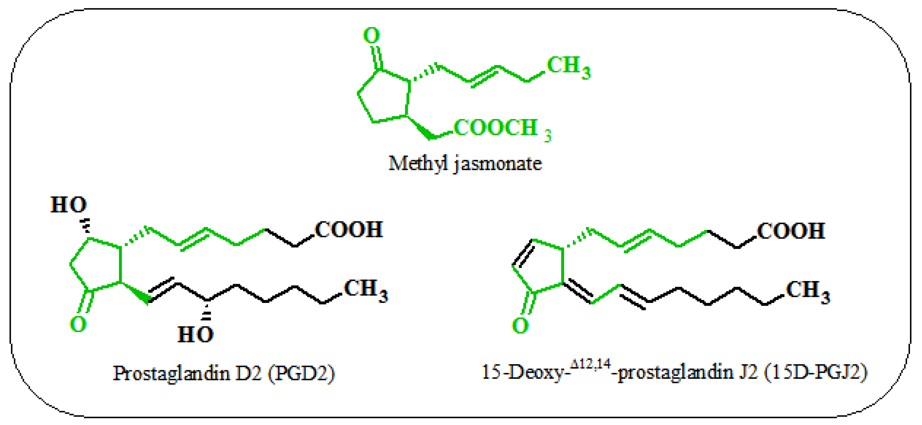
6. Similarities between the Action of Jasmonates on Plant and Animal Cells

There are certain similarities in the action of jasmonate compounds on plant and animal cells. Jasmonic acid leads to cell cycle arrest in the G2 phase of tobacco cells. This process results from the inhibition of the activity of proteins necessary for the initiation of mitosis (CDK B and cyclin B1) [28]. It also mediates in the programmed cell death in Arabidopsis protoplasts [60]. In the case of methyl jasmonate, it blocks the cell cycle in the G0-G1 phase in Taxus cuspidate cells in over 70%. [61]. On the other hand, rice cells exposed to jasmonate compounds exhibited a high activity of MAP kinases (OsMSRMK2, OsBIMK1, OsMSRMK3 oraz kinazy MAPK OsEDR1) [16], which are involved in the response to stress and pathogens. However, the action of jasmonates on neoplastic cells (MOLT-4; A549) activates some MAP kinases (c-Jun N-terminal kinase (JNK) i p38) [62].
It was found that methyl ester of jasmonic acid increases the level of ROS, thus leading to the death of some types of cancer cells [5]. Moreover, in plant cells, MeJA induces the production of hydrogen peroxide in a NAD(P)H dependent process [2]. In contrast, jasmonic acid leads to singlet oxygen-dependent death of Arabidopsis thaliana cells [63]. The increased level of ROS, induced by jasmonate compounds, is also responsible for the synthesis of heat shock proteins, both in plant and animal cells. It has been shown that methyl ester of jasmonic acid induces the synthesis of HSP72 protein in glioblastoma cells, in addition to activating the synthesis of low molecular weight HSP in Douglas fir seeds [16]. Jasmonates can induce similar metabolic, signaling, and stress-related modifications (which can lead to reduced growth and viability) in plant cells as well as in neoplastic animal cells. This suggests that as data concerning the effects of jasmonates in plant cells accumulates, it may provide predictive information that would prove necessary for the design of new anti-cancer drugs.
Jasmonates are capable of inhibiting, inducing, or stimulating changes in plants at the morphological, physiological, cellular, and molecular levels. The most important reason for studying the biological activity of jasmonic acid and its derivatives in mammals, other than hormonal activity in plants, is the structural similarity of these compounds to prostaglandins, especially those exhibiting anti-inflammatory activity, including prostaglandin D2 (PGD2) and 15-deoxy-Δ12,14- prostaglandin J2 (15d-PGJ2) (Figure 5) [64].

7. Biological Activity of Jasmonates and Their Derivatives

7.1. Anti-Inflammatory

The first studies on the potential anti-inflammatory activity of jasmonate compounds concerned MJ derived from Gracilaria verrucosa and showed that the effectiveness of MJ was comparable with or more effective than that of prostaglandin compounds [65]. The study demonstrated the inhibitory effect of MJ on the production of pro-inflammatory mediators (NO, IL-6 and TNF-α) in lipopolysaccharide activated RAW 264.7 mouse macrophages. However, the growing interest in jasmonic acid as a potential therapeutic agent led to the synthesis of new derivatives in order to obtain more active compounds. The basic structural modification changing the activity of jasmonates was the introduction of a double bond in the cyclopentyl ring. Chemically, α,β-unsaturated carbonyl compounds are electrophilic centers that are highly susceptible to addition reactions with nucleophiles such as free sulfhydryl groups of reduced glutathione or cysteine residues in proteins. Thus, prostaglandins are compounds in which cyclopentenone is the pharmacophore responsible for the biological activity of these compounds [66]. Therefore, the introduction of unsaturated bonds into the structure of methyl jasmonate with the formation of methyl 4,5-didehydrojasmonate (DHJM) resulted in the formation of compounds showing higher anti-inflammatory activity than similar prostaglandins [62]. Other modifications were the introduction of various halogens at the C3 position of α-cyclopentenone while maintaining the ring double bond and/or replacing the ester residue with an amide. It was found that the presence of a cyclopentenone fragment is necessary to maintain anti-inflammatory activity, while the presence of halogens increases this activity even with the simultaneous presence of saturated bonds in the side chain [65]. The α-haloenones formed in this manner also showed a higher anti-inflammatory activity than the natural prostaglandins. In 2012, Dang et al. synthesized a series of derivatives with various fragments of jasmonate esters, evaluated their resistance to hydrolysis and converted them into derivatives with a chlorine atom in the position of α-cyclopentenone [67]. The most active analogs in this series were t-butyl and hydroxyethyl esters, which was confirmed by the fact that the chain branching and the increased hydrophilicity in relation to the methyl moiety in MJ affect the anti-inflammatory activity (Table 2 and Figure 6).

7.2. Anticancer

Regardless of their anti-inflammatory properties, it has been shown that jasmonates have the properties characteristic for anticancer drugs. Their action is characterized by high selectivity in relation to neoplastic cells and effectiveness against neoplastic cells resistant to classic anticancer drugs. It has been found that jasmonic acid and jasmonate can increase the survival of animals and humans with lymphoma and induce apoptosis/necrosis of human cells of leukemia, lung, colon, prostate cancer, neuroblastoma, breast cancer, and melanoma, as well as chronic lymphocytic leukemia cells [71]. It is believed that there are three potential mechanisms of action of jasmonates: (i) bioenergy involving ATP depletion through mitochondrial disturbance, (ii) induction of redifferentiation by the activation of the MAPK kinase cascade, and (iii) induction of cell apoptosis by ROS generation (Table 3) [16].
Cellular ATP depletion induced by MJ is caused by a reduction of the interaction of hexokinase-2 enzyme with the VDAC (voltage-dependent anion-selective channel 1) protein, resulting in a decrease in the efficiency of glycolysis and, consequently, in the level of cellular ATP. Prolonged opening of VDAC channels changes the permeability of the mitochondrial membrane, disperses the membrane potential, increases osmotic edema, and releases pro-inflammatory factors, including cytochrome c, leading to cell death [5,16,72] As HK levels are up to 200 times higher in malignant tumor cells in comparison with normal cells [83], the enzyme is an excellent molecular target for potential anti-neoplastic agents. The specific binding of jasmonate to HK has been confirmed in an immunochemical test with the use of surface plasmon resonance and in tests of the VDAC activity of lipid bilayers without inhibiting kinase activity [73]. MJ has been shown to detach HK1 and HK2 from VDAC in a dose-dependent manner in the mitochondrial fraction of CT-26 murine colon carcinoma cells, MOLT-4 human leukemia, BCL1 murine leukemia, B16 murine melanoma, HCC hepatocellular carcinoma (LM3, BEL -7402, Hep3B, SMMC-7721), neuroblastoma (SH-SY5Y), B lymphoma, and liver cancer cells (Hep3B) [5,16,70,71,72,83]. The recently obtained synthetic derivative exhibiting hexokinase-2 inhibitory properties is 3-((3-methyl-1,2,4-oxadiazol-5-yl)methyl)-2-(pent-2-en-1-yl)cyclopentanol. The introduction of 1,2,4-oxodiazole in place of the methyl ester of jasmonate and the reduction of the ketone in cyclopentanone to the hydroxyl group resulted in an active inhibitor that showed cytotoxicity against the following human cell lines: A549 lung cancer and SKOV-3 ovarian cancer [70].
Methyl jasmonate has the ability to induce the Mitogen Activated Protein Kinases (MAPK) pathway, which determines the differentiation of neoplastic cells. Apoptotic cell death has been observed in MOLT-4 leukemia and A549 lung cancer cells treated with MJ by releasing the JNK and p38 proteins, resulting in the activation of the transcription factor AP-1 [75]. Although AP-1 is a protein involved in both apoptosis and differentiation, no effect of AP-1 on apoptosis was found in MOLT-4 and A549 cells. In both MCF-7 (receptor-dependent) and MDA-MB-435 (receptor-independent) breast cancer cell types, methyl jasmonate caused apoptosis but activated the p38 and ERK pathways via MAPK only in receptor-independent cells [76,78]. MJ activated the MAPK pathway in the HL-60 human myelocytic leukemia cell line, which instead of apoptosis caused the cells to differentiate into monocyte-like granulocytes [79]. In the same way, MJ also acts on U937 histiocytic lymphoma and THP-1 acute monocytic leukemia cells, which are the model cells for studying the behavior and differentiation of monocytes [16]. MJ has been shown to inhibit the growth of leukemic blasts and stimulate their differentiation into normal cells as evidenced by the expression of differentiation markers such as NBT reduction of nitrotetrazolium blue (for myelomonocyte differentiation), morphological differentiation into granulocytes, and the expression of CD14 cell differentiation antigens (specific for monocytes) and CD15 (granulocyte-specific) [62].
Another antitumor mechanism of action of jasmonic acid and its methyl ester is the increase in the level of ROS in the following cells: glioblastoma (C6), non-small cell lung cancer (A549 and H520), and uterine cancer (HeLa and CaSki) [76,80,81,82,84,85,86]. In human non-small lung cancer cells (A549) the above compounds increase the expression of pro-apoptotic proteins from the Bcl-2, Bcl-Bax and Xs families [87], while in prostate cancer cells PC-3 increase the expression of the anti-apoptotic protein Bcl-2 [88]. This causes the cell cycle to arrest in the G2-M phase and to activate the executor caspase, then directing cells to apoptosis (Figure 6).
In addition to the fact that jasmonates themselves induce apoptosis in tumor cells, they can also be combined with other anti-tumor agents to achieve synergistic effects. Therefore, studies have been conducted to evaluate the effects of combining MJ with various anticancer compounds [89], that are routinely used in clinical practice: BCNU (carmustine), cisplatin, paclitaxel (taxol) [90], doxorubicin (adriamycin), and 3-bromopyruvate (3-BrP) (Figure 7) [91]. Due to the fact that the target organelles for these drugs are mitochondria, although they act according to different mechanisms, the use of MJ may result in additive efficacy. The use of BCNU alone induces mitochondrial DNA damage [92], while cisplatin and taxol induce mitochondrial membrane depolarization and cytochrome c release [93]. Interactions of these compounds with MJ have been observed in many cell lines of malignant neoplasms, such as: breast, lung, prostate, and pancreatic cancer, as well as leukemia [91]. MJ drastically lowered the IC50 values of the drugs used, at the same time reducing the side effects of these drugs [5]. It has also been found that inhibition of glycolysis by 2DG in 29M4.1 B lymphoma cells enhanced the effect of MJ by drastically reducing cellular ATP levels, this effect was much more potent than the effect induced by MJ alone [70]. The cytotoxic effects of the combination of MJ (1 mM) and 2DG (5 mM) have also been confirmed in the following tumor cells: CT26, D122 and MCF-7 [94,95]. The use of MJ with cyclophosphamide as a routine treatment for breast cancer in tumor-bearing mice resulted in a reduction in tumor volume and an increase in tumor growth inhibition. Moreover, the applied therapy had no significant side effects on kidneys, liver, the immune system, and body weight [91].
Recently, it has also been shown that MJ effectively interacts with cisplatin and radiotherapy in the treatment of cervical cancer cells by significantly reducing the doses of radiation and cisplatin required to inhibit cell survival [5,96]. The study showed for the first time that alpha-radiation selectively reduces cell viability and cervical cancer cell survival.
Figure 7. Therapeutic effects of combining MJ with anticancer compounds.
Ijms 22 08437 g007

7.3. Cosmetic Activities

Methyl jasmonate is a fragrance ingredient used in many fragrance products. It is a colorless oily liquid with a strong sweet floral-herbal odor, representing the typical background notes of jasmine absolute. The smell of jasmine oil is largely due to (1R,2S)-(+)-methyl Z-epijasmonate, which is one of the main components of the oil obtained from jasmine flowers and constitutes 2–3% of the weight of its composition. It can be found in fragrances used in cosmetics, perfumes, shampoos, and soaps, as well as in non-cosmetic products such as cleaners and detergents [8,97]. Studies on albino rabbits and guinea pigs which were topically administered methyl jasmonate (2 g/kg and 10% MJ solution, respectively) showed that it did not irritate the skin. At a concentration of 10%, it also did not irritate human skin, which was confirmed by a 3-week study on 50 volunteers [98].
Regardless of the above, jasmonates have also been used in cosmetic agents responsible for soothing skin irritations, stimulating the exfoliation and renewal of the epidermis, and demonstrating the ability to regulate the activity of the sebaceous glands in the skin [97]. A new derivative of jasmonic acid, i.e., tetrahydrojasmonic acid (trade name LR2412) has been used, among others, in skin care cosmetics. The effect of LR2412, (1 and 10 µm) was investigated on an in vitro reconstructed skin model, EpiskinTM. LR2412 has an anti-aging effect because it stimulates the synthesis of the following enzymes involved in the production of acid: hyaluronan synthase 2 and hyaluronate synthase 3. As a consequence, the following were observed: an increase in hyaluronic acid deposits in the basal and suprabasal layers of the epidermis, stimulation of the deposition of laminin-5, collagen IV, and fibrillin near the dermal-epidermal junctions. It was found that a tetrahydrojasmonic acid molecule has the ability to penetrate the epidermis and the upper layers of the dermis. The research results presented by Bouloc et al. prove that tetrahydrojasmonic acid in combination with retinol yields good results in the treatment of the signs of photoaging. It was also found that the composition containing 0.2% of retinol and 2% of LR2412 is better tolerated by the skin of patients compared to the preparation containing only a 0.025% of tretinoin [99].
The N-terminal conjugate of jasmonic acid with the YPFF-NH2 peptide may also have a potential cosmetic application, but apart from the synthesis, no biological research is available yet [97]. Commercially available acetyl-YPFF tetrapeptide, when applied to the skin, weakens the stimulation of nerve endings, resulting in a reduction of skin hypersensitivity, while jasmonic acid stimulates the renewal of the epidermis. The authors suggest that this combination may be responsible for the reduction of the visible effects of aging by the peptide derivative of jasmonic acid.
Jasmonate derivatives containing heteroatoms in the cyclopentyl ring (N, O) in place of some carbon atoms have been tested for their odor properties. The unsaturated cis double bond, present on the alkyl side chain at position 2 in natural cis-jasmone, plays a very important role as far as the fragrance properties are concerned, and the introduction of the double bond into the heterocyclic jasmone analogs, both cis- and trans-, leads to an increase in odor intensity relative to their saturated heteroanalogs [100]. These derivatives have also been tested (e.g., 4-methyl-3-(2-pentenyl)-2-oxazolidinone) as potential antimicrobial compounds (Staphylococcus aureus MRSA, Enterococcus faecalis, Klebsiella pneumoniae, Escherichia coli, Salmonella enteritidis, Pseudomonas aeruginosa, Candida albicans, Aspergillus flavus, and Microsporum gypseum), but unfortunately all of them showed less activity than the parent jasmonates (Figure 6) [101].

8. In Vivo Effect of Methyl Jasmonate and Synthetic Tetrabromojasmonate

Methyl jasmonate is considered safe for humans based on the widespread consumption of plants. There are no reports on the toxicity of this substance by any route of exposure and it has been recognized by the Food and Agriculture/World Health Organization (FAO/WHO) as a food additive in the group of flavoring agents [102]. Due to the number of in vitro tests confirming the fact that MJ does not have cytotoxic effect on healthy cells, only a few in vivo tests are known (Table 4).
One of the first reports of in vivo studies concerned methyl jasmonate administered to C57BL/6J mice infected with EL-4 leukemia cells. Their survival time was significantly longer compared to mice not treated with jasmonate [103].
Subsequent studies focused on the effects of MJ and its synthetic derivative methyl 5,7,9,10-tetrabromojasmonate (TBMJ), on lung metastases in C57BL mice injected with B16-F10 melanoma cells, at doses of 40 and 75 mg/kg MJ and 20 mg/kg TBMJ of body weight respectively, which were considered non-toxic [69]. The synthetic compound proved to be significantly more potent than MJ in vivo as it generated a similar inhibitory effect on tumor growth at doses per mole that were approximately nine times lower.
Lopes et al. confirmed that MJ in concentrations of 1–10 µM inhibits angiogenesis in the chorioallantoic (CAM) membrane of the chicken embryo [104]. Methyl jasmonate was applied topically to neoplastic and pre-cancerous lesions in eight patients, in the form of an oily solution containing 1 mg of the substance per 1 mL of solution [105]. It did not cause any significant local or systemic side effects. Three patients showed positive responses. Two patients recovered completely and one relapsed three months after treatment.
The potential of MJ as a new therapeutic agent against multiple myeloma in vivo was also considered [106]. Mice were injected with MM.1S cells and treated with 1000 mg MJ/kg body weight for 8 weeks. Tumor growth was monitored by physical parameters (weight loss, apparent tumor weights, hind limb paralysis) and overall survival was taken as the study endpoint. After 150 days, 15/15 control mice treated with the carrier only died, while 8/15 MJ-treated mice survived.
To confirm the protective role of MJ in lipopolysaccharide-induced arthritis in Wistar rats, MJ was administered intraplantally at two doses (20, 40 mg/kg), with indomethacin as the standard. By behavioral evaluation of thermal hyperalgesia of the paw and tail, MJ was found to reduce hyperalgesia to hot and cold stimuli as a withdrawal latency [107].
In the mouse thymus-derived tumor called Dalton’s Lymphoma (DL) [108] investigated whether intra-tumor administration of MJ may result in an increase of tumor growth impedance. The result was a delayed tumor growth associated with prolonged survival of the tumor bearing mice. The MJ-dependent tumor growth delay was associated with a decrease in the blood supply to the tumor environment, cell cycle arrest, increased induction of apoptosis and necrosis, deregulation of glucose and lipid metabolism, increased fragility of cancer cell membranes, and altered cytokine composition in the tumor.

9. Conclusions

Due to the synergistic activity of substances isolated from plants, herbal medicines have a fairly wide range of action, but the isolated individual substances may have different effects. Jasmonic acid, as well as its natural and synthetic derivatives, is one of the few examples of a single compound of natural origin that shows a wide range of activity. The presented paper aims to identify jasmonates as potential therapeutics.
The interest in jasmonates and their derivatives stems from their structural similarity to prostaglandins, and this feature shows potential for the use of this compound as a therapeutic agent. Methyl jasmonate and other new derivatives show a higher anti-inflammatory activity than natural prostaglandins. Jasmonates exhibit the perfect characteristics of anti-cancer agents due to the fact that they act on cancer cells but not on healthy cells. At the same time, they perfectly interact with known chemotherapeutic agents, enhancing their effect. Jasmonates and their derivatives inhibit in vitro proliferation of neoplastic cells such as: myeloid leukemia, breast, prostate, melanoma, stomach, cervix, colon, large intestine, hepatoma, lung, neuroblastoma, sarcoma, and lymphoblastic leukemia. The mechanisms explaining the anti-cancer activity of jasmon compounds proposed are based on: changes in the ATP levels in the cell; induction of redifferentiation through MAPK activity; and ROS-induced apoptosis.
Unfortunately, so far there have been no documented clinical trials focused on jasmonates as therapeutics, apart from patents that report their potential use. However, it must be remembered that some diseases result from the impairment of those mechanisms that are responsible for the beneficial effects of jasmonates on cancer cells. ROS production, widely reported in the literature, may be one such example. The mechanism involving the stimulation of ROS production by jasmonates, resulting in apoptosis of neoplastic cells, may be the cause of the intensification of oxidative stress affecting the development of other diseases.
In the authors’ opinion, it is too early to discuss the effects of jasmonates on the whole body, due to the fact that the therapeutic doses of jasmonates–and thus their side effects–are not yet known. Since jasmonate works according to various mechanisms, it is uncertain whether these will cause treatment-excluding effects in various, often comorbid, diseases.

Author Contributions

I.J.-K. and A.M. writing—review and editing; I.J.-K. supervision. All authors have read and agreed to the published version of the manuscript.

Funding

This research received no external funding.

Conflicts of Interest

The authors declare no conflict of interest.

References

  1. Saniewski, M. The Role of Jasmonates in Ethylene Biosynthesis. In Biology and Biotechnology of the Plant Hormone Ethylene; Kanellis, A.K., Chang, C., Kende, H., Grierson, D., Eds.; NATO ASI Series; Springer: Dordrecht, The Netherlands, 1997; pp. 39–45. ISBN 978-94-011-5546-5. [Google Scholar]
  2. Ho, T.-T.; Murthy, H.N.; Park, S.-Y. Methyl Jasmonate Induced Oxidative Stress and Accumulation of Secondary Metabolites in Plant Cell and Organ Cultures. Int. J. Mol. Sci. 2020, 21, 716. [Google Scholar] [CrossRef] [Green Version]
  3. Félix, C.; Salvatore, M.M.; DellaGreca, M.; Meneses, R.; Duarte, A.S.; Salvatore, F.; Naviglio, D.; Gallo, M.; Jorrín-Novo, J.V.; Alves, A.; et al. Production of Toxic Metabolites by Two Strains of Lasiodiplodia Theobromae, Isolated from a Coconut Tree and a Human Patient. Mycologia 2018, 110, 642–653. [Google Scholar] [CrossRef]
  4. Demole, E.; Lederer, E.; Mercier, D. Isolement et Détermination de La Structure Du Jasmonate de Méthyle, Constituant Odorant Caractéristique de l’essence de Jasmin. Helv. Chim. Acta 1962, 45, 675–685. [Google Scholar] [CrossRef]
  5. Cesari, I.M.; Carvalho, E.; Figueiredo Rodrigues, M.; Mendonça, B.D.S.; Amôedo, N.D.; Rumjanek, F.D. Methyl Jasmonate: Putative Mechanisms of Action on Cancer Cells Cycle, Metabolism, and Apoptosis. Int. J. Cell Biol. 2014, 2014, 572097. [Google Scholar] [CrossRef] [PubMed]
  6. Wilmowicz, E.; Frankowski, K.; Sidłowska, M.; Kućko, A.; Kesy, J.; Gasiorowski, A.; Glazińska, P.; Kopcewicz, J. Jasmonate biosynthesis--the latest discoveries. Postepy Biochem. 2012, 58, 26–33. [Google Scholar] [PubMed]
  7. Wasternack, C.; Strnad, M. Jasmonates: News on Occurrence, Biosynthesis, Metabolism and Action of an Ancient Group of Signaling Compounds. Int. J. Mol. Sci. 2018, 19, 2539. [Google Scholar] [CrossRef] [Green Version]
  8. Ghasemi Pirbalouti, A.; Sajjadi, S.E.; Parang, K. A Review (Research and Patents) on Jasmonic Acid and Its Derivatives. Arch. Pharm. 2014, 347, 229–239. [Google Scholar] [CrossRef]
  9. Wasternack, C. Jasmonates: An Update on Biosynthesis, Signal Transduction and Action in Plant Stress Response, Growth and Development. Ann. Bot. 2007, 100, 681–697. [Google Scholar] [CrossRef] [Green Version]
  10. Wang, J.; Song, L.; Gong, X.; Xu, J.; Li, M. Functions of Jasmonic Acid in Plant Regulation and Response to Abiotic Stress. Int. J. Mol. Sci. 2020, 21, 1446. [Google Scholar] [CrossRef] [Green Version]
  11. Wasternack, C.; Kombrink, E. Jasmonates: Structural Requirements for Lipid-Derived Signals Active in Plant Stress Responses and Development. ACS Chem. Biol. 2010, 5, 63–77. [Google Scholar] [CrossRef]
  12. Han, Y.; Bai, Y.; Xiao, Y.; Du, F.; Liang, Y.; Tan, Z.; Zhao, M.; Liu, H. Simultaneous Discrimination of Jasmonic Acid Stereoisomers by CE-QTOF-MS Employing the Partial Filling Technique. Electrophoresis 2011, 32, 2693–2699. [Google Scholar] [CrossRef] [PubMed]
  13. Holland, C.; Jez, J. Structural Biology of Jasmonic Acid Metabolism and Responses in Plants. In Plant Structural Biology: Hormonal Regulations; Springer: Berlin/Heidelberg, Germany, 2018; pp. 67–82. ISBN 978-3-319-91351-3. [Google Scholar]
  14. Koda, Y.; Kikuta, Y.; Tazaki, H.; Tsujino, Y.; Sakamura, S.; Yoshihara, T. Potato Tuber-Inducing Activities of Jasmonic Acid and Related Compounds. Phytochemistry 1991, 30, 1435–1438. [Google Scholar] [CrossRef]
  15. Krumm, T.; Bandemer, K.; Boland, W. Induction of Volatile Biosynthesis in the Lima Bean (Phaseolus Lunatus) by Leucine- and Isoleucine Conjugates of 1-Oxo- and 1-Hydroxyindan-4-Carboxylic Acid: Evidence for Amino Acid Conjugates of Jasmonic Acid as Intermediates in the Octadecanoid Signalling Pathway. FEBS Lett. 1995, 377, 523–529. [Google Scholar] [CrossRef] [PubMed] [Green Version]
  16. Flescher, E. Jasmonates in Cancer Therapy. Cancer Lett. 2007, 245, 1–10. [Google Scholar] [CrossRef] [PubMed]
  17. Wasternack, C.; Song, S. Jasmonates: Biosynthesis, Metabolism, and Signaling by Proteins Activating and Repressing Transcription. J. Exp. Bot. 2017, 68, 1303–1321. [Google Scholar] [CrossRef]
  18. León, J.; Sánchez-Serrano, J.J. Molecular Biology of Jasmonic Acid Biosynthesis in Plants. Plant Physiol. Biochem. 1999, 37, 373–380. [Google Scholar] [CrossRef]
  19. Ghorbel, M.; Brini, F.; Sharma, A.; Landi, M. Role of Jasmonic Acid in Plants: The Molecular Point of View. Plant Cell Rep. 2021, 5, 1–24. [Google Scholar] [CrossRef]
  20. Larrieu, A.; Vernoux, T. Q&A: How Does Jasmonate Signaling Enable Plants to Adapt and Survive? BMC Biol. 2016, 14, 79. [Google Scholar] [CrossRef]
  21. Cheong, J.-J.; Choi, Y.D. Methyl Jasmonate as a Vital Substance in Plants. Trends Genet. TIG 2003, 19, 409–413. [Google Scholar] [CrossRef]
  22. Enomoto, H.; Miyamoto, K. Unique Localization of Jasmonic Acid-Related Compounds in Developing Phaseolus Vulgaris, L. (Common Bean) Seeds Revealed through Desorption Electrospray Ionization-Mass Spectrometry Imaging. Phytochemistry 2021, 188, 112812. [Google Scholar] [CrossRef]
  23. Wasternack, C.; Strnad, M. Jasmonate Signaling in Plant Stress Responses and Development–Active and Inactive Compounds. New Biotechnol. 2016, 33, 604–613. [Google Scholar] [CrossRef]
  24. Wasternack, C. How Jasmonates Earned Their Laurels: Past and Present. J. Plant Growth Regul. 2015, 34, 761–794. [Google Scholar] [CrossRef]
  25. Chen, R.; Jiang, H.; Li, L.; Zhai, Q.; Qi, L.; Zhou, W.; Liu, X.; Li, H.; Zheng, W.; Sun, J.; et al. The Arabidopsis Mediator Subunit MED25 Differentially Regulates Jasmonate and Abscisic Acid Signaling through Interacting with the MYC2 and ABI5 Transcription Factors. Plant Cell 2012, 24, 2898–2916. [Google Scholar] [CrossRef] [Green Version]
  26. Staswick, P.E.; Tiryaki, I. The Oxylipin Signal Jasmonic Acid Is Activated by an Enzyme That Conjugates It to Isoleucine in Arabidopsis. Plant Cell 2004, 16, 2117–2127. [Google Scholar] [CrossRef] [Green Version]
  27. Suza, W.P.; Staswick, P.E. The Role of JAR1 in Jasmonoyl-L-Isoleucine Production during Arabidopsis Wound Response. Planta 2008, 227, 1221–1232. [Google Scholar] [CrossRef] [PubMed]
  28. Swiatek, A.; Azmi, A.; Stals, H.; Inzé, D.; Van Onckelen, H. Jasmonic Acid Prevents the Accumulation of Cyclin B1;1 and CDK-B in Synchronized Tobacco BY-2 Cells. FEBS Lett. 2004, 572, 118–122. [Google Scholar] [CrossRef] [PubMed] [Green Version]
  29. Koch, T.; Bandemer, K.; Boland, W. Biosynthesis of Cis-Jasmone: A Pathway for the Inactivation and the Disposal of the Plant Stress Hormone Jasmonic Acid to the Gas Phase? Helv. Chim. Acta 1997, 80, 838–850. [Google Scholar] [CrossRef]
  30. Gidda, S.K.; Miersch, O.; Levitin, A.; Schmidt, J.; Wasternack, C.; Varin, L. Biochemical and Molecular Characterization of a Hydroxyjasmonate Sulfotransferase from Arabidopsis Thaliana. J. Biol. Chem. 2003, 278, 17895–17900. [Google Scholar] [CrossRef] [PubMed] [Green Version]
  31. Schaller, F.; Schaller, A.; Stintzi, A. Biosynthesis and Metabolism of Jasmonates. J. Plant Growth Regul. 2004, 23, 179–199. [Google Scholar] [CrossRef]
  32. Piotrowska, A.; Bajguz, A. Conjugates of Abscisic Acid, Brassinosteroids, Ethylene, Gibberellins, and Jasmonates. Phytochemistry 2011, 72, 2097–2112. [Google Scholar] [CrossRef]
  33. Antognoni, F.; Faudale, M.; Poli, F.; Biondi, S. Methyl Jasmonate Differentially Affects Tocopherol Content and Tyrosine Amino Transferase Activity in Cultured Cells of Amaranthus Caudatus and Chenopodium Quinoa. Plant Biol. 2009, 11, 161–169. [Google Scholar] [CrossRef]
  34. Luo, H.; He, W.; Li, D.; Bao, Y.; Riaz, A.; Xiao, Y.; Song, J.; Liu, C. Effect of Methyl Jasmonate on Carotenoids Biosynthesis in Germinated Maize Kernels. Food Chem. 2020, 307, 125525. [Google Scholar] [CrossRef] [PubMed]
  35. Saniewski, M.; Czapski, J. The Effect of Methyl Jasmonate on Lycopene and β-Carotene Accumulation in Ripening Red Tomatoes. Experientia 1983, 39, 1373–1374. [Google Scholar] [CrossRef]
  36. Horbowicz, M.; Kosson, R.; Wiczkowski, W.; Koczkodaj, D.; Mitrus, J. The Effect of Methyl Jasmonate on Accumulation of 2-Phenylethylamine and Putrescine in Seedlings of Common Buckwheat (Fagopyrum Esculentum). Acta Physiol. Plant. 2011, 33, 897–903. [Google Scholar] [CrossRef]
  37. Liu, L.; Li, H.; Zeng, H.; Cai, Q.; Zhou, X.; Yin, C. Exogenous Jasmonic Acid and Cytokinin Antagonistically Regulate Rice Flag Leaf Senescence by Mediating Chlorophyll Degradation, Membrane Deterioration, and Senescence-Associated Genes Expression. J. Plant Growth Regul. 2016, 35, 366–376. [Google Scholar] [CrossRef]
  38. Staswick, P.E.; Huang, J.-F.; Rhee, Y. Nitrogen and Methyl Jasmonate Induction of Soybean Vegetative Storage Protein Genes 1. Plant Physiol. 1991, 96, 130–136. [Google Scholar] [CrossRef] [Green Version]
  39. Rohwer, C.L.; Erwin, J.E. Horticultural Applications of Jasmonates: A Review. J. Hortic. Sci. Biotechnol. 2008, 83, 283–304. [Google Scholar] [CrossRef]
  40. Farhangi-Abriz, S.; Ghassemi-Golezani, K. Jasmonates: Mechanisms and Functions in Abiotic Stress Tolerance of Plants. Biocatal. Agric. Biotechnol. 2019, 20, 101210. [Google Scholar] [CrossRef]
  41. Maslenkova, L.T.; Miteva, T.S.; Popova, L.P. Changes in the Polypeptide Patterns of Barley Seedlings Exposed to Jasmonic Acid and Salinity. Plant Physiol. 1992, 98, 700–707. [Google Scholar] [CrossRef] [PubMed] [Green Version]
  42. León-Morcillo, R.J.; Ángel, J.; Martín, R.; Vierheilig, H.; Ocampo, J.A.; García-Garrido, J.M. Late Activation of the 9-Oxylipin Pathway during Arbuscular Mycorrhiza Formation in Tomato and Its Regulation by Jasmonate Signalling. J. Exp. Bot. 2012, 63, 3545–3558. [Google Scholar] [CrossRef]
  43. Hause, B.; Mrosk, C.; Isayenkov, S.; Strack, D. Jasmonates in Arbuscular Mycorrhizal Interactions. Phytochemistry 2007, 68, 101–110. [Google Scholar] [CrossRef] [PubMed]
  44. Chen, J.; Yan, Z.; Li, X. Effect of Methyl Jasmonate on Cadmium Uptake and Antioxidative Capacity in Kandelia Obovata Seedlings under Cadmium Stress. Ecotoxicol. Environ. Saf. 2014, 104, 349–356. [Google Scholar] [CrossRef] [PubMed]
  45. Maurer, F.; Müller, S.; Bauer, P. Suppression of Fe Deficiency Gene Expression by Jasmonate. Plant Physiol. Biochem. 2011, 49, 530–536. [Google Scholar] [CrossRef]
  46. Çetinbaş-Genç, A.; Vardar, F. Effect of Methyl Jasmonate on In-Vitro Pollen Germination and Tube Elongation of Pinus Nigra. Protoplasma 2020, 257, 1655–1665. [Google Scholar] [CrossRef] [PubMed]
  47. Muradoğlu, F.; Yıldız, K.; Balta, F. Methyl Jasmonate Influences of Pollen Germination and Pollen Tube Growth of Apricot (Prunus Armeniaca L.). YYU J. Agric. Sci. 2010, 20, 183–188. [Google Scholar]
  48. Schweizer, P.; Gees, R.; Mosinger, E. Effect of Jasmonic Acid on the Interaction of Barley (Hordeum Vulgare L.) with the Powdery Mildew Erysiphe Graminis f.Sp. Hordei. Plant Physiol. 1993, 102, 503–511. [Google Scholar] [CrossRef] [PubMed] [Green Version]
  49. Dziurka, K.; Dziurka, M.; Warchoł, M.; Czyczyło-Mysza, I.; Marcińska, I.; Noga, A.; Kapłoniak, K.; Skrzypek, E. Endogenous Phytohormone Profile during Oat (Avena Sativa L.) Haploid Embryo Development. Vitro Cell. Dev. Biol. Plant 2019, 55, 221–229. [Google Scholar] [CrossRef]
  50. Pigolev, A.; Miroshnichenko, D.; Dolgov, S.; Savchenko, T. Regulation of Sixth Seminal Root Formation by Jasmonate in Triticum Aestivum L. Plants Basel Switz. 2021, 10, 219. [Google Scholar] [CrossRef]
  51. Corbineau, F.; Rudnicki, R.M.; Côme, D. The Effects of Methyl Jasmonate on Sunflower (Helianthus Annuus L.) Seed Germination and Seedling Development. Plant Growth Regul. 1988, 7, 157–169. [Google Scholar] [CrossRef]
  52. Andrade, A.; Escalante, M.; Vigliocco, A.; del Carmen Tordable, M.; Alemano, S. Involvement of Jasmonates in Responses of Sunflower (Helianthus Annuus) Seedlings to Moderate Water Stress. Plant Growth Regul. 2017, 83, 501–511. [Google Scholar] [CrossRef]
  53. Concha, C.M.; Figueroa, N.E.; Poblete, L.A.; Oñate, F.A.; Schwab, W.; Figueroa, C.R. Methyl Jasmonate Treatment Induces Changes in Fruit Ripening by Modifying the Expression of Several Ripening Genes in Fragaria Chiloensis Fruit. Plant Physiol. Biochem. 2013, 70, 433–444. [Google Scholar] [CrossRef]
  54. Fang, C.; Zhang, H.; Wan, J.; Wu, Y.; Li, K.; Jin, C.; Chen, W.; Wang, S.; Wang, W.; Zhang, H.; et al. Control of Leaf Senescence by an MeOH-Jasmonates Cascade That Is Epigenetically Regulated by OsSRT1 in Rice. Mol. Plant 2016, 9, 1366–1378. [Google Scholar] [CrossRef] [Green Version]
  55. ISHIHARA, A.; OGURA, Y.; TEBAYASHI, S.; IWAMURA, H. Jasmonate-Induced Changes in Flavonoid Metabolism in Barley (Hordeum Vulgare) Leaves. Biosci. Biotechnol. Biochem. 2002, 66, 2176–2182. [Google Scholar] [CrossRef] [Green Version]
  56. Shan, X.; Wang, J.; Chua, L.; Jiang, D.; Peng, W.; Xie, D. The Role of Arabidopsis Rubisco Activase in Jasmonate-Induced Leaf Senescence. Plant Physiol. 2011, 155, 751–764. [Google Scholar] [CrossRef] [PubMed] [Green Version]
  57. Zhao, D.; Ma, H. Male Fertility: A Case of Enzyme Identity. Curr. Biol. 2000, 10, R904–R907. [Google Scholar] [CrossRef] [Green Version]
  58. Ruduś, I.; Weiler, E.W.; Kępczyńska, E. Do Stress-Related Phytohormones, Abscisic Acid and Jasmonic Acid Play a Role in the Regulation of Medicago Sativa L. Somatic Embryogenesis? Plant Growth Regul. 2009, 59, 159–169. [Google Scholar] [CrossRef]
  59. Raya-González, J.; Pelagio-Flores, R.; López-Bucio, J. The Jasmonate Receptor COI1 Plays a Role in Jasmonate-Induced Lateral Root Formation and Lateral Root Positioning in Arabidopsis Thaliana. J. Plant Physiol. 2012, 169, 1348–1358. [Google Scholar] [CrossRef]
  60. Asai, T.; Stone, J.M.; Heard, J.E.; Kovtun, Y.; Yorgey, P.; Sheen, J.; Ausubel, F.M. Fumonisin B1-Induced Cell Death in Arabidopsis Protoplasts Requires Jasmonate-, Ethylene-, and Salicylate-Dependent Signaling Pathways. Plant Cell 2000, 12, 1823–1836. [Google Scholar] [CrossRef] [PubMed] [Green Version]
  61. Naill, M.C.; Roberts, S.C. Cell Cycle Analysis of Taxus Suspension Cultures at the Single Cell Level as an Indicator of Culture Heterogeneity. Biotechnol. Bioeng. 2005, 90, 491–500. [Google Scholar] [CrossRef] [PubMed]
  62. Ishii, Y.; Kiyota, H.; Sakai, S.; Honma, Y. Induction of Differentiation of Human Myeloid Leukemia Cells by Jasmonates, Plant Hormones. Leukemia 2004, 18, 1413–1419. [Google Scholar] [CrossRef] [PubMed] [Green Version]
  63. Danon, A.; Miersch, O.; Felix, G.; Camp, R.G.L.; Apel, K. Concurrent Activation of Cell Death-Regulating Signaling Pathways by Singlet Oxygen in Arabidopsis Thaliana. Plant J. Cell Mol. Biol. 2005, 41, 68–80. [Google Scholar] [CrossRef] [PubMed]
  64. Conti, M. Cyclopentenone: A Special Moiety for Anticancer Drug Design. Anticancer Drugs 2006, 17, 1017–1022. [Google Scholar] [CrossRef] [PubMed]
  65. Dang, H.T.; Lee, H.J.; Yoo, E.S.; Hong, J.; Bao, B.; Choi, J.S.; Jung, J.H. New Jasmonate Analogues as Potential Anti-Inflammatory Agents. Bioorg. Med. Chem. 2008, 16, 10228–10235. [Google Scholar] [CrossRef] [PubMed]
  66. Jarocka-Karpowicz, I.; Markowska, A. Jasmonate Compounds and Their Derivatives in the Regulation of the Neoplastic Processes. Molecules 2021, 26, 2901. [Google Scholar] [CrossRef]
  67. Dang, H.T.; Lee, Y.M.; Kang, G.J.; Yoo, E.S.; Hong, J.; Lee, S.M.; Lee, S.K.; Pyee, Y.; Chung, H.-J.; Moon, H.R.; et al. In Vitro Stability and in Vivo Anti-Inflammatory Efficacy of Synthetic Jasmonates. Bioorg. Med. Chem. 2012, 20, 4109–4116. [Google Scholar] [CrossRef] [PubMed]
  68. Lee, H.-J.; Maeng, K.; Dang, H.-T.; Kang, G.-J.; Ryou, C.; Jung, J.H.; Kang, H.-K.; Prchal, J.T.; Yoo, E.-S.; Yoon, D. Anti-Inflammatory Effect of Methyl Dehydrojasmonate (J2) Is Mediated by the NF-ΚB Pathway. J. Mol. Med. Berl. Ger. 2011, 89, 83–90. [Google Scholar] [CrossRef]
  69. Reischer, D.; Heyfets, A.; Shimony, S.; Nordenberg, J.; Kashman, Y.; Flescher, E. Effects of Natural and Novel Synthetic Jasmonates in Experimental Metastatic Melanoma. Br. J. Pharmacol. 2007, 150, 738–749. [Google Scholar] [CrossRef] [Green Version]
  70. Sucu, B.O.; Ipek, O.S.; Kurtulus, S.O.; Yazici, B.E.; Karakas, N.; Guzel, M. Synthesis of Novel Methyl Jasmonate Derivatives and Evaluation of Their Biological Activity in Various Cancer Cell Lines. Bioorganic Chem. 2019, 91, 103146. [Google Scholar] [CrossRef]
  71. Raviv, Z.; Cohen, S.; Reischer-Pelech, D. The Anti-Cancer Activities of Jasmonates. Cancer Chemother. Pharmacol. 2013, 71, 275–285. [Google Scholar] [CrossRef]
  72. Fingrut, O.; Reischer, D.; Rotem, R.; Goldin, N.; Altboum, I.; Zan-Bar, I.; Flescher, E. Jasmonates Induce Nonapoptotic Death in High-Resistance Mutant P53-Expressing B-Lymphoma Cells. Br. J. Pharmacol. 2005, 146, 800–808. [Google Scholar] [CrossRef] [PubMed]
  73. Goldin, N.; Arzoine, L.; Heyfets, A.; Israelson, A.; Zaslavsky, Z.; Bravman, T.; Bronner, V.; Notcovich, A.; Shoshan-Barmatz, V.; Flescher, E. Methyl Jasmonate Binds to and Detaches Mitochondria-Bound Hexokinase. Oncogene 2008, 27, 4636–4643. [Google Scholar] [CrossRef] [Green Version]
  74. Tong, Q.-S.; Jiang, G.-S.; Zheng, L.-D.; Tang, S.-T.; Cai, J.-B.; Liu, Y.; Zeng, F.-Q.; Dong, J.-H. Methyl Jasmonate Downregulates Expression of Proliferating Cell Nuclear Antigen and Induces Apoptosis in Human Neuroblastoma Cell Lines. Anticancer. Drugs 2008, 19, 573–581. [Google Scholar] [CrossRef]
  75. Rotem, R.; Heyfets, A.; Fingrut, O.; Blickstein, D.; Shaklai, M.; Flescher, E. Jasmonates: Novel Anticancer Agents Acting Directly and Selectively on Human Cancer Cell Mitochondria. Cancer Res. 2005, 65, 1984–1993. [Google Scholar] [CrossRef] [Green Version]
  76. Kim, J.H.; Lee, S.Y.; Oh, S.Y.; Han, S.I.; Park, H.G.; Yoo, M.-A.; Kang, H.S. Methyl Jasmonate Induces Apoptosis through Induction of Bax/Bcl-XS and Activation of Caspase-3 via ROS Production in A549 Cells. Oncol. Rep. 2004, 12, 1233–1238. [Google Scholar] [CrossRef]
  77. Yeruva, L.; Elegbede, J.A.; Carper, S.W. Methyl Jasmonate Decreases Membrane Fluidity and Induces Apoptosis through Tumor Necrosis Factor Receptor 1 in Breast Cancer Cells. Anticancer. Drugs 2008, 19, 766–776. [Google Scholar] [CrossRef] [Green Version]
  78. Yeruva, L.; Hall, C.; Elegbede, J.A.; Carper, S.W. Perillyl Alcohol and Methyl Jasmonate Sensitize Cancer Cells to Cisplatin. Anticancer. Drugs 2010, 21, 1–9. [Google Scholar] [CrossRef]
  79. Cohen, S.; Flescher, E. Methyl Jasmonate: A Plant Stress Hormone as an Anti-Cancer Drug. Phytochemistry 2009, 70, 1600–1609. [Google Scholar] [CrossRef]
  80. Oh, S.Y.; Kim, J.H.; Park, M.J.; Kim, S.M.; Yoon, C.S.; Joo, Y.M.; Park, J.S.; Han, S.I.; Park, H.G.; Kang, H.S. Induction of Heat Shock Protein 72 in C6 Glioma Cells by Methyl Jasmonate through ROS-Dependent Heat Shock Factor 1 Activation. Int. J. Mol. Med. 2005, 16, 833–839. [Google Scholar] [CrossRef] [PubMed]
  81. Kniazhanski, T.; Jackman, A.; Heyfets, A.; Gonen, P.; Flescher, E.; Sherman, L. Methyl Jasmonate Induces Cell Death with Mixed Characteristics of Apoptosis and Necrosis in Cervical Cancer Cells. Cancer Lett. 2008, 271, 34–46. [Google Scholar] [CrossRef] [PubMed]
  82. Jiang, G.; Zhao, J.; Xiao, X.; Tao, D.; Gu, C.; Tong, Q.; Luo, B.; Wang, L.; Zeng, F. AN N-Terminal Smac Peptide Sensitizes Human Prostate Carcinoma Cells to Methyl Jasmonate-Induced Apoptosis. Cancer Lett. 2011, 302, 37–46. [Google Scholar] [CrossRef] [PubMed]
  83. Bustamante, E.; Pedersen, P.L. High Aerobic Glycolysis of Rat Hepatoma Cells in Culture: Role of Mitochondrial Hexokinase. Proc. Natl. Acad. Sci. USA 1977, 74, 3735–3739. [Google Scholar] [CrossRef] [PubMed] [Green Version]
  84. Li, J.; Chen, K.; Wang, F.; Dai, W.; Li, S.; Feng, J.; Wu, L.; Liu, T.; Xu, S.; Xia, Y.; et al. Methyl Jasmonate Leads to Necrosis and Apoptosis in Hepatocellular Carcinoma Cells via Inhibition of Glycolysis and Represses Tumor Growth Mice. Oncotarget 2017, 8, 45965–45980. [Google Scholar] [CrossRef] [PubMed]
  85. Francisco-Marquez, M.; Galano, A. The Reactions of Plant Hormones with Reactive Oxygen Species: Chemical Insights at a Molecular Level. J. Mol. Model. 2018, 24, 255. [Google Scholar] [CrossRef]
  86. Besson, J.C.F.; de Carvalho Picoli, C.; Matioli, G.; Natali, M.R.M. Methyl Jasmonate: A Phytohormone with Potential for the Treatment of Inflammatory Bowel Diseases. J. Pharm. Pharmacol. 2018, 70, 178–190. [Google Scholar] [CrossRef] [PubMed] [Green Version]
  87. Choudhari, A.S.; Mandave, P.C.; Deshpande, M.; Ranjekar, P.; Prakash, O. Phytochemicals in Cancer Treatment: From Preclinical Studies to Clinical Practice. Front. Pharmacol. 2019, 10, 1614. [Google Scholar] [CrossRef] [PubMed] [Green Version]
  88. Ezekwudo, D.; Shashidharamurthy, R.; Devineni, D.; Bozeman, E.; Palaniappan, R.; Selvaraj, P. Inhibition of Expression of Anti-Apoptotic Protein Bcl-2 and Induction of Cell Death in Radioresistant Human Prostate Adenocarcinoma Cell Line (PC-3) by Methyl Jasmonate. Cancer Lett. 2008, 270, 277–285. [Google Scholar] [CrossRef]
  89. Zhang, M.; Su, L.; Xiao, Z.; Liu, X.; Liu, X. Methyl Jasmonate Induces Apoptosis and Pro-Apoptotic Autophagy via the ROS Pathway in Human Non-Small Cell Lung Cancer. Am. J. Cancer Res. 2016, 6, 187–199. [Google Scholar]
  90. Škubník, J.; Pavlíčková, V.; Ruml, T.; Rimpelová, S. Current Perspectives on Taxanes: Focus on Their Bioactivity, Delivery and Combination Therapy. Plants Basel Switz. 2021, 10, 569. [Google Scholar] [CrossRef]
  91. Yousefi, S.; Darvishi, P.; Yousefi, Z.; Pourfathollah, A.A. Effect of Methyl Jasmonate and 3-Bromopyruvate Combination Therapy on Mice Bearing the 4 T1 Breast Cancer Cell Line. J. Bioenerg. Biomembr. 2020, 52, 103–111. [Google Scholar] [CrossRef]
  92. Cai, S.; Xu, Y.; Cooper, R.J.; Ferkowicz, M.J.; Hartwell, J.R.; Pollok, K.E.; Kelley, M.R. Mitochondrial Targeting of Human O6-Methylguanine DNA Methyltransferase Protects against Cell Killing by Chemotherapeutic Alkylating Agents. Cancer Res. 2005, 65, 3319–3327. [Google Scholar] [CrossRef] [Green Version]
  93. Wang, X.; Tournier, C. Regulation of Cellular Functions by the ERK5 Signalling Pathway. Cell. Signal. 2006, 18, 753–760. [Google Scholar] [CrossRef]
  94. Heyfets, A.; Flescher, E. Cooperative Cytotoxicity of Methyl Jasmonate with Anti-Cancer Drugs and 2-Deoxy-D-Glucose. Cancer Lett. 2007, 250, 300–310. [Google Scholar] [CrossRef]
  95. Elia, U.; Flescher, E. PI3K/Akt Pathway Activation Attenuates the Cytotoxic Effect of Methyl Jasmonate toward Sarcoma Cells. Neoplasia N. Y. 2008, 10, 1303–1313. [Google Scholar] [CrossRef] [Green Version]
  96. Milrot, E.; Jackman, A.; Flescher, E.; Gonen, P.; Kelson, I.; Keisari, Y.; Sherman, L. Enhanced Killing of Cervical Cancer Cells by Combinations of Methyl Jasmonate with Cisplatin, X or Alpha Radiation. Invest. New Drugs 2013, 31, 333–344. [Google Scholar] [CrossRef] [PubMed]
  97. Kapuścińska, A.; Nowak, I. Wykorzystanie Wybranych Fitohormonów w Przemyśle Kosmetycznym i Farmaceutycznym; Rośliny–przegląd Wybranych Zagadnień: Lublin, Poland, 2016; pp. 160–174. ISBN 978-83-65598-13-4. [Google Scholar]
  98. Scognamiglio, J.; Jones, L.; Letizia, C.S.; Api, A.M. Fragrance Material Review on Methyl Jasmonate. Food Chem. Toxicol. 2012, 50, S572–S576. [Google Scholar] [CrossRef] [PubMed]
  99. Singh, N.; Baby, D.; Rajguru, J.P.; Patil, P.B.; Thakkannavar, S.S.; Pujari, V.B. Inflammation and Cancer. Ann. Afr. Med. 2019, 18, 121–126. [Google Scholar] [CrossRef]
  100. Pawełczyk, A.; Zaprutko, L. Microwave Assisted Synthesis of Unsaturated Jasmone Heterocyclic Analogues as New Fragrant Substances. Eur. J. Med. Chem. 2009, 44, 3032–3039. [Google Scholar] [CrossRef]
  101. Pawełczyk, A.; Sowa-Kasprzak, K.; Michalak, J.; Kędzia, B.; Zaprutko, L. Ocena Aktywności Antybiotycznej Z-Jasmonu Oraz Jego Pochodnych Heterocyklicznych. Postępy Fitoter. 2017, 18, 171–177. [Google Scholar] [CrossRef]
  102. Safety Evaluation of Certain Food Additives: Prepared by the Eighty-Sixth Meeting of the Joint FAO/WHO Expert Committee on Food Additives (JECFA). Available online: https://www.who.int/publications-detail-redirect/9789240004580 (accessed on 30 July 2021).
  103. Fingrut, O.; Flescher, E. Plant Stress Hormones Suppress the Proliferation and Induce Apoptosis in Human Cancer Cells. Leukemia 2002, 16, 608–616. [Google Scholar] [CrossRef] [Green Version]
  104. Pereira Lopes, J.E.F.; Barbosa, M.R.; Stella, C.N.; Santos, W.A.; Pereira, E.M.; Nogueira-Neto, J.; Augusto, E.M.; Silva, L.V.; Smaili, S.S.; Gomes, L.F. In Vivo Anti-Angiogenic Effects Further Support the Promise of the Antineoplasic Activity of Methyl Jasmonate. Braz. J. Biol. Rev. Brasleira Biol. 2010, 70, 443–449. [Google Scholar] [CrossRef] [Green Version]
  105. Palmieri, B.; Iannitti, T.; Capone, S.; Flescher, E. A Preliminary Study of the Local Treatment of Preneoplastic and Malignant Skin Lesions Using Methyl Jasmonate. Eur. Rev. Med. Pharmacol. Sci. 2011, 15, 333–336. [Google Scholar] [PubMed]
  106. Klippel, S.; Jakubikova, J.; Delmore, J.; Ooi, M.; McMillin, D.; Kastritis, E.; Laubach, J.; Richardson, P.G.; Anderson, K.C.; Mitsiades, C.S. Methyljasmonate Displays in Vitro and in Vivo Activity against Multiple Myeloma Cells. Br. J. Haematol. 2012, 159, 340–351. [Google Scholar] [CrossRef] [PubMed]
  107. Gunjegaonkar, S.M.; Shanmugarajan, T.S. Methyl Jasmonate a Stress Phytohormone Attenuates LPS Induced in Vivo and in Vitro Arthritis. Mol. Biol. Rep. 2019, 46, 647–656. [Google Scholar] [CrossRef]
  108. Goel, Y.; Yadav, S.; Pandey, S.K.; Temre, M.K.; Maurya, B.N.; Verma, A.; Kumar, A.; Singh, S.M. Tumor Decelerating and Chemo-Potentiating Action of Methyl Jasmonate on a T Cell Lymphoma In Vivo: Role of Altered Regulation of Metabolism, Cell Survival, Drug Resistance, and Intratumoral Blood Flow. Front. Oncol. 2021, 11, 619351. [Google Scholar] [CrossRef] [PubMed]
Figure 1. Factors influencing the changes in the levels of jasmonate compounds in the plant.
Ijms 22 08437 g001
Figure 2. Structure-activity relationship of jasmonate compounds in relation to jasmonic acid. [↓, decreased activity; ↑, increased activity.; −, inactive; +, active].
Ijms 22 08437 g002
Figure 3. Synthesis of jasmonate compound. Abbreviations for compounds: α-ALA, α-linolenic acid; 13-LOX, 13-lipoxygenase; 13-HPOT,(13S)-hydroperoxyoctadecatrienoic acid; 13-AOS, allene oxide synthase; 12,13-EOT, 12,13-epoxyoctadeca-9,11,15-trienoic acid; AOC, allene oxide cyclase; JMT, jasmonate methyl transferase; GH3, Grechten Hagen 3; OPDA, cis-(+)-12-oxophytodienoic acid; OPR3, 12-oxo-phytodienic acid reductase3; OPC-8, 3-oxo-2-(2-pentenyl)-cyclopentane-1-octanoic acid; ACX1, acyl-CoA oxidase1; MFP, the multifunctional proteins; 3KAT, L-3-ketoacyl-CoA-thiolase; (3R,7R)(-)-JA, (3R,7R)(-) stereoisomer of jasmonic acid; (3R,7S)(+)-JA, (3R,7S)(+) stereoisomer of jasmonic acid.
Ijms 22 08437 g003
Figure 4. Jasmonic acid derivatives and metabolites; green box = derivatives active, blue box = derivatives inactive, orange box = derivatives partially active. 12-HSO3-JA, 12-bisulfate jasmonic acid; 12-HO-JA, 12-hydroxyjasmononic acid; 12-HO-JIle, (12-hydroxyjasmonoyl)isoleucine; 12-HOOC-J-Ile, (12-carboxyjasmonoyl) isoleucine; 12-Glu-JA, 12-glukozylojasmonic acid; J-Ile, jasmonoylisoleucine; 12-Glu-J-Ile, (12-glukosyljasmonoyl)isoleucine; Glu-J, glukosyl ester of jasmonic acid; JM, methyl ester of jasmonic acid; Ile-JM, methyl ester of jasmonoylisoleucine; Ile-Glu-J, glukosyl ester of jasmonoylisoleucine.
Ijms 22 08437 g004
Figure 5. Similarities between jasmonates and prostaglandins.
Ijms 22 08437 g005
Figure 6. Biological activity of new jasmonate derivatives. [↓, decreased activity; ↑, increased activity].
Ijms 22 08437 g006
Table 1. Action of jasmonates in plants.
ActivityPlantAction of JasmonatesLiterature
GerminationPinus nigra
Prunus armeniaca
inhibits pollen germination[46,47]
Plant growthHordeum vulgare
Avena sativa
Triticum aestivum
Helianthus annuus
inhibits the elongation of seedlings
limits elongation of roots and stems
[48,49,50,51,52]
Ripening fruitFragaria chiloensisprevents softening of the fruit[53]
Aging of leavesOryza sativa
Hordeum vulgare
accelerates the breakdown of photosynthetic pigments[54,55]
Organ prolapseArabidopsis Rubiscoaccelerates the fall off of pods and leaves[56]
Male organ fertility in higher plantsArabidopsis thalianareduces male fertility[57]
Overall developmentMedicago sativalengthens shoots
slows down the induction of the embryo somatic differentiation process
[58]
Metabolic processesAvena sativum
Solanum lycopersicum
low concentration-stimulates the development of mycorrhiza
high concentration-reduces the mycorrhization capacity of the roots
[42,43]
Formation of tubers and rootsArabidopsis thalianaincreases the weight of tubers
induces the formation of lateral roots
[59]
Table 2. New jasmonate derivatives with anti-inflammatory activity.
Jasmonates DerivativesCellsConcentration/IC50References
Methyl jasmonateRAW264.7
(macrophage)
50 and 100 μM[65]
methyl 4,5-didehydrojasmonate DHJMRAW264.7
(macrophage)
6.25,
12.5, 25, and 50 μM
[68]
methyl 5,7,9,10-tetrabromojasmonatemelanoma cells
B16-F10
0.042 mM[69]
methyl 5-chloro-4,5-didehydrodihydro-jasmonateRAW264.7
(macrophage)
12.5 and 20 µM[65]
t-butyl 5-chloro-4,5-didehydrodihydro-jasmonateRAW264.7
(macrophage)
3.12, 6.25,
12.5 and 25 μM
[67]
3-((3-methyl-1,2,4-oxadiazol-5-yl) methyl)-2-(pent-2-en-1-yl)cyclo-pentanol A549
SKOV-3
4564 mM
6077 mM
[70]
Table 3. Anticancer mechanism of MJ action.
MJ-Mechanism of Anticancer ActionCancer CellsMJ
IC50
Literature
lymphoma B2 mM[72]
bioenergy involving
ATP depletion via mitochondrial disturbance
mouse colon cancer CT-263 mM (max conc)[73]
human T-lymphoblastic leucemia cell line MOLT-4 3 mM (max conc)[73]
mouse leucemia BCL13 mM (max conc)[73]
mouse melanoma B16 2.6 mM[69]
hepatocellular carcinoma HCC (LM3, BEL-7402, Hep3B, SMMC-7721) 1.65 mM[5]
neuroblastoma SH-SY5Y3 mM (max conc)[74]
liver cancer Hep3B0.5 mM[75]
induction of re-differentiation
by activation of the MAPK kinase cascade
human T-lymphoblastic leucemia cell line MOLT-40.5 mM[75]
lung cancer A5494.937 mM[76]
human breast cancer MCF-72 mM[77]
human melanocytic MDA-MB-4351.9 mM[78]
leukemia HL-60 0.4 mM[79]
induction of apoptosis
by the generation of ROS
glioblastoma C65 mM[80]
non-small cell lung cancer A549 i H520 2 mM and 2.5 mM[78]
cervical carcinoma HeLa, CaSki, SiHa i C33A 3.0 mM, 2.2 mM, 3.3 mM and 1.7 mM[81]
prostate cancer PC-35 mM[82]
Table 4. In vivo effect of methyl jasmonate and synthetic tetrabromojasmonate.
JasmonatesConcentration/
Exposure Time
OrganismEffectsReferences
MJ0.5–3 mM, 24 hC57BL/6 mice bearing EL-4 lymphoma↑ survival time compared to untreated control[105]
MJ
methyl 5,7,9,10-tetrabromojasmonate (synthetic)
40 or 75 mg/kg body weight, 5 days a week, 3 weeks
20 mg/kg body weight,
5 days a week, 3 weeks
B16-F10 cells inoculated i.v. into the tail vein of C57BL mice to produce tumor growth in lungs↓ lung metastasis[70]
MJ1–10 µM, 5 dayschorioallantoic (CAM) membrane of the chicken embryo↓ angiogenesis[106]
MJ1 mg/1 mL, twice daily
on the diseased skin
or mucus for 4 weeks
pre-malignant and malignant skin lesions in 8 patients in the 56–73 age range3 patients showed positive responses
2 patients recovered
1 patient relapse three months after treatment
[107]
MJ1000 mg/kg body weight, 5 days a week over
the next 8 weeks,
multiple myeloma in miceoverall survival for 150 days after MM cell injection[108]
MJ20 and 40 mg/kg
body weight
Wistar rats↓ lipopolysaccharide induced arthritis in rats[107]
MJintra-tumoral
administration
murine thymus-derived tumor Dalton’s Lymphoma↓tumor growth
↑survival of the tumor-bearing mice
[108]
[↓, decreased activity; ↑, increased activity].
Publisher’s Note: MDPI stays neutral with regard to jurisdictional claims in published maps and institutional affiliations.

