Article

Flavonoid and Phenolic Acids Content and In Vitro Study of the Potential Anti-Aging Properties of Eutrema japonicum (Miq.) Koidz Cultivated in Wasabi Farm Poland

by
Katarzyna Szewczyk
1,*,
Wioleta Pietrzak
1,
Katarzyna Klimek
2,
Małgorzata Miazga-Karska
2,
Agnieszka Firlej
3,
Marek Flisiński
3 and
Anna Grzywa-Celińska
4
1
Department of Pharmaceutical Botany, Medical University of Lublin, 1 Chodźki Str., 20-093 Lublin, Poland
2
Department of Biochemistry and Biotechnology, Medical University of Lublin, 1 Chodźki Str., 20-093 Lublin, Poland
3
Wasabi Farm Poland, 5B Słoneczna Str. 26-630 Jedlnia-Letnisko, Poland
4
Chair and Department of Pneumonology, Oncology and Allergology, Medical University of Lublin, 8 Jaczewskiego Str., 20-090 Lublin, Poland
*
Author to whom correspondence should be addressed.
Int. J. Mol. Sci. 2021, 22(12), 6219; https://doi.org/10.3390/ijms22126219
Submission received: 26 May 2021 / Revised: 5 June 2021 / Accepted: 7 June 2021 / Published: 9 June 2021
(This article belongs to the Special Issue Flavonoids)

Abstract

:
Skin aging is a natural, unavoidable, and complex process caused by oxidative stress. As a consequence, it leads to an increase in the activation of extracellular matrix disruption enzymes and DNA damage. The search for natural sources that inhibit these mechanisms can be a good approach to prevent skin aging. The purpose of our study was to evaluate the composition of flavonoids and phenolic acids in the extracts obtained from the flowers, roots, and leaves of Eutrema japonicum cultivated in Poland. Then, the resultant extracts were subjected to an assessment of antioxidant, anti-collagenase, anti-elastase, anti-hyaluronidase, antibacterial, and cytotoxic properties. It was demonstrated that the extract from the flowers had the highest content of flavonoid glycosides (17.15 mg/g DE). This extract showed the greatest antioxidant, anti-collagenase, anti-elastase, and anti-hyaluronidase activities compared to the other samples. Importantly, the collagenase inhibitory activity of this extract (93.34% ± 0.77%) was better than that of positive control epigallocatechin gallate (88.49% ± 0.45%). An undeniable advantage of this extract was also to possess moderate antibacterial properties and no cytotoxicity towards normal human skin fibroblasts. Our results suggest that extracts from E. japonicum flowers may be considered as a promising antiaging compound for applications in cosmetic formulations.

1. Introduction

The plant kingdom is a valuable source of polyphenols, such as flavonoids, which exhibit beneficial antioxidant, antibacterial, antiaging, antiviral, antifungal, and anti-inflammatory effects [1]. Thus, the use of plant extracts in cosmetic formulations has been gaining popularity during the past few years. Their use in cosmetic products was found to be more favorable than the application of single biologically active compounds. The search of natural substances with health-promoting activities, such as antioxidant, anti-elastase, and anti-collagenase properties, as well as without cytotoxic effects on the skin, has gained increasing attention. An example is plants rich in polyphenols that have a protective role in many diseases, such as inflammation, cardiovascular illnesses, and cancers. For these reasons, they are used in the healthcare, food, and cosmetics industries [2]. Flavonoids and phenolic acids, in view of their ROS-scavenging activity and/or ability to chelate metals, are often used in cosmetic, especially antiaging, formulations [3]. Currently, various edible plants can be more and more often found in modern cosmetic preparations [1,3].
Eutrema japonicum (Miq.) Koidz. (syn. Wasabia japonica (Miq.) Matsum., so-called wasabi or Japanese horseradish, belongs to the Brassicaceae family [4]. The taxa is mainly cultivated by either a semiaquatic system or a field one. In nature, it grows on the shaded, wet banks of cold mountain springs and streams [5]. The paste of wasabi roots has been used for a long time in Japanese traditional food such as sushi and sashimi as a pungent spice [6]. Literature studies have shown that E. japonicum contains not only allyl isothiocyanate derivatives [7,8] but, also, flavonoids [9,10], phenylpropanoids [10,11], and carotenoids [12]. Extracts from the leaves and roots of E. japonicum have numerous activities, such as antigenotoxic [13], anti-inflammatory [10,12], antioxidant [10,11], anti-atopic dermatitis [14], antiplatelet [15], anticancer [15,16], and anti-obesity properties [17]. Besides value as a functional and regular food [18,19], the extract of the leaves of wasabi is also used as a cosmetic product, and it is included in the International Nomenclature of Cosmetic Ingredients (INCI) list [20,21].
As a response to the increasing requirements for high-quality and safety cosmetics, there is a growing interest in plant extract products that are rich in many active compounds. In our research, we attempted to demonstrate the benefits arising from the potential use of edible plants, such as Eutrema japonicum. As part of the attempt to discover new functional components for antiaging and/or anti-acne preparations, different parts of E. japonicum cultivated in Poland were investigated. Therefore, the flavonoid and phenolic acid contents were determined, as well as the antioxidant, anti-collagenase, anti-elastase, antimicrobial, and cytotoxic properties.

2. Results and Discussion

2.1. Phytochemical Analysis

The total phenolic content (TPC) was determined using Folin–Ciocalteu reagent, and the results were estimated as gallic acid equivalents (GAE) per g of dry extract (DE) (Table 1). Among the four extracts of E. japonicum, the extract from the flowers (WJA) had the highest phenolic content (187.79 ± 6.86 mg GAE/g DE), followed by annual leaves (WJC) (77.92 ± 4.82 mg GAE/g DE), biennial leaves (WJD) (54.93 ± 0.99 mg GAE/g DE), and roots (WJB) (31.55 ± 2.17 mg GAE/g DE). It is worth noting that the results obtained for the extracts of E. japonicum in our study were better compared to the data presented for the extracts derived from other Wasabi species. For instance, Kang et al. [22] demonstrated that TPC for n-hexane and water fractions of 80% ethanol extract of the leaves of Wasabi koreana were 13.29 ± 0.43 and 26.35 ± 0.35 mg GAE/g of extract, respectively. Kim et al. [23] found lower amounts of phenolic compounds in the water extracts of the leaves (approx. 0.13 mg GAE/g) and of the roots (approx. 0.04 mg GAE/g) of W. koreana. Shin et al. [24] studied the total phenolic content in different organs of wasabi grown in an organic system. They showed that the total phenolic values in the methanol extract or in the boiled water extract of the roots ranged from 5.10 to 7.78 mg of tannic acid equivalent (TAE)/g of the dry weight (dw.) and from 3.40 to 4.64 mg TAE/g dw., respectively. In turn, the methanol extracts of the flowers and leaves possessed phenolic contents equal to 36.44 and 32.01 mg TAE/g dw., respectively. Thus, the results obtained in our study indicated that E. japonicum is a rich source of phenolic compounds.
The total flavonoid content of the different parts of E. japonicum cultivated in Poland was estimated by the previously described colorimetric method [25]. The data were expressed as quercetin equivalents (QE) per g of dry extracts (DE). The results presented in Table 1 showed that, among the examined samples, the flowers (WJA) exhibited the highest content of total flavonoids (45.08 ± 0.18 mg QE/g DE). A comparable content was noted for annual (WJC) and biennial (WJD) leaves (28.81 ± 0.11 and 29.77 ± 0.04 mg QE/g DE, respectively). The data obtained for the roots was five times lower compared to the value noted for the flowers (9.15 ± 0.10 mg QE/g DE). The results obtained in our study for the flowers and leaves were higher than those found by Shin et al. [24]. In their study, a quantitative estimation revealed that a methanol extract from the flowers of W. japonica (grown in an organic system) possessed a 11.52 mg QE/g dw. flavonoid content, followed by the leaves—3.25 mg QE/g dw. and fruits—0.64 mg QE/g dw. In the roots, petioles, and floral stalks, the flavonoid content was not detected. The higher amounts of flavonoids were found in the different extracts from the leaves of W. koreana (30.16 ± 0.37 to 104.16 ± 8.93 mg NE/g [22].
The total phenolic acids content (TPAC) in the studied extracts were presented in Table 1. The amounts ranged from 4.34 ± 0.08 to 17.20 ± 0.24 mg CAE/g DE. The highest TPAC content was observed for the flowers (WJA).
In the next step of our study, the phenolic acid and flavonoid compositions of the extracts obtained from E. japonicum were investigated using the LC-MS/MS method. The analysis was carried out using a previously validated and described method [26]. The results of the qualitative and quantitative analyses are presented in Table 2. The sample LC-ESI-MS/MS chromatogram (WJA) and mass spectra of the main compound isosaponarin are displayed in Figures S1 and S2, respectively.
Among the investigated parts of E. japonicum, the flowers (WJA) had the highest total content of phenolic acids and flavonoids. Only six phenolic acids (salicylic acid, p-coumaric acid, caffeic acid, ferulic acid, chlorogenic acid, and cis-sinapic acid) were identified in all the samples. Cis-sinapic acid was the most abundant phenolic acid in all the wasabi extracts (0.09 ± 0.0 to 0.77 ± 0.06 mg per g of DE). Chlorogenic acid was detected in a quantifiable amount in the extracts from the flowers (0.01 ± 0.01 mg/g DE), roots (0.62 ± 0.02 mg/g DE), and biennial leaves (0.42 ± 0.02 mg/g DE).
The flavonoid aglycones (naringenin, apigenin, kaempferol, eriodyctiol, quercetin, taxifolin, isorhamnetin, and luteolin) were mainly observed in the flower extract. However, their amounts were below the limit of quantification (LOQ). Luteolin was observed in a quantifiable amount (0.01 ± 0.00 mg/g DE) only in the extract from the annual leaves (WJC).
Among the obtained extracts, that of the flowers of E. japonicum had the highest total flavonoid glycoside content (17.15 mg/g DE). Quercetin 3-O-rutinoside and isovitexin 4’-O-glucoside were determined in all the studied parts. The isovitexin 4’-O-glucoside content was the highest in the annual leaves and flowers extracts (6.22 ± 0.04 and 6.02 ± 0.10 mg/g DE, respectively). In the flowers extract, great amounts of luteolin 3′,7′-diglucoside and kaempferol 3-O-rutinoside were also observed (4.98 ± 0.23 and 2.50 ± 0.06 mg/g DE, respectively).
According to the literature data, there have not been many reports focusing on the identification of phenolic acids and flavonoids in E. japonicum. However, ten flavonoids (namely, isovitexin, isosaponarin, apigenin, luteolin, isoorientin, 7-O-trans-sinapoylisovitexin, 6″-O-(2-O-trans-sinapoyl-β-D-glucopyranosyl)-7-O-trans-sinapoylisovitexin 4′-O-β-D-glucopyranoside, 7-O-trans-sinapoylisovitexin 4′-O-β-D-glucopyranoside, 7-O-trans-sinapoylisovitexin 4′-O-(6-O-trans-sinapoyl-β-D-glucopyranoside), and 6′’-O-(2-O-trans-sinapoyl-β-D-glucopyranosyl)-7-O-trans-sinapoylisovitexin) were isolated from the fresh leaves of wasabi [9]. Using the HPLC method, Kurata et al. [10] identified luteolin, isorhamnetin-3-glucoside, astragalin, isovitexin, isoorientin, and rutin in the flowers of W. japonica. Moreover, from the leaves of W. japonica cultivated in Japan, isosaponarin [12,20] and isoorientin were isolated [12]. Moreover, Hosoya et al. [11] isolated trans-p-hydroxycinnamic acid, trans-ferulic acid, trans-sinapic acid, 3,4-dimethoxy-trans-cinnamic acid, trans-ferulic acid methyl ester, trans-sinapic acid methyl ester, 3,4-dihydroxy-5-methoxy-trans-cinnamic acid, and 3,4-dihydroxy-5-methoxy-trans-cinnamic acid methyl ester from the leaves of W. japonica. From the roots of W. japonica collected in the Republic of Korea, trans-p-coumaric acid, trans-ferulic acid, benzoic acid, and syringic acid were also isolated [27].
Therefore, our results were in good agreement with previous investigations on the identification of isosaponarin and ferulic acid in the leaves [11,20], isorhamnetin-3-glucoside and astragalin in the flowers [10], and trans-ferulic acid, as well as trans-p-coumaric acid, in the roots [27] of E. japonicum. To the best of our knowledge, the other phenolic acids and flavonoids were identified for the first time in the investigated species.

2.2. Skin-Related Activities

Skin aging is a natural, unavoidable, complex process caused by oxidative stress. As a result, an increase in the activation of extracellular matrix disruption enzymes and DNA damage was observed. Various intrinsic and extrinsic factors are responsible for this process, including genetic, hormonal, and metabolic changes, as well as exposure to environmental stress [28,29]. As various enzymes such as elastase, collagenase, or hyaluronidase are involved in the skin-aging process, plant extracts, as their inhibitors, are desirable. Thus, apart from the qualitative and quantitative analyses of flavonoids and phenolic acids, this study focused on skin-related activities of different parts of E. japonicum cultivated in Poland. In our research, we examined the in vitro antioxidant, anti-collagenase, anti-elastase, anti-hyaluronidase, antibacterial, and cytotoxic activities of different parts of E. japonicum.

2.2.1. Antioxidant Activity

The antioxidant activity was studied on a microplate scale in the cell-free systems. All the samples were studied in a concentration range from 10 to 150 μg/mL. It was demonstrated that all the investigated extracts exhibited the moderate scavenging capacity in a concentration-dependent manner (Table 3). For comparison, the radical scavenging activity of ascorbic acid (AA) was tested in the same conditions (IC50 = 4.92 ± 0.32 μg/mL). The highest DPPH scavenging activity was shown for the extract from the flowers (WJA) (IC50 = 28.72 ± 0.24 μg/mL), followed by annual (WJC) and biennial (WJD) leaves (IC50 = 62.84 ± 0.08 and 69.10 ± 0.16 μg/mL, respectively).
The results of other published reports seem to be difficult to compare due to the other conditions used during the experiments. Nevertheless, antioxidant activity using the DDPH assay was studied for different fractions of 80% ethanol extract of the leaves of W. koreana [22]. The authors found that extracts of wasabi possessed low scavenging effects, with an IC50 value ranging from 522.06 to 12,667.20 μg/mL (the IC50 value for the positive control—ascorbic acid—was 41.93 μg/mL). Kim et al. [23] also studied the antioxidant capacity with a DPPH test of different parts of W. japonica. They found that the leaves and roots had IC50 values of 7.64 ± 0.54 μg/mL and 16.95 ± 0.61 μg/mL, respectively. Eleven compounds isolated from the leaves of W. japonica were also evaluated by the DPPH radical scavenging assay. Ferulic acid, luteolin, isoorientin, and rutin showed a significant inhibitory activity. Importantly, luteolin (IC50 = 4.09 ± 0.28 μg/mL) and isoorientin (IC50 = 6.73 ± 0.45 μg/mL) showed a stronger activity compared to Trolox (IC50 = 6.48 ± 0.73 μg/mL) [10].
Similar to the DPPH test, the ABTS●+ assay revealed that the extract from the flowers (WJA) possessed the strongest ability to scavenge free radicals (IC50 = 11.68 ± 0.47 μg/mL), followed by WJD (IC50 = 28.53 ± 0.72 μg/mL) and WJC (IC50 = 33.25 ± 0.31 μg/mL). The ABTS●+ assay was also used to test the different extracts of W. koreana. Kang and coauthors found that different extracts of W. koreana possessed a low ABTS radical scavenging effect, with IC50 values that ranged from 171.99 to 3103.04 μg/mL (the IC50 for the positive control, ascorbic acid, was 39.07 μg/mL) [22].
It is commonly known that phenolic compounds may reduce oxidative stress by various mechanisms that depend on their chemical structures. One of them is the chelation of metal ions, such as iron, which plays a key role in the production of harmful oxygen species [30]. Under regular conditions, iron is stored and transported by ferritin or transferrin, which prevents the reaction of free iron ions with reactive oxygen species. The iron ions generate OH• radicals, which can react with lipids, causing their peroxidation [31]. Therefore, it is crucial to search for new natural compounds with the potential ability to chelate metal ions.
The chelating capacity was determined based on measurement of the percentage of inhibition of the formation of a ferrozine–Fe2+ complex. The extracts from the flowers (WJA) and biennial (WJD) and annual (WJC) leaves possessed the activity of interfering with the formation of iron and ferrozine complexes, which suggests their high chelating capacity and ability to capture iron ions before ferrozine. The IC50 values for these extracts (12.50 ± 0.36, 13.65 ± 0.29, and 14.27 ± 0.21 µg/mL, respectively) were comparable with that of the positive control—Na2EDTA*2H2O (IC50 = 8.75 ± 0.15 µg/mL). Shin and coauthors also evaluated the metal chelating capacity of different parts of W. japonica. They found that the highest chelating activity was expressed by a boiled water extract of the leaves (76.9%), followed by fruits (68.9%), flowers (62.9%), and floral stalks (38.5%) [24].
The radical scavenging activities of the phenylpropanoid derivatives isolated from the leaves of W. japonica were also evaluated against superoxide anion radicals using an electron spin resonance (ESR) method. The results of this study showed that 1-(3″,4″-dihydroxy-5″-methoxy)-O-trans-cinnamoyl-2′-O-trans-sinapoyl gentiobiose, 1-O-trans-caffeoyl-2′-O-trans-sinapoyl gentiobiose, 1,2′-di-(3″,4″-dihydroxy-5″-methoxy)-O-trans-cinnamoyl gentiobiose, 1-(3″,4″-dihydroxy-5′′-methoxy)-O-trans-cinnamoyl-2′-O-trans-feruloyl gentiobiose, 3,4-dihydroxy-5-methoxy-trans-cinnamic acid, and 3,4-dihydroxy-5-methoxy-trans-cinnamic acid methyl ester exhibited IC50 values close to 29.0, 85.0, 8.0, 17.0, 36.0, and 31.0 lM, respectively. It is worth underlining that these values were significantly lower compared to the result obtained for the positive control—ascorbic acid (IC50 = 140 lM) [11].

2.2.2. Anti-Collagenase Activity

Collagen, a dominant constituent of normal human dermis that is mainly responsible for its structural stability. Its reduction is started by collagenases, which split interstitial collagens [32]. The inhibition of this enzyme activity can decrease the collagen degradation and, thus, delay wrinkle formation in aging skin. The anti-collagenase effect of the extracts from the flowers, roots, and leaves of E. japonicum was measured using C. histolyticum collagenase. The results are presented in Table 4.
All the investigated extracts of E. japonicum inhibited more than 70% of the collagenase activity. The extract from the flowers (93.34% ± 0.77%) and from the biennial leaves (90.16% ± 0.51%) possessed a significantly higher activity compared to the positive control—EGCG (88.49% ± 0.45%).
Numerous studies have been demonstrated that flavonoids and phenolic acids are mostly the compounds responsible for the inhibition of collagenase [33,34].

2.2.3. Anti-Elastase Activity

In normal adult skin, the elastin dominates, representing over 90% of the total content of the developed elastic fiber. This protein is primarily responsible for the elasticity of skin [32]. Since there are many reports showing that skin aging is directly related to the breakdown of elastin by the enzyme elastase [33], the elastase inhibitory activity was also determined for the E. japonicum extracts.
The analysis was achieved using a N-Succinyl-Ala-Ala-Ala-p-nitroanilide (substrate molecule) and elastase obtained from a porcine pancreas (enzyme). EGCG was used as a positive control, and it inhibited 91.03% ± 0.18% of the enzyme activity. All the extracts showed similar properties, but the highest activity was observed for the extract from the roots—WJB (90.18% ± 0.54%). All the results of the anti-elastase activity are presented in Table 4.

2.2.4. Anti-Hyaluronidase Activity

Hyaluronic acid, which is located at the periphery of the collagen and elastin fibers in young skin, disappears in aged skin. The decreases in the hyaluronic acid level, caused by an increase in the hyaluronidase activity, can result in changes in the aged skin, such as a reduced turgidity, wrinkling, and altered elasticity. Thus, hyaluronidase inhibitors are useful ingredients, as they have antiaging effects on the skin [35,36].
In our study, the extract from the flowers (WJA) exhibited the best hyaluronidase inhibition activity (47.32% ± 0.53%) compared to other samples. Most importantly, such activity was comparable with that of the positive standard—EGCG (62.90% ± 0.12%). In turn, the hyaluronidase inhibition activity for the extracts from the roots (WJB) and annual and biennial leaves (WJC and WJD) was 13.46% ± 0.22%, 28.95% ± 0.69%, and 25.78% ± 0.18%, respectively.
To the best of our knowledge, previous studies have not reported the in vitro anti-collagenase, anti-elastase, and anti-hyaluronidase activities of E. japonicum. Although the influence of isosaponarin isolated from the leaves of W. japonica on collagen synthesis in human fibroblasts was investigated. The authors found that this flavone glycoside increased the type I collagen production at the mRNA gene level [20].

2.2.5. Antibacterial Activity

All the samples (extracts and standards) were preliminary tested for their antibacterial activity by the modified disc diffusion method, determining the zones of bacterial growth inhibition. The size of a bacterial growth inhibition zone is directly proportional to the degree of sensitivity of the bacteria against the tested drug. The larger the inhibition zone, the higher the sensitivity of the bacteria against the analyzed agent.
The data indicated that only two studied extracts had strong inhibition activity against Gram-positive, Gram-negative, and aerobic or microaerobic bacterial strains. Among the four E. japonicum extracts, only WJB and WJA possessed antibacterial activity (Table 5). This activity was especially focused on the microaerobic strains: S. mutans, S. sanguinis, P. acnes PCM 2400, and P. acnes PCM 2334, with the zones of growth inhibition ranging from 16 to 21 mm. Such a narrow spectrum of antimicrobial activity makes it possible to target the therapeutic use of the WJA and WJB extracts against anaerobic pathogens. In turn, two other extracts (WJC and WJD) showed low or no activity against the tested bacterial strains.
In turn, the phenolic acids and flavonoid standards used in our experiment (quercetin—K1; luteolin—K2; gallic acid—K3; p-coumaric acid—K4; caffeic acid—K5; vanillic acid—K6), except for K3, had no activity against the tested bacterial strains. K3 showed a broad spectrum of activity against all the strains, ranging from 10 mm to 36 mm.
Moreover, the MIC (minimum inhibitory concentration) of the samples was determined for active extracts, which exhibited the bacterial growth inhibition zones. In the MIC test (Table 6), the activity of the E. japonicum extracts was determined in the range of 250–500 μg/mL against the anaerobic strains. The remaining sample determinations against the combination of microorganisms showed no significant activity. Additionally, to distinguish the inhibiting and killing abilities of the tested extracts, the MBC (minimal bactericidal concentration) was determined.
The MIC is the lowest concentration of drug that inhibits the bacterial growth, observed as no turbidity in the culture media. Additionally, the MBC is the lowest concentration that kills the bacteria. It is well-known that bacteriostatic antimicrobial agents have MBC/MIC ratios greater than or equal to 16, while bactericidal antimicrobial agents possess MBC/MIC ratios less than or equal to 4 [37]. In this study, the MBC/MIC ratios for the tested strains (Table 6) indicated that the studied extracts (WJA, WJC, and K3) exhibited a bacteriostatic mode of action against the tested pathogens.
No previous studies have been reported about the antibacterial activity of E. japonicum against the bacterial strains tested in this research. However, in a previous study, an antimicrobial protein (WjAMP-1), purified from W. japonica leaves, showed an inhibition of growth of Escherichia coli (IC50 = 8 μg/mL) [38]. Moreover, the roots, stems, and leaves of W. japonica collected in Korea and Japan showed bactericidal activities against H. pylori strain NCTC 11637 (reference strain), YS 27 (from a duodenal ulcer patient), and YS 50 (from stomach cancer) [39].

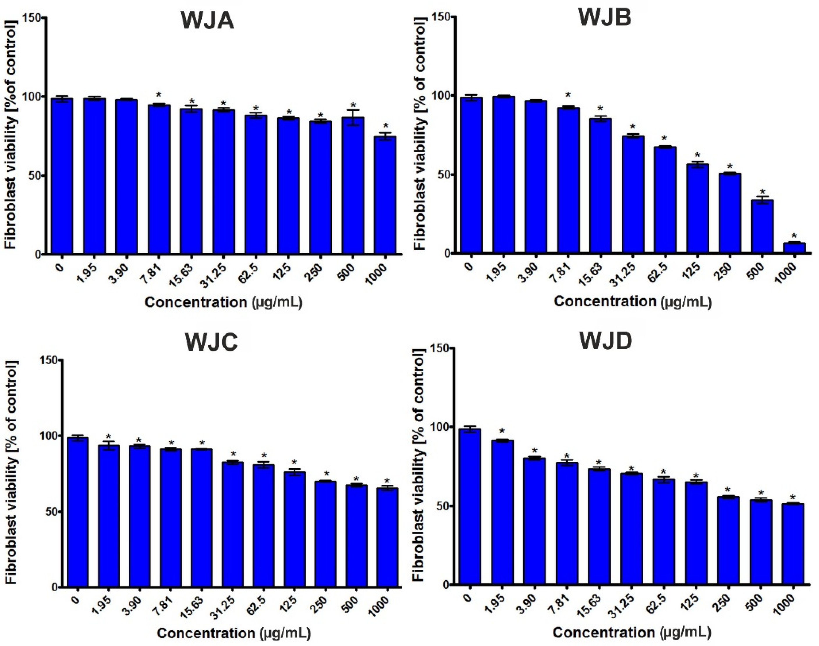
2.2.6. Cytotoxic Activity

The human skin fibroblast viability after a 24-h incubation with the tested extracts is presented in Figure 1. Taking into account the obtained results, it was observed that the BJ cells showed different responses to the extracts in concentration-dependent manner. In general, most of investigated extracts were nontoxic, with CC50 values above 1000 μg/mL (Table 7). Nevertheless, in the case of the WJB extract, it was found that it slightly inhibited the BJ cell viability (Figure 1), with a CC50 value close to 254 μg/mL (Table 7). Thus, it was proven that the tested extracts (apart from WJB) were nontoxic towards normal human fibroblasts, and as a consequence, they can be considered very promising substances for further investigations.
In the previous research, the cytotoxic activities of nine compounds isolated from the roots of W. japonica, collected in the Republic of Korea, were evaluated against A549 (non-small-cell lung adenocarcinoma), SKOV-3 (ovary malignant ascites), SK-MEL-2 (skin melanoma), and BT549 (invasive ductal carcinoma) cell lines using the sulforhodamine B (SRB) bioassay. In this research, the authors found that trans-p-coumaric acid possessed a cytotoxic activity against the BT549 cell line, with an IC50 value equal to 10 μM. The other tested compounds (wasabiside A-E, trans-ferulic acid, benzoic acid, and syringic acid) were inactive (IC50 > 10 μM) against all the tested cancer cell lines used [27].

3. Materials and Methods

3.1. Chemicals and Reagents

Ascorbic acid, collagenase from Clostridium histolyticum, 2,2-diphenyl-1-picrylhydrazyl radical (DPPH), 2,2′-azino-bis-(3-ethyl-benzothiazole-6-sulfonic acid) (ABTS●+), elastase from a porcine pancreas, (-)-epigallocatechin gallate (EGCG), hyaluronidase from the bovine tests, hyaluronic acid sodium salt from a rooster comb, Folin–Ciocalteu reagent, ethylenediaminetetraacetic acid, disodium dihydrate (Na2EDTA*2H2O), Tricine (≥99%; titration), N-[3-(2-Furyl)acryloyl]-Leu-Gly-Pro-Ala (FALGPA), and N-Succinyl-Ala-Ala-Ala-p-nitroanilide (SANA) were obtained from Sigma-Aldrich (Steinheim, Germany). Phosphate-buffered saline (PBS) was purchased from Gibco (Carlsbad, CA, USA). Reference substances were supplied by ChromaDex (Irvine, CA, USA), while acetonitrile, formic acid, and water for LC analysis were supplied by Merck (Darmstadt, Germany). All other chemicals were of analytical grade and were obtained from the Polish Chemical Reagent Company (POCH, Gliwice, Poland).

3.2. Plant Material

The flowers, leaves (annual and biennial), and roots of Eutrema japonicum (Miq.) Koidz. were collected from the Wasabi Farm Poland in Jedlnia Letnisko (Poland) at an altitude of 160.3 m a.m.s.l., coordinates 51°43′52″ N, 21°32′27″ E, in January 2021. Taxonomical identification was confirmed by Prof. K. Szewczyk, the botanist from the Department of Pharmaceutical Botany (Medical University in Lublin, Poland). Voucher specimen was deposited in the Department of Pharmaceutical Botany (EJ-250121).

3.3. Preparation of the Extracts

The plant materials were dried in the shade at 24 °C (±0.5 °C) to achieve a constant weight [40]. Extracts were prepared using a mixture of methanol–acetone–water (3:1:1, v/v/v; 3 × 100 mL) and were then sonicated at a controlled temperature (40 ± 2 °C) for 30 min [25]. The combined extracts were filtered, concentrated under reduced pressure, and, after freezing, lyophilized in a vacuum concentrator (Free Zone 1 apparatus; Labconco, Kansas City, MO, USA) to obtain dried residues. Dry extracts were weighted and stored in a freezer at −20 °C. The following yields were obtained: flowers (WJA)—1.17 g, roots (WJB)—3.41 g, annual leaves (WJC)—2.41 g, and biennial leaves (WJD)—4.71 g.

3.4. Total Flavonoid and Phenolic and Phenolic Acid Contents

Total flavonoid (TFC) and total phenolic contents (TPC) were established using the colorimetric assays, as described previously [25]. The absorbances were measured at 430 and 680 nm, respectively, using a Pro 200F Elisa Reader (Tecan Group Ltd., Männedorf, Switzerland). TPC was estimated from the calibration curve (R2 = 0.9845) using gallic acid as a standard (concentration range 0.002–0.1 mg/mL). The results were expressed as the mg of gallic acid equivalents (GAE) per 1 g of dry extract (DE). TFC was estimated from the calibrated curve (R2 = 0.995) using quercetin (0.004–0.11 mg/mL) as the standard. The results were expressed as the mg of quercetin equivalents (QE) per 1 g of DE. The total phenolic acids (TPAC) content was assessed using Arnov’s reagent, as described in Polish Pharmacopoeia IX (an official translation of PhEur 7.0) [40]. The absorbance was measured at 490 nm. The TPAC was estimated from the calibration curve (R2 = 0.9999) using caffeic acid as a standard in the concentration of 3.36–23.52 μg/mL. The results were expressed as the mg of caffeic acid equivalents (CAE) per 1 g of DE.

3.5. LC-ESI-MS/MS Analysis

The Agilent 1200 Series HPLC system (Agilent Technologies, Palo Alto, CA, USA) coupled to a 3200 QTRAP mass spectrometer (AB Sciex, Redwood City, CA, USA) was used for the qualitative and quantitative analyses of flavonoids and phenolic acids in different parts of the E. japonicum extracts. The separation of the analyzed compounds, injected in a 3-µL amount, was performed on a Zorbax SB-C18 analytical column (2.1 × 100 mm, 1.8 µm; Agilent Technologies, Palo Alto, CA, USA) at 25 °C. Elution was carried out using solvent A (0.1% HCOOH in water) and solvent B (0.1% HCOOH in acetonitrile). The following gradient elution program was used: 0–2 min—20% B, 3 to 4 min—25% B, 5 to 6 min—35% B, 5 to 6 min—35% B, 8–12 min—65% B, 14–16 min—80% B, and 20–28 min—20% B. The flow rate was 300 µL/min. The mass spectra of the analyzed compounds were acquired in the negative ESI mode, and the optimum values of the source parameters were as follows: capillary temperature 450°C, nebulizer gas 50 psi, curtain gas 30 psi, and source voltage −4500 V for phenolic acids and flavonoid glycosides and capillary temperature 550 °C, nebulizer gas 30 psi, curtain gas 20 psi, and source voltage −4500 V for the flavonoid aglycones analysis. Details of the LC-ESI-MS/MS analysis are presented in Table 8 and Table 9. Analyst 1.5 software (AB Sciex, Redwood City, CA, USA) was used for the analysis and data acquisition.

3.6. Antioxidant Activity

All antioxidant and enzyme inhibitory assays were done in 96-well plates (Nunclon, Nunc, Roskilde, Denmark) using an Infinite Pro 200F Elisa Reader (Tecan Group Ltd., Männedorf, Switzerland). The experiments were performed in triplicate.

3.6.1. DPPH• Assay

The 2,2-diphenyl-1-picryl-hydrazyl (DPPH•) free radical scavenging activity of the E. japonicum extracts and positive control—ascorbic acid (AA)—was studied using the method described previously [25] but with some modifications. After 30 min of incubation at 28 °C, the decrease in DPPH• absorbance, caused by the tested extracts, was measured at 517 nm. The results were expressed as the values of the IC50.

3.6.2. ABTS●+ Assay

The ABTS●+ decolorization assay was the second method applied for an assessment of the antioxidant activity [25]. The absorbance was measured at 734 nm. Trolox was used as a positive control. The results were expressed as the values of the IC50.

3.6.3. Metal Chelating Activity (CHEL)

The metal chelating activity was established using the method described by Guo et al. [41], modified in our previous study [25,42]. The absorbance was measured at 562 nm. As a positive control, Na2EDTA*2H2O was used. The results were expressed as the IC50 values of the E. japonicum extracts based on the concentration–inhibition curves.

3.7. Enzyme Inhibitory Activity

3.7.1. Anti-Elastase Activity

Anti-elastase activity was measured spectrophotometrically according to Chiocchio et al. [43]. Porcine pancreatic elastase (3.33 mg/mL; 25 µL) and E. japonicum samples (100 µg/mL) were incubated in Tris-buffer (0.2 mM, pH 8.0) for 10 min at 29 °C. To start the reaction, N-Succinyl-Ala-Ala-Ala-p-nitroanilide (2 mM; 125 µL) as a substrate was added. After 15 min of incubation, the absorbance was measured at 485 nm. Epigallocatechin gallate (100 µg/mL) was used as a positive control.

3.7.2. Anti-Collagenase Activity

Anti-collagenase activity was studied using N-[3-(2-furyl) acryloyl]-Leu-Gly-Pro-Ala (FALGPA) as a substrate and was measured according to Mandrone et al. [44]. Collagenase from Clostridium histolyticum (20 mU), dissolved in Tricine buffer (pH 7.5, 0.05 M, containing 0.4-M natrium chloride and 0.01-M calcium chloride), and E. japonicum samples (100 µg/mL) were incubated for 10 min at 35 °C. To start the reaction, FALGPA (1 mM) was added. After 15 min of incubation at 29 °C, the absorbance was measured at 345 nm. Epigallocatechin gallate (100 µg/mL) was used as a positive control.

3.7.3. Anti-Hyaluronidase Activity

Anti-hyaluronidase activity was established using the method described by Liyanaarachchi et al. [45]. After 20 min of incubation at 37 °C, the absorbance was measured at 585 nm. Epigallocatechin gallate (100 µg/mL) was used as a positive control.

3.8. Antimicrobial Activity

3.8.1. Bacterial Strains

All the tested extracts were screened for their in vitro antibacterial activity against strains: aerobic Gram-positive Staphylococcus aureus ATCC 25923, Staphylococcus epidermidis ATCC 12228, aerobic Gram-negative Escherichia coli ATCC 25992, Pseudomonas aeruginosa ATCC 27853, microaerobic Gram-positive Propionibacterium acnes PCM 2400, Propionibacterium acnes PCM 2334, S. mutans PCM 2502, and S. sanguinis PCM 2335. For determination of the antibacterial activity, Mueller–Hinton agar or broth (MH-agar or MH-broth) for aerobic and Brain–Heart Infusion agar or broth (BHI-agar or BHI-broth) for microaerobic strains were used. Bacterial inoculum was prepared by subculturing microorganisms into MH-agar or BHI-agar at 37 °C for 24 h or 48 h, respectively. The growth was harvested using 5 mL of 0.9% NaCl and diluted to 0.5 McFarland (1.5 × 10⁸ CFU/mL (CFU: colony-forming unit)).

3.8.2. Agar Disc Diffusion Assay

The antibacterial character of all the samples was initially determined by a disc diffusion assay [46]. The bacterial inoculum was spread on the agar surface of the Petri plates by applying a cotton swab. The solutions of all the tested samples (100 µg/mL in DMSO) were placed on inoculated Petri plates. The plates with MH-agar (for aerobic strains) were incubated for 24 h at 37 °C, while the plates with BHI-agar were incubated for 48 h at 37 °C. After incubation, the diameter of the growth inhibition zone around the samples was measured.

3.8.3. MIC (Minimum Inhibitory Concentration) and MBC (Minimal Bactericidal Concentration) Analysis

The minimum inhibitory concentration (MIC) of the extracts was determined for the bacterial strains, which exhibited the bacterial growth inhibition zones. The test was performed using double-serial microdilution in the 96-well microtiter plates, according to the CLSI method but with some modifications [47]. Two hundred microliters of broth were pipetted into each well. The double-serial dilution of the tested derivatives was performed in the test wells, resulting in concentrations that ranged from 2000 µg/mL to 0.098 µg/mL. Finally, 2 µL of tested bacteria inoculum were added to the wells (except for the negative sterility control and blanc dye control). The tests were performed at 37 °C for 24 h (aerobic strains) or for 48 h (microaerobic strains). After incubation, the panel was digitally analyzed at 600 nm using the microplate reader BioTek™ Synergy™ H4 Hybrid Microplate Reader (Thermo Fisher Scientific, Waltham, MA, USA). The growth intensity in the sample well was compared to the negative and positive controls. Additionally, the MBC (minimal bactericidal concentration) was determined by spreading 10 µL of liquid from a clear sample well on the agar medium. The plates were incubated for 24 h at 37 °C, and the MBC was defined as the lowest concentration of sample without bacterial growth. Each experiment was repeated in triplicate.

3.9. Cytotoxicity Evaluation

The cytotoxic effect of the tested extracts was assessed in accordance with our procedure described earlier [48,49]. The experiment was carried out using the BJ cell line (normal human skin fibroblasts, ATCC CRL-2522TM). Briefly, the BJ cells were seeded on a 96-well plate at a concentration of 1.5 × 10⁴ cells/well and incubated for 24 h at 37 °C in a humidified atmosphere (5% CO2, 95% air). On the next day, stock solutions of extracts (100 mg/mL) in dimethyl sulfoxide (DMSO) were obtained, and then, two-fold serial dilutions of extracts (1000–1.95 μg/mL) in the culture medium were prepared. Subsequently, the culture medium was replaced by an appropriate dilution of extracts, and the treated BJ cells were incubated for 24 h at 37 °C, 5% CO2, 95% air. Cytotoxicity of the tested extracts was evaluated using the MTT assay [50]. Based on the obtained results, the values of the half-maximum cytotoxic concentration (CC50) were determined (4-parameter nonlinear regression analyses, GraphPad Prism 5, version 5.04, Software, GraphPad Software, San Diego, CA, USA). The CC50 refers to a concentration of extract required for the reduction of BJ cell viability to 50%.

3.10. Statistical Analysis

The results were expressed as the mean values ± standard deviations (SD) of three independent experiments. The data from the cell culture experiments were subjected to a statistical analysis using an unpaired Student’s t-test, and the differences were considered significant when p < 0.05 (GraphPad Prism 5, version 5.04 Software, GraphPad Software, San Diego, CA, USA).

4. Conclusions

In this in vitro study, we characterized the composition and comprehensively evaluated the biological activities of the extracts from the flowers (WJA), roots (WJB), and leaves—annual (WJC), as well as biennial (WJD)—of Eutrema japonicum cultivated at Wasabi Farm Poland. Thus, the content of flavonoids and phenolic acids in the extracts was determined, as well as the antioxidant, anti-collagenase, anti-elastase, anti-hyaluronidase, antimicrobial, and cytotoxic properties of such extracts.
Among the tested samples, the WJA extract possessed the most desirable biological activities. It exhibited the greatest antioxidant features, as proven by the DPPH assay, ABTS●+ test, and Metal Chelating Activity test. Moreover, it had the greatest anti-collagenase and anti-hyaluronidase features compared to the other investigated extracts. The determined MIC and MBC values proved that the WJA extract also exhibited the most promising antibacterial activity, as it possessed bacteriostatic activity against the anaerobic pathogens. In turn, the cell culture experiments indicated that the WJA extract was noncytotoxic towards normal skin human fibroblasts. The beneficial biological properties of the WJA extract seemed to be correlated with its composition. We proved that WJA is rich in flavonoids, especially isovitexin 4’-O-glucoside, luteolin 3′,7′-diglucoside, and kaempferol 3-O-rutinoside. It is worth underlining that the aforementioned properties revealed in this study allowed us to suppose that the WJA extract has “multidirectional” activity. Thus, considering a broad spectrum of activities of such an extract, it seems to be a promising antiaging agent. For this reason, the WJA extract will be subjected to further in vivo studying in order to confirm its beneficial activity.

Supplementary Materials

The following are available online at https://www.mdpi.com/article/10.3390/ijms22126219/s1. Figure S1. The chromatogram in the Multiple Reaction Monitoring (MRM) mode of phenolic acids, flavonoid aglycones and glycosides in the WJA extract: 1-chlorogenic acid; 2- p-coumaric acid; 3- sinapic acid; 4 – isosaponarin; 5 - luteolin 3’,7’-diglucoside; 6 – rutin; 7 – isovitexin/vitexin; 8 -luteolin-7-O-glucoside; 9 - isoquercetin; 10 - kaempferol-3-O-rutinoside; 11-narcissoside; 12 - astragalin; 13 - isorhamnetin-3-O-glucoside; 14 - apigenin 7-O-glucoside. Figure S2. Mass spectra of isosaponarin by ESI in negative mode.

Author Contributions

Conceptualization, K.S., A.F. and M.F.; methodology, K.S., W.P., M.M.-K. and K.K.; formal analysis, K.S., W.P., M.M.-K. and K.K.; resources, K.S., A.F. and M.F.; data curation, K.S., W.P., M.M.-K. and K.K.; writing—original draft preparation, K.S. and A.G.-C.; writing—review and editing, K.S., M.M.-K. and K.K.; and supervision, K.S. and K.K. All authors have read and agreed to the published version of the manuscript.

Funding

This research was funded by the Ministry of Science and Higher Education in Poland DS45 and DS2 projects of the Medical University of Lublin. This paper was developed using the equipment purchased with agreement No. POPW.01.03.00-06-010/09-00, Operational Program Development of Eastern Poland 2007-2013, Priority Axis I, Modern Economy, Operations 1.3. Innovations Promotion.

Institutional Review Board Statement

Not applicable.

Informed Consent Statement

Not applicable.

Data Availability Statement

Data available on request.

Conflicts of Interest

The authors declare no conflict of interest.

References

  1. Nizioł-Łukaszewska, Z.; Bujak, T. Ocena właściwości kosmetyków myjących zawierających ekstrakt wodny z czarnuszki siewnej (Nigella sativa L.). In Rośliny w Nowoczesnej Kosmetologii; Kiełtyka-Dadasiewicz, A., Ed.; Wydawnictwo Akade-mickie Wyższej Szkoły Społeczno-Przyrodniczej im; Wincentego Pola w Lublinie: Lublin, Poland, 2016; pp. 93–105. [Google Scholar]
  2. Kumar, N.; Goel, N. Phenolic acids: Natural versatile molecules with promising therapeutic applications. Biotechnol. Rep. 2019, 24, e00370. [Google Scholar] [CrossRef]
  3. Ribeiro, A.S.; Estanqueiro, M.; Oliveira, M.B.; Lobo, J.M.S. Main Benefits and Applicability of Plant Extracts in Skin Care Products. Cosmetics 2015, 2, 48–65. [Google Scholar] [CrossRef] [Green Version]
  4. The Plant List, Version 1.1. Available online: http://www.theplantlist.org/ (accessed on 3 May 2021).
  5. Miles, C.; Chadwick, C. Growing Wasabi in the Pacific Northwest. Farming the Northwest; PNW0605; Washington State University: Washington, DC, USA, 2008; pp. 1–12. [Google Scholar]
  6. Kojima, M.; Uchida, M.; Akahori, Y. Studies on the Volatile Components of Wasabia japonica, Brassica juncea and Cocholearia armoracia by Gas Chromatography-Mass Spectrometry. I. Determination of Low Mass Volatile Components. Yakugaku Zasshi 1973, 93, 453–459. [Google Scholar] [CrossRef] [Green Version]
  7. Etoh, H.; Nishimura, A.; Takasawa, R.; Yagi, A.; Saito, K.; Sakata, K.; Kishima, I.; Ina, K. ω-methylsulfinylalkyl isothiocyanates in wasabi, Wasabia japonica Matsum. Agric. Biol. Chem. 1990, 54, 1587–1589. [Google Scholar] [CrossRef]
  8. Kumagai, H.; Kashima, N.; Seki, T.; Sakurai, H.; Ishii, K.; Ariga, T. Analysis of volatile components in essential oil of upland Wasabi and their inhibitory effects on platelet aggregation. Biosci. Biotech. Biochem. 1994, 58, 2131–2135. [Google Scholar] [CrossRef]
  9. Hosoya, T.; Yun, Y.S.; Kunugi, A. Five novel flavonoids from Wasabia japonica. Tetrahedron 2005, 61, 7037–7044. [Google Scholar] [CrossRef]
  10. Kurata, T.; Misawa, N.; Hosoya, T.; Yamada-Kato, T.; Okunishi, I.; Kumazawa, S. Isolation and Identification of Components from Wasabi (Wasabia japonica Matsumura) Flowers and Investigation of Their Antioxidant and Anti-inflammatory Activities. Food Sci. Technol. Res. 2019, 25, 449–457. [Google Scholar] [CrossRef]
  11. Hosoya, T.; Yun, Y.S.; Kunugi, A. Antioxidant phenylpropanoid glycosides from the leaves of Wasabia japonica. Phytochemistry 2008, 69, 827–832. [Google Scholar] [CrossRef]
  12. Yoshida, S.; Hosoya, T.; Inui, S.; Masuda, H.; Kumazawa, S. Component Analysis of Wasabi Leaves and an Evaluation of their Anti-inflammatory Activity. Food Sci. Technol. Res. 2015, 21, 247–253. [Google Scholar] [CrossRef] [Green Version]
  13. Shimamura, Y.; Iio, M.; Urahira, T.; Masuda, S. Inhibitory effects of Japanese horseradish (Wasabia japonica) on the formation and genotoxicity of a potent carcinogen, acrylamide. J. Sci. Food Agric. 2016, 97, 2419–2425. [Google Scholar] [CrossRef]
  14. Nagai, M.; Okunishi, I. The effect of wasabi rhizome extract on atopic dermatitis-like symptoms in HR-1 hairless mice. J. Nutr. Sci. Vitaminol. 2009, 55, 195–200. [Google Scholar] [CrossRef] [Green Version]
  15. Morimitsu, Y.; Hayashi, K.; Nakagawa, Y.; Horio, F.; Uchida, K.; Osawa, T. Antiplatelet and anticancer isothiocyanates in Japanese domestic horseradish, wasabi. BioFactors 2000, 13, 271–276. [Google Scholar] [CrossRef]
  16. Fuke, Y.; Haga, Y.; Ono, H.; Nomura, T.; Ryoyama, K. Anti-carcinogenic activity of 6-methylsulfinylhexyl isothiocyanate-, an active anti-proliferative principal of wasabi (Eutrema wasabi Maxim.). Cytotechnology 1997, 25, 197–203. [Google Scholar] [CrossRef] [PubMed]
  17. Yamasaki, M.; Ogawa, T.; Wang, L.; Katsube, T.; Yamasaki, Y.; Sun, X.; Shiwaku, K. Anti-obesity effects of hot water extract from Wasabi (Wasabia japonica Matsum.) leaves in mice fed high-fat diets. Nutr. Res. Pract. 2013, 7, 267–272. [Google Scholar] [CrossRef] [PubMed] [Green Version]
  18. Wang, X.; Zhou, H.; Liu, X. Preliminary study on the functional elements in the mixture of wasabi leaf and leaf stalk and its catharsis effect. J. Chongqing Med. Univ. 2011, 36, 1347–1349. [Google Scholar]
  19. Kinae, N.; Masuda, H.; Shin, I.S.; Furugori, M.; Shimoi, K. Functional properties of wasabi and horseradish. BioFactors 2000, 13, 265–269. [Google Scholar] [CrossRef]
  20. Nagai, M.; Akita, K.; Yamada, K.; Okunishi, I. The effect of isosaponarin isolated from wasabi leaf on collagen synthesis in human fibroblasts and its underlying mechanism. J. Nat. Med. 2010, 64, 305–312. [Google Scholar] [CrossRef] [PubMed]
  21. Gottschalck, T.E.; Bailey, J.E. International Cosmetic Ingredient Dictionary and Handbook, 10th ed.; Introduction INCI Name Monographs A–K; The Cosmetics, Toiletries and Fragrances Association; CTFA: Washington, DC, USA, 2004; Volume 1, p. 1983. [Google Scholar]
  22. Kang, D.Y.; Hitayezu, E.; Han, I.H.; Kim, J.S.; Jo, Y.M.; Kang, Y.-H. Physicochemical characteristics and antioxidant activities of solvent fractions from ethanol extract of Wasabia koreana Nakai leaf. Korean J. Food Preserv. 2019, 26, 586–593. [Google Scholar] [CrossRef]
  23. Kim, D.-S.; Kim, H.S.; Lee, J.; Pan, J.H.; Kim, Y.J.; Kim, J.K.; Woo, S.; Shin, E.-C. Wasabia koreana Nakai: A Preliminary Study on Nutrients and Chemical Compounds That May Impact Sensory Properties. Molecules 2018, 23, 2512. [Google Scholar] [CrossRef] [Green Version]
  24. Shin, S.W.; Ghimeray, A.K.; Park, C.H. Investigation of total phenolic, total flavonoid, antioxidant and allyl isothiocyanate content in the different organs of Wasabia japonica grown in an organic system. Afr. J. Tradit. Complement. Altern. Med. 2014, 11, 38–45. [Google Scholar] [CrossRef] [Green Version]
  25. Szewczyk, K.; Bogucka-Kocka, A.; Vorobets, N.; Grzywa-Celińska, A.; Granica, S. Phenolic Composition of the Leaves of Pyrola rotundifolia L. and Their Antioxidant and Cytotoxic Activity. Molecules 2020, 25, 1749. [Google Scholar] [CrossRef] [Green Version]
  26. Bogucka-Kocka, A.; Vorobets, N.; Chrząszcz, M.; Pietrzak, W.; Szewczyk, K. Polyphenol Composition of Extracts of the Fruits of Laserpitium Krapffii Crantz and Their Antioxidant and Cytotoxic Activity. Antioxidants 2019, 8, 363. [Google Scholar] [CrossRef] [Green Version]
  27. Kim, C.S.; Subedi, L.; Kwon, O.W.; Park, H.B.; Kim, S.Y.; Choi, S.U.; Lee, K.R. Wasabisides A–E, Lignan Glycosides from the Roots of Wasabia japonica. J. Nat. Prod. 2016, 79, 2652–2657. [Google Scholar] [CrossRef] [PubMed]
  28. Boran, R. Investigations of anti-aging potential of Hypericum origanifolium Willd. for skincare formulations. Ind. Crop. Prod. 2018, 118, 290–295. [Google Scholar] [CrossRef]
  29. Narayanaswamy, R.; Ismail, I.S. Cosmetic potential of Southeast Asian herbs: An overview. Phytochem. Rev. 2015, 14, 419–428. [Google Scholar] [CrossRef]
  30. Kurutas, E.B. The importance of antioxidants which play the role in cellular response against oxidative/nitrosative stress: Current state. Nutr. J. 2015, 15, 1–22. [Google Scholar] [CrossRef] [PubMed] [Green Version]
  31. Ferrali, M.; Signorini, C.; Caciotti, B.; Sugherini, L.; Ciccoli, L.; Giachetti, D.; Comporti, M. Protection against oxidative damage of erythrocyte membrane by the flavonoid quercetin and its relation to iron chelating activity. FEBS Lett. 1997, 416, 123–129. [Google Scholar] [CrossRef] [Green Version]
  32. Uitto, J. Connective tissue biochemistry of the aging dermis: Age-associated alternations in collagen and elastin. Clin. Geriatr. Med. 1989, 5, 127–148. [Google Scholar] [CrossRef]
  33. Ersoy, E.; Ozkan, E.E.; Boga, M.; Yilmaz, M.A.; Mat, A. Anti-aging potential and anti-tyrosinase activity of three Hypericum species with focus on phytochemical composition by LC–MS/MS. Ind. Crop. Prod. 2019, 141, 111735. [Google Scholar] [CrossRef]
  34. Wittenauer, J.; Mäckle, S.; Sussmann, D.; Schweiggert-Weisz, U.; Carle, R. Inhibitory effects of polyphenols from grape pomace extract on collagenase and elastase activity. Fitoterapia 2015, 101, 179–187. [Google Scholar] [CrossRef]
  35. Bauman, L. Skin ageing and its treatment. J. Pathol. 2007, 211, 241–251. [Google Scholar] [CrossRef]
  36. Barla, F.; Higashijima, H.; Funai, S.; Sugimoto, K.; Harada, N.; Yamaji, R.; Fujita, T.; Nakano, Y.; Inui, H. Inhibitive Effects of Alkyl Gallates on Hyaluronidase and Collagenase. Biosci. Biotechnol. Biochem. 2009, 73, 2335–2337. [Google Scholar] [CrossRef] [PubMed]
  37. O’Neill, A.J.; Chopra, I. Preclinical evaluation of novel antibacterial agents by microbiological and molecular techniques. Expert Opin. Investig. Drugs 2004, 13, 1045–1063. [Google Scholar] [CrossRef] [PubMed]
  38. Kiba, A.; Saitoh, H.; Nishihara, M.; Omiya, K.; Yamamura, S. C-Terminal Domain of a Hevein-Like Protein from Wasabia japonica has Potent Antimicrobial Activity. Plant Cell Physiol. 2003, 44, 296–303. [Google Scholar] [CrossRef]
  39. Shin, I.S.; Masuda, H.; Naohide, K. Bactericidal activity of wasabi (Wasabia japonica) against Helicobacter pylori. Int. J. Food Microbiol. 2004, 94, 255–261. [Google Scholar] [CrossRef]
  40. Polish Pharmacopoeia IX. In PTFarm; Polish Pharmaceutical Society: Warsaw, Poland, 2011; p. 150.
  41. Guo, J.-T.; Lee, H.-L.; Chiang, S.-H.; Lin, F.-I.; Chang, C.-Y. Antioxidant Properties of the Extracts from Different Parts of Broccoli in Taiwan. J. Food Drug Anal. 2001, 9, 96–101. [Google Scholar] [CrossRef]
  42. Nowak, R.; Szewczyk, K.; Gawlik-Dziki, U.; Rzymowska, J.; Komsta, Ł. Antioxidative and cytotoxic potential of some Che-nopodium L. species growing in Poland. Saudi J. Biol. Sci. 2016, 23, 15–23. [Google Scholar] [CrossRef] [Green Version]
  43. Chiocchio, I.; Mandrone, M.; Sanna, C.; Maxia, A.; Tacchini, M.; Poli, F. Screening of a hundred plant extracts as tyrosinase and elastase inhibitors, two enzymatic targets of cosmetic interest. Ind. Crop. Prod. 2018, 122, 498–505. [Google Scholar] [CrossRef]
  44. Mandrone, M.; Lorenzi, B.; Venditti, A.; Guarcini, L.; Bianco, A.; Sanna, C.; Ballero, M.; Poli, F.; Antognoni, F. Antioxidant and anti-collagenase activity of Hypericum hircinum L. Ind. Crop. Prod. 2015, 76, 402–408. [Google Scholar] [CrossRef]
  45. Liyanaarachchi, G.D.; Samarasekera, J.K.R.R.; Mahanama, K.R.R.; Hemalal, K.D.P. Tyrosinase, elastase, hyaluronidase, in-hibitory and antioxidant activity of Sri Lankan medicinal plants for novel cosmeceuticals. Ind. Crop. Prod. 2018, 111, 597–605. [Google Scholar] [CrossRef]
  46. Murray, P.R.; Baron, E.J.; Pfaller, M.A.; Tenover, F.C.; Yolke, R.H. Manual of Clinical Microbiology, 6th ed.; Mosby Year Book: London, UK, 1995; pp. 143–260. ISBN 13-978-1555810863. [Google Scholar]
  47. Clinical Laboratory Standards Institute. Performance Standards for Antimicrobial Susceptibility Testing. Eighteenth International Supplement; CLSI document M7-MIC; Clinical Laboratory Standards Institute: Wayne, PA, USA, 2008. [Google Scholar]
  48. Miazga-Karska, M.; Szewczyk, K.; Klimek, K.; Ginalska, G. In vitro activity of peptide fractions from Impatiens glandulifera against caries causing bacteria. Acta Pol. Pharm. 2017, 74, 710–714. [Google Scholar] [PubMed]
  49. Chrząszcz, M.; Miazga-Karska, M.; Klimek, K.; Granica, S.; Tchórzewska, D.; Ginalska, G.; Szewczyk, K. Extracts from Cephalaria uralensis (Murray) Roem. & Schult. and Cephalaria gigantea (Ledeb.) Bobrov as Potential Agents for Treatment of Acne Vulgaris: Chemical Characterization and In Vitro Biological Evaluation. Antioxidants 2020, 9, 796. [Google Scholar] [CrossRef]
  50. Pitucha, M.; Woś, M.; Miazga-Karska, M.; Klimek, K.; Mirosław, B.; Pachuta-Stec, A.; Gładysz, A.; Ginalska, G. Synthesis, antibacterial and antiproliferative potential of some new 1-pyridinecarbonyl-4-substituted thiosemicarbazide derivatives. Med. Chem. Res. 2016, 25, 1666–1677. [Google Scholar] [CrossRef] [PubMed] [Green Version]
Figure 1. Fibroblast viability (BJ cell line ATTC CRL-2522TM) after a 24-h incubation with the WJA, WJB, WJC, and WJD extracts. * Significantly different results compared to the control (culture medium without extracts—0 μg/mL), unpaired t-test; p < 0.05.
Ijms 22 06219 g001
Table 1. The total content of phenolic (TPC), flavonoid (TFC), and phenolic acids (TPAC) in the flowers (WJA), roots (WJB), and leaves (annual—WJC and biennial—WJD) of E. japonicum. DE—dry extract; GAE—Gallic Acid Equivalent; CAE—Caffeic Acid Equivalent; QE—Quercetin Equivalent. Values were presented as the mean ± standard deviation (n = 9).
SampleTotal Phenolic Content
(mg GAE/g DE)
Total Phenolic Acids
(mg CAE/g DE)
Total Flavonoid Content
(mg QE/g DE)
WJA187.79 ± 6.8617.20 ± 0.2445.08 ± 0.18
WJB31.55 ± 2.174.34 ± 0.089.15 ± 0.10
WJC77.92 ± 4.8213.84 ± 0.0928.81 ± 0.11
WJD54.93 ± 0.999.70 ± 0.2029.77 ± 0.04
Table 2. Content of phenolic acids and flavonoids in the leaves, roots, and flowers of E. japonicum. nd—not detected; LOQ—limit of quantification; DE—dry extract.
NoCompoundAmounts (mg/g DE)
WJAWJBWJCWJD
1salicylic acid<LOQ<LOQ<LOQnd
2p-coumaric acid0.10 ± 0.02nd0.01 ± 0.01<LOQ
3caffeic acid<LOQnd<LOQ<LOQ
4ferulic acid<LOQnd1.21 ± 0.07<LOQ
5chlorogenic acid0.01 ± 0.010.62 ± 0.02nd0.42 ± 0.02
6cis-sinapic acid0.67 ± 0.070.09 ± 0.000.77 ± 0.060.56 ± 0.01
7apigenin<LOQnd<LOQ<LOQ
8naringenin<LOQndndnd
9kaempferol<LOQndndnd
10eriodictiol<LOQndndnd
11quercetin<LOQndndnd
12taxifolin<LOQndndnd
13isorhamnetin<LOQndndnd
14luteolin<LOQnd0.01 ± 0.00<LOQ
15apigenin 7-O-glucoside0.02 ± 85.0nd<LOQnd
16naringenin 7-O-glucoside<LOQndndnd
17apigenin 6-C-glucoside/apigenin 8-C-glucoside0.52 ± 0.07nd2.57 ± 0.010.70 ± 0.09
18luteolin 7-O-glucoside0.44 ± 0.12nd0.18 ± 0.000.13 ± 0.02
19kaempferol 3-O-glucoside0.38 ± 0.01nd<LOQnd
20quercetin 3-O-glucoside0.53 ± 0.07nd<LOQ<LOQ
21isorhamnetin 3-O-glucoside0.48 ± 0.02ndndnd
22kaempferol 3-O-rutinoside2.50 ± 0.06nd<LOQnd
23quercetin 3-O-rutinoside0.74 ± 0.03<LOQ<LOQ<LOQ
24luteolin 3′,7′-diglucoside4.98 ± 0.23nd3.51 ± 0.132.74 ± 0.10
25isorhamnetin 3-O-rutinoside0.54 ± 0.04ndndnd
26isovitexin 4’-O-glucoside6.02 ± 0.10<LOQ6.22 ± 0.045.99 ± 0.22
Table 3. The IC50 values determined in the antioxidant tests. Data were expressed as the mean ± SD, n = 3. AA—ascorbic acid; Na2EDTA*2H2O—ethylenediaminetetraacetic acid, disodium dihydrate; nt—not tested. The flowers (WJA), roots (WJB), and leaves (annual—WJC and biennial—WJD) of E. japonicum. DPPH—2,2-diphenyl-1-picryl-hydrazyl radical; ABTS—2,2′-azino-bis-(3-ethyl-benzothiazole-6-sulfonic acid; CHEL—metal chelating activity. * Statistically significant differences compared to AA; # statistically significant differences compared to Na2EDTA*2H2O, p < 0.05.
SampleIC50
DPPH (μg/mL)ABTS (μg/mL)CHEL (μg/mL)
WJA28.72 ± 0.24 *11.68 ± 0.4712.50 ± 0.36 #
WJB134.30 ± 1.75 *65.18 ± 0.7332.08 ± 0.13 #
WJC62.84 ± 0.08 *33.25 ± 0.3114.27 ± 0.21 #
WJD69.10 ± 0.16 *28.53 ± 0.7213.65 ± 0.29 #
AA4.92 ± 0.32ntnt
Troloxnt3.14 ± 0.17nt
Na2EDTA*2H2Ontnt8.75 ± 0.15
Table 4. Anti-collagenase, anti-elastase, and anti-hyaluronidase activities of the flowers (WJA), roots (WJB), and leaves (annual—WJC and biennial—WJD) of E. japonicum. * Statistically significant differences compared to epigallocatechin gallate (EGCG), p < 0.05.
SampleInhibition (%)
Collagenase InhibitionElastase InhibitionHyaluronidase Inhibition
WJA93.34 ± 0.77 *88.93 ± 0.16 *47.32 ± 0.53 *
WJB78.42 ± 0.25 *90.18 ± 0.54 *13.46 ± 0.22 *
WJC87.51 ± 0.83 *84.90 ± 0.6028.95 ± 0.69 *
WJD90.16 ± 0.51 *88.89 ± 0.36 *25.78 ± 0.18 *
EGCG88.49 ± 0.4591.03 ± 0.1862.90 ± 0.12
Table 5. Zones of bacterial growth inhibition expressing the antibacterial activity of the E. japonicum extracts. Extracts from the flowers (WJA), roots (WJB), and leaves (annual—WJC and biennial—WJD) of E. japonicum. K1—quercetin; K2—luteolin; K3—gallic acid; K4—p-coumaric acid; K5—caffeic acid; K6—vanillic acid.
Bacterial StrainsZones of Bacterial Growth Inhibition (mm)
WJAWJBWJCWJDK1K2K3K4K5K6
S. aureus ATCC 2592300000015000
S. epidermidis ATCC 12228000000360280
E. coli ATCC 2599276000012844
P. aeruginosa ATCC 2785300000010644
S. mutans PCM 250216.516000016000
P. acnes PCM 24001619.51580020000
P. acnes PCM 2334172116104022000
S. sanguinis PCM 23351718000018000
Table 6. Minimum inhibitory concentration (MIC (μg/mL)) and MBC/MIC ratio as the antibacterial activity of the E. japonicum extracts.
Bacterial StrainsSample
WJAWJBK3
S. aureus ATCC 25923--125
MBC/MIC 16
S. epidermidis ATCC 12228--31.25
MBC/MIC--8
E. coli ATCC 2599210001000500
MBC/MIC--8
P. aeruginosa ATCC 27853--1000
MBC/MIC---
S. mutans PCM 2502500500125
MBC/MIC-88
P. acnes PCM 240050025062.5
MBC/MIC888
P. acnes PCM 233425012562.5
MBC/MIC8168
S. sanguinis PCM 233550025062.5
MBC/MIC-88
Table 7. The half-maximal cytotoxic concentrations (CC50) of the tested extracts. The CC50 values were determined against normal human skin fibroblasts (BJ cell line ATTC CRL-2522TM) after a 24-h incubation.
ExtractCC50 (μg/mL)
WJA>1000
WJB245.45
WJC>1000
WJD>1000
Table 8. LC-ESI-MS/MS analytical results of the phenolic acids and flavonoids.
CompoundRetention Time (min)Q1/Q3 (m/z)DP (V)EP (V)CEP (V)CE (eV)CXP (V)
Phenolic acids
5-caffeoylquinic acid (chlorogenic acid)9.30352.9/190.8−35−4.5−16−24−2
10.42352.9/84.9−35−4.5−16−600
Caffeic acid11.38178.7/88.9−30−6.5−12−460
178.7/134.9−30−6.5−12−160
4-Hydroxycinnamic acid (p-coumaric acid)14.10162.7/119−30−8−12−140
162.7/93−30−8−12−440
Sinapic acid14.47222.8/121−35−8.5−10−360
14.94222.8/148.9−35−8.5−10−200
Ferulic acid14.80192.8/133.9−25−11.5−14−160
15.22192.8/177.9−25−11.5−14−12−2
Salicylic acid17.91136.8/93−35−4−10−16−2
136.8/75−35−4−10−480
Flavonoid aglycones
Naringenin14.52270.8/119−50−11.5−12−340
270.8/150.9−50−11.5−12−220
Taxifolin15.15302.7/124.9−45−3.5−18−260
302.7/284.8−45−3.5−18−14−4
Luteolin17.82284.7/132.9−75−9−18−380
284.7/150.9−75−9−18−260
Eriodyctiol17.89286.7/134.9−45−6−12−320
286.7/150.9−45−6−12−18−2
Quercetin17.94300.7/150.9−60−2.5−12−260
300.7/178.8−60−2.5−12−20−2
Apigenin18.64268.8/117−70−9.5−12−440
268.8/106.8−70−9.5−12−340
Kaempferol18.85284.7/116.8−70−5−12−460
284.7/93−70−5−12−520
Isorhamnetin18.99314.7/299.7−65−2.5−26−20−4
314.7/150.9−65−2.5−26−300
Flavonoid glycosides
Isovitexin 4’-O-glucoside (Isosaponarin)8.82593.2/282.1−25−10−34−30−3
593.2/473.1−25−10−34−30−3
Luteolin 3′,7′-diglucoside11.28609.1/285−70−7.5−28−50−4
609.1/447−70−7.5−28−32−18
Quercetin 3-O-rutinoside (Rutin)11.99608.7/299.6−90−8−30−46−4
608.7/270.9−90−8−30−60−4
Apigenin 6-C-glucoside (Isovitexin)12.38430.8/310.9−65−4.5−18−28−4
430.8/340.9−65−4.5−18−26−14
Apigenin 8-C-glucoside (Vitexin)12.40430.8/310.9−75−4.5−20−26−4
430.8/340.9−75−4.5−20−34−14
Luteolin 7-O-glucoside (Luteoloside)12.87446.8/284.8−70−10.5−20−30−4
446.8/132.9−70−10.5−20−780
Quercetin 3-O-glucoside (Isoquercetin)13.00462.7/299.7−85−1.5−20−30−4
462.7/270.7−85−1.5−20−44−4
Kaempferol 3-O-rutinoside (Nicotiflorin)13.31592.7/284.8−65−12−30−38−2
592.7/226.7−65−12−30−68−2
Isorhamnetin 3-O-rutinoside (Narcissoside)13.52622.8/314.9−90−4.5−30−40−4
622.8/298.8−90−4.5−30−52−4
Kaempferol 3-O-glucoside (Astragalin)14.66446.7/226.8−75−9−20−54−2
446.7/254.8−75−9−20−40−2
Isorhamnetin 3-O-glucoside14.76476.8/313.9−95−10−22−30−4
476.8/270.9−95−10−22−44−4
Apigenin 7-O-glucoside (Apigetrin and Cosmosiin)14.91430.7/267.7−70−9−20−38−4
430.7/116.9−70−9−20−840
Naringenin 7-O-glucoside15.12432.7/270.8−40−8.5−20−22−4
432.7/118.9−40−8.5−20−640
Table 9. Analytical results of the LC-ESI-MS/MS quantitative method of the phenolic acids and flavonoids. Limit of quantification (LOQ), limit of detection (LOD), and calibration curve parameters.
CompoundLOD (ng/mL)LOQ (ng/mL)R2Linearity Range (ng/mL)
Phenolic acids
5-Caffeoylquinic acid (chlorogenic acid)701500.9989150–15,000
Caffeic acid2004000.9991400–20,000
4-Hydroxycinnamic acid (p-coumaric acid)502000.9990500–15,000
Sinapic acid50010000.99891000–30,000
Ferulic acid100015000.99851830–35,000
Salicylic acid3005000.99741500–15,000
Flavonoid aglycones
Naringenin1503000.9982600–6000
Taxifolin10200.998440–10,000
Luteolin5150.998750–1500
Eriodyctiol25500.9985100–6600
Quercetin501000.9982100–6600
Apigenin15220.9970100–4000
Kaempferol30500.9971150–3000
Isorhamnetin3005000.99901000–5000
Flavonoid glycosides
Isovitexin 4’-O-glucoside (Isosaponarin)1002500.9958500–10,000
Luteolin 3′,7′-diglucoside2505000.99801000–25,000
Quercetin 3-O-rutinoside (Rutin)1002500.99802000–28,800
Apigenin 6-C-glucoside (Isovitexin)1002500.99911500–50,000
Apigenin 8-C-glucoside (Vitexin)1002000.99812000–50,000
Luteolin 7-O-glucoside (Luteoloside)501000.9982200–25,000
Quercetin 3-O-glucoside (Isoquercetin)1002500.99752500–50,000
Kaempferol 3-O-rutinoside (Nicotiflorin)601200.9957150–60,000
Isorhamnetin 3-O-rutinoside (Narcissoside)501000.9981150–25,000
Kaempferol 3-O-glucoside (Astragalin)1002500.99741500–25,000
Isorhamnetin 3-O-glucoside1001500.99692000–35,000
Apigenin 7-O-glucoside (Apigetrin, Cosmosiin)1002500.99891500–25,000
Naringenin 7-O-glucoside1002000.9990250–25,000
Publisher’s Note: MDPI stays neutral with regard to jurisdictional claims in published maps and institutional affiliations.

