Wnt-Independent and Wnt-Dependent Effects of APC Loss on the Chemotherapeutic Response


1
Department of Biological Sciences, University of Notre Dame, Notre Dame, IN 46617, USA
2
Mike and Josie Harper Cancer Research Institute, South Bend, IN 46617, USA
3
Department of Biochemistry and Molecular Biology, Indiana University School of Medicine-South Bend, South Bend, IN 46617, USA
*
Author to whom correspondence should be addressed.
Int. J. Mol. Sci. 2020, 21(21), 7844; https://doi.org/10.3390/ijms21217844
Submission received: 30 September 2020
/
Revised: 20 October 2020
/
Accepted: 20 October 2020
/
Published: 22 October 2020
(This article belongs to the Special Issue The Wnt Signaling Pathway in Cancer)
Abstract
:Resistance to chemotherapy occurs through mechanisms within the epithelial tumor cells or through interactions with components of the tumor microenvironment (TME). Chemoresistance and the development of recurrent tumors are two of the leading factors of cancer-related deaths. The Adenomatous Polyposis Coli (APC) tumor suppressor is lost in many different cancers, including colorectal, breast, and prostate cancer, and its loss correlates with a decreased overall survival in cancer patients. While APC is commonly known for its role as a negative regulator of the WNT pathway, APC has numerous binding partners and functional roles. Through APC’s interactions with DNA repair proteins, DNA replication proteins, tubulin, and other components, recent evidence has shown that APC regulates the chemotherapy response in cancer cells. In this review article, we provide an overview of some of the cellular processes in which APC participates and how they impact chemoresistance through both epithelial- and TME-derived mechanisms.
1. Introduction
Chemotherapy is the standard of care for most cancer types. The cytotoxic agents involved have different modes of action, including the disruption of DNA processes, microtubule networks, metabolism, and major signaling pathways. There are different classes of chemotherapeutics: alkylating agents; antimetabolites; anti-tumor antibiotics; topoisomerase inhibitors; mitotic inhibitors; and corticosteroids. There are also targeted therapies, hormone therapies, and immunotherapies. While the use of chemotherapeutic drugs demonstrated success in the clinic, patients often develop resistance to these treatment options [1]. Resistance can be classified as either intrinsic or acquired. Intrinsic resistance occurs when tumors exhibit resistance to the initial treatment and acquired resistance occurs after exposure to treatment [1,2]. Cancer cells have developed diverse methods to combat chemotherapy, depending on the drug’s mode of action.
Genetic and epigenetic changes that alter drug influx and efflux, DNA repair, metabolism, cancer stemness, cell–cell interactions, and apoptosis cause a decreased drug response in patients. As described in this review article, the tumor suppressor Adenomatous Polyposis Coli (APC) influences a number of these drug-resistant phenotypes. We will discuss the use of APC as both a marker for the development of drug resistance and as a potential therapeutic target. Exploring the numerous processes in which APC participates will guide how the loss of APC can contribute to a decreased drug sensitivity. Here, we demonstrate that APC affects numerous chemoresistant pathways, altering responses to different classes of chemotherapeutic agents through epithelial- and tumor microenvironment (TME)-derived mechanisms.
2. Adenomatous Polyposis Coli and Cancer
APC is located on the long arm of chromosome 5. In the 1990s, APC was identified as the cause of familial adenomatous polyposis (FAP), which is a heritable disorder that causes numerous adenomas throughout the intestinal tract [3]. It was discovered that nonsense mutations in the APC gene resulting in a truncated protein were the driver of FAP. FAP patients present with hundreds of adenomatous polyps in their colon and many of these polyps will develop into cancer, providing an early demonstration of the role of APC in cancer [3,4].
In colorectal cancer (CRC), the dominant driver is the mutation of APC, which often occurs in the mutation cluster region (MCR), resulting in a truncated protein. Truncated APC demonstrates alternative functions, which will not be discussed in this review, but have recently been reviewed by Shay et al. [5]. Up to 90% of colon cancer cases have a mutation in APC, making it an important target in colon cancer therapeutics [6,7]. APC loss through mutation or promoter hypermethylation occurs in many different cancer types, including colon, prostate, breast, and non-small cell lung cancers [8,9,10,11,12]. The loss of APC through the upregulation of miR-135 contributes to colorectal, breast, and gastric cancer development [13,14,15]. This makes APC a viable therapeutic target in these cancer types. In addition, it was previously shown that the loss of APC resulted in a decrease in overall survival in non-small cell lung cancer (NSCLC) and breast cancer [9,16]. This not only demonstrates that the loss of APC could serve as a therapeutic marker, but also lends the question of why patients who present with APC-deficient tumors have a worse prognosis than APC-competent tumors.
3. APC Affects Multiple Cellular Processes
APC is a large (310 kDa), multifunctional protein affecting many cellular processes, including proliferation, migration, DNA repair, and chromosomal segregation [17]. APC contains multiple functional domains, as determined by their different binding partners, including the oligomerization domain, armadillo repeats, 15 amino acid repeats, 20 amino acid repeats, SAMP (amino acids: serine, alanine, methionine, proline) repeats, the basic domain, the EB1-binding domain, and the PDZ-binding domain. In this review, we will discuss some of the most important interactions regulated by APC affecting chemosensitivity.
One of the most studied roles of APC is its regulation of the Wnt/β-catenin signaling pathway. APC is a member of the β-catenin destruction complex, along with glycogen synthase kinase 3β (GSK3β), Axin, and casein kinase 1 (CK1), which allows for β-catenin to be targeted for proteasomal degradation. Without functional APC, β-catenin translocates into the nucleus and binds to a member of the TCF/LEF transcription factor family, activating the expression of many genes, including the S-phase regulators c-myc and cyclin D1, in order to promote proliferation. Mutations in APC causing dysregulated Wnt signaling are the primary mechanism for hyperproliferation in CRC, where Wnt signaling is essential in intestinal stem cell maintenance. Stem cells display an increased expression of Wnt proteins, but the essential role of Wnt in stem cells was first shown when TCF4 loss depleted the stem cell population [18]. In addition, Sato et al. made the remarkable discovery that Lgr5+ intestinal stem cells can organize into crypt-villus structures, which recapitulate the intestinal structure [19]. Uncontrolled Wnt signaling in granulocyte-macrophage progenitor cells enhanced the self-renewal and leukemic potential [20]. In addition to the multiple findings of Wnt directly impacting stem cells and cancer stem cells (CSCs), we made the novel observation that the loss of APC results in an increase in stem cells, independent of Wnt activation [21]. Our lab previously created cells from primary mammary tumors isolated from a mouse mammary tumor virus-polyoma middle T (MMTV-PyMT) transgenic mouse crossed with an ApcMin/+ mouse (further referred to as MMTV-PyMT;ApcMin/+ cells). In this model, we showed that there was no activation of the Wnt pathway and no loss of heterozygosity of APC. These two observations suggest that even minor changes to APC expression have a significant impact on the tumor phenotype. Using this model, we showed that APC loss resulted in an increase of aldehyde dehydrogenase (ALDH)-high cells compared to control cells. We have also shown enhanced mammosphere development in a model of APC-knockdown human breast cancer cells (unpublished results). Overall, these data suggest that APC regulates stem cell proliferation.
In addition to Wnt-mediated cell cycle control, APC can also regulate cell cycle progression through the G2/M transition via interactions with Topoisomerase IIα, which is a regulator of the G2/M checkpoint. This interaction is essential in maintaining euploidy, where the loss of APC is deleterious to the chromosomal integrity [22,23,24]. Genomic instability is a fundamental hallmark of cancer and the role of APC in stabilizing microtubules (MTs) is essential to maintaining chromosomal stability. The loss of APC increases errors in mitotic spindle formation and chromosomal segregation [24,25,26]. APC is in the class of MT-associated proteins known as plus end-binding proteins (+TIPs) that regulate MT plus end dynamics, and interacts with the fellow +TIP, end-binding protein (EB1), which is important in maintaining proper chromosome alignment [27]. It was previously shown that MTs bound with APC exhibited increased growth and decreased transition states between growth and shortening [28]. The C-terminus of APC binds MTs, and truncations of APC in cancer suggest a loss of APC–MT interactions and a decreased MT stability, which promote tumor development.
In addition to the interaction between APC and MTs, recent evidence that APC also binds directly to actin suggests that APC could act as a regulator between MT dynamics and actin-based protrusions [29]. APC-deficient adenomas were previously shown to exhibit nondirected cell migration along the crypt-villus axis [30]. In addition, APC has been shown to interact with the guanine nucleotide exchange factor (GEF) known as Asef, stimulating Rac1 activation, membrane ruffling, lamellipodia formation, and cell migration [31,32]. The mouse mammary carcinoma cells referred to as 4T07 showed APC/β-catenin complexes at membrane protrusions. APC knockdown in the 4T07 cells resulted in decreased cell migration that was independent of Wnt pathway activation [33]. In a model of normal epithelium using MDCK cells, our lab showed that APC regulates cell migration through increased β1 integrin expression [34]. Together, this demonstrates APC’s function in maintaining cellular motility, with APC as a mediator between actin-microtubule crosstalk and allowing directional migration, indicating that APC also has a vital role in migration, which is an early step in metastasis [35,36]. The occurrence of metastasis, which is characterized by invasion and motility, is a leading culprit of cancer-related deaths.
APC can either block or enhance DNA repair, depending on the type of DNA damage, the repair pathway involved, and the stage of tumorigenesis. APC’s role in DNA repair was first identified in long patch base excision repair (LP-BER), which is important in repairing abasic DNA damage. APC binds to DNA polymerase β (Pol-β), Flap endonuclease 1 (Fen-1), and AP endonuclease 1 (APE1), blocking LP-BER. In addition, APC interacts with replication protein A 32 (RPA32) during replication stress to promote ataxia telangiectasia and Rad3-related protein (ATR)-dependent phosphorylation of RPA32, checkpoint kinase 1 (Chk1), and phosphorylated histone H2AX (γ-H2AX), thereby regulating cell cycle reentry [37]. APC also associates with the DNA-dependent protein kinase catalytic subunits (DNA-PKcs) at the chromatin in response to double-stranded breaks, regulating non-homologous end joining (NHEJ) repair [38]. Using primary mammary tumor cells isolated from a MMTV-PyMT transgenic mouse crossed with an ApcMin/+ mouse, we made the novel observation that the loss of APC in MMTV-PyMT;ApcMin/+ mouse mammary cells decreased doxorubicin-induced DNA double-stranded breaks compared to wild-type specimens [39]. These roles of APC in different DNA repair pathways can not only promote tumorigenesis by increasing the genomic instability, but also have implications for DNA damaging anti-cancer agents.
Overall, APC interacts with numerous binding partners affecting cellular processes that are required to maintain cellular homeostasis. While APC’s role in contributing to cancer development is well-established, more recent data have shown that the APC status may also influence cancer treatment.
4. APC Loss and Epithelial-Derived Chemoresistance
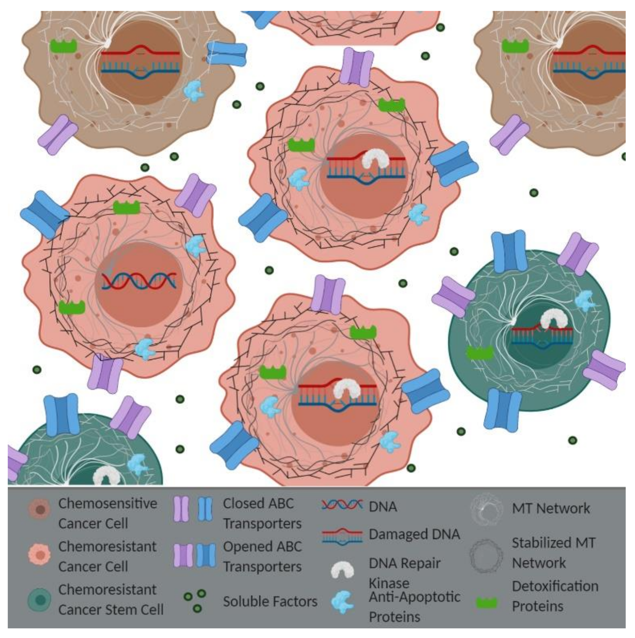
The development of resistance to standard chemotherapies is inevitable. Intracellular factors (i.e., efflux pumps or anti-apoptotic proteins) were originally believed to be the primary way in which cancer cells survived treatment (Figure 1). One of the most notable ways that chemoresistance occurs is through the overexpression of ATP binding cassette (ABC) transporters. The ABC transporters are membrane export pumps that efflux chemotherapeutics out of the cancer cell, thereby preventing drug-induced apoptosis. These transporters include the well-known pumps multidrug resistance 1 (MDR1) and multidrug resistance-associated protein 1 (MRP1). The expression of the MDR1 transporter is increased when APC is mutated [21,40], which may be a result of the Wnt/β-catenin signaling pathway, given that the MDR1 gene promoter contains multiple binding elements for β-catenin/TCF4 [40,41]. The inhibition of Wnt signaling and suppression of MDR1 by overexpressing miR-506 re-sensitized CRC cells to oxaliplatin [42]. Silencing the long non-coding ribonucleic acid-homeobox transcript antisense ribonucleic acid (lncRNA-HOTAIR) in NSCLC cells also inhibited Wnt signaling, decreased the expression of MDR1 and MRP1, and decreased the cisplatin sensitivity [43]. Similarly, overexpressing β-catenin in oral squamous cell carcinoma cells caused cisplatin resistance by increasing the expression of MDR1 and MRP1 [44]. In addition to APC working through the Wnt pathway to increase MDR1, our lab has shown that the loss of APC can function independently of the Wnt pathway, through upregulation of the signal transducer and activator of transcription 3 (STAT3), to induce MDR1 expression [21,45]. Therefore, we and others have demonstrated that APC loss may influence chemoresistance by altering MDR1 through both Wnt-dependent and -independent mechanisms. While these ABC transporters have been shown to contribute to resistance, they unfortunately have not shown much success as therapeutic targets to prevent resistance in the clinic. Toxicity and interactions with other drugs used by the patients were found to be issues in clinical trials. Future drug development for ABC transport modulators includes improving drug delivery and preventing compensation from other drug exporters [46].
Many chemotherapies work by causing DNA damage and subsequent cell death; however, cancer cells have developed enhanced DNA repair pathways to repair the therapy-induced damage and avoid apoptosis. Depending on the type of damage inflicted, repair occurs through different signaling pathways. Oxidation, deamination, and alkylation damage are repaired via BER. 5-fluorouracil (5-FU) is a structural analog of uracil and thymine, and when uracil is processed, it creates an abasic site that is repaired via BER. Using CRC cells with different APC expression levels, it was shown that knocking down APC in HCT-116 cells, which have WT APC, resulted in resistance to 5-FU. In a complementary experiment, introducing full-length APC in LOVO cells, which have mutant APC, resulted in sensitivity to 5-FU [47,48]. It was also shown that APC regulation of the 5-FU therapeutic response stemmed from APC’s role in LP-BER [49]. Recently, the development of poly (ADP-Ribose) polymerase (PARP) inhibitors for treatment in BRCA-mutated cancers has highlighted the potential targeting of DNA repair proteins as a therapeutic approach. PARP inhibitors have improved patient prognoses; however, it was recently shown that Wnt signaling can regulate the response to PARP inhibitors in ovarian cancer [50,51]. Furthermore, it was found that APC was necessary to induce BER in metastatic breast cancer stem cells (mBCSCs) following treatment with the PARP inhibitor ABT-888 in mBCSCs pre-treated with the small molecule Quinacrine. While the knockdown of APC in mBCSCs decreased DNA damage, increased BER activity, and reduced apoptosis, the overexpression of APC in BT20 breast cancer cells displayed the opposite affects [52]. The double-stranded breaks that are created by chemotherapeutic agents are repaired through either NHEJ or homologous recombination (HR), in which APC is known to interact with RPA32 and DNA-PKcs. It was also shown that inhibitors for the DNA repair kinases ataxia telangiectasia mutated (ATM) and DNA-PK restored doxorubicin-induced apoptosis in doxorubicin-resistant APC-deficient breast cancer cells [39]. The role of APC in multiple DNA repair pathways demonstrates APC’s potential as a therapeutic marker like BRCA.
The recognition that altered metabolic pathways in cancer cells contribute to chemoresistance was only established within the last decade. [53]. Cancer cells prefer aerobic glycolysis, referred to as the Warburg effect, partially because of mitochondrial dysfunction disrupting pyruvate metabolism. It was recently shown that APC regulated the expression of the mitochondrial pyruvate carrier (MPC), affecting pyruvate metabolism and promoting tumor development [54,55]. Inhibiting MPC activity using UK5099 reduced the sensitivity to cisplatin compared to untreated LNCaP prostate cancer cells by inducing a stem-like phenotype [56]. These studies suggest that APC could work through MPC to alter the response to chemotherapy. In addition to MPC, glutathione metabolism is an important contributor to cisplatin resistance in NSCLC cells [57,58]. Glutathione is necessary in detoxification systems that combat reactive oxygen species (ROS) production induced by chemotherapeutic drugs. Cancer cells have increased detoxification and antioxidant systems to combat this ROS, such as glutathione S-transferase (GST), glutathione peroxidase (GSH-PX), and superoxide dismutase (SOD) [59]. Giera et al. demonstrated that the constitutive activation of β-catenin increased GST isoforms in mouse hepatomas, which can conjugate a myriad of hydrophobic and electrophilic molecules to glutathione, allowing for drug exportation [60,61]. It was also shown that glutamine activated the Wnt pathway and increased the expression of GSH-PX and SOD in Alzheimer’s disease. Inhibition of the Wnt pathway prevented these glutamine-induced antioxidants [62]. These Wnt-dependent processes downstream of glutathione are important because the utilization of glutathione synthesis inhibitors increases the sensitivity to cisplatin in cervical cancer [63]. Therefore, metabolic changes preventing chemotherapy efficacy represent another target for combination therapy for improving patient prognoses. Taken together, these studies suggest that APC’s role in Wnt regulation could contribute to resistance through alterations in glutathione-dependent detoxification systems; targeting these chemoresistant mechanisms could be a therapeutic target in APC-deficient cancers.
Cancer stems cells (CSCs) are naturally more resistant to chemotherapy due to their slower cycling time, increased expression of ABC transporters, and enhanced DNA repair [64]. Inhibiting Wnt signaling decreased CSC marker proteins while increasing the sensitivity to 5-FU in CRC cells [65]. In addition, chemotherapeutics can induce cellular senescence and stemness via Wnt activation [66]. CSCs also produce less ROS while increasing antioxidant systems to combat any oxidative stress. Decreasing ROS scavenging in CSCs increased the sensitivity to radiation therapy [67,68]. Chemotherapy-resistant MMTV-PyMT;ApcMin/+ cells lacking elevated Wnt signaling displayed increased numbers of ALDH-high cells, which is a marker of CSCs [21] and correlates with chemoresistance [69]. This suggests that APC has functions supporting stemness, independent of β-catenin. The use of an ALDH inhibitor increased the sensitivity of chemotherapy and radiotherapy in ALDHhigh/CD44+ breast cancer cells [70]. Therefore, APC regulation of CSCs may provide information on how to eliminate the chemoresistant CSC population.
The taxanes, which are a type of mitotic inhibitor, stabilize microtubules (MTs), disrupting mitosis and cell cycle progression. Directly preventing taxane’s mode of action by preventing the taxane stabilization of MTs is one mechanism leading to taxane resistance, which can be accomplished through alterations to the MT network, including microtubule-associated proteins (MAPs) such as APC. APC’s role in stabilizing MTs, independent of its interaction with the plus end MT binding protein EB1, can regulate the cell’s response to paclitaxel [71]. Low doses of paclitaxel in APC-deficient intestinal enterocytes failed to decrease the number of mitotic cells compared to wild-type enterocytes [72]. Resistance to paclitaxel was also seen in breast cells lacking APC [21]. APC loss decreases the efficacy of microtubule-stabilizing agents; however, the use of the MT-destabilizing agent vinorelbine preferentially killed APC-deficient U2OS cells through its ability to induce apoptosis during interphase, as well as mitosis [73]. This demonstrates the need to consider the molecular tumor profile to establish an appropriate therapeutic approach.
The ultimate goal of chemotherapy is to induce apoptosis in cancer cells. Therefore, the mis-regulation of pro- and anti-apoptosis proteins can induce chemoresistance. Bladder cancer patients who were resistant to cisplatin treatment also showed an increased nuclear expression of survivin, which is a Wnt target that is an inhibitor of the apoptosis protein (IAP) [74]. The co-expression of APC and AXIN employed to inhibit Wnt signaling reduced survivin expression and inhibited cell growth [75]. As survivin is primarily expressed in cancer cells, inhibitors are currently being evaluated as a single agent or in combination [76,77]. Another family of proteins regulating apoptosis are the B-cell lymphoma 2 (BCL-2) family proteins, which are crucial in the mitochondrial death pathway. The loss of APC increased the expression of the pro-survival protein BCL-2, but not MCL-1, in MMTV-PyMT;ApcMin/+ cells [45]. BCL-2 inhibitors are clinically being investigated as a potential combination therapy. Venclexta, which is a BCL-2 small molecule inhibitor and FDA approved for treatment in lymphocytic leukemia, is therapeutically efficacious in refractory and relapsed chronic lymphocytic leukemia, including in combating resistance [78]. An increased expression of the downstream apoptotic mediators caspase 3, 7, and 9 was found in Apc-mutated colon cancer [79], suggesting that the loss of APC increases the apoptotic response. This demonstrates that APC regulates the expression of caspases and other apoptotic proteins.
The heterogeneous nature of tumors also contributes to chemotherapy resistance through cell–cell interactions. Wnt activation through miR-103/107 targeting Axin2 promoted stemness and chemoresistance [80]. MiR-130a induced cisplatin resistance in hepatocellular carcinoma cells (HCCs) by inhibiting the tumor suppressor gene RUNX3, thereby activating Wnt [81]. In addition, miR-92a is upregulated in 5-FU-resistant CRC cells, and an ectopic expression of miR-92a induced chemoresistance in CRC cells through increased Wnt activation by targeting negative regulators of Wnt signaling. Interleukin-6 (IL-6)/STAT3 was found to increase miR-92a expression, promoting a stem-like phenotype to induce resistance [82]. Our lab demonstrated that doxorubicin resistance in MMTV-PyMT;ApcMin/+ cells was mediated by increased STAT3 expression, independent of the Wnt pathway [45]. The upregulation of miR-135 induced paclitaxel resistance via downregulating APC in lung, uterine, and breast cancer cells [83,84]. Recently, the re-expression of APC was shown using the lncRNA SMAD5-AS1, which acts as a competitive endogenous RNA to miR-135, preventing APC inhibition, suggesting the use of lncRNAs for restoring gene expression [85]. APC loss mediates chemoresistance through cell–cell interactions via Wnt-dependent and -independent mechanisms.
5. APC Loss and Tumor Microenvironment-Derived Chemoresistance
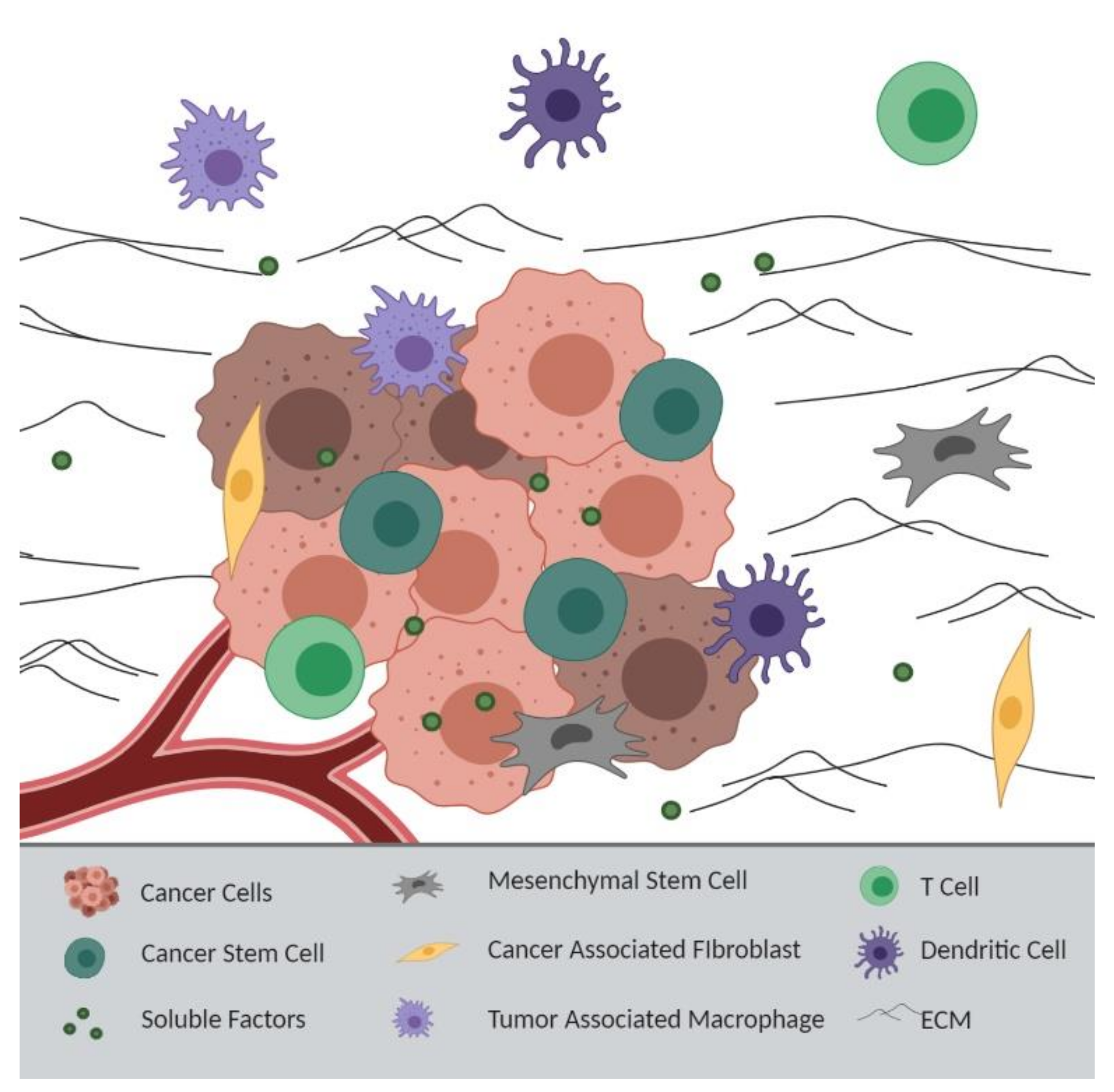
Within the past decade, the shifted emphasis onto the tumor microenvironment (TME) has demonstrated that extracellular factors support the development of chemoresistance [86]. The heterogeneous tumor landscape, which is comprised of both cellular components (cancer, endothelial, and stromal cells) and noncellular components (the extracellular matrix (ECM) and soluble factors), creates barriers that promote chemoresistance. Cancer cells can influence the TME to support their own survival. Cells that are normally anti-tumorigenic cells, including fibroblasts and macrophages, can be converted into tumor-supporting cells, such as cancer-associated fibroblasts (CAFs) and tumor-associated macrophages (TAMs) [86]. Exosomes, which are small lipid vesicles containing proteins and genetic material, are secreted by different types of cells into the TME, inducing pro-invasion and survival signaling in cancer cells [87]. In addition, inflammatory factors in the TME mediate epithelial–mesenchymal transition (EMT), metastasis, and resistance through the expansion and recruitment of CAFs and immune cells such as macrophages [88]. Together, the TME and tumor cells crosstalk to support cancer cell survival (Figure 2).
A defining feature of the tumor landscape is pockets of hypoxia, demanding increased vascular formation or angiogenesis to support the metabolic need of cancer cells. Many drugs induce cell death through the production of free radicals and oxidative stress and therefore require oxygen to induce apoptosis. Hypoxic glioblastoma cells released exosomes containing miR-301a into the TME, activating Wnt signaling and decreasing the sensitivity to radiation [89]. In addition, APC has an antagonistic relationship with hypoxia-inducible factor-1α (HIF-1α), where HIF-1α represses the transcription of APC and APC loss increases HIF-1α [90]. In a hypoxic model of diffuse large B-cell lymphoma, a reduced HIF-1α expression resulted in enhanced doxorubicin-induced apoptosis [91]. HIF-1α inhibition also increased the chemosensitivity under hypoxic conditions in acute lymphocytic leukemia cells [92]. Reduced angiogenesis to tumors impeded drug delivery, as well as free radical formation from oxygen depletion. APC binds to the Rac-specific GEF Asef, enhancing Asef activity [32]. The APC/Asef complex increased endothelial cell migration and tube formation through increased basic fibroblast growth factor (bFGF) and vascular endothelial growth factor (VEGF). Asef−/− mice also displayed reduced microvessel formation [93]. This suggests that APC/Asef could be necessary for angiogenesis to supply tumor cells with an adequate blood supply.
Mesenchymal stromal/stem cells (MSCs) are recruited to tumors and release soluble factors to the cancer cells [86]. Human umbilical cord-derived MSCs co-cultured with a human cholangiocarcinoma cell line produced conditioned media that contributed to drug resistance and metastasis through increased Wnt activation and the upregulation of Wnt target genes (matrix metalloproteinase 2 (MMP2), cyclin D1, and c-myc) [94]. Irradiated MSCs supported cancer stemness in HCCs via Wnt activation, contributing to radiation resistance [95]. Moreover, FAP patients often present with desmoid tumors that originate from mesenchymal cells because APC loss dysregulates Wnt signaling and increases growth in MSCs [96]. Therefore, APC regulates the growth of MSCs, which contribute to a chemoresistant phenotype. Interestingly, some studies have suggested that MSCs are differentiated into CAFs, but this is still debated [86].
Some of the most studied aspects of the TME contributing to resistance are CAFs and their transport and the release of soluble factors. Exosomes secreted from CAFs released Wnt ligands into the TME, which activated Wnt signaling and drug resistance in differentiated CRC cells. In contrast, inhibiting Wnt secretion in Wnt3a overexpressing CAFs prevented resistance [97]. CAFs express the lncRNA colorectal cancer-associated lncRNA (CCAL), which activates Wnt signaling to promote oxaliplatin resistance in CRC cells [98]. CCAL-induced Wnt activation also increased MDR1 expression [99], which may allow for alterations in drug efflux. In CRC cells, CAFs transfer exosomes to increase miR-92a-3p, which activates Wnt signaling, preventing mitochondrial apoptosis and increasing stemness, EMT, and 5-FU/Oxaliplatin resistance [100]. Following chemotherapy or radiation, fibroblast-secreted WNT16B activated Wnt in primary prostate cells through NF-κB to promote a mesenchymal phenotype and evade apoptosis [101]. While these data point to a role for Wnt signaling, an inference could be made that one method of Wnt signaling activation is the loss of APC, which could lead to the activation of the same resistance mechanisms. The stromal depletion of APC in the uterine stroma induced a myofibroblast-like phenotype, similar to the phenotype of CAFs, contributing to endometrial hyperplasia and carcinogenesis through unopposed estrogen signaling in endometrial cells [102]. These studies demonstrate that an APC-deficient stroma can increase tumorgenicity and chemosensitivity.
TAMs also secrete soluble factors into the TME that influence the therapeutic response [103]. Crosstalk between tumor cells and TAMs or M2-like macrophages promotes cancer cell survival. Not only do M2 macrophages secrete Wnt ligands to stimulate Wnt signaling in epithelial tumor cells, inducing stemness and the EMT phenotype, but Wnt6 expression in granulomatous lesions drives macrophage polarization toward an M2-like phenotype, which was also seen in HCCs [104,105]. HCCs were also shown to secrete Wnt ligands to activate M2 macrophages into TAMs [106]. This demonstrates a positive feedback loop between cells, where stromal Wnt drives cancer progression and cancer cell-derived Wnt stimulates a tumor-supporting microenvironment. TAM-secreted CCL5 promoted migration and EMT in prostate cancer cells, while also supporting CSC self-renewal by activating β-catenin/STAT3 [107]. In addition, TAMs release the cytokine IL-6, reducing the 5-FU response in CRC subcutaneous tumors via IL-6/STAT3 [108]. We found a loss of APC-induced doxorubicin resistance through an increased expression of STAT3 in MMTV-PyMT;ApcMin/+ cells [45]. ApcMin/+ mice have an increased expression of the M1 macrophage marker IL-23 and the M2 macrophage markers IL-13 and CCL17. The knock-out of monocyte chemoattractant protein 1 (MCP-1), which is an important chemokine for macrophage recruitment, decreased the expression of IL-1 and IL-6 in polyps of ApcMin/+ mice. No change was observed in β-catenin staining between ApcMin/+ and ApcMin/+/MCP-1−/− mice, demonstrating that macrophage recruitment in ApcMin/+ mice is independent of Wnt activation [109]. The loss of APC and subsequent Wnt signaling support macrophage infiltration into the tumor, which negatively impacts the drug response.
The newest promising therapy option in oncology is immunotherapy. Tumor cells can affect the TME by preventing immune cell recruitment to the tumor, rendering immunotherapy ineffective. Metastatic melanoma cells with active Wnt signaling displayed a decreased expression of the chemokine CCL4, preventing CD103+ dendritic cell (DC) recruitment and the subsequent activation of CD8+ cytotoxic T-cells. The direct injection of DCs increased the sensitivity to the immune checkpoint blockade [110,111]. This demonstrates that Wnt activation preventing DC recruitment can affect the response to immunotherapy. Similarly, Wnt signaling activation in HCCs reduced DC recruitment and the efficacy of the immune checkpoint blockade anti-PD-1 [112]. Wnt inhibitors are being evaluated, in combination with immune checkpoint inhibitors, such as anti-PDL-1, in preclinical trials [64]. Apc-deficient mice exhibited an impaired differentiation of T regulatory cells (Tregs) and high levels of the immune suppressive cytokine IL-10 because APC loss reduced the nuclear localization of Nuclear Factor of Activated T Cells (NFAT) [113]. This immune alteration was shown to occur partially outside of APC’s role in Wnt regulation. CD4+ T cell development was also impaired in ApcMin/+ mice [114], demonstrating that APC loss alters the immune cell response, which may reduce the immunotherapy efficacy [115]. Overall, APC should be considered as its own therapeutic marker because of APC’s Wnt-dependent and -independent roles.
6. Conclusions
APC is lost in numerous cancer types, through either mutations or promoter hypermethylation. More recent studies have shown that APC loss can cause resistance and may be used to predict the response to chemotherapy. The loss of APC and lack of control of the APC-mediated cellular functions present a therapeutic challenge given that the restoration of APC has been clinically challenging. Resistance can occur intracellularly, intercellularly (with TME), or through a relationship between the cells and the ECM. APC loss affects many cellular processes and may thus be responsible for multiple mechanisms of chemotherapy resistance. Future studies need to address whether APC can be used as a therapeutic marker. While the role of Wnt activation contributing to chemoresistance is well-studied, the Wnt-independent roles of APC are largely ignored. These Wnt-independent mechanisms demonstrate the importance of APC as a unique therapeutic marker outside of Wnt/β-catenin activation. Furthermore, the role of APC in the TME and how this affects the therapeutic response is understudied. Understanding how APC imparts chemoresistance will be imperative in discovering combination therapies to overcome resistance and improve patient outcomes.
Author Contributions
Manuscript written by C.D.S. with support from J.R.P. All authors have read and agreed to the published version of the manuscript.
Funding
Work in the lab was funded by an A.C.S. Research Scholar Grant awarded to J.R.P.
Acknowledgments
Figures were made using BioRender.
Conflicts of Interest
The authors declare no conflict of interest.
Abbreviations
| +TIPs | Plus end-binding proteins |
| 5-FU | 5-fluorouracil |
| ABC | ATP binding cassette |
| ALDH | Aldehyde dehydrogenase |
| APC | Adenomatous polyposis coli |
| APE1 | AP endonuclease 1 |
| ATM | Ataxia telangiectasia mutated |
| ATR | Ataxia telangiectasia and Rad3-related protein |
| BCL-2 | B cell lymphoma 2 |
| bFGF | Basic fibroblast growth factor |
| CAFs | Cancer-associated fibroblast |
| CCAL | Colorectal cancer-associated lncRNA |
| Chk1 | Checkpoint kinase 1 |
| CK1 | Casein kinase 1 |
| CRC | Colorectal cancer |
| CSCs | Cancer stem cells |
| DC | Dendritic cell |
| DNA-PKcs | DNA-dependent protein kinase catalytic subunits |
| EB1 | End-binding proteins |
| ECM | Extracellular matrix |
| EMT | Epithelial–mesenchymal transition |
| FAP | Familial adenomatous polyposis |
| Fen-1 | Flap endonuclease 1 |
| GEF | Guanine nucleotide exchange factor |
| GSH-PX | Glutathione peroxidase |
| GSK3β | Glycogen synthase kinase 3β |
| GST | Glutathione S-transferase |
| HCCs | Hepatocellular carcinoma cells |
| HIF-1α | Hypoxia-inducible factor-1α |
| HR | Homologous recombination |
| IL-6 | Interleukin-6 |
| LncRNA-HOTAIR | Long non-coding ribonucleic acid-homeobox transcript antisense ribonucleic acid |
| LP-BER | Long patch-base excision repair |
| MAPs | Microtubule-associated proteins |
| mBCSCs | Metastatic breast cancer stem cells |
| MCP-1 | Monocyte chemoattractant protein 1 |
| MCR | Mutation cluster region |
| MDR1 | Multidrug resistance 1 |
| MMP2 | Matrix metalloproteinase 2 |
| MMTV-PyMT | Mouse mammary tumor virus-polyoma middle T |
| MPC | Mitochondrial pyruvate carrier |
| MRP1 | Multidrug resistance-associated protein 1 |
| MSCs | Mesenchymal stromal/stem cells |
| MTs | Microtubules |
| NFAT | Nuclear factor of activated T cells |
| NHEJ | Non-homologous end joining (NHEJ) |
| NSCLC | Non-small cell lung cancer |
| PARP | Poly (ADP-Ribose) polymerase |
| Pol-β | Polymerase β |
| ROS | Reactive oxygen species |
| RPA32 SAMP | Replication protein A 32 Serine, Alanine, Methionine, Proline |
| STAT3 | Signal transducer and activator of transcription 3 |
| SOD | Superoxide dismutase |
| TAMs | Tumor-associated macrophages |
| TME | Tumor microenvironment |
| Tregs | T regulatory cells |
| VEGF | Vascular endothelial growth factor |
| γ-H2AX | Phosphorylated histone H2AX |
References
- Ji, X.; Lu, Y.; Tian, H.; Meng, X.; Wei, M.; Cho, W.C. Chemoresistance mechanisms of breast cancer and their countermeasures. Biomed. Pharmacother. 2019, 114, 108800. [Google Scholar] [CrossRef] [PubMed]
- Perez, E.A. Impact, mechanisms, and novel chemotherapy strategies for overcoming resistance to anthracyclines and taxanes in metastatic breast cancer. Breast Cancer Res. Treat. 2008, 114, 195. [Google Scholar] [CrossRef] [PubMed]
- Groden, J.; Thliveris, A.; Samowitz, W.; Carlson, M.; Gelbert, L.; Albertsen, H.; Joslyn, G.; Stevens, J.; Spirio, L.; Robertson, M.; et al. Identification and characterization of the familial adenomatous polyposis coli gene. Cell 1991, 66, 589–600. [Google Scholar] [CrossRef]
- Waller, A.; Findeis, S.; Lee, M.J. Familial Adenomatous Polyposis. J. Pediatr. Genet. 2016, 5, 78–83. [Google Scholar] [CrossRef] [PubMed] [Green Version]
- Zhang, L.; Shay, J.W. Multiple Roles of APC and its Therapeutic Implications in Colorectal Cancer. J. Natl. Cancer Inst. 2017, 109, djw332. [Google Scholar] [CrossRef] [Green Version]
- Muzny, D.M.; Bainbridge, M.N.; Chang, K.; Dinh, H.H.; Drummond, J.A.; Fowler, G.; Kovar, C.L.; Lewis, L.R.; Morgan, M.B.; Newsham, I.F.; et al. Comprehensive molecular characterization of human colon and rectal cancer. Nature 2012, 487, 330–337. [Google Scholar] [CrossRef] [Green Version]
- Yuan, S.; Tao, F.; Zhang, X.; Zhang, Y.; Sun, X.; Wu, D. Role of Wnt/β-Catenin Signaling in the Chemoresistance Modulation of Colorectal Cancer. Biomed. Res. Int. 2020, 2020, 9390878. [Google Scholar] [CrossRef] [Green Version]
- Arnold, C.N.; Goel, A.; Niedzwiecki, D.; Dowell, J.M.; Wasserman, L.; Compton, C.; Mayer, R.J.; Bertagnolli, M.M.; Boland, C.R. APC promoter hypermethylation contributes to the loss of APC expression in colorectal cancers with allelic loss on 5q. Cancer Biol. 2004, 3, 960–964. [Google Scholar] [CrossRef] [Green Version]
- Brabender, J.; Usadel, H.; Danenberg, K.D.; Metzger, R.; Schneider, P.M.; Lord, R.V.; Wickramasinghe, K.; Lum, C.E.; Park, J.; Salonga, D.; et al. Adenomatous polyposis coli gene promoter hypermethylation in non-small cell lung cancer is associated with survival. Oncogene 2001, 20, 3528–3532. [Google Scholar] [CrossRef] [Green Version]
- Esteller, M.; Sparks, A.; Toyota, M.; Sanchez-Cespedes, M.; Capella, G.; Peinado, M.A.; Gonzalez, S.; Tarafa, G.; Sidransky, D.; Meltzer, S.J.; et al. Analysis of adenomatous polyposis coli promoter hypermethylation in human cancer. Cancer Res. 2000, 60, 4366–4371. [Google Scholar]
- Jin, Z.; Tamura, G.; Tsuchiya, T.; Sakata, K.; Kashiwaba, M.; Osakabe, M.; Motoyama, T. Adenomatous polyposis coli (APC) gene promoter hypermethylation in primary breast cancers. Br. J. Cancer 2001, 85, 69–73. [Google Scholar] [CrossRef] [Green Version]
- Li, B.-Q.; Liu, P.-P.; Zhang, C.-H. Correlation between the methylation of APC gene promoter and colon cancer. Oncol. Lett. 2017, 14, 2315–2319. [Google Scholar] [CrossRef] [PubMed] [Green Version]
- Nagel, R.; le Sage, C.; Diosdado, B.; van der Waal, M.; Oude Vrielink, J.A.F.; Bolijn, A.; Meijer, G.A.; Agami, R. Regulation of the Adenomatous Polyposis Coli Gene by the miR-135 Family in Colorectal Cancer. Cancer Res. 2008, 68, 5795–5802. [Google Scholar] [CrossRef] [PubMed] [Green Version]
- Jiang, D.; Zhou, B.; Xiong, Y.; Cai, H. miR-135 regulated breast cancer proliferation and epithelial-mesenchymal transition acts by the Wnt/β-catenin signaling pathway. Int. J. Mol. Med. 2019, 43, 1623–1634. [Google Scholar] [CrossRef] [PubMed]
- Wang, L.-p.; Ma, X.-q.; Cai, J.-c. [Clinicopathological significance and function of miR-135b in the occurrence and development of gastric cancer]. Zhonghua Yi Xue Za Zhi 2012, 92, 3269–3273. [Google Scholar] [PubMed]
- Mukherjee, N.; Bhattacharya, N.; Alam, N.; Roy, A.; Roychoudhury, S.; Panda, C.K. Subtype-specific alterations of the Wnt signaling pathway in breast cancer: Clinical and prognostic significance. Cancer Sci. 2012, 103, 210–220. [Google Scholar] [CrossRef]
- Prosperi, J.R.; Goss, K.H. Wnt Pathway-Independent Activities of the APC Tumor Suppressor. In Tumor Suppressors; Susan, D.N., Ed.; Nova Science Publishers, Inc.: Hauppauge, NY, USA, 2011. [Google Scholar]
- Korinek, V.; Barker, N.; Moerer, P.; van Donselaar, E.; Huls, G.; Peters, P.J.; Clevers, H. Depletion of epithelial stem-cell compartments in the small intestine of mice lacking Tcf-4. Nat. Genet. 1998, 19, 379–383. [Google Scholar] [CrossRef]
- Sato, T.; Vries, R.G.; Snippert, H.J.; van de Wetering, M.; Barker, N.; Stange, D.E.; van Es, J.H.; Abo, A.; Kujala, P.; Peters, P.J.; et al. Single Lgr5 stem cells build crypt-villus structures in vitro without a mesenchymal niche. Nature 2009, 459, 262–265. [Google Scholar] [CrossRef]
- Jamieson, C.H.M.; Ailles, L.E.; Dylla, S.J.; Muijtjens, M.; Jones, C.; Zehnder, J.L.; Gotlib, J.; Li, K.; Manz, M.G.; Keating, A.; et al. Granulocyte–Macrophage Progenitors as Candidate Leukemic Stem Cells in Blast-Crisis CML. N. Engl. J. Med. 2004, 351, 657–667. [Google Scholar] [CrossRef] [Green Version]
- VanKlompenberg, M.K.; Bedalov, C.O.; Soto, K.F.; Prosperi, J.R. APC selectively mediates response to chemotherapeutic agents in breast cancer. BMC Cancer 2015, 15, 457. [Google Scholar] [CrossRef] [Green Version]
- Wang, Y.; Azuma, Y.; Moore, D.; Osheroff, N.; Neufeld, K.L. Interaction between tumor suppressor adenomatous polyposis coli and topoisomerase IIalpha: Implication for the G2/M transition. Mol. Biol. Cell 2008, 19, 4076–4085. [Google Scholar] [CrossRef] [PubMed] [Green Version]
- Wang, Y.; Coffey, R.J.; Osheroff, N.; Neufeld, K.L. Topoisomerase IIalpha binding domains of adenomatous polyposis coli influence cell cycle progression and aneuploidy. PLoS ONE 2010, 5, e9994. [Google Scholar] [CrossRef]
- Fodde, R.; Kuipers, J.; Rosenberg, C.; Smits, R.; Kielman, M.; Gaspar, C.; van Es, J.H.; Breukel, C.; Wiegant, J.; Giles, R.H.; et al. Mutations in the APC tumour suppressor gene cause chromosomal instability. Nat. Cell Biol. 2001, 3, 433–438. [Google Scholar]
- Dikovskaya, D.; Newton, I.P.; Nathke, I.S. The adenomatous polyposis coli protein is required for the formation of robust spindles formed in CSF Xenopus extracts. Mol. Biol. Cell 2004, 15, 2978–2991. [Google Scholar]
- Kaplan, K.B.; Burds, A.A.; Swedlow, J.R.; Bekir, S.S.; Sorger, P.K.; Nathke, I.S. A role for the Adenomatous Polyposis Coli protein in chromosome segregation. Nat. Cell Biol. 2001, 3, 429–432. [Google Scholar]
- Green, R.A.; Wollman, R.; Kaplan, K.B. APC and EB1 function together in mitosis to regulate spindle dynamics and chromosome alignment. Mol. Biol. Cell 2005, 16, 4609–4622. [Google Scholar] [PubMed] [Green Version]
- Kita, K.; Wittmann, T.; Nathke, I.S.; Waterman-Storer, C.M. Adenomatous polyposis coli on microtubule plus ends in cell extensions can promote microtubule net growth with or without EB1. Mol. Biol. Cell 2006, 17, 2331–2345. [Google Scholar]
- Efimova, N.; Yang, C.; Chia, J.X.; Li, N.; Lengner, C.J.; Neufeld, K.L.; Svitkina, T.M. Branched actin networks are assembled on microtubules by adenomatous polyposis coli for targeted membrane protrusion. J. Cell Biol. 2020, 219. [Google Scholar] [CrossRef]
- Oshima, H.; Oshima, M.; Kobayashi, M.; Tsutsumi, M.; Taketo, M.M. Morphological and Molecular Processes of Polyp Formation in ApcΔ716 Knockout Mice. 1997, 57, 1644–1649. Cancer Res. 1997, 57, 1644–1649. [Google Scholar]
- Kawasaki, Y.; Sato, R.; Akiyama, T. Mutated APC and Asef are involved in the migration of colorectal tumour cells. Nat. Cell Biol. 2003, 5, 211–215. [Google Scholar]
- Kawasaki, Y.; Senda, T.; Ishidate, T.; Koyama, R.; Morishita, T.; Iwayama, Y.; Higuchi, O.; Akiyama, T. Asef, a link between the tumor suppressor APC and G-protein signaling. Science 2000, 289, 1194–1197. [Google Scholar] [CrossRef]
- Odenwald, M.A.; Prosperi, J.R.; Goss, K.H. APC/β-catenin-rich complexes at membrane protrusions regulate mammary tumor cell migration and mesenchymal morphology. BMC Cancer 2013, 13, 12. [Google Scholar] [CrossRef] [Green Version]
- Lesko, A.C.; Prosperi, J.R. Epithelial Membrane Protein 2 and beta1 integrin signaling regulate APC-mediated processes. Exp. Cell Res. 2017, 350, 190–198. [Google Scholar] [CrossRef] [PubMed]
- Kroboth, K.; Newton, I.P.; Kita, K.; Dikovskaya, D.; Zumbrunn, J.; Waterman-Storer, C.M.; Nathke, I.S. Lack of adenomatous polyposis coli protein correlates with a decrease in cell migration and overall changes in microtubule stability. Mol. Biol. Cell 2007, 18, 910–918. [Google Scholar] [CrossRef] [PubMed]
- Welch, D.R.; Hurst, D.R. Defining the Hallmarks of Metastasis. Cancer Res. 2019, 79, 3011–3027. [Google Scholar] [CrossRef]
- Brocardo, M.G.; Borowiec, J.A.; Henderson, B.R. Adenomatous polyposis coli protein regulates the cellular response to DNA replication stress. Int. J. Biochem. Cell Biol. 2011, 43, 1354–1364. [Google Scholar] [CrossRef]
- Kouzmenko, A.P.; Takeyama, K.; Kawasaki, Y.; Akiyama, T.; Kato, S. Truncation mutations abolish chromatin-associated activities of adenomatous polyposis coli. Oncogene 2008, 27, 4888–4899. [Google Scholar] [CrossRef] [PubMed] [Green Version]
- Stefanski, C.; Keffler, K.; McClintock, S.; Milac, L.; Prosperi, J. APC loss affects DNA damage repair causing doxorubicin resistance in breast cancer cells. Neoplasia 2019, 21, 1143–1150. [Google Scholar] [CrossRef] [PubMed]
- Yamada, T.; Mori, Y.; Hayashi, R.; Takada, M.; Ino, Y.; Naishiro, Y.; Kondo, T.; Hirohashi, S. Suppression of intestinal polyposis in Mdr1-deficient ApcMin/+ mice. Cancer Res. 2003, 63, 895–901. [Google Scholar]
- Yamada, T.; Takaoka, A.S.; Naishiro, Y.; Hayashi, R.; Maruyama, K.; Maesawa, C.; Ochiai, A.; Hirohashi, S. Transactivation of the Multidrug Resistance 1 Gene by T-Cell Factor 4/β-Catenin Complex in Early Colorectal Carcinogenesis. Cancer Res. 2000, 60, 4761–4766. [Google Scholar]
- Zhou, H.; Lin, C.; Zhang, Y.; Zhang, X.; Zhang, C.; Zhang, P.; Xie, X.; Ren, Z. miR-506 enhances the sensitivity of human colorectal cancer cells to oxaliplatin by suppressing MDR1/P-gp expression. Cell Prolif. 2017, 50, e12341. [Google Scholar] [CrossRef]
- Guo, F.; Cao, Z.; Guo, H.; Li, S. The action mechanism of lncRNA-HOTAIR on the drug resistance of non-small cell lung cancer by regulating Wnt signaling pathway. Exp. Med. 2018, 15, 4885–4889. [Google Scholar] [CrossRef] [PubMed] [Green Version]
- Li, L.; Liu, H.-C.; Wang, C.; Liu, X.; Hu, F.-C.; Xie, N.; Lü, L.; Chen, X.; Huang, H.-Z. Overexpression of β-Catenin Induces Cisplatin Resistance in Oral Squamous Cell Carcinoma. Biomed. Res. Int. 2016, 2016, 5378567. [Google Scholar] [CrossRef] [Green Version]
- VanKlompenberg, M.K.; Leyden, E.; Arnason, A.H.; Zhang, J.T.; Stefanski, C.D.; Prosperi, J.R. APC loss in breast cancer leads to doxorubicin resistance via STAT3 activation. Oncotarget 2017, 8, 102868–102879. [Google Scholar] [CrossRef] [PubMed] [Green Version]
- Choi, Y.H.; Yu, A.-M. ABC transporters in multidrug resistance and pharmacokinetics, and strategies for drug development. Curr. Pharm. Des. 2014, 20, 793–807. [Google Scholar] [CrossRef]
- Wyatt, M.D.; Wilson, D.M., 3rd. Participation of DNA repair in the response to 5-fluorouracil. Cell Mol. Life Sci. 2009, 66, 788–799. [Google Scholar] [CrossRef] [PubMed] [Green Version]
- Krokan, H.E.; Bjørås, M. Base excision repair. Cold Spring Harb. Perspect. Biol. 2013, 5, a012583. [Google Scholar] [CrossRef]
- Das, D.; Preet, R.; Mohapatra, P.; Satapathy, S.R.; Siddharth, S.; Tamir, T.; Jain, V.; Bharatam, P.V.; Wyatt, M.D.; Kundu, C.N. 5-Fluorouracil mediated anti-cancer activity in colon cancer cells is through the induction of Adenomatous Polyposis Coli: Implication of the long-patch base excision repair pathway. DNA Repair 2014, 24, 15–25. [Google Scholar] [CrossRef] [Green Version]
- Yamamoto, T.M.; McMellen, A.; Watson, Z.L.; Aguilera, J.; Ferguson, R.; Nurmemmedov, E.; Thakar, T.; Moldovan, G.-L.; Kim, H.; Cittelly, D.M.; et al. Activation of Wnt signaling promotes olaparib resistant ovarian cancer. Mol. Carcinog. 2019, 58, 1770–1782. [Google Scholar] [CrossRef]
- Fukumoto, T.; Zhu, H.; Nacarelli, T.; Karakashev, S.; Fatkhutdinov, N.; Wu, S.; Liu, P.; Kossenkov, A.V.; Showe, L.C.; Jean, S.; et al. N(6)-Methylation of Adenosine of FZD10 mRNA Contributes to PARP Inhibitor Resistance. Cancer Res. 2019, 79, 2812–2820. [Google Scholar] [CrossRef] [Green Version]
- Siddharth, S.; Nayak, D.; Nayak, A.; Das, S.; Kundu, C.N. ABT-888 and quinacrine induced apoptosis in metastatic breast cancer stem cells by inhibiting base excision repair via adenomatous polyposis coli. DNA Repair 2016, 45, 44–55. [Google Scholar] [CrossRef] [PubMed]
- Zaal, E.A.; Berkers, C.R. The Influence of Metabolism on Drug Response in Cancer. Front. Oncol. 2018, 8, 500. [Google Scholar] [CrossRef] [PubMed]
- Sandoval, I.T.; Delacruz, R.G.C.; Miller, B.N.; Hill, S.; Olson, K.A.; Gabriel, A.E.; Boyd, K.; Satterfield, C.; Van Remmen, H.; Rutter, J.; et al. A metabolic switch controls intestinal differentiation downstream of Adenomatous polyposis coli (APC). Elife 2017, 6, e22706. [Google Scholar] [CrossRef] [Green Version]
- Bensard, C.L.; Wisidagama, D.R.; Olson, K.A.; Berg, J.A.; Krah, N.M.; Schell, J.C.; Nowinski, S.M.; Fogarty, S.; Bott, A.J.; Wei, P.; et al. Regulation of Tumor Initiation by the Mitochondrial Pyruvate Carrier. Cell Metab. 2020, 31, 284–300.e287. [Google Scholar] [CrossRef]
- Zhong, Y.; Li, X.; Yu, D.; Li, X.; Li, Y.; Long, Y.; Yuan, Y.; Ji, Z.; Zhang, M.; Wen, J.-G.; et al. Application of mitochondrial pyruvate carrier blocker UK5099 creates metabolic reprogram and greater stem-like properties in LnCap prostate cancer cells in vitro. Oncotarget 2015, 6, 37758–37769. [Google Scholar] [CrossRef] [Green Version]
- Shi, Y.; Wang, Y.; Huang, W.; Wang, Y.; Wang, R.; Yuan, Y. Integration of Metabolomics and Transcriptomics to Reveal Metabolic Characteristics and Key Targets Associated with Cisplatin Resistance in Nonsmall Cell Lung Cancer. J. Proteome Res. 2019, 18, 3259–3267. [Google Scholar] [CrossRef]
- Guo, W.; Tan, H.-Y.; Chen, F.; Wang, N.; Feng, Y. Targeting Cancer Metabolism to Resensitize Chemotherapy: Potential Development of Cancer Chemosensitizers from Traditional Chinese Medicines. Cancers 2020, 12, 404. [Google Scholar] [CrossRef] [PubMed] [Green Version]
- Perillo, B.; Di Donato, M.; Pezone, A.; Di Zazzo, E.; Giovannelli, P.; Galasso, G.; Castoria, G.; Migliaccio, A. ROS in cancer therapy: The bright side of the moon. Exp. Mol. Med. 2020, 52, 192–203. [Google Scholar] [CrossRef] [PubMed]
- Giera, S.; Braeuning, A.; Köhle, C.; Bursch, W.; Metzger, U.; Buchmann, A.; Schwarz, M. Wnt/β-Catenin Signaling Activates and Determines Hepatic Zonal Expression of Glutathione S-Transferases in Mouse Liver. Toxicol. Sci. 2010, 115, 22–33. [Google Scholar] [CrossRef] [Green Version]
- Allocati, N.; Masulli, M.; Di Ilio, C.; Federici, L. Glutathione transferases: Substrates, inihibitors and pro-drugs in cancer and neurodegenerative diseases. Oncogenesis 2018, 7, 1–15. [Google Scholar] [CrossRef] [PubMed]
- Wang, Y.; Wang, Q.; Li, J.; Lu, G.; Liu, Z. Glutamine Improves Oxidative Stress through the Wnt3a/β-Catenin Signaling Pathway in Alzheimer’s Disease In Vitro and In Vivo. Biomed. Res. Int. 2019, 2019, 4690280. [Google Scholar] [CrossRef] [PubMed] [Green Version]
- Rashmi, R.; Huang, X.; Floberg, J.M.; Elhammali, A.E.; McCormick, M.L.; Patti, G.J.; Spitz, D.R.; Schwarz, J.K. Radioresistant Cervical Cancers Are Sensitive to Inhibition of Glycolysis and Redox Metabolism. Cancer Res. 2018, 78, 1392–1403. [Google Scholar] [CrossRef] [PubMed] [Green Version]
- Zhong, Z.; Virshup, D.M. Wnt signaling and drug resistance in cancer. Mol. Pharmacol. 2020, 97, 72–89. [Google Scholar] [CrossRef] [Green Version]
- Urushibara, S.; Tsubota, T.; Asai, R.; Azumi, J.; Ashida, K.; Fujiwara, Y.; Shiota, G. WNT/β-Catenin Signaling Inhibitor IC-2 Suppresses Sphere Formation and Sensitizes Colorectal Cancer Cells to 5-Fluorouracil. Anticancer Res. 2017, 37, 4085–4091. [Google Scholar] [PubMed]
- Milanovic, M.; Fan, D.N.Y.; Belenki, D.; Däbritz, J.H.M.; Zhao, Z.; Yu, Y.; Dörr, J.R.; Dimitrova, L.; Lenze, D.; Monteiro Barbosa, I.A.; et al. Senescence-associated reprogramming promotes cancer stemness. Nature 2018, 553, 96–100. [Google Scholar] [CrossRef] [Green Version]
- Diehn, M.; Cho, R.W.; Lobo, N.A.; Kalisky, T.; Dorie, M.J.; Kulp, A.N.; Qian, D.; Lam, J.S.; Ailles, L.E.; Wong, M.; et al. Association of reactive oxygen species levels and radioresistance in cancer stem cells. Nature 2009, 458, 780–783. [Google Scholar] [CrossRef]
- Das, P.K.; Islam, F.; Lam, A.K. The Roles of Cancer Stem Cells and Therapy Resistance in Colorectal Carcinoma. Cells 2020, 9, 1392. [Google Scholar] [CrossRef]
- Ginestier, C.; Hur, M.H.; Charafe-Jauffret, E.; Monville, F.; Dutcher, J.; Brown, M.; Jacquemier, J.; Viens, P.; Kleer, C.G.; Liu, S.; et al. ALDH1 is a marker of normal and malignant human mammary stem cells and a predictor of poor clinical outcome. Cell Stem Cell 2007, 1, 555–567. [Google Scholar] [CrossRef] [Green Version]
- Croker, A.K.; Allan, A.L. Inhibition of aldehyde dehydrogenase (ALDH) activity reduces chemotherapy and radiation resistance of stem-like ALDHhiCD44+ human breast cancer cells. Breast Cancer Res. Treat. 2012, 133, 75–87. [Google Scholar] [CrossRef]
- Xu, J.-H.; Hu, S.-L.; Shen, G.-D.; Shen, G. Tumor suppressor genes and their underlying interactions in paclitaxel resistance in cancer therapy. Cancer Cell Int. 2016, 16, 13. [Google Scholar] [CrossRef] [Green Version]
- Radulescu, S.; Ridgway, R.A.; Appleton, P.; Kroboth, K.; Patel, S.; Woodgett, J.; Taylor, S.; Nathke, I.S.; Sansom, O.J. Defining the role of APC in the mitotic spindle checkpoint in vivo: APC-deficient cells are resistant to Taxol. Oncogene 2010, 29, 6418–6427. [Google Scholar] [CrossRef]
- Klotz, D.M.; Nelson, S.A.; Kroboth, K.; Newton, I.P.; Radulescu, S.; Ridgway, R.A.; Sansom, O.J.; Appleton, P.L.; Nathke, I.S. The microtubule poison vinorelbine kills cells independently of mitotic arrest and targets cells lacking the APC tumour suppressor more effectively. J. Cell Sci. 2012, 125, 887–895. [Google Scholar] [CrossRef] [PubMed] [Green Version]
- Krafft, U.; Tschirdewahn, S.; Hess, J.; Harke, N.N.; Hadaschik, B.; Olah, C.; Krege, S.; Nyirády, P.; Szendröi, A.; Szücs, M.; et al. Validation of survivin and HMGA2 as biomarkers for cisplatin resistance in bladder cancer. Urol. Oncol. Semin. Orig. Investig. 2019, 37, 810.e7–810.e15. [Google Scholar] [CrossRef] [PubMed]
- Xu, M.; Liu, X.; Xu, Y.; Zhu, S.; Gao, Y. Co-expression of Axin and APC gene fragments inhibits colorectal cancer cell growth via regulation of the Wnt signaling pathway. Mol Med. Rep. 2017, 16, 3783–3790. [Google Scholar] [CrossRef] [Green Version]
- Reed, J.C.; Wilson, D.B. Cancer Immunotherapy Targeting Survivin. Clin. Cancer Res. 2003, 9, 6310. [Google Scholar] [PubMed]
- Khan, Z.; Khan, A.A.; Yadav, H.; Prasad, G.B.K.S.; Bisen, P.S. Survivin, a molecular target for therapeutic interventions in squamous cell carcinoma. Cell Mol. Biol. Lett. 2017, 22, 8. [Google Scholar] [CrossRef] [PubMed] [Green Version]
- García-Aranda, M.; Pérez-Ruiz, E.; Redondo, M. Bcl-2 Inhibition to Overcome Resistance to Chemo- and Immunotherapy. Int. J. Mol. Sci. 2018, 19, 3950. [Google Scholar] [CrossRef] [Green Version]
- Chen, T.; Yang, I.; Irby, R.; Shain, K.H.; Wang, H.G.; Quackenbush, J.; Coppola, D.; Cheng, J.Q.; Yeatman, T.J. Regulation of Caspase Expression and Apoptosis by Adenomatous Polyposis Coli. Cancer Res. 2003, 63, 4368–4374. [Google Scholar] [PubMed]
- Chen, H.-Y.; Lang, Y.-D.; Lin, H.-N.; Liu, Y.-R.; Liao, C.-C.; Nana, A.W.; Yen, Y.; Chen, R.-H. miR-103/107 prolong Wnt/β-catenin signaling and colorectal cancer stemness by targeting Axin2. Sci. Rep. 2019, 9, 9687. [Google Scholar] [CrossRef] [Green Version]
- Xu, N.; Shen, C.; Luo, Y.; Xia, L.; Xue, F.; Xia, Q.; Zhang, J. Upregulated miR-130a increases drug resistance by regulating RUNX3 and Wnt signaling in cisplatin-treated HCC cell. Biochem. Biophys. Res. Commun. 2012, 425, 468–472. [Google Scholar] [CrossRef]
- Zhang, G.-J.; Li, L.-F.; Yang, G.-D.; Xia, S.-S.; Wang, R.; Leng, Z.-W.; Liu, Z.-L.; Tian, H.-P.; He, Y.; Meng, C.-Y.; et al. MiR-92a promotes stem cell-like properties by activating Wnt/β-catenin signaling in colorectal cancer. Oncotarget 2017, 8, 101760–101770. [Google Scholar] [CrossRef]
- Holleman, A.; Chung, I.; Olsen, R.R.; Kwak, B.; Mizokami, A.; Saijo, N.; Parissenti, A.; Duan, Z.; Voest, E.E.; Zetter, B.R. miR-135a contributes to paclitaxel resistance in tumor cells both in vitro and in vivo. Oncogene 2011, 30, 4386–4398. [Google Scholar] [CrossRef] [PubMed] [Green Version]
- Lv, Z.-D.; Xin, H.-N.; Yang, Z.-C.; Wang, W.-J.; Dong, J.-J.; Jin, L.-Y.; Li, F.-N. miR-135b promotes proliferation and metastasis by targeting APC in triple-negative breast cancer. J. Cell. Physiol. 2019, 234, 10819–10826. [Google Scholar] [CrossRef]
- Zhao, C.-C.; Jiao, Y.; Zhang, Y.-Y.; Ning, J.; Zhang, Y.-R.; Xu, J.; Wei, W.; Kang-Sheng, G. Lnc SMAD5-AS1 as ceRNA inhibit proliferation of diffuse large B cell lymphoma via Wnt/β-catenin pathway by sponging miR-135b-5p to elevate expression of APC. Cell Death Dis. 2019, 10, 1–15. [Google Scholar] [CrossRef]
- Senthebane, D.A.; Rowe, A.; Thomford, N.E.; Shipanga, H.; Munro, D.; Mazeedi, M.A.M.A.; Almazyadi, H.A.M.; Kallmeyer, K.; Dandara, C.; Pepper, M.S.; et al. The Role of Tumor Microenvironment in Chemoresistance: To Survive, Keep Your Enemies Closer. Int. J. Mol. Sci. 2017, 18, 1586. [Google Scholar] [CrossRef]
- Steinbichler, T.B.; Dudás, J.; Skvortsov, S.; Ganswindt, U.; Riechelmann, H.; Skvortsova, I.-I. Therapy resistance mediated by exosomes. Mol. Cancer 2019, 18, 58. [Google Scholar] [CrossRef] [PubMed]
- Erin, N.; Grahovac, J.; Brozovic, A.; Efferth, T. Tumor microenvironment and epithelial mesenchymal transition as targets to overcome tumor multidrug resistance. Drug Resist. Updates 2020, 53, 100715. [Google Scholar] [CrossRef] [PubMed]
- Yue, X.; Lan, F.; Xia, T. Hypoxic Glioma Cell-Secreted Exosomal miR-301a Activates Wnt/β-catenin Signaling and Promotes Radiation Resistance by Targeting TCEAL7. Mol. Ther. 2019, 27, 1939–1949. [Google Scholar] [CrossRef]
- Newton, I.P.; Kenneth, N.S.; Appleton, P.L.; Nathke, I.; Rocha, S. Adenomatous polyposis coli and hypoxia-inducible factor-1{alpha} have an antagonistic connection. Mol. Biol. Cell 2010, 21, 3630–3638. [Google Scholar] [CrossRef] [Green Version]
- Wei, Y.; Zeng, H.; Wei, X.; Huang, W.; Song, J.; Zheng, J.; Feng, R. HIF-1a Regulating the Chemoresistance By Upregulating the Expression of xCT and GCLM in the Diffuse Large B Cell Lymphoma. Blood 2019, 134, 5236. [Google Scholar] [CrossRef]
- Frolova, O.; Samudio, I.; Benito, J.M.; Jacamo, R.; Kornblau, S.M.; Markovic, A.; Schober, W.; Lu, H.; Qiu, Y.H.; Buglio, D.; et al. Regulation of HIF-1α signaling and chemoresistance in acute lymphocytic leukemia under hypoxic conditions of the bone marrow microenvironment. Cancer Biol. 2012, 13, 858–870. [Google Scholar] [CrossRef] [PubMed]
- Kawasaki, Y.; Jigami, T.; Furukawa, S.; Sagara, M.; Echizen, K.; Shibata, Y.; Sato, R.; Akiyama, T. The adenomatous polyposis coli-associated guanine nucleotide exchange factor Asef is involved in angiogenesis. J. Biol. Chem. 2010, 285, 1199–1207. [Google Scholar] [CrossRef] [Green Version]
- Wang, W.; Zhong, W.; Yuan, J.; Yan, C.; Hu, S.; Tong, Y.; Mao, Y.; Hu, T.; Zhang, B.; Song, G. Involvement of Wnt/β-catenin signaling in the mesenchymal stem cells promote metastatic growth and chemoresistance of cholangiocarcinoma. Oncotarget 2015, 6, 42276–42289. [Google Scholar] [CrossRef] [Green Version]
- Hou, J.; Zhao, N.; Zhu, P.; Chang, J.; Du, Y.; Shen, W. Irradiated mesenchymal stem cells support stemness maintenance of hepatocellular carcinoma stem cells through Wnt/β-catenin signaling pathway. Cell Biosci. 2020, 10, 93. [Google Scholar] [CrossRef]
- Carothers, A.M.; Rizvi, H.; Hasson, R.M.; Heit, Y.I.; Davids, J.S.; Bertagnolli, M.M.; Cho, N.L. Mesenchymal stromal cell mutations and wound healing contribute to the etiology of desmoid tumors. Cancer Res. 2012, 72, 346–355. [Google Scholar] [CrossRef] [PubMed] [Green Version]
- Hu, Y.B.; Yan, C.; Mu, L.; Mi, Y.L.; Zhao, H.; Hu, H.; Li, X.L.; Tao, D.D.; Wu, Y.Q.; Gong, J.P.; et al. Exosomal Wnt-induced dedifferentiation of colorectal cancer cells contributes to chemotherapy resistance. Oncogene 2019, 38, 1951–1965. [Google Scholar] [CrossRef] [Green Version]
- Deng, X.; Ruan, H.; Zhang, X.; Xu, X.; Zhu, Y.; Peng, H.; Zhang, X.; Kong, F.; Guan, M. Long noncoding RNA CCAL transferred from fibroblasts by exosomes promotes chemoresistance of colorectal cancer cells. Int. J. Cancer 2020, 146, 1700–1716. [Google Scholar] [CrossRef] [PubMed]
- Ma, Y.; Yang, Y.; Wang, F.; Moyer, M.-P.; Wei, Q.; Zhang, P.; Yang, Z.; Liu, W.; Zhang, H.; Chen, N.; et al. Long non-coding RNA CCAL regulates colorectal cancer progression by activating Wnt/β-catenin signalling pathway via suppression of activator protein 2α. Gut 2016, 65, 1494–1504. [Google Scholar] [CrossRef]
- Hu, J.L.; Wang, W.; Lan, X.L.; Zeng, Z.C.; Liang, Y.S.; Yan, Y.R.; Song, F.Y.; Wang, F.F.; Zhu, X.H.; Liao, W.J.; et al. CAFs secreted exosomes promote metastasis and chemotherapy resistance by enhancing cell stemness and epithelial-mesenchymal transition in colorectal cancer. Mol. Cancer 2019, 18, 91. [Google Scholar] [CrossRef] [PubMed] [Green Version]
- Sun, Y.; Campisi, J.; Higano, C.; Beer, T.M.; Porter, P.; Coleman, I.; True, L.; Nelson, P.S. Treatment-induced damage to the tumor microenvironment promotes prostate cancer therapy resistance through WNT16B. Nat. Med. 2012, 18, 1359–1368. [Google Scholar] [CrossRef]
- Tanwar, P.S.; Zhang, L.; Roberts, D.J.; Teixeira, J.M. Stromal deletion of the APC tumor suppressor in mice triggers development of endometrial cancer. Cancer Res. 2011, 71, 1584–1596. [Google Scholar] [CrossRef] [PubMed] [Green Version]
- Ireland, L.V.; Mielgo, A. Macrophages and Fibroblasts, Key Players in Cancer Chemoresistance. Front. Cell Dev. Biol. 2018, 6, 131. [Google Scholar] [CrossRef] [Green Version]
- Schaale, K.; Brandenburg, J.; Kispert, A.; Leitges, M.; Ehlers, S.; Reiling, N. Wnt6 Is Expressed in Granulomatous Lesions of Mycobacterium tuberculosis–Infected Mice and Is Involved in Macrophage Differentiation and Proliferation. J. Immunol. 2013, 191, 5182–5195. [Google Scholar] [CrossRef] [PubMed] [Green Version]
- Cosín-Roger, J.; Ortiz-Masiá, D.; Calatayud, S.; Hernández, C.; Alvarez, A.; Hinojosa, J.; Esplugues, J.V.; Barrachina, M.D. M2 macrophages activate WNT signaling pathway in epithelial cells: Relevance in ulcerative colitis. PLoS ONE 2013, 8, e78128. [Google Scholar] [CrossRef]
- Yang, Y.; Ye, Y.-C.; Chen, Y.; Zhao, J.-L.; Gao, C.-C.; Han, H.; Liu, W.-C.; Qin, H.-Y. Crosstalk between hepatic tumor cells and macrophages via Wnt/β-catenin signaling promotes M2-like macrophage polarization and reinforces tumor malignant behaviors. Cell Death Dis. 2018, 9, 793. [Google Scholar] [CrossRef] [PubMed]
- Huang, R.; Wang, S.; Wang, N.; Zheng, Y.; Zhou, J.; Yang, B.; Wang, X.; Zhang, J.; Guo, L.; Wang, S.; et al. CCL5 derived from tumor-associated macrophages promotes prostate cancer stem cells and metastasis via activating β-catenin/STAT3 signaling. Cell Death Dis. 2020, 11, 234. [Google Scholar] [CrossRef] [PubMed]
- Yin, Y.; Yao, S.; Hu, Y.; Feng, Y.; Li, M.; Bian, Z.; Zhang, J.; Qin, Y.; Qi, X.; Zhou, L.; et al. The Immune-microenvironment Confers Chemoresistance of Colorectal Cancer through Macrophage-Derived IL6. Clin. Cancer Res. 2017, 23, 7375. [Google Scholar] [CrossRef] [Green Version]
- McClellan, J.L.; Davis, J.M.; Steiner, J.L.; Enos, R.T.; Jung, S.H.; Carson, J.A.; Pena, M.M.; Carnevale, K.A.; Berger, F.G.; Murphy, E.A. Linking tumor-associated macrophages, inflammation, and intestinal tumorigenesis: Role of MCP-1. Am. J. Physiol. Gastrointest. Liver Physiol. 2012, 303, G1087–G1095. [Google Scholar] [CrossRef] [Green Version]
- Spranger, S.; Gajewski, T.F. A new paradigm for tumor immune escape: β-catenin-driven immune exclusion. J. Immunother. Cancer 2015, 3, 1–3. [Google Scholar] [CrossRef] [Green Version]
- Spranger, S.; Bao, R.; Gajewski, T.F. Melanoma-intrinsic β-catenin signalling prevents anti-tumour immunity. Nature 2015, 523, 231–235. [Google Scholar] [CrossRef]
- Ruiz de Galarreta, M.; Bresnahan, E.; Molina-Sánchez, P.; Lindblad, K.E.; Maier, B.; Sia, D.; Puigvehi, M.; Miguela, V.; Casanova-Acebes, M.; Dhainaut, M.; et al. β-Catenin Activation Promotes Immune Escape and Resistance to Anti-PD-1 Therapy in Hepatocellular Carcinoma. Cancer Discov. 2019, 9, 1124–1141. [Google Scholar] [CrossRef] [PubMed]
- Agüera-González, S.; Burton, O.T.; Vázquez-Chávez, E.; Cuche, C.; Herit, F.; Bouchet, J.; Lasserre, R.; del Río-Iñiguez, I.; Di Bartolo, V.; Alcover, A. Adenomatous Polyposis Coli Defines Treg Differentiation and Anti-inflammatory Function through Microtubule-Mediated NFAT Localization. Cell Rep. 2017, 21, 181–194. [Google Scholar] [CrossRef] [PubMed] [Green Version]
- Chae, W.-J.; Bothwell, A.L.M. Spontaneous Intestinal Tumorigenesis in Apc (/Min+) Mice Requires Altered T Cell Development with IL-17A. J. Immunol. Res. 2015, 2015, 860106. [Google Scholar] [CrossRef] [PubMed] [Green Version]
- Borst, J.; Ahrends, T.; Bąbała, N.; Melief, C.J.M.; Kastenmüller, W. CD4+ T cell help in cancer immunology and immunotherapy. Nat. Rev. Immunol. 2018, 18, 635–647. [Google Scholar] [CrossRef]
Figure 1.
Epithelial-Derived Resistance: Intracellular resistance can occur though different mechanisms. Active (open) ATP binding cassette (ABC) transporters on the chemoresistant cells are responsible for the efflux of drugs. Decreased drug-induced DNA damage and enhanced DNA repair kinase expression and/or activity prevent the induction of apoptosis. An increased expression of anti-apoptotic proteins, alterations in detoxification systems, altered microtubule (MT) networks, and various cell–cell interactions can also contribute to drug resistance. Images made with BioRender.

Figure 2.
Tumor Microenvironment-Derived Resistance: Intercellular crosstalk between cancer cells and the tumor microenvironment (TME), including mesenchymal stem cells, cancer-associated fibroblasts, tumor-associated macrophages, T cells, and dendritic cells, promotes chemoresistance through the release of soluble factors. The extracellular matrix (ECM) can also act as a barrier to chemotherapeutics, and similarly, a limited blood supply to the tumor can prevent drug accumulation at the tumor site. Images made with BioRender.
Publisher’s Note: MDPI stays neutral with regard to jurisdictional claims in published maps and institutional affiliations. |
© 2020 by the authors. Licensee MDPI, Basel, Switzerland. This article is an open access article distributed under the terms and conditions of the Creative Commons Attribution (CC BY) license (http://creativecommons.org/licenses/by/4.0/).
Share and Cite
MDPI and ACS Style
Stefanski, C.D.; Prosperi, J.R. Wnt-Independent and Wnt-Dependent Effects of APC Loss on the Chemotherapeutic Response. Int. J. Mol. Sci. 2020, 21, 7844. https://doi.org/10.3390/ijms21217844
AMA Style
Stefanski CD, Prosperi JR. Wnt-Independent and Wnt-Dependent Effects of APC Loss on the Chemotherapeutic Response. International Journal of Molecular Sciences. 2020; 21(21):7844. https://doi.org/10.3390/ijms21217844
Chicago/Turabian StyleStefanski, Casey D., and Jenifer R. Prosperi. 2020. "Wnt-Independent and Wnt-Dependent Effects of APC Loss on the Chemotherapeutic Response" International Journal of Molecular Sciences 21, no. 21: 7844. https://doi.org/10.3390/ijms21217844
Note that from the first issue of 2016, this journal uses article numbers instead of page numbers. See further details here.