Article

Preparation of Biphenyl-Conjugated Bromotyrosine for Inhibition of PD-1/PD-L1 Immune Checkpoint Interactions

1
Nano Medical Engineering Laboratory, RIKEN Cluster of Pioneering Research, 2-1 Hirosawa, Wako, Saitama 351-0198, Japan
2
Department of Life Science and Technology, School of Life Science and Technology, Tokyo Institute of Technology, 4259 Nagatsuta-cho, Midori-ku, Yokohama 226-8503, Japan
3
Emergent Bioengineering Materials Research Team, RIKEN Center for Emergent Matter Science, 2-1 Hirosawa, Wako, Saitama 351-0198, Japan
*
Authors to whom correspondence should be addressed.
Int. J. Mol. Sci. 2020, 21(10), 3639; https://doi.org/10.3390/ijms21103639
Submission received: 7 April 2020 / Revised: 14 May 2020 / Accepted: 17 May 2020 / Published: 21 May 2020

Abstract

:
Cancer immunotherapy has been revolutionized by the development of monoclonal antibodies (mAbs) that inhibit interactions between immune checkpoint molecules, such as programmed cell-death 1 (PD-1), and its ligand PD-L1. However, mAb-based drugs have some drawbacks, including poor tumor penetration and high production costs, which could potentially be overcome by small molecule drugs. BMS-8, one of the potent small molecule drugs, induces homodimerization of PD-L1, thereby inhibiting its binding to PD-1. Our assay system revealed that BMS-8 inhibited the PD-1/PD-L1 interaction with IC50 of 7.2 μM. To improve the IC50 value, we designed and synthesized a small molecule based on the molecular structure of BMS-8 by in silico simulation. As a result, we successfully prepared a biphenyl-conjugated bromotyrosine (X) with IC50 of 1.5 μM, which was about five times improved from BMS-8. We further prepared amino acid conjugates of X (amino-X), to elucidate a correlation between the docking modes of the amino-Xs and IC50 values. The results suggested that the displacement of amino-Xs from the BMS-8 in the pocket of PD-L1 homodimer correlated with IC50 values. This observation provides us a further insight how to derivatize X for better inhibitory effect.

Graphical Abstract

1. Introduction

Immunotherapy has recently emerged as a fourth modality for cancer therapy, together with surgery, chemotherapy, and radiation therapy [1,2,3,4]. The immunotherapy promotes T-cells to kill cancer cells by the blockade of immune checkpoint pathways [5,6]. One of the major immune checkpoint pathways is inactivated by the binding of programmed cell-death 1 (PD-1) [7], which is largely expressed on T cells, and its ligand PD-L1 [3,8,9], which is mainly expressed on antigen-presenting cells under physiological conditions but is upregulated on cancer cells [10]. PD-L1 binding to PD-1 suppresses T-cell function, including cytolytic activity, leading to downregulation of the anti-tumor immune response [2,5]. Another immune checkpoint is mediated by binding of the ligands B7-1/2 (CD80, CD86) on activated antigen-presenting cells or cancer cells to cytotoxic T-lymphocyte-associated protein 4 (CTLA-4) on T cells, which also suppresses T-cell activity [11,12]. Identification of these immunosuppressive pathways led to the development of monoclonal antibody (mAb)-based cancer therapies that inhibit PD-1/PD-L1 or CTLA-4/B7 pathways, thereby reinvigorating the host anti-tumor immune response [2,13,14,15,16,17]. Among the therapies currently approved for clinical use are the anti-CTLA-4 mAb ipilimumab (Yervoy®), which was the first immune checkpoint inhibitor to demonstrate an anti-cancer effect [18,19], and the anti-PD-1 mAb nivolumab (Opdivo®) [20]. In addition to these and other approved mAb-based immune checkpoint inhibitors [21], many others are currently in clinical trials for various cancers and immune-based diseases [22,23,24,25].
Protein-based drugs such as mAbs have some important drawbacks, such as high production costs associated with the preparation of biologicals [26], poor tumor penetration due to their large molecular weights (~150 kDa) [27], and unexpected post-translational glycosylation patterns [28]. Small molecule drugs, which are generally orally active and can overcome many of the challenges associated with protein drugs, are therefore being pursued as attractive alternative immune checkpoint inhibitors [28,29].
Until now, Bristol-Myers Squibb (BMS) has disclosed the patent claim [30] with structures of a number of BMS compounds, which are the potential inhibitors of the PD-1/PD-L1 pathway. Previous works have shown that one of the BMS compounds, BMS-8, binds directly to PD-L1 and induces formation of PD-L1 homodimers, which in turn prevents the interaction with PD-1 [31]. In the patent claims, the homogenous time-resolved fluorescence (HTRF) assay report that BMS-8 has a sub μM order of IC50, 0.146 μM [30], with other BMS compounds [32]. In this study, however, our amplified luminescence proximity homogeneous assay (Alpha) measured the IC50 of BMS-8 as 7.2 μM. Therefore, we aimed to prepare higher affinity compounds by taking the advantage of the complex structure of BMS-8/PD-L1 [31] with in silico simulation [33,34,35]. Figure 1 shows our strategies to improve the affinity of BMS-8. We used fragmented structures of 3-hydroxymethyl-2-methylbiphenyl (1) and 3-bromotyrosine (2). After conjugation of 1 and 2, a biphenyl-conjugated bromotyrosine (denoted as X) was synthesized. Because an amino and carboxyl group included in X, it could be conjugated to various amino acids. [36,37]. During the procedures, we employed in silico simulation and IC50 assay to reveal molecular mechanism of the inhibition.

2. Results

2.1. In Silico Docking Simulation and Organic Chemistry Synthesis of a Biphenyl-Conjugated Bromotyrosine

We designed a biphenyl-conjugated bromotyrosine (denoted as X), based on the BMS-8. We docked X into the crystal structure of BMS-8/PD-L1AB complex (PDB ID: 5J8O) [31] using ICM 3.8-7 software (Molsoft L.L.C., San Diego, CA, USA) [33,34,35], without guidance and induced fitting to avoid over-fitting. We obtained the docking score of −42.96 for X, which was the same order of BMS-8, −49.5 (Table 1). Based on the scores, we confirmed the potential of X for inhibition. Therefore, we synthesized X by the organic chemistry procedures. Scheme 1 shows the synthetic route for a biphenyl-bromotyrosine 6. Full synthesis details are provided in Materials and Methods. The C- and N-terminals of 3-bromotyrosine (2) were first protected by tert-butyl and fluorenylmethyloxycarbonyl (Fmoc) groups, respectively, to produce the amino acid 4, which was then reacted with 3-hydroxymethyl-2-methylbiphenyl (1) through the Mitsunobu reaction to yield compound 5. Deprotection of the tert-butyl group in compound 5 produced the Fmoc-protected amino acid 6. Deprotection of the Fmoc group in 6 yielded the compound X. Peptide conjugates were obtained by solid-state peptide synthesis using compound 6. 1H NMR spectra of the compounds are shown in Figures S1–S4. A summary of the analytical data for the synthesized compounds is given in Table S1. The analytical data indicate the successful synthesis of X and 29 amino-X derivatives consisting of 2-mers (GX, XG, XS, XR, XA, XW), 3-mers (YXC, WXG, QXQ, CXA, RXN, SXR, NXR, CXR, GXG, XNL, XNH, XHP, XGG), 4-mers (XCSE, XGGG), 5-mers (WRXNN, ERXNK, WRXNQ, XRRRR, XGGGG), 6-mer (XGGGGG), and 7-mers (CERXNKM, FWRXNNI).

2.2. Inhibition Assays of PD-1/PD-L1 Binding by BMS-8 and X

To evaluate the binding affinities of the compounds for PD-L1, we used the amplified luminescence proximity homogeneous assay (Alpha) by using the AlphaLISA® assay kit [38]. This assay is based on photoinduced energy transfer between donor and acceptor beads conjugated to PD-1 and PD-L1, respectively (Figure S6).
The AlphaLISA® assay revealed that the intermediates of X, compounds 1–6, showed a few hundred μM or weaker IC50 values (Figure 3). BMS-8 inhibited the PD-1/PD-L1 interaction with IC50 of 7.2 μM (Figure 2), which was weaker than that previously reported, IC50 of 0.146 μM [30]. On the other hand, nivolumab showed nano-molar order of inhibition (IC50 = 5.1 nM, Figure 2), corresponding to the previously reported value [39], which suggests the validity of our assay system.

2.3. Fragmentation of BMS-8 and Conjugation of Compounds to Prepare X

To prepare higher affinity compounds based on BMS-8, we first considered a scenario that smaller groups of BMS-8, compounds 16 (Scheme 1), showed better inhibitory effect for PD-1/PD-L1 PPI. The docking scores of the compounds, however, were larger than that of BMS-8 (−49.5), suggesting pooper inhibition effect. Actually, AlphaLISA assay revealed that the IC50 values were a few hundred μM, which were much weaker than that of BMS-8 (7.2 μM) (Figure 3).
Therefore, we considered the next scenario of conjugation of compounds; we conjugated compound 4 and compound 1 to prepare biphenyl-bromotyrosine (X), which resembled BMS-8 except the terminal amino- and carboxyl-groups. In turn, X showed a docking score of −42.96, comparable to that of BMS-8 (−49.5). In fact, X inhibited PD-1/PD-L1 PPI with IC50 = 1.5 μM (Figure 4), which was five times better than that of BMS-8 (7.2 μM).

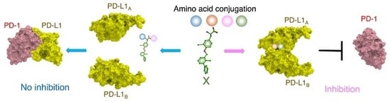
2.4. Docking Simulation and Inhibition Assay of Amino-Xs

The binding mode of the BMS compounds and derivatives to PD-L1 has previously been revealed by X-ray crystallography [31,40,41,42]. BMS compounds induces transient homodimerization of PD-L1AB on the binding, which masks the binding site for PD-1 located in the homodimerization interface. We docked amino-Xs to the crystal structure of BMS-8/PD-L1AB complex (PDB ID: 5J8O) [31], using ICM 3.8-7 software (Molsoft L.L.C., San Diego, CA, USA) [33,34,35], without guidance and induced fitting to avoid over-fitting. After the docking, we calculated the root mean square deviation (RMSD) of distances between atoms in compound BMS-8 and X, excluding Cα, NH2, and COOH atoms (Figure 5).
Table 1 shows the docking scores and RMSD values for amino-Xs docked to PD-L1AB. Also, the IC50 values for the amino-Xs are listed in Table 1. As a result, they suggested some positive correlations. The IC50 values of the 1–2-mer amino-Xs showed moderate correlations with both the RMSDs (CC 0.67, Table 2) and the scores (CC 0.40, Table 2). However, these correlations weakened as the number of conjugated amino acids increased (RMSD from 0.67 to 0 and CC 0.40 to −0.20, Table 2). These results suggest that the current in silico docking worked better for amino-Xs conjugated with shorter amino acids.
To discuss the correlations further, we compared the docking structures of X (IC50 = 1.5 μM), XG (IC50 = 2.1 μM), and GX (IC50 = 448.5 μM).
We compared the binding modes of BMS-8 and X in the pocket of PD-L1AB homodimer (Figure 6). BMS-8, with IC50 of 7.2 μM (Figure 2), binds the pocket with a hydrogen bind to Q66A and a hydrophobic interaction with V68A (Figure 6A), respectively. On the other hand, X forms a hydrogen bond with the hydroxy group of the side chain of Y56A, which stabilizes the binding (Figure 6A), with IC50 of 1.5 μM (Figure 4). The superposition of X onto BMS-8 showed an RMSD displacement of 0.40 Å (Figure 6B) We conclude that binding of X would not markedly impede PD-L1 homodimerization, which is consistent with its relatively low IC50 value of 1.5 μM (Figure 4). These results suggest that we can improve an IC50 value by substituting the six-membered group of BMS-8 with some proper groups, leading to rearrangement of interactions around it. Besides, smaller displacement of biphenyl-bromotyrosine portion shown by RMSD is preferable for higher affinity.
Modeling of XG identified two potential hydrogen bonds between the N-terminal of XG and the side chain of Q66A and between the carboxyl group of Gly and R125B in the side chain (Figure 7A). The RMSD between XG and BMS-8 was 0.28 Å (Figure 7B), which suggested that the IC50 value of XG would be similar to that of X. Indeed, XG had a measured IC50 for PD-1/PD-L1 binding of 2.1 μM (Figure 7C). X and XG potentially have the inhibitory effect for PD-1/PD-L1 interaction because KD between PD-1 and PD-L1 are reported as 6.4 μM [43].
GX docking into the binding pocket of the PD-L1 homodimer revealed two hydrogen bonds formed between GX amino groups and carbonyl group of Y123B (Figure 8A). As a result, the calculated RMSD between GX and BMS-8 was 0.52 Å (Figure 8B), which was larger than the RMSD of X and XG. This observation suggests that GX binding might sterically hinder PD-L1 homodimerization, leading to poorer inhibition of PD-1/PD-L1 binding. Consistent with this, the measured IC50 for GX was 448.5 μM (Figure 8C), which was several hundred times higher than those for X and XG ( Figure 4; Figure 7C). It is possible that the larger displacement of X of GX caused to deform the pocket of the PD-L1 homodimer, leading to the weaker inhibition of GX than those of X and XG.
The X portion of BMS-8 without Cα, NH2, COOH atoms formed hydrophobic interactions in the crystal structure (PDB ID: 5J8O), with residues I54A, Y56A, V68A, M115A, I116A, S117A, A121A, D122A, I54B, Y56B, M115B, I116B, S117B, A121B, D122B, and Y123B of the PD-L1 homodimer (Figure 9A). The space-filling representation of X shows the adherent interaction mode to the binding pocket (Figure 9B,C). The intermediate compounds of BMS-8, compounds 16 (Scheme 1) showed a poor ability to inhibit PD-1/PD-L1 binding (Figure 3), which was probably due to insufficient hydrophobic filling of the compounds in the binding pocket.
Collectively, our results suggest that the larger displacement of amino-Xs from BMS-8 prevents PD-L1A/PD-L1B homodimer formation. The docking simulations suggest that X and GX promote homodimerization of PD-L1, resulting in low IC50 values, whereas the larger displacement of amino-Xs prevents PD-L1 homodimer formation and increase the IC50 values.
The results of this study advance our understanding of how small molecule compounds could be rationally designed to inhibit PD-1/PD-L1 interactions with high affinity. In silico docking simulations have typically shown that target proteins have stable binding pockets during ligand binding, even allowing for some local flexibility of the side chains within the pockets [37,44]. In that scenario, binding scores generally correlate well with experimentally determined inhibitor activity [45]. However, binding of X and amino-X in the PD-L1 pocket occurs through strict interactions, indicating that even a slight displacement of the X conformation leads to deformation of the PD-L1 homodimer, which deceases the inhibitory effect. Consistent with this, the amino-Xs with shorter amino acid conjugates showed moderate positive correlations between the measured IC50 values and RMSDs in the no template/flexible docking mode, whereas the correlation was weakened by further amino acid addition.

3. Materials and Methods

3.1. Materials for Organic Chemistry Synthesis

Sodium chloride (NaCl), lysozyme, monosodium phosphate (NaH2PO4), imidazole, glycerol, reduced glutathione, oxidized glutathione, methanol, dimethyl sulfoxide (DMSO), trifluoroacetic acid (TFA), tert-butyl acetate, perchloric acid (HClO4), hydrochloric acid (HCl), sodium carbonate, ethyl acetate, sodium sulfate, hexane, sodium hydrogen carbonate (NaHCO3), acetone, triphenyl phosphine (Ph3P), anhydrous dichloromethane (CH2Cl2), and anhydrous tetrahydrofuran (THF) were purchased from Wako Pure Chemical Industries Ltd. (Osaka, Japan). 3-Bromo-tyrosine, 3-hydroxymethyl-2-methylbiphenyl, and diisopropyl azodicarboxylate (DIAD; 40% in toluene, approximately 1.9 mol L−1) were purchased from Tokyo Chemical Industry Co., Ltd. (Tokyo, Japan). Magnesium sulfate and CH2Cl2 were purchased from Junsei Chemical Co., Ltd. (Tokyo, Japan). Deuterochloroform (CDCl3) was purchased from Isotec, Inc. (Miamisburg, OH, USA), and N-[(9H-fluoren-9-ylmethoxy) carbonyloxy] succinimide (Fmoc-Osu) was purchased from Watanabe Chemical Industries, Ltd. (Hiroshima, Japan).

3.2. Synthesis of a Biphenyl-Conjugated Bromotyrosine

Ijms 21 03639 i001
(S)-tert-Butyl 2-amino-3-(3-bromo-4-hydroxyphenyl) propanoate (3). A suspension of 3-bromotyrosine (2; 1.0 g, 3.9 mmol) in tert-butyl acetate (16 mL, 92 mmol) was cooled to 0 °C, and stirred for 30 min. HClO4 (0.5 mL, 7.7 mmol) was then slowly added to the suspension at 0 °C, and the reaction mixture was warmed to 25 °C and stirred for 24 h. The mixture was washed with water and 1N HCl, and the aqueous phase was brought to pH 9 using sodium carbonate and then extracted with ethyl acetate. The resulting organic phase was washed with water and dried with sodium sulfate. The solvent was evaporated under reduced pressure, yielding an oily compound. This crude product was washed with cold hexane and then dried under reduced pressure to yield compound 3 (0.57 g, 47%). 1H-NMR (400 MHz, CDCl3): δ = 1.41 (s, 9H, –OC(CH3)3), 2.73 (dd, 1H, J = 14.4, 8.0 Hz, HOPh(Br)–CH2CH(NH2)–), 2.93 (dd, 1H, J = 13.6, 5.2 Hz, HOPh(Br)–CH2CH(NH2)–), 3.57 (dd, 1H, J = 7.2, 5.6 Hz, HOPh(Br)–CH2CH(NH2)–), 3.70 (m, 3H, HOPh(Br)–CH2CH(NH2)–), 6.70 (d, 1H, J = 8.0 Hz, aromatic ring), 6.94 (dd, 1H, J = 8.4, 2.0 Hz, aromatic ring), 7.26 (d, 1H, J = 1.6 Hz, aromatic ring).
Ijms 21 03639 i002
(S)-tert-Butyl 2-({[(9H-fluoren-9-yl)methoxy]carbonyl}amino)-3-(3-bromo-4-hydroxyphenyl)propanoate (4). A suspension of 3 (0.5 g, 1.6 mmol) and NaHCO3 (0.27 g, 3.2 mmol) in water (20 mL) was cooled to 0 °C. Fmoc-Osu (1.1 g, 3.2 mmol) in acetone (40 mL) was added to the suspension slowly, and the reaction mixture was then stirred at 25 °C for 15 h. The solvent was removed and washed with 1N HCl and water. After drying under vacuum, the crude product was purified by column chromatography on silica gel (eluent: ethyl acetate/hexane = 1:3 v/v) to yield compound 4 (0.71 g, 84%). 1H-NMR (400 MHz, CDCl3): δ = 1.42 (s, 9H, –OC(CH3)3), 3.00 (d, 2H, J = 5.6 Hz, HOPh(Br)–CH2CH(NHCOOCH2CH–)–), 4.21 (t, 1H, J = 7.2 Hz, HOPh(Br)–CH2CH(NHCOOCH2CH–)–), 4.33 (dd, 1H, J = 10.4, 7.2 Hz, HOPh(Br)–CH2CH(NHCOOCH2CH–)–), 4.43–5.00 (m, 2H, HOPh(Br)–CH2CH(NHCOOCH2CH–)–), 5.29 (d, 1H, J = 8.0 Hz, HOPh(Br)–CH2CH(NHCOOCH2CH–)–), 5.43 (s, 1H, HOPh(Br)–CH2CH(NHCOOCH2CH–)–), 6.91 (d, 1H, J = 8.4 Hz, aromatic ring), 6.96 (d, 1H, J = 9.2 Hz, aromatic ring), 7.26–7.33 (m, 3H, aromatic ring), 7.40 (dd, 2H, J = 7.4, 7.4 Hz, aromatic ring), 7.57 (dd, 2H, J = 6.2, 6.2 Hz, aromatic ring), 7.76 (d, 2H, J = 7.2 Hz, aromatic ring); high resolution mass spectrometry (HRMS) calculated for C28H28BrNO5 ([M + H]+): 538.1224, found: 538.1224.
Ijms 21 03639 i003
(S)-tert-Butyl 2-({[(9H-fluoren-9-yl)methoxy]carbonyl}amino)-3-{3-bromo-4-[(2-methyl-1,1’-biphenyl-3-yl)methoxy]phenyl}propanoate (5). To a solution of 4 (0.1 g, 0.19 mmol), 3-hydroxymethyl-2-methylbiphenyl (1; 39 mg, 0.20 mmol), and triphenyl phosphine (57 mg, 0.20 mmol) in anhydrous THF (10 mL), DIAD (0.1 mL, 0.22 mmol) was added at 0 °C under argon, and the reaction mixture was stirred at 0 °C for 12 h under argon. The organic phase was extracted with CH2Cl2 and dried over anhydrous magnesium sulfate. The solvent was then evaporated under reduced pressure, with the temperature kept below 30 °C. The crude product was purified by column chromatography on silica gel (eluent: ethyl acetate/hexane = 1:4 v/v) to yield compound 5 (0.09 g, 66%). 1H-NMR (400 MHz, CDCl3): δ = 1.46 (s, 9H, –OC(CH3)3), 2.27 (s, 3H, Biphenyl(CH3)–CH2OPh(Br)–CH2CH(NHCOOCH2CH–)–), 3.05 (d, 2H, J = 5.6 Hz, Biphenyl(CH3)–CH2OPh(Br)–CH2CH(NHCOOCH2CH–)–), 4.23 (t, 1H, J = 7.6 Hz, Biphenyl(CH3)–CH2OPh(Br)–CH2CH(NHCOOCH2CH–)–), 4.34 (dd, 1H, J = 6.8, 6.8 Hz, Biphenyl(CH3)–CH2OPh(Br)–CH2CH(NHCOOCH2CH–)–), 4.46-4.56 (m, 2H, Biphenyl(CH3)–CH2OPh(Br)–CH2CH(NHCOOCH2CH–)–), 5.13 (s, 2H, Biphenyl(CH3)-CH2OPh(Br)-CH2CH(NHCOOCH2CH–)–), 5.37 (d, 1H, J = 7.6 Hz, Biphenyl(CH3)–CH2OPh(Br)-CH2CH(NHCOOCH2CH–)–), 6.92 (d, 1H, J = 8.0 Hz, aromatic ring), 7.05 (d, 1H, J = 7.2 Hz, aromatic ring), 7.19–7.45 (m, 14H, aromatic ring), 7.53 (d, 1H, J = 6.8 Hz, aromatic ring), 7.60 (dd, 2H, J = 6.4, 6.4 Hz, aromatic ring), 7.77 (d, 2H, J = 7.2 Hz, aromatic ring); HRMS calculated for C42H40BrNO5 ([M + H]+): 718.2163, found: 718.2164.
Ijms 21 03639 i004
(S)-2-({[(9H-fluoren-9-yl)methoxy]carbonyl}amino)-3-{3-bromo-4-[(2-methyl-1,1’-biphenyl-3-yl)methoxy]phenyl}propanoic acid (6). A solution of 5 (3.9 g, 5.42 mmol) in anhydrous CH2Cl2 (36 mL) was stirred at 0 °C under argon for 15 min. TFA (1.3 mL, 16.6 mmol) was added dropwise to the solution at 0 °C, and the reaction mixture was stirred at 25 °C under argon. After 6 h, TFA (1.5 mL, 19.5 mmol) was added to the reaction mixture, which was then stirred at 25 °C for 18 h under argon. The solvent was removed under reduced pressure, with the temperature kept below 40 °C. The crude product was purified by column chromatography on silica gel (eluent: CH2Cl2/methanol = 97:3 v/v) to yield compound 6 (3.2 g, 85%). 1H-NMR (400 MHz, CDCl3): δ = 2.24 (s, 3H, Biphenyl(CH3)–CH2OPh(Br)–CH2CH(NHCOOCH2CH–)–), 3.05 (dd, 1H, J = 14.0, 6.0 Hz, Biphenyl(CH3)–CH2OPh(Br)–CH2CH(NHCOOCH2CH–)–), 3.15 (dd, 1H, J = 14.8, 5.2 Hz, Biphenyl(CH3)–CH2OPh(Br)–CH2CH(NHCOOCH2CH–)–), 4.21 (t, 1H, J = 6.8 Hz, Biphenyl(CH3)–CH2OPh(Br)–CH2CH(NHCOOCH2CH–)–), 4.36 (dd, 1H, J = 6.8, 6.8 Hz, Biphenyl(CH3)–CH2OPh(Br)–CH2CH(NHCOOCH2CH–)–), 4.46 (dd, 1H, J = 10.0, 7.2 Hz, Biphenyl(CH3)–CH2OPh(Br)–CH2CH(NHCOOCH2CH–)–), 4.66 (dd, 1H, J = 13.2, 6.0 Hz, Biphenyl(CH3)–CH2OPh(Br)–CH2CH(NHCOOCH2CH–)–), 5.09 (s, 2H, Biphenyl(CH3)-CH2OPh(Br)–CH2CH(NHCOOCH2CH–)–), 5.23 (d, 1H, J = 8.4 Hz, Biphenyl(CH3)-CH2OPh(Br)–CH2CH(NHCOOCH2CH–)–), 6.91 (d, 1H, J = 8.8 Hz, aromatic ring), 7.03 (d, 1H, J = 7.6 Hz, aromatic ring), 7.21–7.55 (m, 15H, aromatic ring), 7.74 (d, 2H, J = 7.2 Hz, aromatic ring); HRMS calculated for C38H32BrNO5 ([M + H]+): 662.1537, found: 662.1520.

3.3. Solid-State Peptide Synthesis

Amino-Xs were synthesized using an automated peptide synthesizer (MultiPep CF, INTAVIS Bioanalytical Instruments AG, Cologne, Germany). The synthetic protocol for glycine-conjugated peptide XG was as follows: Fmoc-protected glycine attached to a polystyrene resin (Fmoc-Gly NovaSyn TGT, Merck KGaA, Darmstadt, Germany) was deprotected by piperidine (20% in N-methylpyrrolidone (NMP). The resulting resin was reacted with 6 (99 mg, 0.14 mmol), 1-[bis(dimethylamino)methylene]-1H-benzotriazolium 3-oxide hexafluorophosphate (HBTU; 150 μL, 0.5 M in N,N-dimethylformamide (DMF), N-methylmorpholine (45 μL, 4.0 M in DMF) in NMP (8 μL) for 45 min. After washing, the N-α-protecting group of Fmoc in compound 6 was deprotected by piperidine (20% in NMP). Finally, the obtained peptide was cleaved from the resin using TFA (95% in water), yielding XG. Other peptides were synthesized using a similar method. (S)-2-amino-3-[3-bromo-4-{(2-methyl-1,1′-biphenyl-3-yl)methoxy}phenyl]propanoic acid (X) was obtained by deprotection of Fmoc in 6 using piperidine (20% in NMP).

3.4. Characterization

The synthesized compounds were identified using 1H NMR spectroscopy (JNM–ECZ400R, JEOL Ltd., Tokyo, Japan) and HRMS (QSTAR Elite, AB SCIEX, Framingham, MA, USA).

3.5. Determination of the IC50 Value by AlphaLISA®

3.5.1. Principle of the Competitive Binding Assay

The binding affinity of the inhibitors to PD-L1 were measured using the AlphaLISA® assay kit (AL356 HV/C/F, PerkinElmer) according to the manufacturer’s instructions, with the anti-PD-1 mAb nivolumab (Selleck Chemicals, Houston, TX, USA) included as a positive control [41]. In this assay, direct binding of an inhibitor to PD-L1 is detected by photoinduced energy transfer (Figure S6). Biotin-conjugated PD-1 is attached to streptavidin-coated donor beads and histidine (His)-tagged PD-L1 is attached to anti-His-conjugated acceptor beads. Photoexcitation of the donor beads at 680 nm yields singlet oxygen. If PD-L1–PD-1 binding is successful, energy is transferred through singlet oxygen, leading to an increase in fluorescence intensity at 615 nm (Figure S6).

3.5.2. Preparation of Samples

BMS-8 was purchased from AA Blocks LLC (San Diego, CA, USA). Stock solutions of inhibitors in DMSO (stock solution A, 5 mM) were serially diluted (Figure S5A) to obtain 10 assay solutions (1–10) with concentrations ranging from 5.0 mM to 2.6 nM (Table S2). An aliquot of solution 1–10 (2 μL) was mixed with His-tagged PD-L1 (25 nM, 2 μL), biotin-conjugated PD-1 (25 nM, 2 μL), anti-His acceptor beads (0.55 g L–1, 2 μL), and streptavidin-coated donor beads (1.1 g L−1, 2 μL) (Figure S5B) in a final volume of 10 μL and incubated at 25 °C for 90 min. Positive and negative technical controls were included in parallel. Positive controls contained buffer (2 μL) in place of solution 1–10, and negative controls contained only the beads (2 μL each) and buffer (6 μL).

3.5.3. AlphaLISA® Measurement and Analysis

The reaction samples (10 μM) were placed in a 384-well microplate and photoirradiated at 680 nm from the top. Fluorescence at 615 nm was detected using an EnSpire multimode plate reader (Perkin Elmer, Waltham, MA, USA). IC50 values were estimated from a sigmoidal curve of fluorescence intensity vs. inhibitor concentration using a relative weighting method (1/Y2 weighting) with GraphPad Prism 8 (GraphPad Software Inc., San Diego, CA, USA).

3.6. Docking Simulation of Compounds

The docking simulation software ICM 3.8-7 [33] was used to investigate the binding modes of X and amino-Xs to the PD-L1 homodimer complexed with BMS-8 (PDB ID: 5J8O) [31]. We performed docking without template docking [37] or introducing flexibility [37] to avoid over-fitting of the ligands into the pocket. The docking simulation supposed Monte Carlo pseudo-Brownian motion [46]. In the simulation, the score suggests goodness of docking, defined as follows [45]:
Score = ΔEIntFF + TΔSTor + α1ΔEHBond + α2ΔEHBDesol + α3ΔEsolEl + α4ΔEHPob + α5Qsize
where α1–α5 = weight, ΔEIntFF = ligand–target van der Waals interactions and internal force field energy of the ligand, TΔSTor = free energy changes due to conformational energy loss upon ligand binding, ΔEHBond = hydrogen bonding interactions, ΔEHBDesol = hydrogen bond donor–acceptor desolvation energy, ΔEsolEl = solvation electrostatic energy upon ligand binding, ΔEHPob = hydrophobic free energy gain, and Qsize = a size correction term proportional to the number of ligand atoms [45,47,48]. We calculated RMSD values by using CORREL function in the Microsoft Excel.

4. Conclusions

This study reports that we prepared the new biphenyl-conjugated bromotyrosine, which inhibits the PD-1/PD-L1 interaction with better effect than that of BMS-8. In addition, the amino-Xs, which are conjugates of X with a variety of amino acids, provide the molecular mechanism how amino acid modifications of X affects inhibition of PD-1/PD-L1 interactions. Binding of the X without the Cα, NH2, and COOH atoms portion of amino-Xs into the PD-L1 binding pocket is required to promote transient homodimerization of PD-L1A/PD-L1B, leading to formation of a stable ternary complex composed of X and PD-L1AB. Amino acid conjugation, however, alters the X docking conformation in the PD-L1 pocket, reducing the IC50 values dramatically. We conclude that improper interactions between amino acids conjugated to X and those in the binding pocket induced displacement of the compounds, thereby reducing inhibitory effect. In the future, we plan to design conjugates with amino acids that do not disturb the conformation of X in the PD-L1 binding pocket.

Supplementary Materials

The following are available online at https://www.mdpi.com/1422-0067/21/10/3639/s1.

Author Contributions

H.M., M.K., Y.I., and E.K. conceived the project. E.-H.K. and M.K. conducted the organic syntheses. H.M. and E.-H.K. performed the in silico simulations. E.-H.K., R.D., and H.M. measured the IC50 values by the AlphaLISA®. E.-H.K., M.K., and H.M. wrote the manuscript. All of the authors discussed the results and approved the content of the final manuscript. All authors have read and agreed to the published version of the manuscript.

Funding

This study was partly supported by the Incentive Research Fund of RIKEN (FY2017-2018, H.M. and M.K.) and the Japan Agency for Medical Research and Development (AMED: FY2018-2020, Y.I.). E.-H.K. was financially supported by the Junior Research Associate Program at RIKEN.

Acknowledgments

We thank the Bio-material Analysis Support Unit, RIKEN Center for Brain Science for synthesis of biphenyl-conjugated amino acids. We thank Anne M. O’Rourke, from Edanz Group (www.edanzediting.com/ac) for editing a draft of this manuscript.

Conflicts of Interest

The authors declare no conflicts of interest.

Abbreviations

PD-1Programmed cell death 1
PD-L1Programmed cell death-ligand 1
PD-L1APD-L1 chain A
PD-L1BPD-L1 chain B
PD-L1ABHomodimer of PD-L1A/PD-L1B chains
AlphaAmplified Luminescence Proximity Homogeneous Assay
Xbiphenyl-conjugated bromotyrosine
Amino-XAmino acid conjugated-X
MALDI-TOF MSMatrix assisted laser desorption/ionization-time of flight mass spectrometry
RMSDRoot mean square deviation
KDEquilibrium dissociation constant
IC5050% maximal inhibitory concentration
CCCorrelation coefficient
HTRFHomogenous Time-Resolved Fluorescence

References

  1. Rius, M.; Lyko, F. Epigenetic cancer therapy: Rationales, targets and drugs. Oncogene 2011, 31, 4257–4265. [Google Scholar] [CrossRef] [PubMed] [Green Version]
  2. Postow, M.A.; Callahan, M.K.; Wolchok, J.D. Immune checkpoint blockade in cancer therapy. J. Clin. Oncol. 2015, 33, 1974–1982. [Google Scholar] [CrossRef] [Green Version]
  3. Lee, L.; Gupta, M.; Sahasranaman, S. Immune Checkpoint inhibitors: An introduction to the next-generation cancer immunotherapy. J. Clin. Pharmacol. 2016, 56, 157–169. [Google Scholar] [CrossRef] [PubMed] [Green Version]
  4. Hoos, A. Development of immuno-oncology drugs-from CTLA4 to PD1 to the next generations. Nat. Rev. Drug Discov. 2016, 15, 235–247. [Google Scholar] [CrossRef] [PubMed]
  5. Iwai, Y.; Ishida, M.; Tanaka, Y.; Okazaki, T.; Honjo, T.; Minato, N. Involvement of PD-L1 on tumor cells in the escape from host immune system and tumor immunotherapy by PD-L1 blockade. Proc. Natl. Acad. Sci. USA 2002, 99, 12293–12297. [Google Scholar] [CrossRef] [PubMed] [Green Version]
  6. Mellman, I.; Coukos, G.; Dranoff, G. Cancer immunotherapy comes of age. Nature 2011, 480, 480–489. [Google Scholar] [CrossRef]
  7. Ishida, Y.; Agata, Y.; Shibahara, K.; Honjo, T. Induced expression of PD-1, a novel member of the immunoglobulin gene superfamily, upon programmed cell death. EMBO J. 1992, 11, 3887–3895. [Google Scholar] [CrossRef]
  8. Dong, H.; Zhu, G.; Tamada, K.; Chen, L. B7-H1, a third member of the B7 family, co-stimulates T-cell proliferation and interleukin-10 secretion. Nat. Med. 1999, 5, 1365–1369. [Google Scholar] [CrossRef]
  9. Freeman, G.J.; Long, A.J.; Iwai, Y.; Bourque, K.; Chernova, T.; Nishimura, H.; Fitz, L.J.; Malenkovich, N.; Okazaki, T.; Byrne, M.C.; et al. Engagement of the PD-1 immunoinhibitory receptor by a novel B7 family member leads to negative regulation of lymphocyte activation. J. Exp. Med. 2000, 192, 1027–1034. [Google Scholar] [CrossRef] [Green Version]
  10. Dong, H.D.; Strome, S.E.; Salomao, D.R.; Tamura, H.; Hirano, F.; Flies, D.B.; Roche, P.C.; Lu, J.; Zhu, G.F.; Tamada, K.; et al. Tumor-associated B7-H1 promotes T-cell apoptosis: A potential mechanism of immune evasion. Nat. Med. 2002, 8, 793–800. [Google Scholar] [CrossRef]
  11. Lee, K.M.; Chuang, E.; Griffin, M.; Khattri, R.; Hong, D.K.; Zhang, W.; Straus, D.; Samelson, L.E.; Thompson, C.B.; Bluestone, J.A. Molecular basis of T cell inactivation by CTLA-4. Science 1998, 282, 2263–2266. [Google Scholar] [CrossRef] [PubMed] [Green Version]
  12. Stamper, C.C.; Zhang, Y.; Tobin, J.F.; Erbe, D.V.; Ikemizu, S.; Davis, S.J.; Stahl, M.L.; Seehra, J.; Somers, W.S.; Mosyak, L. Crystal structure of the B7-1/CTLA-4 complex that inhibits human immune responses. Nature 2001, 410, 608–611. [Google Scholar] [CrossRef] [PubMed]
  13. Drake, C.G. Basic overview of current immunotherapy approaches in urologic malignancy. Urol. Oncol. Semin. Orig. Investig. 2006, 24, 413–418. [Google Scholar] [CrossRef]
  14. Weiner, L.M.; Surana, R.; Wang, S. Monoclonal antibodies: Versatile platforms for cancer immunotherapy. Nat. Rev. Immunol. 2010, 10, 317–327. [Google Scholar] [CrossRef] [PubMed] [Green Version]
  15. Guo, Y.-T.; Hou, Q.-Y.; Wang, N. Monoclonal antibodies in cancer therapy. Clin. Oncol. Cancer Res. 2011, 8, 215–219. [Google Scholar] [CrossRef]
  16. Pento, J.T. Monoclonal Antibodies for the Treatment of Cancer. Anticancer Res. 2017, 37, 5935–5939. [Google Scholar] [CrossRef] [Green Version]
  17. Reichert, J.M. Monoclonal Antibodies as Innovative Therapeutics. Curr. Pharm. Biotechnol. 2008, 9, 423–430. [Google Scholar] [CrossRef]
  18. Leach, D.R.; Krummel, M.F.; Allison, J.P. Enhancement of antitumor immunity by CTLA-4 blockade. Science 1996, 271, 1734–1736. [Google Scholar] [CrossRef] [Green Version]
  19. Hodi, F.S.; O’Day, S.J.; McDermott, D.F.; Weber, R.W.; Sosman, J.A.; Haanen, J.B.; Gonzalez, R.; Robert, C.; Schadendorf, D.; Hassel, J.C.; et al. Improved survival with ipilimumab in patients with metastatic melanoma. New Engl. J. Med. 2010, 363, 711–723. [Google Scholar] [CrossRef]
  20. Wang, C.Y.; Thudium, K.B.; Han, M.H.; Wang, X.T.; Huang, H.C.; Feingersh, D.; Garcia, C.; Wu, Y.; Kuhne, M.; Srinivasan, M.; et al. In vitro characterization of the anti-PD-1 antibody nivolumab, BMS-936558, and in vivo toxicology in non-human primates. Cancer Immunol. Res. 2014, 2, 846–856. [Google Scholar] [CrossRef] [Green Version]
  21. Kimiz-Gebologlu, I.; Gulce-Iz, S.; Biray-Avci, C. Monoclonal antibodies in cancer immunotherapy. Mol. Boil. Rep. 2018, 45, 2935–2940. [Google Scholar] [CrossRef] [PubMed]
  22. Suzuki, S.; Ishida, T.; Yoshikawa, K.; Ueda, R. Current status of immunotherapy. Jpn. J. Clin. Oncol. 2016, 46, 191–203. [Google Scholar] [CrossRef] [PubMed] [Green Version]
  23. Zhang, H.; Chen, J. Current status and future directions of cancer immunotherapy. J. Cancer 2018, 9, 1773–1781. [Google Scholar] [CrossRef] [PubMed] [Green Version]
  24. Sharma, P.; Hu-Lieskovan, S.; Wargo, J.A.; Ribas, A. Primary, adaptive, and acquired resistance to cancer immunotherapy. Cell 2017, 168, 707–723. [Google Scholar] [CrossRef] [PubMed] [Green Version]
  25. El-Osta, H.; Shahid, K.; Mills, G.M.; Peddi, P. Immune checkpoint inhibitors: The new frontier in non-small-cell lung cancer treatment. Onco Targets Ther. 2016, 9, 5101–5116. [Google Scholar] [CrossRef] [Green Version]
  26. Shukla, A.A.; Thommes, J. Recent advances in large-scale production of monoclonal antibodies and related proteins. Trends Biotechnol. 2010, 28, 253–261. [Google Scholar] [CrossRef]
  27. Chames, P.; Van Regenmortel, M.; Weiss, E.; Baty, D. Therapeutic antibodies: Successes, limitations and hopes for the future. Br. J. Pharmacol. 2009, 157, 220–233. [Google Scholar] [CrossRef]
  28. Bojadzic, D.; Buchwald, P. Toward small-molecule inhibition of protein-protein interactions: general aspects and recent progress in targeting costimulatory and coinhibitory (immune checkpoint) interactions. Curr. Top. Med. Chem. 2018, 18, 674–699. [Google Scholar] [CrossRef]
  29. Golani, L.K.; Wallace-Povirk, A.; Deis, S.M.; Wong, J.; Ke, J.; Gu, X.; Raghavan, S.; Wilson, M.R.; Li, X.; Polin, L.; et al. Tumor targeting with novel 6-substituted pyrrolo [2,3-d] pyrimidine antifolates with heteroatom bridge substitutions via cellular uptake by folate receptor alpha and the proton-coupled folate transporter and inhibition of de novo purine nucleotide biosynthesis. J. Med. Chem. 2016, 59, 7856–7876. [Google Scholar] [CrossRef] [Green Version]
  30. Chupak, L.S.; Zheng, X. Compounds Useful as Immunomodulators. Bristol-Myers Squibb Company. WO2015034820 A1, 12 March 2015. [Google Scholar]
  31. Zak, K.M.; Grudnik, P.; Guzik, K.; Zieba, B.J.; Musielak, B.; Dömling, A.S.S.; Dubin, G.; Holak, T.A. Holak. Structural basis for small molecule targeting of the programmed death ligand 1 (PD-L1). Oncotarget 2016, 7, 30323–30335. [Google Scholar] [CrossRef] [Green Version]
  32. Musielak, B.; Kocik, J.; Skalniak, L.; Magiera-Mularz, K.; Sala, D.; Czub, M.; Stec, M.; Siedlar, M.; Holak, T.A.; Plewka, J. CA-170-A potent small-molecule PD-L1 inhibitor or not? Molecules 2019, 24, 2804. [Google Scholar] [CrossRef] [PubMed] [Green Version]
  33. Abagyan, R.; Totrov, M.; Kuznetsov, D. ICM–A New method for protein modeling and design-applications to docking and structure prediction from the distorted native conformation. J. Comput. Chem. 1994, 15, 488–506. [Google Scholar] [CrossRef]
  34. Bottegoni, G.; Kufareva, I.; Totrov, M.; Abagyan, R. Four-dimensional docking: A fast and accurate account of discrete receptor flexibility in ligand docking. J. Med. Chem. 2009, 52, 397–406. [Google Scholar] [CrossRef] [PubMed] [Green Version]
  35. Bottegoni, G.; Kufareva, I.; Totrov, M.; Abagyan, R. A new method for ligand docking to flexible receptors by dual alanine scanning and refinement (SCARE). J. Comput. Aided. Mol. Des. 2008, 22, 311–325. [Google Scholar] [CrossRef] [Green Version]
  36. Wang, W.; Hirano, Y.; Uzawa, T.; Liu, M.; Taiji, M.; Ito, Y. In vitro selection of a peptide aptamer that potentiates inhibition of cyclin-dependent kinase 2 by purvalanol. MedChemComm 2014, 5, 1400–1403. [Google Scholar] [CrossRef]
  37. Dharmatti, R.; Miyatake, H.; Nandakumar, A.; Ueda, M.; Kobayashi, K.; Kiga, D.; Yamamura, M.; Ito, Y. Enhancement of binding affinity of folate to its receptor by peptide conjugation. Int. J. Mol. Sci. 2019, 20, 2152. [Google Scholar] [CrossRef] [PubMed] [Green Version]
  38. Eglen, R.M.; Reisine, T.; Roby, P.; Rouleau, N.; Illy, C.; Bosse, R.; Bielefeld, M. The use of AlphaScreen technology in HTS: Current status. Curr. Chem. Genom. 2008, 1, 2–10. [Google Scholar] [CrossRef]
  39. Tan, S.; Zhang, H.; Chai, Y.; Song, H.; Tong, Z.; Wang, Q.; Qi, J.; Wong, G.; Zhu, X.; Liu, W.J.; et al. An unexpected N-terminal loop in PD-1 dominates binding by nivolumab. Nat. Commun. 2017, 8, 14369. [Google Scholar] [CrossRef] [Green Version]
  40. Guzik, K.; Zak, K.M.; Grudnik, P.; Magiera, K.; Musielak, B.; Torner, R.; Skalniak, L.; Domling, A.; Dubin, G.; Holak, T.A. Small-molecule inhibitors of the programmed cell death-1/programmed death-ligand 1 (PD-1/PD-L1) interaction via transiently induced protein states and dimerization of PD-L1. J. Med. Chem. 2017, 60, 5857–5867. [Google Scholar] [CrossRef]
  41. Skalniak, L.; Zak, K.M.; Guzik, K.; Magiera, K.; Musielak, B.; Pachota, M.; Szelazek, B.; Kocik, J.; Grudnik, P.; Tomala, M.; et al. Small-molecule inhibitors of PD-1/PD-L1 immune checkpoint alleviate the PD-L1-induced exhaustion of T-cells. Oncotarget 2017, 8, 72167–72181. [Google Scholar] [CrossRef] [Green Version]
  42. Magiera-Mularz, K.; Skalniak, L.; Zak, K.M.; Musielak, B.; Rudzinska-Szostak, E.; Berlicki, L.; Kocik, J.; Grudnik, P.; Sala, D.; Zarganes-Tzitzikas, T.; et al. Bioactive macrocyclic inhibitors of the PD-1/PD-L1 immune checkpoint. Angew. Chem. Int. Ed. 2017, 56, 13732–13735. [Google Scholar] [CrossRef] [PubMed]
  43. Lazar-Molnar, E.; Scandiuzzi, L.; Basu, I.; Quinn, T.; Sylvestre, E.; Palmieri, E.; Ramagopal, U.A.; Nathenson, S.G.; Guha, C.; Almo, S.C. Structure-guided development of a high-affinity human Programmed Cell Death-1: Implications for tumor immunotherapy. EBioMedicine 2017, 17, 30–44. [Google Scholar] [CrossRef] [Green Version]
  44. Wang, W.; Hirano, Y.; Uzawa, T.; Taiji, M.; Ito, Y. Peptide-assisted enhancement of inhibitory effects of small molecular inhibitors for kinases. Bull. Chem. Soc. Jpn. 2016, 89, 444–446. [Google Scholar] [CrossRef]
  45. Neves, M.A.; Totrov, M.; Abagyan, R. Docking and scoring with ICM: The benchmarking results and strategies for improvement. J. Comput. Mol. Des. 2012, 26, 675–686. [Google Scholar] [CrossRef] [PubMed] [Green Version]
  46. Abagyan, R.; Totrov, M. Biased probability monte-carlo conformational searches and electrostatic calculations for peptides and proteins. J. Mol. Biol. 1994, 235, 983–1002. [Google Scholar] [CrossRef] [PubMed] [Green Version]
  47. Totrov, M.; Abagyan, R. Flexible protein-ligand docking by global energy optimization in internal coordinates. Proteins 1997, 29 (Suppl. 1), 215–220. [Google Scholar] [CrossRef]
  48. Schapira, M.; Totrov, M.; Abagyan, R. Prediction of the binding energy for small molecules, peptides and proteins. J. Mol. Recognit. 1999, 12, 177–190. [Google Scholar] [CrossRef]
Figure 1. Strategies to improve inhibitory effect of BMS-8. 3-hydroxymethyl-2-methylbiphenyl (1) and 3-bromotyrosine (2) were selected as fragmented structures. A biphenyl-conjugated bromotyrosine X was synthesized after conjugation of 1 and 2. We conjugated a variety of amino acids as additions, to the amino- and carboxyl-groups of X to reveal molecular mechanism of the inhibition.
Ijms 21 03639 g001
Scheme 1. Synthetic scheme for the biphenyl-conjugated bromotyrosine 6.
Ijms 21 03639 sch001
Figure 2. Inhibition of PD-1/PD-L1 interaction by BMS-8 and nivolumab measured by the AlphaLISA® assay.
Ijms 21 03639 g002
Figure 3. Docking scores, IC50 values and measurements of compounds 1–6. All compounds showed larger scores than that of X (score = −42.96) with a few hundred μM of IC50 values.
Ijms 21 03639 g003
Figure 4. AlphaLISA assay of X. X shows IC50 = 1.5 μM with docking score = −42.96.
Ijms 21 03639 g004
Figure 5. Root mean square deviation (RMSD) calculation between amino-X and BMS-8 bound to PD-L1AB homodimer. After docking of amino-X (in this case, WXG), we calculated RMSD between a part of WXG (excluding Cα, amino-group and carboxyl-group) and the corresponding part of BMS-8, as shown by the red dotted-rectangle.
Ijms 21 03639 g005
Figure 6. Docking conformations of BMS-8 and X. (A) The docking modes of BMS-8 and X were revealed by the X-ray crystallography and in silico docking simulation, respectively, which the 2D binding pictures. The 2D figures show that biphenyl portions of the ligands bind into the pocket by hydrophobic interactions shown in light-green color. In contrast, the amino cation at the six-membered ring of BMS-8 makes a hydrogen bond with the sidechain of Q66A in cyan color. In addition, the six-membered ring makes hydrophobic interaction with V68A. On the other hand, amino-group of bromo-tyrosine in X makes a hydrogen bonding to the hydroxyl group of Y56A colored in cyan, without other hydrophobic interaction, as shown in the 2D picture. (B) BMS-8 and X without Cα, NH2, and COOH superposed each other with RMSD of 0.40 Å.
Ijms 21 03639 g006
Figure 7. In silico binding mode of XG. (A) Behavior of XG in the binding pocket of the PD-L1AB homodimer. (B) Superposition of XG onto BMS-8. The RMSD for displacement was 0.28 Å. (C) IC50 of XG for PD-1/PD-L1 binding.
Ijms 21 03639 g007
Figure 8. In silico binding mode of GX. (A) Behavior of GX in the binding pocket of the PD-L1AB homodimer. (B) Superposition of GX onto BMS-8. The RMSD for displacement was 0.52 Å RMSD. (C) IC50 of GX for PD-1/PD-L1 binding.
Ijms 21 03639 g008
Figure 9. Schematic drawing and space-filling representation of BMS-8 binding in the binding pocket of the PD-L1 homodimer. In (A–C), violet represents X without Cα, NH2, COOH atoms. (A) Binding mode of BMS-8 in the pocket of the PD-L1AB homodimer (PDB ID:5J8O). Yellow and cyan represent PD-L1A and PD-L1B side chains, respectively. (B) Space-filling representation of BMS-8 bound to the surface of PD-L1A (yellow) and contact area with PD-L1B (cyan). (C) Space-filling representation of BMS-8 bound to the surface of PD-L1B (cyan) and contact area with PD-L1A (yellow).
Ijms 21 03639 g009
Table 1. Docking simulation and IC50 measurements of BMS-8 and amino-Xs.
Amino Acid LengthSequenceScoreRMSD (Å)IC50 (μM)
-BMS-8−49.5-7.2
1X−42.960.401.5
2GX−41.00.52448.5
XG−46.90.282.1
XS−42.10.602655.0
XR−45.70.37892.0
XA−43.10.4722.3
XW−43.30.51845.0
3YXC−37.10.46465.0
WXG−50.60.48404.8
QXQ−37.70.731961.0
CXA−42.00.48665.0
RXN−40.30.63405.3
SXR−36.70.58796.0
NXR−50.30.46982.0
CXR−41.50.54550.0
GXG−43.60.39676.0
XNL−43.00.58855.0
XNH−40.70.50313.0
XHP−33.50.55359.0
XGG−36.10.576505.0
4XCSE−32.60.451555.0
XGGG−53.30.516766.0
5WRXNN−38.10.38157.4
ERXNK−21.30.4815.6
WRXNQ−19.40.49163.2
XRRRR−28.30.45435.6
XGGGG−41.80.75647.5
6XGGGGG−45.30.48846.0
7CERXNKM4.651.80308.2
FWRXNNI−7.300.41311.8
Table 2. Correlation coefficients (CC) for IC50 vs. Score and IC50 vs. RMSD.
LengthCC of Score/IC50CC of RMSD/IC50
1-20.400.67
1-30.350.37
1-400.28
1-7−0.200
CC values were calculated by the Microsoft Excel.

