Article

ORESARA15 Acts Synergistically with ANGUSTIFOLIA3 and Separately from AINTEGUMENTA to Promote Cell Proliferation during Leaf Growth

1
Department of Molecular Genetics, Dong-A University, Busan 49315, Korea
2
Subtropical Horticulture Research Institute, Jeju National University, Jeju 63243, Korea
3
Department of Applied Bioscience, Graduate School of Natural Science, Dong-A University, Busan 49315, Korea
*
Author to whom correspondence should be addressed.
Int. J. Mol. Sci. 2020, 21(1), 241; https://doi.org/10.3390/ijms21010241
Submission received: 29 October 2019 / Revised: 24 December 2019 / Accepted: 25 December 2019 / Published: 29 December 2019
(This article belongs to the Special Issue Plant Cell and Organism Development)

Abstract

:
Developing leaves undergo sequential coordinated cell proliferation and cell expansion to determine their final size and shape. Although several important regulators of cell proliferation have been reported, the gene network regulating leaf developmental processes remains unclear. Previously, we showed that ORESARA15 (ORE15) positively regulates the rate and duration of cell proliferation by promoting the expression of direct targets, GROWTH-REGULATING FACTOR (GRF) transcription factors, during leaf growth. In the current study, we examined the spatiotemporal patterns of ORE15 expression and determined that ORE15 expression partially overlapped with AN3/GIF1 and ANT expression along the midvein in the proximal region of the leaf blade in young leaves. Genetic analysis revealed that ORE15 may function synergistically with AN3 to control leaf growth as a positive regulator of cell proliferation. Our molecular and genetic studies are the first to suggest the importance of functional redundancies between ORE15 and AN3, and between AN3 and ANT in cell proliferation regulatory pathway during leaf growth.

Graphical Abstract

1. Introduction

Organ size is an important factor determining plant architecture and influencing adaptations to the environment. The final size of a plant organ is determined by two processes: Cell division and cell expansion. Cell proliferation is tightly regulated by many elements including genetic factors and phytohormones; however, the complete network of regulatory factors involved in cell proliferation during leaf development remains to be elucidated. Several studies have discovered individual cell cycle-related and cell proliferation regulatory factors, and their functional network has been summarized [1,2].
D-type cyclins, known as the CYCD family, are prime integrators for the cell cycle G1-to-S phase transition during organ growth. Overexpression of CYCD3;1 induces ectopic cell division and inhibits cell expansion and endoreduplication [3,4]. CYCD3;1 is required for cell number control in developing organs by regulating the duration of the mitotic phase and the timing of the transition to the endocycle. Many studies have suggested that CYCD3;1 acts to determine cell cycle progression and maintain cell number balance. Another positive regulator, the AP2-type transcription factor AINTEGUMENTA (ANT), enhances cell proliferation by prolonging the duration of CYCD3;1 expression and promoting an unknown cellular growth pathway, resulting in smaller organs and fewer cells in loss of function (LOF) mutants, whereas larger organs and more cells result from the period of extended growth in gain of function (GOF) mutants [5,6]. Overexpression of the novel auxin-inducible gene ARGOS has been shown to extend ANT and CYCD3;1 expression, ultimately enlarging the plant. These results suggest that ARGOS regulates cell proliferation and leaf growth through the ANT–CYCD3;1 pathway [7]. However, CYCD3;1 activity is insufficient to influence organ growth [4], and combined mutation of members of the CYCD3 family does not abolish organ growth [6]. The exact links between ANT and cellular growth remain unknown.
Another important cell proliferation regulator of organ growth, ANGUSTIFOLIA3/GRF-INTERACTING FACTOR1 (AN3/GIF1), is a member of the transcriptional coactivator family in Arabidopsis [8,9]. AN3/GIF1 is recruited by DNA-binding factors such as GROWTH REGULATOR FACTORs (GRFs) to activate target gene expression [10,11]. Mutations in GRF3, GRF4, or GRF5 have been shown to decrease cell proliferation activity, thereby reducing leaf size [12,13]. GIFs have been found to redundantly regulate leaf blade development through cell proliferation regulation [14,15,16], acting in a non-autonomous manner [17]. Recent studies have reported that the AN3/GIF1 protein recruits SWITCH/SUCROSE NONFEREMNTING (SWI/SNF) chromatin remodeling complexes to regulate leaf development [17,18,19].
The TCP (TEOSINTE BRANCHED 1, CYCLOIDEA, and PROLIFERATING CELL FACTORS 1 AND 2) family members have also been identified as cell proliferation regulators in meristems and organ primordia [20]. Recent findings have revealed a second class of TCPs, whose expression is regulated by miRNA319, which plays important roles in multiple developmental processes including leaf growth, morphogenesis, and senescence [21,22,23]. TCP4 affects cell proliferation regulation via the induction of miRNA396, which represses the GRF family, and by direct connection with KIP-RELATED PROTEIN 1 (KRP1) which terminates the cell cycle and induces the transition from mitotic cell cycle to the endocycle causing an increase in DNA ploidy by directly interacting with cell cycle genes [2,24]. Previously, we isolated and characterized ORESARA 15 (ORE15) encoding a plant A/T-rich sequence-and zinc-binding protein (PLATZ) family transcription factor and determined that ORE15 enhances leaf growth by promoting the rate and duration of cell proliferation in its early stage and suppresses leaf senescence in its late stage by modulating the miRNA396–GRF–GIF regulatory pathway [25]. It has been proposed that ORE15 may act as a transcriptional activator since it positively regulates the expression of GRF1 and GRF4 through direct binding to their promoters [25]. Together, these results suggest that ORE15 may work together with GRF–GIF to control the cell proliferation in leaf growth.
Although several important regulators involved in the control of cell proliferation have been reported, the molecular basis of the regulatory gene network during leaf developmental processes remains unclear. In this study, our characterization of cell proliferation during leaf growth using LOF and GOF mutations of ORE15 suggests that ORE15 positively regulates cell proliferation during leaf growth. We also generated double mutants of cell proliferation-related genes, ANT, and AN3/GIF1 using a combination of LOF and GOF ORE15 mutants and performed genetic and anatomical analyses to explore the relationship among ORE15, AN3/GIF1, and ANT in cell proliferation regulatory pathways during leaf growth.

2. Results

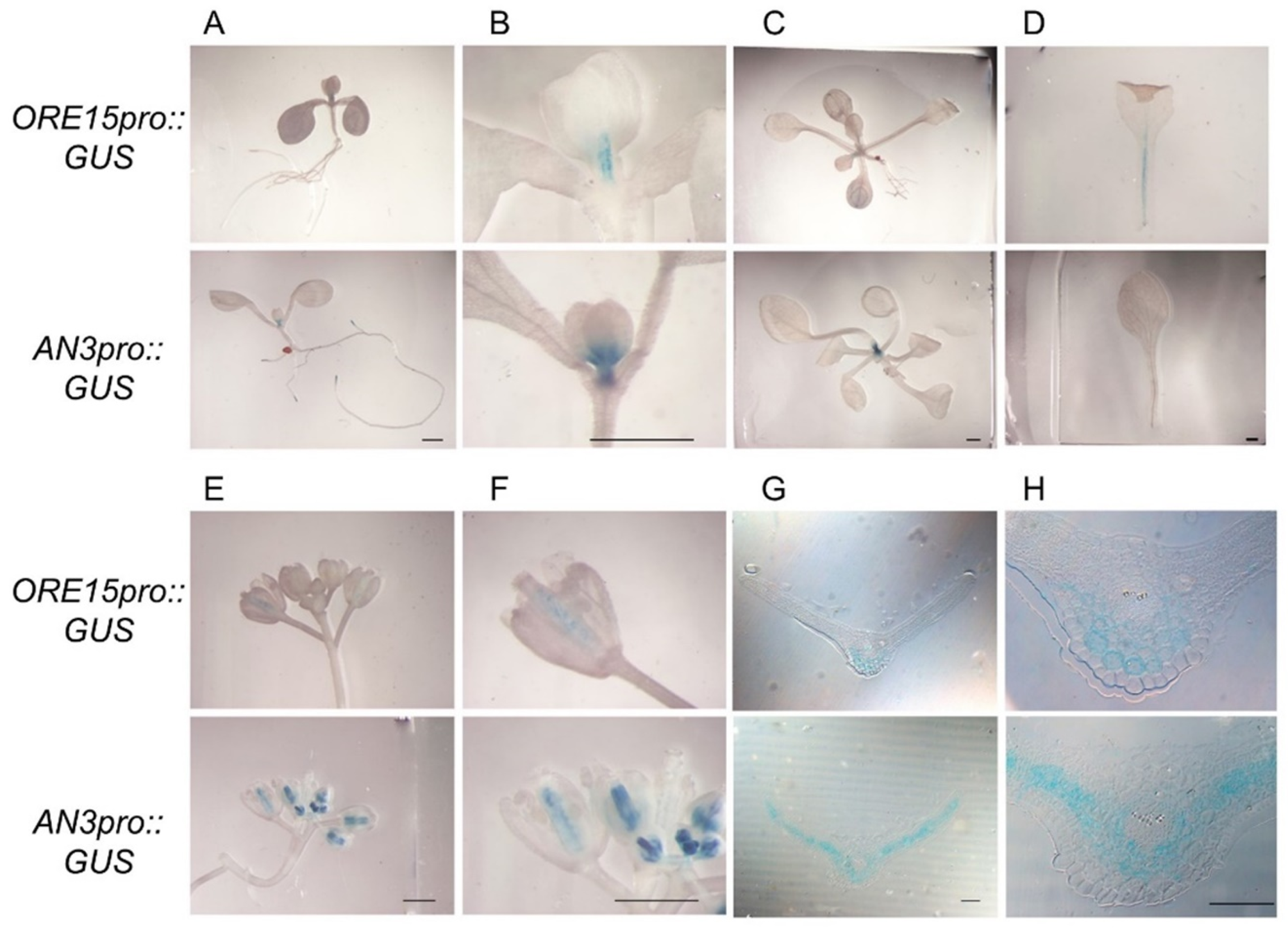
2.1. ORE15 Is Expressed in the Proximal Region of the Leaf Blade and in Petioles of Young Leaves

Our previous study demonstrated that ORE15 enhances leaf growth by promoting cell proliferation in the early stage of leaf development by modulating the GRF–GIF [25]. ORE15 also bound directly to the promoters of the GRF1 and GRF4 genes, which play roles in cell proliferation in leaf primordia [8,9,25], but did not bind directly to ANT and AN3/GIF1 promoter. To further explore ORE15 function in organ growth, we determined the temporal and spatial expression patterns of ORE15 using a promoter::β-glucuronidase (GUS) approach. ORE15 was expressed in the proximal part of the leaf blade and in leaf petioles (Figure 1A–D). Strong ORE15 expression was observed mainly along the midveins of young leaves (i.e., the first to fifth leaves) produced at 14 days after sowing (DAS) and throughout the petiole (Figure 1A–D). Similar spatial expression of ORE15 along the midvein was observed during the cell proliferation stage in leaves (Figure 1A–D). However, ORE15 expression was not detected either in shoot apical meristem or leaf primordia (Figure 1A–C). ORE15 expression was detected in the carpels of mature flowers (Figure 1E,F), but not in young flowers, mature leaves, or roots. Our expression analysis of ORE15 supported previous findings obtained by reverse transcription polymerase chain reaction (RT-PCR) gene expression analysis [25].
AN3/GIF1 was strongly expressed in the proximal region of the leaf during the cell proliferation stage (Figure 1A–C), which is consistent with previous findings [9]. AN3 was also expressed in the carpels of both young and mature flowers (Figure 1E,F). ANT, another important regulator of cell proliferation, has been reported to be expressed in active cell proliferation regions including young leaf blades, veins of leaves and stems, pistils, and meristems [26]. A study of GRF1 and GRF4, which were direct downstream targets of ORE15 [25], also showed very similar expression patterns to those of ORE15 [9,27]. To compare the expression domains of ORE15 and AN3/GIF1 in detail, we conducted histochemical analysis of GUS-stained leaves of transgenic plants. An examination of transverse sections of cell proliferating leaves of ORE15promoter::GUS transgenic plants showed that ORE15 was expressed strongly in the abaxial parenchyma cells around midvein in the proximal region of the leaf blade (Figure 1B,D,G,H). Transverse sections of GUS-stained leaves from AN3promoter::GUS transgenic plants showed that AN3 was expressed in mesophyll cells in the proximal region of the leaf blade of young leaves (Figure 1G,H), which was consistent with a previous report [17]. These results indicate that spatiotemporal expression patterns of ORE15 partially overlap with those of AN3/GIF1 in the abaxial parenchyma cells around midvein in the proximal region of the leaf blade. Together, these expression findings supported the idea of genetic interaction between the ORE15 and GRF–GIF complex, especially AN3/GIF1, GRF1, and GRF4 in the regulation of cell proliferation.

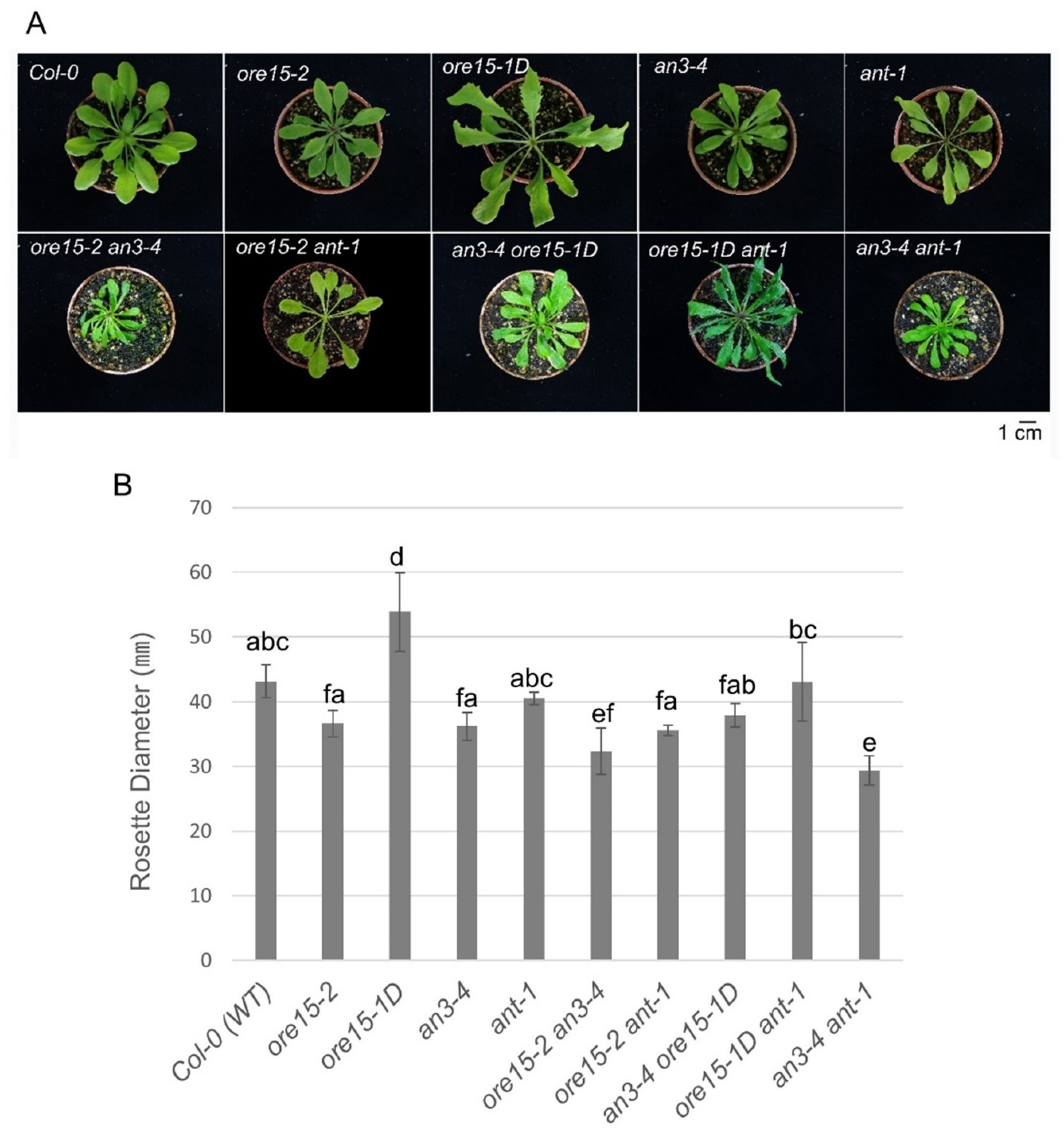
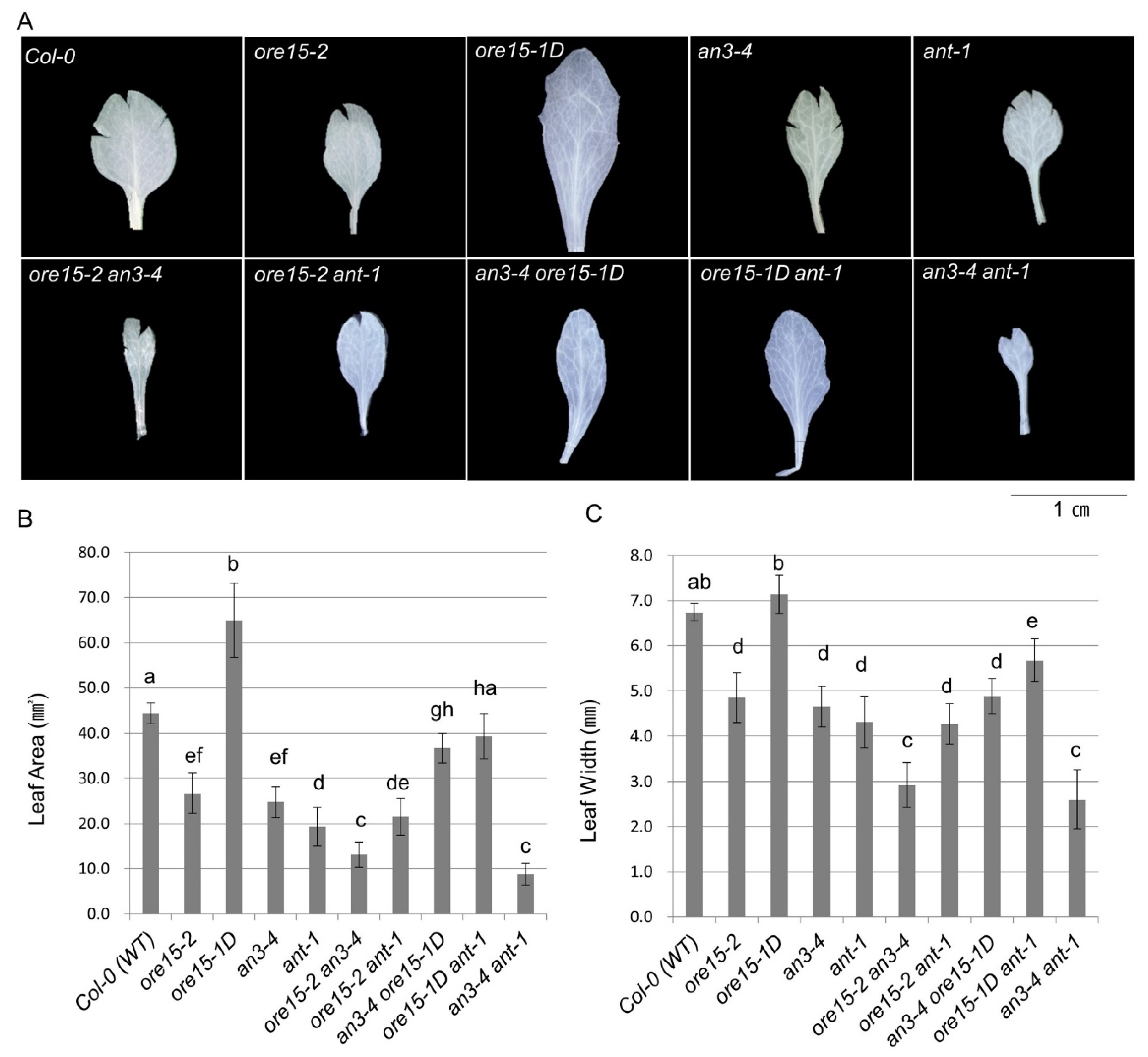
2.2. ORE15 Acts Synergically with AN3 and Separately from ANT to Promote Leaf Growth

In a previous study, we elucidated the regulatory relationship between ORE15 and the GRF–GIF regulatory module at the genetic level. The results of our expression study indicated that ANT expression was downregulated by ORE15 [25].
To identify the genetic relationship between ORE15, AN3, and ANT, we generated ore15-2 an3-4, ore15-1D an3-4, ore15-2 ant-1, and ore15-1D ant-1 by crossing a combination of LOF and GOF mutants and measured mature third leaves in the resulting mutant lines. We found that ore15-2 an3-4 plants had much smaller leaves and cell numbers than each single mutant (Figure 2, Figure 3 and Figure 4) [25]. ore15-1D an3-4 plants showed median size of leaves compared with the two parental lines (Figure 2 and Figure 3). The number of palisade cells produced in leaves of the ore15-1D an3-4 double mutant were 34.0%, 203.8%, and 102.2% of those produced in ore15-1D, an3-4, and Columbia-0 (Col-0) leaves, respectively (Figure 4), indicating the genetic interaction between ORE15 and AN3. Thus, ORE15 may function synergistically with AN3 to control leaf growth as a positive regulator of cell proliferation.
Both ore15-2 and ant-1 mutants produced smaller leaves than Col-0 (Figure 2 and Figure 3). The LOF double mutant ore15-2 ant-1 produced slightly smaller and slightly lager leaf area than ore15-2 and ant-1 single mutant, respectively (Figure 3, Table S1). ore15-1D ant-1 showed an intermediate phenotype of each single mutant, producing smaller and larger leaves than ore15-1D and ant-1, respectively (Figure 3). In addition, ore15-1D ant-1 leaves showed serrated phenotype, the same as ore15-1D leaves (Figure 2A). These results suggest that ORE15 may act separately from the ANT pathway to promote leaf growth.

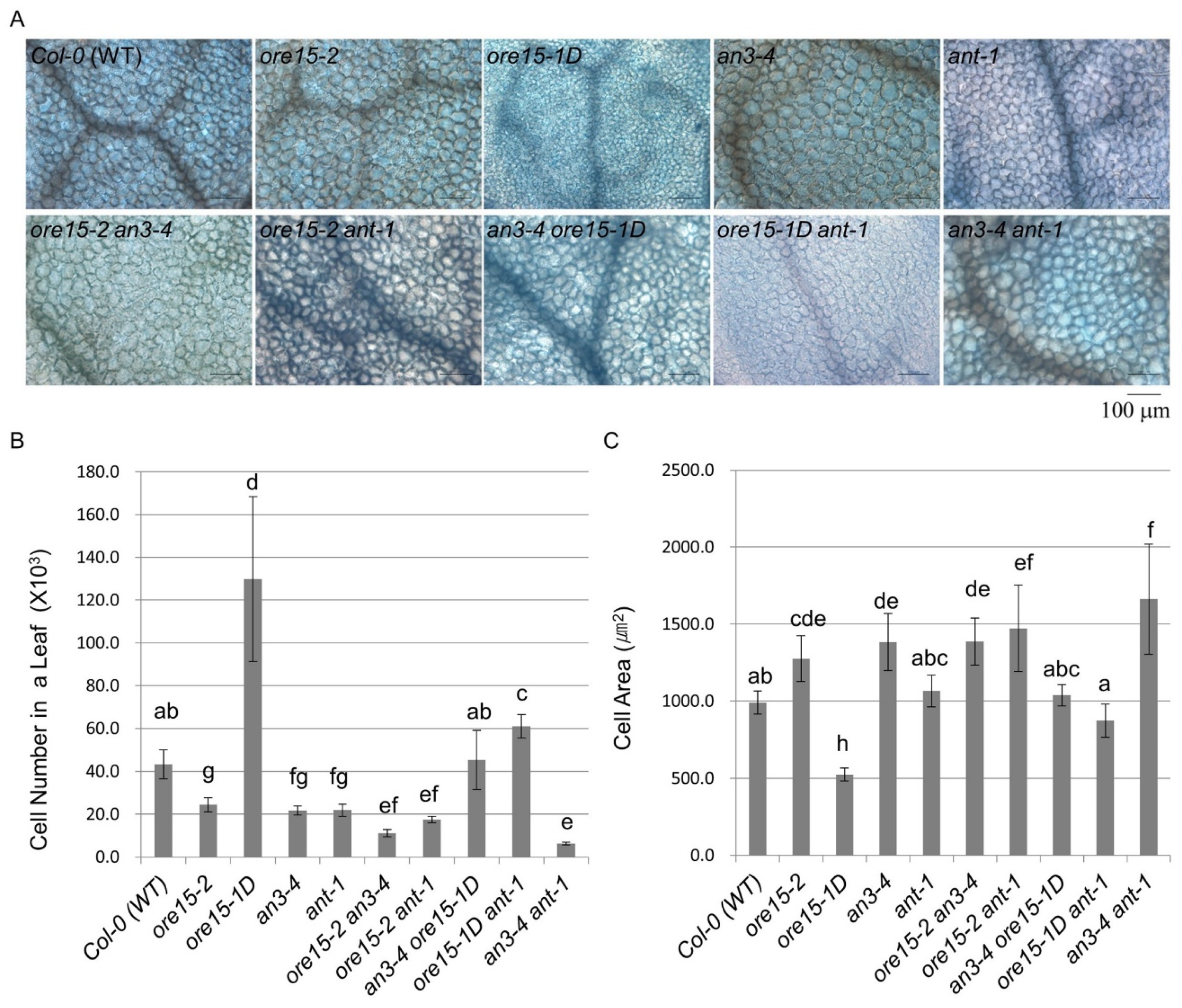
2.3. ORE15 Regulates Cell Proliferation during Leaf Growth Independently to ANT

Leaf growth is regulated by cell proliferation and cell expansion [28,29]. As previously reported, ORE15 promotes leaf growth by enhancing the rate and duration of cell proliferation. In our previous study, the ore15-2 an3-4 double mutant produced significantly smaller leaves and cell numbers, indicating a composite regulatory interaction between ORE15 and AN3/GIF1 in the regulation of leaf growth [25].
To further elucidate a potential regulatory interaction between ORE15 and ANT, we determined the number and area of leaf cells in mature third leaves collected from GOF and LOF mutants of ORE15 and ANT (Figure 4, Table S2). The numbers of palisade cells produced in leaves of the ore15-2 ant-1 LOF double mutant were 71.9%, 80.3%, and 40.7% of those produced in ore15-2, ant-1, and Col-0 leaves, respectively (Figure 4 and Figure S1). The ore15-1D ant-1 double mutant produced fewer cells (46.9%) than ore15-1D but more cells (278.1%) than ant-1 (Figure 4 and Figure S1), indicating an independent gene action. Together, our results suggest that ORE15 may act as a positive regulator promoting cell proliferation, separately from the ANT pathway during leaf growth (Figure 6).
On the other hand, the ore15-1D mutant produced smaller cells than Col-0; however, ore15-2 produced larger cells than Col-0 (Figure 4), indicating that ORE15 may negatively regulate cell area during leaf growth. Since endoreduplication is a determinant of cell expansion [30,31], we performed flow cytometric analysis of mature leaves of Col-0, ore15-2, and ore15-1D. Ploidy levels of ore15-2 and ore15-1D were slightly higher and lower than that of Col-0 (Figure S2). These results indicate that ORE15 may be a minor factor influencing negative promotion of endoreduplication in leaves. The ore15-2 ant-1 LOF double mutant produced cells that were 115.5% and 138.2% larger than those of ore15-2 and ant-1, respectively (Figure 4). By contrast, ore15-1D ant-1 produced the median cell area among each single mutant (Figure 4). These results indicate that ORE15 negatively regulates cell expansion, which may be a secondary effect of the ORE15- and ANT- cell proliferation pathway or a compensation process due to inhibition of cell division in the leaf organ [32].

2.4. AN3 Genetically Interacts with ANT to Promote Cell Proliferation during Leaf Growth

Our previous studies suggested that ORE15 plays a role in the AN3/GIF1-mediated cell proliferation regulatory pathway. The results of the current study indicate that ORE15 regulates cell proliferation during leaf growth, independently to ANT. To explore the molecular mechanism of cell proliferation in leaf growth, we analyzed the genetic interaction between AN3 and ANT using double mutants generated by crossing an3-4 and ant-1. Interestingly, leaves were significantly smaller in the an3-4 ant-1 LOF double mutant than in each single mutant (Figure 2 and Figure 3). These results indicate that AN3 and ANT synergistically interact with each other to control leaf growth.
To further elucidate the relationship between AN3 and ANT in the cell proliferation pathway, we examined the number and area of leaf cells in mature third leaves of double mutants (Figure 4). The an3-4 ant-1 double mutant produced significantly fewer cells than each single mutant (29.2%, 28.9%) and Col-0 (14.6%) (Figure 4 and Figure S1). Cells produced by an3-4 ant-1 were much larger than those of either an3-4 or ant-1 (Figure 4), under compensation process due to reduced cell division [32]. These results suggest that AN3 and ANT synergistically control cell proliferation, indicating that AN3 genetically interacts with ANT for regulating cell proliferation during leaf growth.

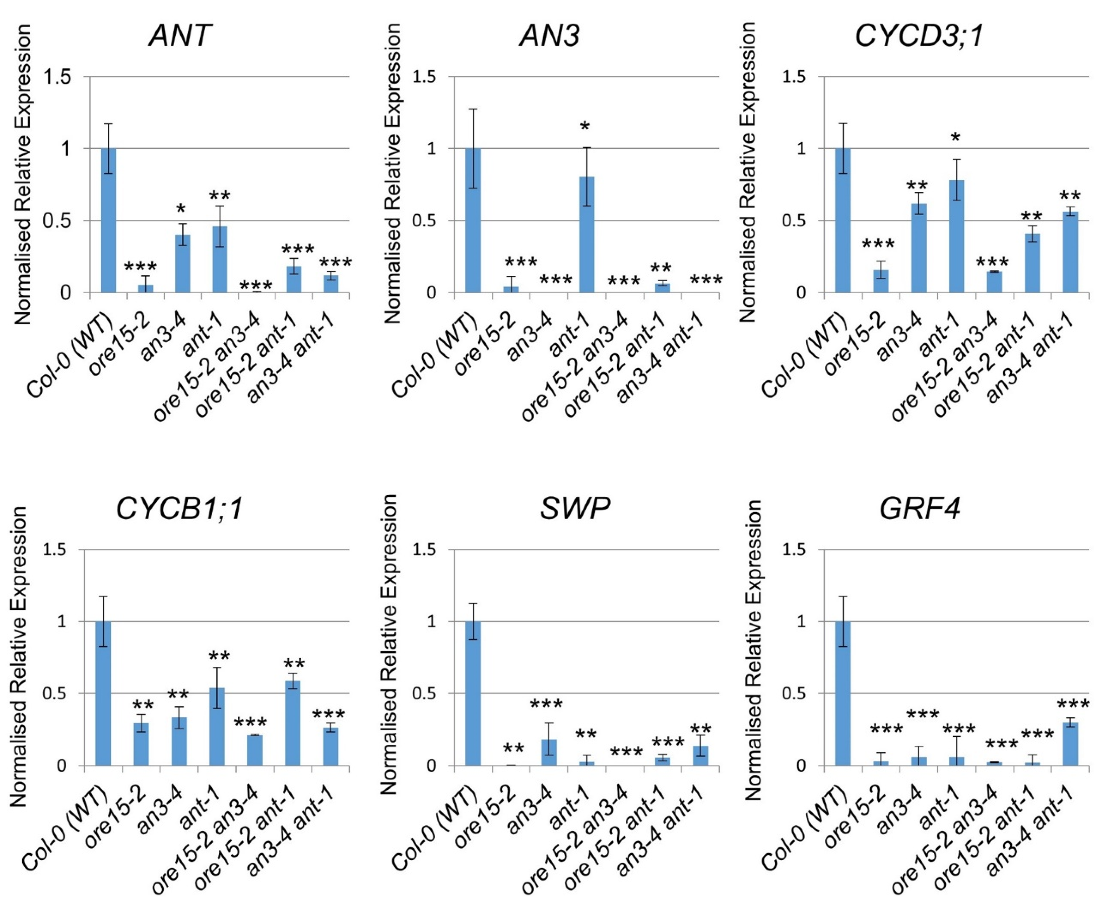
2.5. ORE15 Influences Cell Proliferation by Affecting Genes That Regulate Cell Division
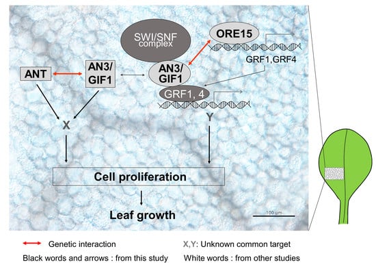
To gain further insight into the molecular mechanisms by which AN3 and ANT mediate the cell proliferation regulatory pathways underlying leaf growth, we analyzed expression levels of several leaf growth factors controlling cell proliferation or cell division in the third leaf blade during the cell proliferation stage among LOF single and double mutants using quantitative RT-PCR (Figure 5). The expression of AN3 and ANT was strongly reduced in ore15-2 and all double mutants compared with Col-0 (Figure 5), supporting the idea of potential gene interaction among ORE15, AN3, and ANT. In addition, the expression of ANT in an3-4 and that of AN3 in ant-1 was strongly reduced (Figure 5), also supporting our hypothesis of interaction between ANT and AN3.
Cyclin B1;1 (CYCB1;1) expression was reduced in all mutants compared with Col-0, although differences were detected in the extent of reduction (Figure 5). CYCD3;1 expression was greatly reduced in ore15-2, ore15-2 ant-1, an3-4 ant-1, and ore15-2 an3-4 LOF double mutants, indicating that ORE15 and the ANT pathway coordinate to regulate cell proliferation. GRF4 expression was also greatly reduced in all single and double mutants compared with Col-0 (Figure 5), supporting a relationship between ORE15 and the GRF–GIF. The expression of STRUWWELPETER (SWP), which encodes a protein similar to subunits of the mediator complex [33], was greatly reduced in all single and double mutants compared with Col-0 (Figure 5), indicating that SWP might have an important role in cell proliferation regulation during leaf growth.

3. Discussion

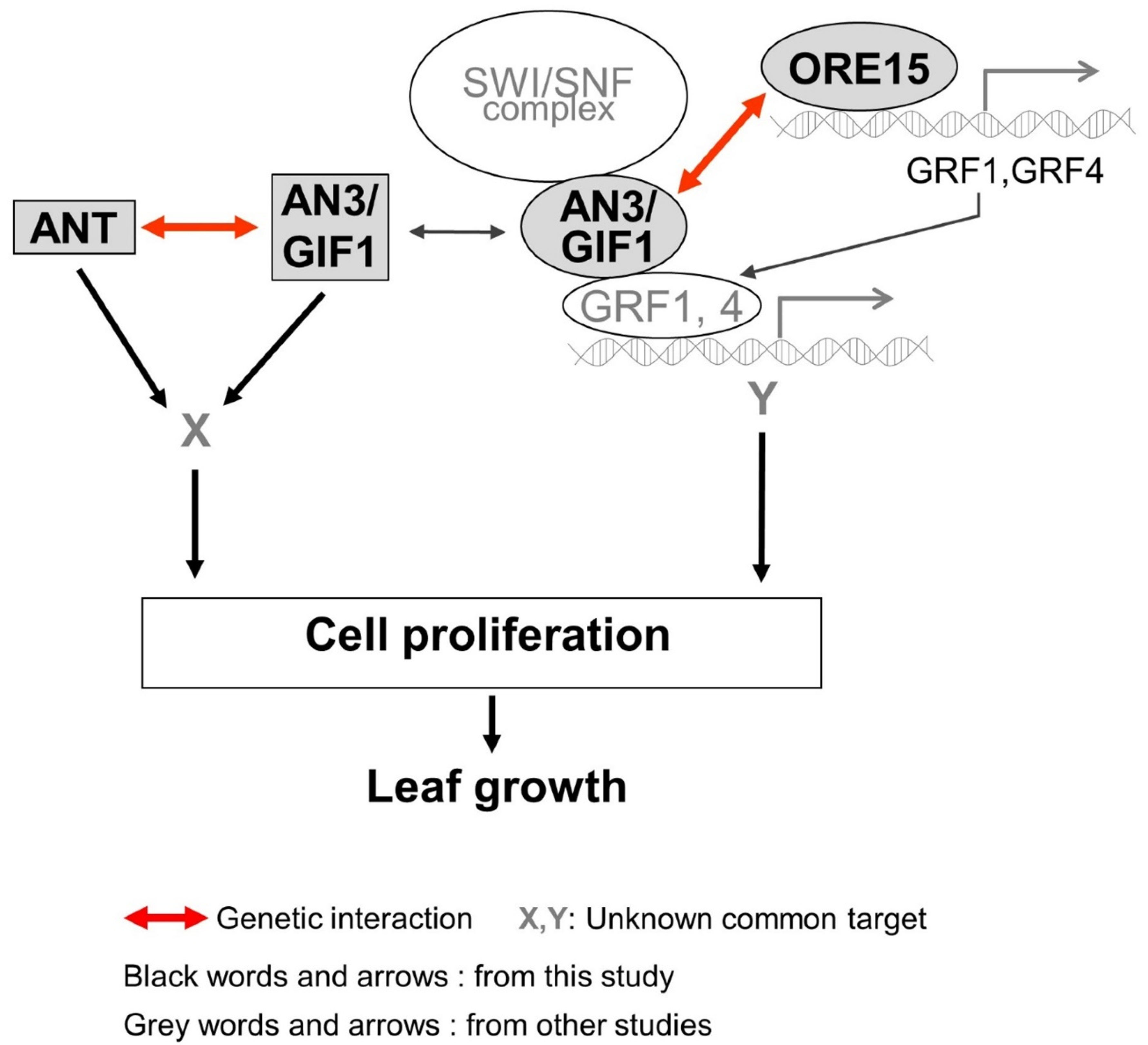
3.1. ORE15 May Act to Maintain AN3 and ANT Expression in the Later Stage of Cell Proliferation during Leaf Growth

In this study, the ore15-1D and ore15-2 mutants produced larger and smaller leaves, respectively, than the Col-0 (Figure 2 and Figure 3), consistent with the findings of our previous study [25]. Kinematic analysis of leaf growth showed a higher rate of cell proliferation in ore15-1D in early stage of leaves compared with those in Col-0 [25]. Anatomical analysis also showed that ORE15 mediated the cell proliferating phase of early stage in leaf growth [25].
Our ORE15promoter::GUS analysis indicated that ORE15 was expressed in the abaxial parenchyma cells along the midvein region of the proximal part of the leaf blade and throughout the leaf petiole (Figure 1). On the other hand, AN3 was expressed in the adaxial and abaxial parenchyma cells along the midvein region of the proximal part of the leaf blade (Figure 1). In spite of the different spatial expression patterns in adaxial parenchyma cells between ORE15 and AN3, two genes showed genetic interaction in cell proliferation. This discrepancy could be explained by non-cell autonomous regulation of AN3 [17] and the possibility of ORE15 action in non-cell autonomous manner. Further studies are required to clarify the detail of interaction between ORE15 and AN3 in leaf growth. In addition, the temporal expression pattern of ORE15 partially overlapped with that of AN3 (Figure 1) and ANT, as also shown previously [9,17,26]. ORE15 expression was not detected in leaf primordia. On the other hand, strong AN3 and ANT expressions were observed in the proximal region of leaf primordia and young leaves (Figure 1A–C) [9,17,26]. A chromatin immunoprecipitation (ChIP) study of GRF1 and GRF4 promoters, which were found to be putative targets of the ORE15 gene in our previous study [25], showed very similar expression patterns to ORE15 [9]. Moreover, GRF1 and GRF4 expression was weaker than AN3 expression in the proximal region of leaf primordia [9]. These results indicated that ORE15 may function to maintain AN3 and ANT expression in the later stage of cell proliferation for leaf growth. Together, our result of the spatiotemporally overlapping expression of ORE15 and the GIF–GRF complex, especially AN3/GIF1, GRF1, and GRF4, supports our working model of the cell proliferation regulation to promote leaf growth.

3.2. Functional Redendancy between ORE15 and AN3, and between AN3 and ANT in Cell Proliferation Regulatory Pathways during Leaf Growth

Our previous study indicated that ORE15 enhances leaf growth by promoting cell proliferation in the early stage of leaf development by modulating the AN3/GIF1–GRF and delays leaf senescence at the mature stage of leaf development [25]. In this study, the more severe leaf size defect and more reduced cell number in leaves in ore15-2 an3-4 double mutant (Figure 3 and Figure 4) suggest that there is some functional redundancy between ORE15 and AN3 and that these two proteins work in parallel pathways that promote leaf growth (Figure 6). These pathways might converge on a common node, perhaps by regulating some common target gene [34]. ORE15 also enhances the transcription of ANT, CycD3;1, and CycB1:1 (Figure 5), which are involved in promoting cell division activity [25]. The ARGOS–ANT–CYCD3 pathway is another well characterized pathway involved in cell proliferation-mediated leaf growth [5,7,35].
Therefore, we analyzed the relationship between ORE15 and another important regulator, ANT. The double mutant ore15-2 ant-1 had more reduced cell number in leaves than each single mutant (Figure 4), indicating that ORE15 may act separately from ANT in the cell proliferation regulatory pathway (Figure 6). The serrated leaves in the ore15-1D ant-1 mutant also support that ORE15 and ANT might work separately in leaf growth.
To date, the relationship between ANT and AN3 for proper cell proliferation during leaf development has remained unknown. Recently, it was reported that gif1/an3 ant-1 double mutant had small plants with fewer cells than its parental lines and displayed remarkable synergism in leaf development in supplementary data [16], indicating the possible genetic interaction between AN3/GIF1 and ANT. In the current study, the an3-4 ant-1 double mutant produced narrower and smaller leaves than all single mutants (Figure 2 and Figure 3). In addition, the an3-4 ant-1 double mutant produced significantly reduced cell number than all single mutants (Figure 4), indicating that these two genes may act synergistically as regulators of the cell proliferation pathway. However, the molecular mechanism of interaction between these two factors is still unclear. Together, these findings provide the first evidence to suggest that there is some functional redundancy between AN3 and ANT in leaf growth. Thus, these two proteins work in parallel and convergent pathways to regulate an unknown common target gene in cell proliferation (Figure 6). In our previous work, we suggested that ORE15 delays the onset of senescence and regulates cell proliferation [25]. Our previous genetic analyses suggested that ORE15 primarily utilizes AN3/GIF1 in the regulation of leaf senescence. It has been reported that ANT plays a negative role in regulating leaf senescence, acting downstream of ARF2 [36], suggesting a possible regulatory relationship between AN3/GIF1 and ANT in leaf senescence.
It has also been suggested that cells in the leaf blade and petiole originate from a common proliferative region located at the leaf blade/petiole junction [37]. Our expression analysis showed strong expression of ORE15 in the proximal part of the leaf blade and throughout the leaf petiole (Figure 1), partially overlapping with the expression of AN3 and ANT. Strong expression of AN3 and GRF family gene was also observed in the junction between the leaf blade and petiole in the current study (Figure 1) and in previous reports [9,15,37]. Our future work will aim to resolve the molecular mechanisms by which ORE15 coordinates the GRF–GIF pathway to regulate cell proliferation in the leaf blade and petiole, as well as leaf longevity.

4. Materials and Methods

4.1. Plant Material and Growth Conditions

The wild type Arabidopsis thaliana used in this study was Columbia-0 (Col-0). All mutants and transgenic plants in this study were originated from Col-0. The ore15-2 and ore15-1D mutants were described previously [25]. ore15-2 (SALK_029507) is a T-DNA insertional mutant, in which T-DNA was inserted in the first intron of the ORE15 (At1g31040) and was obtained from the Salk collection. ore15-1Dominant (ore15-1D) was isolated from the T-DNA insert lines of the activation tagging vector, pSKI015 with tetrameric CaMV 35S enhancer repeats.
The an3-4 and ant-1 mutants were described previously [10,33]. an3-4 has large deletionsin transcribed regions of the AN3 (At5g26840) locus, which occurred by X-ray irradiation. The AN3promoter::β-glucuronidase (GUS) transgenic plants were kindly provided by G. Horiguchi [9]. Since ant-1 homozygote is a female sterile plant and shows no visible defects at the early seedling stage, we isolated homozygotes by genomic PCR analysis to detect ant-1-associated nucleotide deletions as previously described [38]. ant-1 has a 22 nucleotide deletion (from cDNA nucleotides 803 to 824) within the open reading frame of the ANT (At4g37750) locus, producing truncated ANT protein. For ORE15 expression analysis using GUS assay, about 1.5 kb of DNA fragment of the upstream region of ORE15 gene were amplified from genomic DNA by PCR. The forward primer with SacI overhang and reverse primer with SpeI overhang at the 5′- end (Table S3) were used and then cloned into the pBluescript (-) vector. The ORE15 promoter fragment was inserted by replacing the CaMV 35S promoter into pB2GW7 harboring GUS gene with SacI and SpeI restriction digestion. Transgenic plants were generated by the Agrobacterium-mediated floral dipping methods. Detailed information about these primers is listed in Supplementary Table S3. The plant seeds were surface sterilized, germinated on a Murashige and Skoog (MS) medium [39], and transferred to soil 3 weeks after sowing. Plants were grown at 22 °C under long daylight conditions (70–90 μE/m2·s, 16 h light/8 h dark cycle).

4.2. Histochemical Staining for GUS Activity and Anatomy of Leaves

GUS staining of transgenic plants harboring ORE15promoter::GUS or AN3promoter::GUS were performed by GUS assay method as described previously [40]. For anatomical analysis, samples were embedded in Technovit 7100 resin (Kulzer & Co. GmbH, Wehrheim, Germany) and examined as described previously [41]. Sectional slices were observed by a light microscopy (Axiskop2, Carl Zeiss, Jena, Germany). Rosette diameter was measured using the length from tip to tip of the longest rosette leaves from at least five plants at 35 DAS. Measurement of cell number and cell area for leaf growth analysis was performed as described previously [9,25]. At least seven mature third leaves were collected from WT and mutants on 21 DAS and used for measurement. To measure leaf area and width, the curling leaves were made flat by cutting the rolled margin of leaf blades as shown in Figure 3. Paradermal images of leaf cells from collected leaves were observed using a DIC optic by light microscopy, and number and area of leaf cells were determined as described previously [25]. Data was analyzed using the NIH IMAGE program imageJ (1.51j8, NIH, Bethesda, MD, USA) and statistically analyzed using Statistical Package for the Social Science (SPSS 13.0, SPSS Inc., Chicago, IL, USA) program.

4.3. RNA Isolation and Analysis of Gene Expression

Total RNA was isolated from the cell proliferating leaves—the third to fifth leaves of 13 DAS—using the RNeasy mini kit (Qiagen, Hilden, Germany) and cDNA was synthesized using the Reverse TraAce-a-First strand cDNA synthesis kit (TOYOBO, Tokyo, Japan) for reverse transcription. Primers used for qRT-PCR are listed in Supplementary Table S3. The qRT-PCR was performed using SYBR® Premix Ex TaqTM II (TAKARA, Otsu, Japan) with three technical replicates as described previously [25] and Bio-Rad CFX96TM Real-Time System (Bio-Rad, Hercules, CA, USA). The relative expression level of each gene was calculated using the ΔΔCq method and the TUB4 gene was used as a control [41].

Supplementary Materials

Supplementary materials can be found at https://www.mdpi.com/1422-0067/21/1/241/s1. Figure S1: The relative alteration in cell number in a leaf from LOF and GOF single mutants and the combined mutants as in the corresponding control, Col-0 (WT), Figure S2: Distribution of DNA content in third leaves from Col-0 (WT), ore15-2 mutant, and ore15-1D mutant plants during leaf development, Table S1: The area and width of mature 3rd leaves of Col-0 (WT), LOF and GOF single mutants, and the combined double mutants at 21 DAS, Table S2: The total number and area of palisade mesophyll cells in mature 3rd leaves from Col-0 (WT), LOF and GOF single mutants, and the combined double mutants at 21 DAS, Table S3: List of oligonucleotide primers used in this study.

Author Contributions

Conceptualization, G.-T.K.; data curation, G.-T.K., S.E.J., and J.H.K.; formal analysis, S.E.J., J.Y.H., and T.T.H.L.; funding acquisition, G.-T.K., S.E.J., and J.H.K.; investigation, G.-T.K., S.E.J., and J.H.K.; methodology, S.E.J., J.H.K., J.Y.H., and T.T.H.L.; software, S.E.J., J.Y.H., and T.T.H.L.; supervision, G.-T.K.; validation, G.-T.K.; visualization, S.E.J., J.Y.H., and T.T.H.L.; writing—original draft, G.-T.K. and S.E.J.; writing—review and editing, G.-T.K. and S.E.J. All authors have read and agreed to the published version of the manuscript.

Funding

This research was supported by the mid-carrier Researcher Program (2017R1A2B4012714) through the National Research Foundation of Korea (NRF) funded by the Ministry of Science and ICT (MSIT), the Woo Jang-Choon Project (PJ01093902) of the Rural Development Administration, and the Research Fellow Program (2017R1A6A3A11028940), and Basic Science Research Program (2019R1A6A1A11052070) through the NRF funded by the Ministry of Education.

Acknowledgments

We thank G. Horiguchi (Rikkyo University, Japan) for providing AN3promoter::GUS transgenic seeds.

Conflicts of Interest

The authors declare no conflict of interest. The funders had no role in the design of the study; in the collection, analyses, or interpretation of data; in the writing of the manuscript, or in the decision to publish the results.

Abbreviations

Col-0Columbia-0
ORE15ORESARA15
AN3ANGUSTIFOLIA3
ANTAINTEGUMENTA
GIFGRFINTERACTING FACTOR
GRFGROWTH-REGULATING FACTOR
TCPTEOSINTE BRANCHED 1, CYCLOIDEA, and PROLIFERATING CELL FACTORS 1 and 2
KRP1Kip-related protein 1
WTWild type
LOFLoss of function
GOFGain of function
SWPSTRUWWELPETER

References

  1. Breuninger, H.; Lenhard, M. Control of Tissue and Organ Growth in Plants. In Current Topics in Developmental Biology; Timmermans, M., Ed.; Elsevier: Amsterdam, The Netherlands, 2010; Volume 91, pp. 185–220. [Google Scholar] [CrossRef]
  2. Sablowski, R.; Carnier Dornelas, M. Interplay between Cell Growth and Cell Cycle in Plants. J. Exp. Bot. 2014, 65, 2703–2714. [Google Scholar] [CrossRef] [PubMed] [Green Version]
  3. Schnittger, A.; Schobinger, U.; Bouyer, D.; Weinl, C.; Stierhof, Y.D.; Hulskamp, M. Ectopic D-Type Cyclin Expression Induces Not Only DNA Replication but Also Cell Division in Arabidopsis Trichomes. Proc. Natl. Acad. Sci. USA 2002, 99, 6410–6415. [Google Scholar] [CrossRef] [PubMed] [Green Version]
  4. Dewitte, W.; Riou-Khamlichi, C.; Scofield, S.; Healy, J.M.S.; Jacqmard, A.; Kilby, N.J.; Murray, J.A.H. Altered Cell Cycle Distribution, Hyperplasia, and Inhibited Differentiation in Arabidopsis Caused by the D-Type Cyclin CYCD3. Plant Cell 2003, 15, 79–92. [Google Scholar] [CrossRef] [PubMed] [Green Version]
  5. Mizukami, Y.; Fischer, R.L. Plant Organ Size Control: AINTEGUMENTA Regulates Growth and Cell Numbers during Organogenesis. Proc. Natl. Acad. Sci. USA 2000, 97, 942–947. [Google Scholar] [CrossRef] [PubMed] [Green Version]
  6. Dewitte, W.; Scofield, S.; Alcasabas, A.A.; Maughan, S.C.; Menges, M.; Braun, N.; Collins, C.; Nieuwland, J.; Prinsen, E.; Sundaresan, V.; et al. Arabidopsis CYCD3 D-Type Cyclins Link Cell Proliferation and Endocycles and Are Rate-Limiting for Cytokinin Responses. Proc. Natl. Acad. Sci. USA 2007, 104, 14537–14542. [Google Scholar] [CrossRef] [Green Version]
  7. Hu, Y.; Xie, Q.; Chua, N.H. The Arabidopsis Auxin-Inducible Gene ARGOS Controls Lateral Organ Size. Plant Cell 2003, 15, 1951–1961. [Google Scholar] [CrossRef] [Green Version]
  8. Kim, J.H.; Kende, H. A Transcriptional Coactivator, AtGIF1, Is Involved in Regulating Leaf Growth and Morphology in Arabidopsis. Proc. Natl. Acad. Sci. USA 2004, 101, 13374–13379. [Google Scholar] [CrossRef] [Green Version]
  9. Horiguchi, G.; Kim, G.T.; Tsukaya, H. The Transcription Factor AtGRF5 and the Transcription Coactivator AN3 Regulate Cell Proliferation in Leaf Primordia of Arabidopsis thaliana: Control of Leaf Shape and Size by AtGRF5 and AN3. Plant J. 2005, 43, 68–78. [Google Scholar] [CrossRef]
  10. Kim, J.H. Biological Roles and an Evolutionary Sketch of the GRF–GIF Transcriptional Complex in Plants. BMB Rep. 2019, 52, 227–238. [Google Scholar] [CrossRef] [Green Version]
  11. Kim, J.H.; Tsukaya, H. Regulation of Plant Growth and Development by the GROWTH-REGULATING FACTOR and GRF-INTERACTING FACTOR Duo. J. Exp. Bot. 2015, 66, 6093–6107. [Google Scholar] [CrossRef] [Green Version]
  12. Debernardi, J.M.; Mecchia, M.A.; Vercruyssen, L.; Smaczniak, C.; Kaufmann, K.; Inze, D.; Rodriguez, R.E.; Palatnik, J.F. Post-Transcriptional Control of GRF Transcription Factors by MicroRNA MiR396 and GIF Co-Activator Affects Leaf Size and Longevity. Plant J. 2014, 79, 413–426. [Google Scholar] [CrossRef] [PubMed]
  13. Vercruyssen, L.; Tognetti, V.B.; Gonzalez, N.; Van Dingenen, J.; De Milde, L.; Bielach, A.; De Rycke, R.; Van Breusegem, F.; Inzé, D. GROWTH REGULATING FACTOR5 Stimulates Arabidopsis Chloroplast Division, Photosynthesis, and Leaf Longevity. Plant Physiol. 2015, 167, 817–832. [Google Scholar] [CrossRef] [PubMed] [Green Version]
  14. Lee, B.H.; Ko, J.H.; Lee, S.; Lee, Y.; Pak, J.H.; Kim, J.H. The Arabidopsis GRF-INTERACTING FACTOR Gene Family Performs an Overlapping Function in Determining Organ Size as Well as Multiple Developmental Properties. Plant Physiol. 2009, 151, 655–668. [Google Scholar] [CrossRef] [PubMed] [Green Version]
  15. Lee, B.H.; Kim, J.H. Spatio-Temporal Distribution Patterns of GRF-INTERACTING FACTOR Expression and Leaf Size Control. Plant Signal. Behav. 2014, 9, e29697. [Google Scholar] [CrossRef] [PubMed] [Green Version]
  16. Lee, B.H.; Wynn, A.N.; Franks, R.G.; Hwang, Y.; Lim, J.; Kim, J.H. The Arabidopsis thaliana GRF-INTERACTING FACTOR Gene Family Plays an Essential Role in Control of Male and Female Reproductive Development. Dev. Biol. 2014, 386, 12–24. [Google Scholar] [CrossRef] [PubMed] [Green Version]
  17. Kawade, K.; Horiguchi, G.; Usami, T.; Hirai, M.Y.; Tsukaya, H. ANGUSTIFOLIA3 Signaling Coordinates Proliferation between Clonally Distinct Cells in Leaves. Curr. Biol. 2013, 23, 788–792. [Google Scholar] [CrossRef] [Green Version]
  18. Zhang, F.; Wang, H.; Kalve, S.; Wolabu, T.W.; Nakashima, J.; Golz, J.F.; Tadege, M. Control of Leaf Blade Outgrowth and Floral Organ Development by LEUNIG, ANGUSTIFOLIA 3 and WOX Transcriptional Regulators. New Phytol. 2019, 223, 2024–2038. [Google Scholar] [CrossRef]
  19. Vercruyssen, L.; Verkest, A.; Gonzalez, N.; Heyndrickx, K.S.; Eeckhout, D.; Han, S.K.; Jegu, T.; Archacki, R.; Van Leene, J.; Andriankaja, M.; et al. ANGUSTIFOLIA3 binds to SWI/SNF chromatin remodeling complex to regulate transcription during Arabidopsis leaf development. Plant Cell 2014, 26, 210–229. [Google Scholar] [CrossRef] [Green Version]
  20. Martin-Trillo, M.; Cubas, P. TCP genes: A family snapshot ten years later. Trends Plant Sci. 2010, 15, 31–39. [Google Scholar] [CrossRef]
  21. Efroni, I.; Blum, E.; Goldshmidt, A.; Eshed, Y. A Protracted and Dynamic Maturation Schedule Underlies Arabidopsis Leaf Development. Plant Cell 2008, 20, 2293–2306. [Google Scholar] [CrossRef] [Green Version]
  22. Nag, A.; King, S.; Jack, T. MiR319a Targeting of TCP4 Is Critical for Petal Growth and Development in Arabidopsis. Proc. Natl. Acad. Sci. USA 2009, 106, 22534–22539. [Google Scholar] [CrossRef] [PubMed] [Green Version]
  23. Schommer, C.; Debernardi, J.M.; Bresso, E.G.; Rodriguez, R.E.; Palatnik, J.F. Repression of Cell Proliferation by MiR319-Regulated TCP4. Mol. Plant 2014, 7, 1533–1544. [Google Scholar] [CrossRef] [PubMed] [Green Version]
  24. Rodriguez, R.E.; Mecchia, M.A.; Debernardi, J.M.; Schommer, C.; Weigel, D.; Palatnik, J.F. Control of Cell Proliferation in Arabidopsis thaliana by MicroRNA MiR396. Development 2010, 137, 103–112. [Google Scholar] [CrossRef] [PubMed] [Green Version]
  25. Kim, J.H.; Kim, J.; Jun, S.E.; Park, S.; Timilsina, R.; Kwon, D.S.; Kim, Y.; Park, S.J.; Hwang, J.Y.; Nam, H.G.; et al. ORESARA15, a PLATZ Transcription Factor, Mediates Leaf Growth and Senescence in Arabidopsis. New Phytol. 2018, 220, 609–623. [Google Scholar] [CrossRef] [Green Version]
  26. Meng, L.S.; Wang, Y.B.; Yao, S.Q.; Liu, A. Arabidopsis AINTEGUMENTA mediates salt tolerance by trans-repressing SCABP8. J. Cell Sci. 2015, 128, 2919–2927. [Google Scholar] [CrossRef] [Green Version]
  27. Lee, S.J.; Lee, B.H.; Jung, J.H.; Park, S.K.; Song, J.T.; Kim, J.H. GROWTH-REGULATING FACTOR and GRF-INTERACTING FACTOR Specify Meristematic Cells of Gynoecia and Anthers. Plant Physiol. 2018, 176, 717–729. [Google Scholar] [CrossRef] [Green Version]
  28. Vanhaeren, H.; Inze, D.; Gonzalez, N. Plant Growth beyond Limits. Trends Plant Sci. 2018, 176, 717–729. [Google Scholar] [CrossRef]
  29. Tsukaya, H. Leaf Development. In The Arabidopsis Book; American Society of Plant Biologists: Rockville, MD, USA, 2013; Volume 11, p. e0163. [Google Scholar]
  30. Sugimoto-Shirasu, K.; Roberts, K. “Big it up”: Endoreduplication and cell-size control in plants. Curr. Opin. Plant Biol. 2003, 6, 544–553. [Google Scholar] [CrossRef]
  31. De Veylder, L.; Larkin, J.C.; Schnittger, A. Molecular control of function endoreduplication in development and physiology. Trends Plant Sci. 2011, 16, 624–634. [Google Scholar] [CrossRef]
  32. Hisanaga, T.; Kawade, K.; Tsukaya, H. Compensation: A key to clarifying the organ-level regulation of lateral organ size in plants. J. Exp. Bot. 2015, 66, 1055–1063. [Google Scholar] [CrossRef] [Green Version]
  33. Autran, D.; Belcram, J.C.; Beemster, G.T.; Kronenberger, J.; Grandjean, O.; Inzé, D.; Traas, J. Cell numbers and leaf development in Arabidopsis: A functional analysis of the STRUWWELPETER gene. EMBO J. 2002, 21, 6036–6049. [Google Scholar] [CrossRef] [PubMed] [Green Version]
  34. Pérez-Pérez, J.M.; Candela, H.; Micol, J.L. Understanding synergy in genetic interactions. Trends Genet. 2009, 25, 368–376. [Google Scholar] [CrossRef] [PubMed]
  35. Powell, A.E.; Lenhard, M. Control of Organ Size in Plants. Curr. Biol. 2012, 22, R360–R367. [Google Scholar] [CrossRef] [PubMed] [Green Version]
  36. Feng, G.; Xu, Q.; Wang, Z.; Zhuoma, Q. AINTEGUMENTA Negatively Regulates Age-Dependent Leaf Senescence Downstream of AUXIN RESPONSE FACTOR 2 in Arabidopsis thaliana. Plant Biotechnol. 2016, 33, 71–76. [Google Scholar] [CrossRef] [Green Version]
  37. Ichihashi, Y.; Kawade, K.; Usami, T.; Horiguchi, G.; Takahashi, T.; Tsukaya, H. Key Proliferative Activity in the Junction between the Leaf Blade and Leaf Petiole of Arabidopsis. Plant Physiol. 2011, 157, 1151–1162. [Google Scholar] [CrossRef] [Green Version]
  38. Kang, J.; Mizukami, Y.; Wang, H.; Fowke, L.; Dengler, N.G. Modification of Cell Proliferation Patterns Alters Leaf Vein Architecture in Arabidopsis thaliana. Planta 2007, 226, 1207–1218. [Google Scholar] [CrossRef]
  39. Murashige, T.; Skoog, F. A revised medium for rapid growth and bioassays with tobacco tissue culture. Physiol. Plant. 1962, 15, 473–497. [Google Scholar] [CrossRef]
  40. Jun, S.E.; Okushima, Y.; Nam, J.; Umeda, M.; Kim, G.T. Kip-Related Protein 3 Is Required for Control of Endoreduplication in the Shoot Apical Meristem and Leaves of Arabidopsis. Mol. Cells 2013, 35, 47–53. [Google Scholar] [CrossRef] [Green Version]
  41. Kim, G.T.; Tsukaya, H.; Uchimiya, H. The ROTUNDIFOLIA3 gene of Arabidopsis thaliana encodes a new member of the cytochrome P-450 family that is required for the regulated polar elongation of leaf cells. Genes Dev. 1998, 12, 2381–2391. [Google Scholar] [CrossRef] [Green Version]
Figure 1. Spatial and temporal specific expression of ORE15 and AN3 determined by promoter::β-glucuronidase (GUS) assay of transgenic plants harboring the ORE15promoter::GUS and AN3promoter::GUS construct. (A) Seedling with the first pair of true leaves at seven days after sowing (DAS). (B) Magnified view of (A). (C) Seedling at 14 DAS. (D) First true leaf pair in the juvenile phase at 14 DAS. (E) Young floral bud. (F) Mature flower. Scale bar = 10 mm. (G) Transverse section of the proximal part of leaf blades from GUS-stained third leaf at 14 DAS. (H) Magnified view of (G). Scale bar = 100 μm.
Ijms 21 00241 g001
Figure 2. Plant morphology and rosette diameter of the Arabidopsis Columbia-0 (Col-0) wild type (WT), single loss of function (LOF) and gain of function (GOF) mutants. (A) Plant phenotypes of the ore15-2, ore15-1D, an3-4, ant-1, and combined double mutants, ore15-2 an3-4, ore15-2 ant-1, an3-4 ore15-1D, ore15-1D ant-1, and an3-4 ant-1 at 35 DAS. ore15-2 (SALK_029507) is a T-DNA insertional mutant and ore15-1D is a dominant mutant isolated from activation tagging lines, in which T-DNA was inserted in the first intron and 3′ UTR of ORE15, respectively. an3-4 has a large deletion in the AN3 locus and ant-1 has a 22-nucleotide deletion in the second exon of the ANT locus. Scale bar = 1 cm. (B) The widest diameter of rosette leaves. Data are means ± standard error (SE) (n = 5). Means were compared using one-way analysis of variance (ANOVA) followed by Duncan’s multiple range test. Different lowercase letters indicate significantly different means between genotypes.
Ijms 21 00241 g002
Figure 3. Leaf morphology of the mature third leaf from Col-0 (WT), single mutant, and double mutant plants at 21 DAS. (A) Phenotype of the mature third leaf detached from Col-0, single mutant, and double mutant plants at 21 DAS. Scale bar = 1 cm. (B) Leaf area. (C) Leaf width. Data are means ± standard error (SE) (7 ≤ n ≤ 10). Means were compared using one-way analysis of variance (ANOVA) followed by Duncan’s multiple range test. Different lowercase letters indicate significantly different means between genotypes.
Ijms 21 00241 g003
Figure 4. Statistical analysis of leaf palisade mesophyll cell number and area in mature third leaves from Col-0 (WT), single mutant, and double mutant plants at 21 DAS. (A) Paradermal images of palisade mesophyll cells on mature third leaves. Scale bar = 100 µm. (B) Cell number. (C) Cell area. Data are means ± SE (4 ≤ n ≤ 7). Means were compared using one-way ANOVA followed by Duncan’s multiple range test. Different lowercase letters indicate significantly different means between genotypes.
Ijms 21 00241 g004
Figure 5. Expression of cell proliferation-related genes in the blades of cell proliferating leaves (third to fifth leaves) of Col-0 (WT), single mutant, and double mutant plants at 13 DAS. The transcript abundance of each gene was analyzed by quantitative reverse transcription polymerase chain reaction (qRT-PCR) using primers specific to each gene, normalized to TUB4, and are shown as values relative to each level of the Col-0 (WT). Data are representative of three technical replicates. Plotted values are means ± SE (n = 3). A statistical analysis was performed using a one-way ANOVA. Asterisks indicate significant difference from Col-0 (WT) plants (*, p < 0.1; **, p < 0.05; ***, p < 0.01).
Ijms 21 00241 g005
Figure 6. A working model of the genetic interaction between ORE15, AN3, and ANT for cell proliferation regulatory pathway during leaf growth. Two functionally redundant parallel pathways might converge on common nodes, by regulating unknown target X, Y genes.
Ijms 21 00241 g006

