The Intercalation of CORM-2 with Pharmaceutical Clay Montmorillonite (MMT) Aids for Therapeutic Carbon Monoxide Release

 , ,
, , 
Abstract
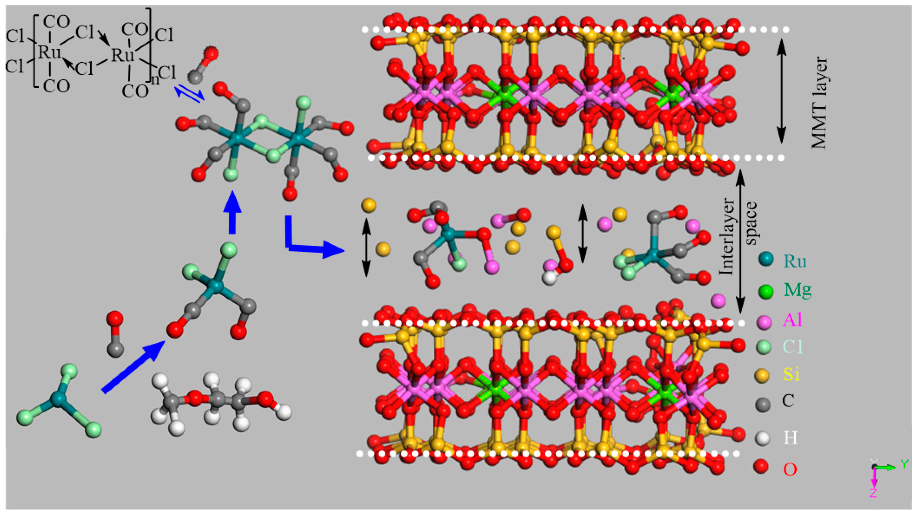
1. Introduction
2. Characterization Techniques
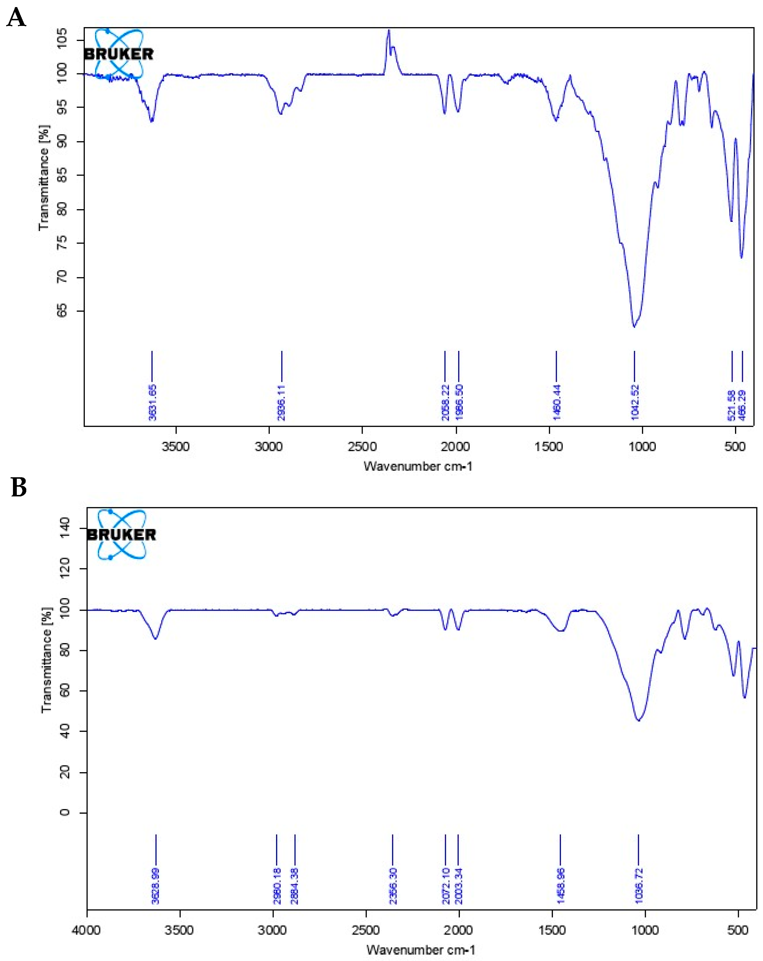
2.1. Infrared Spectroscopy (IR)
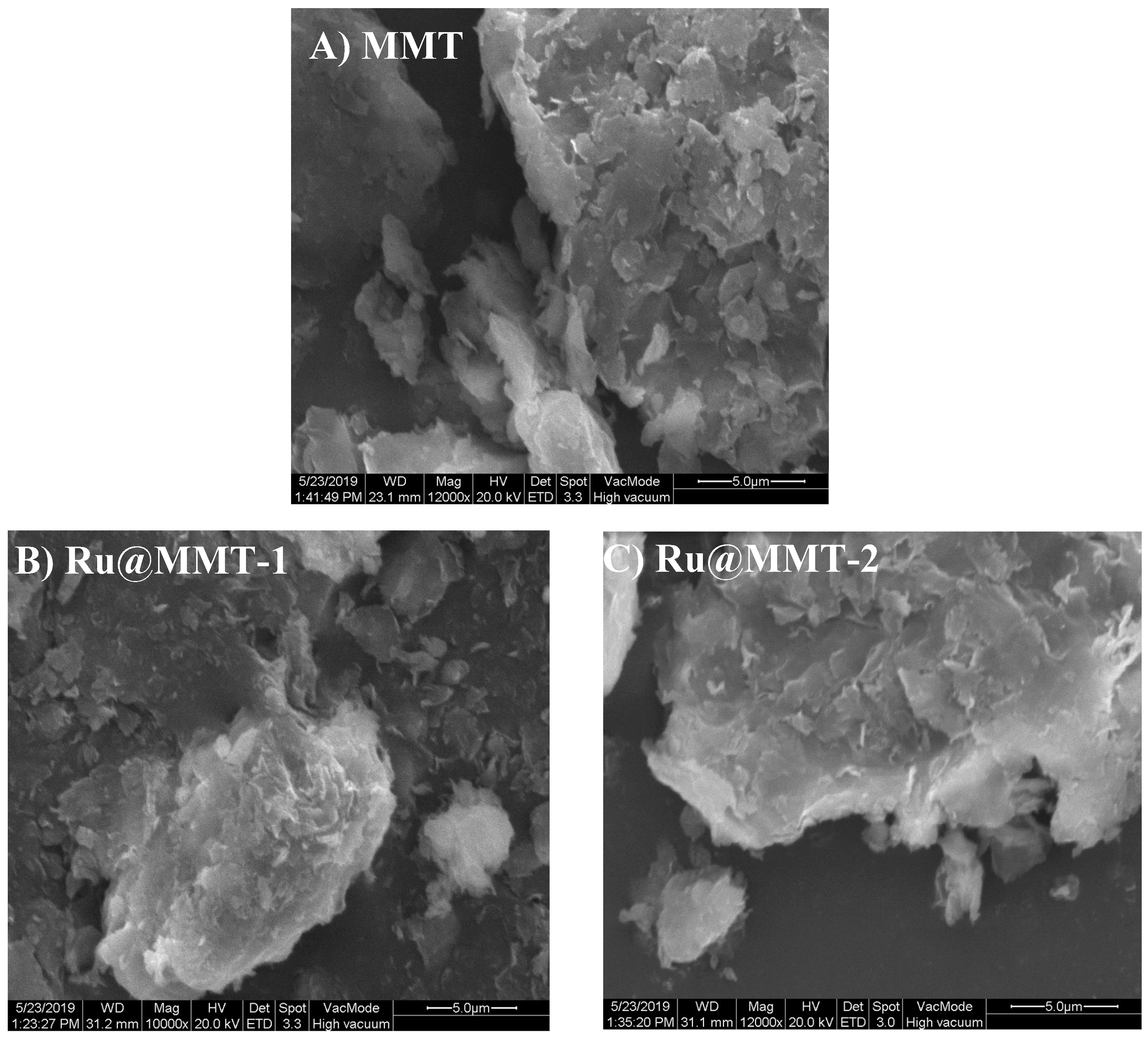
2.2. Scanning Electron Microscopy (SEM)
2.3. Transmission Electron Microscope (TEM)
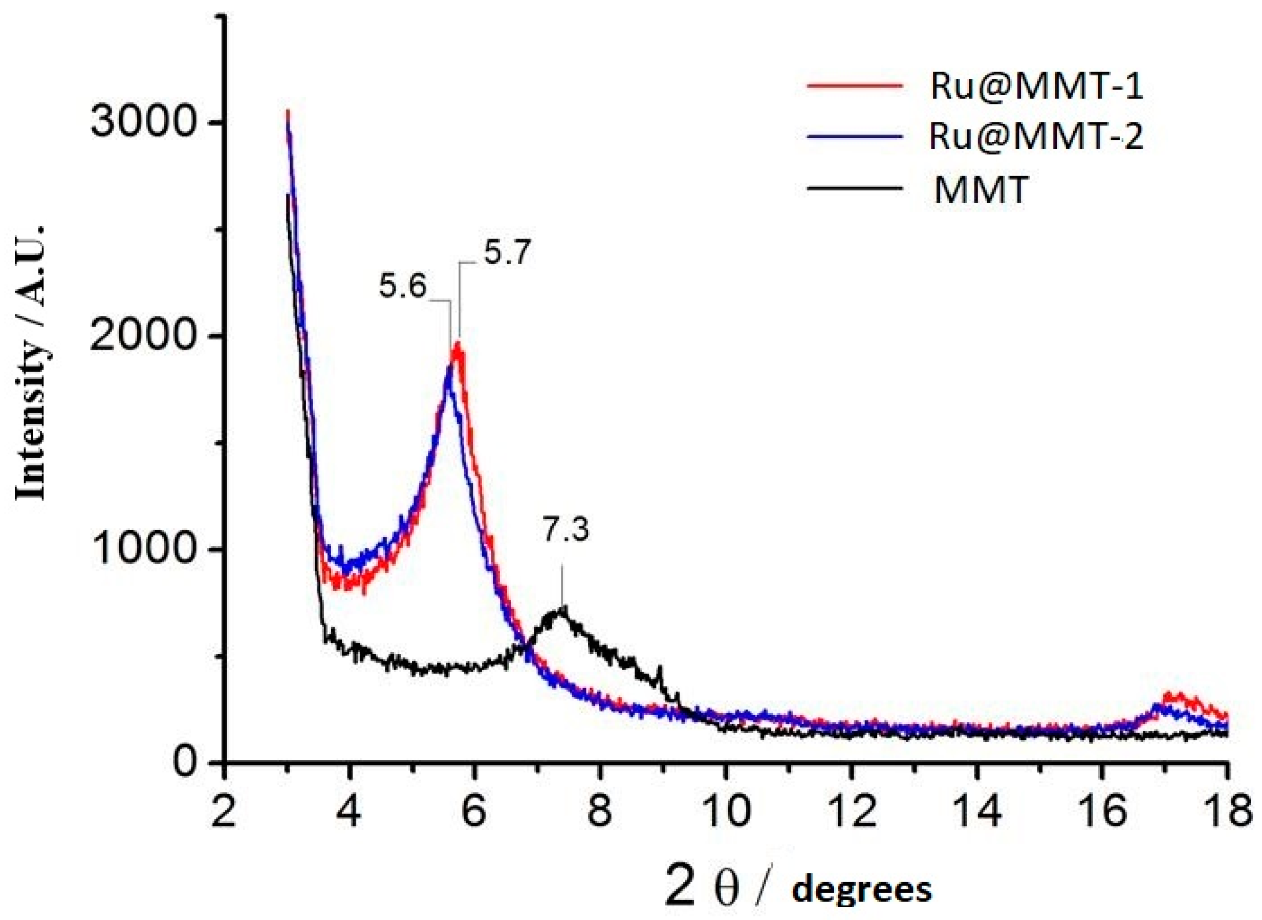
2.4. X-Ray Diffraction (X-ray)
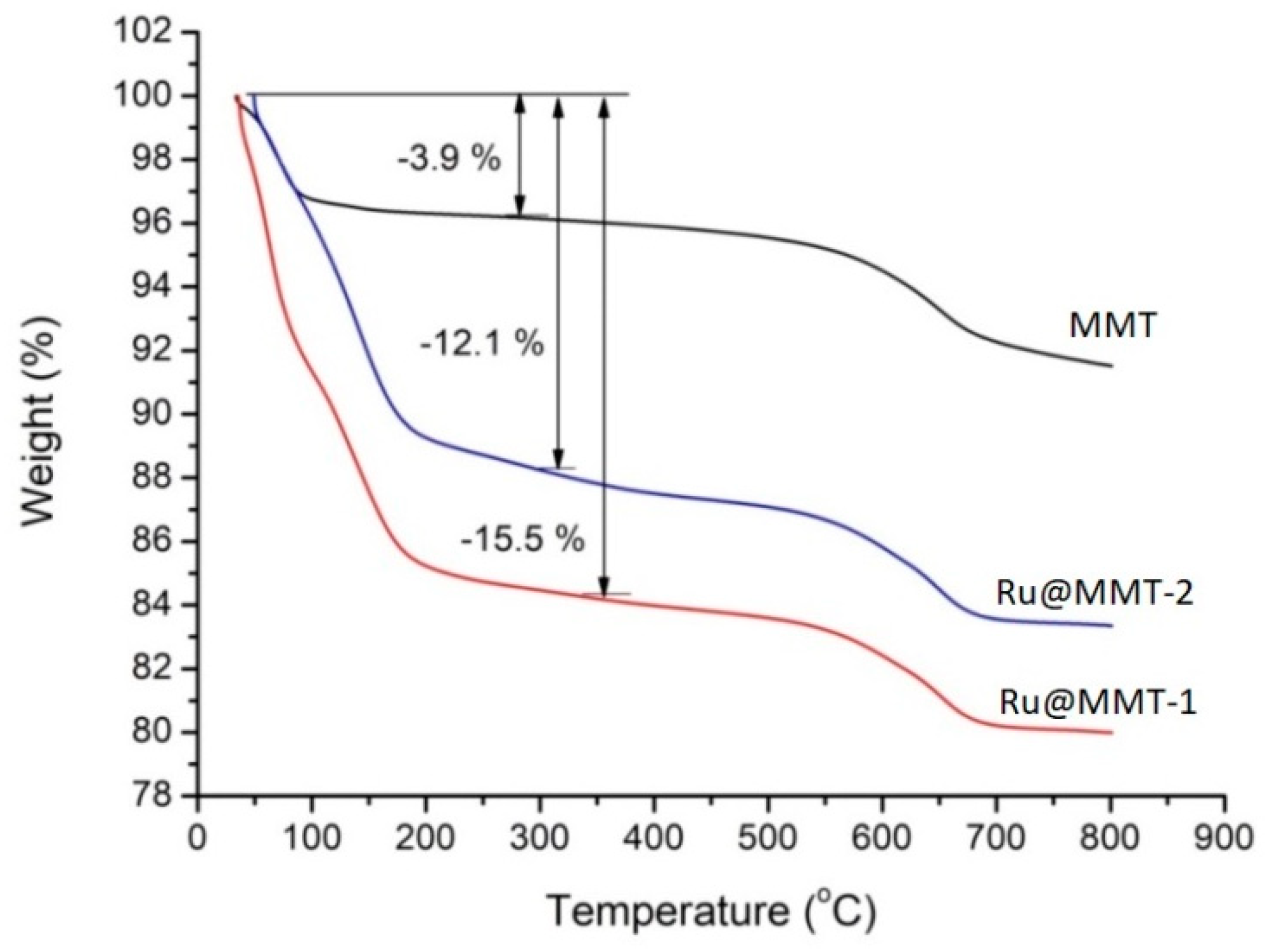
2.5. Thermogravimetric Analysis (TGA)
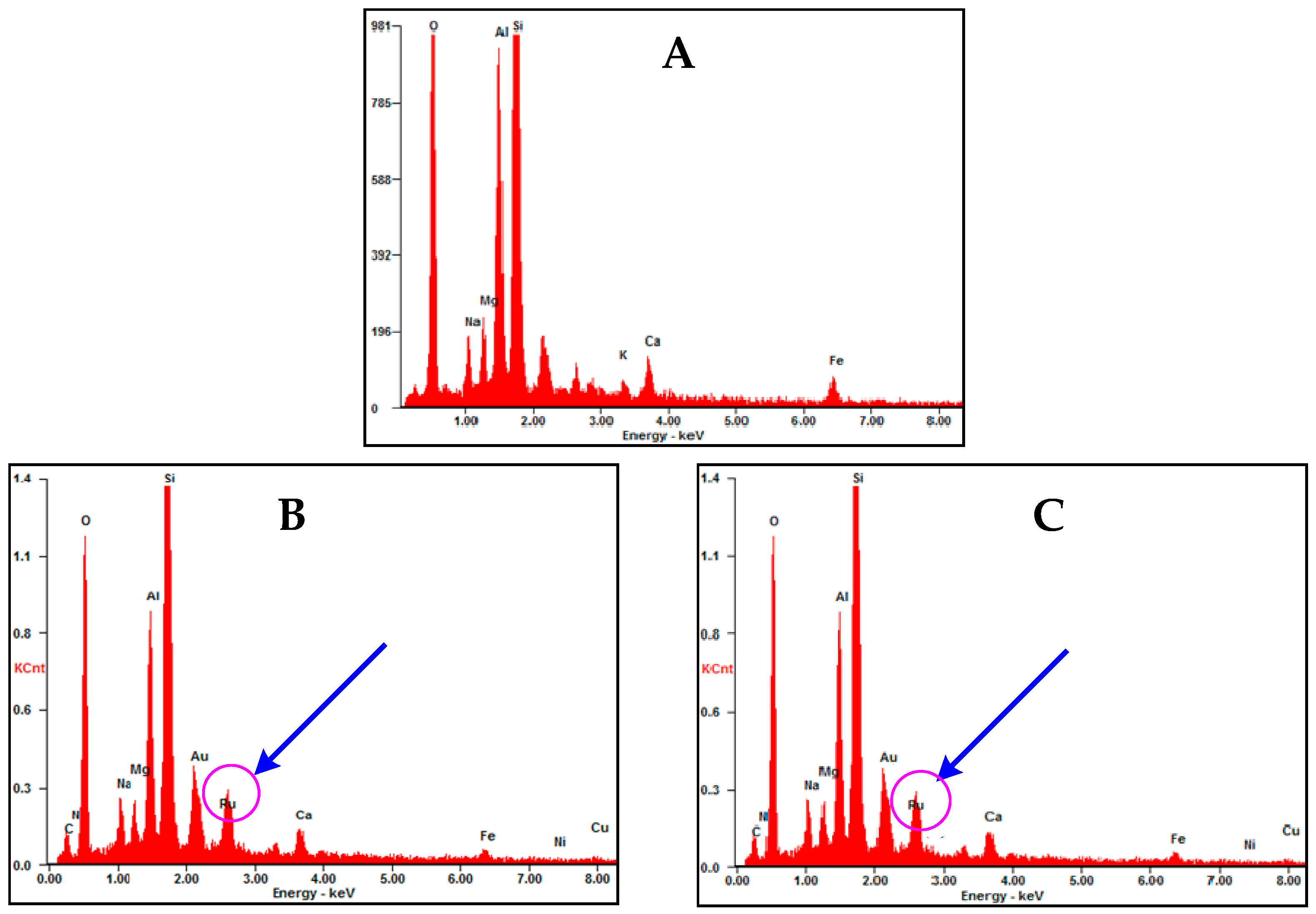
2.6. Energy-Dispersive X-Ray Spectroscopy (EDX)
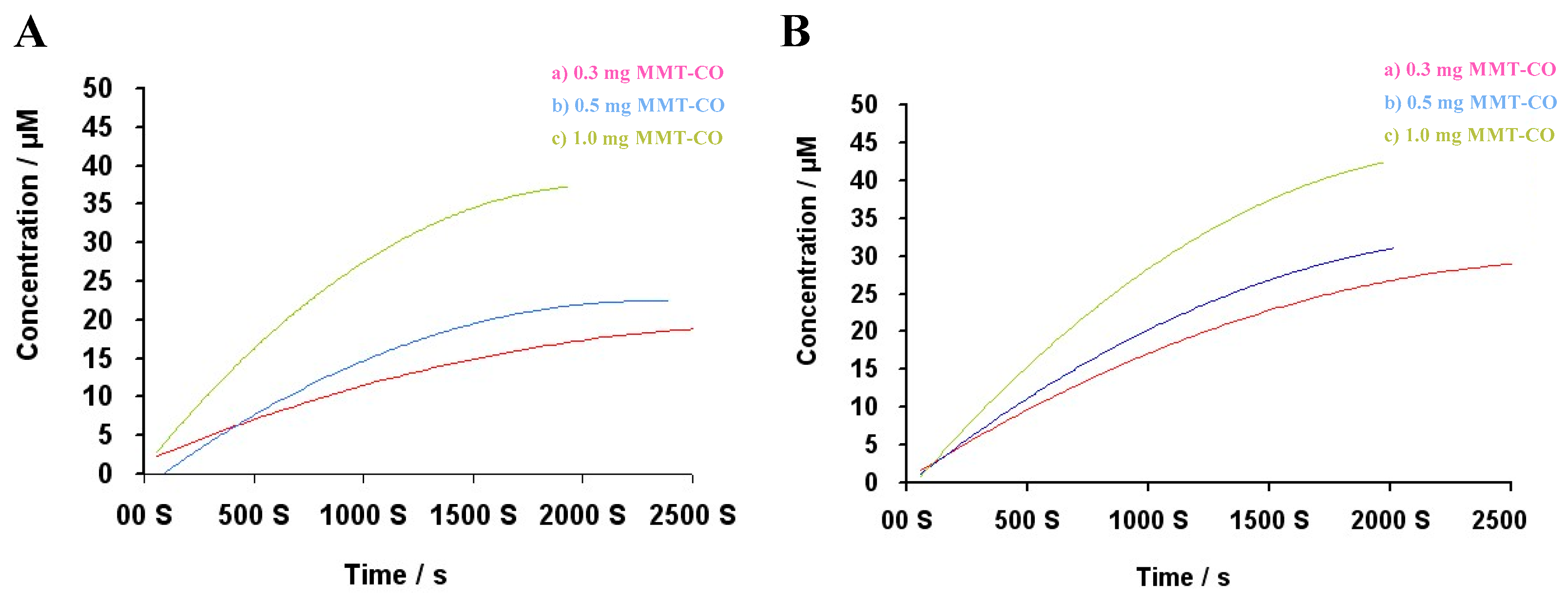
2.7. Myoglobin Assay (Mb)
3. Discussion
4. Materials and Synthesis Procedure
4.1. Materials
4.2. Characterization
4.2.1. General Remarks
4.2.2. Preparation Ru@MMT-1
4.2.3. Preparation Ru@MMT-2
4.3. Myoglobin Assay for CO Kinetics Profile
5. Conclusions
Author Contributions
Funding
Acknowledgments
Conflicts of Interest
References
- Raub, J.A.; Mathieu-Nolf, M.; Hampson, N.B.; Thom, S.R. Carbon monoxide poisoning—a public health perspective. Toxicology 2000, 145, 1–14. [Google Scholar] [CrossRef]
- Haldane, J.B.S. Carbon monoxide as a tissue poison. Biochem. J. 1927, 21, 1068–1075. [Google Scholar] [CrossRef] [PubMed]
- Douglas, C.G.; Haldane, J.S.; Haldane, J.B.S. The laws of combination of haemoglobin with carbon monoxide and oxygen. J. Physiol. (Oxford, Oxf. UK) 1912, 44, 275–304. [Google Scholar] [CrossRef]
- Jiang, Y.h. Relationship between endogenous carbon monoxide production and disease. J. Mod. Lab. Med. 2013, 28, 22–24. [Google Scholar] [CrossRef]
- Leffler, C.W.; Parfenova, H.; Jaggar, J.H. Carbon monoxide as an endogenous vascular modulator. Am. J. Physiol. Heart Circ. Physiol. 2011, 301, H1–H11. [Google Scholar] [CrossRef] [PubMed]
- Kim, H.-H.; Choi, S. Therapeutic Aspects of Carbon Monoxide in Cardiovascular Disease. Int. J. Mol. Sci. 2018, 19, 2381. [Google Scholar] [CrossRef]
- Wang, L.; Yan, L.; Liu, J.; Chen, C.; Zhao, Y. Quantification of Nanomaterial/Nanomedicine Trafficking in Vivo. Anal. Chem. 2018, 90, 589–614. [Google Scholar] [CrossRef]
- Palao, E.; Slanina, T.; Muchová, L.; Šolomek, T.; Vítek, L.; Klán, P. Transition-Metal-Free CO-Releasing BODIPY Derivatives Activatable by Visible to NIR Light as Promising Bioactive Molecules. J. Am. Chem. Soc. 2016, 138, 126–133. [Google Scholar] [CrossRef]
- Ling, K.; Men, F.; Wang, W.-C.; Zhou, Y.-Q.; Zhang, H.-W.; Ye, D.-W. Carbon Monoxide and Its Controlled Release: Therapeutic Application, Detection, and Development of Carbon Monoxide Releasing Molecules (CORMs). J. Med. Chem. 2018, 61, 2611–2635. [Google Scholar] [CrossRef]
- Foresti, R.; Bani-Hani, M.G.; Motterlini, R. Use of carbon monoxide as a therapeutic agent: Promises and challenges. Intensive Care Med. 2008, 34, 649–658. [Google Scholar] [CrossRef]
- Abraham, D.J.; Rotella, D.P. Medicinal Chemistry and Therapeutic Applications of the Gasotransmitters NO, CO, and H2S and their Prodrugs. In Burger’s Medicinal Chemistry, Drug Discovery, and Development, 7th ed.; John Wiley & Sons, Inc.: New Jersey, NJ, USA, 2010. [Google Scholar] [CrossRef]
- Szabo, C. Gaseotransmitters: New frontiers for translational science. Sci. Transl. Med. 2010, 2, 59ps54. [Google Scholar] [CrossRef] [PubMed]
- Yang, S.; Chen, M.; Zhou, L.; Zhang, G.; Gao, Z.; Zhang, W. Photo-activated CO-releasing molecules (PhotoCORMs) of robust sawhorse scaffolds [μ2-OOCR1, η1-NH2CHR2(C=O] OCH3, Ru(I)2CO4]. Dalton Trans. 2016, 45, 3727–3733. [Google Scholar] [CrossRef] [PubMed]
- He, Q.; Kiesewetter, D.O.; Qu, Y.; Fu, X.; Fan, J.; Huang, P.; Liu, Y.; Zhu, G.; Liu, Y.; Qian, Z.; et al. NIR-Responsive On-Demand Release of CO from Metal Carbonyl-Caged Graphene Oxide Nanomedicine. Adv. Mater. (Deerfield Beach Fla.) 2015, 27, 6741–6746. [Google Scholar] [CrossRef] [PubMed]
- Yin, H.; Fang, J.; Liao, L.; Nakamura, H.; Maeda, H. Styrene-maleic acid copolymer-encapsulated CORM2, a water-soluble carbon monoxide (CO) donor with a constant CO-releasing property, exhibits therapeutic potential for inflammatory bowel disease. J. Controll. Release Off. J. Controll. Release Soc. 2014, 187, 14–21. [Google Scholar] [CrossRef] [PubMed]
- Peng, P.; Wang, C.; Shi, Z.; Johns, V.K.; Ma, L.; Oyer, J.; Copik, A.; Igarashi, R.; Liao, Y. Visible-light activatable organic CO-releasing molecules (PhotoCORMs) that simultaneously generate fluorophores. Org. Biomol. Chem. 2013, 11, 6671–6674. [Google Scholar] [CrossRef] [PubMed]
- Hasegawa, U.; van der Vlies, A.J.; Simeoni, E.; Wandrey, C.; Hubbell, J.A. Carbon monoxide-releasing micelles for immunotherapy. J. Am. Chem. Soc. 2010, 132, 18273–18280. [Google Scholar] [CrossRef]
- Pai, S.; Radacki, K.; Schatzschneider, U. Sonogashira, CuAAC, and Oxime Ligations for the Synthesis of MnI Tricarbonyl PhotoCORM Peptide Conjugates. Eur. J. Inorg. Chem. 2014, 2014, 2886–2895. [Google Scholar] [CrossRef]
- Pfeiffer, H.; Rojas, A.; Niesel, J.; Schatzschneider, U. Sonogashira and “Click” reactions for the N-terminal and side-chain functionalization of peptides with [Mn(CO)3(tpm)]+-based CO releasing molecules (tpm = tris(pyrazolyl)methane). Dalton Trans. (Camb. Engl. 2003) 2009, 4292–4298. [Google Scholar] [CrossRef]
- Jackson, C.S.; Schmitt, S.; Dou, Q.P.; Kodanko, J.J. Synthesis, characterization, and reactivity of the stable iron carbonyl complex [Fe(CO)(N4Py)](ClO4)2: Photoactivated carbon monoxide release, growth inhibitory activity, and peptide ligation. Inorg. Chem. 2011, 50, 5336–5338. [Google Scholar] [CrossRef]
- Matson, J.B.; Webber, M.J.; Tamboli, V.K.; Weber, B.; Stupp, S.I. A peptide-based material for therapeutic carbon monoxide delivery. Soft Matter 2012, 8, 6689–6692. [Google Scholar] [CrossRef]
- Bischof, C.; Joshi, T.; Dimri, A.; Spiccia, L.; Schatzschneider, U. Synthesis, spectroscopic properties, and photoinduced CO-release studies of functionalized ruthenium(II) polypyridyl complexes: Versatile building blocks for development of CORM-peptide nucleic acid bioconjugates. Inorg. Chem. 2013, 52, 9297–9308. [Google Scholar] [CrossRef] [PubMed]
- Pfeiffer, H.; Sowik, T.; Schatzschneider, U. Bioorthogonal oxime ligation of a Mo(CO)4(N–N) CO-releasing molecule (CORM) to a TGF β-binding peptide. J. Organomet. Chem. 2013, 734, 17–24. [Google Scholar] [CrossRef]
- Zobi, F.; Blacque, O.; Jacobs, R.A.; Schaub, M.C.; Bogdanova, A.Y. 17 e−rhenium dicarbonyl CO-releasing molecules on a cobalamin scaffold for biological application. Dalton Trans. 2012, 41, 370–378. [Google Scholar] [CrossRef] [PubMed]
- Wilson, J.L.; Fayad Kobeissi, S.; Oudir, S.; Haas, B.; Michel, B.; Dubois Rande, J.L.; Ollivier, A.; Martens, T.; Rivard, M.; Motterlini, R.; et al. Design and synthesis of new hybrid molecules that activate the transcription factor Nrf2 and simultaneously release carbon monoxide. Chemistry (Weinh. der Bergstr. Ger.) 2014, 20, 14698–14704. [Google Scholar] [CrossRef] [PubMed]
- Pena, A.C.; Penacho, N.; Mancio-Silva, L.; Neres, R.; Seixas, J.D.; Fernandes, A.C.; Romao, C.C.; Mota, M.M.; Bernardes, G.J.; Pamplona, A. A novel carbon monoxide-releasing molecule fully protects mice from severe malaria. Antimicrob. Agents Chemother. 2012, 56, 1281–1290. [Google Scholar] [CrossRef] [PubMed]
- Marques, A.R.; Kromer, L.; Gallo, D.J.; Penacho, N.; Rodrigues, S.S.; Seixas, J.D.; Bernardes, G.J.L.; Reis, P.M.; Otterbein, S.L.; Ruggieri, R.A.; et al. Generation of Carbon Monoxide Releasing Molecules (CO-RMs) as Drug Candidates for the Treatment of Acute Liver Injury: Targeting of CO-RMs to the Liver. Organometallics 2012, 31, 5810–5822. [Google Scholar] [CrossRef]
- Kunz, P.C.; Brückmann, N.E.; Spingler, B. Towards Polymer Diagnostic Agents—Copolymers of N-(2-Hydroxypropyl)methacrylamide and Bis(2-pyridylmethyl)-4-vinylbenzylamine: Synthesis, Characterisation and Re(CO)3-Labelling. Eur. J. Inorg. Chem. 2007, 2007, 394–399. [Google Scholar] [CrossRef]
- Brückmann, N.E.; Wahl, M.; Reiß, G.J.; Kohns, M.; Wätjen, W.; Kunz, P.C. Polymer Conjugates of Photoinducible CO-Releasing Molecules. Eur. J. Inorg. Chem. 2011, 2011, 4571–4577. [Google Scholar] [CrossRef]
- Tabe, H.; Fujita, K.; Abe, S.; Tsujimoto, M.; Kuchimaru, T.; Kizaka-Kondoh, S.; Takano, M.; Kitagawa, S.; Ueno, T. Preparation of a Cross-Linked Porous Protein Crystal Containing Ru Carbonyl Complexes as a CO-Releasing Extracellular Scaffold. Inorg. Chem. 2015, 54, 215–220. [Google Scholar] [CrossRef]
- Tabe, H.; Shimoi, T.; Fujita, K.; Abe, S.; Ijiri, H.; Tsujimoto, M.; Kuchimaru, T.; Kizaka-Kondo, S.; Mori, H.; Kitagawa, S.; et al. Design of a CO-releasing Extracellular Scaffold Using in Vivo Protein Crystals. Chem. Lett. 2015, 44, 342–344. [Google Scholar] [CrossRef]
- Chaves-Ferreira, M.; Albuquerque, I.S.; Matak-Vinkovic, D.; Coelho, A.C.; Carvalho, S.M.; Saraiva, L.M.; Romao, C.C.; Bernardes, G.J. Spontaneous CO release from RuII(CO)2-protein complexes in aqueous solution, cells, and mice. Angew. Chem. (Int. Ed. Engl.) 2015, 54, 1172–1175. [Google Scholar] [CrossRef] [PubMed]
- Albuquerque, I.S.; Jeremias, H.F.; Chaves-Ferreira, M.; Matak-Vinkovic, D.; Boutureira, O.; Romão, C.C.; Bernardes, G.J.L. An artificial CO-releasing metalloprotein built by histidine-selective metallation. Chem. Commun. 2015, 51, 3993–3996. [Google Scholar] [CrossRef] [PubMed]
- Fujita, K.; Tanaka, Y.; Sho, T.; Ozeki, S.; Abe, S.; Hikage, T.; Kuchimaru, T.; Kizaka-Kondoh, S.; Ueno, T. Intracellular CO release from composite of ferritin and ruthenium carbonyl complexes. J. Am. Chem. Soc. 2014, 136, 16902–16908. [Google Scholar] [CrossRef] [PubMed]
- Diring, S.; Carné-Sánchez, A.; Zhang, J.; Ikemura, S.; Kim, C.; Inaba, H.; Kitagawa, S.; Furukawa, S. Light responsive metal–organic frameworks as controllable CO-releasing cell culture substrates. Chem. Sci. 2017, 8, 2381–2386. [Google Scholar] [CrossRef] [PubMed]
- Dördelmann, G.; Pfeiffer, H.; Birkner, A.; Schatzschneider, U. Silicium Dioxide Nanoparticles As Carriers for Photoactivatable CO-Releasing Molecules (PhotoCORMs). Inorg. Chem. 2011, 50, 4362–4367. [Google Scholar] [CrossRef] [PubMed]
- Gonzales, M.A.; Han, H.; Moyes, A.; Radinos, A.; Hobbs, A.J.; Coombs, N.; Oliver, S.R.J.; Mascharak, P.K. Light-triggered carbon monoxide delivery with Al-MCM-41-based nanoparticles bearing a designed manganese carbonyl complex. J. Mater. Chem. B 2014, 2, 2107–2113. [Google Scholar] [CrossRef]
- Dordelmann, G.; Meinhardt, T.; Sowik, T.; Krueger, A.; Schatzschneider, U. CuAAC click functionalization of azide-modified nanodiamond with a photoactivatable CO-releasing molecule (PhotoCORM) based on [Mn(CO)3(tpm)]+. Chem. Commun. (Camb. Engl.) 2012, 48, 11528–11530. [Google Scholar] [CrossRef] [PubMed]
- Kunz, P.C.; Meyer, H.; Barthel, J.; Sollazzo, S.; Schmidt, A.M.; Janiak, C. Metal carbonyls supported on iron oxide nanoparticles to trigger the CO-gasotransmitter release by magnetic heating. Chem. Commun. 2013, 49, 4896–4898. [Google Scholar] [CrossRef]
- Govender, P.; Pai, S.; Schatzschneider, U.; Smith, G.S. Next generation PhotoCORMs: Polynuclear tricarbonylmanganese(I)-functionalized polypyridyl metallodendrimers. Inorg. Chem. 2013, 52, 5470–5478. [Google Scholar] [CrossRef]
- Krupskaya, V.V.; Zakusin, S.V.; Tyupina, E.A.; Dorzhieva, O.V.; Zhukhlistov, A.P.; Belousov, P.E.; Timofeeva, M.N. Experimental Study of Montmorillonite Structure and Transformation of Its Properties under Treatment with Inorganic Acid Solutions. Minerals 2017, 7, 49. [Google Scholar] [CrossRef]
- Tunc, S.; Duman, O. The effect of different molecular weight of poly(ethylene glycol) on the electrokinetic and rheological properties of Na-bentonite suspensions. Colloids Surf. A Physicochem. Eng. Asp. 2008, 317, 93–99. [Google Scholar] [CrossRef]
- Chang, F.-R.C.; Skipper, N.T.; Sposito, G. Computer Simulation of Interlayer Molecular Structure in Sodium Montmorillonite Hydrates. Langmuir 1995, 11, 2734–2741. [Google Scholar] [CrossRef]
- Song, S.; Jia, F.; Peng, C. Study On Decomposition of Goethite/Siderite in Thermal Modification Through Xrd, Sem and Tga Measurements. Surf. Rev. Lett. 2014, 21, 1450019. [Google Scholar] [CrossRef]
- Li, H.; Song, S.; Dong, X.; Min, F.; Zhao, Y.; Peng, C.; Nahmad, Y. Molecular Dynamics Study of Crystalline Swelling of Montmorillonite as Affected by Interlayer Cation Hydration. JOM 2018, 70, 479–484. [Google Scholar] [CrossRef]
- Faizan, M.; Muhammad, N.; Niazi, K.U.K.; Hu, Y.; Wang, Y.; Wu, Y.; Sun, H.; Liu, R.; Dong, W.; Zhang, W.; et al. CO-Releasing Materials: An Emphasis on Therapeutic Implications, as Release and Subsequent Cytotoxicity Are the Part of Therapy. Materials 2019, 12, 1643. [Google Scholar] [CrossRef] [PubMed]
- Deng, C.-l.; Yu, M.-a.; Wang, X.; Liao, Y.-l. Research progress of montmorillonite as pharmaceutical excipients and drug carriers. Guisuanyan Tongbao 2013, 32, 414–418. [Google Scholar]
- Sun, K. Composite Adsorbent Containing Montmorillonite and Its Preparation Method and Its Application in Treating Pharmaceutical and Personal Care Products. Patent CN108126672A, 8 June 2018. [Google Scholar]
- Bekaroğlu, M.G.; Nurili, F.; İşçi, S. Montmorillonite as imaging and drug delivery agent for cancer therapy. Appl. Clay Sci. 2018, 162, 469–477. [Google Scholar] [CrossRef]
- ul Haque, S.; Nasar, A. Montmorillonite clay nanocomposites for drug delivery. In Applications of Nanocomposite Materials in Drug Delivery; Inamuddin, Asiri, A.M., Mohammad, A., Eds.; Woodhead Publishing: Cambridge, UK, 2018; pp. 633–648. [Google Scholar] [CrossRef]
- Ding, F.; Gao, M.; Shen, T.; Zeng, H.; Xiang, Y. Comparative study of organo-vermiculite, organo-montmorillonite and organo-silica nanosheets functionalized by an ether-spacer-containing Gemini surfactant: Congo red adsorption and wettability. Chem. Eng. J. 2018, 349, 388–396. [Google Scholar] [CrossRef]
- Barraqué, F.; Montes, M.L.; Fernández, M.A.; Mercader, R.C.; Candal, R.J.; Torres Sánchez, R.M. Synthesis and characterization of magnetic-montmorillonite and magnetic-organo-montmorillonite: Surface sites involved on cobalt sorption. J. Magn. Magn. Mater. 2018, 466, 376–384. [Google Scholar] [CrossRef]
- Sunil, B.H.; Pushpalatha, M.; Basavaprasad, V.M.; Huvanna, T.P. Modified nano-clay formulation and their application. Int. J. Chem. Stud. 2018, 6, 1–6. [Google Scholar]
- Liu, X.; Zhu, J.; Liu, L.; Liang, T.; Chen, Y.; Tian, T.; Li, T.; Qiao, Y.; Zhang, Y.; He, J.; et al. Nano Clay and Desulfurization ash Solidified Emulsified Asphalt Mixture and Preparation Method Thereof. Patent CN106746933A, 31 May 2017. [Google Scholar]
- Wypych, F. Chemical Modification of Clay Surfaces. In Interface Science and Technology; Wypych, F., Satyanarayana, K.G., Eds.; Elsevier: Amsterdam, The Netherlands, 2004; Volume 1, pp. 1–56. [Google Scholar]
- Fu, X.; Qutubuddin, S. Polymer–clay nanocomposites: Exfoliation of organophilic montmorillonite nanolayers in polystyrene. Polymer 2001, 42, 807–813. [Google Scholar] [CrossRef]
- Ray, S.S.; Okamoto, M. Polymer/layered silicate nanocomposites: A review from preparation to processing. Prog. Polym. Sci. 2003, 28, 1539–1641. [Google Scholar] [CrossRef]
- Wang, Z.; Wang, X.; Li, G.; Zhang, Z. Enhanced exfoliation of montmorillonite prepared by hydrothermal method. Appl. Clay Sci. 2008, 42, 146–150. [Google Scholar] [CrossRef]
- Dos Anjos, V.E.; Abate, G.; Grassi, M.T. Determination of labile species of As(V), Ba, Cd, Co, Cr(III), Cu, Mn, Ni, Pb, Sr, V(V), and Zn in natural waters using diffusive gradients in thin-film (DGT) devices modified with montmorillonite. Anal. Bioanal. Chem. 2017, 409, 1963–1972. [Google Scholar] [CrossRef] [PubMed]
- Prayongphan, S.; Ichikawa, Y.; Kawamura, K.; Suzuki, S.; Chae, B.-G. Diffusion with micro-sorption in bentonite: evaluation by molecular dynamics and homogenization analysis. Comput. Mech. 2006, 37, 369–380. [Google Scholar] [CrossRef]
- Norrish, K. Crystalline Swelling of Montmorillonite: Manner of Swelling of Montmorillonite. Nature 1954, 173, 256. [Google Scholar] [CrossRef]
- Usuki, A.; Kawasumi, M.; Kojima, Y.; Okada, A.; Kurauchi, T.; Kamigaito, O. Swelling behavior of montmorillonite cation exchanged for ω-amino acids by ε-caprolactam. J. Mater. Res. 1993, 8, 1174–1178. [Google Scholar] [CrossRef]
- Cottet, L.; Almeida, C.A.P.; Naidek, N.; Viante, M.F.; Lopes, M.C.; Debacher, N.A. Adsorption characteristics of montmorillonite clay modified with iron oxide with respect to methylene blue in aqueous media. Appl. Clay Sci. 2014, 95, 25–31. [Google Scholar] [CrossRef]
- Atkin, A.J.; Lynam, J.M.; Moulton, B.E.; Sawle, P.; Motterlini, R.; Boyle, N.M.; Pryce, M.T.; Fairlamb, I.J. Modification of the deoxy-myoglobin/carbonmonoxy-myoglobin UV-vis assay for reliable determination of CO-release rates from organometallic carbonyl complexes. Dalton Trans. (Camb. Engl. 2003) 2011, 40, 5755–5761. [Google Scholar] [CrossRef]
- McLean, S.; Mann, B.E.; Poole, R.K. Sulfite species enhance carbon monoxide release from CO-releasing molecules: implications for the deoxymyoglobin assay of activity. Anal. Biochem. 2012, 427, 36–40. [Google Scholar] [CrossRef]
- Kautz, A.C.; Kunz, P.C.; Janiak, C. CO-releasing molecule (CORM) conjugate systems. Dalton Trans. (Camb. Engl. 2003) 2016, 45, 18045–18063. [Google Scholar] [CrossRef] [PubMed]


| Sr. No. | Strategic Advantages | CO | CORMs | CORMats (This Work) |
|---|---|---|---|---|
| 1 | Therapeutic ways | Direct insertion | Indirect insertion | Indirect insertion |
| 2 | Constructability | Not recommended | Possible | Possible |
| 3 | Controllability | Proper arrangement required | Possible | Possible |
| 4 | Administration capability | Proper arrangement (In-need hospital) | Itinerant | Itinerant |
| 5 | Loading capability | High | Low | Controllable |
| 6 | Specific equipment | Yes | No special requirement (Just orally intake) | No special requirement (Just orally intake) |
| 7 | Targeting tissue facility | Nearly impossible | Feasible, moderate Control | Feasible, more Control |
| 8 | Tissue selectivity | Not prefer | Prefer | Confident |
| 9 | Toxicity of MMCs | Not present | Difficult to control | Reduced toxicity |
| 10 | Tissue receptor | Impossible | Need special arrangement | Easy to modified |
| Mass Percentage [Wt%] 1 | MMT 2 | Before Degradation | After Degradation | ||
|---|---|---|---|---|---|
| Ru@MMT-1 | Ru@MMT-2 | Ru@MMT-1 | Ru@MMT-2 | ||
| Mg | 2.26 | 1.81 | 1.41 | 1.99 | 1.87 |
| Al | 10.54 | 8.99 | 7.34 | 12.44 | 13.01 |
| Si | 34.40 | 29.72 | 25.76 | 40.15 | 38.04 |
| Ru | 0.00 | 5.38 | 4.18 | 3.81 | 2.71 |
| Elements 1 | Montmorillonite 2 | Ru@MMT-1 | Ru@MMT-2 | |||
|---|---|---|---|---|---|---|
| Wt [%] | At [%] | Wt [%] | At [%] | Wt [%] | At [%] | |
| O K | 43.20 | 58.07 | 35.55 | 55.33 | 33.20 | 46.29 |
| Na K | 02.41 | 02.25 | 02.97 | 03.21 | 02.40 | 02.33 |
| Mg K | 02.26 | 02.00 | 01.81 | 01.85 | 01.41 | 01.30 |
| Al K | 10.54 | 08.40 | 08.99 | 08.30 | 07.34 | 06.07 |
| Si K | 34.40 | 26.34 | 29.72 | 26.35 | 25.76 | 20.46 |
| Pd L | 01.35 | 00.27 | 00.63 | 00.15 | 00.15 | 00.03 |
| Ca K | 01.98 | 01.06 | 02.06 | 01.28 | 01.52 | 00.85 |
| Fe K | 03.15 | 01.21 | 01.53 | 00.68 | 01.27 | 00.51 |
| Ru L | 0.00 | 0.00 | 05.38 | 01.33 | 04.18 | 00.92 |
© 2019 by the authors. Licensee MDPI, Basel, Switzerland. This article is an open access article distributed under the terms and conditions of the Creative Commons Attribution (CC BY) license (http://creativecommons.org/licenses/by/4.0/).
Share and Cite
Faizan, M.; Niazi, K.U.K.; Muhammad, N.; Hu, Y.; Wang, Y.; Lin, D.; Liu, Y.; Zhang, W.; Gao, Z. The Intercalation of CORM-2 with Pharmaceutical Clay Montmorillonite (MMT) Aids for Therapeutic Carbon Monoxide Release. Int. J. Mol. Sci. 2019, 20, 3453. https://doi.org/10.3390/ijms20143453
Faizan M, Niazi KUK, Muhammad N, Hu Y, Wang Y, Lin D, Liu Y, Zhang W, Gao Z. The Intercalation of CORM-2 with Pharmaceutical Clay Montmorillonite (MMT) Aids for Therapeutic Carbon Monoxide Release. International Journal of Molecular Sciences. 2019; 20(14):3453. https://doi.org/10.3390/ijms20143453
Chicago/Turabian StyleFaizan, Muhammad, Kifayat Ullah Khan Niazi, Niaz Muhammad, Yongxia Hu, Yanyan Wang, Dezhi Lin, Yuanyuan Liu, Weiqiang Zhang, and Ziwei Gao. 2019. "The Intercalation of CORM-2 with Pharmaceutical Clay Montmorillonite (MMT) Aids for Therapeutic Carbon Monoxide Release" International Journal of Molecular Sciences 20, no. 14: 3453. https://doi.org/10.3390/ijms20143453
APA StyleFaizan, M., Niazi, K. U. K., Muhammad, N., Hu, Y., Wang, Y., Lin, D., Liu, Y., Zhang, W., & Gao, Z. (2019). The Intercalation of CORM-2 with Pharmaceutical Clay Montmorillonite (MMT) Aids for Therapeutic Carbon Monoxide Release. International Journal of Molecular Sciences, 20(14), 3453. https://doi.org/10.3390/ijms20143453