Protective Effect of Peony (Paeonia ostii) Flower Extract Against Tape Stripping-Induced Skin Barrier Impairment in Mice

 , ,
, ,
Abstract
1. Introduction
2. Results
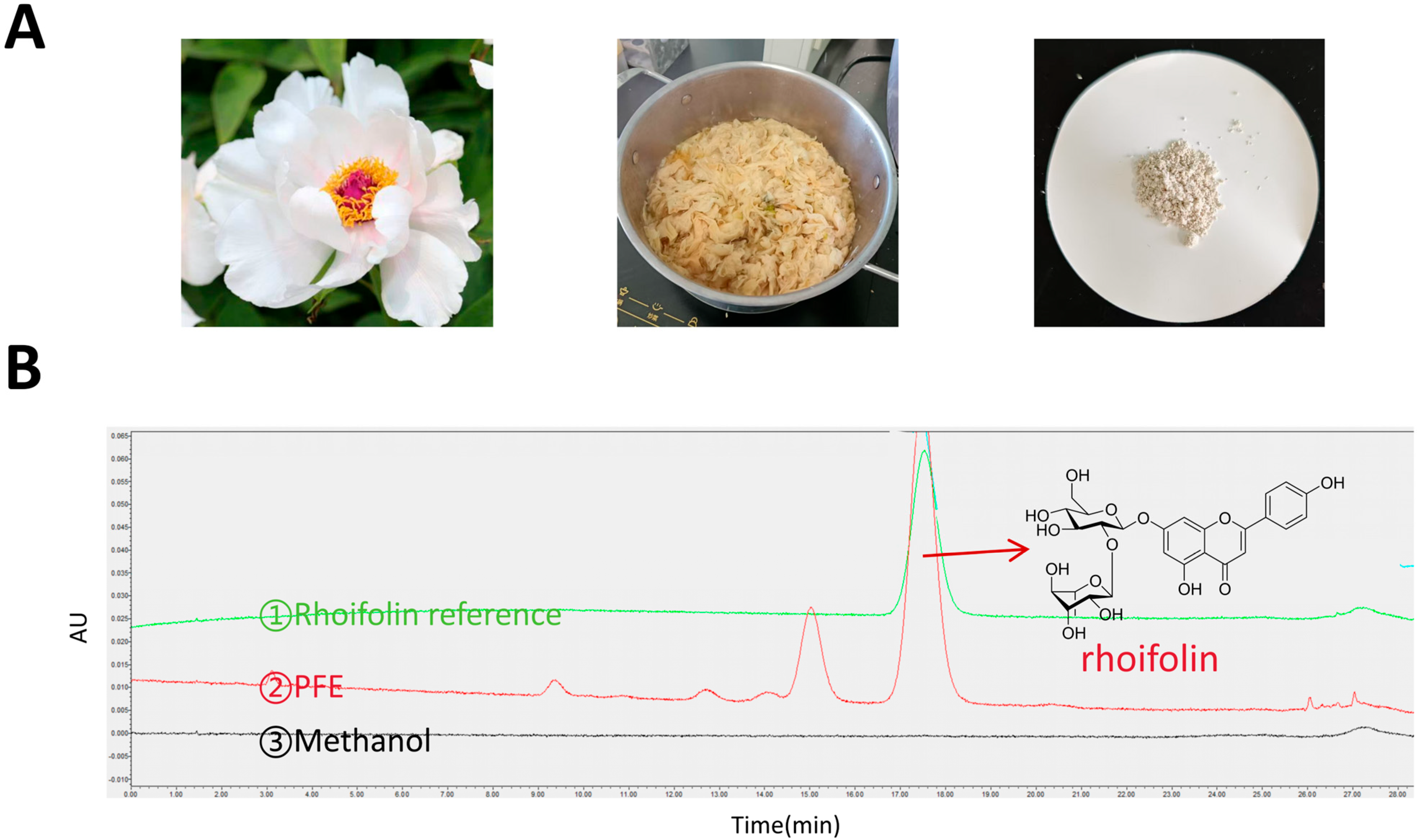
2.1. Preparation and HPLC Analysis of PFE
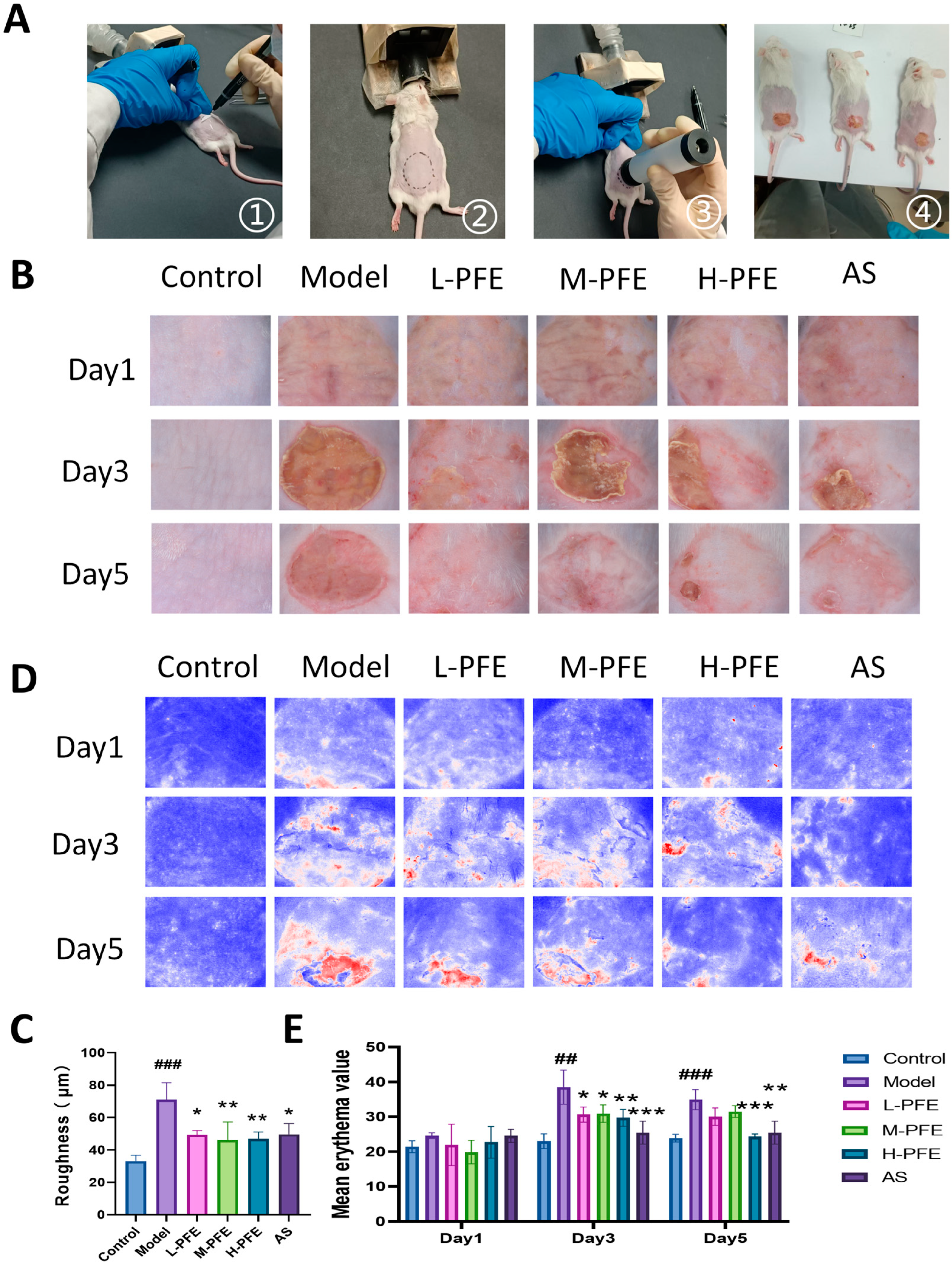
2.2. PFE Facilitated the Restoration of the Dorsal Skin Appearance
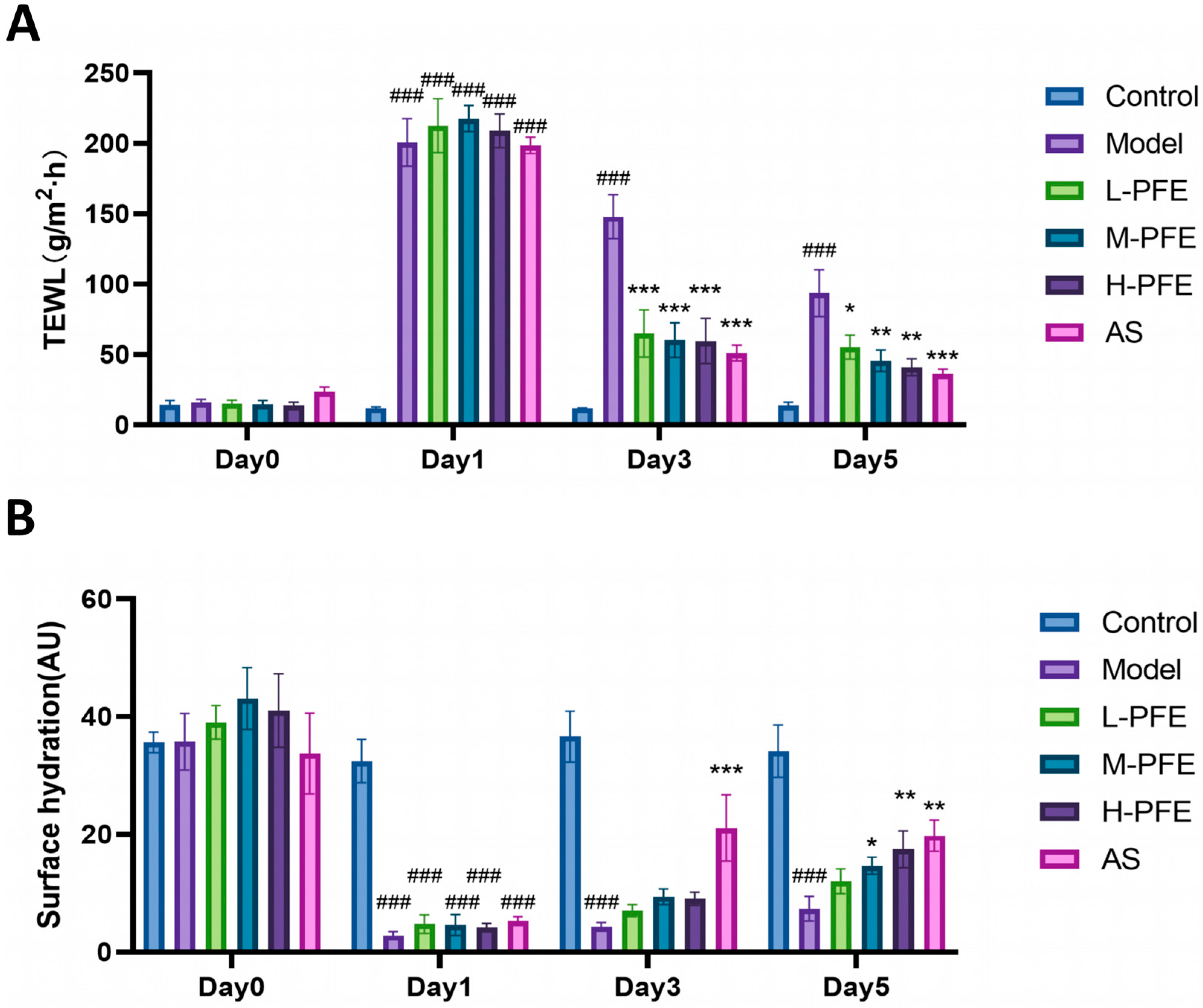
2.3. PFE Reduced TEWL and Increased SH of the Dorsal Skin
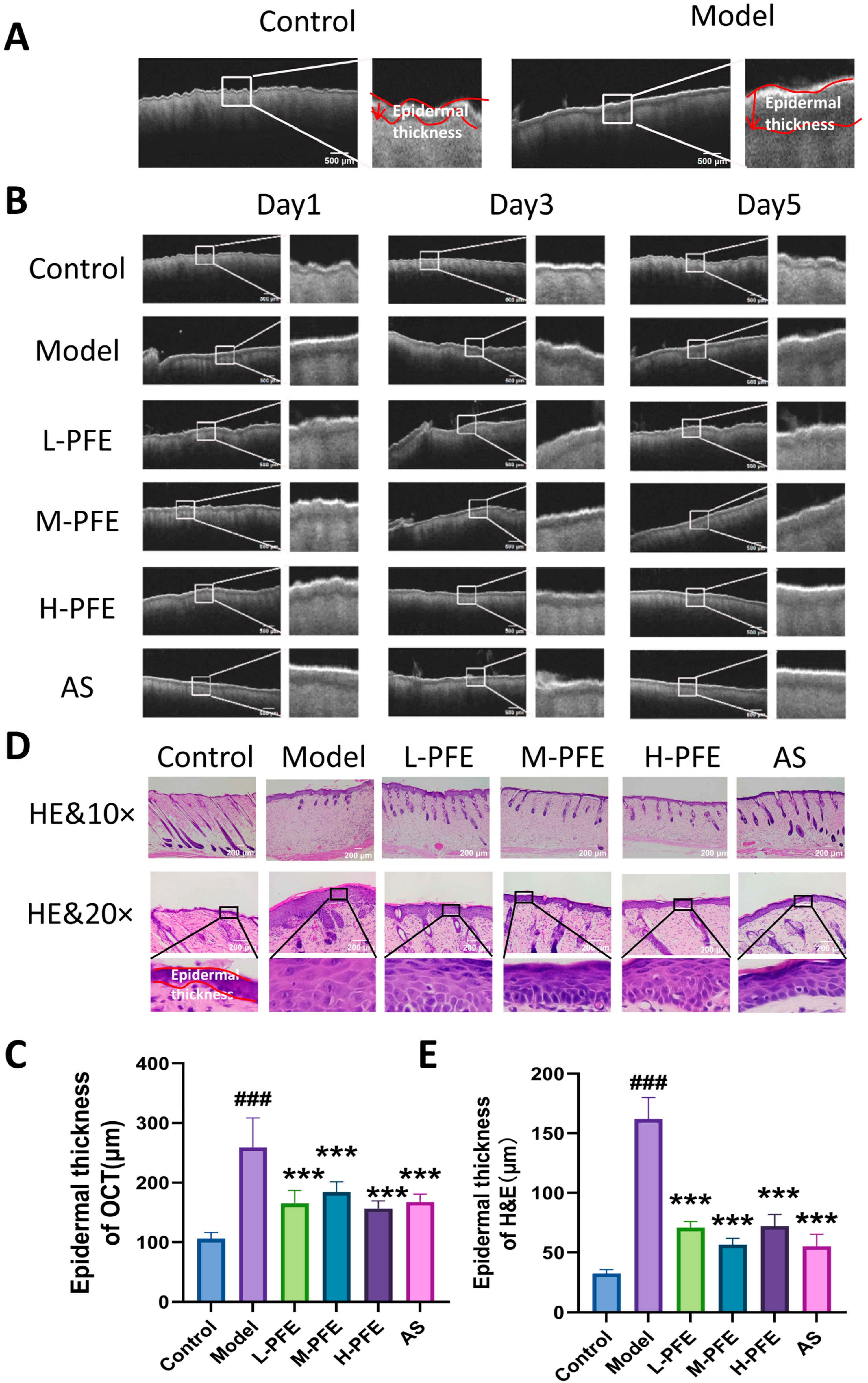
2.4. PFE Facilitated the Repair of Edematous Conditions and the Epidermal Tissue Structure
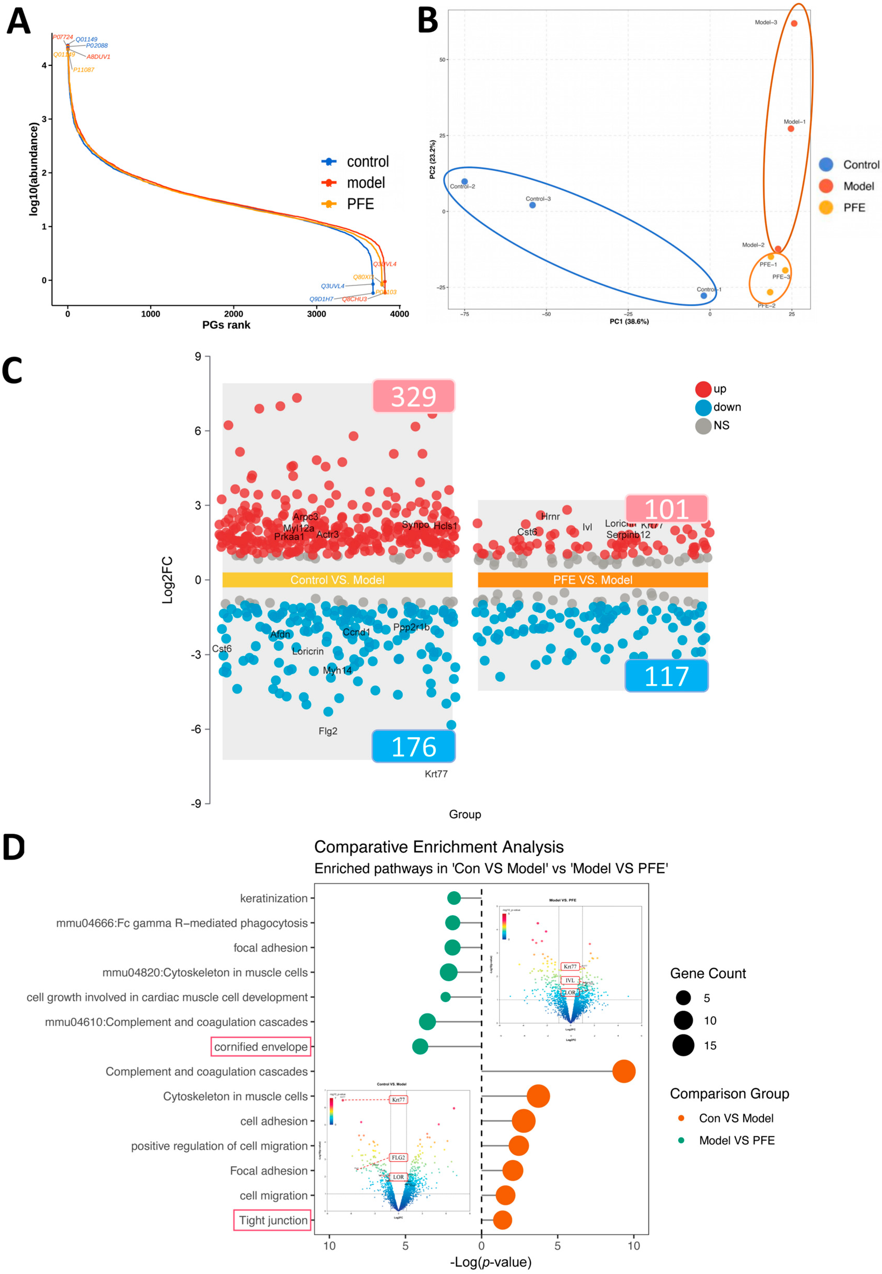
2.5. Proteomic Changes in Skin Following the PFE Intervention
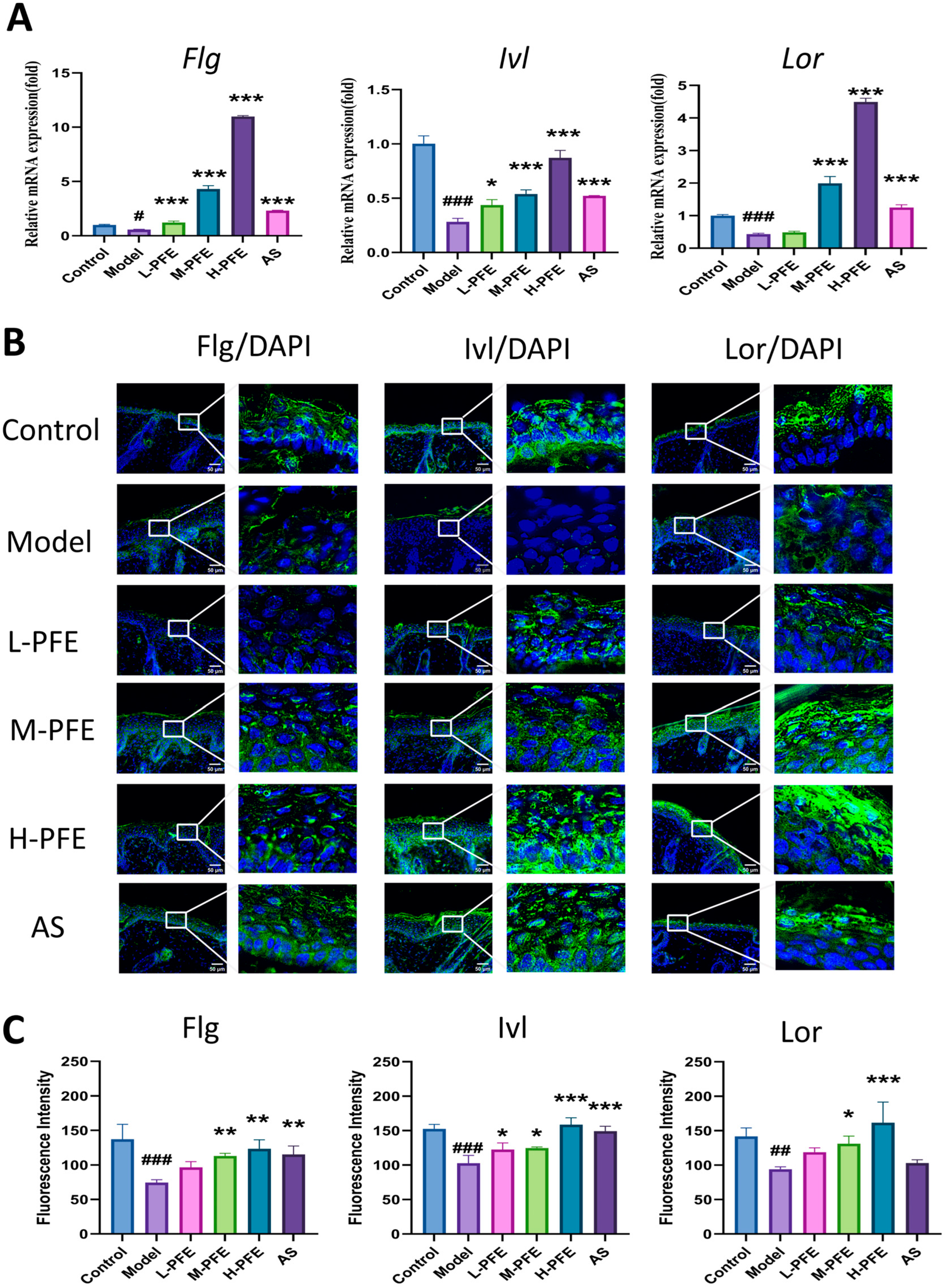
2.6. PFE Enhanced Both mRNA and Protein Expression of Flg, Ivl, and Lor in the Tape Stripping Mouse Model
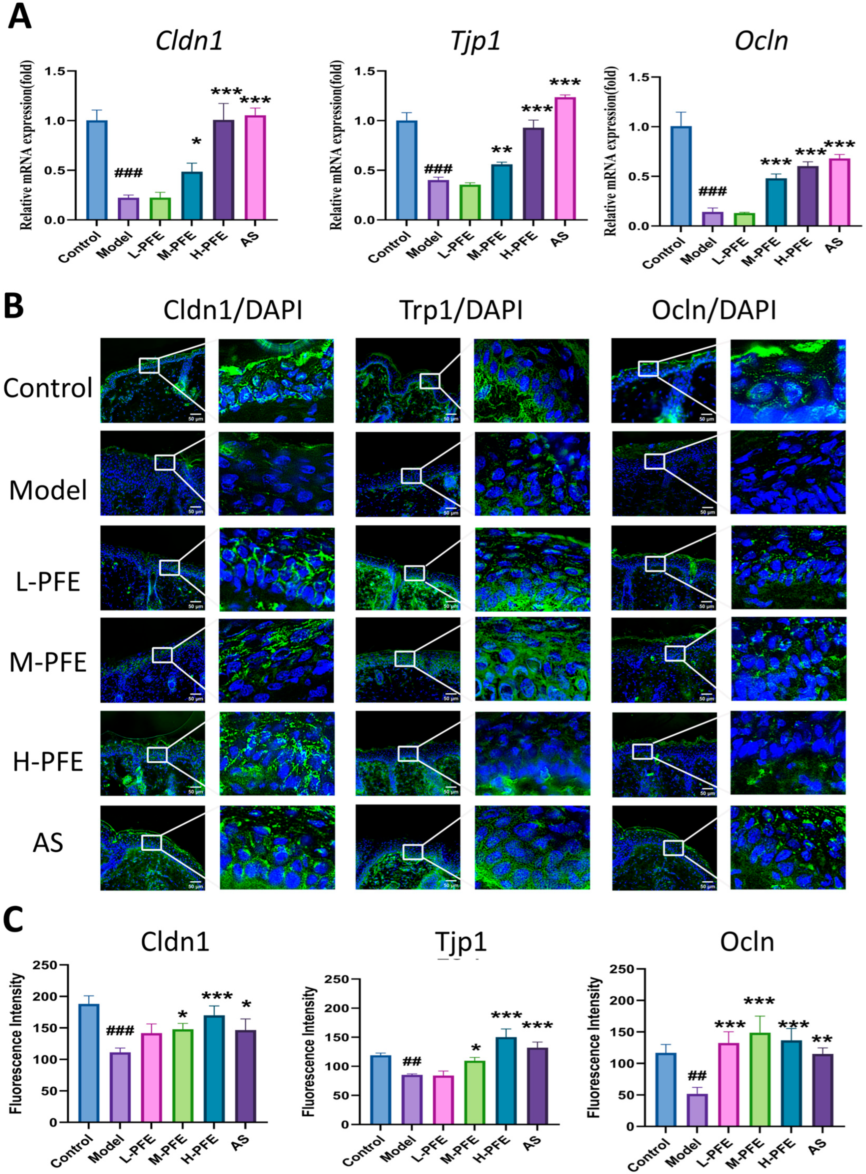
2.7. PFE Enhanced Both mRNA and Protein Expression of Cldn1, Tjp1, and Ivl in the Tape Stripping Mouse Model
3. Discussion
4. Materials and Methods
4.1. Chemicals and Reagents
4.2. Animals and Treatment Conditions
4.3. Epidermal Barrier Disruption
4.4. Preparation of the Hydrogel
4.5. Preparation of PFE
4.6. Experimental Design
4.7. Histological Analysis
4.8. Assessment of Skin Surface Characteristics
4.9. Immunofluorescence Analysis
4.10. qRT-PCR
4.11. Proteome Analysis
4.12. Statistical Analysis
5. Conclusions
Author Contributions
Funding
Institutional Review Board Statement
Informed Consent Statement
Data Availability Statement
Acknowledgments
Conflicts of Interest
Abbreviations
| TEWL | transepidermal water loss |
| PFE | peony flower extract |
| IF | immunofluorescence |
| CE | cornified envelope |
| TJs | tight junctions |
| FASP | filter-aided sample preparation |
| PPI | protein-protein interaction |
| TCM | traditional Chinese medicine |
| Flg | filaggrin |
| Ivl | involucrin |
| Lor | loricrin |
| Cldn1 | claudin-1 |
| Tjp1 | zonula occludens-1 |
| Ocln | occludin |
| DAPI | 4′,6-diamidino-2-phenylindole |
| FA | Formic acid |
| HPLC | high-performance liquid chromatography |
References
- Kim, S.; Jang, J.E.; Kim, J.; Lee, Y.I.; Lee, D.W.; Song, S.Y.; Lee, J.H. Enhanced barrier functions and anti-inflammatory effect of cultured coconut extract on human skin. Food Chem. Toxicol. 2017, 106, 367–375. [Google Scholar] [CrossRef]
- Bäsler, K.; Bergmann, S.; Heisig, M.; Naegel, A.; Zorn-Kruppa, M.; Brandner, J.M. The role of tight junctions in skin barrier function and dermal absorption. J. Control. Release 2016, 242, 105–118. [Google Scholar] [CrossRef]
- Blank, I.H. Further observations on factors which influence the water content of the stratum corneum. J. Investig. Dermatol. 1953, 21, 259–271. [Google Scholar] [CrossRef] [PubMed]
- Elias, P.M.; Goerke, J.; Friend, D.S. Mammalian epidermal barrier layer lipids: Composition and influence on structure. J. Investig. Dermatol. 1977, 69, 535–546. [Google Scholar] [CrossRef] [PubMed]
- Menon, G.K.; Cleary, G.W.; Lane, M.E. The structure and function of the stratum corneum. Int. J. Pharm. 2012, 435, 3–9. [Google Scholar] [CrossRef] [PubMed]
- Yosipovitch, G.; Misery, L.; Proksch, E.; Metz, M.; Ständer, S.; Schmelz, M. Skin barrier damage and itch: Review of mechanisms, topical management and future directions. Acta Derm. Venereol. 2019, 99, 1201–1209. [Google Scholar] [CrossRef]
- Kim, B.E.; Leung, D.Y. Significance of Skin Barrier Dysfunction in Atopic Dermatitis. Allergy Asthma Immunol. Res. 2018, 10, 207–215. [Google Scholar] [CrossRef]
- Singkhorn, S.; Tantisira, M.H.; Tanasawet, S.; Hutamekalin, P.; Wongtawatchai, T.; Sukketsiri, W. Induction of keratinocyte migration by ECa 233 is mediated through FAK/Akt, ERK, and p38 MAPK signaling. Phytother. Res. 2018, 32, 1397–1403. [Google Scholar] [CrossRef]
- Oryan, A.; Alemzadeh, E.; Mohammadi, A.A.; Moshiri, A. Healing potential of injectable Aloe vera hydrogel loaded by adipose-derived stem cell in skin tissue-engineering in a rat burn wound model. Cell Tissue Res. 2019, 377, 215–227. [Google Scholar] [CrossRef]
- Lee, K.-S.; Lee, S.; Wang, H.; Lee, G.; Kim, S.; Ryu, Y.-H.; Chang, N.H.; Kang, Y.-W. Analysis of Skin Regeneration and Barrier-Improvement Efficacy of Polydeoxyribonucleotide Isolated from Panax Ginseng (C.A. Mey.) Adventitious Root. Molecules 2023, 28, 7240. [Google Scholar] [CrossRef]
- Shedoeva, A.; Leavesley, D.; Upton, Z.; Fan, C. Wound healing and the use of medicinal plants. Evid. Based Complement. Altern. Med. 2019, 2019, 2684108. [Google Scholar] [CrossRef]
- Wang, R.; Lechtenberg, M.; Sendker, J.; Petereit, F.; Deters, A.; Hensel, A. Wound-healing plants from TCM: In vitro investigations on selected TCM plants and their influence on human dermal fibroblasts and keratinocytes. Fitoterapia 2013, 84, 308–317. [Google Scholar] [CrossRef]
- Peng, L.-P.; Cai, C.-F.; Zhong, Y.; Xu, X.-X.; Xian, H.-L.; Cheng, F.-Y.; Mao, J.-F. Genetic analyses reveal independent domestication origins of the emerging oil crop Paeonia ostii, a tree peony with a long-term cultivation history. Sci. Rep. 2017, 7, 5340. [Google Scholar] [CrossRef]
- Announcement on the Approval of 8 New Food Ingredients Including Euglena gracilis. Chin. J. Food Hyg. 2014, 26, 91.
- Lu, Y.; Jiang, Z.; Yan, W.; Duan, H. Genetic toxicological study of Danfeng peony flowers. J. Food Saf. Qual. Insp. 2019, 10, 3552–3556. [Google Scholar]
- Li, Z.; Ma, Y.; Li, F.; Wei, Y.; Zhang, L.; Yu, L.; Chen, L.; Wang, X.; Ning, E.; Zhang, L.; et al. Quality Evaluation of Peony Petals Based on the Chromatographic Fingerprints and Simultaneous Determination of Sixteen Bioactive Constituents Using UPLC-DAD-MS/MS. Molecules 2023, 28, 7741. [Google Scholar] [CrossRef] [PubMed]
- Zheng, Y.; Li, P.; Shen, J.; Yang, K.; Wu, X.; Wang, Y.; Yuan, Y.-H.; Xiao, P.; He, C. Comprehensive comparison of different parts of Paeonia ostii, a food-medicine plant, based on untargeted metabolomics, quantitative analysis, and bioactivity analysis. Front. Plant Sci. 2023, 14, 1243724. [Google Scholar] [CrossRef] [PubMed]
- Du Plessis, J.; Stefaniak, A.; Eloff, F.; John, S.; Agner, T.; Chou, T.-C.; Nixon, R.; Steiner, M.; Franken, A.; Kudla, I.; et al. International guidelines for the in vivo assessment of skin properties in non-clinical settings: Part 2. transepidermal water loss and skin hydration. Skin. Res. Technol. 2013, 19, 265–278. [Google Scholar] [CrossRef]
- Gao, Y.; Wang, X.; Chen, S.; Li, S.; Liu, X. Acute skin barrier disruption with repeated tape stripping: An In Vivo model for damage skin barrier. Ski. Res. Technol. 2012, 19, 162–168. [Google Scholar] [CrossRef]
- Ishitsuka, Y.; Roop, D.R. Loricrin: Past, Present, and Future. Int. J. Mol. Sci. 2020, 21, 2271. [Google Scholar] [CrossRef]
- Furue, M. Regulation of filaggrin, loricrin, and involucrin by IL-4, IL-13, IL-17A, IL-22, AHR, and NRF2: Pathogenic implications in Atopic Dermatitis. Int. J. Mol. Sci. 2020, 21, 5382. [Google Scholar] [CrossRef]
- Chelakkot, C.; Ghim, J.; Ryu, S.H. Mechanisms regulating intestinal barrier integrity and its pathological implications. Exp. Mol. Med. 2018, 50, 1–9. [Google Scholar] [CrossRef] [PubMed]
- Jensen, J.M.; Proksch, E. The skin’s barrier. G. Ital. Dermatol. Venereol. 2009, 144, 689–700. [Google Scholar] [PubMed]
- Baker, P.; Huang, C.; Radi, R.; Moll, S.B.; Jules, E.; Arbiser, J.L. Skin Barrier Function: The Interplay of Physical, Chemical, and Immunologic Properties. Cells 2023, 12, 2745. [Google Scholar] [CrossRef]
- Lee, H.W.; Ju, Y.J.; Choi, S.; Rhew, K.; Sevilleno, S.S.; Choi, M.S. Atopic Dermatitis Management: From Conventional Therapies to Biomarker-Driven Treatment Approaches. Biomol. Ther. 2025, 33, 813–829. [Google Scholar] [CrossRef] [PubMed]
- Chung, H.S.; Hwang, H.S. Therapeutic potential of phytochemical luteolin in restoring skin barrier by inhibiting antimicrobial peptides associated with TRAF6/TAK1/IKK/IκB in psoriasis-like HaCaT models. Food Sci. Biotechnol. 2025, 34, 2909–2921. [Google Scholar] [CrossRef]
- Yang, Y.; Sun, J.; You, H.; Sun, Y.; Song, Y.; Shen, Z.; Liu, T.; Guan, D.; Zhou, Y.; Cheng, S.; et al. Aloe-emodin relieves allergic contact dermatitis pruritus by inhibiting mast cell degranulation. Immunol. Lett. 2024, 270, 106902. [Google Scholar] [CrossRef]
- Deng, Y.; Wang, F.; He, L. Skin Barrier Dysfunction in Acne Vulgaris: Pathogenesis and Therapeutic Approaches. Med. Sci. Monit. 2024, 30, e945336. [Google Scholar] [CrossRef]
- Guo, L.; Yin, Z.; Wen, L.; Xin, J.; Gao, X.; Zheng, X. Flower extracts from Paeonia decomposita and Paeonia ostii inhibit melanin synthesis via cAMP-CREB-associated melanogenesis signaling pathways in murine B16 melanoma cells. J. Food Biochem. 2019, 43, e12777. [Google Scholar] [CrossRef]
- Yang, J.; Wang, C.; Li, N.; Wu, L.; Huang, Z.; Hu, Z.; Li, X.; Qu, Z. Phytochemicals and anti-tyrosinase activities of Paeonia ostii leaves and roots. Plant Physiol. Biochem. 2022, 181, 50–60. [Google Scholar] [CrossRef]
- Ding, H.-Y.; Chou, T.-H.; Lin, R.-J.; Chan, L.-P.; Wang, G.-H.; Liang, C.-H. Antioxidant and antimelanogenic behaviors of Paeonia suffruticosa. Plant Foods Hum. Nutr. 2011, 66, 275–284. [Google Scholar] [CrossRef]
- Usui, K.; Nakashima, C.; Takahashi, S.; Okada, T.; Ishida, Y.; Nakajima, S.; Kitoh, A.; Nomura, T.; Dainichi, T.; Honda, T.; et al. TRPV1-positive sensory nerves and neuropeptides are involved in epidermal barrier repair after tape stripping in mice. J. Allergy Clin. Immunol. 2024, 153, 868–873.e4. [Google Scholar] [CrossRef]
- Wang, Q.; Zhang, H.; Wang, X.; Ma, C.; Zhang, J.; Wu, J.; Li, L.; Lu, Y.; Wei, J.; Han, L. Amygdalin alleviates psoriasis-like lesions by improving skin barrier function. Arch. Dermatol. Res. 2024, 317, 115. [Google Scholar] [CrossRef] [PubMed]
- Proksch, E.; Soeberdt, M.; Neumann, C.; Kilic, A.; Abels, C. Modulators of the endocannabinoid system influence skin barrier repair, epidermal proliferation, differentiation and inflammation in a mouse model. Exp. Dermatol. 2019, 28, 1058–1065. [Google Scholar] [CrossRef] [PubMed]
- Gao, S.; Chen, Y.; Zhao, J.; Jing, R.; Guo, K.; Wang, L.; Li, X.; Li, C.; Hu, Z.; Xu, N. Oat β-glucan ameliorates epidermal barrier disruption by upregulating the expression of CaSR through dectin-1-mediated ERK and p38 signaling pathways. Int. J. Biol. Macromol. 2021, 185, 876–889. [Google Scholar] [CrossRef] [PubMed]
- Li, Z.; Jiang, R.; Wang, M.; Zhai, L.; Liu, J.; Xu, X.; Sun, L.; Zhao, D. Ginsenosides repair UVB-induced skin barrier damage in BALB/c hairless mice and HaCaT keratinocytes. J. Ginseng Res. 2022, 46, 115–125. [Google Scholar] [CrossRef]
- Strasser, B.; Mlitz, V.; Hermann, M.; Rice, R.H.; Eigenheer, R.A.; Alibardi, L.; Tschachler, E.; Eckhart, L. Evolutionary origin and diversification of epidermal barrier proteins in amniotes. Mol. Biol. Evol. 2014, 31, 3194–3205. [Google Scholar] [CrossRef]
- Jarnik, M.; de Viragh, P.A.; Schärer, E.; Bundman, D.; Simon, M.N.; Roop, D.R.; Steven, A.C. Quasi-Normal cornified cell envelopes in loricrin knockout mice imply the existence of a loricrin backup system. J. Investig. Dermatol. 2002, 118, 102–109. [Google Scholar] [CrossRef]
- Kypriotou, M.; Huber, M.; Hohl, D. The human epidermal differentiation complex: Cornified envelope precursors, S100 proteins and the ‘fused genes’ family. Exp. Dermatol. 2012, 21, 643–649. [Google Scholar] [CrossRef]
- Osawa, R.; Akiyama, M.; Shimizu, H. Filaggrin gene defects and the risk of developing allergic disorders. Allergol. Int. 2011, 60, 1–9. [Google Scholar] [CrossRef]
- Kirschner, N.; Rosenthal, R.; Günzel, D.; Moll, I.; Brandner, J.M. Tight junctions and differentiation—A chicken or the egg question? Exp. Dermatol. 2012, 21, 171–175. [Google Scholar] [CrossRef]
- González-Mariscal, L.; Domínguez-Calderón, A.; Raya-Sandino, A.; Ortega-Olvera, J.M.; Vargas-Sierra, O.; Martínez-Revollar, G. Tight junctions and the regulation of gene expression. Semin. Cell Dev. Biol. 2014, 36, 213–223. [Google Scholar] [CrossRef] [PubMed]
- Matter, K.; Aijaz, S.; Tsapara, A.; Balda, M.S. Mammalian tight junctions in the regulation of epithelial differentiation and proliferation. Curr. Opin. Cell Biol. 2005, 17, 453–458. [Google Scholar] [CrossRef] [PubMed]
- Furuse, M.; Hata, M.; Furuse, K.; Yoshida, Y.; Haratake, A.; Sugitani, Y.; Noda, T.; Kubo, A.; Tsukita, S. Claudin-based tight junctions are crucial for the mammalian epidermal barrier: A lesson from claudin-1-deficient mice. J. Cell Biol. 2002, 156, 1099–1111. [Google Scholar] [CrossRef] [PubMed]
- Tokumasu, R.; Yamaga, K.; Yamazaki, Y.; Murota, H.; Suzuki, K.; Tamura, A.; Bando, K.; Furuta, Y.; Katayama, I.; Tsukita, S. Dose-dependent role of claudin-1 in vivo in orchestrating features of atopic dermatitis. Proc. Natl. Acad. Sci. USA 2016, 113, E4061–E4068. [Google Scholar] [CrossRef]
- Fukuda, K.; Ito, Y.; Amagai, M. Barrier Integrity and Immunity: Exploring the Cutaneous Front Line in Health and Disease. Annu. Rev. Immunol. 2025, 43, 219–252. [Google Scholar] [CrossRef]
- Kubo, A.; Nagao, K.; Amagai, M. Epidermal barrier dysfunction and cutaneous sensitization in atopic diseases. J. Clin. Investig. 2012, 122, 440–447. [Google Scholar] [CrossRef]
- Antonov, D.; Schliemann, S.; Elsner, P. Methods for the Assessment of Barrier Function. Curr. Probl. Dermatol. 2016, 49, 61–70. [Google Scholar] [CrossRef]



| Gene | Primer Direction | Sequence (5′→3′) |
|---|---|---|
| Flg | Forward | GCAAGATCAGGCTCAGGAGGAAG |
| Reverse | AGAATGGACTTGGCTGTCACTGG | |
| Lor | Forward | GGCGGCGGCGGCTATTATAG |
| Reverse | GGAACCACCTCCATAGGAACCAC | |
| Ivl | Forward | GGGACCTCTCAAGACTGTGTGTTC |
| Reverse | AGACCTGGCATTGTGTAGGATGTG | |
| Tjp1 | Forward | ACCCGAAACTGATGCTGTGGATAG |
| Reverse | GCTGGCTGGCTGTACTGTGAG | |
| Cldn1 | Forward | GTGTCCTACTTTCCTGCTCCTGTC |
| Reverse | AGAAGGTGTTGGCTTGGGATAAGG | |
| Ocln | Forward | CCTCTGACCTTGAGTGTGGATGAC |
| Reverse | TCCTCTTGCCCTTTCCTGCTTTC | |
| Gapdh | Forward | AGAAGGTGGTGAAGCAGGCATC |
| Reverse | CGAAGGTGGAAGAGTGGGAGTTG |
Disclaimer/Publisher’s Note: The statements, opinions and data contained in all publications are solely those of the individual author(s) and contributor(s) and not of MDPI and/or the editor(s). MDPI and/or the editor(s) disclaim responsibility for any injury to people or property resulting from any ideas, methods, instructions or products referred to in the content. |
© 2025 by the authors. Licensee MDPI, Basel, Switzerland. This article is an open access article distributed under the terms and conditions of the Creative Commons Attribution (CC BY) license.
Share and Cite
Yang, R.; Yang, J.; He, G.; Zhang, Y.; Jiang, X.; Wang, J.; Yang, H.; Shang, C. Protective Effect of Peony (Paeonia ostii) Flower Extract Against Tape Stripping-Induced Skin Barrier Impairment in Mice. Molecules 2026, 31, 62. https://doi.org/10.3390/molecules31010062
Yang R, Yang J, He G, Zhang Y, Jiang X, Wang J, Yang H, Shang C. Protective Effect of Peony (Paeonia ostii) Flower Extract Against Tape Stripping-Induced Skin Barrier Impairment in Mice. Molecules. 2026; 31(1):62. https://doi.org/10.3390/molecules31010062
Chicago/Turabian StyleYang, Ruiying, Jicheng Yang, Gaiying He, Yusheng Zhang, Xue Jiang, Jiyong Wang, Hongjun Yang, and Chengxiang Shang. 2026. "Protective Effect of Peony (Paeonia ostii) Flower Extract Against Tape Stripping-Induced Skin Barrier Impairment in Mice" Molecules 31, no. 1: 62. https://doi.org/10.3390/molecules31010062
APA StyleYang, R., Yang, J., He, G., Zhang, Y., Jiang, X., Wang, J., Yang, H., & Shang, C. (2026). Protective Effect of Peony (Paeonia ostii) Flower Extract Against Tape Stripping-Induced Skin Barrier Impairment in Mice. Molecules, 31(1), 62. https://doi.org/10.3390/molecules31010062