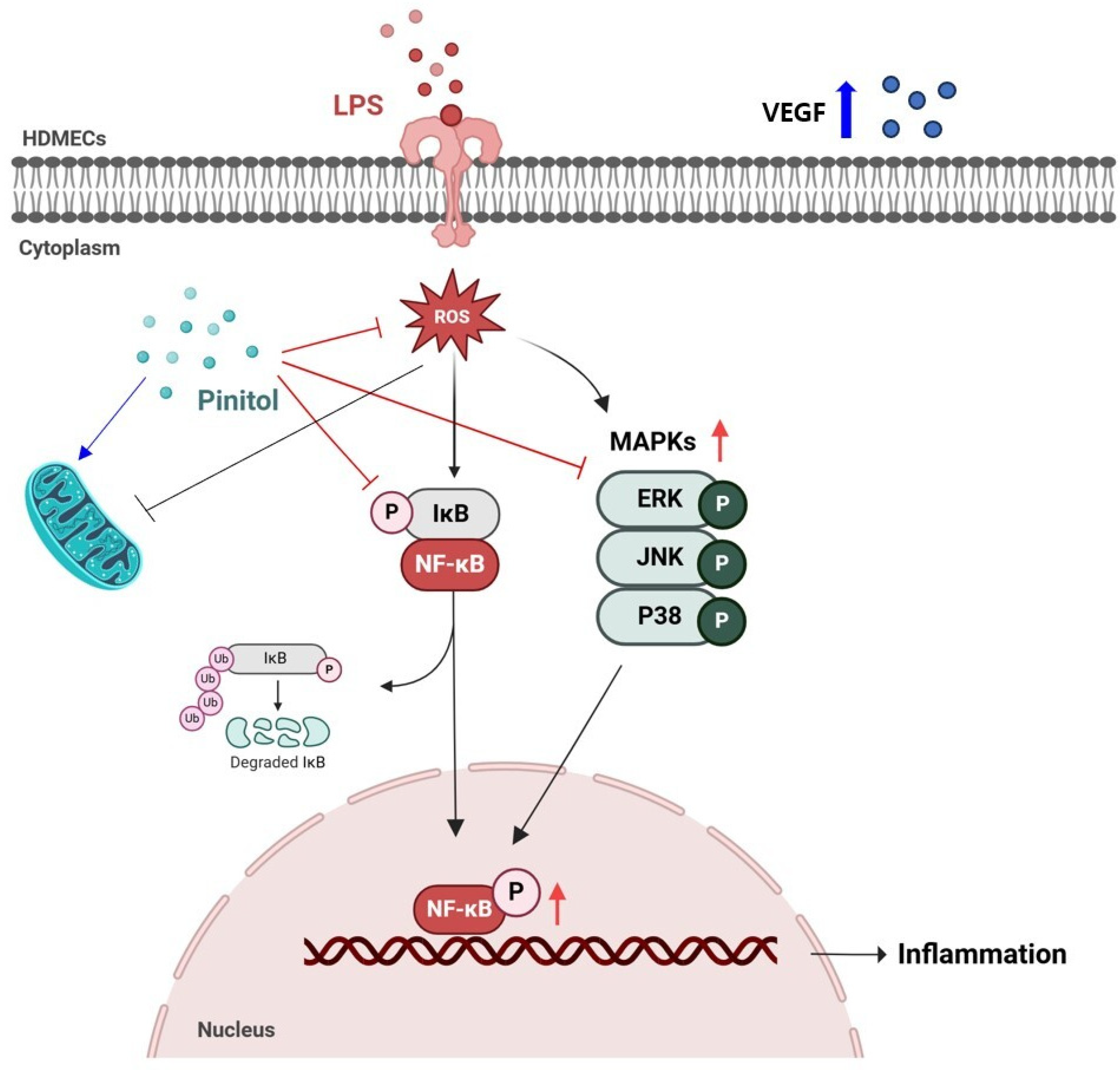
Pinitol Improves Lipopolysaccharide-Induced Cellular Damage in Human Dermal Microvascular Endothelial Cells


Abstract
Share and Cite
Go, M.Y.; Kim, J.; Jeon, C.Y.; Kim, M.; Shin, D.W. Pinitol Improves Lipopolysaccharide-Induced Cellular Damage in Human Dermal Microvascular Endothelial Cells. Molecules 2025, 30, 1513. https://doi.org/10.3390/molecules30071513
Go MY, Kim J, Jeon CY, Kim M, Shin DW. Pinitol Improves Lipopolysaccharide-Induced Cellular Damage in Human Dermal Microvascular Endothelial Cells. Molecules. 2025; 30(7):1513. https://doi.org/10.3390/molecules30071513
Chicago/Turabian StyleGo, Min Young, Jinsick Kim, Chae Young Jeon, Mujun Kim, and Dong Wook Shin. 2025. "Pinitol Improves Lipopolysaccharide-Induced Cellular Damage in Human Dermal Microvascular Endothelial Cells" Molecules 30, no. 7: 1513. https://doi.org/10.3390/molecules30071513
APA StyleGo, M. Y., Kim, J., Jeon, C. Y., Kim, M., & Shin, D. W. (2025). Pinitol Improves Lipopolysaccharide-Induced Cellular Damage in Human Dermal Microvascular Endothelial Cells. Molecules, 30(7), 1513. https://doi.org/10.3390/molecules30071513