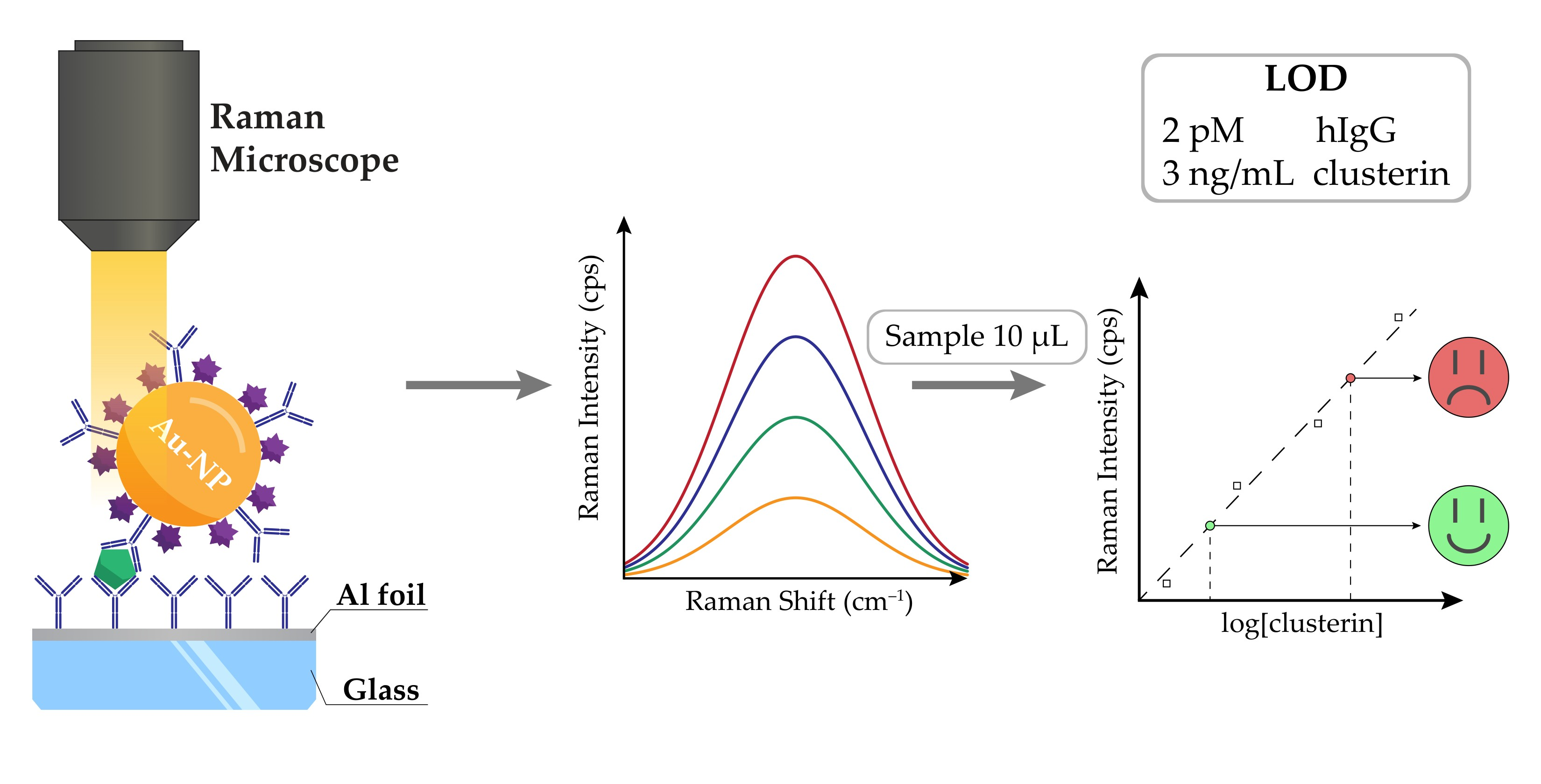
Detection of Biomarker Clusterin in SERS Immunoassays on Al Foil After Substrate Selection and Assay Optimization with Fluorescently Labeled Antibodies


Abstract
Share and Cite
Mergenbayeva, S.; Terzapulo, X.; Bukasov, R. Detection of Biomarker Clusterin in SERS Immunoassays on Al Foil After Substrate Selection and Assay Optimization with Fluorescently Labeled Antibodies. Molecules 2025, 30, 3974. https://doi.org/10.3390/molecules30193974
Mergenbayeva S, Terzapulo X, Bukasov R. Detection of Biomarker Clusterin in SERS Immunoassays on Al Foil After Substrate Selection and Assay Optimization with Fluorescently Labeled Antibodies. Molecules. 2025; 30(19):3974. https://doi.org/10.3390/molecules30193974
Chicago/Turabian StyleMergenbayeva, Saule, Xeniya Terzapulo, and Rostislav Bukasov. 2025. "Detection of Biomarker Clusterin in SERS Immunoassays on Al Foil After Substrate Selection and Assay Optimization with Fluorescently Labeled Antibodies" Molecules 30, no. 19: 3974. https://doi.org/10.3390/molecules30193974
APA StyleMergenbayeva, S., Terzapulo, X., & Bukasov, R. (2025). Detection of Biomarker Clusterin in SERS Immunoassays on Al Foil After Substrate Selection and Assay Optimization with Fluorescently Labeled Antibodies. Molecules, 30(19), 3974. https://doi.org/10.3390/molecules30193974