Review

Phospholipase A2—A Significant Bio-Active Molecule in Honeybee (Apis mellifera L.) Venom

Department of Cell and Molecular Biology, Faculty of Medicine, “Iuliu Hațieganu” University of Medicine and Pharmacy, 6. Louis Pasteur St., 400349 Cluj-Napoca, Romania
*
Author to whom correspondence should be addressed.
Molecules 2025, 30(12), 2623; https://doi.org/10.3390/molecules30122623
Submission received: 30 April 2025 / Revised: 11 June 2025 / Accepted: 16 June 2025 / Published: 17 June 2025

Abstract

:
Phospholipase A2 (PLA2) is a prevalent molecule in the honeybee venom. Its importance is reflected by the number of scientists focused on studying it from various points of view. This review summarises a significant amount of data concerning this fascinating substance. Firstly, the origin and occurrence of PLA2, with similarities and differences among species or populations of bees are highlighted. Next, its synthesis, post-translational processing and structural features are described, followed by the PLA2 availability. In a larger section, the multiple effects of honeybee venom PLA2 are detailed, starting with the main ability as an enzyme to interact with biological membranes and to hydrolyse the sn-2 ester bond in 1,2-diacyl-sn-3-phosphoglycerides; the docking process, the substrate binding and the catalytic steps are analysed too. Then, the pro-/anti-inflammatory effect and allergenic property, the anticoagulant effect and the involvement of PLA2 in apoptosis are revised. Selected antiviral, antibiotic and antitumoral effects of PLA2, as well as its use in immunotherapy are mentioned as beneficial applications. Additionally, the mechanisms of toxicity of PLA2 are presented in detail. Finally, a number of anti-PLA2 compounds are enumerated. In each section, the features of the honeybee venom molecule are discussed in relation to PLA2s from other species.

1. Introduction

Venoms are complex mixtures of many inorganic and organic molecules, produced and used by numerous animals, very distant on the evolutionary scale. The venom is used either as a defensive weapon—to fight against predators, or for attack—to immobilise and digest prey [1]. Bees use their venom mostly to defend the hive, and therefore they developed a complicated sting apparatus and a particular strategy, based on high coordination [2]. Also, their venom was adapted to this purpose. Its chemical composition and the amount released during stinging, along with a relatively reduced level of aggressivity (except for the Africanized bees—which resulted after human intervention) contribute to discouraging their mammalian enemies, rather than killing them [3,4].
Among the multiple substances present in the dry (crystallised) honeybee (Apis mellifera L.) venom (HBV), phospholipase A2 (PLA2) stands as the second one in terms of concentration and roles, after a peptide named melittin. Neumann et al. [5] characterised the HBV for the first time and, a few years later, Habermann and Neumann [6] reported the presence of PLA2 in the HBV. Then, after Habermann published his famous paper in Science [7], the interest of scientists for this valuable natural compound increased exponentially, and the 1990s represented the golden decade in the study of HBV, mainly of HBV PLA2 (hvPLA2). The methods to study the HBV and its components evolved from filter paper electrophoresis, diversified and culminated with high-performance liquid chromatography (HPLC) and cloning of the hvPLA2 in bacteria [8]. In the meantime, the methods used for studying the effects of HBV or hvPLA2 alone advanced from blood testing to the production of monoclonal antibodies, electrophysiology and transmission electron microscopy. Even though the number of publications on this topic decreased during the last 10–15 years, the subject still remains of actual scientific interest, due to the new medical relevance of the hvPLA2 [9,10,11,12].
In our laboratory, there is a long-standing interest (originating from the above-mentioned golden decade) in the study of histological, histochemical, ultrastructural and physiological effects of HBV in different organs or tissues, in experimental conditions. All this activity was concretised in the publication of 16 papers in Romanian journals not indexed in WOS (between 1996 and 2005), and later in other 6 papers in WOS-indexed journals. Among the formers, some reported interesting effects of hvPLA2 in vivo [13,14] and in vitro [15]. As a logical consequence, an important amount of scientific literature regarding hvPLA2 has been accumulated, from different databases and using various keywords, that represents the foundation of the present work. We analyse here the hvPLA2 structure and its many effects in relation to other similar molecules belonging to the same family, but present in other species. Finally, a number of anti-PLA2 molecules are discussed.

2. PLA2—A Common Molecule in Different Organisms

PLA2, also called phosphatidilcoline-2-acyl hydrolase, or lecithinase A, is an enzyme commonly found in living beings. Historically, it was first isolated from the pancreas and described by Bókay in 1878 [16]. A few decades later, PLA2 was also identified in cobra venom, and, in the following years, in many invertebrate and vertebrate species [17,18,19]. Despite their very different origins, PLA2s display similar structures, and, more importantly, they share a common function: the capacity to catalyse the hydrolysis of the sn-2 ester bond in 1,2-diacyl-sn-3-phosphoglycerides (PLA2 EC 3.1.1.4. [20]), thus generating free fatty acids and lysophospholipids. Apart from PLA2, other two enzymes with similar activities exist: PLB (that hydrolyses phospholipids at either the sn-1 or sn-2 positions) and PLC (that removes the phosphate group from phospholipids) [21].
PLA2s have been categorised into 11 classes and 23 subclasses, based on several criteria, such as their source, structural features and effects [18], and later into 15 groups. They cluster in five main categories or types: secreted PLA2s (sPLA2s), cytosolic, Ca2+-independent, platelet-activating factor acetyl hydrolase, and lysosomal PLA2s (for an outstanding review of PLA2 classification and tools used for this, see [22]).
So far, more than 150 different PLA2s have been characterised in the venoms of different species [23]. Venom PLA2s are all Ca2+-dependent sPLA2s, relatively small molecules ranging between 13 and 19 kDa. sPLA2s comprise 17 known distinct isoenzymes [24,25,26]. The hvPLA2 is a sPLA2 that belongs to class IIIA [18,27,28,29,30].

3. PLA2 Is a Major Molecule of HBV

3.1. Bees and Their Venoms

Michener [31] classified bees (insects from the order Hymenoptera) in 17,533 species from around the world. They were grouped into 443 genera differentiated into diverse lineages, with or without venom. The most relevant are the 11 venom-producing species of genus Apis (family Apidae), including A. mellifera (the European honeybee, with 33 subspecies), A. cerana (the Asian honeybee), A. florea (dwarf honeybee), and A. dorsata (giant honeybee) [31,32].
The venom is used by worker bees to defend themselves and their colony, while queens only use it during fights with other queens [33]. Due to its beneficial properties, the bee venom was considered for centuries—particularly in Eastern Europe and Asia—as a traditional drug in the treatment of certain arthritic ailments [34,35,36]. In trying to explain the venom action on the human or animal body, several prominent scientists have studied its composition. The venom secreted by bees is a highly concentrated mixture of at least 18 organic and inorganic substances including proteins (enzymes), peptides, amino acids, sugars, lipids and acids [5,7,37,38,39,40,41], most of them with biochemical or pharmacological activity [7,42,43].

3.2. hvPLA2—Differences

hvPLA2 has been identified as an important molecule accounting for 11–15% of the dry venom of worker bees [6,7]. Like the other components of HBV, hvPLA2 is synthesised by cylindrical secretory cells in the bee venom’s main gland. This gland is thin, convoluted, and bifurcated, located in the abdomen between the rectum and the ovaries, and attached to the sting apparatus [37,44,45]. After synthesis, HBV is collected and stored in a reservoir of this gland. According to Owen and Bridges [46], the secretory cells undergo ultrastructural changes in adult bees, a process that is preceded by a decrease of 50% in the protein content of the venom glands; these data were further confirmed by Abreu et al. [47]. Additionally, studies by Owen et al. [48], and Li et al. [49] showed that PLA2 is synthesised in A. mellifera mainly after hatching, and within 10 days it reaches its maximal level in the venom reservoir of worker bees (about 40 μg); this level is then maintained throughout the rest of their life. Li et al. [49] also reported undetectable PLA2 mRNA during the later adult stage of A. cerana, suggesting either an arrest in PLA2 synthesis, or a gland involution. The synthesis and storage of PLA2 in the venom sac in A. cerana follow the same steps as in A. mellifera, with maximal values found in the 10th day. However, the enzyme was detected in a lower amount in comparison to A. mellifera—only 10–12 μg [49]. These data were consistent with the differences in the total venom content of the sac of bees from the two species: 138 μg [49], or 147 μg [50] in A. mellifera, and 43 μg in A. cerana [49], respectively. Furthermore, Palma and Brochetto-Braga [51] observed both qualitative and quantitative biochemical differences between the venom of 3 races of A. mellifera: A. m. ligustica, A. m. adansonii, and A. m. scutellata (Africanized bees). Apart from this, Schumacher et al. [52] reported a high variability in venom quantity among individual mature bees, and across different colonies, with an average of 134.1 μg (range 38–330 μg) in A. m. mellifera and 98.2 μg (range 34–200 μg) in A. m. scutellata. The PLA2 content also showed the same variability: 11.1% (1.8–27.4%) from dry weight in A. m. mellifera, and 18.1% (range 5.1–31%) in A. m. scutellata. Additionally, small seasonal differences in PLA2 concentration were observed in A. m. mellifera [53] and in A. m. scutellata [54], along with variations based on the age of the bee [48,55].
These differences are mainly thought to be due to the involvement of A. mellifera in the defence of their bigger colonies (against large mammals and other vertebrates) as compared to A. cerana and to the higher fighting skills of the European bees [49]. On the other hand, in the Africanized bees (recognised as the most aggressive), multiple stinging, rather than increased venom amount, potency or delivery seems to be the cause of the serious reactions following their attacks [50].
Unlike in workers, it was found that queen venom contains much less PLA2 [56]. Thus, it was concluded that different control mechanisms influence the production of PLA2 in these castes of identical genotype. The genes encoding the PLA2 from the venom of different species of bees have been sequenced and cloned (A. mellifera and A. cerana by Shen et al. [57], A. cerana by Li et al. [49] that also reported differences from A. mellifera). Moreover, the genes encoding PLA2 from many other species of animals (with or without venoms) have been studied so far [58,59,60].

3.3. hvPLA2—Synthesis and Proteolysis

In A. mellifera, hvPLA2, encoded by a single-copy gene/haploid genome, is synthesised in the rough endoplasmic reticulum as a bigger precursor of 167 amino acids, with a molecular weight of 19.058 kDa [61]:
  • MQVVLGSLFL10 LLLSTSHGWQ20 IRDRIGDNEL30 EERIIYPGTL40 WCGHGNKSSG50 PNELGRFKHT60 DACCRTHDMC70 PDVMSAGESK80 HGLTNTASHT90 RLSCDCDDKF100 YDCLKNSADT110 ISSYFVGKMY120 FNLIDTKCYK130 LEHPVTGCGE140 RTEGRCLHYT150 VDKSKPKVYQ160 WFDLRKY167
This precursor is next processed to the mature form by proteolytic cleavage: the signal peptide of 18 amino acids (1–18), and then a smaller propeptide of 15 amino acids (19–33) are removed; thus, the secreted protein has 134 amino acids (numbered in Figure 1 from 34 to 167) and a molecular weight of 15.249 kDa [61]. Also, in [61] the lengths of the signal and of the mature protein are available.
Additional experimental evidence showed 31 sites of proteolysis in the structure of the hvPLA2. But, while Habermann [7], King [62] and Annand et al. [63] found 14 chymotryptic and 17 tryptic attack points, Shipolini et al. reported 13 and 18, respectively [64].
These data were obtained in two different ways. One method, used by Kuchler et al. [65] was based on analysis of the cDNA for PLA2 from BV glands. The other method involved studying the purified secreted hvPLA2: Scott et al. [66] and Shipolini et al. [64] reported a molecule of 128 amino acids, while studies of Habermann [7] and Annand et al. [63] showed 129 amino acids forming the polypeptide chain. Apart from two N-D replacements, the amino acid sequence determined by Kuchler et al. [65] is identical to the one previously reported by Shipolini et al. [64] in the N- and C-terminal regions, while several differences have been recorded in the central part of the molecule.
All these small discrepancies could stem from the different resolutions of the methods employed by the researchers.
The primary structure of hvPLA2 was found to be similar in a ratio of 31% to the group III of human cellular sPLA2s [67,68,69]. On the other hand, in its N-terminal region 60% of the amino acid sequence is common with those of PLA2s from the venoms of the Rhopilema nomadica medusa and of the Heloderma lizard [70]. While sPLA2s in groups I and II are highly homologous [30], hvPLA2 shares little similarity with them, except for a few regions—the catalytic site, the Ca2+ binding loop, and certain cysteine residues [65]. Kuchler et al. [65] also discussed other aspects concerning the homology of different domains of the hvPLA2 with their counterparts in vertebrate PLA2s and concluded that the common features are typical for proteins having a common evolutionary origin.

3.4. hvPLA2—Glycosylation

hvPLA2 is a glycoprotein resulting from the co-translational N46-glycosylation (see Figure 1). The sugar motif consists of mannose, N-acetylglucosamine, fucose (that is alpha 1–6 and/or alpha 1–3 linked to the N-acetylglucosamine), galactose and a low amount of N-acetylgalactosamine (accounting for about 10% of the PLA2 oligosaccharides) [62,64,66,71,72,73,74].
According to Li et al. [49], the glycosylation pattern in A. mellifera results in the formation of four types of PLA2s, with different molecular weights, but Altmann et al. [75] characterised three hvPLA2 isoforms in this species, of about 16, 18 and 20 kDa. The differences between them were mainly given by their glycosylation patterns: the isoform with the lowest molecular weight is not glycosylated in N13 as the other two are, while the heaviest contains N-acetylgalactosamine. Shipolini et al. calculated a molecular weight of 15.800 kDa for the glycosylated PLA2 in A. m. mellifera [64]. The controversial data could result from the accuracy of the working conditions as well as from the different resolutions of the methods employed. Comparatively, the glycoprotein has 15.249 kDa in A. m. carnica [76]. In A. cerana, the glycosylation leads to the occurrence of an enzyme with a higher molecular size, often correlated with ageing. However, this glycosylation process was found to be different between the two species [49].

3.5. hvPLA2—Folding

Li et al. [49] showed complex patterns of post-translational modifications of PLA2, identical among the individuals of A. mellifera. In A. cerana, nevertheless, the post-translational modifications were found to differ within the same colony, between different colonies and in comparison to A. mellifera.
The N-terminal segment of hvPLA2 arrives, after the three-dimensional folding, at the central part of the enzyme, which represents the catalytic centre, also containing the Ca2+ binding domain with a conserved XCGXG motif [63,77] (see Figure 1 amino acids 41–45). Because both consist of hydrophobic amino acids, they participate in the formation of a hydrophobic channel at this level [78].
Ten cysteine residues contribute to the particular folding that generates the secondary and tertiary structure of PLA2. They form 5 disulfide bonds in positions: 42–64, 63–103, 70–96, 94–128, and 138–146 [63,65,66,79] (as depicted in Figure 1). This leads to one of the main differences from other classes of PLA2 that contain seven disulfide bonds with another pattern [63,80]. Furthermore, the sizes and shapes of the amino acids also lead to the formation of three α-helix regions: K58-H67; C94-N106; I111-N122 (see Figure 1 and [61]). On the contrary, hvPLA2 has many similarities with snake PLA2s (for review, see Montecucco et al., 2008 [81]). The crystal structure of hvPLA2 has also been well documented by comparison with the bovine pancreatic PLA2 [66], or with the enzyme from the Chinese cobra (Naja naja atra) [82]. For an instructive 3D representation of the hvPLA2, see [61]. X-ray crystallography has revealed a similar catalytic domain with other classes of PLAs [66].

3.6. Availability

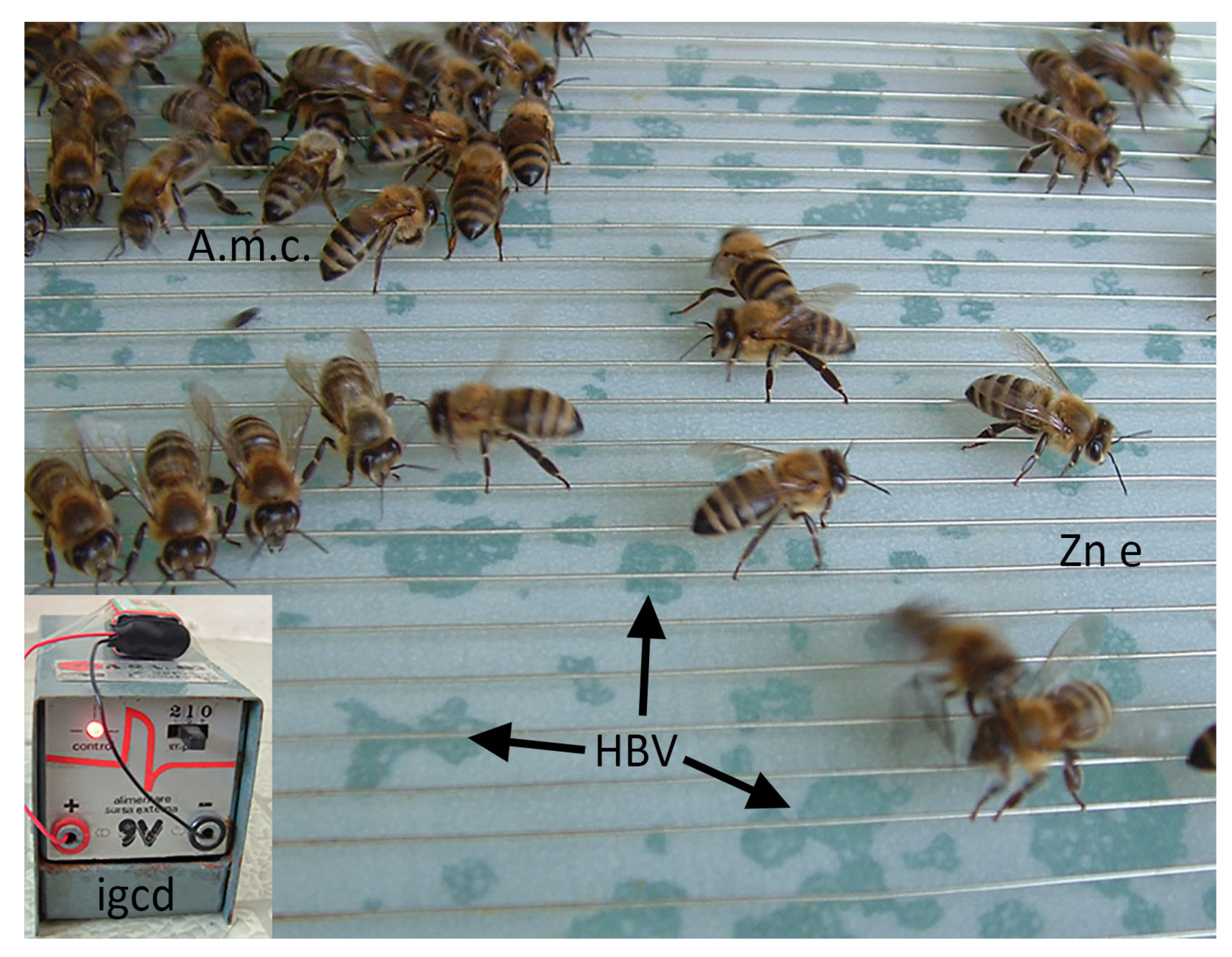
Even though the hvPLA2 is secreted by bees in low amounts, their extremely high number allows the collection of reasonable quantities of venom, using, as a main standardised method, the electric stimulation of the stinging reflex of bees [47,83], as represented in Figure 2. Recent data indicated that the amount of venom correlated with the position of the bee venom collector frame [84], as well as with the temperature [85]. Scaccabarozzi also reported differences in HBV weight and protein diversity caused by both temperature and other ecological factors [86]. From the dry HBV, the enzyme can be further purified with high yield through preparative HPLC [87]. An alternative way of directly obtaining the hvPLA2 is by bioengineering based on cloning the bee PLA2 gene in the bacteria E. coli [8]. The commercially available lyophilised powder containing hvPLA2 is useful for accurate experimental activities since it is very stable at −20 °C [87].
The aqueous formulation could also be used, but the enzyme stability is lower at the same temperature [88]. The catalytic activity of the hvPLA2 is documented: “One unit will hydrolyze 1.0 μmol of soybean L-α-phosphatidylcholine to L-α-lysophosphatidylcholine and a fatty acid per min at pH 8.9 at 25 °C” [87]. On the other hand, Nair et al. studied the thermal stability of hvPLA2 and noted a maximum activity at 65 °C using egg yolk as substrate and at 60 °C in the presence of dicaproyl and dipalmitoyl lecithin [89].
In our previous studies, we tested the lyophilised hvPLA2 (product number P9279, m.w. 14,500 Da, purity 1956.57 units/mg protein) from Sigma-Aldrich (St. Louis, MO, USA). The obtained results published so far (as well as others in analysis) were impressive, helping our progress in understanding the effects of the HBV on the mammalian organism.

4. Effects of hvPLA2

In consistency with its occurrence in so many different classes of organisms so distant phylogenetically, the functions of PLA2s are very diverse. The most studied PLA2s are those from venomous insects, playing various toxic roles, and those from mammals. For the latter ones, it was noted their involvement in a range of cellular processes, from lipid digestion [90] to inflammation [91,92], or control of cellular proliferation [93]. Since the purpose of this review is to deal mainly with hvPLA2, we hereby discuss its functions (Figure 3), but in relation to other PLA2s.

4.1. Breakdown of Membranes

One of the first noted effects of the HBV was its ability to induce haemolysis and cell membrane permeability [94,95,96]. This feature is conferred by PLA2, as well as by other substances, especially by melittin—a peptide which is the main component of bee venom. hvPLA2 has harmful effects on the complex lipids in animal or human organisms [7,41].
Thus, its final effect is damaging the cell or its organelles. It has low activity in the presence of phospholipid monomers, but it is activated by high concentrations of substrate molecules—micelles, cell membranes, vesicles with phospholipid bilayers, etc. [19]. In cells, the sn-2 position of phospholipids often contains polyunsaturated fatty acids that, when released by the PLA2-catalysed hydrolysis, can be further metabolised into eicosanoids and other bioactive lipid mediators [97]. Additionally, the resulting lysophospholipids may play various significant biological roles [98].

4.2. Interaction with Membranes

The interaction of PLA2 with membranes has been studied on enzymes from various organisms, and the comparison of hvPLA2 with other phospholipase structures provides compelling evidence for a common catalytic mechanism [66,82,99,100].
hvPLA2 is a basic, water-soluble protein, that must attach itself to membranes in order to interact with phospholipids and to hydrolyse them. Its solubility in water and its activation by phospholipids in high concentrations have suggested a mechanism of enzymatic catalysis occurring in two phases. The first step is the interfacial binding of the enzyme, followed, in the next step, by the catalytic reaction. Therefore, from a functional point of view, PLA2 displays two distinct, very important domains. The first one, responsible for substrate recognition, surrounds the second one, the catalytic site; both are oriented in the same direction [28,101].
The contact of PLA2 with membranes triggers specific modifications in the three-dimensional structure of the enzyme, particularly in the N-terminal region. While this region of the molecule is randomly unfolded in water, it turns into an α-helix structure when binding to the phospholipid surface [19,28]. Based on studies of enzymatic kinetics performed using an electron paramagnetic resonance spectroscopy technique, Lin et al. [101] established that PLA2 attaches to the water-lipid interface, without integrating into membranes. By measuring the lateral pressure of the phospholipids in the monolayer exposed to PLA2, they found minor modifications after interfacial binding, with values comparable to those of the intact layer. These data suggest only a reduced infiltration of PLA2 in the outer membrane monolayer. One year later, Ball et al. [102], followed by Ahmed et al. [103] developed a model for hvPLA2 docking on membranes. This model postulates that the acyl residue behaves like a hydrophobic anchor: it penetrates the lipid surface and partially inserts itself into the bilayer (as represented in Figure 4). Thus, through this substrate-binding process, the lytic power of PLA2 is significantly increased. Notably, this first association of PLA2s with membranes is mainly based on hydrophobic affinities and interactions, and not on electrostatic ones [19,28,101,104,105]. Jackman et al. [106] suggested that the initial hvPLA2 docking can be followed by a “lipid desorption”, process determined by electrostatic interactions, thus leading to membrane lysis. Additionally, Maggio [107] showed that external electrostatic fields, as well as the charges of the lipid molecules of the membrane, can influence the enzyme’s activity. Experiments with amino acid replacements in the structure of hvPLA2 proved that changes in the charge of the residues at the membrane-binding site can also affect how strongly the enzyme interacts with the substrate [108], the mutant enzyme with anionic amino acids in the binding domain attaching more tightly. Furthermore, the actual catalytic activity seems to be enhanced by the presence of a long-chain fatty acid in the binding site consecutive to conformational changes in the protein and not due to hydrophobic interactions with the substrate [109]. The enzyme is next involved, in the presence of 2 Ca2+ ions [78,82,94], in multiple interactions with phospholipids, during which the hydrophobic channel expands, and the interfacial binding domain is remodelled.
Y57 of the human sPLA2 plays an important role in this process [78]. Ball et al. [102] confirmed that the Ca2+-binding region, which is rich in hydrophobic residues [110], also contributes to the process of enzyme docking to the membrane. The remaining chain lines up parallel to the lipid bilayer, in the proximity of the polar headgroups of the phospholipids. Even though Ca2+ ions are not necessary in the first step of PLA2 docking, they represent an important cofactor required for the attachment of phospholipid molecules to the catalytic domain of the enzyme, as well as for the initiation of the enzymatic reaction [28,105]. The results of Scott et al. [66] indicated that cobra venom PLA2 facilitates “substrate diffusion from the interfacial binding surface to the catalytic site rather than an allosteric change in the enzyme’s structure”. For the PLA2 from another snake (Agkistrodon contortrix laticinctus), it was shown by Ambrosio et al. [111] that a fatty acid adsorbed to the active site triggers conformational changes in the C-terminal region, responsible for the Ca2+-independent activity of the enzyme. Data reported by Nabemoto et al. [112] suggest that sPLA2s are inactive in the first stages of interaction with membranes and their activation is triggered by a modification of their disulfide bonds.
Studies performed on different types of liposomes have revealed further aspects of PLA2 binding to membrane surfaces. Thus, Ghomashchi et al. [104] reported that hvPLA2 binds preferentially to anionic vesicles versus phosphatidylcholine vesicles. Holopainen et al. [113] have shown that the lytic effect of PLA2 is extremely powerful in the case of unilamellar liposomes, where a pronounced tendency of aggregation of the neighbouring vesicles was also observed. The multilamellar liposomes displayed only minor variations in size, thus suggesting that the hydrolytic effect of the enzyme only concerns the outer monolayer. Furthermore, it was observed that PLA2 is temperature-dependant: its lytic activity is significantly elevated in liposomes in liquid crystal state, and irrelevant in liposomes in gel state. Another modulating factor is the lateral equilibrium pressure of lipid molecules in membranes. This remark can be supported by the lack of modifications in phospholipid monolayers at surface pressures of over 30 mN/m; in the gel state, the lateral equilibrium pressure exceeds the capacity of the enzyme to insert into lipid layers [113]. Billy et al. [114] obtained similar results working on washed platelets.

4.3. Mechanism of Phospholipids Hydrolysis

The catalysing mechanism of hvPLA2 has been well documented [66,78]. X-ray crystallography data revealed a similar catalytic centre with other classes of PLAs, proving their common action mechanism [66]. Thunnissen et al. [115] described the changes in the crystalline three-dimensional structure of PLA2 during the process of substrate binding to the catalytic domain. After docking on the membrane, the catalytic site of the hvPLA2 surrounds the hydrophilic group of a single phospholipid molecule via specific interactions. Ambrosio et al. [111] have shown that the position initially occupied by the sulphate ions in a modified hvPLA2 corresponds to that where the phosphate group of the phospholipid temporarily attaches. They also reported significant conformational changes in the Y52, K53, and H68 residues involved in this process for the serpent A. contortrix laticinctus (broad-banded copperhead) PLA2.
R and K residues (in a total number of 18 in hvPLA2 and 23 in the human sPLA2 from certain cells during inflammation) play important roles in the interaction with lipid membranes and in the formation of protein-lipid complexes. Replacement through reversible mutations of the cationic residues R7 and K10 from the interfacial binding area of the human cellular sPLA2 with E was followed only by a moderate decrease of its capacity to bind the lipidic substrate from phospholipid anionic vesicles. These data, also confirmed by certain experimental results of enzyme binding to micelles (phospholipid-detergent), support the finding that a higher number of R and K residues are involved in this kind of protein-lipid assembling [116].
The active catalytic site in the hvPLA2 molecule is located in its central region [66,67,117]. Annand et al. [63] showed the paramount importance of three amino acids in the active site of hvPLA2 (H67, D97 and Y120, see Figure 1). A key function is however performed by the H. Its capital role was proved using mutant, inactive enzymes, with H67 replaced by another amino acid. On the other hand, the enzymes without the two other essential amino acids (D97 and Y120) still maintained an efficient catalytic activity. Histidine probably also acts as a Brönsted base to deprotonate water, while D and Y establish hydrogen bonds with H67. On the other hand, Ghomashchi et al. [104] reported that the deletion of the large β-loop (residues 132–151) had no obvious consequences on interfacial binding and catalysis of hvPLA2.
This mechanism, described for the hvPLA2, was found to be similar for the sPLA2s in other species [110]. Thus, sPLA2s typically found in mammalian secretory fluids and snake venoms, have the active catalytic site made of H48 and D99 residues, as well as a D in position 49, which also binds the Ca2+ ions [82,118]. The replacement of the D49 by a lysine residue resulted in a catalytically inactive enzyme [119,120].
In their outstanding review, Berg et al. [28] summarised a high number of mutant variants of hvPLA2 as well as of sPLA2s from various species, emphasising their roles for a better understanding of the catalytic effect of the enzyme. Moreover, Schneider et al. [121] reported no differences in the catalytic activity of glycosylated (native, secreted form) and non-glycosylated (after enzymatic removal) hvPLA2.
The enzyme uses a H+ to trigger a nucleophilic attack on the sn-2 bond (β-ester bond) in the target phospholipid from the plasma membrane, leading to the release of unsaturated free fatty acids (arachidonic acid, among others) and lysophospholipids. The latters include lysophosphatidylcholine or lysolecithin—another “structural poison” with lytic action upon the neighbouring cells, maintaining an avalanche effect. As mentioned before, one unit of hvPLA2 produces one fatty acid each minute [7,19,38,82,117,122,123].
The enzyme displays a high affinity for many natural substrates, such as phosphatidylcholine, phosphatidylethanolamine, as well as for their plasmalogen analogues. By measuring the haemolytic power of the resulting lysolecithin, the quantitative estimation of hvPLA2 activity could be performed. However, this method is not an absolute one, because the intact remaining lecithin, as well as the cholesterol from membranes, are known as inhibitors of the haemolysis produced by lysolecithin [38].
The resulting metabolites (the fatty acid and the lysolecithin) behave as secondary messengers at low concentrations [124,125], while at high concentrations they are cytotoxic [124].

4.4. Pro-Inflammatory Effect

Another role of PLA2 is the production of inflammatory mediators [17,126,127]. The fatty acid and the lysolecithin resulting from the enzymatic activity of hvPLA2 can further serve as precursors for generating eicosanoids [126,128], platelet-activating factor, and lysophosphatidic acid, which in turn modulate many cellular processes [129]. Farooqui et al. [130] reviewed the multiple biological effects of all these molecules. Studies on mammalian sPLA2 revealed that it indirectly triggers the inflammatory process through the release of fatty acids. This, in turn, leads to increased vascular permeability, hemodynamic alterations and, further, organ injury [131]. Wery et al. [110] proved the presence of sPLA2 in various inflammatory exudates, in platelets and in the synovial fluid of patients suffering from rheumatoid arthritis. Furthermore, it induced tracheal inflammation and mucus production in an in vivo model of asthma [132]. Additionally, PLA2 was observed to also behave as an acute phase reactant, with clinical studies on post-surgical patients showing an immediate increase in the concentration of the enzyme, with a maximum in the second day post-operative [133].

4.5. PLA2—Allergen of Bee Venom

Many different populations (mainly in Europe, Asia and parts of Africa) have come in contact with bees and their venom since the dawn of humankind. Therefore, they learned how to avoid or to deal with them, and, at a certain moment in our distant history, how to take important advantages from apiculture. Apart from the intense local pain generated by a bee sting, PLA2 is the major allergen factor of the venom, alongside hyaluronidase and melittin [39,41,134,135,136]. Generally, after an accidental exposure to hvPLA2, the human body reacts by producing IgG1 anti-PLA2 antibodies with low affinity. Repeated contact with the HBV in beekeepers and venom-treated patients (see Section 4.8 and Section 5) results in an increase in IgG4 anti-PLA2 antibodies with high affinity [137]. On the other hand, in very rare situations, IgE anti-PLA2 antibodies trigger allergy in HBV-sensitive patients [138]. These outcomes were also reported after in vitro tests using human leukocytes [136]. Additionally, PLA2 is responsible for IgE-mediated anaphylactic shock [45,139].
Taking into account the correlation between the whole content [47], and the PLA2 content [49] of the A. mellifera venom gland and the bee’s age, a differentiated immune response occurs after stinging by young or adult bees. Analysing IgG4 polyclonal antibodies from beekeepers (highly and most frequently exposed to the HBV), Schneider et al. [121] showed that from the hvPLA2 only specific epitopes (not the entire molecule) are responsible for its actual immunogenicity. Experimental data revealed that in the hvPLA2 molecule, there are four such epitopes. The first one is the region containing the glycosylated N46, and the other three are PLA73-96, PLA107-125 and PLA140-164 [140,141]. An important role in the modulation of the immune answer to this powerful allergen is played by the enzyme catalytic site. This involvement was proved by Annand et al. [63] who tested the response of basophils and the synthesis of IgG and IgE using both normal and mutant hvPLA2s. Changes in charge, amino acid sequence or conformation of hvPLA2 lead to variations in its allergenic activity [135]. Whether the glycosylation does or does not account for the immune response could be considered at least controversial, and more experimental and clinical data (on larger study groups) is required. Thus, it was shown that the sugar residues from the hvPLA2 were less important [142] or not involved at all in the allergenicity [121]. On the other hand, Prenner et al. [143] concluded that the hvPLA2 fucosilated N-acetylglucosamine residue could play an immunogenic role. Also, Dudler et al. [144] analysed the specificity of the human T cell response against hvPLA2 and identified some T cell clones activated by the entire enzyme, but not by its non-glycosylated variants. Additionally, the active hvPLA2 induced an IgE response in mice, while the mutant, inactive form of the enzyme did not [145]. Similar results were also reported by Förster et al. [8], on both natural hvPLA2 and recombinant PLA2 produced in Escherichia coli. In the same line, it has been shown that most individuals with HBV allergy (96%) also react to the recombinant PLA2 produced in Escherichia coli [146]. For detailed reviews see Perez-Riverol et al. [147] with roles of hvPLA2 (and from other species) in allergy, and Blaser et al. [148] for the immunology of hvPLA2 in allergic and non-allergic subjects.

4.6. Anticoagulant Effect

Once arrived in the blood flow, apart from its haemolytic activity, hvPLA2 also possesses another converging, anticoagulant effect [114,149]. Venom PLA2s have been classified by Boffa et al. [150] into 3 categories, based on their anticoagulant activities: strong, moderate (including hvPLA2) and weak. Comparing different phospholipases, Kini and Evans [151] pointed out a part of the molecule situated between residues 54 and 57, which is responsible for this effect. In strongly anticoagulant PLA2s, this region is positively charged and contains 4×K residues located on the surface of the molecule, while in the enzymes from the other categories, it is negatively charged [151]. Interestingly, while all strong anticoagulant enzymes are basic proteins, not all basic PLA2s have strong anticoagulant action [150,152]. Thus, the exact location of the basic residues is the determining factor, rather than its overall basicity [151]. In hvPLA2, the region thought responsible for its anticoagulant effect contains 3 positively charged residues and has a basic character that explains the moderate effect of the enzyme [151]. Condrea et al. [153,154] have shown that alterations of this region of the molecule can lead to a loss of its anticoagulant properties. Billy et al. [114] noted that, in activated platelets, cobra venom PLA2 inhibited the production of thrombin and the annexin-V binding; however, there was no observable effect on clotting or the activation of platelets. Mukherjee et al. [155], also working on cobra venom, proved its inhibitory potential on thrombin and factor Xa, non-enzymatically, and without relying on phospholipids. Kini [156] reviewed the various action mechanisms of anticoagulant snake venom PLA2s. Studying HBV, Ouyang et al. [157] also pointed out the same inhibition of the activation of prothrombin; however, they concluded that this effect could be attributed to the suppression of the procoagulant properties of intact phospholipids.

4.7. PLA2 and Apoptosis

hvPLA2 can be involved in the first steps of apoptosis, after a mechanism similar to that of the tumour necrosis factor (TNF) in some leukaemic cell lines. Melittin (as well as TNF) has the ability to activate the hvPLA2 and also the cytosolic PLA2 (from mammalian cells). Participation of PLA2 in apoptosis has not been fully elucidated. It is assumed that a crucial role is performed by the arachidonic acid release from membrane phospholipids. It is then metabolised (via lipooxygenase and cyclooxygenase) with the production of free radical oxygen species, involved in cytolysis. PLA2 is also responsible for ceramide production, which in turn has an important role in the TNF or the HIV-induced apoptosis [158]. Anyway, it was proved that prevention of PLA2 activation was followed by resistance of the studied cell lines to TNF [159,160].
Additional experimental data showed the relevance of cellular PLA2 (cPLA2) in apoptosis. Thus, the inhibition of PLA2 further inhibited DNA fragmentation [161]. Other in vitro studies showed increased activity of cPLA2 in HeLa and breast carcinoma cells consecutive to induction of apoptosis with TNFα [159,162] (observation consistent with the report of Wu et al. [160]), or in neurons exposed to oxidative stress and β-amyloid [163].
On the other hand, Kim et al. [164] showed that the hvPLA2 injected intraperitoneally (0.2 mg/kg) significantly alleviated the diethyl 1,4-dihydro-2,4,6-trimethylpyridine-3,5-dicarboxylate diet-induced apoptosis in mice, associated with an important reduction in the cleaved caspase-3 levels. Unfortunately, the paper did not explain the molecular mechanisms involved in this protection. The same authors further claimed that the hvPLA2 reduced hepatic fibrosis and demonstrated the anti-inflammatory effect by reducing the number of neutrophils, F4/80+ macrophages and CD4+ T-cells in the liver tissue in the same experimental conditions, but such results should be considered with some reserve since they incorrectly compared Portal regions with central regions of lobules. Another study demonstrated that hvPLA2 prevented in vitro the apoptosis of spleen regulatory T cells via a concomitant decrease in their caspase-3 expression and of annexin V-positive early apoptotic populations [165]. Also, the regulatory T cells treated with hvPLA2 showed a higher expression of programmed cell death-1 protein (that also helps cancer cells to elude the immune response [166]) and cytotoxic T-lymphocyte-associated protein-4 (an inhibitory receptor from the CD28 immunoglobulin subfamily (reviewed in [167])).
It is difficult to comment on the diverging data reported by different groups of researchers, but it is very likely that progress and higher availability of laboratory tools aimed to investigate the molecular mechanisms of the various cell death subroutines (as recommended by the Nomenclature Committee on Cell Death 2018—[168]) will eventually solve this problem too.

4.8. Antiviral, Antibiotic and Antitumoral Effects of hvPLA2

Given the presence of phospholipids in the viral envelope and in the membranes of pathogenic microorganisms and PLA2’s ability to hydrolyse them, numerous studies have been conducted, assessing the utility of the enzyme in fighting infections or cancer. Fenard et al. reported the successful use of hvPLA2 [169], or of a peptide (p3bv) derived from this enzyme [170] in preventing HIV entry into the cell and its further multiplication. However, according to these authors, the peptide p3bv proved to be less effective as compared to the entire molecule. The same mechanism was described by Santos et al. [171], studying the antiviral effect of Crotalus durissus terrificus PLA2 on the Chikungunya virus. hvPLA2 had a moderate virucidal effect on Flaviviridae (hepatitis C, dengue and Japanese encephalitis virus) at relatively low concentrations, that did not cause cytotoxicity or haemolysis [172]. Further studies on the same virus family showed a more potent virucidal activity of various snake venom PLA2s [172,173]. Muller et al. [174] noted that PLA2 only affects enveloped viruses, proving that its action mechanism depends on the presence of glycerophospholipids in the viral envelope. For a review of the antiviral effects of HBV and its components, including hvPLA2, see Yaacoub et al. [9].
On the other hand, hvPLA2 has an antimicrobial effect (but quite a reduced one), acting on the membranes of Gram-positive bacteria [105,116,175], at a comparable efficiency to the homologous enzyme isolated from cobra venom, but around 100 times less potent than sPLA2 of human cells [176]. In this case, the presence of Ca2+ was shown to be the modulating factor of enzyme activity [105]. Boutrin et al. [177] proved the bacteriostatic and bactericidal effect of hvPLA2 on Gram-negative bacteria, with minimum bactericidal concentrations in 2 h cultures being 10−5–10−6 mg/mL. Additionally, hvPLA2 even in very low concentrations had a lethal effect on Trypanosoma brucei brucei; this effect could be explained by direct membrane lysis or by interference with Ca2+ levels [177]. Furthermore, PLA2s have antimalarial properties [178,179]. Deregnaucourt and Schrével [178] showed that both toxic and non-toxic sPLA2s had a lethal effect on the intraerythrocytic development of Plasmodium falciparum via serum phospholipid hydrolysis; interestingly, hvPLA2 was stage-specific, only being active in the 19–26 h period of the 48 h cycle of the parasite. This effect is not linked to the actual binding of the enzyme to erythrocyte membranes, but rather to the presence of serum lipoproteins that are hydrolysed by PLA2 into arachidonic, linoleic, or docosahexaenoic acid (toxic extracellular compounds that induced the direct degeneration of Plasmodium without damaging the host cell membrane) [179]. Moreira et al. [180] showed that, through the expression of the hvPLA2 gene in transgenic mosquitoes, the development and transmission of the parasite were seriously impaired, leading to a possible way of controlling this disease.
Finally, PLA2s also have antitumor effects. The toxic and cytostatic effects of the hvPLA2 were demonstrated in vitro on various lines of tumoral cells, such as MCF-7 cells [181], or renal cancer cells [182]. hvPLA2 generated a moderate inhibition of their proliferation; however, its effect was much enhanced by the co-administration of phosphatidylinositol-(3,4)-bisphosphate [183]. Further studies using phosphatidylinositol homologues proved that the 3-phosphorylated ones had the strongest antitumoral properties when combined with hvPLA2; those with a polyunsaturated fatty acid as an acyl group had a reduced synergy with hvPLA2 [182]. Furthermore, since HBV displays a marked synergic effect of hvPLA2 and melittin [184], it can serve as an even more powerful antitumoral agent [185,186,187]. For reviews on the applications of HBV in cancer therapy see [10,12,188]. With an interesting approach, Shi et al. [189] reviewed the pharmacological effects and mechanisms of the HBV (including the hvPLA2) and its main components in inflammatory diseases, pain and neurological disorders, microbial diseases, cancer, and in liver, kidney, lung and muscle injury. Other PLA2s have also been observed to have similar properties: the enzyme obtained from Bothrops jararacussu venom exhibited a strong antitumoral and antimetastatic effect on triple-negative breast cancer cells, through numerous mechanisms, including induction of apoptosis and autophagy, decreasing cell proliferation and migration, as well as blocking the epithelial–mesenchymal transition [190]. Bazaa et al. [191] demonstrated that the Macrovipera lebetina transmediterranea venom PLA2 was a powerful inhibitor of both adhesion and migration of human tumour cells, an effect that was mediated by α5β1 and αv-containing integrins and did not rely on the integrity of the catalytic centre of the molecule. Furthermore, peptides derived from the C-terminal region of snake venom PLA2s, despite lacking catalytic activity, proved to be potent cytotoxic agents, with an efficiency comparable to that of paclitaxel in murine models of breast tumours [192].

4.9. Toxicity of hvPLA2

As the second most prevalent active molecule, hvPLA2 highly contributes to HBV toxicity. With low toxicity when pure, it is activated by high doses of melittin [193,194,195,196], the main component of the HBV, and turns into a major haemolytic factor [7,45,197]. This is an illustrative example of how natural selection facilitated the synthesis of two very different molecules (a protein and a peptide) by the same insect organism and with the same purpose.
hvPLA2 has a similar lytic activity to that of the PLA2 from cobra venom, more powerful as compared to the enzyme isolated from the viper venom [7], and approximately 100 times more potent than that of human sPLA2s [176]. Within the genus Apis, the venom of A. mellifera (tested from three populations, A. dorsata, A. cerana, A. florea) demonstrated similar lethal activity toward mice [33]. When comparing the toxicity of European and Africanized HBV, the LD50s were also similar [50,198]. Therefore, it remains possible that the activity of hvPLA2 from all these bee species could be similar. While Ownby et al. [195] used the hvPLA2 in a dose of 4 mg/kg with intramuscular administration in mice, we calculated and tested on rats an LD50 of 9.3 mg PLA2/kg via subcutaneous route [13,14]. Other researchers reported in mice detailed effects of snake venom PLA2 from Indian cobra (Naja naja naja) venom (LD50 of 2.4 mg/kg) [199] or crossed pit viper (Bothrops alternatus) venom (LD50 of 140 µg/kg) [200], in both cases administered by intraperitoneal route. The differences in the tested doses also confirm the different degrees of lethality of the enzyme from different genera or species.
PLA2 toxicity was first explained by its enzymatic effect on the membrane phospholipids. Thus, the membrane integrity is compromised, with consequences on the functions of the targeted cells. Moreover, the fatty acids and the lysophospholipids produced in high amounts by the enzymatic digestion propagate the membrane damages by detergent-like effects; the arachidonic acid triggers an accentuated oxidative stress followed by increased lipid peroxidation and by oxidative damage to membrane proteins [124,201]. All the structural alterations of the membranes affect their permeability, mainly for Ca2+ ions, whose cytosolic increase could trigger cytoskeletal changes, lipolysis and proteolysis [124].
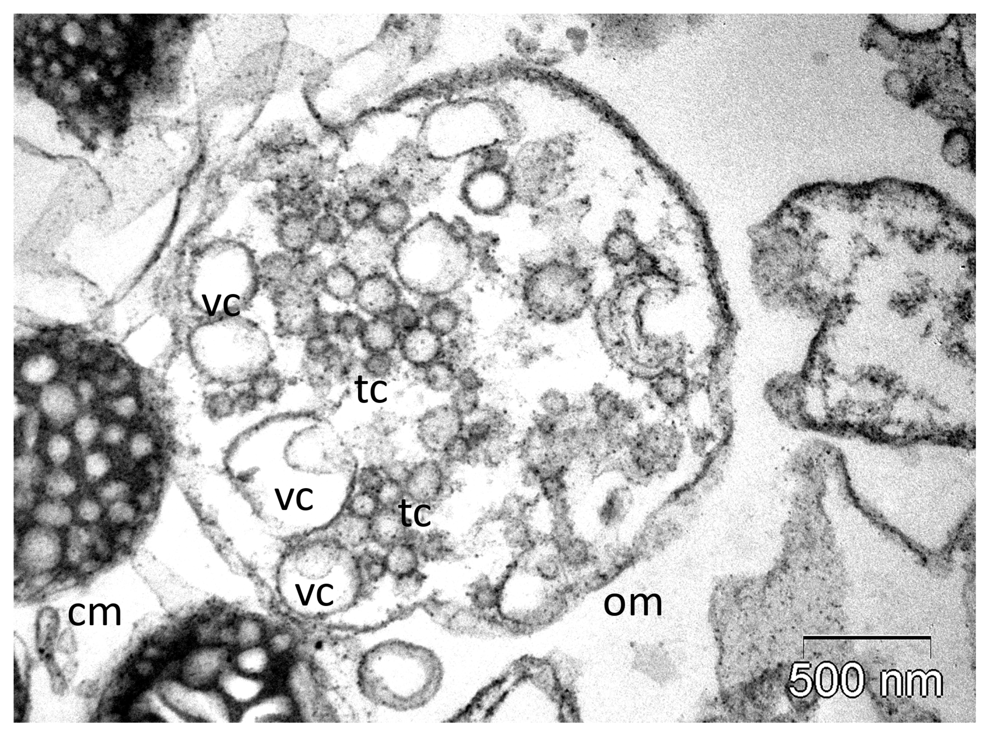
As a cytolytic factor, PLA2 is involved in mast cell lysis, and therefore, in histamine [202] and serotonin [38] release. Furthermore, it causes K+ release from skeletal muscle cells, and epinephrine release from adrenal medulla [38]. PLA2 activation also results in the inhibition of oxidative phosphorylation. It attacks the succinate-dehydrogenase (and other enzymes catalysing metabolic dehydrogenation), as well as the respiratory chain-containing membrane. We previously reported the ability of the hvPLA2 to induce dose-dependent fusions of the adrenocortical mitochondria cristae in vitro, resulting in large vesicular cristae (some with 2–3 membranes, as can be observed in Figure 5), that further collapsed at higher doses [15]. Similar but less pronounced ultrastructural alterations of the adrenocortical mitochondrial cristae (along with other cellular responses) were found when we tested in vivo the subcutaneous administration of hvPLA2 (in a dose of 9.3 mg PLA2/kg) [14]. PLA2 inhibits the tissular thromboplastin and destroys the lysosomes. Moreover, the free fatty acids released during the phospholipid breakdown interfere as well with oxidative phosphorylation; they also produce changes in membrane permeability by different mechanisms [130,201]. All these effects may participate in tissue damage resulting after the local application of venom [7,38].
PLA2 displays a marked neurotoxicity. In contrast to other enzymes (for example PLA2 isolated from viper venoms—Vipera ammodytes meridionalis or Vipera russelli formosensis), which act in combination with other molecules [203], hvPLA2 exerts its neurotoxic action mostly alone. On the brain cell membranes, there are located receptors to which the hvPLA2 binds with high affinity [204,205]. These receptors, called N-type receptors, are believed to represent binding targets for endogenous sPLA2s, but they also have an important role in the progress of the molecular phenomena involved in PLA2 neurotoxicity.
Continuing their studies in this direction, the same research group reported interesting in vivo responses of neurons to the hvPLA2, via N-type receptors [205,206].
Experiments with mutated PLA2 (with different sequences of amino acids at different levels of the enzyme) showed that the hvPLA2 high affinity for the N-type receptors is essentially provided by the amino acids located in the interfacial binding domain (amino acids 109–124), in the Ca2+ binding region, and at the N-terminal end, respectively [123]. On the other hand, the same authors noted that mutant hvPLA2 molecules (with low affinity for these receptors) are devoid of neurotoxicity even though some of them still maintain a high enzymatic activity [123].
PLA2 neurotoxicity also results, as discussed above, from the direct action of the enzyme on the phospholipids from the membranes of the various structures participating in the blood–brain barrier and from neuronal membranes, respectively. All the damage and cascade events are eventually responsible for the structural and functional modifications of neurons. We found important ultrastructural alterations of the frontal cortex neurons consecutive to experimental testing of hvPLA2 (unpublished results), facilitated by a compromised blood–brain barrier. Some of these changes were consistent with those (histological, histochemical, ultrastructural and physiological) reported by us consecutive to the experimental administration of HBV [207]. Apart from affecting the central nervous system, the HBV molecules play an important role at the level of the peripheral nervous system as well. In the same paper, we also mentioned the immediate reactions of rats to the high dose of HBV including increased aggressiveness, motor agitation and uncoordinated movements, abnormal discharges (at 40–45 min after the HBV injections), piloerection, lethargy and generalised convulsions crisis (after 1.5 h) [207]. Such symptoms were previously described consecutive to the intraperitoneal administration of hvPLA2 in mice (at an LD50 of 2.4 mg/kg) [199], or when hvPLA2 was injected in rats via the intracerebroventricular route [208]. Pungerčar and Križaj [209] attributed many of these toxic effects of the hvPLA2 to its action on the membranes of motoneurons participating in the neuromuscular junctions. The hvPLA2 neurotoxicity was further demonstrated in vitro. Thus, it was shown that, once it arrived in synaptosomes, hvPLA2 caused the uncoupling of mitochondria [210], with a decrease in the ATP synthesis, and subsequent increase in cytosolic Ca2+ ions that modulated the frequency of neurotransmitter release.
In snake venom envenomation, the cancelling of neuromuscular transmission results in respiratory failure, paralysis and eventually death, PLA2 blocking both cholinergic synapses (in skeletal muscles), and non-cholinergic neurons (from the cerebral cortex, hippocampus and granular layer of the cerebellum [209]).
Another mechanism explaining the cellular toxicity of hvPLA2 is based on its property to bind to calmodulin (cytosolic receptor for Ca2+) [211] and, this way, to modulate many metabolic pathways. However, this mechanism requires more investigations.
On the other hand, the arachidonic acid released during the enzymatic action of PLA2 on membranes stimulates glycogenolysis in cortical astrocytes [212], in order to energetically sustain the resistance of glial and neuronal cells against the deleterious effects of PLA2. Katsuki and Okuda [213] showed that the arachidonic acid also contributes to neurodegeneration. Farooqui et al. [130] reviewed more molecular mechanisms involved in PLA2 neurotoxicity.
However, there is contrasting data revealing a potential neuroprotective effect of hvPLA2 in a mouse model of Alzheimer’s disease, induced by lipopolysaccharide injection [214]. Injected intraperitoneally, the enzyme was shown to reduce the memory impairment and amyloidogenesis, through the inhibition of nuclear factor kappa B, glial fibrillary acidic protein and allograft inflammatory factor 1 expression, as well as through reduced cytokine release. Additionally, hvPLA2 increased the number of regulatory T cells, which in turn led to a reduction in microglial activation. Converging results were also reported by the same authors in vitro, using BV-2 cells.
hvPLA2 is also a myotoxic compound. Apart from the N-type receptors located on the neuronal plasma membranes, experimental data proved a potent expression of M-type receptors for sPLA2s in muscular tissues (skeletal and smooth muscles, myocardium). In addition to this, the presence of certain plasma PLA2-binding proteins suggests that the bee enzyme is involved in vivo in a series of other still unknown physiological processes [63,68,123].
Studies on mice showed myotoxic effects of the hvPLA2 injected intramuscularly, consisting in extensive ultrastructural alterations that culminated with cellular necrosis [195], or degenerated muscle fibres and necrosis around the injection site, causing respiratory distress and paralysis [199]. Gutiérrez and Ownby [215] explained in detail the mechanisms of PLA2s (including the hvPLA2) myotoxicity, emphasising the differences between the local and systemic effects.
Working with snake and mammal pancreatic PLA2s, Lambeau et al. [216] revealed that G30, and D49 as well as L31 are essential for PLA2 binding to the M-type receptors. Furthermore, Lambeau et al. [204] showed that the receptors for sPLA2s differ in different tissues, being responsible for “tissue-dependent pharmacological profiles” and that in the skeletal muscles, their density is age-dependent.
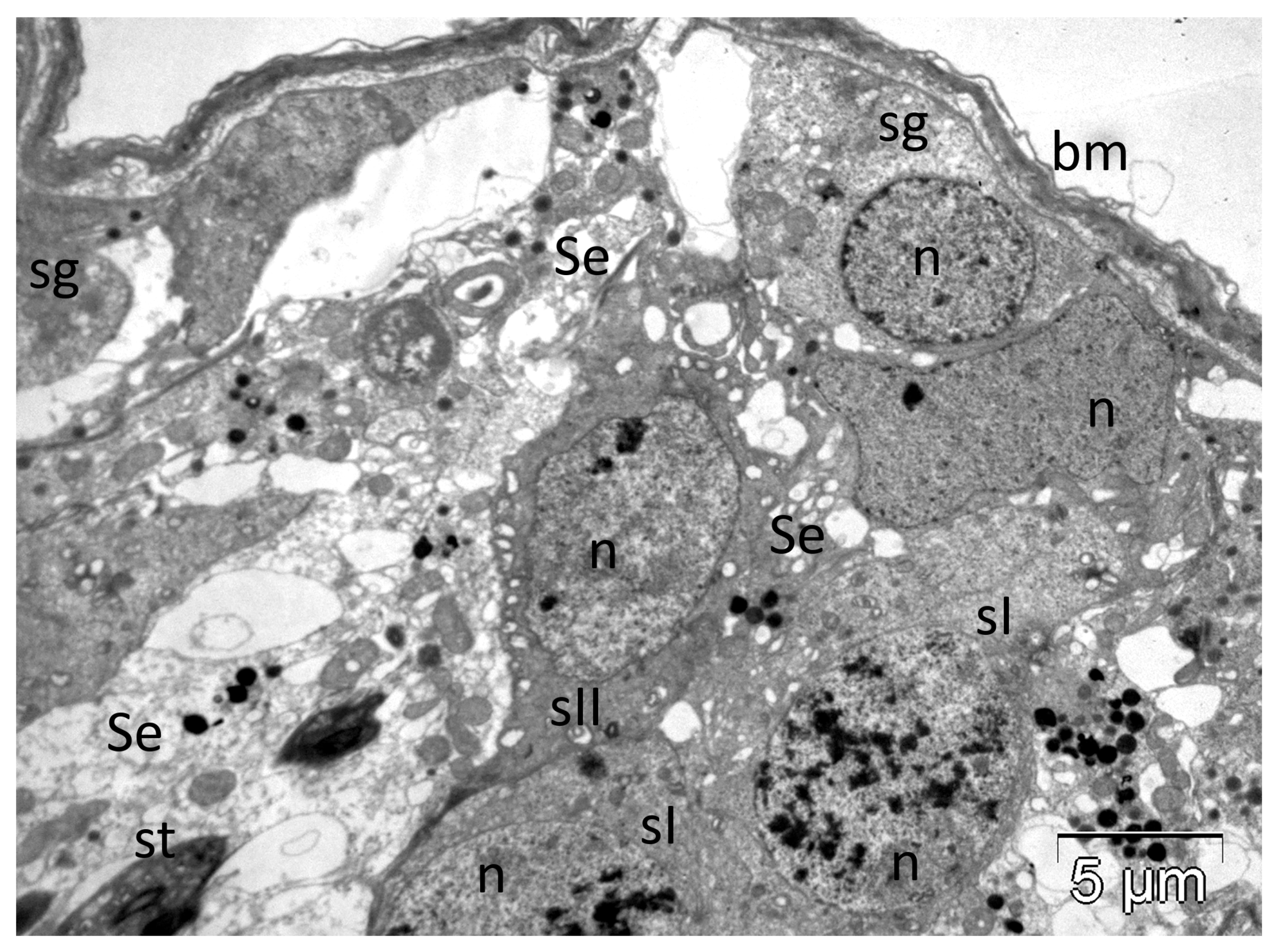
hvPLA2 triggers toxic effects in other organs too. The M-type receptors for sPLA2s were found not only on the membranes of muscle cells but also in kidney, lung, liver and pancreas [63,68,123,204], with roles that remain to be elucidated. We found important ultrastructural changes produced by the experimental administration of the hvPLA2 in several organs of rats. To date, we have demonstrated the ability of the hvPLA2 to damage the interstitial Leydig cells and to cancel the permeability of the testicular barrier, thus interfering with the normal progression of spermatogenesis. The latter effect was facilitated via extensive ultrastructural damage of the Sertoli cells [13], as shown in Figure 6. In the adrenocortical cells, apart from the above-mentioned changes in mitochondria, the hvPLA2 also triggered dose-dependent alterations of nuclei and smooth endoplasmic reticulum, associated in some cases with plasma membrane rupture and the release of organelles outside the cells [14].
However, low doses of hvPLA2 (0.2 mg/kg injected intraperitoneally) seem to have a hepatoprotective effect against the acetaminophen (500 mg/kg)-induced acute liver injury [217] in mice. To demonstrate this effect, the authors also tested and showed reduced protection of hvPLA2 in regulatory T cells-depleted mice, establishing a correlation between this cellular population and the normal functional parameters of the liver.
As a main constituent of HBV, hvPLA2 also highly contributes to the overall effectiveness of the venom consecutive to massive attacks of both European and Africanized honeybees on humans [3,218,219,220,221], or animals (dogs [222,223], or horses [224,225]).
The toxic effects debuted in humans with local reactions and culminated with extensive haemolysis [7], circulatory and respiratory impairment, acute renal failure (after 200–500 stings in adults) and death, after more than 500 stings (Refs. [4,218,219,226], and reviewed by Schmidt [227]).

5. Immunotherapy

As mentioned above, hvPLA2 is a powerful allergen. Thus, one of its main medical applications is in immunotherapy. Early studies showed that doses of 100 μg of HBV, administered constantly, over a period of several years, provided protection against allergic reactions [52] and induced the production of IgG, while decreasing IgE levels [228,229]. Patient immunisation using only hvPLA2 was proved equally efficient in conferring the same protective effect against bee stings [230]. Müller et al. [139] tested 3 peptides derived from hvPLA2 that contained its epitopes and noted a similar success of the immunotherapy as that obtained using whole HBV. Furthermore, their results showed a decrease in the frequency of allergic side effects when using the peptides, since they lacked catalytic activity [139]. Additionally, Nair et al. [231] proved that there was no cross-protection given by IgG antibodies against hvPLA2. However, antibodies against bald-face hornet venom offered protection against hvPLA2, as well as PLA2s from yellow hornet or yellow jacket.
In an interesting approach, Jilek et al. [232] assessed the effectiveness of genic therapy in vivo, on mice injected with plasmids carrying a PLA2 gene sequence. They reported long-time low levels of IgE and IgG1 (and increased levels of IgG2a and IgG3) consecutive to the administration of the recombinant plasmids, both before and after the mice sensitization to hvPLA2. They also demonstrated that the prophylactic treatment was more effective than the therapeutic one, this way opening the possibility of using such DNA vaccines for human immunotherapy.

6. Anti-PLA2 Molecules

Due to the numerous adverse effects of PLA2s, the inhibitory action of various substances has been extensively studied. These molecules act on different, specific, targets to inactivate the enzyme. Several natural compounds with such properties have been identified, including manoalide, luffariellolide, and scalaradial, isolated from marine organisms [233]. The latter was proved to have a two-step action mechanism, with an initial non-covalent attachment, followed by a covalent modification of hvPLA2 near its substrate-binding site [234], namely of K127 [104]. In 1995, De Rosa et al. [235] discovered and isolated from a species of marine sponge, Fasciospongia cavernosa, a compound (cacospongionolide B) with a strong anti-inflammatory activity mainly given by its capacity to inhibit PLA2. Because of its importance, not only as a possible anti-venom agent but also with applications in different inflammatory affections such as asthma, psoriasis or rheumatoid arthritis, analogues of this natural product were synthesised, that preserve the inhibitory activity on the hvPLA2 [236]. Antimicrobial peptides, such as magainin 2, indolicidin, and temporins B and L, all derived from natural sources, were also observed to lower the catalytic activity of hvPLA2 [237]. Furthermore, numerous other proteins (lipocortin, bovine serum albumin, myoglobin, lysozyme) showed an inhibitory effect, but only in experiments using low substrate liposomes [238]. Hains et al. [239] purified another PLA2 inhibitor from the serum of the Australian tiger snake (Notechis ater), composed of two protein subunits that form a complex of approximately 110 kDa; upon further experiments, this molecule was observed to block all the different PLA2 enzymes tested. A similar behaviour was noted in proteins from the blood of different cobra species [240,241]. On the other hand, another molecule isolated from Crotalus durissus terrificus serum was proved to have a specific action only against the snake PLA2 neurotoxin (by binding to its active site) and not against hvPLA2 or other PLA2s [242,243,244]. In the serum of Agkistrodon blomhoffii siniticus, three proteins were described, the first two with inhibitory effects on PLA2s from Crotalidae venom (either acidic or basic), and another one that blocked all PLA2s tested, including hvPLA2 [245]. Noetzel et al. [203] also reported a series of substances, including vitamin E, with anti-PLA2 effect on the enzyme secreted by other species. Apart from the exogenous PLA2 inhibitors, it has been observed that crotapotin, a peptide that is naturally complexed to the Crotalus durissus terrificus venom PLA2, acts as an intrinsic enzymatic inhibitor; its action, however, is not restricted to the sPLA2 of the same species, as studies have noted its potency on hvPLA2 and on other snake PLA2s [246]. Many arthropod venoms, including HBV, as well as snake venoms, contain citrate that acts as an endogenous inhibitor of the enzyme, mainly through its ability to bind Ca2+ [247,248]. Synthetic calcium chelators, such as EDTA, can also reduce the activity of Ca2+-dependent PLA2s [249]. Indoxan was proved to be effective in blocking PLA2-induced cell damage, through reduced generation of arachidonic acid and prostaglandin D2 [250]. A similar mechanism was also reported for nordihydroguaiaretic acid and aristolochic acid [251]. Heparin is another molecule with a powerful, non-specific, inhibitory action [246,252]. Furthermore, numerous other chemical substances with the same property, such as benzenesulfonamides [253] or 4-bromophenacyl bromide [195,254] have been described in the literature. The role of these inhibitors is paramount, since through their development the roles of different PLA2 classes could be determined [255,256,257]. Crous et al. [258] reviewed the interactions of the human and snake sPLA2s with several inhibitors using X-ray crystal structures and discussed potential therapeutic applications of these inhibitors.

7. Concluding Remarks

In conclusion, PLA2 is one of the main toxic components of the HBV, commonly found in many other animal species as well, being partially responsible for the venom activity. It is a molecule of high interest for the scientific community (including our department) due to its diverse effects. Far from simply hydrolysing phospholipids, this enzyme plays numerous important biological roles. It interacts non-specifically or specifically with membranes, by mechanisms discussed in detail, being both a powerful toxin and an immune response modulator. The diverse effects of the hvPLA2 recommend it as a promising candidate for various medical applications. This molecule also represents a powerful tool in basic research, useful for deepening our understanding of the protein-lipid and protein–protein interactions in biological membranes. The importance of PLA2 is reflected by the number of studies focused on finding inhibitors for counteracting its toxicity. Analysing the literature data, we noted that some aspects (the structure and the molecular mechanism) were more intensively studied than others. Therefore, more attention should be paid to the clinically relevant ones such as the anticoagulant effects of the hvPLA2 (important for patients predisposed to thrombotic events), immunotherapy or apoptosis. For future research perspectives, we plan to deepen the understanding of hvPLA2 interaction with cellular structures in vivo by revealing ultrastructural responses of other tissues.

Author Contributions

Conceptualization, A.F.; resources, M.M. and A.F.; writing—original draft preparation, A.F.; writing—review and editing, M.M. and A.F. All authors have read and agreed to the published version of the manuscript.

Funding

This research received no external funding.

Institutional Review Board Statement

Not applicable.

Informed Consent Statement

Not applicable.

Data Availability Statement

No new data were created or analyzed in this study.

Conflicts of Interest

The authors declare no conflicts of interest.

Abbreviations

The following abbreviations are used in this manuscript:
A.m.Apis mellifera
HBVhoneybee venom
hv PLA2honeybee venom phospholipase A2
HPLChigh performance liquid chromatography
PLA2phospholipase A2
TNFtumour necrosis factor

References

  1. Bücherl, W. Introduction. In Venomous Animals and Their Venoms; Bücherl, W., Buckley, E., Eds.; Academic Press: New York, NY, USA, 1971; Volume 1, pp. xix–xxii. [Google Scholar] [CrossRef]
  2. Maschwitz, U.W.J.; Kloft, W. Morphology and function of the venom apparatus of insects—Bees, wasps, ants and caterpillars. In Venomous Animals and Their Venoms; Bücherl, W., Buckley, E., Eds.; Academic Press: New York, NY, USA, 1971; Volume 1, pp. 1–60. [Google Scholar] [CrossRef]
  3. Veena, V.; Mehta, H.; Dhingra, P.; Ballani, A. Multiple bee stings and acute kidney injury: A case report. Cureus 2024, 16, e66488. [Google Scholar] [CrossRef]
  4. Rahimian, R.; Shirazi, F.M.; Schmidt, J.O.; Klotz, S.A. Honeybee stings in the era of killer bees: Anaphylaxis and toxic envenomation. Am. J. Med. 2020, 133, 621–626. [Google Scholar] [CrossRef] [PubMed]
  5. Neumann, W.; Habermann, E.; Amend, G. Zur Papierelektrophoresis den Fraktionierung tierischer Gifte. Naturwissenschaften 1952, 39, 286–287. [Google Scholar] [CrossRef]
  6. Habermann, E.; Neumann, W. Reinigung der Phosphalipase A des Bienegiftes. Biochem. Z. 1957, 328, 465–473. [Google Scholar]
  7. Habermann, E. Bee and wasp venoms. The biochemistry and pharmacology of their peptides and enzymes are reviewed. Science 1972, 177, 314–322. [Google Scholar] [CrossRef] [PubMed]
  8. Förster, E.; Dudler, T.; Gmachl, M.; Aberer, W.; Urbanek, R.; Suter, M. Natural and recombinant enzymatically active or inactive bee venom phospholipase A2 has the same potency to release histamine from basophils in patients with Hymenoptera allergy. J. Allergy Clin. Immunol. 1995, 95, 1229–1235. [Google Scholar] [CrossRef]
  9. Yaacoub, C.; Wehbe, R.; Roufayel, R.; Fajloun, Z.; Coutard, B. Bee venom and its two main components—Melittin and phospholipase A2—As promising antiviral drug candidates. Pathogens 2023, 12, 1354. [Google Scholar] [CrossRef]
  10. Gajski, G.; Leonova, E.; Sjakste, N. Bee venom: Composition and anticancer properties. Toxins 2024, 16, 117. [Google Scholar] [CrossRef]
  11. Sadek, K.M.; Shib, N.A.; Taher, E.S.; Rashed, F.; Shukry, M.; Atia, G.A.; Taymour, N.; El-Nablaway, M.; Ibrahim, A.M.; Ramadan, M.M.; et al. Harnessing the power of bee venom for therapeutic and regenerative medical applications: An updated review. Front. Pharmacol. 2024, 18, 1412245. [Google Scholar] [CrossRef]
  12. El-Arabey, A.A.; Ghramh, H.A. Bee venom: Yesterday’s enemy becomes modern medicine for skin cancer. Exp. Cell Res. 2025, 445, 114435. [Google Scholar] [CrossRef]
  13. Tilinca, M.; Florea, A. Ultrastructural analysis of early toxic effects produced by bee venom phospholipase A2 and melittin in Sertoli cells in rats. Toxicon 2018, 141, 94–103. [Google Scholar] [CrossRef]
  14. Harfmann, D.; Florea, A. Experimental envenomation with honeybee venom melittin and phospholipase A2 induced multiple ultrastructural changes in adrenocortical mitochondria. Toxicon 2023, 229, 107136. [Google Scholar] [CrossRef] [PubMed]
  15. Florea, A.; Varga, A.P.; Matei, H.V. Ultrastructural variability of mitochondrial cristae induced in vitro by bee (Apis mellifera) venom and its derivatives, melittin and phospholipase A2, in isolated rat adrenocortical mitochondria. Micron 2018, 112, 42–54. [Google Scholar] [CrossRef] [PubMed]
  16. Bókay, A. Ueber die Verdaulichkeit des Nucleins und Lecithins. Biol. Chem. 1878, 1, 157–164. [Google Scholar] [CrossRef]
  17. Dennis, E.A. Phospholipases. In The Enzymes, 3rd ed.; Boyer, P.D., Ed.; Academic Press: New York, NY, USA, 1983; Volume 16, pp. 307–353. [Google Scholar]
  18. Six, D.A.; Dennis, E.A. The expanding superfamily of phospholipase A2 enzymes: Classification and characterisation. Biochim. Biophys. Acta-Mol. Cell Biol. Lipids 2000, 1488, 1–19. [Google Scholar] [CrossRef]
  19. Wilton, D.C.; Waite, M. Phospholipases. In Biochemistry of Lipids, Lipoproteins and Membranes, 4th ed.; Vance, D.E., Vance, J.E., Eds.; Elsevier Science: Oxford, UK, 2002; pp. 291–314. [Google Scholar]
  20. Apis mellifera (Honey Bee): 100577717. Available online: https://www.genome.jp/entry/AME+100577717+406141+409277+409307+409614+410570+552242+724436+725668 (accessed on 8 April 2025).
  21. Thiyagaraj, A.; Gupta, A.; Salunkhe, S.; Sharathkumar, R.M. Role of phospholipases in chronic inflammatory diseases: A systems biology perspective (Chapter 13). In Phospholipases in Physiology and Pathology; Chakraborti, S., Ed.; Academic Press: Cambridge, MA, USA, 2023; Volume 6, pp. 219–236. [Google Scholar] [CrossRef]
  22. Schaloske, R.H.; Dennis, E.A. The phospholipase A2 superfamily and its group numbering system. Biochim. Biophys. Acta-Mol. Cell Biol. Lipids 2006, 1761, 1246–1259. [Google Scholar] [CrossRef]
  23. Valentin, E.; Lambeau, G. What can venom phospholipases A2 tell us about the functional diversity of mammalian secreted phospholipases A2? Biochimie 2000, 82, 815–831. [Google Scholar] [CrossRef]
  24. Ho, I.C.; Arm, J.P.; Bingham, C.O., 3rd; Choi, A.; Austen, K.F.; Glimcher, L.H. A novel group of phospholipase A2s preferentially expressed in type 2 helper T cells. J. Biol. Chem. 2001, 276, 18321–18326. [Google Scholar] [CrossRef]
  25. Murakami, M.; Kudo, I. Phospholipase A2. J. Biochem. 2002, 131, 285–292. [Google Scholar] [CrossRef]
  26. Rouault, M.; Bollinger, J.G.; Lazdunski, M.; Gelb, M.H.; Lambeau, G. Novel mammalian group XII secreted phospholipase A2 lacking enzymatic activity. Biochemistry 2003, 42, 11494–11503. [Google Scholar] [CrossRef]
  27. Ackermann, E.J.; Dennis, E.A. Mammalian calcium-independent phospholipase A2. Biochim. Biophys. Acta-Lipids Lipid Metab. 1995, 1259, 125–136. [Google Scholar] [CrossRef]
  28. Berg, O.G.; Gelb, M.H.; Tsai, M.D.; Jain, M.K. Interfacial enzymology: The secreted phospholipase A2-paradigm. Chem. Rev. 2001, 101, 2613–2653. [Google Scholar] [CrossRef]
  29. Bon, C.; Choumet, V.; Delot, E.; Faure, G.; Robbe-Vincent, A.; Saliou, B. Different evolution of phospholipase A2 neurotoxins (β-neurotoxins) from Elapidae and Viperidae snakes. Ann. N. Y. Acad. Sci. 1994, 710, 142–148. [Google Scholar] [CrossRef] [PubMed]
  30. Dennis, E.A. Diversity of group types, regulation, and function of phospholipase A2. J. Biol. Chem. 1994, 269, 13057–13060. [Google Scholar] [CrossRef] [PubMed]
  31. Michener, C.D. The Bees of the World, 2nd ed.; Johns Hopkins University Press: Baltimore, MD, USA, 2007. [Google Scholar]
  32. Crane, E. Apis species (Honey bees). In Encyclopedia of Insects, 2nd ed.; Resh, V.H., Cardé, R.T., Eds.; Academic Press: Cambridge, MA, USA; Elsevier: San Diego, CA, USA, 2009; pp. 31–32. [Google Scholar]
  33. Schmidt, J.O. Toxicology of venom from the honeybee genus Apis. Toxicon 1995, 33, 917–927. [Google Scholar] [CrossRef] [PubMed]
  34. Habermann, E. Chemistry, pharmacology and toxicology of bee, wasp and hornet venoms. In Venomous Animals and Their Venoms; Bücherl, W., Buckley, E.E., Eds.; Academic Press: New York, NY, USA, 1971; pp. 61–94. [Google Scholar] [CrossRef]
  35. Kang, S.S.; Pak, S.C.; Choi, S.H. The effect of whole bee venom on arthritis. Am. J. Chin. Med. 2002, 30, 73–84. [Google Scholar] [CrossRef]
  36. Kwon, Y.B.; Lee, H.J.; Han, H.J.; Mar, W.C.; Kang, S.K.; Yoon, O.B.; Beitz, A.J.; Lee, J.H. The water-soluble fraction of bee venom produces antinociceptive and anti-inflammatory effects on rheumatoid arthritis in rats. Life Sci. 2002, 71, 191–204. [Google Scholar] [CrossRef]
  37. Bücherl, W.; Buckley, E. Venomous Animals and Their Venoms; Academic Press: New York, NY, USA, 1971; pp. 2–38. [Google Scholar] [CrossRef]
  38. Habermann, E. Chimie, pharmacologie et toxicologie du venin. In Traité de Biologie de L’abeille; Chauvin, R., Ed.; Masson ETC: Paris, France, 1968; pp. 363–387. [Google Scholar]
  39. Shipolini, R.A. Biochemistry of bee venom. In Handbook of Natural Toxins; Tu, A.T., Ed.; Marcel Dekker Inc.: New York, NY, USA, 1984; Volume 2, pp. 49–85. [Google Scholar]
  40. Banks, B.E.C.; Shipolini, R.A. Chemistry and pharmacology of honey-bee venom. In Venoms of the Hymenoptera: Biochemical, Pharmacological and Behavioural Aspects; Piek, T., Ed.; Academic Press: Orlando, FL, USA, 1986; pp. 329–416. [Google Scholar]
  41. Schmidt, J.O. Bee products: Chemical composition and application. In Bee Products; Mizrahi, A., Lensky, Y., Eds.; Plenum Press: New York, NY, USA, 1996; pp. 15–26. [Google Scholar]
  42. Chauvin, R. Action du venin sur l’homme. In Traité de Biologie de L’abeille; Chauvin, R., Ed.; Masson ETC: Paris, France, 1968; pp. 160–161. [Google Scholar]
  43. Oh, M.M.; Power, J.M.; Thompson, L.T.; Disterhoft, J.F. Apamin increases excitability of CA1 hippocampal pyramidal neurons. Neurosci. Res. Commun. 2000, 27, 135–141. [Google Scholar] [CrossRef]
  44. Bridges, A.R.; Owen, M.D. The morphology of the honey bee (Apis mellifera L.) venom gland and reservoir. J. Morphol. 1984, 181, 69–86. [Google Scholar] [CrossRef]
  45. Vetter, R.S.; Visscher, P.K. Bites and stings of medically important venomous arthropods. Int. J. Dermatol. 1998, 37, 481–496. [Google Scholar] [CrossRef]
  46. Owen, M.D.; Bridges, A.R. Aging in the venom glands of queen and worker honey bees (Apis mellifera L.): Some morphological and chemical observations. Toxicon 1976, 14, 1–5. [Google Scholar] [CrossRef]
  47. Abreu, R.M.M.; Silva de Moraes, R.L.M.; Malaspina, O. Histological aspects and protein content of Apis mellifera L. worker venom glands: The effect of electrical shocks in summer and winter. J. Venom. Anim. Toxins 2000, 6, 87–98. [Google Scholar] [CrossRef]
  48. Owen, D.M.; Pfaff, L.A.; Reisman, R.E.; Wypych, J. Phospholipase A2 in venom extracts from honey bees (Apis mellifera L.) of different ages. Toxicon 1990, 28, 813–820. [Google Scholar] [CrossRef]
  49. Li, J.H.; Zhang, C.X.; Shen, L.R.; Tang, Z.H.; Cheng, J.A. Expression and regulation of phospholipase A2 in venom gland of the Chinese honeybee, Apis cerana cerana. Arch. Insect Biochem. Physiol. 2005, 60, 1–12. [Google Scholar] [CrossRef] [PubMed]
  50. Schumacher, M.J.; Schmidt, J.O.; Egen, N.B.; Lowry, J.E. Quantity, analysis and lethality of European and Africanized honey bee venoms. Am. J. Trop. Med. Hyg. 1990, 43, 79–86. [Google Scholar] [CrossRef] [PubMed]
  51. Palma, M.S.; Brochetto-Braga, M.R. Biochemical variability between venoms from different honey-bee (Apis mellifera) races. Comp. Biochem. Physiol. Part. C Pharmacol. Toxicol. Endocrinol. 1993, 106, 423–427. [Google Scholar] [CrossRef]
  52. Schumacher, M.J.; Schmidt, J.O.; Egen, N.B.; Dilon, K.A. Biochemical variability of venoms from individual European and Africanized honey bees. J. Allergy Clin. Immunol. 1992, 90, 59–65. [Google Scholar] [CrossRef]
  53. Benton, A.W.; Patton, R.L. A qualitative analysis of the proteins in the venom of honey bees. J. Insect Physiol. 1965, 11, 1359–1364. [Google Scholar] [CrossRef]
  54. Ferreira Junior, R.S.; Sciani, J.M.; Marques-Porto, R.; Junior, A.L.; Orsi, R.O.; Barraviera, B.; Pimenta, D.C. Africanized honey bee (Apis mellifera) venom profiling: Seasonal variation of melittin and phospholipase A2 levels. Toxicon 2010, 56, 355–362. [Google Scholar] [CrossRef]
  55. Owen, M.D. Relationship between age and hyaluronidase activity in the venoms of queen and worker honey bees (Apis mellifera L.). Toxicon 1979, 17, 94–98. [Google Scholar] [CrossRef]
  56. Marz, R.; Mollay, C.; Kreil, G. Queen bee venom contains much less phospholipase than worker bee venom. Insect Biochem. 1981, 11, 685–690. [Google Scholar] [CrossRef]
  57. Shen, L.S.; Zhang, C.Z.; Cheng, J.A. Cloning and sequencing of gene encoding phospholipase A2 from the venom of Apis cerana cerana and A. mellifera. J. Agric. Biotechnol. 2002, 10, 29–32. [Google Scholar]
  58. Ryu, Y.; Oh, Y.; Yoon, J.; Cho, W.; Baek, K. Molecular characterization of a gene encoding the Drosophila melanogaster phospholipase A2. Biochim. Biophys. Acta-Gene Struct. Expr. 2003, 1628, 206–210. [Google Scholar] [CrossRef]
  59. Fry, B.G.; Roelants, K.; Champagne, D.E.; Scheib, H.; Tyndall, J.D.; King, G.F.; Nevalainen, T.J.; Norman, J.A.; Lewis, R.J.; Norton, R.S.; et al. The toxicogenomic multiverse: Convergent recruitment of proteins into animal venoms. Annu. Rev. Genom. Hum. Genet. 2009, 10, 483–511. [Google Scholar] [CrossRef]
  60. Walker, A.A.; Robinson, S.D.; Yeates, D.K.; Jin, J.; Baumann, K.; Dobson, J.; Fry, B.G.; King, G.F. Entomo-venomics: The evolution, biology and biochemistry of insect venoms. Toxicon 2018, 154, 15–27. [Google Scholar] [CrossRef]
  61. Phospholipase A2—Apis mellifera (Honeybee) | UniProtKB. Available online: http://www.uniprot.org/uniprot/P00630 (accessed on 18 March 2025).
  62. King, T.P. Insect venom allergens. In Molecular Approaches to the Study of Allergens; Baldo, B.A., Ed.; Karger: Basel, Switzerland, 1990; Volume 28, pp. 84–100. [Google Scholar]
  63. Annand, R.R.; Kontoyianni, M.; Penzotti, J.E.; Dudler, T.; Lybrand, T.P.; Gelb, M.H. Active site of bee venom phospholipase A2: The role of histidine-34, aspartate-64 and tyrosine-87. Biochemistry 1996, 35, 4591–4601. [Google Scholar] [CrossRef] [PubMed]
  64. Shipolini, R.A.; Callewaert, G.L.; Cottrell, R.C.; Vernon, C.A. The amino-acid sequence and carbohydrate content of phospholipase A2 from bee venom. Eur. J. Biochem. 1974, 48, 465–476. [Google Scholar] [CrossRef]
  65. Kuchler, K.; Gmachl, M.; Sippl, M.J.; Kreil, G. Analysis of the cDNA for phospholipase A2 from honeybee venom glands. The deduced amino acid sequence reveals homology to the corresponding vertebrate enzymes. Eur. J. Biochem. 1989, 184, 249–254. [Google Scholar] [CrossRef] [PubMed]
  66. Scott, D.L.; Otwinowski, Z.; Gelb, M.H.; Sigler, P.B. Crystal structure of bee-venom phospholipase A2 in a complex with a transition-state analogue. Science 1990, 250, 1563–1566. [Google Scholar] [CrossRef]
  67. Valentin, E.; Ghomashchi, F.; Gelb, M.H.; Lazdunski, M.; Lambeau, G. Novel human secreted phospholipase A2 with homology to the group III bee venom enzyme. J. Biol. Chem. 2000, 275, 7492–7496. [Google Scholar] [CrossRef]
  68. Hanasaki, K.; Arita, H. Biological and pathological functions of novel types of secretory phospholipase A2s and their receptors (Review). Ann. Rep. Shionogi Res. Lab. 2002, 52, 1–22. [Google Scholar]
  69. Sun, G.Y.; Shelat, P.B.; Jensen, M.B.; He, Y.; Sun, A.Y.; Simonyi, A. Phospholipases A2 and inflammatory responses in the central nervous system. Neuromol. Med. 2010, 12, 133–148. [Google Scholar] [CrossRef] [PubMed]
  70. Lotan, A.; Fishman, L.; Loya, Y.; Zlotkin, E. Delivery of a nematocyst toxin. Nature 1995, 375, 456. [Google Scholar] [CrossRef]
  71. Weber, A.; Marz, L.; Altmann, F. Characteristics of the asparagine-linked oligosaccharide from honey-bee venom phospholipase A2. Evidence for the presence of terminal N-acetylglucosamine and fucose in an insect glycoprotein. Comp. Biochem. Phys. Part B Comp. Biochem. 1986, 83, 321–324. [Google Scholar] [CrossRef]
  72. Staudacher, E.; Altmann, F.; Marz, L.; Hard, K.; Kamerling, J.P.; Vliegenthart, J.F. Alpha 1-6(alpha 1-3)-difucosylation of the asparagine-bound N-acetylglucosamine in honeybee venom phospholipase A2. Glycoconj. J. 1992, 9, 82–85. [Google Scholar] [CrossRef]
  73. Kubelka, V.; Altmann, F.; Staudacher, E.; Tretter, V.; Marz, L.; Hard, K.; Kamerling, J.P.; Vliegenthart, J.F. Primary structures of the N-linked carbohydrate chains from honeybee venom phospholipase A2. Eur. J. Biochem. 1993, 213, 1193–1204. [Google Scholar] [CrossRef]
  74. Haslam, S.M.; Reason, A.J.; Morris, H.R.; Dell, A. Core fucosylation of honeybee venom phospholipase A2. Glycobiology 1994, 4, 105–106. [Google Scholar] [CrossRef] [PubMed]
  75. Altmann, F.; Kubelka, V.; Staudacher, E.; Uhl, K.; März, L. Characterization of the isoforms of phospholipase A2 from honeybee venom. Insect Biochem. 1991, 21, 467–472. [Google Scholar] [CrossRef]
  76. Peiren, N.; Vanrobaeys, F.; de Graaf, D.C.; Devreese, B.; Van Beeumenb, J.; Jacobs, F.J. The protein composition of honeybee venom reconsidered by a proteomic approach. Biochim. Biophys. Acta-Proteins Proteom. 2005, 1752, 1–5. [Google Scholar] [CrossRef]
  77. Murakami, M.; Sato, H.; Miki, Y.; Yamamoto, K.; Taketomi, Y. A new era of secreted phospholipase A2. J. Lipid Res. 2015, 56, 1248–1261. [Google Scholar] [CrossRef]
  78. Scott, D.L.; White, S.P.; Browning, J.L.; Rosa, J.J.; Gelb, M.H.; Sigler, P.B. Structures of free and inhibited human secretory phospholipase A2 from inflammatory exudate. Science 1991, 254, 1007–1010. [Google Scholar] [CrossRef] [PubMed]
  79. Shipolini, R.A.; Doonan, S.; Vernon, C.A. The disulphide bridges of phospholipase A2 from bee venom. Eur. J. Biochem. 1974, 48, 477–483. [Google Scholar] [CrossRef]
  80. Arni, R.K.; Ward, R.J. Phospholipase A2—A structural review. Toxicon 1996, 34, 827–841. [Google Scholar] [CrossRef] [PubMed]
  81. Montecucco, C.; Gutiérrez, J.M.; Lomonte, B. Cellular pathology induced by snake venom phospholipase A2 myotoxins and neurotoxins: Common aspects of their mechanisms of action. Cell. Mol. Life Sci. 2008, 65, 2897–2912. [Google Scholar] [CrossRef]
  82. Scott, D.L.; White, S.P.; Otwinowski, Z.; Yuan, W.; Gelb, M.H.; Sigler, P.B. Interfacial catalysis: The mechanism of phospholipase A2. Science 1990, 250, 1541–1546. [Google Scholar] [CrossRef]
  83. Gelderblom, R. Collecting bee venom—The other way. Hivelights 1997, 13, 12–13. [Google Scholar]
  84. Abd El-Wahed, A.A.; Ghazala, N.A.; Elfeel, M.A.; Sayed, S.S. Bee venom extraction methods: A comparative study on quantity and honey bee performance. J. Apic. Res. 2025, 1–9. [Google Scholar] [CrossRef]
  85. Ali, E.A.; Saad, A.; Younis, M.S.; Metwally, A.A.A. Using sensing technology inside beehive to determine the optimal conditions affecting the demand for bee (Apis mellifera L.) venom collector device. Int. J. Eng. Invent. 2023, 12, 76–85. [Google Scholar]
  86. Scaccabarozzi, D.; Dods, K.; Le, T.T.; Gummer, J.P.A.; Lussu, M.; Milne, L.; Campbell, T.; Wafujian, B.P.; Priddis, C. Factors driving the compositional diversity of Apis mellifera bee venom from a Corymbia calophylla (marri) ecosystem, Southwestern Australia. PLoS ONE 2021, 16, e0253838. [Google Scholar] [CrossRef]
  87. P9279 Phospholipase A2 from Honey Bee Venom (Apis mellifera). Available online: https://www.sigmaaldrich.com/RO/en/product/sigma/p9279#product-documentation (accessed on 18 March 2025).
  88. Bee Venom PLA 2 Control. Available online: https://www.caymanchem.com/product/765016/bee-venom-pla2-control?srsltid=AfmBOopZ3xtGtm45Q2cojgo5jgoslc5LnhE7UmT0rsaNVbAnkUqL9fnX (accessed on 18 March 2025).
  89. Nair, C.; Hermans, J.; Munjal, D.; Elliott, W.B. Temperature stability of phospholipase A activity—I Bee (Apis mellifera) venom phospholipase A2. Toxicon 1976, 14, 35–42. [Google Scholar] [CrossRef]
  90. De Haas, G.H.; Postema, N.M.; Nieuwenhuizen, W.; Van Deenen, M. Purification and properties of an anionic zymogen of phospholipase a from porcine pancreas. Biochim. Biophys. Acta-Enzymol. 1968, 159, 118–129. [Google Scholar] [CrossRef] [PubMed]
  91. Pruzanski, W.; Vadas, P. Phospholipase A2—A mediator between proximal and distal effectors of inflammation. Immunol. Today 1991, 12, 143–146. [Google Scholar] [CrossRef]
  92. Kudo, I.; Murakami, M.; Hara, S.; Inoue, K. Mammalian non-pancreatic phospholipases A2. Biochim. Biophys. Acta-Lipids Lipid Metab. 1993, 1170, 217–231. [Google Scholar] [CrossRef]
  93. Arita, H.; Hanasaki, K.; Nakano, T.; Oka, S.; Teraoka, H.; Matsumoto, K. Novel proliferative effect of phospholipase A2 in Swiss 3T3 cells via specific binding site. J. Biol. Chem. 1991, 266, 19139–19141. [Google Scholar] [CrossRef] [PubMed]
  94. Alder, G.M.; Arnold, W.M.; Bashford, C.L.; Drake, A.F.; Pasternak, C.A.; Zimmermann, U. Divalent cation-sensitive pores formed by natural and synthetic melittin and by Triton X-100. Biochim. Biophys. Acta-Biomembr. 1991, 1061, 111–120. [Google Scholar] [CrossRef]
  95. Blondelle, S.E.; Houghten, R.A. Probing the relationships between the structure and hemolytic activity of melittin with a complete set of leucine substitution analogs. Peptide Res. 1991, 4, 12–18. [Google Scholar]
  96. Diaz-Achirica, P. Permeabilization of the mitochondrial inner membrane by short cecropin-A-melittin hybrid peptides. Eur. J. Biochem. 1994, 224, 257–263. [Google Scholar] [CrossRef]
  97. Funk, C.D. Prostaglandins and leukotrienes: Advances in eicosanoid biology. Science 2001, 294, 1871–1875. [Google Scholar] [CrossRef]
  98. Rivera, R.; Chun, J. Biological effects of lysophospholipids. Rev. Physiol. Biochem. Pharmacol. 2008, 160, 25–46. [Google Scholar] [CrossRef]
  99. Gelb, M.H.; Jain, M.K.; Hanel, A.M.; Berg, O.G. Interfacial enzymology of glycerolipid hydrolases: Lessons from secreted phospholipases A2. Annu. Rev. Biochem. 1995, 64, 653–688. [Google Scholar] [CrossRef]
  100. Scott, D.L.; Mandel, A.M.; Sigler, P.B.; Honig, B. The Electrostatic Basis for the Interfacial Binding of Secretory Phospholipases A2. Biophys. J. 1994, 67, 493–504. [Google Scholar] [CrossRef]
  101. Lin, Y.; Nielsen, R.; Murray, D.; Hubbell, W.L.; Mailer, C.; Robinson, B.H.; Gelb, M.H. Docking phospholipase A2 on membranes using electrostatic potential-modulated spin relaxation magnetic resonance. Science 1998, 279, 1925–1929. [Google Scholar] [CrossRef]
  102. Ball, A.; Nielsen, R.; Gelb, M.H.; Robinson, B.H. Interfacial membrane docking of cytosolic phospholipase A2 C2 domain using electrostatic potential-modulated spin relaxation magnetic resonance. Proc. Natl. Acad. Sci. USA 1999, 96, 6637–6642. [Google Scholar] [CrossRef]
  103. Ahmed, T.; Kelly, S.M.; Price, N.C.; Lawrence, A.J. Activation of phospholipase A2 by long chain fatty acyl groups involves a novel unstable linkage. J. Biochem. 2000, 127, 871–875. [Google Scholar] [CrossRef]
  104. Ghomashchi, F.; Lin, Y.; Hixon, M.S.; Yu, B.Z.; Annand, R.; Jain, M.K.; Gelb, M.H. Interfacial recognition by bee venom phospholipase A2: Insights into nonelectrostatic molecular determinants by charge reversal mutagenesis. Biochemistry 1998, 37, 6697–6710. [Google Scholar] [CrossRef] [PubMed]
  105. Koprivnjak, T.; Peschel, A.; Gelb, M.H.; Liang, N.S.; Weis, J.P. Role of charge properties of bacterial envelope in bactericidal action of human Group IIA phospholipase A2 against Staphylococcus aureus. J. Biol. Chem. 2002, 277, 47636–47644. [Google Scholar] [CrossRef] [PubMed]
  106. Jackman, J.A.; Cho, N.J.; Duran, R.S.; Frank, C.W. Interfacial binding dynamics of bee venom phospholipase A2 investigated by dynamic light scattering and quartz crystal microbalance. Langmuir 2010, 26, 4103–4112. [Google Scholar] [CrossRef]
  107. Maggio, B. Modulation of phospholipase A2 by electrostatic fields and dipole potential of glycosphingolipids in monolayers. J. Lipid Res. 1999, 40, 930–939. [Google Scholar] [CrossRef] [PubMed]
  108. Bollinger, J.G.; Diraviyam, K.; Ghomashchi, F.; Murray, D.; Gelb, M.H. Interfacial binding of bee venom secreted phospholipase A2 to membranes occurs predominantly by a nonelectrostatic mechanism. Biochemistry 2004, 43, 13293–13304. [Google Scholar] [CrossRef]
  109. Ahmed, T.; Kelly, S.M.; Lawrence, A.J.; Mezna, M.; Price, N.C. Conformational changes associated with activation of bee venom phospholipase A2. J. Biochem. 1996, 120, 1224–1231. [Google Scholar] [CrossRef]
  110. Wery, J.P.; Schevitz, R.W.; Clawson, D.K.; Bobbitt, J.L.; Dow, E.R.; Gamboa, G.; Goodson, T., Jr.; Hermann, R.B.; Kramer, R.M.; McClure, D.B. Structure of recombinant human rheumatoid arthritic synovial fluid phospholipase A2 at 2.2 A resolution. Nature 1991, 352, 79–82. [Google Scholar] [CrossRef] [PubMed]
  111. Ambrosio, A.L.; Nonato, M.C.; de Araújo, H.S.; Arni, R.; Ward, R.J.; Ownby, C.L.; de Souza, D.H.; Garratt, R.C. A molecular mechanism for Lys49-phospholipase A2 activity based on ligand-induced conformational change. J. Biol. Chem. 2005, 280, 7326–7335. [Google Scholar] [CrossRef] [PubMed]
  112. Nabemoto, M.; Ohsawa, K.; Nakamura, H.; Hirabayashi, T.; Saito, T.; Okuma, Y.; Nomura, Y.; Murayama, T. Reversible activation of secretory phospholipase A2 by sulfhydryl reagents. Arch. Biochem. Biophys. 2005, 436, 145–153. [Google Scholar] [CrossRef] [PubMed]
  113. Holopainen, J.M.; Angelova, M.I.; Söderlund, T.; Kinnunen, P.K.J. Macroscopic consequences of the action of phospholipase C on giant unilamellar liposomes. Biophys. J. 2002, 83, 932–943. [Google Scholar] [CrossRef]
  114. Billy, D.; Speijer, H.; Zwaal, R.F.A.; Hack, E.C.; Hermens, W.T. Anticoagulant and membrane-degrading effects of secretory (non-pancreatic) phospholipase A2 are inhibited in plasma. Thromb. Haemost. 2002, 87, 978–984. [Google Scholar] [CrossRef]
  115. Thunnissen, M.M.G.M.; Ab, E.; Kalk, K.H.; Drenth, J.; Dijkstra, B.W.; Kuipers, O.P.; Dijkman, R.; De Haas, G.H.; Verheij, H.M. X-ray structure of phospholipase A2 complexed with a substrate-derived inhibitor. Nature 1990, 347, 689–691. [Google Scholar] [CrossRef]
  116. Canaan, S.; Nielsen, R.; Ghomashchi, F.; Robinson, B.H.; Gelb, M.H. Unusual mode of binding of human group IIA secreted phospholipase A2 to anionic interfaces as studied by continuous wave and time domain electron paramagnetic resonance spectroscopy. J. Biol. Chem. 2002, 277, 30984–30990. [Google Scholar] [CrossRef]
  117. Murakami, M.; Masuda, S.; Shimbara, S.; Bezzine, S.; Lazdunski, M.; Lambeau, G.; Gelb, M.H.; Matsukura, S.; Kokubu, F.; Adachi, M.; et al. Cellular arachidonate-releasing function of novel classes of secretory phospholipase A2s (Groups III and XII). J. Biol. Chem. 2003, 278, 10657–10667. [Google Scholar] [CrossRef]
  118. Verheij, H.M.; Volwerk, J.J.; Jansen, E.H.J.M.; Puyk, W.C.; Dijkistra, B.W.; Drenth, J.; de Haas, G.H. Methylation of Histidine-48 in Pancreatic Phospholipase A2: Role of Histidine and Calcium Ion in the Catalytic Mechanism. Biochemistry 1980, 19, 743–750. [Google Scholar] [CrossRef]
  119. Maraganore, J.M.; Merutka, G.; Cho, W.; Werlches, W.; Kezdy, F.J.; Heinrickson, R.L. A new class of phospholipases A2 with lysine in place of aspartate 49. Functional consequences for calcium and substrate binding. J. Biol. Chem. 1984, 259, 13839–13843. [Google Scholar] [CrossRef]
  120. de Araújo, H.S.; White, S.P.; Ownby, C.L. cDNA cloning and sequence analysis of a lysine-49 phospholipase A2 myotoxin from Agkistrodon contortrix laticinctus snake venom. Arch. Biochem. Biophys. 1996, 326, 21–30. [Google Scholar] [CrossRef]
  121. Schneider, T.; Lang, A.B.; Carballido, J.M.; Santamaria Babi, L.F.; Dudler, T.; Kägi, M.K.; Blaser, K.; Suter, M. Human monoclonal or polyclonal antibodies recognize predominantly discontinuous epitopes on bee venom phospholipase A2. J. Allergy Clin. Immunol. 1994, 94, 61–70. [Google Scholar] [CrossRef] [PubMed]
  122. Murakami, M.; Yoshihara, K.; Shimbara, S.; Lambeau, G.; Gelb, M.H.; Singer, A.G.; Sawada, M.; Inagaki, N.; Nagai, H.; Ishihara, M.; et al. Cellular arachidonate-releasing function and inflammation-associated expression of group IIF secretory phospholipase A2. J. Biol. Chem. 2002, 277, 19145–19155. [Google Scholar] [CrossRef] [PubMed]
  123. Nicolas, J.P.; Lin, Y.; Lambeau, G.; Ghomashchi, F.; Lazdunski, M.; Gelb, M.H. Localization of structural elements of bee venom phospholipase A2 involved in N-type receptor binding and neurotoxicity. J. Biol. Chem. 1997, 272, 7173–7181. [Google Scholar] [CrossRef]
  124. Farooqui, A.A.; Horrocks, L.A. Excitatory amino acid receptors, neural membrane phospholipid metabolism and neurological disorders. Brain Res. Rev. 1991, 16, 171–191. [Google Scholar] [CrossRef]
  125. Berk, P.D.; Stump, D.D. Mechanisms of cellular uptake of long chain free fatty acids. Mol. Cell. Biochem. 1999, 192, 17–31. [Google Scholar] [CrossRef] [PubMed]
  126. Dennis, E.A. The regulation of eicosanoid production: Role of phospholipases and inhibitors. Bio/Technology 1987, 5, 1294–1300. [Google Scholar] [CrossRef]
  127. Burke, J.E.; Dennis, E.A. Phospholipase A2 structure/function, mechanism, and signaling. J. Lipid Res. 2009, 50, S237–S242. [Google Scholar] [CrossRef]
  128. Austin, S.C.; Funk, C.D. Insight into prostaglandin, leukotriene, and other eicosanoid functions using mice with targeted gene disruptions. Prostaglandins Other Lipid Mediat. 1999, 58, 231–252. [Google Scholar] [CrossRef]
  129. Fourcade, O.; Simon, M.F.; Viodé, C.; Rugani, N.; Leballe, F.; Ragab, A.; Fournié, B.; Sarda, L.; Chap, H. Secretory phospholipase A2 generates the novel lipid mediator lysophosphatidic acid in membrane microvesicles shed from activated cells. Cell 1995, 80, 919–927. [Google Scholar] [CrossRef]
  130. Farooqui, A.A.; Yang, H.C.; Rosenberger, T.A.; Horrocks, L.A. Phospholipase A2 and its role in brain tissue. J. Neurochem. 1997, 69, 889–901. [Google Scholar] [CrossRef]
  131. Sitprijaa, V.; Suteparak, S. Animal toxins: An overview. Asian Biomed. 2008, 2, 451–457. [Google Scholar]
  132. Okamoto, K.; Kim, J.S.; Rubin, B.K. Secretory phospholipases A2 stimulate mucus secretion, induce airway inflammation, and produce secretory hyperresponsiveness to neutrophil elastase in ferret trachea. Am. J. Physiol. Lung Cell. Mol. Physiol. 2007, 292, 62–67. [Google Scholar] [CrossRef] [PubMed]
  133. Ogawa, M.; Arakawa, H.; Yamashita, S.; Sakamoto, K.; Ikei, S. Postoperative elevations of serum interleukin 6 and group II phospholipase A2: Group II phospholipase A2 in serum is an acute phase reactant. Res. Commun. Chem. Pathol. Pharmacol. 1992, 75, 109–115. [Google Scholar]
  134. Hoffman, D.R.; Shipman, W.H. Allergens in bee venom: I. Separation and identification of the major allergens. J. Allergy Clin. Immunol. 1976, 58, 551–562. [Google Scholar] [CrossRef] [PubMed]
  135. King, T.P.; Sobotka, A.K.; Kochoumian, L.; Lichtenstein, L.M. Allergens of honey bee venom. Arch. Biochem. Biophys. 1976, 172, 661–671. [Google Scholar] [CrossRef]
  136. Sobotka, A.K.; Franklin, R.M.; Adkinson, N.F., Jr.; Valentine, M.; Baer, H.; Lichtenstein, L.M. Allergy to insect stings: II. Phospholipase A: The major allergen in honeybee venom. J. Allergy Clin. Immunol. 1976, 57, 29–40. [Google Scholar] [CrossRef]
  137. Aalberse, R.C.; van der Gaag, R.; van Leeuwen, J. Serologic aspects of IgG4 antibodies. J. Immunol. 1983, 130, 722–726. [Google Scholar] [CrossRef]
  138. Müller, U.; Fricker, M.; Wymann, D.; Blaser, K.; Crameri, R. Increased specificity of diagnostic tests with recombinant major bee venom allergen phospholipase A2. Clin. Exp. Allergy 1997, 27, 915–920. [Google Scholar] [CrossRef]
  139. Müller, U.; Akdis, C.A.; Fricker, M.; Akdis, M.; Blesken, T.; Bettens, F.; Blaser, K. Successful immunotherapy with T-cell epitope peptides of bee venom phospholipase A2 induces specific T-cell anergy in patients allergic to bee venom. J. Allergy Clin. Immunol. 1998, 101, 747–754. [Google Scholar] [CrossRef]
  140. Carballido, J.M.; Carballido-Perrig, N.; Kägi, M.K.; Meloen, R.H.; Wüthrich, B.; Heusser, C.H.; Blaser, K. T cell epitope specificity in human allergic and nonallergic subjects to bee venom phospholipase A2. J. Immunol. 1993, 150 (8 Pt 1), 3582–3591. [Google Scholar] [CrossRef] [PubMed]
  141. Kämmerer, R.; Kettner, A.; Chvatchko, Y.; Dufour, N.; Tiercy, J.M.; Corradin, G.; Spertini, F. Delineation of PLA2 epitopes using short or long overlapping synthetic peptides: Interest for specific immunotherapy. Clin. Exp. Allergy 1997, 27, 1016–1026. [Google Scholar] [CrossRef] [PubMed]
  142. Okano, M.; Nishizaki, K.; Satoskar, A.R.; Yoshino, T.; Masuda, Y.; Harn, D.A. Involvement of carbohydrate on phospholipase A2, a bee-venom allergen, in in vivo antigenspecific IgE synthesis in mice. Allergy 1999, 54, 811–818. [Google Scholar] [CrossRef]
  143. Prenner, C.; Mach, L.; Glossl, J.; Marz, L. The antigenicity of the carbohydrate moiety of an insect glycoprotein, honey-bee (Apis mellifera) venom phospholipase A2. The role of alpha 1,3-fucosylation of the asparagine-bound Nacetylglucosamine. Biochem. J. 1992, 284, 377–380. [Google Scholar] [CrossRef] [PubMed]
  144. Dudler, T.; Altmann, F.; Carballido, J.M.; Blaser, K. Carbohydrate-dependent, HLA class II-restricted, human T cell response to the bee venom allergen phospholipase A2 in allergic patients. Eur. J. Immunol. 1995, 25, 538–542. [Google Scholar] [CrossRef]
  145. Dudler, T.; Machado, D.C.; Kolbe, L.; Annand, R.R.; Rhodes, N.; Gelb, M.H.; Koelsch, K.; Suter, M.; Helm, B.A. A link between catalytic activity, IgE-independent mast cell activation, and allergenicity of bee venom phospholipase A2. J. Immunol. 1995, 155, 2605–2613. [Google Scholar] [CrossRef]
  146. Müller, U.; Dudler, T.; Schneider, T.; Crameri, R.; Fischer, H.; Skrbic, D.; Maibach, R.; Blaser, K.; Suter, M. Type I skin reactivity to native and recombinant phospholipase A2 from honey bee venom is similar. J. Allergy Clin. Immunol. 1995, 96, 395–402. [Google Scholar] [CrossRef]
  147. Perez-Riverol, A.; Lasa, A.M.; Dos Santos-Pinto, J.R.A.; Palma, M.S. Insect venom phospholipases A1 and A2: Roles in the envenoming process and allergy. Insect Biochem. Mol. Biol. 2019, 105, 10–24. [Google Scholar] [CrossRef]
  148. Blaser, K.; Carballido, J.M.; Faith, A.; Crameri, R.; Akdis, C.A. Determinants and mechanisms of human immune responses to bee venom phospholipase A2. Int. Arch. Allergy Immunol. 1998, 117, 1–10. [Google Scholar] [CrossRef]
  149. Petroianu, G.; Lin, J.; Helfrich, U.; Maleck, W.; Rufer, R. Phospholipase A2-induced coagulation abnormalities after bee sting. Ann. J. Emerg. Med. 2000, 18, 22–27. [Google Scholar] [CrossRef]
  150. Boffa, M.C.; Rothen, C.; Verheij, H.M.; Verger, R.; De Haas, G.H. Classification of phospholipases A2 based upon their anticoagulant activity and penetration ability into phospholipid monolayers. Nat. Toxins 1980, 131–138. [Google Scholar] [CrossRef]
  151. Kini, R.M.; Evans, H.J. Structure-function relationships of phospholipases. The anticoagulant region of phospholipases A. J. Biol. Chem. 1987, 262, 14402–14407. [Google Scholar] [CrossRef] [PubMed]
  152. Verheij, H.M.; Boffa, M.C.; Rothen, C.; Bryckaert, M.C.; Verger, R.; de Haas, G.H. Correlation of enzymatic activity and anticoagulant properties of phospholipase A2. Eur. J. Biochem. 1980, 112, 25–32. [Google Scholar] [CrossRef]
  153. Condrea, E.; Fletcher, J.E.; Rapuano, B.E.; Yang, C.C.; Rosenberg, P. Effect of modification of one histidine residue on the enzymatic and pharmacological properties of a toxic phospholipase A2 from Naja nigricollis snake venom and less toxic phospholipases A2 from Hemachatus haemachatus and Naja naja atra snake venoms. Toxicon 1981, 19, 61–71. [Google Scholar] [CrossRef]
  154. Condrea, E.; Rapuano, B.E.; Fletcher, J.E.; Yang, C.C.; Rosenberg, P. Ethoxyformylation and guanidination of snake venom phospholipases A2: Effects on enzymatic activity, lethality and some pharmacological properties. Toxicon 1983, 21, 209–218. [Google Scholar] [CrossRef] [PubMed]
  155. Mukherjee, A.K.; Kalita, B.; Thakur, R. Two acidic, anticoagulant PLA2 isoenzymes purified from the venom of monocled cobra Naja kaouthia exhibit different potency to inhibit thrombin and factor Xa via phospholipids independent, non-enzymatic mechanism. PLoS ONE 2014, 9, e101334. [Google Scholar] [CrossRef]
  156. Kini, R.M. Structure-function relationships and mechanism of anticoagulant phospholipase A2 enzymes from snake venoms. Toxicon 2005, 45, 1147–1161. [Google Scholar] [CrossRef]
  157. Ouyang, C.; Lin, S.C.; Teng, C.M. Anticoagulant properties of Apis mellifera (honey bee) venom. Toxicon 1979, 17, 197–201. [Google Scholar] [CrossRef]
  158. Obeid, L.M.; Linardic, C.M.; Karolak, L.A.; Hannun, Y.A. Programmed cell death induced by ceramide. Science 1993, 259, 1769–1771. [Google Scholar] [CrossRef]
  159. MacEwan, D.J. Elevated cPLA2 levels as a mechanism by which the p70 TNF and p75 NGF receptors enhance apoptosis. FEBS Lett. 1996, 379, 77–81. [Google Scholar] [CrossRef]
  160. Wu, M.X.; Ao, Z.; Prasad, K.V.; Wu, R.; Schlossman, S.F. IEX-1L, an apoptosis inhibitor involved in NF-kappaB-mediated cell survival. Science 1998, 281, 998–1001. [Google Scholar] [CrossRef] [PubMed]
  161. Agarwal, M.L.; Larkin, H.E.; Zaidi, S.I.; Mukhtar, H.; Oleinick, N.L. Phospholipase activation triggers apoptosis in photosensitized mouse lymphoma cells. Cancer Res. 1993, 53, 5897–5902. [Google Scholar]
  162. Jäättelä, M.; Benedict, M.; Tewari, M.; Shayman, J.A.; Dixit, V.M. Bcl-x and Bcl-2 inhibit TNF and Fas-induced apoptosis and activation of phospholipase A2 in breast carcinoma cells. Oncogene 1995, 10, 2297–2305. [Google Scholar]
  163. Hatanaka, Y.; Suzuki, K.; Kawasaki, Y.; Endo, Y.; Taniguchi, N.; Takei, N. A role of peroxides in Ca2+ ionophore-induced apoptosis in cultured rat cortical neurons. Biochem. Biophys. Res. Commun. 1996, 227, 513–518. [Google Scholar] [CrossRef] [PubMed]
  164. Kim, J.Y.; Jang, H.J.; Leem, J.; Kim, G.M. Protective effects of bee venom-derived phospholipase A2 against cholestatic liver disease in mice. Biomedicines 2021, 9, 992. [Google Scholar] [CrossRef] [PubMed]
  165. Baek, H.; Park, S.Y.; Ku, S.J.; Ryu, K.; Kim, Y.; Bae, H.; Lee, Y.S. Bee venom phospholipase A2 induces regulatory T cell populations by suppressing apoptotic signaling pathway. Toxins 2020, 12, 198. [Google Scholar] [CrossRef]
  166. Chen, M.; Bie, L.; Ying, J. Cancer cell-intrinsic PD-1: Its role in malignant progression and immunotherapy. Biomed. Pharmacother. 2023, 167, 115514. [Google Scholar] [CrossRef]
  167. Van Coillie, S.; Wiernicki, B.; Xu, J. Molecular and cellular functions of CTLA-4. Adv. Exp. Med. Biol. 2020, 1248, 7–32. [Google Scholar] [CrossRef] [PubMed]
  168. Galluzzi, L.; Vitale, I.; Aaronson, S.A.; Abrams, J.M.; Adam, D.; Agostinis, P.; Alnemri, E.S.; Altucci, L.; Amelio, I.; Andrews, D.W.; et al. Molecular mechanisms of cell death: Recommendations of the Nomenclature Committee on Cell Death 2018. Cell Death Differ. 2018, 25, 486–541. [Google Scholar] [CrossRef]
  169. Fenard, D.; Lambeau, G.; Valentin, E.; Lefebvre, J.C.; Lazdunski, M.; Doglio, A. Secreted phospholipases A2, a new class of HIV inhibitors that block virus entry into host cells. J. Clin. Investig. 1999, 104, 611–618. [Google Scholar] [CrossRef]
  170. Fenard, D.; Lambeau, G.; Maurin, T.; Lefebvre, J.C.; Doglio, A. A peptide derived from bee venom-secreted phospholipase A2 inhibits replication of T-cell tropic HIV-1 strains via interaction with the CXCR4 chemokine receptor. Mol. Pharmacol. 2001, 60, 341–347. [Google Scholar] [CrossRef] [PubMed]
  171. Santos, I.A.; Shimizu, J.F.; de Oliveira, D.M.; Martins, D.O.S.; Cardoso-Sousa, L.; Cintra, A.C.O.; Aquino, V.H.; Sampaio, S.V.; Nicolau-Junior, N.; Sabino-Silva, R.; et al. Chikungunya virus entry is strongly inhibited by phospholipase A2 isolated from the venom of Crotalus durissus terrificus. Sci. Rep. 2021, 11, 8717. [Google Scholar] [CrossRef] [PubMed]
  172. Chen, M.; Aoki-Utsubo, C.; Kameoka, M.; Deng, L.; Terada, Y.; Kamitani, W.; Sato, K.; Koyanagi, Y.; Hijikata, M.; Shindo, K.; et al. Broad-spectrum antiviral agents: Secreted phospholipase A2 targets viral envelope lipid bilayers derived from the endoplasmic reticulum membrane. Sci. Rep. 2017, 7, 15931. [Google Scholar] [CrossRef] [PubMed]
  173. Muller, V.D.; Russo, R.R.; Cintra, A.C.; Sartim, M.A.; Alves-Paiva, R.M.; Figueiredo, L.T.; Sampaio, S.V.; Aquino, V.H. Crotoxin and phospholipases A2 from Crotalus durissus terrificus showed antiviral activity against dengue and yellow fever viruses. Toxicon 2012, 59, 507–515. [Google Scholar] [CrossRef]
  174. Muller, V.D.; Soares, R.O.; dos Santos, N.N., Jr.; Trabuco, A.C.; Cintra, A.C.; Figueiredo, L.T.; Caliri, A.; Sampaio, S.V.; Aquino, V.H. Phospholipase A2 isolated from the venom of Crotalus durissus terrificus inactivates dengue virus and other enveloped viruses by disrupting the viral envelope. PLoS ONE 2014, 9, e112351. [Google Scholar] [CrossRef]
  175. Perumal Samy, R.; Gopalakrishnakone, P.; Thwin, M.M.; Chow, T.K.; Bow, H.; Yap, E.H.; Thong, T.W. Antibacterial activity of snake, scorpion and bee venoms: A comparison with purified venom phospholipase A2 enzymes. J. Appl. Microbiol. 2007, 102, 50–59. [Google Scholar] [CrossRef]
  176. Beers, S.A.; Buckland, A.G.; Koduri, R.S.; Cho, W.; Gelb, M.H.; Wilton, D.C. The antibacterial properties of secreted phospholipases A2. A major physiological role for the group IIa enzyme that depends on the very high pI of the enzyme to allow penetration of the bacterial cell wall. J. Biol. Chem. 2002, 277, 1788–1793. [Google Scholar] [CrossRef]
  177. Boutrin, M.C.F.; Foster, H.A.; Pentreath, V.W. The effects of bee (Apis mellifera) venom phospholipase A2 on Trypanosoma brucei brucei and enterobacteria. Exp. Parasitol. 2008, 119, 246–251. [Google Scholar] [CrossRef]
  178. Deregnaucourt, C.; Schrével, J. Bee venom phospholipase A2 induces stage-specific growth arrest of the intraerythrocytic Plasmodium falciparum via modifications of human serum components. J. Biol. Chem. 2000, 275, 39973–39980. [Google Scholar] [CrossRef]
  179. Guillaume, C.; Calzada, C.; Lagarde, M.; Schrével, J.; Deregnaucourt, C. Interplay between lipoproteins and bee venom phospholipase A2 in relation to their anti-Plasmodium toxicity. J. Lipid Res. 2006, 47, 1493–1506. [Google Scholar] [CrossRef]
  180. Moreira, L.A.; Ito, J.; Ghosh, A.; Devenport, M.; Zieler, H.; Abraham, E.G.; Crisanti, A.; Nolan, T.; Catteruccia, F.; Jacobs-Lorena, M. Bee venom phospholipase inhibits malaria parasite development in transgenic mosquitoes. J. Biol. Chem. 2002, 277, 40839–40843. [Google Scholar] [CrossRef] [PubMed]
  181. Nevalainen, T.J.; Martikainen, P.M. Different toxicity of pancreatic, snake and bee venom phospholipase A2 on MCF-7 cells grown in culture. Toxicon 1992, 30, 539–540. [Google Scholar]
  182. Putz, T.; Ramoner, R.; Gander, H.; Rahm, A.; Bartsch, G.; Bernardo, K.; Ramsay, S.; Thurnher, M. Bee venom secretory phospholipase A2 and phosphatidylinositol-homologues cooperatively disrupt membrane integrity, abrogate signal transduction and inhibit proliferation of renal cancer cells. Cancer Immunol. Immunother. 2007, 56, 627–640. [Google Scholar] [CrossRef]
  183. Putz, T.; Ramoner, R.; Gander, H.; Rahm, A.; Bartsch, G.; Thurnher, M. Antitumor action and immune activation through cooperation of bee venom secretory phospholipase A2 and phosphatidylinositol-(3,4)-bisphosphate. Cancer Immunol. Immunother. 2006, 55, 1374–1383. [Google Scholar] [CrossRef]
  184. Mollay, C.; Kreil, G. Enhancement of bee venom phospholipase A2 activity by melittin, direct lytic factor from cobra venom and polymyxin B. FEBS Lett. 1974, 46, 141–144. [Google Scholar] [CrossRef] [PubMed]
  185. Ip, S.W.; Wei, H.C.; Lin, J.P.; Kuo, H.M.; Liu, K.C.; Hsu, S.C.; Yang, J.S.; Mei, D.; Chiu, T.H.; Han, S.M.; et al. Bee venom induced cell cycle arrest and apoptosis in human cervical epidermoid carcinoma Ca Ski cells. Anticancer Res. 2008, 28, 833–842. [Google Scholar]
  186. Duffy, C.; Sorolla, A.; Wang, E.; Golden, E.; Woodward, E.; Davern, K.; Ho, D.; Johnstone, E.; Pfleger, K.; Redfern, A.; et al. Honeybee venom and melittin suppress growth factor receptor activation in HER2-enriched and triple-negative breast cancer. NPJ Precis. Oncol. 2020, 4, 24. [Google Scholar] [CrossRef] [PubMed]
  187. Yaacoub, C.; Rifi, M.; El-Obeid, D.; Mawlawi, H.; Sabatier, J.M.; Coutard, B.; Fajloun, Z. The cytotoxic effect of Apis mellifera venom with a synergistic potential of its two main components-melittin and PLA2-on colon cancer HCT116 cell lines. Molecules 2021, 26, 2264. [Google Scholar] [CrossRef]
  188. Oršolić, N. Bee venom in cancer therapy. Cancer Metastasis Rev. 2012, 31, 173–194. [Google Scholar] [CrossRef]
  189. Shi, P.; Xie, S.; Yang, J.; Zhang, Y.; Han, S.; Su, S.; Yao, H. Pharmacological effects and mechanisms of bee venom and its main components: Recent progress and perspective. Front. Pharmacol. 2022, 13, 1001553. [Google Scholar] [CrossRef]
  190. de Vasconcelos Azevedo, F.V.P.; Zóia, M.A.P.; Lopes, D.S.; Gimenes, S.N.; Vecchi, L.; Alves, P.T.; Rodrigues, R.S.; Silva, A.C.A.; Yoneyama, K.A.G.; Goulart, L.R.; et al. Antitumor and antimetastatic effects of PLA2-BthTX-II from Bothrops jararacussu venom on human breast cancer cells. Int. J. Biol. Macromol. 2019, 135, 261–273. [Google Scholar] [CrossRef]
  191. Bazaa, A.; Luis, J.; Srairi-Abid, N.; Kallech-Ziri, O.; Kessentini-Zouari, R.; Defilles, C.; Lissitzky, J.C.; El Ayeb, M.; Marrakchi, N. MVL-PLA2, a phospholipase A2 from Macrovipera lebetina transmediterranea venom, inhibits tumor cells adhesion and migration. Matrix Biol. 2009, 28, 188–193. [Google Scholar] [CrossRef]
  192. Araya, C.; Lomonte, B. Antitumor effects of cationic synthetic peptides derived from Lys49 phospholipase A2 homologues of snake venoms. Cell Biol. Int. 2007, 31, 263–268. [Google Scholar] [CrossRef] [PubMed]
  193. Yunes, R.; Goldhammer, A.R.; Garner, W.K.; Cordes, E.H. Phospholipases: Melittin facilitation of bee venom phospholipase A2-catalyzed hydrolysis of unsonicated lecithin liposomes. Arch. Biochem. Biophys. 1977, 183, 105–112. [Google Scholar] [CrossRef]
  194. Cajal, Y.; Jain, M.K. Synergism between mellitin and phospholipase A2 from bee venom: Apparent activation by intervesicle exchange of phospholipids. Biochemistry 1997, 36, 3882–3893. [Google Scholar] [CrossRef] [PubMed]
  195. Ownby, C.L.; Powell, J.R.; Jiang, M.S.; Fletcher, J.E. Melittin and phospholipase A2 from bee (Apis mellifera) venom cause necrosis of murine skeletal muscle in vivo. Toxicon 1997, 35, 67–80. [Google Scholar] [CrossRef] [PubMed]
  196. Koumanov, K.; Momchilova, A.; Wolf, C. Bimodal regulatory effect of melittin and phospholipase A2-activating protein on human type II secretory phospholipase A2. Cell Biol. Int. 2003, 27, 871–877. [Google Scholar] [CrossRef]
  197. Lee, S.Y.; Park, H.S.; Lee, S.J.; Choi, M.U. Melittin exerts multiple effects on the release of free fatty acids from L1210 cells: Lack of selective activation of phospholipase A2 by melittin. Arch. Biochem. Biophys. 2001, 389, 57–67. [Google Scholar] [CrossRef]
  198. Schumacher, M.J.; Schmidt, J.O.; Egen, N.B. Lethality of “killer” bee stings. Nature 1989, 337, 413. [Google Scholar] [CrossRef]
  199. Bhat, M.K.; Gowda, T.V. Purification and characterization of a myotoxic phospholipase A2 from Indian cobra (Naja naja naja) venom. Toxicon 1989, 27, 861–873. [Google Scholar] [CrossRef]
  200. Nisenbom, H.E.; Perazzo, J.C.; Monserrat, A.J.; Vidal, J.C. Contribution of phospholipase A2 to the lethal potency of Bothrops alternatus (víbora de la cruz) venom. Toxicon 1986, 24, 807–817. [Google Scholar] [CrossRef] [PubMed]
  201. Keyser, D.O.; Alger, B.E. Arachidonic acid modulates hippocampal calcium current via protein kinase C and oxygen radicals. Neuron 1990, 5, 545–553. [Google Scholar] [CrossRef]
  202. Rothschild, A.M. Histamine release by bee venom phospholipase A and mellitin in the rat. Br. J. Pharmacol. Chemother. 1965, 25, 59–66. [Google Scholar] [CrossRef]
  203. Noetzel, C.; Chandra, V.; Perbandt, M.; Rajashankar, K.; Singh, T.; Aleksiev, B.; Kalkura, N.; Genov, N.; Betzel, C. Enzymatic activity and inhibition of the neurotoxic complex vipoxin from the venom of Vipera ammodytes meridionalis. Z. Naturforsch 2002, 57, 1078–1083. [Google Scholar] [CrossRef]
  204. Lambeau, G.; Lazdunski, M.; Barhanin, J. Properties of receptors for neurotoxic phospholipases A2 in different tissues. Neurochem. Res. 1991, 16, 651–658. [Google Scholar] [CrossRef]
  205. Lambeau, G.; Lazdunski, M. Receptors for a growing family of secreted phospholipases A2. Trends Pharmacol. Sci. 1999, 20, 162–170. [Google Scholar] [CrossRef]
  206. Valentin, E.; Lambeau, G. Increasing molecular diversity of secreted phospholipases A2 and their receptors and binding proteins. Biochim. Biophys. Acta-Mol. Cell Biol. Lipids 2000, 1488, 59–70. [Google Scholar] [CrossRef] [PubMed]
  207. Florea, A.; Puică, C.; Vinţan, M.; Benga, I.; Crăciun, C. Electrophysiological and structural aspects in the frontal cortex after the bee (Apis mellifera) venom experimental treatment. Cell. Mol. Neurobiol. 2011, 31, 701–714. [Google Scholar] [CrossRef] [PubMed]
  208. Gandolfo, G.; Lambeau, G.; Lazdunski, M.; Gottesmann, C. Effects on behaviour and EEG of single chain Phospholipases A2 from snake and bee venoms injected into rat brain: Search for a functional antagonism. Pharmacol. Toxicol. 1996, 78, 341–347. [Google Scholar] [CrossRef]
  209. Pungerčar, J.; Križaj, I. Understanding the molecular mechanism underlying the presynaptic toxicity of secreted phospholipases A2. Toxicon 2007, 50, 871–892. [Google Scholar] [CrossRef]
  210. Nicholls, D.; Snelling, R.; Dolly, J.O. Bioenergetic actions of β-bungarotoxin, dendrotoxin and bee-venom phospholipase A2 on guinea-pig synaptosomes. Biochem. J. 1985, 229, 653–662. [Google Scholar] [CrossRef] [PubMed]
  211. Sribar, J.; Copic, A.; Paris, A.; Sherman, N.E.; Gubensek, F.; Fox, J.W.; Krizaj, I. A high affinity acceptor for phospholipase A2 with neurotoxic activity is a calmodulin. J. Biol. Chem. 2001, 276, 12493–12496. [Google Scholar] [CrossRef] [PubMed]
  212. Sorg, O.; Pellerin, L.; Stolz, M.; Beggah, S.; Magistretti, P.J. Adenosine triphosphate and arachidonic acid stimulate glycogenolysis in primary cultures of mouse cerebral cortical astrocytes. Neurosci. Lett. 1995, 188, 109–112. [Google Scholar] [CrossRef] [PubMed]
  213. Katsuki, H.; Okuda, S. Arachidonic acid as a neurotoxic and neurotrophic substance. Prog. Neurobiol. 1995, 46, 607–636. [Google Scholar] [CrossRef]
  214. Ham, H.J.; Han, J.H.; Lee, Y.S.; Kim, K.C.; Yun, J.; Kang, S.K.; Park, Y.; Kim, S.H.; Hong, J.T. Bee venom soluble phospholipase A2 exerts neuroprotective effects in a lipopolysaccharide-induced mouse model of Alzheimer’s disease via inhibition of nuclear factor-kappa B. Front. Aging Neurosci. 2019, 11, 287. [Google Scholar] [CrossRef]
  215. Gutiérrez, J.M.; Ownby, C.L. Skeletal muscle degeneration induced by venom phospholipases A2: Insights into the mechanisms of local and systemic myotoxicity. Toxicon 2003, 42, 915–931. [Google Scholar] [CrossRef]
  216. Lambeau, G.; Ancian, P.; Nicolas, J.P.; Beiboers, S.H.W.; Moinier, D.; Verheij, H.; Lazdunski, M. Structural elements of secretory phospholipases A2 involved in the binding to M-type receptors. J. Biol. Chem. 1995, 270, 5534–5540. [Google Scholar] [CrossRef]
  217. Kim, H.; Keum, D.J.; Kwak, J.W.; Chung, H.S.; Bae, H. Bee venom phospholipase A2 protects against acetaminophen-induced acute liver injury by modulating regulatory T cells and IL-10 in mice. PLoS ONE 2014, 9, e114726. [Google Scholar] [CrossRef]
  218. Kolecki, P. Delayed toxic reaction following massive bee envenomation. Ann. Emerg. Med. 1999, 33, 114–116. [Google Scholar] [CrossRef]
  219. Bresolin, N.L.; Carvalho, F.L.C.; Goes, J.E.C.; Fernandes, V.R.; Barotto, A.M. Acute renal failure following massive attack by Africanized bee stings. Pediatr. Nephrol. 2002, 17, 625–627. [Google Scholar] [CrossRef]
  220. Daher, E.D.; Silva, G.B.; Bezerra, G.P.; Pontes, L.B.; Martins, A.M.C.; Guimaraes, J.A. Acute renal failure after massive honeybee stings. Rev. Inst. Med. Trop. S. Paulo 2003, 45, 45–50. [Google Scholar] [CrossRef] [PubMed]
  221. Odinaka, K.K.; Achigbu, K.; Ike, I.; Iregbu, F. Bee sting envenomation resulting in gross haematuria in an eight-year-old Nigerian male with sickle cell anaemia: A case report. Niger. Med. J. 2015, 56, 69–70. [Google Scholar] [CrossRef]
  222. Cowell, A.K.; Cowell, R.L.; Tyler, R.D.; Nieves, M.A. Severe systemic reactions to Hymenoptera stings in three dogs. J. Am. Vet. Med. Assoc. 1991, 198, 1014–1016. [Google Scholar] [CrossRef]
  223. Oliveira, E.C.; Pedroso, P.M.O.; Meirelles, A.E.W.B.; Pescador, C.A.; Gouvea, A.S.; Driemeier, D. Pathological findings in dogs after multiple Africanized bee stings. Toxicon 2007, 49, 1214–1218. [Google Scholar] [CrossRef] [PubMed]
  224. Sandhu, B.S.; Sood, N.K.; Gupta, K.; Singh, A. Honey bee (Apis mellifera) envenomation induced death in a mare. Ind. J. Vet. Pathol. 2011, 35, 77–79. [Google Scholar]
  225. Lewis, N.; Racklyeft, D.J. Mass envenomation of a mare and foal by bees. Aust. Vet. J. 2014, 92, 141–148. [Google Scholar] [CrossRef]
  226. França, F.O.; Benvenuti, L.A.; Fan, H.W.; Dos Santos, D.R.; Hain, S.H.; Picchi-Martins, F.R.; Cardoso, J.L.; Kamiguti, A.S.; Theakston, R.D.; Warrell, D.A. Severe and fatal mass attacks by ’killer’ bees (Africanized honey bees-Apis mellifera scutellata) in Brazil: Clinicopathological studies with measurement of serum venom concentrations. Q. J. Med. 1994, 87, 269–282. [Google Scholar]
  227. Schmidt, J.O. Clinical consequences of toxic envenomations by Hymenoptera. Toxicon 2018, 150, 96–104. [Google Scholar] [CrossRef] [PubMed]
  228. Romero, O.; Bustos, G.J., Jr.; Bustos, G.D.; Azar, M.; Bustos, G.J. IgG1, IgG4 and IgE antibodies anti honey bee venom produced during honey bee venom immunotherapy. J. Allergy Clin. Immunol. 1991, 87, 198. [Google Scholar] [CrossRef]
  229. Akdis, C.A.; Akdis, M.; Blesken, T.; Wymann, D.; Alkan, S.S.; Müller, U.; Blaser, K. Epitope-specific T cell tolerance to phospholipase A2 in bee venom immunotherapy and recovery by IL-2 and IL-15 in vitro. J. Clin. Investig. 1996, 98, 1676–1683. [Google Scholar] [CrossRef]
  230. Yunginger, J.W.; Dahlberg, M.E.; Jones, R.T.; Parker, J.L.; Carey, T.L. Diagnostic and therapeutic importance of individual honeybee venom allergens. J. Allergy Clin. Immunol. 1982, 69, 139. [Google Scholar] [CrossRef]
  231. Nair, B.C.; Nair, C.; Denne, S.; Wypych, J.; Arbesman, C.E.; Elliott, W.B. Immunologic comparison of phospholipases A present in Hymenoptera insect venoms. J. Allergy Clin. Immunol. 1976, 58, 101–109. [Google Scholar] [CrossRef] [PubMed]
  232. Jilek, S.; Barbey, C.; Spertini, F.; Corthésy, B. Antigen-independent suppression of the allergic immune response to bee venom phospholipase A2 by DNA vaccination in CBA/J mice. J. Immunol. 2001, 166, 3612–3621. [Google Scholar] [CrossRef] [PubMed]
  233. Potts, B.C.M.; Faulkner, D.J.; De Carvalho, S.M.; Jacobs, R.S. Chemical mechanism of inactivation of bee venom phospholipase A2 by the marine natural products manoalide, luffariellolide, and scalaradial. J. Am. Chem. Soc. 1992, 114, 5093–5100. [Google Scholar] [CrossRef]
  234. de Carvalho, M.S.; Jacobs, R.S. Two-step inactivation of bee venom phospholipase A2 by scalaradial. Biochem. Pharmacol. 1991, 42, 1621–1626. [Google Scholar] [CrossRef]
  235. De Rosa, S.; Crispino, A.; De Giulio, A.; Iodice, C.; Pronzato, R.; Zavodnik, N. Cacospongionolide B, a new sesterterpene from the sponge Fasciospongia cavernosa. J. Nat. Prod. 1995, 58, 1776–1780. [Google Scholar] [CrossRef]
  236. Cheung, A.K.; Snapper, M.L. Total syntheses of (+)- and (−)-cacospongionolide B:  New insight into structural requirements for phospholipase A2 inhibition. J. Am. Chem. Soc. 2002, 124, 11584–11585. [Google Scholar] [CrossRef]
  237. Zhao, H.; Kinnunen, P.K. Modulation of the activity of secretory phospholipase A2 by antimicrobial peptides. Antimicrob. Agents Chemother. 2003, 47, 965–971. [Google Scholar] [CrossRef]
  238. Conricode, K.M.; Ochs, R.S. Mechanism for the inhibitory and stimulatory actions of proteins on the activity of phospholipase A2. Biochim. Biophys. Acta-Lipids Lipid Metab. 1989, 1003, 36–43. [Google Scholar] [CrossRef]
  239. Hains, P.G.; Sung, K.L.; Tseng, A.; Broady, K.W. Functional characteristics of a phospholipase A2 inhibitor from Notechis ater serum. J. Biol. Chem. 2000, 275, 983–991. [Google Scholar] [CrossRef]
  240. Shao, J.; Shen, H.; Havsteen, B. Purification, characterization and binding interactions of the Chinese-cobra (Naja naja atra) serum antitoxic protein CSAP. Biochem. J. 1993, 293, 559–566. [Google Scholar] [CrossRef] [PubMed]
  241. Ohkura, N.; Inoue, S.; Ikeda, K.; Hayashi, K. Isolation and characterization of a phospholipase A2 inhibitor from the blood plasma of the Thailand cobra Naja naja kaouthia. Biochem. Biophys. Res. Commun. 1994, 200, 784–788. [Google Scholar] [CrossRef] [PubMed]
  242. Fortes-Dias, C.L.; Lin, Y.; Ewell, J.; Diniz, C.R.; Liou, T.Y. A phospholipase A2 inhibitor from the plasma of the South American rattlesnake (Crotalus durissus terrificus). J. Biol. Chem. 1994, 269, 15646–15651. [Google Scholar] [CrossRef]
  243. Perales, J.; Villela, C.; Domont, G.B.; Choumet, V.; Saliou, B.; Moussatché, H.; Bon, C.; Faure, G. Molecular structure and mechanism of action of the crotoxin inhibitor from Crotalus durissus terrificus serum. Eur. J. Biochem. 1995, 227, 19–26. [Google Scholar] [CrossRef] [PubMed]
  244. Faure, G.; Villela, C.; Perales, J.; Bon, C. Interaction of the neurotoxic and nontoxic secretory phospholipases A2 with the crotoxin inhibitor from Crotalus serum. Eur. J. Biochem. 2000, 267, 4799–4808. [Google Scholar] [CrossRef]
  245. Ohkura, N.; Okuhara, H.; Inoue, S.; Ikeda, K.; Hayashi, K. Purification and characterization of three distinct types of phospholipase A2 inhibitors from the blood plasma of the Chinese mamushi, Agkistrodon blomhoffii siniticus. Biochem. J. 1997, 325, 527–531. [Google Scholar] [CrossRef]
  246. Landucci, E.C.T.; Toyama, M.H.; Marangoni, S.; Benedito, O.; Giuseppe, C.; Antunes, E.; de Nucci, G. Effect of crotapotin and heparin on the rat paw oedema induced by different secretory phospholipases A2. Toxicon 2000, 38, 199–208. [Google Scholar] [CrossRef]
  247. Francis, B.; Seebart, C.; Kaiser, I.I. Citrate is an endogenous inhibitor of snake venom enzymes by metal-ion chelation. Toxicon 1992, 30, 1239–1246. [Google Scholar] [CrossRef]
  248. Fenton, A.W.; West, P.R.; Odell, G.V.; Hudiburg, S.M.; Ownby, C.L.; Mills, J.N.; Scroggins, B.T.; Shannon, S.B. Arthropod venom citrate inhibits phospholipase A2. Toxicon 1995, 33, 763–770. [Google Scholar] [CrossRef]
  249. Dempsey, C.E. The actions of melittin on membranes. Biochim. Biophys. Acta-Rev. Biomembr. 1990, 1031, 143–161. [Google Scholar] [CrossRef]
  250. Yagami, T.; Ueda, K.; Asakura, K.; Hata, S.; Kuroda, T.; Sakaeda, T.; Takasu, N.; Tanaka, K.; Gemba, T.; Hori, Y. Human group IIA secretory phospholipase A2 induces neuronal cell death via apoptosis. Mol. Pharmacol. 2002, 61, 114–126. [Google Scholar] [CrossRef] [PubMed]
  251. Hölscher, C.; Rose, S.P. Inhibitors of phospholipase A2 produce amnesia for a passive avoidance task in the chick. Behav. Neural Biol. 1994, 61, 225–232. [Google Scholar] [CrossRef]
  252. Yang, H.C.; Farooqui, A.A.; Horrocks, L.A. Effects of glycosaminoglycans and glycosphingolipids on cytosolic phospholipases A2 from bovine brain. Biochem. J. 1994, 299, 91–95. [Google Scholar] [CrossRef]
  253. Farooqui, A.A.; Yang, H.C.; Horrocks, L.A. Plasmalogens, phospholipases A2, and signal transduction. Brain Res. Rev. 1995, 21, 152–161. [Google Scholar] [CrossRef] [PubMed]
  254. Ross, B.M.; Kim, D.K.; Bonventre, J.V.; Kish, S.J. Characterization of a novel phospholipase A2 activity in human brain. J. Neurochem. 1995, 64, 2213–2221. [Google Scholar] [CrossRef] [PubMed]
  255. Barbour, S.; Dennis, E.A. Antisense inhibition of group II phospholipase A2 expression blocks the production of prostaglandin E2 by P388Dl cells. J. Biol. Chem. 1993, 268, 875–882. [Google Scholar] [CrossRef]
  256. Balsinde, J.; Barbour, S.E.; Bianco, I.D.; Dennis, E.A. Arachidonic acid mobilization in P388D1 macrophages is controlled by two distinct Ca2+-dependent phospholipase A2s. Proc. Natl. Acad. Sci. USA 1994, 91, 11060–11064. [Google Scholar] [CrossRef]
  257. Balsinde, J.; Dennis, E.A. Distinct roles in signal transduction for each of the phospholipase A2 enzymes present in P388D1 macrophages. J. Biol. Chem. 1996, 271, 6758–6765. [Google Scholar] [CrossRef]
  258. Crous, C.; Petzer, A.; Petzer, J.P. Interactions of small molecule inhibitors with secreted phospholipase A2: A review of the structural data. Chem. Biol. Drug Des. 2024, 103, e14466. [Google Scholar] [CrossRef]
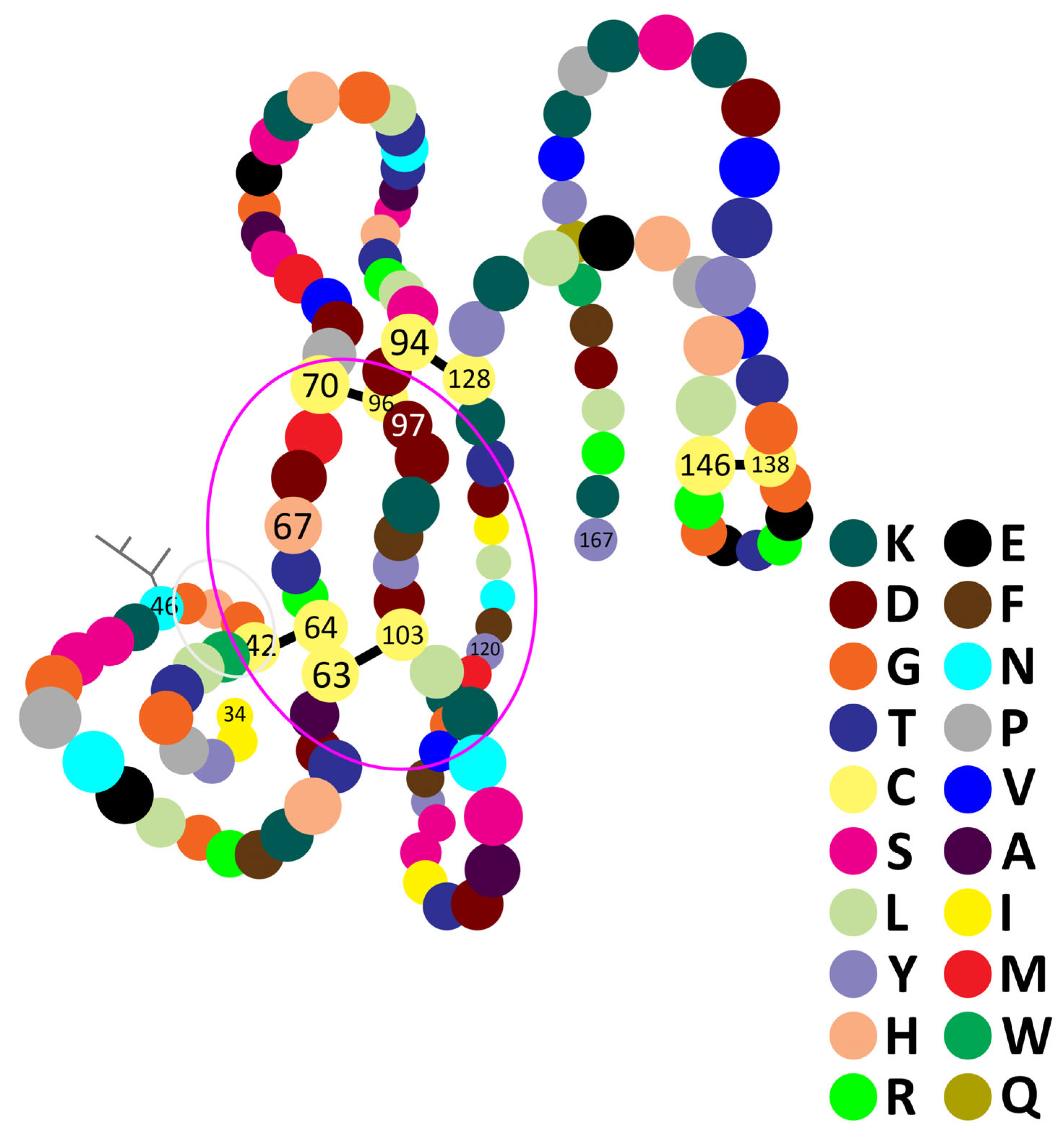
Figure 1. Primary, secondary and tertiary structures of the honeybee venom phospholipase A2 (hvPLA2) imagined based on the numerous structures available in [61]. See the text for the significance of numbers. Black lines, disulfide bonds; branched lines, glycosylation motif; grey circle, Ca2+-binding domain; pink circle, possible area of the catalytic site.
Molecules 30 02623 g001
Figure 2. Venom collection device using the electric stimulation of bees. A.m.c., Apis mellifera carpatica; HBV, honeybee venom under a latex membrane; igcd, impulse generator of the collecting device; Zn e, zinc electrodes. (personal archive A.F.).
Molecules 30 02623 g002
Figure 3. Main effects of the hvPLA2.
Molecules 30 02623 g003
Figure 4. Interaction of the hvPLA2 with a biological membrane.
Molecules 30 02623 g004
Figure 5. Severe ultrastructural alterations of orthodox mitochondria isolated from rat adrenocortical cells, generated in vitro by experimental administration of a high dose of hvPLA2 (personal archive A.F.). cm, condensed mitochondrion; om, orthodox mitochondrion; tc, tubular cristae; vc, vesicular cristae (abnormal) (personal archive A.F.).
Molecules 30 02623 g005
Figure 6. Severe ultrastructural alterations of Sertoli cells in a seminiferous tubule, generated in vivo by experimental administration of a high dose of hvPLA2 in rats. bm, basement membrane; n, nucleus; sI, primary spermatocyte; sII, secondary spermatocyte; Se, Sertoli cell; sg, spermatogonia. (personal archive A.F.).
Molecules 30 02623 g006
Disclaimer/Publisher’s Note: The statements, opinions and data contained in all publications are solely those of the individual author(s) and contributor(s) and not of MDPI and/or the editor(s). MDPI and/or the editor(s) disclaim responsibility for any injury to people or property resulting from any ideas, methods, instructions or products referred to in the content.