Article

Identification of Marker Compounds and In Vitro Toxicity Evaluation of Two Portuguese Asphodelus Leaf Extracts

1
Research Institute for Medicines (iMed.ULisboa), Faculty of Pharmacy, Universidade de Lisboa, 1649-003 Lisbon, Portugal
2
National Reference Laboratory of Antibiotic Resistances and Healthcare-Associated Infections, Department of Infectious Diseases, National Institute of Health Dr. Ricardo Jorge, 1649-016 Lisbon, Portugal
3
MEtRICs/Chemistry Department, Nova School of Science and Technology, Universidade Nova de Lisboa, 2829-516 Caparica, Portugal
*
Author to whom correspondence should be addressed.
Molecules 2023, 28(5), 2372; https://doi.org/10.3390/molecules28052372
Submission received: 31 January 2023 / Revised: 27 February 2023 / Accepted: 1 March 2023 / Published: 4 March 2023
(This article belongs to the Special Issue Discovery of Bioactive Ingredients from Natural Products III)

Abstract

:
The leaves of Asphodelus bento-rainhae subsp. bento-rainhae, an endemic Portuguese species, and Asphodelus macrocarpus subsp. macrocarpus have been used as food, and traditionally as medicine, for treating ulcers, urinary tract, and inflammatory disorders. The present study aims to establish the phytochemical profile of the main secondary metabolites, together with the antimicrobial, antioxidant and toxicity assessments of both Asphodelus leaf 70% ethanol extracts. Phytochemical screenings were conducted by the TLC and LC-UV/DAD-ESI/MS chromatographic technique, and quantification of the leading chemical classes was performed by spectrophotometric methods. Liquid-liquid partitions of crude extracts were obtained using ethyl ether, ethyl acetate, and water. For in vitro evaluations of antimicrobial activity, the broth microdilution method, and for the antioxidant activity, the FRAP and DPPH methods were used. Genotoxicity and cytotoxicity were assessed by Ames and MTT tests, respectively. Twelve known compounds including neochlorogenic acid, chlorogenic acid, caffeic acid, isoorientin, p-coumaric acid, isovitexin, ferulic acid, luteolin, aloe-emodin, diosmetin, chrysophanol, and β-sitosterol were identified as the main marker compounds, and terpenoids and condensed tannins were found to be the major class of secondary metabolites of both medicinal plants. The ethyl ether fractions demonstrated the highest antibacterial activity against all the Gram-positive microorganisms, (MIC value of 62 to 1000 µg/mL), with aloe-emodin as one of the main marker compounds highly active against Staphylococcus epidermidis (MIC value of 0.8 to 1.6 µg/mL). Ethyl acetate fractions exhibited the highest antioxidant activity (IC50 of 800 to 1200 µg/mL, respectively). No cytotoxicity (up to 1000 µg/mL) or genotoxicity/mutagenicity (up to 5 mg/plate, with/without metabolic activation) were detected. The obtained results contribute to the knowledge of the value and safety of the studied species as herbal medicines.

1. Introduction

Medicinal plants have been used as potential functional foods or resources to prevent various diseases worldwide in different traditional medicine systems. Medicinal plants and their respective phytochemicals, mainly secondary metabolites, are used not only to combat specific nutrient deficiencies, but to sustain secure food and primary healthcare medicines [1].
The species Asphodelus L. (Asphodelaceae) is consumed in large quantities in the cuisines (e.g., soups, pastries, etc.) of several countries and cultures. The leaves of Asphodelus aestivus Brot., for instance, are commonly consumed as a cooked vegetable dish in Turkey, where they are known as “çiriş otu” [2]. In Puglia, on the southeast coast of Italy, burrata cheese is always wrapped in Asphodelus ramosus L. leaves to indicate the freshness of the cheese before it dries out [3]. In addition to their nutritional value, Asphodelus spp. leaves are widely used in traditional medicine to treat ulcers and urinary and inflammatory disorders [4]. In North African countries and the Iberian Peninsula, decoctions of leaves and stems have also been used to treat withering and paralysis [5,6]. Previously reported phytochemical studies of Asphodelus spp. extracts from leaves and aerial parts have revealed the presence of phenolic acids [7,8], flavonoids [6,7,8,9,10,11], and anthracene derivatives [8,12,13,14,15,16] as the main chemical classes of their marker secondary metabolites. Several in vitro and in vivo biological activities of Asphodelus spp. leaf and aerial parts extracts have been reported and documented for their antimicrobial [7,15,17,18,19,20], antioxidant [2,19,21,22,23], and antitumoral [7,15,21,24] activity [4].
Asphodelus bento-rainhae subsp. bento-rainhae P. Silva is an endemic species from Serra da Gardunha and is considered as “vulnerable” on the Red List of Threatened Species of the International Union for the Conservation of Nature (IUCN), and co-exists with Asphodelus macrocarpus subsp. macrocarpus Parl. in the same geographical area. They are known by the common Portuguese name “abrotea” (Ancient Greek: Ἀβρότονον), and their leaf is used as fertilizer and fodder in Portugal [25]. To date, no data related to the phytochemical characterization, pre-clinical safety, and biological potential of Asphodelus bento-rainhae and Asphodelus macrocarpus leaves have been found in the literature. Therefore, the present study was conducted to identify the main chemical constituents, antimicrobial and antioxidant activities of leaf extracts of these species along with their in vitro toxicity assessments, using samples collected at different times of the year to determine the most appropriate period for the collection of material and to contribute to the knowledge of safety and their value as herbal medicinal products.

2. Results and Discussion

2.1. Drug-Extract Ratio

The drug−70% ethanol extract ratio for Asphodelus bento-rainhae leaf (AbL) were 4.5: 1 and 4.8: 1 for the first (AbLa) and second (AbLb) collection seasons, respectively. For Asphodelus macrocarpus leaf (AmL), these values were obtained as 1:2.9 for the first (AmLa) and 1:6.3 for the second (AmLb) collection season.

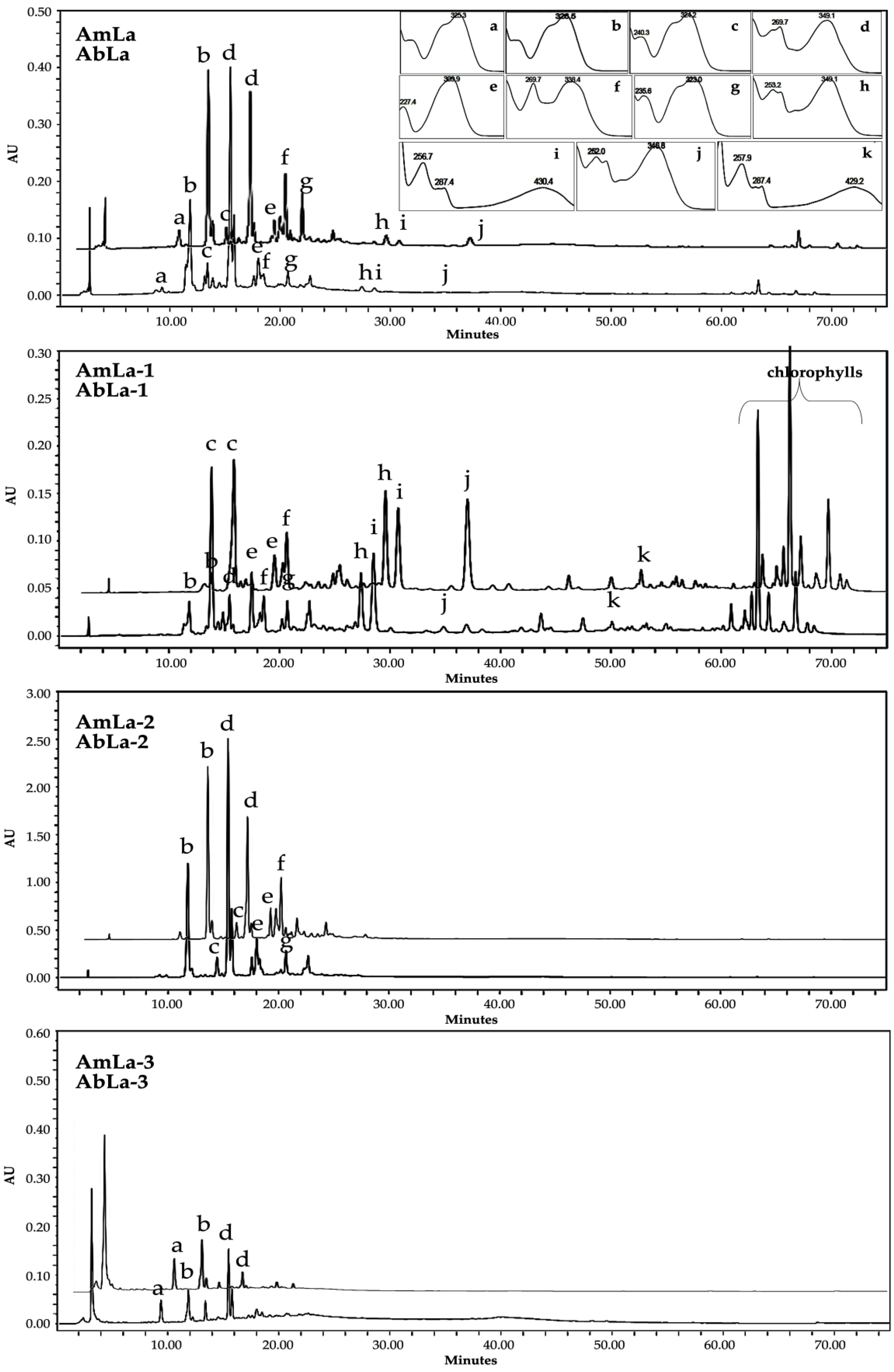
2.2. Phytochemical Analysis

Thin-layer chromatography (TLC), followed by high-performance liquid chromatography (HPLC) coupled to a photodiode detector (UV/DAD), and electrospray ionization spectrometry (ESI/MS) techniques were applied for the rapid and reliable detection of several samples. The obtained chromatographic profiles of Asphodelus bento-rainhae and Asphodelus macrocarpus leaf extracts (AbLa, and AmLa, respectively) and their subsequent liquid-liquid partition with increasing polarity solvents, namely ethyl ether (AbLa-1, AmLa-1), ethyl acetate (AbLa-2, AmLa-2) and water (AbLa-3, AmLa-3), showed qualitative similarity in their chemical composition, characterized by the presence of terpenoids, phenolic acids, flavonoids, and anthracene derivatives. Based on both TLC and HPLC spectral analysis, using the authentic standards (co-chromatography) and comparison with literature data (Figure 1), twelve known compounds, namely, neochlorogenic acid (a), chlorogenic acid (b), caffeic acid (c), isoorientin (d), p-coumaric acid (e), isovitexin (f), ferulic acid (g), luteolin (h), aloe-emodin (i), diosmetin (j), chrysophanol (k), and β-sitosterol (l) were identified as major marker compounds of both species (Table 1, Figure 2).
Previously reported phytochemical studies of Asphodelus spp. revealed the presence of chlorogenic acid in the leaf and aerial part extracts of Asphodelus aestivus Brot. [8] and Asphodelus ramosus L. [26], while caffeic acid was only reported from the flower extract of A. ramosus [27].
Isoorientin from Asphodelus aestivus [8], Asphodelus albus Mill. subsp. delphinensis [10], Asphodelus cerasifer Gay [10,12], Asphodelus microcarpus Salz. et Viv. [6], and Asphodelus ramosus [11], together with isovitexin from Asphodelus aestivus [8] and luteolin from Asphodelus acaulis Desf. [12], Asphodelus albus [10,12], Asphodelus cerasifer [10,12], Asphodelus fistulosus L. [16], Asphodelus macrocarpus Parl. subsp. rubescens [12], Asphodelus microcarpus [7,10], Asphodelus ramosus [10], and Asphodelus tenuifolius Cav. [9] have also been recorded as the most common flavonoids of these species.
Aloe-emodin from A. aestivus [8], A. albus [12,13], A. cerasifer [12], A. fistulosus [14,16], A. macrocarpus subsp. rubescens [12], and A. microcarpus [13,14], as well as chrysophanol from A. albus [12,13], A. fistulosus [14,16], A. macrocarpus subsp. rubescens [12] and A. microcarpus [14,15] have been frequently detected and therefore found to be the most common anthracene derivatives.
β-sitosterol, a common phytosterol, was, however, only found in the root extracts of A. albus, A. microcarpus, and A. tenuifolius [6,28,29,30], and seed extract of A. fistulosus and A. microcarpus [31].
Quantification results of the main chemical classes of secondary metabolites, namely total phenolics (TPC), total flavonoids (TFC), total anthraquinones (TAC), total condensed and hydrolysable tannins (TCTC and THTC, respectively), together with total terpenoids, (TTC) are presented in Table 2.
Concerning the analysis between the different collection seasons for the same species, the results showed that the total content of TCTC and TFC in A. bento-rainhae (p-values: 0.034, 0.01, respectively) and THTC in A. macrocarpus leaf extracts were significantly higher in the first collection season (p-value: 0.01).
The analysis of the results between different species of the same collection season showed that TTC content in the first collection season and TFC content in the second collection season were significantly higher in A. macrocarpus when compared to those of A. bento-rainhae (p-values: 0.0021 and 0.01, respectively). However, TAC, TCTC, and TFC contents in the first collection season (p-value: 0.002, 0.007, and 0.006, respectively) and THTC content in the second collection season (p-value: 0.028) were significantly higher in A. bento-rainhae when compared to those of A. macrocarpus.
The obtained data showed the TCTC (180.96 ± 10.98 and 142.98 ± 6.71 mg CAE/g DW) and TTC (111.72 ± 22.77 and 165.47 ± 26.54 mg OAE/g DW) contents with the highest and TAC (1.07 ± 0.11 and 0.55 ± 0.07 mg RhE/g DW) with the lowest content in comparison to the other quantified chemical classes in both A. bento-rainhae and A. macrocarpus leaf extracts.
Previously reported Asphodelus spp. leaf extracts quantified values of TPC, TFC, and TCTC indicated the important role of solvent selection for the extraction procedure. In fact, A. microcarpus ethanol extract showed a higher amount of TPC and TFC (54.44 ± 13.6 mg GAE/g of DW and 31.13 ± 1.96 mg QUE/g of DW, respectively) in comparison to the aqueous and methanol extracts [21]. However, in A. ramosus, the aqueous extract exhibited a higher amount of TPC (33.51 ± 0.33 mg GAE/g of DW) when compared to the methanol, methanol/water (50%), and ethyl acetate extracts [32]. A. aestivus acetone extract also showed an elevated amount of TFC (17.74 ± 0.46 mg CAE/g of DW) in comparison to the aqueous, ethanol and methanol extracts [2,19]. Contrary to the data mentioned above and that obtained by us, significantly higher amounts of TPC (183.7 ± 3.5, 128.5 ± 2.1 and 109.7 ± 1.5 mg GAE/g of DW) and lower amounts of TCTC (59.8 ± 0.6, 49.2 ± 0.5 and 41.4 ± 0.3 mg CAE/g of DW) were reported from A. tenuifolius methanol, ethanol, and petroleum ether extracts [33]. It was also observed that both TPC and TFC contents have increased with the increase of the extraction temperature in the experiments done with A. ramosus [32].

2.3. Determination of In Vitro Antioxidant Potential

In this study, the antioxidant activity was evaluated by two complementary methods, DPPH assay to determine the 50% inhibition of free radical scavenging activity, and FRAP, which evaluates the reducing potency of the antioxidants to react to the ferric tripyridyltriazine (Fe3+-TPTZ) complex.
Concerning the results shown in Table 3, overall, A. bento-rainhae exhibited stronger antioxidant activity when compared to A. macrocarpus extracts. Among all the tested extracts, ethyl acetate fractions (AbLa-2, AmLa-2) showed the highest antioxidant activity when compared to all the other fractions (IC₅₀: 800 μg/mL and IC₅₀: 1200 μg/mL, respectively). When comparing FRAP and DPPH, the obtained an r value of −0.975, showing a strong correlation between them, validating the results of both techniques, although the data of the FRAP test correlate better with the quantifications data. The classes of compounds that correlate better with the antioxidant power of the extracts are the flavonoids (TFC, r value of 0.943) and phenolic compounds (TPC, r value of 0.949), in which a higher content of these compounds is related to higher antioxidant power. In accordance with these results, phytochemical screenings of the crude extracts and their L-L partitions revealed the presence of homoorientin and chlorogenic acid as the main marker compounds of most active fractions (AbLa-2, AmLa-2).
There is no report on the antioxidant activity of our studied Asphodelus species; however, the previously reported results of DPPH analyses of the other Asphodelus spp. showed that A. microcarpus leaf ethanol and methanol extracts exhibited the highest antioxidant activity (IC50: 55.9 μg/mL and IC50: 98 μg/mL, respectively) [21,23]. On the contrary, A. aestivus leaf methanol extracts noticeably showed a higher antioxidant activity when compared to ethanol extract (IC50: 160 μg/mL and IC50: 9540 μg/mL, respectively) [2,19]. A. tenuifolius leaf methanol extract exhibited the lowest IC50 (18370 μg/mL) levels among the others, including our studied species [22].

2.4. Assessment of the Antibacterial Potential

The in vitro quantitative method of susceptibility testing (determination of MIC values) was used for the evaluation of the antimicrobial potential against both selected Gram-positive and Gram-negative resistant pathogens in this study.
Concerning the obtained results, leaf crude extracts (AbLa, AmLa), and their subsequent ethyl acetate (AbLa-2, AmLa-2) and aqueous (AbLa-3, AmLa-3) L-L partition fractions did not exhibit antimicrobial activity against both Gram-positive and Gram-negative microorganism pathogens at any of the concentrations tested (MIC > 2000 µg/mL). However, as shown in Table 4, only diethyl ether fractions (AbLa-1, AmLa-1) demonstrated considerable antibacterial activity against all the Gram-positive microorganisms, with MIC values ranging from 62 to 1000 µg/mL. In general, A. bento-rainhae exhibited higher activity when compared to A. macrocarpus, and no activity in the tested range of concentrations (MIC > 2000 µg/mL) was found against Gram-negative microorganisms (Escherichia coli, Klebsiella pneumoniae, Pseudomonas aeruginosa, and Acinetobacter baumannii).
Aloe-emodin (compound i, Table 1), identified as one of the main marker compounds of the diethyl ether fraction of both plant extracts, was also tested against the pathogen panel under the study. This compound was found to be highly active against all the Gram-positive strains, particularly against all Staphylococcus epidermidis strains with a MIC between 0.8 to 1.6 µg/mL. In accordance with our results, aloe-emodin was previously reported as a potential antimicrobial that was active against several Gram-positive bacteria [34]; however, in a recent study, aloe-emodin with MIC values of 4 to 32 µg/mL exhibited deformities in the morphology of S. epidermidis cells and the destruction of the selective permeability of the cell membranes [35].
Results of studies involving the determination of the antimicrobial activity of other Asphodelus spp. against a similar pathogen panel revealed their lower antimicrobial potential. For instance, a leaf ethanol extract of A. aestivus exhibited a MIC of 42,000 µg/mL against S. aureus, and of 60,000 µg/mL against Klebsiella pneumoniae [36]. The A. fistulosus leaf ethanolic and aqueous extracts showed activity against S. aureus (MIC 2200 µg/mL and 7600 µg/mL, respectively) [37]. A methanol extract of A. luteus aerial part showed an MIC between 1250 to 2500 µg/mL against methicillin-resistant S. aureus (MRSA) [17]. A methylene-chloride extract of the aerial part of A. tenuifolius was found to be more active against S. aureus (MIC = 1600 µg/mL), Enterococcus faecalis (MIC = 1000 µg/mL), and E. coli (MIC = 1800 µg/mL) in comparison to the n-butanol and ethyl acetate extracts of the same species [9]. Recently, an A. tenuifolius whole plant chloroform extract was shown to be active against S. epidermidis (MIC = 580 µg/mL) [38]. A. microcarpus leaf extracts also showed antimicrobial activity against several Gram-positive strains, with MIC values of 78 to 5000 µg/mL [7,15,17,39,40]. A. bento-rainhae and A. macrocarpus leaf extracts seem to be more active against the tested Gram-positive strains in comparison to the other tested Asphodelus spp. extracts. The antibacterial activity of A. fistulosus leaf aqueous extract against E. coli (MIC = 62 µg/mL) and of A. tenuifolius aerial part methylene-chloride extract against the same microorganism (E. coli, MIC = 1800 µg/mL) and also against P. aeruginosa (MIC = 150 µg/mL) are examples of the few studies relating the antimicrobial activity of Asphodelus spp. to Gram-negative strains.
Overall, the observed antimicrobial activity of both A. bento-rainhae and A. macrocarpus leaf crude extracts were similar to those obtained and reported form the other Asphodelus spp. tested against a similar panel of pathogens. However, the fractionation of crude extracts enabled the detection of significant antimicrobial activity in the diethyl ether L-L partition fractions, quantitatively the richest in 1,8-dihydroxyanthracene derivatives, a known chemical class of secondary metabolites with antimicrobial activity [34].

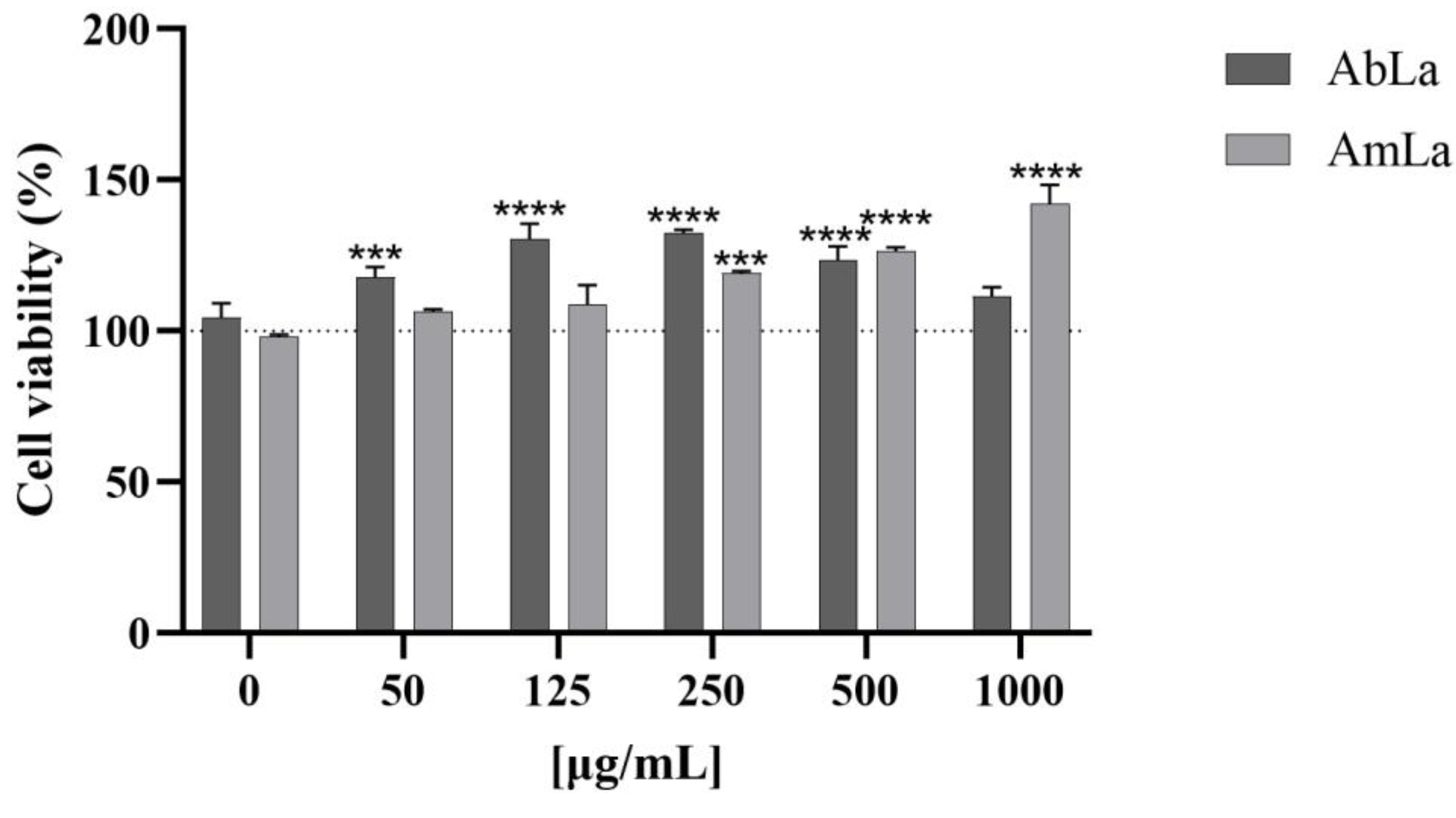
2.5. Pre-Clinical Safety Assessment

Following the guidelines of the genotoxicity by the Ames test, which is commonly used as an initial screen of genotoxicity, for a substance to be considered genotoxic in the test, the number of revertant colonies on the plates containing the test compounds/substance must be more than twice the number of colonies produced on the solvent control plates (i.e., a ratio above 2.0). In addition, a positive dose-response should be evident for the various concentrations of the tested mutagen [41,42]. Since the crude extracts obtained from the first collection season (AbLa, AmLa) exhibited higher contents of the main classes of secondary metabolites, they were subsequently selected for further safety examination.
The obtained results of the Ames test for both AbLa and AmLa extracts are presented in Table 5. Neither extract induced an increase in the number of revertant colonies in any of the tested strains at any tested concentration, with (500, 1250, 2500, and 5000 µg/plate) and without (250, 625, 1250, 2500, 3750, and 5000 µg/plate) metabolic activation, when compared to the negative control. Moreover, cytotoxicity did not occur since there was neither a decrease in the number of spontaneous revertants nor a decrease on the background lawn of the plates at any of the concentrations tested. Therefore, under the conditions of this study, neither extract of the two species showed mutagenic activity.
Our cell viability assay (Figure 3) concurrently indicated that none of the AbLa and AmLa extracts reduced HepG2 viability. The AbLa extract (50–500 µg/mL) enhanced HepG2 viability/proliferation up to ~30% when compared to the 0 µg/mL concentration, whereas the same was observed for AmLa, i.e., it promoted HepG2 viability/proliferation by up to 40%, especially at higher concentrations (250–1000 µg/mL; p < 0.001 and p < 0.0001). Therefore, under the conditions of this study, the extracts of both species did not show mutagenic activity and in vitro cytotoxicity of HepG2, which is crucial to ensure their safety [42,43,44].

3. Materials and Methods

3.1. Chemical and Biological Reagents

Acetone, aluminum chloride, 2-aminoanthracene, 9-aminoacridine hydrochloride monohydrate, ammonium sodium phosphate dibasic tetrahydrate, ascorbic acid, benzo(a)pyrene, chlorogenic acid, chrysophanol, d-(+)-biotin, dimethyl sulfoxide/DMSO, 2,2- diphenyl-1-picrylhydrazyl/DPPH, gallic acid, glucose monohydrate, glucose-6-phosphate, diosmetin, neochlorogenic acid, nicotinamide adenine dinucleotide phosphate (NADP+), 2-nitrofluorene, tert-butyl hydroperoxide/T-BHP, 2,4,6-tris(2-pyridyl)-s-triazine/TPTZ and vanillin were obtained from Sigma-Aldrich (St. Louis, MO, USA). p-Anisaldehyde, ferric chloride hexahydrate, hydrochloric acid, l-histidine monohydrochloride monohydrate, magnesium acetate tetrahydrate, magnesium sulfate heptahydrate, methanol, perchloric acid, potassium iodate, sodium acetate trihydrate, sodium carbonate, sodium hydroxide, and sodium nitrite were purchased from Merck (Darmstadt, Germany). Aloe-emodin, caffeic acid, (+)-catechin, ferulic acid, isoorientin, isovitexin, luteolin, oleanolic acid, p-coumaric acid and rhein were acquired from Extrasynthese (Genay, France). Citric acid monohydrate, di-sodium hydrogen phosphate dihydrate, and sodium dihydrogen phosphate monohydrate were purchased from PanReac AppliChem (Barcelona, Spain). Sodium chloride and di-potassium hydrogen phosphate were from Honeywell Fluka™ (Seelze, Germany). β-sitosterol and 2-aminoethyl diphenylborinate were obtained from Acros organics (Geel, Belgium). Bacto™ agar was acquired from Becton Dickinson & Co (Franklin Lakes, NJ, USA), n-butanol came from Thermo Fisher ScientificTM (Waltham, MA, USA), ethanol (CH3CH2OH) was sourced from Carlo Erba Reagents (Val-de-Reuil, França), ferrous sulfate heptahydrate came from M&B laboratory chemicals (Dagenham, UK), Folin-Ciocalteu was acquired from Biochem chemopharma (Cosne-Cours-sur-Loire, France), glacial acetic acid came from Chem-Lab NV (Zedelgem, Belgium), polyethylene glycol 400/PEG was sourced from VWR Chemicals (Rosny-sous-Bois, France), sulfuric acid (H₂SO₄) was acquired from PanReac AppliChem (Barcelona, Spain), sodium azide came from J.T. Baker Chemical Company (Phillipsburg, NJ, USA) and nutrient broth (NB) Nº 2 was sourced from Oxoid (Basingstoke, UK). Aroclor 1254-induced rat liver S9 was purchased from Trinova Biochem (GmbH, Giessen, Germany). In preparing all solutions, dilutions, and culture media, ultra-pure water from a Milli-Q water purification system, Millipore (Molsheim, France), was used.

3.2. Plant Materials

The leaves of A. bento-rainhae (AbL) and A. macrocarpus (AmL) were collected from Serra da Gardunha, Portugal, first at the early flowering stage (AbLa, AmLa) in Spring, and then for the second time (AbLb, AmLb), during the Summer of 2019. All samples were dried in a well-ventilated dark space at room temperature. Corresponding voucher specimens were deposited in the Laboratory of Pharmacognosy, Department of Pharmacy, Pharmacology and Health Technologies, Faculty of Pharmacy, Universidade de Lisboa (Voucher specimens’ number: OSilva_201901- A. bento rainhae and OSilva_201902- A. macrocarpus).

3.3. Preparation of Extract

Powder of the dried samples was obtained by grinding, and extraction was performed using the maceration method (with a mixture of ethanol/water 70:30) under agitation and filtration (3×, 24 h each). Hydroethanolic extracts were evaporated under reduced pressure at a temperature of less than 40 °C using a rotary evaporator and subsequently freeze-dried. The selected extracts (AbLa, AmLa) were then submitted to liquid-liquid partitioning (L-L), generating the diethyl ether (AbLa-1, AmLa-1), ethyl acetate (AbLa-2, AmLa-2), and aqueous (AbLa-3, AmLa-3) fractions.

3.4. Chromatographic Conditions

Silica gel 60 F254 and 60 RP-18 F254 pre-coated plates (Merck®, Darmstadt, Germany) were used for TLC screenings. Different spray reagents, including anisaldehyde–sulfuric acid for the detection of terpenoids, natural product polyethylene glycol reagent (NP/PEG = NEU) for the detection of phenolics, and potassium hydroxide (KOH) 5% ethanolic solution for the detection of anthracene derivatives [45] were used.
A HPLC-UV/DAD analysis was performed using a Waters Alliance 2690 Separations Module (Waters Corporation, Milford, MA, USA) coupled with a Waters 996 photodiode array detector (UV/DAD) (Waters Corporation, MA, USA). An Atlantis T3 column, RP-18 end-capped (5 µm, 150 × 4.6 mm), connected to a pre-column with the same stationary phase was used. The injection volume was 25 µL with a flow rate of 1 mL/min. A mixture of water + 0.1% formic acid (solvent A) and acetonitrile (solvent B) was used as the mobile phase, and gradients (95% A and 5% B), 20 min (71% A and 29% B), 30 min (67% A and 33% B), 35 min (64% A and 36% B), 45 min (50% A and 50% B), 65 min (100% B) and 75 min (95% A and 5% B) were applied. Crude extracts (20 mg/mL) were solubilized in water and standard solutions were prepared in acetonitrile (1 mg/mL) and filtered through a polytetrafluoroethylene syringe filter (0.2 µm). Data were collected and analyzed using a Waters Millennium® 32 Chromatography Manager (Waters Corporation, Milford, MA, USA). The chromatogram was monitored and registered on Maxplot wavelength (240–650 nm).
An HPLC-MS/ESI analysis was carried out using an HPLC (Waters Alliance 2695), with an autosampler and photodiode array detector (Waters PDA 2996) in tandem with a triple quadrupole mass spectrometer (Micromass® Quatro MicroTM API, Waters®, Drinagh, Ireland) using an electrospray ionization source (ESI) operating in negative mode. A LiChrospher 100 RP-18 (5 µm) 250 × 4 mm column with respective pre-column (Merck, Darmstadt, Germany) was used. A mixture of water + 0.1% formic acid (solvent A) and acetonitrile (solvent B) was used as the mobile phase. Data were acquired and analyzed using MassLynx™ V4.1 software (Waters®, Drinagh, Ireland).
Peaks assignment and the identification of compounds were based on a co-chromatography technique with the comparison of retention times, UV-DAD, and mass spectral data with those of standards and published data.

3.5. Quantification Assays for Determination of the Main Classes of Secondary Metabolites

Total phenolic content (TPC) of the crude extracts was determined using the Folin-Ciocalteu method [46], and an increasing gallic acid calibration curve (10–70 µg/mL) was used to obtain the standard equation of Y = 0.0087X + 0.0264, R2 = 0.994. Total flavonoid content (TFC) was obtained following the method by Olivera et al., 2008 [47], and catechin concentrations (50–200 µg/mL) were used to obtain a standard curve with an equation of Y = 0.0039X + 0.027, R2 = 0.993. Total triterpenoid content (TTC) was assessed using the procedure developed by Chang & Lin, 2012 [48], and oleanolic acid concentrations (100–800 µg/mL in methanol) were used to obtain a standard curve with an equation of Y = 0.0012X + 0.0849, R2 = 0.994. For the determination of the total condensed tannins (TCTC) [46], catechin concentrations (200–2000 µg/mL) were used to obtain a standard curve with an equation of Y = 0.0002X + 0.0324, R2 = 0.981 and for quantification of total hydrolysable tannins (THTC) [49], gallic acid concentrations (100–600 µg/mL) was used to obtain a standard curve with an equation of Y = 0.001X + 0.054, R2 = 0.977. Total anthraquinones content (TAC) was evaluated according to the method described by Sakulpanich & Gritsanapan, 2008 [50], and rhein concentrations (3–18 µg/mL) were used to obtain a standard curve with an equation of Y = 0.0215X−0.0016, R2 = 0.998.
All of the above-mentioned colorimetric techniques were assessed in triplicate for method validation, and a UV-Vis spectrophotometer (Hitachi, U–2000, Tokyo, Japan) was used. Values were obtained using standard equations (where X was the concentration of standard equivalents expressed as milligrams per gram of dried extract and Y was the measured absorbance). All of the obtained data were treated statistically by a one-way analysis of variance (ANOVA) with the Asphodelus species as the source of variance. Once both of the Asphodelus species were collected in two different seasons, the obtained data were also analyzed by ANOVA, with the season as the source of variance. The significant value was set for a p-value < 0.05.

3.6. In Vitro Antimicrobial Activity

The antibacterial assay was carried out by the broth microdilution method [51] in 96-well tissue culture plates (VWR®, Radnor, PA, USA) to determine the activities by testing minimum inhibitory concentrations (MIC) of extracts against twelve reference (ATCC, LGC Standards S.L.U., Barcelona, Spain) and clinical strains (INSA clinical strains collection) of both Gram-positive (Staphylococcus aureus, S. epidermidis, S. saprophyticus, S. haemolyticus) (Table 6) and Gram-negative (Escherichia coli, Klebsiella pneumoniae, Pseudomonas aeruginosa, Acinetobacter baumannii) bacteria representing the antimicrobial resistance status. Samples to be tested were initially prepared in water or DMSO 10% and were screened at the concentration of 2–2000 μg/mL for crude extracts or L-L partitions and 0.2–200 for pure compounds. Serial dilutions were performed in a Mueller-Hinton medium and were distributed (50 μL) in each of the microplate wells using a microplate liquid handler (PrecisionTM BioTek, Winooski, VT, USA).
For the preparation of inoculum from a pure bacterial culture on agar, a suspension in Mueller-Hinton medium (108 CFU/mL) with a turbidity of 0.5 for Gram-negative and 0.25 for Gram-positive bacteria on the McFarland scale (Grant Bio™ DEN-1B, Cambridgeshire, UK) were prepared and stored at 4 °C until use. For MIC determination, the prepared suspensions were diluted at a ratio of 1:10, and from this dilution, 50 µL was added to all the wells. Two controls were included for each extract, fraction or compound, one plate in the absence of the extract solution and the other in the presence of the solvent (DMSO), to verify the absence of contamination and to check the validity of the inoculum. After incubation at 37 °C for 18 h, the plates were read in a lighted place, and the MIC was determined. All experiments were carried out in triplicate, as previously described, to obtain consistent values.

3.7. In Vitro Antioxidant Activity

The antioxidant potential was determined by two methods, initially started by a modified free radical scavenging activity (DPPH method) [52], followed by the ferric reducing antioxidant power test (FRAP assay). DPPH solution (3.9 mL, 6 × 10⁻⁵ M in methanol) was mixed with 100 µL of diluted extracts or standard (ascorbic acid). After 30 min of incubation at room temperature, the absorbance of samples and standard solutions was measured at 517 nm. The percentage of DPPH free radical scavenging activity was calculated using the following formula: % scavenging = [absorbance of control−absorbance of test sample/absorbance of control] × 100. Results were expressed as mean ± standard deviation and presented in inhibitory concentration (IC50 value), representing the sample concentration required to scavenge 50% of the DPPH free radicals.
For the Frap assay [53], 100 µL of plant extracts (1000–5000 μg) were mixed with 3 mL of working FRAP reagent (300 mM acetate buffer pH 3.6, 10 mM TPTZ in 40 mM HCl and 20 mM FeCl3. 6H2O in the ratio of 10:1:1 at the time of use); thereafter, samples were placed in the water bath at 37 °C. The reduction of ferric tripyridyl triazine (Fe III TPTZ) complex to ferrous form (which has an intense blue color) can be monitored by measuring the change in absorption at 593 nm, measured after 4 min. Ascorbic acid concentrations (25–175 µg/mL) were used to obtain a standard curve with an equation of Y = 0.616X−1.1702, R2 = 0.9989. The FRAP reagent was used as a blank, and results were expressed as mmol ascorbic acid/g dry extract. Values were obtained in three sets of experiments and assessed in triplicate for method validation.
To ascertain if both methods were equally valid in measuring the antioxidant activity, they were correlated through the Pearson coefficient index (−1 < r < 1). A Pearson coefficient absolute value higher than 0.9 shows a strong correlation between the two methods. The Pearson index was also used to correlate the data of antioxidant activity with the quantification of the several chemical classes of compounds to ascertain their relationship with antioxidant power. Once both Asphodelus species were collected in two different seasons, the obtained data were also analyzed by ANOVA, with the season as the source of variance. The significance value was set for a p-value < 0.05.

3.8. In Vitro Genotoxicity/Mutagenicity Evaluation by Ames Test

A bacterial reverse mutation test (Ames test), commonly employed as an initial screening of the genotoxicity potential of herbal substances/preparations, was used to detect relevant genetic changes and genotoxic carcinogens [54]. The assessment of mutagenicity was performed according to the OECD No. 471 [55], the ICH S2 (R1) [56] guidelines, and following the published protocols [44], using five Salmonella enterica serovar Typhimurium tester strains (TA98, TA100, TA102, TA1535, and TA1537) in a direct plate incorporation method with and without metabolic activation. TA100, TA98, TA102 and TA1535 were kindly provided by the Genetic Department of the Nova Medical School of the Universidade NOVA de Lisboa (Portugal), having received them from Professor B.N. Ames (Berkeley, CA, USA). TA1537 was from ATCC, NUMBER: 29630™, LOT: 7405375. The strains were inoculated in nutrient broth medium and incubated for 12–16 h, at 37 °C in the dark, shaking at 210 rpm in an orbital incubator, and kept at 4 °C until use.
S9 mix (10%, v/v rat liver S9, 0.4 M MgCl2, 1.65 M KCl, 1 M glucose-6-phosphate, 0.1 M nicotinamide adenine dinucleotide phosphate, and 0.2 M sodium phosphate buffer, pH 7.4) was freshly prepared and kept on ice during the experiment.
The extracts (25 mg/mL) were dissolved in DMSO (up to 30%), which also served as the negative control. An amount of 200 µL of extract dilutions were mixed with 500 µL sodium phosphate buffer (0.1 M, pH 7.4) (assay without metabolic activation) or S9 mix (assay with metabolic activation), 100 µL of the bacterial culture, and 2 mL of melted top-agar, supplemented with 0.05 mM biotin and histidine, at 45 °C. This mixture was then vortexed and plated on Petri dishes with Vogel-Bonner agar medium and supplemented with 2% glucose. After a 48-h incubation at 37 °C, manual counting of His+ revertant colonies for each concentration was performed. All assays were performed in triplicate. The results were expressed as the mean number of revertant colonies with the standard deviation (mean ± SD). The positive controls were sodium azide (SA, 1.5 µg/plate for TA100 and TA1535), 2-nitrofluorene (2-NF, 5 µg/plate for TA98), 9-aminoacridine (9-AA, 100 µg/plate for TA1537), and tert-butyl hydroperoxide (tBHP, 50 µg/plate for TA102) in the assay without metabolic activation, and 2-aminoathracene (2-AA, 2 µg/plate for TA98 and 10 µg/plate for TA102, TA1535 and TA1537) and benzo(a)pyrene (BaP, 5 µg/plate for TA100) in the assay with metabolic activation.

3.9. In Vitro Cytotoxicity Evaluation by MTT Assay

Cytotoxicity was evaluated by the methylthiazolyldiphenyl-tetrazolium bromide (MTT) reduction assay [57] on a human liver cell line HepG2 (ATCC Cat. No. HB-8065, Middlesex, UK). HepG2 were seeded in 96-well plates at a density of 8.5 × 10⁴ cells/cm2 in α-MEM (Sigma-Aldrich®, St. Louis, MO, USA) with 1 mM sodium pyruvate (PAN Biotech, Aidenbach, Germany) and 1% non-essential amino acids (NEAA, PAN Biotech, Aidenbach, Germany) supplemented with 10% fetal bovine serum (FBS, Gibco®- Thermo Fisher ScientificTM (Waltham, MA, USA), in a humidified chamber at 37 °C in a 5% CO2 atmosphere. After 48-h incubation, the cell culture medium was replaced by fresh medium with AbLa and AmLa extracts (9:1) at final concentrations of 50, 125, 250, 500, and 1000 µg/mL. Cells were also incubated with a complete cell culture medium, DMSO 1% and DMSO 20% in α-MEM as a positive, solvent, and negative control, respectively. After 48 h, the cells were carefully washed with 100 μL PBS, and 200 μL 0.5 mg/mL MTT (Sigma- Aldrich®) in a cell culture medium was added. HepG2 were incubated for 3 h in a humidified chamber at 37 °C in a 5% CO2 atmosphere. The purple crystals were solubilized with 200 μL DMSO and measured at 570 nm using a microplate spectrophotometer (SPECTROstar Omega; BMG LabTech, Ortengerg, Germany). The results were expressed as a percentage relative to the solvent control. Four wells were used for each sample, and at least two independent experiments were performed.
Data analysis and graphs were plotted using GraphPad Prism® software (version 9.0.0.121, GraphPad Software, San Diego, CA, USA). Results are presented as mean ± standard deviation. p < 0.05 was considered significant.

4. Conclusions

The weak antimicrobial activity verified with our crude leaf extracts of Asphodelus bento-rainhae and Asphodelus macrocarpus is consistent with the results obtained when testing other Asphodelus spp. against a similar panel of pathogens [4]. However, fractionation of these extracts enabled the detection of significant antimicrobial activity in the diethyl ether L-L partition fractions, quantitatively the richest in 1,8-dihydroxyanthracene derivatives, a known chemical class of secondary metabolites with antimicrobial activity [34]. Furthermore, the well-known antibacterial agent aloe-emodin was identified as the main compound responsible for this activity. Although the in vitro cytotoxicity and mutagenicity of this compound has been reported by others, no cytotoxicity or mutagenic activity was observed in the corresponding extracts and fractions that we tested.
On the other hand, the ethyl acetate L-L partition fractions are quantitatively the richest in phenolic acids and flavonoid derivatives, and showed the highest antioxidant activity, confirming the major role of the different classes of the identified phenolic compounds in the activity of Asphodelus bento-rainhae and Asphodelus macrocarpus leaves as medicinal plants. Moreover, the negative results of the Ames and MTT tests indicate that the hydroethanolic leaf extracts of both species are safe in terms of toxicity, and these data together with the phytochemical profiles will provide appropriate information for inclusion in the future quality monograph of these medicinal plants.

Author Contributions

Conceptualization, M.M. and O.S.; Data curation, M.M., K.L., S.P.C., M.P.D., J.P.M. and O.S.; Formal analysis, M.M., K.L., S.P.C. and M.P.D.; Funding acquisition, M.P.D., B.S.L., M.C. and O.S.; Investigation, M.M., K.L., S.P.C., V.M., M.P.D., J.P.M., M.C. and O.S.; Methodology, M.M., K.L., S.P.C., V.M., M.P.D., J.P.M. and O.S.; Project administration, B.S.L., M.C. and O.S.; Resources, O.S.; Software, M.M., K.L., S.P.C. and J.P.M.; Supervision, B.S.L., M.C. and Olga Silva; Validation, M.M., K.L. and S.P.C.; Writing, original draft, M.M., K.L. and S.P.C.; Writing, review & editing, M.M., K.L., S.P.C., V.M., M.P.D., J.P.M., R.S., I.M.d.S., B.S.L., M.C. and O.S. All authors have read and agreed to the published version of the manuscript.

Funding

This research was funded by the Foundation for Science and Technology (FCT, Portugal) through national funds FCT/MCTES to iMed.ULisboa (UIDP/04138/2020, UIDB/04138/2020) and MEtRICs (UIDP/04077/2020, UIDB/04077/2020) research projects, as well as doctoral scholarship (SFRH/BD/125310/2016) granted to the first author.

Institutional Review Board Statement

Not applicable.

Informed Consent Statement

Not applicable.

Data Availability Statement

Not applicable.

Acknowledgments

We would like to thank Michael Kranendonk and his team from the Centre for Toxicogenomics and Human Health (ToxOmics), NOVA Medical School, Universidade NOVA de Lisboa, for their kind availability and scientific contributions, and Eugénia Ferreira and Paula Nobre for their technical support throughout the laboratory experiments.

Conflicts of Interest

The authors declare that they have no conflicts of interest.

Sample Availability

Samples of the compounds are available from the first and correspondent authors.

References

  1. Sivakumar, D.; Chen, L.; Sultanbawa, Y. A Comprehensive Review on Beneficial Dietary Phytochemicals in Common Traditional Southern African Leafy Vegetables. Food Sci. Nutr. 2018, 6, 714–727. [Google Scholar] [CrossRef] [PubMed] [Green Version]
  2. Peksel, A.; Imamoglu, S.; Altas Kiymaz, N.; Orhan, N. Antioxidant and Radical Scavenging Activities of Asphodelus Aestivus Brot. Extracts. Int. J. Food Prop. 2013, 16, 1339–1350. [Google Scholar] [CrossRef]
  3. Paura, B.; Di Marzio, P. Making a Virtue of Necessity: The Use of Wild Edible Plant Species (Also Toxic) in Bread Making in Times of Famine According to Giovanni Targioni Tozzetti (1766). Biology 2022, 11, 285. [Google Scholar] [CrossRef] [PubMed]
  4. Malmir, M.; Serrano, R.; Caniça, M.; Silva-Lima, B.; Silva, O. A Comprehensive Review on the Medicinal Plants from the Genus Asphodelus. Plants 2018, 7, 20. [Google Scholar] [CrossRef] [Green Version]
  5. Díaz Linfante, Z. Asphodelus L. In Flora Iberica; Talavera, S., Andrés, C., Arista, M., Piedra, M.P.F., Rico, E., Crespo, M.B., Quintanar, A., Herrero, A., Aedo, C., Eds.; Real Jardin Botänico, Consejo Superior de Investigaciones Científicas C.S.I.C.: Madrid, Spain, 2013; ISBN 276-308-152. [Google Scholar]
  6. Hammouda, F.M.; Rizk, A.M.; Ghaleb, H.; Abdel-Gawad, M.M. Chemical and Pharmacological Studies of Asphodelus microcarpus. Planta Med. 1972, 22, 188–195. [Google Scholar] [CrossRef]
  7. Di Petrillo, A.; Fais, A.; Pintus, F.; Santos-Buelga, C.; González-Paramás, A.M.; Piras, V.; Orrù, G.; Mameli, A.; Tramontano, E.; Frau, A. Broad-Range Potential of Asphodelus microcarpus Leaves Extract for Drug Development. BMC Microbiol. 2017, 17, 159. [Google Scholar] [CrossRef] [Green Version]
  8. Çalış, I.; Birincioǧlu, S.S.; Kırmızıbekmez, H.; Pfeiffer, B.; Heilmann, J. Secondary Metabolites from Asphodelus aestivus. Z. Für Naturforsch. B 2006, 61, 1304–1310. [Google Scholar] [CrossRef]
  9. Faidi, K.; Hammami, S.; Ben Salem, A.; El Mokni, R.; Garrab, M.; Mastouri, M.; Gorcii, M.; Trabelsi Ayedi, M.; Taglialatela-Scafati, O.; Mighri, Z. Polyphenol Derivatives from Bioactive Butanol Phase of the Tunisian Narrow-Leaved Asphodel (Asphodelus Tenuifolius Cav., Asphodelaceae). J. Med. Plants Res. 2014, 8, 550–557. [Google Scholar] [CrossRef] [Green Version]
  10. Raynaud Par, J.; Abdel-Gawad, M.M. Contribution à l’étude Chimiotaxinomique Du Genre Asphodelus (Liliaceae); Lyon Linnéenne Society: Lyon, France, 1974. [Google Scholar]
  11. Reynaud, J.; Flament, M.M.; Lussignol, M.; Becchi, M. Flavonoid Content of Asphodelus ramosus (Liliaceae). Can. J. Bot. 1997, 75, 2105–2107. [Google Scholar] [CrossRef]
  12. Williams, C.A. Biosystematics of the Monocotyledoneae—Flavonoid Patterns in Leaves of the Liliaceae. Biochem. Syst. Ecol. 1975, 3, 229–244. [Google Scholar] [CrossRef]
  13. Van Rheede van Oudtshoorn, M.C.B. Chemotaxonomic Investigations in Asphodeleae and Aloineae (Liliaceae). Phytochemistry 1964, 3, 383–390. [Google Scholar] [CrossRef]
  14. Hammouda, F.M.; Rizk, A.M.; El-Nasr, M.M.S.; Asr, E.-N. Anthraquinones of Certain Egyptian Asphodelus Species. Z. Für Naturforsch. C 1974, 29, 351–354. [Google Scholar] [CrossRef] [Green Version]
  15. El-Ghaly, E.-S. Phytochemical and Biological Activities of Asphodelus Microcarpus Leaves. J. Pharmacogn. Phytochem. 2017, 6, 259–264. [Google Scholar]
  16. Abd El-Fattah, H. Chemistry of Asphodelus fistulosus. Int. J. Pharmacogn. 1997, 35, 274–277. [Google Scholar] [CrossRef]
  17. Al-Kayali, R.; Kitaz, A.; Haroun, M. Antibacterial Activity of Asphodelin lutea and Asphodelus microcarpus Against Methicillin Resistant Staphylococcus aureus Isolates. Int. J. Pharmacogn. Phytochem. Res. 2016, 8, 1964–1968. [Google Scholar]
  18. Vaghasiya, Y.; Chanda, S.V. Screening of Methanol and Acetone Extracts of Fourteen Indian Medicinal Plants for Antimicrobial Activity. Turk. J. Biol. 2007, 31, 243–248. [Google Scholar]
  19. Peksel, A. Evaluation of Antioxidant and Antifungal Potential of Asphodelus aestivus Brot. Growing in Turkey. J. Med. Plants Res. 2012, 6, 253–265. [Google Scholar] [CrossRef]
  20. Ali-Shtayeh, M.S.; Abu Ghdeib, S.I. Antifungal Activity of Plant Extracts against Dermatophytes. Mycoses 1999, 42, 665–672. [Google Scholar] [CrossRef]
  21. Di Petrillo, A.; González-Paramás, A.M.; Era, B.; Medda, R.; Pintus, F.; Santos-Buelga, C.; Fais, A. Tyrosinase Inhibition and Antioxidant Properties of Asphodelus microcarpus Extracts. BMC Complement. Altern. Med. 2016, 16, 453. [Google Scholar] [CrossRef] [Green Version]
  22. Al-Laith, A.A.; Alkhuzai, J.; Freije, A. Assessment of Antioxidant Activities of Three Wild Medicinal Plants from Bahrain. Arab. J. Chem. 2019, 12, 2365–2371. [Google Scholar] [CrossRef] [Green Version]
  23. Mayouf, N.; Charef, N.; Saoudi, S.; Baghiani, A.; Khennouf, S.; Arrar, L. Antioxidant and Anti-Inflammatory Effect of Asphodelus microcarpus Methanolic Extracts. J. Ethnopharmacol. 2019, 239, 111914. [Google Scholar] [CrossRef] [PubMed]
  24. Al Groshi, A.; Nahar, L.; Andrew, E.; Auzi, A.; Sarker, S.D.; Ismail, F.M.D. Cytotoxicity of Asphodelus aestivus against Two Human Cancer Cell Lines. Nat. Prod. Chem. Res. 2017, 5, 61. [Google Scholar]
  25. Malmir, M.; Serrano, R.; Lima, K.; Duarte, M.P.; Moreira da Silva, I.; Silva Lima, B.; Caniça, M.; Silva, O. Monographic Quality Parameters and Genotoxicity Assessment of Asphodelus Bento-Rainhae and Asphodelus Macrocarpus Root Tubers as Herbal Medicines. Plants 2022, 11, 3173. [Google Scholar] [CrossRef] [PubMed]
  26. Kitaz, A. Comparison of the Total Phenol, Flavonoid Contents and Antioxidant Activity of Methanolic Roots Extracts of Asphodelus microcarpus and Asphodeline lutea Growing in Syria. Int. J. Pharmacogn. Phytochem. Res. 2017, 9, 159–164. [Google Scholar] [CrossRef] [Green Version]
  27. Chimona, C.; Karioti, A.; Skaltsa, H.; Rhizopoulou, S. Occurrence of Secondary Metabolites in Tepals of Asphodelus Ramosus L. Plant Biosyst.-Int. J. Deal. All Asp. Plant Biol. 2013, 148, 31–34. [Google Scholar] [CrossRef] [Green Version]
  28. Abdel-Gawad, M.M.; Hasan, A.; Raynaud Par, J. Estude de l’insaponifiable et Des Acides Gras Des Tuberculus d’ Asphodelus albus. Fitoterapia 1976, 47, 111–112. [Google Scholar]
  29. Rizk, A.M.; Hammouda, F.M. Phytochemical Studies of Asphodelus microcarpus (Lipids and Carbohydrates). Planta Med. 1970, 18, 168–172. [Google Scholar] [CrossRef]
  30. Abdel-Mogib, M.; Basaif, S. Two New Naphthalene and Anthraquinone Derivatives from Asphodelus tenuifolius. Pharmazie 2002, 57, 286–287. [Google Scholar]
  31. Fell, K.R.; Hammouda, F.M.; Rizk, A.M. The Constituents of the Seeds of Asphodelus microcarpus Viviani and A. Fistulosus L. J. Pharm. Pharmacol. 1968, 20, 646–649. [Google Scholar] [CrossRef]
  32. Apaydin, E.; Arabaci, G. Antioxidant Capacity and Phenolic Compounds with HPLC of Asphodelus Ramosus and Comparison of the Results with Allium Cepa L. and Allium Porrum L. Extracts. Turk. J. Agric. Nat. Sci. 2017, 4, 499–505. [Google Scholar]
  33. Eddine, L.S.; Segni, L.R.O. In Vitro Assays of the Antibacterial and Antioxidant Properties of Extracts from Asphodelus tenuifolius Cav and Its Main Constituents: A Comparative Study. Int. J. Pharm. Clin. Res. 2015, 7, 119–125. [Google Scholar]
  34. Malmir, M.; Serrano, R.; Silva, O. Anthraquinones as Potential Antimicrobial Agents—A Review. In Antimicrobial Research: Novel Bioknowledge and Educational Programs; Mendez-Vilas, A., Ed.; Formatex: Badajoz, Spain, 2017; pp. 55–61. [Google Scholar]
  35. Li, T.; Lu, Y.; Zhang, H.; Wang, L.; Beier, R.C.; Jin, Y.; Wang, W.; Li, H.; Hou, X. Antibacterial Activity and Membrane-Targeting Mechanism of Aloe-Emodin Against Staphylococcus epidermidis. Front. Microbiol. 2021, 12, 621866. [Google Scholar] [CrossRef]
  36. Oskay, M.; Aktaş, K.; Sari, D.; Azeri, C. A Comparative Study of Antimicrobial Activity Using Well and Disk Diffusion Method on Asphodelus aestivus (Liliaceae). Ekoloji 2007, 16, 62–65. [Google Scholar]
  37. Al-Qudah, M.M.A. Antibacterial Effect of Asphodelus Fistulosus Aqueous and Ethanolic Crude Extracts on Gram Positive and Gram-Negative Bacteria. Braz. J. Biol. 2022, 84, 1–9. [Google Scholar] [CrossRef]
  38. Khalfaoui, A.; Noumi, E.; Belaabed, S.; Aouadi, K.; Lamjed, B.; Adnan, M.; Defant, A.; Kadri, A.; Snoussi, M.; Khan, M.A.; et al. LC-ESI/MS-Phytochemical Profiling with Antioxidant, Antibacterial, Antifungal, Antiviral and In Silico Pharmacological Properties of Algerian Asphodelus tenuifolius (Cav.) Organic Extracts. Antioxidants 2021, 10, 628. [Google Scholar] [CrossRef]
  39. Alhage, J.; Elbitar, H. In Vitro Screening for Antioxidant and Antimicrobial Properties of Three Lebanese Medicinal Plants Crude Extracts. Pharmacogn. Res. 2019, 11, 127. [Google Scholar] [CrossRef]
  40. Abuhamdah, S. Phytochemical Investigations and Antibacterial Activity of Selected Medicinal Plants from Jordan. Eur. J. Med. Plants 2013, 3, 394–404. [Google Scholar] [CrossRef]
  41. Shin, K.Y.; Won, B.Y.; Ha, H.J.; Yun, Y.S.; Lee, H.G. Genotoxicity Studies on the Root Extract of Polygala Tenuifolia Willdenow. Regul. Toxicol. Pharmacol. 2015, 71, 365–370. [Google Scholar] [CrossRef]
  42. Kelber, O.; Wegener, T.; Steinhoff, B.; Staiger, C.; Wiesner, J.; Knöss, W.; Kraft, K. Assessment of Genotoxicity of Herbal Medicinal Products: Application of the “Bracketing and Matrixing” Concept Using the Example of Valerianae Radix (Valerian Root). Phytomedicine 2014, 21, 1124–1129. [Google Scholar] [CrossRef] [Green Version]
  43. Mortelmans, K.; Zeiger, E. The Ames Salmonella/Microsome Mutagenicity Assay. Mutat. Res.-Fundam. Mol. Mech. Mutagen. 2000, 455, 29–60. [Google Scholar] [CrossRef]
  44. Maron, D.M.; Ames, B.N. Revised Methods for the Salmonella Mutagenicity Test. Mutat. Res. Environ. Mutagen. Relat. Subj. 1983, 113, 173–215. [Google Scholar] [CrossRef] [PubMed]
  45. Wagner, H.; Bladt, S. Plant Drug Analysis: A Thin Layer Chromatography Atlas, 2nd ed.; Springer: Berlin/Heidelberg, Germany, 1996. [Google Scholar]
  46. Scalbert, A.; Monties, B.; Janin, G. Tannins in Wood: Comparison of Different Estimation Methods. J. Agric. Food Chem. 1989, 37, 1324–1329. [Google Scholar] [CrossRef]
  47. Olivera, D.F.; Viña, S.Z.; Marani, C.M.; Ferreyra, R.M.; Mugridge, A.; Chaves, A.R.; Mascheroni, R.H. Effect of Blanching on the Quality of Brussels Sprouts (Brassica oleracea L. Gemmifera DC) after Frozen Storage. J. Food Eng. 2008, 84, 148–155. [Google Scholar] [CrossRef]
  48. Chang, C.L.; Lin, C.S. Phytochemical Composition, Antioxidant Activity, and Neuroprotective Effect of Terminalia Chebula retzius Extracts. Evid.-Based Complement. Altern. Med. 2012, 125247. [Google Scholar] [CrossRef] [Green Version]
  49. Wilfred Vermerris, R.N. Phenolic Compound Biochemistry; Springer: Dordrecht, The Netherlands, 2006; ISBN 978-1-4020-5163-0. [Google Scholar]
  50. Sakulpanich, A.; Gritsanapan, W. Extraction Method for High Content of Anthraquinones from Cassia Fistula Pods. J. Health Res. 2008, 22, 167–172. [Google Scholar]
  51. CLSI. Methods for Dilution Antimicrobial Susceptibility Tests for Bacteria That Grow Aerobically, Approved Standard, 9th ed.; CLSI document M07-A9; CLSI: Wayne, PA, USA, 2012; Volume 32, ISBN 1-56238-783-9. [Google Scholar]
  52. Brand-Williams, W.; Cuvelier, M.E.; Berset, C. Use of a Free Radical Method to Evaluate Antioxidant Activity. LWT—Food Sci. Technol. 1995, 28, 25–30. [Google Scholar] [CrossRef]
  53. Benzie, I.F.F.; Strain, J.J. The Ferric Reducing Ability of Plasma (FRAP) as a Measure of “Antioxidant Power”: The FRAP Assay. Anal. Biochem. 1996, 239, 70–76. [Google Scholar] [CrossRef] [Green Version]
  54. Bocayuva Tavares, G.D.; Fortes Aiub, C.A.; Felzenszwalb, I.; Carrão Dantas, E.K.; Araújo-Lima, C.F.; Siqueira Júnior, C.L. In Vitro Biochemical Characterization and Genotoxicity Assessment of Sapindus saponaria Seed Extract. J. Ethnopharmacol. 2021, 276, 114170. [Google Scholar] [CrossRef]
  55. OECD (Organisation for Economic Co-operation and Development). Guideline for Testing of Chemicals: No.471-Bacterial Reverse Mutation Test; OECD: Paris, France, 2020; ISBN 9789264071247. [Google Scholar]
  56. ICH (International Conference on Harmonization). S2(R1) Guidance on Genotoxicity Testing and Data Interpretation for Pharmaceuticals Intended for Human Use; Step 4 Version of November; ICH: Geneva, Switzerland, 2011. [Google Scholar]
  57. Santos, J.M.; Camões, S.P.; Filipe, E.; Cipriano, M.; Barcia, R.N.; Filipe, M.; Teixeira, M.; Simões, S.; Gaspar, M.; Mosqueira, D.; et al. Three-Dimensional Spheroid Cell Culture of Umbilical Cord Tissue-Derived Mesenchymal Stromal Cells Leads to Enhanced Paracrine Induction of Wound Healing. Stem Cell Res. Ther. 2015, 6, 90. [Google Scholar] [CrossRef]
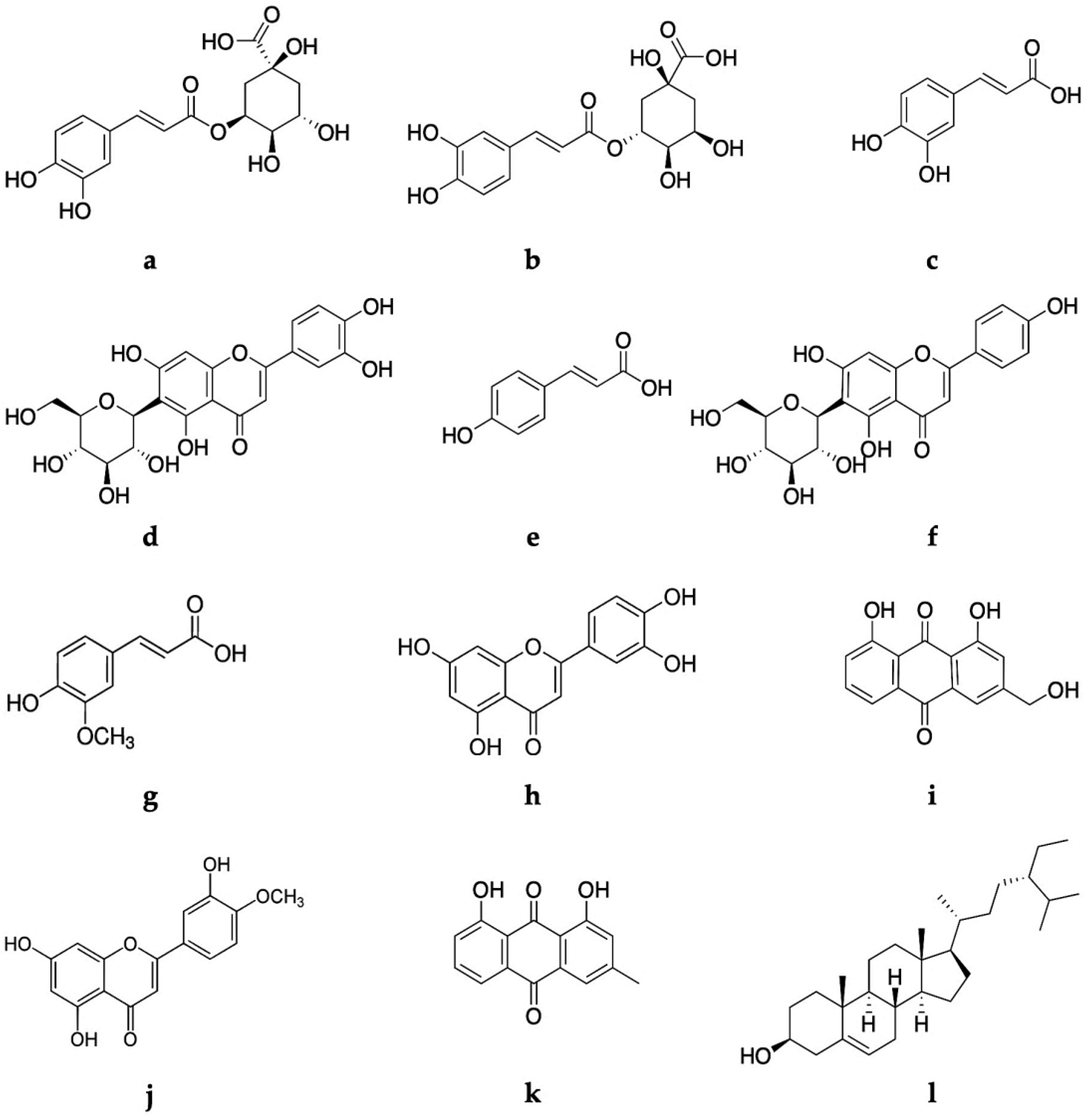
 [Green Version]
Figure 1. Comparative HPLC-UV/DAD chromatographic profiles of marker secondary metabolites of A. bento-rainhae and A. macrocarpus leaf crude extracts and their subsequent L-L partitions. Abbreviations: AbLa: A. bento-rainhae leaf first collection, AmLa: A. macrocarpus leaf first collection, (−1): ethyl ether fractions, (−2): ethyl acetate fractions, and (−3): aqueous fractions.
Molecules 28 02372 g001
Figure 2. Structures of the marker secondary metabolites (a to l) from A. bento-rainhae and A. macrocarpus leaf extracts mentioned in Table 1.
Molecules 28 02372 g002
Figure 3. HepG2 viability after 48 h of incubation with AbLa and AmLa extracts evaluated by MTT reduction assay. Data are shown as the percentage of solvent control (dashed line) and as mean ± standard deviation; n = 2–5. *** p < 0.001; **** p < 0.0001.
Molecules 28 02372 g003
Table 1. Characterization of the peaks of interest obtained from A. bento-rainhae and A. macrocarpus leaf crude extracts and their subsequent L-L partitions.
PeaktR (min)λmax (nm)[M-H](m/z)MS/MS (m/z)Identified Compound
a9.29325.3353191 (100), 179 (3)* neochlorogenic acid
b11.79326.5353191 (100), 179 (67)chlorogenic acid
c13.82240.3, 324.2179135 (100)caffeic acid
d15.43269.7, 349.1447357 (43), 327 (100), 297 (76)isoorientin
e17.46227.4, 309.9163119 (100)p-coumaric acid
f18.55269.7, 338.4431341 (23), 311 (72), 283 (100)isovitexin
g20.53235.6, 323.0193178 (62), 149 (68), 134 (100) ferulic acid
h27.38253.2, 349.1285175 (13), 151 (100), 133 (22)luteolin
i28.73256.7, 287.4, 430.4269239 (100)aloe-emodin
j34.78252.0, 346.8299284 (100)diosmetin
k50.46257.9, 287.4, 429.2253225 (100)chrysophanol
Abbreviations: tR: Retention time, λmax: wavelength. * neochlorogenic acid is a synonym name of 5-O-caffeoylquinic acid.
Table 2. Quantification of the principal classes of secondary metabolites of A. bento-rainhae and A. macrocarpus leaf crude extracts.
AssaysAbLaAbLbAmLaAmLb
Mean ± SDMean ± SDMean ± SDMean ± SD
TPC
(mg GAE/g dried extract)
(mg GAE/g dried leaf)

44.16 ± 21.62
9.23 ± 4.52

38.83 ± 17.1
8.57 ± 3.78

37.15 ± 14.32
12.63 ± 5.38

38.28 ± 15.63
6.09 ± 2.49
TFC
(mg CAE/g dried extract)
(mg CAE/g dried leaf)

* 40.79 ± 4.45
8.16 ± 0.89

29.56 ± 1.43
6.53 ± 0.32

33.46 ± 0.89
5.32 ± 0.14

* 35.52 ± 1.51
12.08 ± 0.51
TAC
(mg RhE/g dried extract)
(mg RhE/g dried leaf)

* 1.16 ± 0.13
0.24 ± 0.05

1.07 ± 0.11
0.24 ± 0.04

0.55 ± 0.07
0.19 ± 0.02

0.81 ± 0.09
0.13 ± 0.01
TCTC
(mg CAE/g dried extract)
(mg CAE/g dried leaf)

* 180.96 ± 10.98
37.82 ± 2.30

149.71 ± 12.98
33.06 ± 2.87

132.60 ± 2.73
45.09 ± 0.93

142.98 ± 6.71
22.73 ± 1.07
THTC
(mg GAE/g dried extract)
(mg GAE/g dried leaf)

67.61 ± 9.22
14.13 ± 1.93

* 55.16 ± 6.64
12.18 ± 1.47

60.53 ± 8.04
20.58 ± 2.74

37.03 ± 3.87
5.89 ± 0.62
TTC
(mg OAE/g dried extract)
(mg OAE/g dried leaf)

111.72 ± 22.77
23.35 ± 4.76

88.78 ± 23.22
19.60 ± 5.13

* 165.47 ± 26.54
56.26 ± 9.03

125.74 ± 20.72
19.99 ± 3.29
Abbreviations: AbLa: A. bento-rainhae leaf first collection, AbLb: A. bento-rainhae leaf second collection, AmLa: A. macrocarpus leaf first collection, AmLb: A. macrocarpus leaf second collection, TPC: total phenolic content, TFC: total flavonoid content, TAC: total anthraquinones content, TCTC: total condensed tannin content, THTC: total hydrolysable tannin content, TTC: total triterpenoid content, GAE: gallic acid equivalents, CAE: catechin equivalents, RhE: rhein equivalents, OAE: oleanolic acid equivalents. * Significantly higher content (p-value < 0.05) when compared between different species of the same collection season analyzed by ANOVA test.
Table 3. In vitro determination of the antioxidant activity of A. bento-rainhae and A. macrocarpus leaf crude extracts and their subsequent L-L partitions.
Extracts CodeAssays
DPPH
(IC50 μg/mL)
FRAP
(mmol AA/g Dry Extract)
AbLa20000.337 ± 0.042
AbLb25400.306 ± 0.023
AmLa29900.280 ± 0.046
AmLb30700.271 ± 0.072
AbLa-12950Nd
AbLa-2800Nd
AbLa-32910Nd
AmLa-13009Nd
AmLa-21200Nd
AmLa-34000Nd
AA83Nd
Abbreviations: AbLa: A. bento-rainhae leaf first collection extract, AbLb: A. bento-rainhae leaf second collection extract, AmLa: A. macrocarpus leaf first collection extract, AmLb: A. macrocarpus leaf second collection extract, DPPH: 2,2-diphenyl-1-picrylhydrazyl, IC50: The half maximal inhibitory concentration, FRAP: Ferric reducing antioxidant power, AA: ascorbic acid, Nd: not determined.
Table 4. In vitro antimicrobial activity of A. bento-rainhae and A. macrocarpus leaf etheric L-L partition extracts against Gram-positive strains.
Bacteria (Gram +)MIC (µg/mL)
AbLa-1AmLa-1Aloe-Emodin
S. aureus ATCC 292135005003.2
S. aureus CQINSA49236212550
S. aureus INSArefV5005001.6
S. aureus INSA93625025012.5
S. aureus INSA8961251253.2
S. saprophyticus INSA842125250100
S. saprophyticus INSA8671000100025
S. epidermidis INSA7962505001.6
S. epidermidis INSA9582505000.8
S. epidermidis INSA9601251251.6
S. haemolyticus INSA98212512525
S. haemolyticus INSA98412512512.5
Abbreviations: AbLa: A. bento-rainhae leaf first collection extract, AmLa: A. macrocarpus leaf first collection extract, ATCC: American Type Culture Collection, INSA, Instituto Nacional de Saúde clinical strains collection, MIC: minimum inhibitory concentration.
Table 5. Mutagenicity of A. bento-rainhae and A. macrocarpus leaf crude extracts in the bacterial reverse mutation test (Ames Test).
AbLa
µg/Plate
Number of Revertant Colonies Without Metabolic Activation, Mean (n = 3) ± Standard Deviation (SD)
TA98TA100TA102TA1535TA1537
25017 ± 4160 ± 7355 ± 1319 ± 410 ± 1
62520 ± 4158 ± 5349 ± 3424 ± 110 ± 2
125017 ± 2182 ± 16429 ± 2520 ± 17 ± 1
250021 ± 2178 ± 8458 ± 1622 ± 28 ± 2
375024 ± 3175 ± 19472 ± 2921 ± 49 ± 3
500024 ± 2175 ± 14485 ± 3118 ± 113 ± 3
AmLa
µg/plate
25017 ± 2186 ± 10357 ± 1422 ± 39 ± 1
62520 ± 2155 ± 15365 ± 320 ± 39 ± 2
125022 ± 5150 ± 5394 ± 816 ± 110 ± 5
250021 ± 3170 ± 15441 ± 217 ± 312 ± 5
375024 ± 5168 ± 4454 ± 2417 ± 38 ± 2
500023 ± 3165 ± 20407 ± 2824 ± 215 ± 1
NC19 ± 2156 ± 17320 ± 421 ± 37 ± 1
PC2-NFSAtBHPSA9-AA
488 ± 301048 ± 43881 ± 26827 ± 131354 ± 5
AbLa
µg/plate
Number of revertant colonies with metabolic activation, mean (n = 3) ± standard deviation (SD)
500Nd166 ± 22221 ± 1619 ± 415 ± 1
125063 ± 6164 ± 9248 ± 1115 ± 716 ± 1
250059 ± 5174 ± 4248 ± 1117 ± 211 ± 1
500052 ± 6178 ± 15254 ± 1215 ± 116 ± 1
NC44 ± 8157 ± 6172 ± 211 ± 212 ± 1
PC2-AABaP2-AA2-AA2-AA
832 ± 35947 ± 148732 ± 12266 ± 1306 ± 50
Abbreviations: AbLa: A. bento-rainhae leaf first collection extract, AmLa: A. macrocarpus leaf first collection extract, Nd: not determined, NC: negative control/solvent control (DMSO 30%), PC: positive control reference, 2-NF: 2-nitrofluorene, SA: sodium azide, tBHP: tert-butyl hydroperoxide, 9-AA: 9-aminoacridine, 2-AA: 2-aminoathracene, BaP: benzo(a)pyrene.
Table 6. Composition of the Gram-positive pathogen panel under study.
Bacteria (Gram +) Demonstration of Resistance to the Antibiotics
CXTCPFXDAPERYFAGNLzdOXAPCNTECTETVAN
S. aureus ATCC 29,213 SMS
S. aureus CQINSA4923RR RSRSRRSSS
S. aureus INSArefVR R R
S. aureus INSA936 R
S. aureus INSA896 RR R R
S. saprophyticus INSA842 RR
S. saprophyticus INSA867 R
S. epidermidis INSA796 RR R R
S. epidermidis INSA958 R R
S. epidermidis INSA960 R
S. haemolyticus INSA982 RR R
S. haemolyticus INSA984 RRR
Abbreviations: ATCC: American Type Culture Collection, INSA: Instituto Nacional de Saúde clinical strains collection, CXT: cefoxitin, CPFX: ciprofloxacin, DAP: daptomycin, ERY: erythromycin, FA: fusidic acid, GEN: gentamicin, Lzd: linezolid, OXA: oxacillin, PCN: penicillin, TEC: teicoplanin, TET: tetracycline, Van: vancomycin, MR: methicillin-sensitive, S: sensitive, R: resistant.
Disclaimer/Publisher’s Note: The statements, opinions and data contained in all publications are solely those of the individual author(s) and contributor(s) and not of MDPI and/or the editor(s). MDPI and/or the editor(s) disclaim responsibility for any injury to people or property resulting from any ideas, methods, instructions or products referred to in the content.

