Correction: Sun et al. Ergosterol Isolated from Antrodia camphorata Suppresses LPS-Induced Neuroinflammatory Responses in Microglia Cells and ICR Mice. Molecules 2023, 28, 2406

 , and
, and 
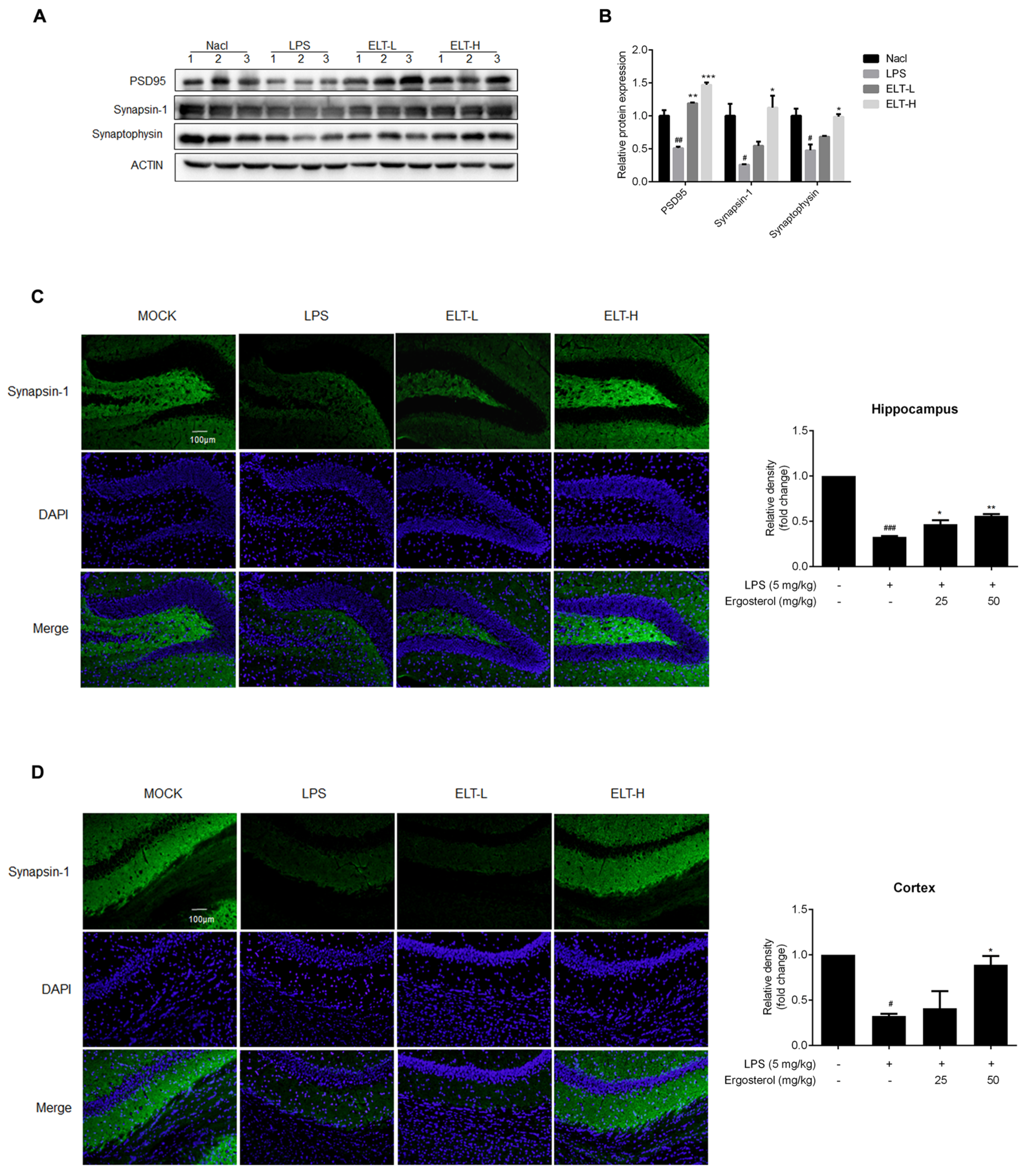
Error in Figure
Reference
- Sun, P.; Li, W.; Guo, J.; Peng, Q.; Ye, X.; Hu, S.; Liu, Y.; Liu, W.; Chen, H.; Qiao, J.; et al. Ergosterol Isolated from Antrodia camphorata Suppresses LPS-Induced Neuroinflammatory Responses in Microglia Cells and ICR Mice. Molecules 2023, 28, 2406. [Google Scholar] [CrossRef] [PubMed]

Disclaimer/Publisher’s Note: The statements, opinions and data contained in all publications are solely those of the individual author(s) and contributor(s) and not of MDPI and/or the editor(s). MDPI and/or the editor(s) disclaim responsibility for any injury to people or property resulting from any ideas, methods, instructions or products referred to in the content. |
© 2023 by the authors. Licensee MDPI, Basel, Switzerland. This article is an open access article distributed under the terms and conditions of the Creative Commons Attribution (CC BY) license (https://creativecommons.org/licenses/by/4.0/).
Share and Cite
Sun, P.; Li, W.; Guo, J.; Peng, Q.; Ye, X.; Hu, S.; Liu, Y.; Liu, W.; Chen, H.; Qiao, J.; et al. Correction: Sun et al. Ergosterol Isolated from Antrodia camphorata Suppresses LPS-Induced Neuroinflammatory Responses in Microglia Cells and ICR Mice. Molecules 2023, 28, 2406. Molecules 2023, 28, 7236. https://doi.org/10.3390/molecules28217236
Sun P, Li W, Guo J, Peng Q, Ye X, Hu S, Liu Y, Liu W, Chen H, Qiao J, et al. Correction: Sun et al. Ergosterol Isolated from Antrodia camphorata Suppresses LPS-Induced Neuroinflammatory Responses in Microglia Cells and ICR Mice. Molecules 2023, 28, 2406. Molecules. 2023; 28(21):7236. https://doi.org/10.3390/molecules28217236
Chicago/Turabian StyleSun, Ping, Weiling Li, Jiazheng Guo, Qian Peng, Xiansheng Ye, Song Hu, Yuchen Liu, Wei Liu, Haifeng Chen, Jialu Qiao, and et al. 2023. "Correction: Sun et al. Ergosterol Isolated from Antrodia camphorata Suppresses LPS-Induced Neuroinflammatory Responses in Microglia Cells and ICR Mice. Molecules 2023, 28, 2406" Molecules 28, no. 21: 7236. https://doi.org/10.3390/molecules28217236
APA StyleSun, P., Li, W., Guo, J., Peng, Q., Ye, X., Hu, S., Liu, Y., Liu, W., Chen, H., Qiao, J., & Sun, B. (2023). Correction: Sun et al. Ergosterol Isolated from Antrodia camphorata Suppresses LPS-Induced Neuroinflammatory Responses in Microglia Cells and ICR Mice. Molecules 2023, 28, 2406. Molecules, 28(21), 7236. https://doi.org/10.3390/molecules28217236