Article

Chemical Composition, Phytotoxic and Antibiofilm Activity of Seven Eucalyptus Species from Tunisia

1
Faculty of Science, Bizerte, Zarzouna 7021, Tunisia
2
Department of Pharmacy, University of Salerno, 84084 Fisciano, Italy
3
Laboratory of Management and Valorization of Forest Resources, National Institute of Researches on Rural Engineering, Water and Forests, P.B. 10, Ariana 2080, Tunisia
4
Laboratory of Biotechnology and Nuclear Technology, National Center of Nuclear Sana Science and Technology, Sidi Thabet, B.P. 72, Ariana 2020, Tunisia
5
Insitute of Food Science, CNR-ISA, 83100 Avellino, Italy
*
Author to whom correspondence should be addressed.
These authors contributed equally to this work.
Molecules 2022, 27(23), 8227; https://doi.org/10.3390/molecules27238227
Submission received: 23 October 2022 / Revised: 11 November 2022 / Accepted: 22 November 2022 / Published: 25 November 2022
(This article belongs to the Special Issue Essential Oils II)

Abstract

:
This study was carried out to characterize the chemical composition of the essential oils from seven Eucalyptus species (E. griffithsii, E. hemiphloia, E. lesouefii, E. longicornis, E. pyriformis, E. viminalis, and E. wandoo), as well as their phytotoxic and antibacterial activities. The essential oils were analyzed by GC/MS and the potential in vitro phytotoxicity was evaluated against germination and radical elongation of Raphanus sativus, Lolium multiflorum, and Sinapis arvensis seeds. The antibiofilm activity was studied against both Gram-negative (Pseudomonas aeruginosa, Escherichia coli and Acinetobacter baumannii) and Gram-positive (Staphylococcus aureus and Listeria monocytogenes) bacteria. The inhibition of biofilm formation and its metabolism was determined at different times. Eucalyptol was the most abundant component in all essential oils studied (ranging from 40.8% for E. lesouefii EO to 73.6% for E. wandoo) except for that of E. pyriformis where it was present but at 15.1%. E. pyriformis was the most active against both germination and radical elongation of S. arvensis. The action of all essential oils proved to be highly effective in inhibiting the bacterial adhesion process of the five strains considered. In light of these results, these essential oils could have potential applications both in the agricultural and health fields.

1. Introduction

The Eucalyptus genus (Myrtaceae) is composed of evergreen aromatic flowering trees and comprises about 800 species. It is a native to Australia although Eucalyptus species are now distributed in almost all parts of the world [1].
Different parts of the plant have been used to produce essential oils (EOs), but in the leaves these oils were most plentiful [2]. The EOs from Eucalyptus species rich in eucalyptol have traditionally been utilized as disinfectants, insect repellents, febrifuges and to treat respiratory illnesses [3]. Nowadays, they are used in many pharmaceutical and cosmetics industries [2].
Moreover, the identification of bioactive allelochemicals in several Eucalyptus species EOs developed research towards potential natural herbicidal compounds for weed management [4,5,6,7]. Nowadays, only 8% of conventional herbicides derive from natural sources even though natural product-based herbicides are considered safer than conventional agrochemicals [8]. However, beyond human and environmental toxicity, many weed plants are developing resistance to classical synthetic herbicide; therefore, it appears necessary to increase the study of new natural herbicidal substances [9].
On the other hand, biofilm formation and drug resistance found in both Gram-negative and Gram-positive bacteria represent problems for human health [10,11]. Several studies have demonstrated the antimicrobial properties of the EOs from Eucalyptus species [12,13] but few studies investigated their anti-biofilm activity [14].
The present study was carried out to characterize the chemical composition of the EOs from seven Eucalyptus species (E. griffithsii Maiden, E. hemiphloia Benth. (E. moluccana Wall. ex Roxb.), E. lesouefii Maiden, E. longicornis (F. Muell.) Maiden, E. pyriformis Turcz., E. viminalis Labill. and E. wandoo Blakely), as well as their phytotoxic and antibiofilm activities. The potential in vitro phytotoxicity was evaluated against germination and radical elongation of Raphanus sativus L. (radish), Lolium multiflorum Lam. (Italian ryegrass), and Sinapis arvensis L. (wild mustard) seeds, whereas the antibiofilm activity was evaluated against two Gram-positive (S. aureus and L. monocytogenes) and Gram-negative (E. coli, P. aeruginosa and A. baumannii) pathogenic strains.

2. Results and Discussion

2.1. Chemical Composition

The EO yields from leaves of the Eucalyptus species are shown in Table 1, ranging from 1.3 (E. wandoo) to 3.6% (E. hemiploia). The available literature reports yields of 5.2 and 2.0%, respectively for the EOs from leaves of Tunisian E. lesouefii and E. wandoo [15]. The yields of E. griffithsii, E. hemiphloia, E. pyriformis, E. viminalis were higher than those reported for E. griffithsii from Australia (0.34%) [16], E. hemiploia from Morocco (1.24%) [17], E. pyriformis from Australia (1.53%) [18], and E. viminalis from Portugal (1.1%) [19]. No studies reported the EO yield of E. lesouefii.
The percent compositions of the EOs are reported in Table 2. The compounds are listed according to their elution order on a HP-5MS column. The oxygenated monoterpenes were the main constituents in all EOs, with a percentage ranging from 53.9 (E. pyriformis) to 92.8% (E. longicornis). The highest amount of hydrocarbon monoterpenes was found in E. pyriformis EO (36.2%), while sesquiterpene hydrocarbons were present in very low quantity and only in E. lesouefii (0.1%), E. pyriformis (0.7%), E. viminalis (1.2%) and E. wandoo (0.9%) EOs. Oxygenated sesquiterpenes were most abundant in E. wandoo EO (5.8%).
Eucalyptol was the most abundant component in all EOs, ranging from 40.8 (E. lesouefii) to 73.6% (E. wandoo), except for E. pyriformis EO, where it is present at 15.1%.
Forty three components were identified in the EO of E. griffithsii, accounting for 96.7% of the total oil. Eucalyptol (48.9%), α-pinene (9.8%), o-cymene (4.2%) and trans-pinocarveol (4.6%) were the main components. These data agree with a previous analysis of EO from E. griffithsii from Iraq, where eucalyptol and α-pinene were reported in lower amounts (26.1 and 7.9%, respectively) and trans-pinocarveol in a higher quantity (10.6%) with respect to our sample [16]. Other compounds, in amounts greater than 1%, were neo-verbenol (2.9%), α-terpineol (1.9%), cumin aldehyde (1.9%), terpinene-4-ol (1.8%), dihydrocarveol (1.6%), pinocarvone (1.3%), p-menth-1-en-7-al (1.2%) and carvacrol (1.1%).
In the EO of E. hemiphloia, 35 components were identified, accounting for 97.5% of the total EO. Eucalyptol (63.2%), p-cymene (5.5%) and iso-menthol (4.6%) were the main compounds. Zrira and coworkers reported eucalyptol as the principal constituent (44.2%) of a Moroccan EO, but the other main components were α-pinene (7.8%), β-pinene (5.8%), α-terpineol (7.0%) that in our EO were present in smaller amounts or totally absent [17].
The EO from E. lesouefii showed the presence of 24 components, accounting for 96.8% of the total EO. Eucalyptol (40.8%), α-pinene (14.6%), p-cymene (14.3%), trans-pinocarveol (4.9%), β-pinene (4.4%) were the principal components. These results agree in part with Ameur and coworkers that reported eucalyptol (38.0%), α-pinene (12.8%), p-cymene (7.7%), trans-pinocarveol (3.2%), β-pinene (10.9%) and spathulenol (4.6%) as the main components of an EO from the North West of Tunisia [15].
Twenty-one compounds were identified in the EO of E. longicornis, accounting for 97.6% of the total EO, with eucalyptol (84.2%), α-pinene (3.5%), and trans-pinocarveol (4.2%) as the main components. To the best of our knowledge, the chemical composition of the EO of E. longicornis has not yet been reported.
In the EO of E. pyriformis, 46 components were identified, accounting for 97.3% of the total EO, in which the main constituent was p-cymene (28.8%). eucalyptol (15.1%), m-cymen-8-ol (8.8%), trans-pinocarveol (4.4%), sabina ketone (3.9%), β-pinene (3.5%) and α-pinene (1.9%) were the other principal components. Instead, Bignell and coworkers reported eucalyptol as the principal constituent (39.2%) of an EO from South Australia, with aromadendrene (5.2%), β-eudesmol (4.7%), limonene (2.4%), trans-pinocarveol (1.7%) and α- eudesmol (1.7%) [18]. Among these compounds, aromadendrene, limonene and β-eudesmol were totally absent in our sample.
The EO from E. viminalis showed the presence of 31 components, accounting for 96.6% of the total EO. Eucalyptol (68.1%), α-pinene (7.2%), trans-pinocarveol (3.9%), spathulenol (5.1%) were the principal components. Other compounds present in lesser amounts were sabina ketone (1.2%), pinocarvone (1.2%), terpinen-4-ol (1.1%) and cryptone (1.4%). These results agree in part with Elaissi and coworkers who showed in an EO from the North of Tunisia the presence of eucalyptol (62.7%), α-pinene (1.7%), trans-pinocarveol (2.3%) and p-cymene (1.3%), the last absent in our sample, and the presence of some oxygenated sesquiterpenes (globulol, viridiflorol and β-eudesmol), totally absent in our sample [20]. Moreover, an Iranian EO showed eucalyptol (57.8%), α-pinene (13.4%), limonene (5.4%), and globulol (3.0%) as the main constituents [21]. Limonene and globulol were absent in our EO.
In the EO of E. wandoo, 25 components were identified, accounting for 98.0% of the total EO, with eucalyptol (73.6%), trans-pinocarveol (6.1%), α-pinene (4.3%) as the principal constituents. Other components in lesser amounts were α-terpinyl acetate (1.9%), pinocarvone (1.3%) and the sesquiterpenes, spathulenol (2.1%), β-eudesmol (1.5%), rosifoliol (1.0%) and α-eudesmol (0.9%). The literature reports an Algerian EO much poorer in eucalyptol and p-cymene (14.9 and 9.0%, respectively), but with a high content of benzaldehyde (32.3%) absent in the sample analyzed [22]. Instead, a Tunisian EO showed a qualitative composition similar to that of our EO, but with different percentages of the individual constituents [15].
In general, the research agrees with the available literature; however, some different compositions have been found, in particular with regard to the composition of the EO of E. pyriformis. It is interesting to note that both genetic and environmental factors can influence the composition of the essential oils of species grown outside their original area.

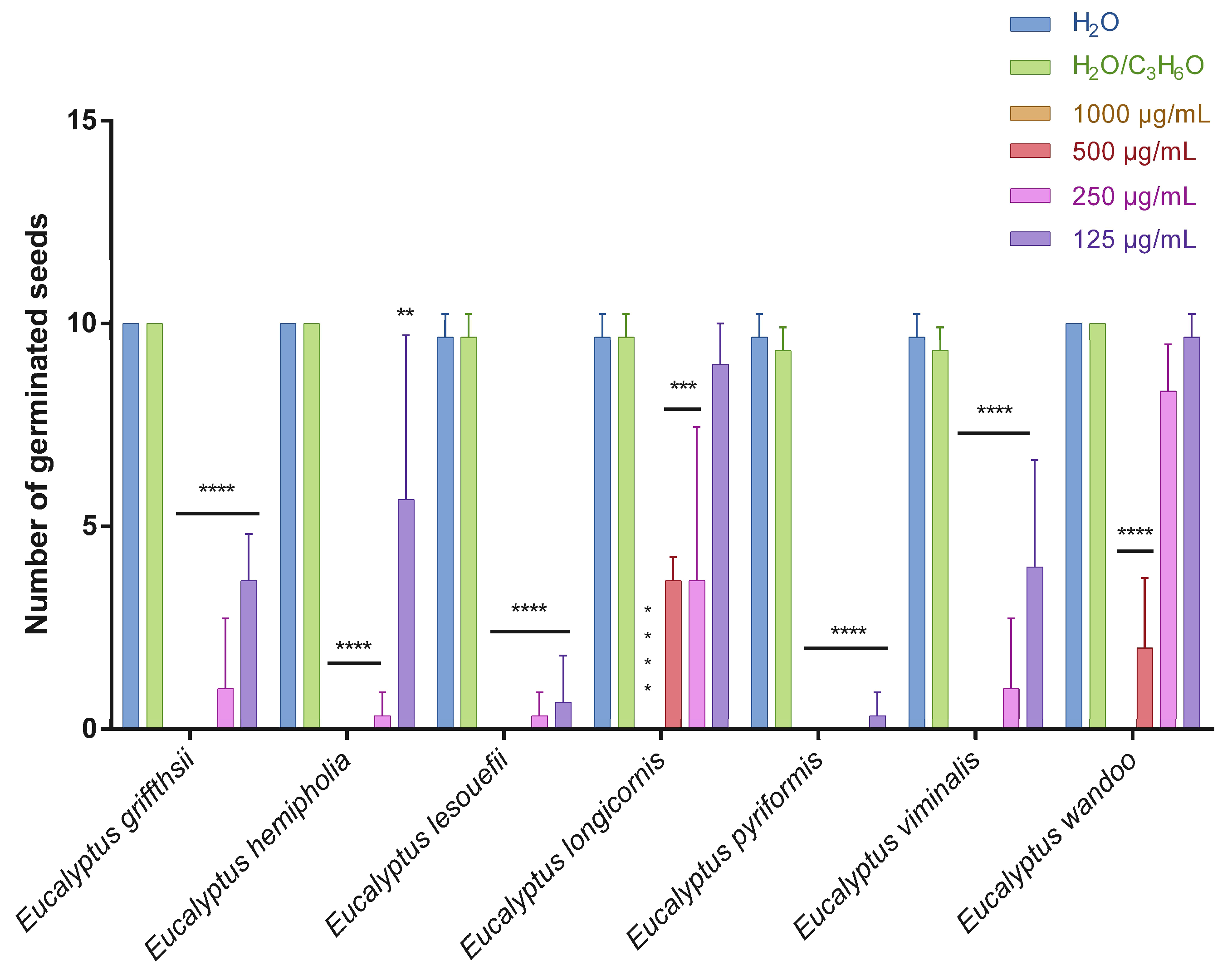
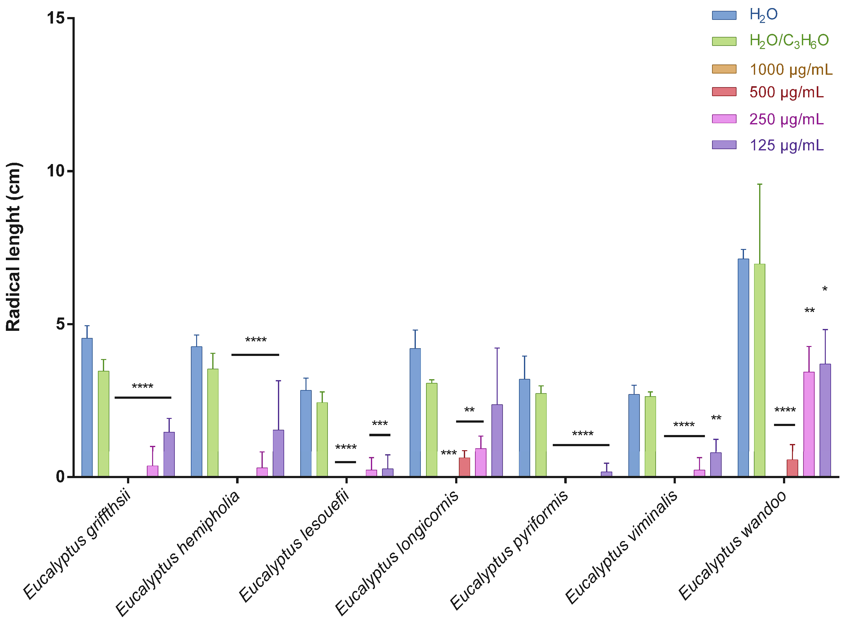
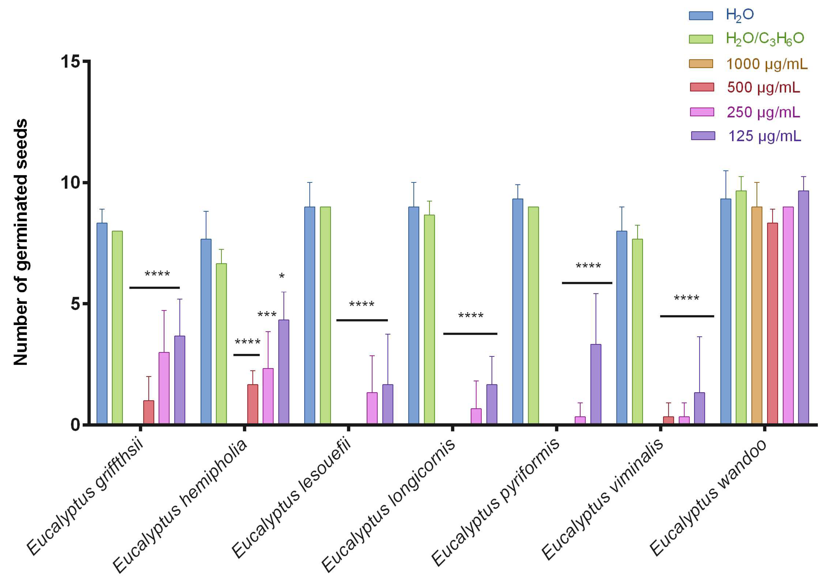
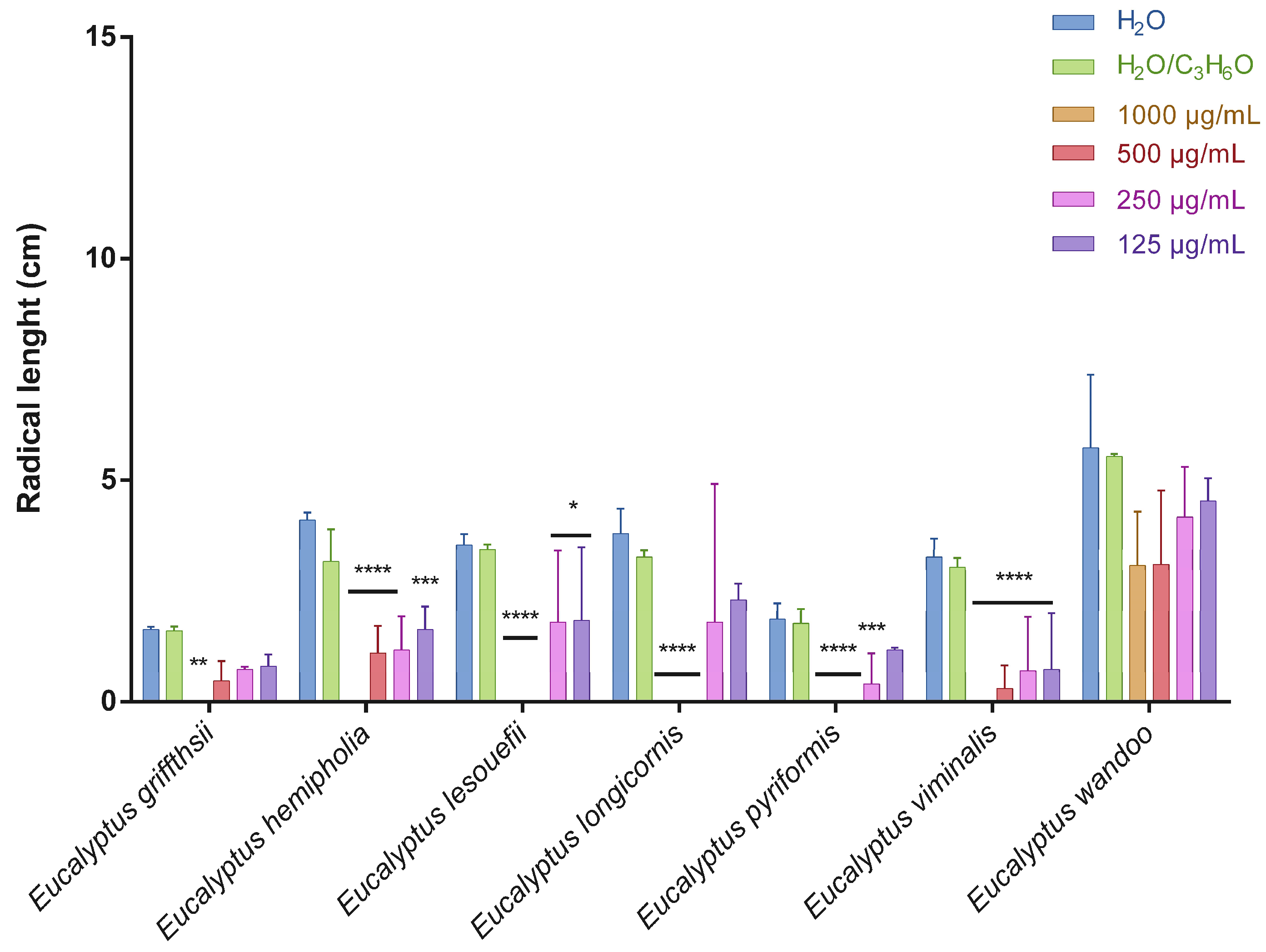
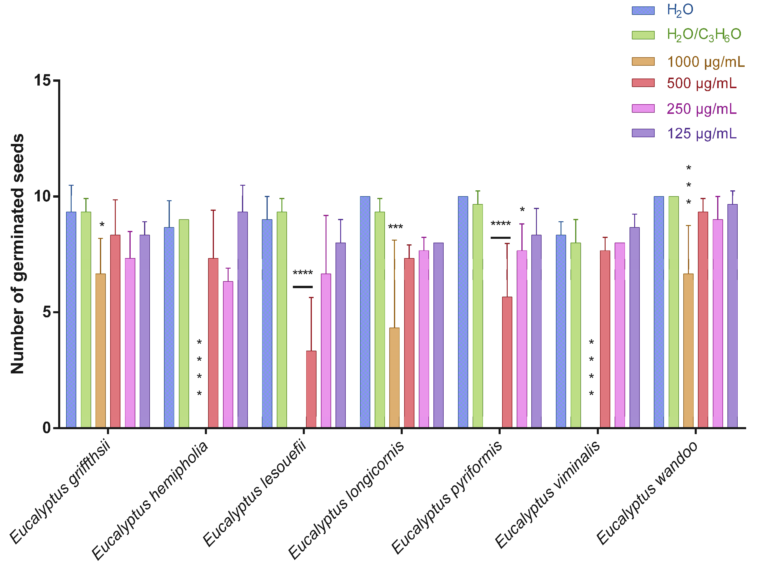
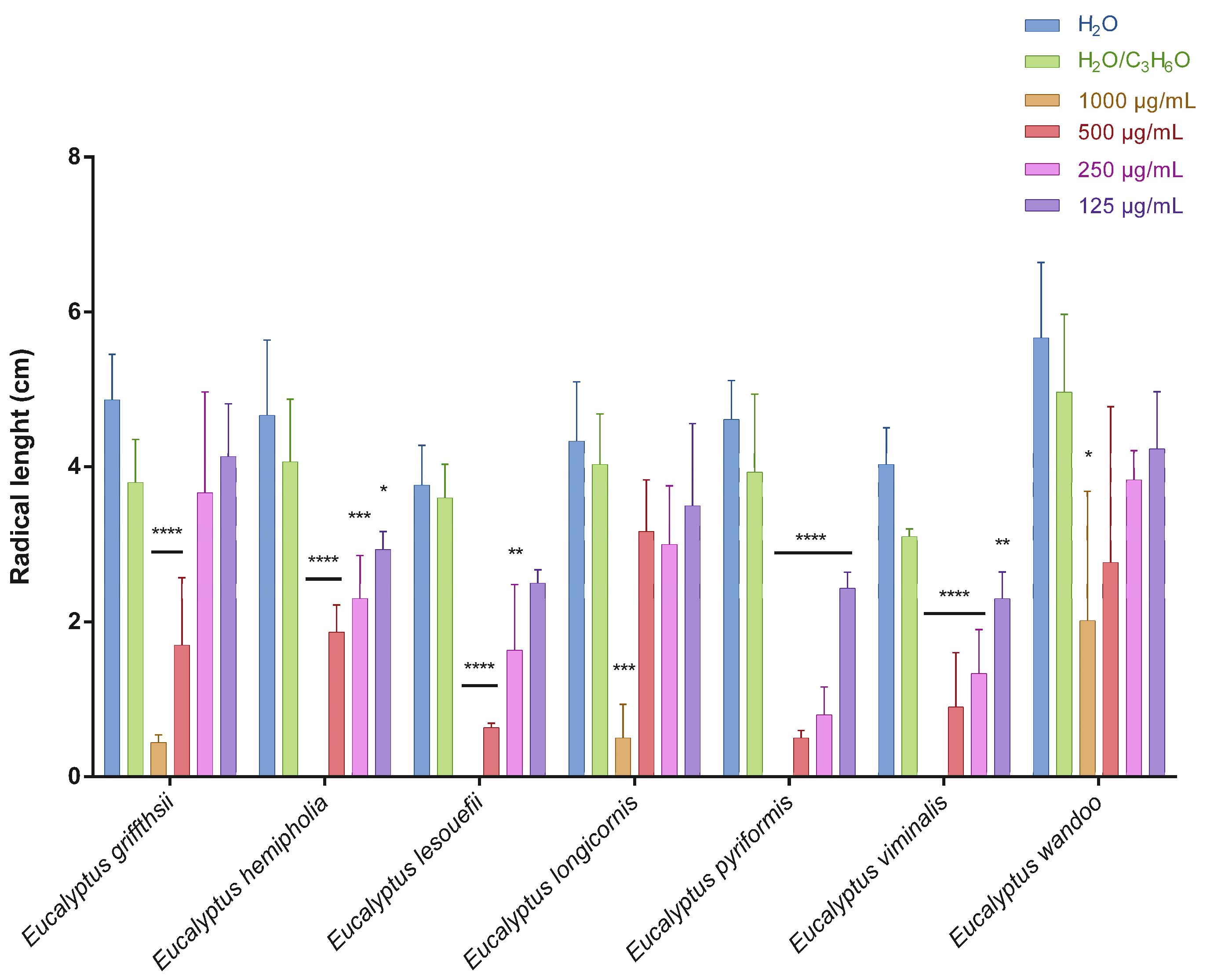
2.2. Phytotoxic Activity

This is the first manuscript that investigates the phytotoxicity of these Eucalyptus EOs against germination and radical elongation of S. arvensis, R. sativus and L. multiflorum seeds. Figure 1, Figure 2, Figure 3, Figure 4, Figure 5 and Figure 6 represented the number of germinated seeds and the radicle length in cm of these seeds after treatment with the EOs.
The germination and radical elongation of R. sativus seeds were significantly inhibited by all doses of all essential oils tested, except for E. wandoo EO. In fact, this last EO never inhibited germination or radical elongation to a percentage greater than 50%.
The EOs from E. lesouefii and E. pyriformis were the most active against germination and radical elongation of S. arvensis. In fact, at the lowest doses tested (125 µg/mL), the inhibition of germination was 93.1 and 96.6%, respectively, and the inhibition of radical elongation was 90.6 and 94.8%, respectively. E. griffithsii and E. viminalis EOs were the second most active against S. arvensis germination with an inhibition > 50% at 125 µg/mL. Instead, the others EOs, at the lowest dose tested, showed a low inhibition of S. arvensis germination ranging from 3.3 to 43.4%. Moreover, the second most active EOs on radical elongation of S. arvensis were E. griffithsii, E. hemiphloia and E. viminalis. In fact, at 125 µg/mL, these EOs showed an inhibition on radical growth of 67.6%, 64.1% and 70.4%, respectively. E. longicornis and E. wandoo EOs were the less active, also against radical elongation of S. arvensis seeds.
E. hemiphloia, E. lesouefii, E. pyriformis, E. viminalis EOs totally inhibited the germination and radical elongation of L. multiflorum, at the highest dose tested. E. lesouefii EO was the most active against L. multiflorum germination with a percentage inhibition of 63% at 500 µg/mL. Instead, E. pyriformis EO showed a better activity against L. multiflorum radical growth with a percentage inhibition of 82.7% at 250 µg/mL.
To the best of our knowledge, the phytotoxic activity on S. arvensis, R. sativus and L. multiflorum seeds of these Eucalyptus species has not been reported.
Many authors reported the phytotoxic and allelopathic effects of several Eucalyptus EOs such as E. citriodora Hook, E. nicholii Maiden and Blakely, E. globulus Labill. and E. tereticornis Sm. against germination and growth of many crops and weeds [5,6,24,25].
The phytotoxicity was probably due to the presence of eucalyptol: as suggested in the literature this compound can causes decreased germination, inhibiting mitochondrial respiration, mitosis and DNA synthesis [4,26].
However, the phytotoxicity of the EO of E. pyrifiormis can be attributed to other constituents or to a synergism between them.
Our results appear relevant; in fact, all EOs affected germination and radical elongation of S. arvensis, one of the most prevalent weed species in wheat in the Middle Black Sea Region of Turkey [27]. Moreover, the EOs were active against L. multiflorum, one of the weeds that are evolving resistance to herbicides [28].
The results obtained may open the way for the direct application of essential oils studied in sustainable agricultural practices or in the identification of new lead compounds for new agrochemicals.

2.3. Antibacterial Activity

Table 3 shows the minimal inhibitory concentration of the Eucalyptus EOs necessary to inhibit the bacterial growth of the five pathogens used in our study.
These MIC values helped us to appraise the aptitude of the seven EOs to affect the bacterial biofilm (Table 4) and to alter the metabolism of the sessile bacterial cells (Table 5).
As reported in Table 3, the MIC values varied between 25 and 38 µL/mL Based on such results, two sub-lethal doses (10 and 20 µL/mL) were used in the antibiofilm tests.
The action of all EOs proved to be highly effective in inhibiting the bacterial adhesion process, prodrome to the biofilm constitution of the five pathogens used in our experiments. The inhibition percentages at the highest concentration (20 µL/mL), were never lower than 58% (EV vs. S. aureus). The EOs resulted in an inhibition even up to 95.04% (EP vs. S. aureus), and in many cases the inhibition was higher than 80%. Overall, all the strains were sensitive to the action of the EOs. It is interesting to note that in some cases, when the EOs were added 24 h after the beginning of the bacterial growth process, when the bacteria were allowed to consolidate the biofilm on the walls of the multiwells, the inhibitory effectiveness of the EOs remained practically intact. Indeed, with 10 µL/mL of the EO of E viminalis, the inhibitory efficacy increased by more than ten times, from 4.36 to 50.77%. Furthermore, the EO of E. wandoo was more effective against S. aureus than in the adhesion event. At the same concentration, the inhibitory efficacy of this EO was practically doubled, from 59.16 to 94.55%.
The EOs were active inhibitors of the sessile cell metabolism, at the early stage of bacterial biofilm formation, and the inhibitory activity on adhesion processes was mainly caused by the action exerted on cell metabolism. It is the case, for example, of A. baumannii, against which, with a few exceptions, the inhibition rates resulted in no less than 53.64% (E. viminalis), and which reached more than 83% (E. griffithsii, E. hemiphloia, and E. pyriformis). In the case of E. coli, the higher concentration of EOs used, provoked an inhibition as high as 90.7% (E. lesoufii) and never less than 63.66% (E. wandoo). In general, however, all EOs showed apparent inhibitory efficacy on the cellular metabolism of all strains. Such efficacy was also observed in tests performed on mature biofilms. Although it decreased in some cases (e.g., E. griffithsii vs. E. coli and L. monocytogenes, or E. hemiphloia and E. viminalis vs. A. baumannii, E. coli, L. monocytogenes, and P. aeruginosa), the strength of action of the EOs on sessile cell metabolism in mature biofilms was maintained. This shows that the inhibitory activity of these EOs was primarily explicated by acting on cell metabolism. The low inhibitory activity recorded in the MTT test, but high in the crystal violet test, may mean that the EOs probably acted by other inhibitory mechanisms (e.g., on bacterial cell walls, nucleic acids), as widely demonstrated for Eucalyptus EOs [29,30] and other phytochemicals [31]. The EOs we analyzed exhibited high inhibitory activity, demonstrating that they could act both in the initial bacterial cell adhesion events and on mature biofilms. This again shows the extreme versatility of these EOs against Gram-positive and Gram-negative pathogens. Luìs and coworkers showed the antibacterial and anti-quorum sensing activity of the EOs of E. globulus Labill. and E. radiata A. Cunn. ex DC. against A. baumannii, thus indicating how these EOs can block the processes leading to bacterial biofilm adhesion and formation from the very beginning [11]. E. globulus EO has been shown to act on the biofilm of E. coli, resulting in 62% inhibition [32], and P. aeruginosa [33]. The inhibitory efficacy of the studied EOs may help to expand the versatility in the use of Eucalyptus EOs. The EO of E. globulus counteracted the formation of biofilms of cariogenic bacteria [34], including S. aureus, against which the EOs we analyzed showed an effective inhibitory action not only in blocking the biofilm from the outset but, where it was already established, acting with an inhibitory vigor that reached up to 95%. Future works will study, at the molecular level, the genes involved in the bacterial adhesion and how, and if, there is a change from the immature biofilm to its mature form.

3. Material and Methods

3.1. Plant Materials

Leaves of the seven Eucalyptus species were harvested from different Tunisian arboretums located in different regions (Table 6). For each species, five samples harvested from more than five different trees were collected and mixed for homogenization. The leaves were stored in a dry place for fifteen days. Specimens were identified at the Regional Station of the National research institute of rural engineering, Waters and Forests (INRGREF).

3.2. Extraction

One hundred grams of dried leaves were submitted to water distillation (500 mL of water) for 4 h, using a Clevenger-type apparatus. The EOs were solubilized in n-hexane, dried in a N2 atmosphere, and stored in amber vials in the refrigerator at 4 °C.

3.3. Analysis of the Essential Oils

Analytical gas chromatography (GC) was carried out on a Perkin-Elmer Sigma-115 gas chromatograph (Perkin Elmer, Waltham, MA, USA) equipped with a flame ionization detector (FID) and a data handling processor. The separation was achieved using a HP-5 MS fused-silica capillary column (30 m × 0.25 mm i.d., 0.25 μm film thickness, Agilent, Roma, Italy). Column temperature: 40 °C, with 5 min initial hold, and then to 270 °C at 2 °C/min, 270 °C (20 min); injection mode, splitless (1 μL of a 1:1000 n-hexane solution).; injector and detector temperatures were 250 °C and 290 °C, respectively. The analysis was also run using a fused silica HP Innowax polyethylene glycol capillary column (50 m × 0.20 mm i.d., 0.25 μm film thickness, Agilent, Roma, Italy). In both cases, helium was used as the carrier gas (1.0 mL/min). GC/MS analyses were performed on an Agilent 6850 Ser. II apparatus (Agilent, Roma, Italy), fitted with a fused silica DB-5 capillary column (30 m × 0.25 mm i.d., 0.33 μm film thickness, Agilent, Roma, Italy), coupled to an Agilent Mass Selective Detector MSD 5973; ionization energy voltage 70 eV; electron multiplier voltage energy 2000 V. Mass spectra (MS) were scanned in the range 40–500 amu, scan time 5 scans/s. Gas chromatographic conditions were as reported in the previous paragraph; transfer line temperature, 295 °C. Most constituents were identified by GC by comparison of their Kovats retention indices (Ri), determined relative to the retention times (tR) of n-alkanes (C10–C35), with either those of the literature [35,36,37] and mass spectra on both columns or those of authentic compounds available in our laboratories by means of NIST 02 and Wiley 275 libraries [38]. The component relative concentrations were obtained by peak area normalization. No response factors were calculated.

3.4. Phytotoxic Activity

The phytotoxic activity was evaluated on germination and radical elongation of the seeds of Lolium multiflorum Lam., Raphanus sativus L., and Sinapis arvensis L., often used for their easy and well-known germinability. S. arvensis seeds were collected from wild populations in Sidi Ismail, Beja, Tunisia on June 2021. R. sativus seeds were purchased from Blumen group s.r.l. (Bologna, Italy) whereas L. multiflorum seeds were purchased from Fratelli Ingegnoli s.p.a. (Milano, Italy). The seeds were sterilized in 95% ethanol for 15 s and sown in Petri dishes (Ø = 90 mm), on three layers of Whatman filter paper, impregnated with 7 mL of deionized water used as first control to verify the germinability of the seeds, 7 mL of a water–acetone mixture (99.5:0.5, v/v) as second control (because the EOs were dissolved in this mixture for their lipophilicity) or 7 mL of different doses (1000, 500, 250, and 100 mg/mL) of the EOs. Controls, carried out with the water–acetone mixture alone, showed no differences in comparison with control in water alone. The germination conditions were 20 ± 1 °C, with a natural photoperiod. Seed germination was observed in the Petri dishes every 24 h. A seed was considered germinated when the protrusion of the root became evident [39]. On the fifth day (after 120 h) the effects on radicle elongation were measured in cm. Each determination was repeated three times, using Petri dishes containing 10 seeds each. Data are expressed as the mean ± SD for both germination and radicle elongation.

3.5. Antimicrobial Activity

3.5.1. Microorganisms and Culture Conditions

Acinetobacter baumannii ATCC 19606, Pseudomonas aeruginosa DSM 50,071 and Escherichia coli DSM 8579 (Gram-negative strains), Staphylococcus aureus subsp. Aureus Rosebach ATCC 25923, and Listeria monocytogenes ATCC 7644 (Gram-positive strains) were used. Bacteria were grown in Luria broth for 18 h at 37 °C before the analysis. A. baumannii was cultured at 35 °C under the same conditions.

3.5.2. Minimal Inhibitory Concentration (MIC)

The essential oils and DMSO were sterilized by ultrafiltration before use. The method using resazurin, developed by Sarker and coworkers [40], with some modifications was used to assess the MIC of the EOs in 96 microtiter-plates. Resazurin is an oxidation–reduction indicator, generally used to evaluate cell growth. It is a blue non-fluorescent and non-toxic dye, which becomes pink and fluorescent when is subjected to a reduction through the action of the oxidoreductases present within viable cells and is transformed to resorufin. This last molecule is further reduced to hydroresorufin that appears (uncolored and non-fluorescent). A resazurin solution was prepared by dissolving 270 mg in 40 mL of sterilized deionized water. One hundred μL of samples in DMSO (1:10 v/v) were added to the first row of the plate. To all other wells, 50 μL of Luria–Bertani broth or normal sterile solution was included. Serial descending concentrations of samples were performed, and each well was supplemented with 10 μL of resazurin indicator solution. Thirty μL of 3.3× strength isosensitized broth and 10 μL of bacterial suspension (5 × 10⁶ cfu/mL) were added in each well. The plates were then closed with parafilm to prevent dehydration. The wide-spectrum conventional antibiotic tetracycline, previously suspended in DMSO, and used in other similar research [41,42] was added in a column of the plate as positive control. Luria–Bertani broth containing resazurin and bacteria, without samples, was considered as the negative control. The plates were incubated at 37 °C (or at 35 °C for A. baumannii) for 24 h. The color change was then assessed visually. Any color changes from purple to pink or colorless were recorded as positive. The lowest concentration of EOs which could prevent the solution from changing from dark purple to pink was considered as the MIC value.

3.5.3. Biofilm Inhibitory Activity

The inhibitory activity of the EOs on the bacterial adhesion was assessed using flat-bottomed 96-well microtiter plates [42]. Bacterial cultures were adjusted to 0.5 McFarland with fresh culture broth. Then, 10 µL of the bacterial cultures and 10 or 20 µL/mL of the EOs were included in each well. The volume of each well was brought to 250 µL final volume with different amounts of Luria–Bertani broth. The plates were covered with parafilm tape to prevent evaporation and incubated for 48 h at 37 °C (or at 35 °C for A. baumannii). Succeeding the elimination of the planktonic cells, sessile cells were washed twice with sterile PBS. After PBS removal, the plates were left under a laminar flow hood for ten minutes. Two hundred µL of methanol was included in each well, to fix the sessile cells, and eliminated after 15 min. The plates were left to dry the samples and 200 µL of 2% w/v crystal violet solution/well was used for 20 min to stain the sessile cells. After 20 min, the staining solution was removed; plates were gently washed with sterile PBS and samples dried. The bound dye’s release was obtained by adding 200 µL of glacial acetic acid 20% w/v. The absorbance was measured at λ = 540 nm (Cary Varian, Milano, Italy). The percent value of adhesion was assessed with respect to the control (formed by the cells grown without the presence of the samples, where a value of 0% inhibition was considered). Triplicate tests were performed, and the average results were calculated for reproducibility.

3.5.4. Activity on Mature Bacterial Biofilm

The inhibitory activity of the EOs on the mature biofilms was assessed using the tests described above, on bacterial cultures grown for 24 h. After 24 h of bacterial growth, the planktonic cells were removed, and the two concentrations of the EOs, 10 and 20 μL/mL, and Luria–Bertani broth were added, and a final volume of 250 μL/well was reached. Plates were kept for 24 h at 37 °C or 35 °C, depending on the strains. The other experimental steps, and the calculation of the percent value of inhibition with respect to the untreated bacteria, were performed as previously described.

3.5.5. Effects of the EOs on Cell Metabolic Activity within the Biofilm

The 3-(4,5-dimethylthiazol-2-yl)-2,5-diphenyltetrazolium bromide (MTT) colorimetric method [42] was used to evaluate the effect of the EOs on the metabolic activity of the bacterial cells. Two concentrations (10 and 20 μL/mL) of the EOs, which were added at the beginning of the bacterial growth and after 24 h of incubation, were used. After 48 h total incubation, planktonic cells were removed; addition of 150 μL of PBS and 30 μL of 0.3% MTT (Sigma, Milano, Italy) was followed by the incubation of the microplates for two hours at 37 °C (35 °C for A. baumannii). The MTT solution was removed, two washing steps were carried out with 200 μL of sterile physiological solution, and 200 μL of dimethyl sulfoxide (DMSO) was added to permit the suspension of the formazan crystals, assessed after two hours at λ = 570 nm (Cary Varian, Milano, Italy).

3.6. Statistical Analysis

All experiments were carried out in triplicate. Data from each experiment were statistically analyzed using GraphPad Prism 6.0 software (GraphPad Software Inc., San Diego, CA, USA) followed by comparison of means (two-way ANOVA) using Dunnett’s multiple comparisons test, at the significance level of p < 0.05.

4. Conclusions

This research has allowed the chemical study of the EOs of seven species of Eucalyptus grown outside their area of origin and to help in the study of the chemodiversity of a complex genus such as the Eucalyptus genus.
The results evidenced the phytotoxic activity of the seven Eucalyptus species, already reported for other Eucalyptus species. Moreover, all EOs could be considered as inhibitory to the bacterial adhesion and the cellular metabolism of all the pathogenic strains used in our experiments. This efficacy was also observed in tests performed on mature biofilms. Considering these results, further investigations on these EOs, such as the molecular analysis of the genes involved in the various steps of adhesion and mature biofilm formation, as well as the assessment of toxicity of the EOs, could provide useful in applications for both the agronomic management and the treatment of human microbial infections.

Author Contributions

Conceptualization, I.A. and V.D.F.; methodology, I.A., L.H., V.D.F. and F.N.; investigation, F.P., L.C., S.K., F.M., M.K. and F.F.; resources, V.D.F. and L.H.; data curation, F.P. and F.N.; writing—original draft preparation, I.A., L.H., F.P. and F.N.; writing—review and editing, F.P., F.N. and V.D.F. All authors have read and agreed to the published version of the manuscript.

Funding

This research received no external funding.

Data Availability Statement

Not applicable.

Conflicts of Interest

The authors declare no conflict of interest.

Sample Availability

Samples of the Eucalyptus essential oils are available from the authors.

References

  1. Konoshima, T.; Takasaki, M. Chemistry and bioactivity of the non-volatile constituents of eucalyptus. In Eucalyptus: The Genus Eucalyptus; Coppen, J.J.W., Ed.; Taylor & Francis: London, UK, New York, NY, USA, 2002; pp. 269–290. [Google Scholar]
  2. Kandasamy, O.S.; Mohamed Yassin, M.; Babu, R.C. Biology, ecology, silviculture, and potential uses of Eucalyptus—An overview. J. Med. Aromat. Plant Sci. 2000, 22, 330–335. [Google Scholar]
  3. Zhang, J.; An, M.; Wu, H.; Stanton, R.; Lemerle, D. Chemistry and bioactivity of Eucalyptus essential oils. Allelopathy J. 2010, 25, 313–330. [Google Scholar]
  4. Romagni, J.G.; Allen, S.N.; Dayan, F.E. Allelopathic effects of volatile cineoles on two weedy plant species. J. Chem. Ecol. 2000, 26, 303–313. [Google Scholar] [CrossRef]
  5. Batish, D.R.; Singh, H.P.; Nidhi, S.; Shalinder, K.; Kohli, R.K. Chemical composition and inhibitory activity of essential oil from decaying leaves of Eucalyptus citriodora. Z. Naturforsch. 2006, 61, 52–56. [Google Scholar] [CrossRef]
  6. Kaur, S.; Pal Singh, H.; Batish, D.; Kumar Kholi, R. Role of monoterpenes in Eucalyptus communities. Curr. Bioact. Compd. 2012, 8, 101–107. [Google Scholar] [CrossRef]
  7. Ibáñez, M.D.; Blázquez, M.A. Phytotoxicity of Essential Oils on Selected Weeds: Potential Hazard on Food Crops. Plants 2018, 7, 79. [Google Scholar] [CrossRef] [Green Version]
  8. Dayan, F.E.; Duke, S.O. Natural compounds as next-generation herbicides. Plant Physiol. 2014, 166, 1090–1105. [Google Scholar] [CrossRef] [Green Version]
  9. Flamini, G. Natural herbicides as a safer and more environmentally friendly approach to weed control: A review of the literature since 2000. Stud. Nat. Prod. Chem. 2012, 38, 353–396. [Google Scholar]
  10. Elaissi, A.; Rouis, Z.; Salem, N.A.B. Chemical composition of 8 eucalyptus species’ essential oils and the evaluation of their antibacterial, antifungal and antiviral activities. BMC Complement. Altern. Med. 2012, 12, 81. [Google Scholar] [CrossRef] [Green Version]
  11. Luís, Â.; Duarte, A.; Gominho, J.; Domingues, F.; Duarte, A.P. Chemical composition, antioxidant, antibacterial and anti-quorum sensing activities of Eucalyptus globulus and Eucalyptus radiata essential oils. Ind. Crops Prod. 2016, 79, 274–282. [Google Scholar] [CrossRef]
  12. Sumitra, S.; Sharma, S.K. Antibacterial activity of essential oil and root extract of Eucalyptus teriticornis. Indian J. Nat. Prod. Resour. 2005, 21, 16–17. [Google Scholar]
  13. Cermelli, C.; Fabio, A.; Fabio, G.; Quaglio, P. Effect of eucalyptus essential oil on respiratory bacteria and viruses. Cur. Microbiol. 2008, 56, 89–92. [Google Scholar] [CrossRef] [PubMed]
  14. Caputo, L.; Smeriglio, A.; Trombetta, D.; Cornara, L.; Trevena, G.; Valussi, M.; Fratianni, F.; De Feo, V.; Nazzaro, F. Chemical Composition and Biological Activities of the Essential Oils of Leptospermum petersonii and Eucalyptus gunnii. Front. Microbiol. 2020, 11, 409. [Google Scholar] [CrossRef] [PubMed] [Green Version]
  15. Ameur, E.; Sarra, M.; Yosra, D.; Mariem, K.; Nabil, A.; Lynen, F.; Larbi, K.M. Chemical composition of essential oils of eight Tunisian Eucalyptus species and their antibacterial activity against strains responsible for otitis. BMC Complement. Altern. Med. 2021, 21, 209. [Google Scholar]
  16. Bignell, C.M.; Dunlop, P.J.; Brophy, J.J.; Jackson, J.F. Volatile leaf oils of some south-western and southern Australian species of the genus Eucalyptus. Part I. Subgenus Symphyomyrtus, Section Dumaria, Series Incrassatae. Flavour Fragr. J. 1994, 9, 113–117. [Google Scholar] [CrossRef]
  17. Zrira, S.S.; Benjilali, B.B.; Fechtal, M.M.; Richard, H.H. Essential oils of twenty-seven Eucalyptus species grown in Morocco. J. Essent. Oil Res. 1992, 4, 259–264. [Google Scholar] [CrossRef]
  18. Bignell, C.M.; Dunlop, P.J.; Brophy, J.J.; Jackson, J.F. Volatile leaf oils of some south-western and Southern Australian species of the genus Eucalyptus. Part III—Subgenus Symphyomyrtus, Section Bisectaria, Series Macrocarpae. Flavour Fragr. J. 1994, 9, 309–313. [Google Scholar] [CrossRef]
  19. Faria, J.M.S.; Sanches, J.; Lima, A.S.; Mendes, M.D.; Leiria, R.; Geraldes, D.A.; Figueiredo, A.C.; Trindade, H.; Pedro, L.G.; Barroso, J.G. Eucalyptus from Mata Experimental do Escaroupim (Portugal): Evaluation of the essential oil composition from sixteen species. Acta Hortic. 2011, 925, 61–66. [Google Scholar] [CrossRef]
  20. Elaissi, A.; Salah, K.H.; Mabrouk, S.; Larbi, K.M.; Chemli, R.; Harzallah-Skhiri, F. Antibacterial activity and chemical composition of 20 Eucalyptus species’ essential oils. Food Chem. 2011, 129, 1427–1434. [Google Scholar] [CrossRef]
  21. Maghsoodlou, M.T.; Kazemipoor, N.; Valizadeh, J.; Falak Nezhad Seifi, M.; Rahneshan, N. Essential oil composition of Eucalyptus microtheca and Eucalyptus viminalis. Avicenna J. Phytomed. 2015, 5, 540–552. [Google Scholar]
  22. Foudil-Cherif, Y.; Meklati, B.Y.; Verzera, A.; Mondello, L.; Dugo, G. Chemical Examination of Essential Oils from the Leaves of Nine Eucalyptus Species Growing in Algeria. J. Essent. Oil Res. 2000, 12, 186–191. [Google Scholar] [CrossRef]
  23. Adams, R.P. Identification of Essential Oil Components by Gas Chromatography/Mass Spectroscopy, 4th ed.; Allured Publishing Corporation: Carol Stream, IL, USA, 2007. [Google Scholar]
  24. Ramezani, S.; Saharkhiz, M.J.; Ramezani, F.; Fotokian, M.H. Use of essential oils as bioherbicides. J. Essent. Oil-Bear Plants 2008, 11, 319–327. [Google Scholar] [CrossRef]
  25. Rasaeifar, M.; Hosseini, N.; Asl, N.; Zandi, P.; Aghdam, A. Allelopathic effect of Eucalyptus globulus’ essential oil on seed germination and seedling establishment of Amaranthus blitoides and Cyndon dactylon. Trakia J. Sci. 2013, 11, 73–81. [Google Scholar]
  26. Macias, F.A.; Molinillo, J.M.; Varela, R.M.; Galindo, J.C. Allelopathy—A natural alternative for weed control. Pest Manag. Sci. 2007, 63, 327–348. [Google Scholar] [CrossRef] [PubMed]
  27. Mennan, H. Economic thresholds of Sinapis arvensis (wild mustard) in winter wheat fields. Pak. J. Agron. 2003, 2, 34–39. [Google Scholar] [CrossRef]
  28. Khammassi, M.; Polito, F.; Amri, I.; Khedhri, S.; Hamrouni, L.; Nazzaro, F.; Fratianni, F.; De Feo, V. Chemical Composition and Phytotoxic, Antibacterial and Antibiofilm Activity of the Essential Oils of Eucalyptus occidentalis, E. striaticalyx and E. stricklandii. Molecules 2022, 27, 5820. [Google Scholar] [CrossRef]
  29. Heap, I. The International Survey of Herbicide Resistant Weeds 2016. Available online: http://www.weedscience.org (accessed on 12 September 2022).
  30. Nazzaro, F.; Fratianni, F.; De Martino, L.; Coppola, R.; De Feo, V. Effect of essential oils on pathogenic bacteria. Pharmaceuticals 2013, 6, 1451–1474. [Google Scholar] [CrossRef]
  31. Nazzaro, F.; Fratianni, F.; Coppola, R. Quorum sensing and phytochemicals. Int. J. Mol. Sci. 2013, 14, 12607–12619. [Google Scholar] [CrossRef] [Green Version]
  32. Dohare, S.; Dubey, S.D.; Kalia, M.; Verma, P.; Pandey, H.; Singh, N.K.; Agarwal, V. Anti-biofilm activity of Eucalyptus globulus oil encapsulated silica nanoparticles against E. coli biofilm. Int. J. Pharm. Sci. 2014, 5, 5011. [Google Scholar]
  33. Sambyal, S.S.; Sharma, P.; Shrivastava, D. Anti-biofilm activity of selected plant essential oils against Pseudomonas aeruginosa and Staphylococcus aureus. Int. J. Curr. Microbiol. Appl. Sci. 2017, 6, 444–450. [Google Scholar] [CrossRef] [Green Version]
  34. de Oliveira Carvalho, I.; Purgato, G.A.; Píccolo, M.S.; Pizziolo, V.R.; Coelho, R.R.; Diaz-Muñoz, G.; Diaz, M.A.N. In vitro anticariogenic and antibiofilm activities of toothpastes formulated with essential oils. Arch. Oral Biol. 2020, 117, 104834. [Google Scholar] [CrossRef] [PubMed]
  35. Jennings, W.; Shibamoto, T. Qualitative Analysis of Flavour and Fragrance Volatiles by Glass Capillary Gas Chromatography; Academic Press: New York, NY, USA, 1980. [Google Scholar]
  36. Davies, N.W. Gas chromatographic retention indices of monoterpenes and sesquiterpenes on methyl silicone and Carbowax 20M phases. J. Chromatogr. 1990, 503, 1–24. [Google Scholar] [CrossRef]
  37. Goodner, K.L. Practical retention index models of OV-101, DB-1, DB-5, and DB-Wax for flavor and fragrance compounds. LWT Food Sci. Technol. 2008, 41, 951–958. [Google Scholar] [CrossRef]
  38. McLafferty, F.W. Wiley Registry of Mass Spectral Data, with NIST Spectral Data CDRom, 7th ed.; John Wiley & Sons: New York, NY, USA, 1998. [Google Scholar]
  39. Bewley, J.D. Seed germination and dormancy. Plant Cell 1997, 9, 1055–1066. [Google Scholar] [CrossRef] [PubMed] [Green Version]
  40. Sarker, S.D.; Nahar, L.; Kumarasamy, Y. Microtitre plate-based antibacterial assay incorporating resazurin as an indicator of cell growth, and its application in the in vitro antibacterial screening of phytochemicals. Methods 2007, 42, 321–324. [Google Scholar] [CrossRef] [PubMed]
  41. Nazzaro, F.; Polito, F.; Amato, G.; Caputo, L.; Francolino, R.; d’Acierno, A.; Fratianni, F.; Candido, V.; Coppola, R.; De Feo, V. Chemical composition of essential oils and bulbs and aerial parts of two cultivars of Allium sativum and their biofilm activity against food and nosocomial pathogens. Antibiotics 2022, 11, 724. [Google Scholar] [CrossRef]
  42. Fratianni, F.; d’Acierno, A.; Ombra, M.N.; Amato, G.; De Feo, V.; Ayala-Zaval, J.F.; Coppola, R.; Nazzaro, F. Fatty acid composition and in vitro anti-inflammatory activity of five cold-pressed Prunus seed oils, and their anti-biofilm effect against pathogenic bacteria. Front. Nutr. 2021, 8, 775751. [Google Scholar] [CrossRef]
Figure 1. Number of germinated seeds of Sinapis arvensis seeds after treatment with different concentrations (125–1000 μg/mL) of Eucalyptus essential oils measured 120 h after sowing. Results are the mean ± standard deviation (n = 3) of three independent experiments. * p < 0.05; ** p < 0.01, *** p < 0.001, **** p < 0.0001 compared to control (ANOVA followed by Dunnett’s multiple comparison test).
Molecules 27 08227 g001
Figure 2. Radical length of Sinapis arvensis seeds after treatment with different concentrations (125–1000 μg/mL) of Eucalyptus essential oils measured 120 h after sowing. Results are the mean ± standard deviation (n = 3) of three independent experiments. * p < 0.05; ** p < 0.01, *** p < 0.001, **** p < 0.0001 compared to control (ANOVA followed by Dunnett’s multiple comparison test).
Molecules 27 08227 g002
Figure 3. Number of germinated seeds of Raphanus sativus seeds after treatment with different concentrations (125–1000 μg/mL) of Eucalyptus essential oils measured 120 h after sowing. Results are the mean ± standard deviation (n = 3) of three independent experiments. * p < 0.05; *** p < 0.001, **** p < 0.0001 compared to control (ANOVA followed by Dunnett’s multiple comparison test).
Molecules 27 08227 g003
Figure 4. Radical length of Raphanus sativus seeds after treatment with different concentrations (125–1000 μg/mL) of Eucalyptus essential oils measured 120 h after sowing. Results are the mean ± standard deviation (n = 3) of three independent experiments. * p < 0.05; ** p < 0.01, *** p < 0.001, **** p < 0.0001 compared to control (ANOVA followed by Dunnett’s multiple comparison test).
Molecules 27 08227 g004
Figure 5. Number of germinated seeds of Lolium multiflorum seeds after treatment with different concentrations (125–1000 μg/mL) of Eucalyptus essential oils measured 120 h after sowing. Results are the mean ± standard deviation (n = 3) of three independent experiments. * p < 0.05; *** p < 0.001, **** p < 0.0001 compared to control (ANOVA followed by Dunnett’s multiple comparison test).
Molecules 27 08227 g005
Figure 6. Radical length of Lolium multiflorum seeds after treatment with different concentrations (125–1000 μg/mL) of Eucalyptus essential oils measured 120 h after sowing. Results are the mean ± standard deviation (n = 3) of three independent experiments. * p < 0.05; ** p < 0.01, *** p < 0.001, **** p < 0.0001 compared to control (ANOVA followed by Dunnett’s multiple comparison test).
Molecules 27 08227 g006
Table 1. Essential Oil Yields of seven Eucalyptus Species.
Yield (%)
Eucalyptus griffithsii1.8
Eucalyptus hemiphloia3.6
Eucalyptus lesouefii3.2
Eucalyptus longicornis1.7
Eucalyptus pyriformis2.8
Eucalyptus viminalis1.8
Eucalyptus wandoo1.3
Table 2. Chemical composition of the EOs [E. griffithsii (EG), E. hemiphloia (EH), E. lesoufii (ELE), E. longicornis (ELO), E. pyriformis (EP), E. viminalis (EV), E. wandoo (EW)+].
NCompound NameEGEHELEELOEPEVEWKI aKI bIdentification c
1α-Thujene0.1------8599301, 2, 3
2α-Pinene9.80.614.63.51.97.24.38649391, 2, 3
3Camphene0.2-0.20.20.10.10.18769541, 2, 3
4Thuja-2,4(10)-diene----t--8829601, 2
5β-Pinene3.80.14.40.13.50.1-9029791, 2, 3
6Myrcene-0.1-----9239901, 2, 3
7δ-3-Carene0.1------93010021, 2, 3
8α-Phellandrene-0.50.2-0.30.1-93110111, 2, 3
9α-Terpinene0.10.3--0.4--94410171, 2, 3
10o-Cymene4.2------95110241, 2, 3
11p-Cymene-5.514.3-28.8-0.695210261, 2, 3
12Eucalyptol48.963.240.884.215.168.173.695810311, 2, 3
13(Z)-β-Ocimene----0.4--98310371, 2, 3
14γ-Terpinene0.61.30.5 --1.198410591, 2, 3
15cis-Sabinene hydrate----0.1--99410701, 2
16cis-Linalool oxide-0.3--0.2--99710721, 2
17Terpinolene0.1------100710881, 2, 3
18p-Mentha-3,8-diene----0.3--100810721, 2
19trans-Linalool oxide-0.3-----100910861, 2
20m-Cymenene----0.5--101010851, 2
21p-Cymenene0.20.3-----101010911, 2, 3
22α-Pinene oxide--0.1----101010991, 2
236-Camphenol-----0.2-101011131, 2
246-Camphenone----0.1-0.1101810961, 2
25Linalool-0.4-----102410961, 2, 3
26cis-Thujone0.40.2--0.4--102211021, 2, 3
27Isoamyl isovalerate0.60.50.40.4---1029 1, 2
28Isopentyl isovalerate----0.1--1029 1, 2
29exo-Fenchol-0.10.60.4-0.40.2103111211, 2, 3
30trans-Thujol0.1---0.4--1033 1, 2, 3
31trans-p-Mentha-2,8-dien-1-ol-3.2-----104011221, 2
32cis-p -Menth-2-en-1-ol0.1---0.3--104111211, 2
33α-Campholenal0.40.40.30.10.80.20,1104311261, 2
34cis- p-Mentha-2,8-dien-1-ol----0.1--104711371, 2
35allo-Ocimene0.3----0.1-105111321, 2, 3
36Terpineol--0.4----105111331, 2, 3
37iso-3-Thujanol----0.7--105211381, 2
38trans-Pinocarveol4.20.34.94.24.43.96.1105511391, 2
39Camphor0.2--0.4---105811461, 2, 3
40trans-p-Menth-2-en-1-ol-0.3--0.3--105911401, 2
41Sabina ketone2.0-1.4-3.91.2-106711591, 2, 3
43cis-Verbenol0.3---0.2--106411411, 2, 3
44Pinocarvone1.30.31.30.91.31.21.3107711641, 2, 3
45Borneol0.60.40.90.50.30.50.4108411691, 2, 3
46Menthol-----0.2-108411711, 2, 3
47p-Mentha-1,5-dien-8-ol---0.20.6--108411701, 2
48Umbellunone0.3------108711711, 2
49trans-2-hydroxy-Pinocamphone------0.7109312501, 2
50Terpinen-4-ol1.81.71.50.131.1-109411771, 2
51neo-Verbenol2.9-1.6----1093 1, 2
52Cryptone-----1.4-109411851, 2
53m-Cymen-8-ol----8.8--109611791, 2
54iso-Menthol -4.6-----109611821, 2
55p-Cymen-8-ol 0.61.0-----109911821, 2, 3
56cis-Pinocarveol---0.4---109811841, 2
58α-Terpinyl acetate ------1.9110113491, 2
59Dihydro carveol1.6-3.9--0.20.1110211931, 2
60α-Terpineol1.91.21.70.8--0.1110211881, 2, 3
61γ-Terpineol----1.91.0-110411991, 2, 3
62trans-Carveol----1.7--110912161, 2
63Myrtenol0.2------111211951, 2, 3
64trans-Pulegol -----0.3-111212141, 2, 3
65Verbenone0.30.8--1.7--111312051, 2, 3
664-Methyleneisophorone----0.7--1118 1, 2
67cis-Carveol0.2------113212291, 2, 3
68cis-p-Mentha-1(7),8-dien-2-ol----0.3--113312301, 2
69(E)-Ocimenone0.3------114112381, 2
70(Z)-Ocimenone -0.3-----114212291, 2
71Pulegone----0.1--114212371, 2, 3
72Cumin aldehyde1.941.0-5.21.0-115012411, 2, 3
73Carvone0.9------115512431, 2, 3
74trans-p-Mentha-1(7),8-dien-2-ol ---0.5-0.7-114311891, 2
75trans-Piperitol acetate----2.2--115413461, 2
76Piperitone0.40.6-----116312521, 2
77cis-p-Mentha-8-thiol-3-one----2.4--118213601, 2
78Perilla aldehyde-2.2-----118212711, 2, 3
79cis-Carvone oxide-----0.3-118312631, 2
80p-Menth-1-en-7-al1.2-0.5--0.2-118412751, 2
81α-Terpinen-7-al 0.20.3-----119812851, 2
82p-Cymen-7-ol 0.6------120412901, 2
83Carvacrol acetate----1.9-0.1120813721, 2
84Carvacrol1.01.8---0.3-123012991, 2, 3
85δ-Elemene--0.1----123013381, 2, 3
86Piperitenone0.3------124413431, 2
87trans-Carvyl acetate ---0.1--0.6124413421, 2
88(E)-Jasmonyl acetate ----0.5--125213981, 2
89allo-Aromadendrene------0.6-132714601, 2, 3
90dehydro-Aromadendrane -----0.4-134814621, 2
91α-Vetispirene -----0.1-137614901, 2
92cis-β-Guaiene ----0.2--138414931, 2
93Viridiflorene-----0.10.6138314961, 2, 3
942-Propyl-heptanol---0.1---1389 1, 2
95Phenetyl pivalate---0.3---1393 1, 2
96Germacrene B------0.3144115611, 2, 3
97Spathulenol1.30.21.2-0.25.12.1145915781, 2, 3
98Caryophyllene oxide-0.2--0.4--146215831, 2, 3
99Guaiol0.2------146416001, 2, 3
100Viridiflorol----0.3-0.2146515921, 2, 3
101Globulol---0.1-0.2-1466 15901, 2, 3
102Cubeban-11-ol------0.1147415951, 2, 3
103Rosifoliol-----0.11.0149616001, 2, 3
1041-epi-Cubenol ----0.1--150216281, 2, 3
105α-Eudesmol----0.2-0.9152416531, 2, 3
106β-Eudesmol---0.1--1.5152616501, 2, 3
Total96.797.596.897.697.396.698.0
Monoterpene hydrocarbons19.59.134.23.836.27.66.1
Oxygenated monoterpenes73.281.359.992.853.981.485.2
Sesquiterpene hydrocarbons--0.1-0.71.20.9
Oxygenated sesquiterpenes1.50.41.20.21.25.45.8
Other2.56.71.40.85.31-
a Kovats index determined relative to the tR of a series of n-alkanes (C10-C35) on an HP-5MS column; b Kovats index from literature [23]; c Identification method: 1 = linear retention index; 2 = identification based on the comparison of mass spectra; 3 = Co-injection with standard compounds; t = traces, less than 0.1%; - = absent.
Table 3. MIC (µL/mL) of the EOs required to inhibit the growth of A. baumannii, E. coli, L. monocytogenes, P. aeruginosa and S. aureus. Tetracycline was used as a positive control.
MIC (µL/mL)
A. baumanniiE. coliL. monocytogenesP. aeruginosaS. aureus
E. griffithsii25 ± 1 **25 ± 126 ± 126 ± 125 ± 1
E. hemiphloia26 ± 226 ± 1 *28 ± 1 **30 ± 2 **28 ± 1 ***
E. lesoufii28 ± 1 **28 ± 1 ***26 ± 127 ± 227 ± 1 **
E. longicornis30 ± 1 ***30 ± 1 ****38 ± 2 ****27 ± 235 ± 1 ****
E. pyriformis25 ± 125 ± 126 ± 226 ± 126 ± 2 *
E. viminalis30 ± 1 ***28 ± 1 ***25 ± 125 ± 130 ± 1 ****
E. wandoo26 ± 129 ± 1 ****35 ± 1 ****26 ± 129 ± 1 ****
Tetracycline24 ± 223 ± 123 ± 124 ± 223 ± 1
Data are expressed as the mean ± SD of three experiments; * p < 0.05; ** p < 0.01, *** p < 0.001, **** p < 0.0001 compared to control (ANOVA followed by Dunnett’s multiple comparison test).
Table 4. Inhibitory activity of the EOs on the biofilm, at 0 and 24 h.
Time 0A. baumanniiE. coliL. monocytogenesP. aeruginosaS. aureus
EG
10 µL/mL
82.48 ± 0.2 ****85.44 ± 0.2 ****85.03 ± 0.1 ****89.62 ± 0.2 ****87.17 ± 0.1 ****
EG
20 µL/mL
92.53 ± 0.1 ****86.39 ± 0.2 ****87.56 ± 0.2 ****91.40 ± 0.1 ****93.73 ± 0.1 ****
EH
10 µL/mL
37.53 ± 0.2 ****47.00 ± 0.3 ****77.59 ± 0.3 ****78.81 ± 0.2 ****84.23 ± 0.1 ****
EH
20 µL/mL
74.98 ± 0.3 ****78.44 ± 0.4 ****81.06 ± 0.3 ****79.71 ± 0.3 ****84.99 ± 0.2 ****
EP
10 µL/mL
92.08 ± 0.2 ****89.71 ± 0.2 ****79.13 ± 0.2 ****84.81 ± 0.1 ****88.51 ± 0.1 ****
EP
20 µL/mL
92.37 ± 0.1 ****94.39 ± 0.1 ****86.65 ± 0.2 ****90.64 ± 0.1 ****95.04 ± 0.2 ****
ELE
10 µL/mL
73.89 ± 0.3 ****60.58 ± 0.3 ****0.00 ± 0.0064.08 ± 0.3 ****77.32 ± 0.2 ****
ELE
20 µL/mL
79.61 ± 0.2 ****89.92 ± 0.2 ****92.14 ± 0.1 ****89.48 ± 0.1 ****86.03 ± 0.2 ****
ELO
10 µL/mL
84.82 ± 0.1 ****85.64 ± 0.2 ****82.19 ± 0.3 ****80.91 ± 0.2 ****62.11 ± 0.3 ****
ELO
20 µL/mL
86.09 ± 0.1 ****88.16 ± 0.1 ****88.14 ± 0.3 ****87.38 ± 0.2 ****73.98 ± 0.2 ****
EV
10 µL/mL
59.39 ± 0.1 ****54.29 ± 0.2 ****0.00 ± 0.0083.31 ± 0.3 ****4.36 ± 0.1 ****
EV
20 µL/mL
63.16 ± 0.2 ****77.07 ± 0.2 ****98.67 ± 0.1 ****93.70 ± 0.1 ****58.84 ± 0.2 ****
EW
10 µL/mL
89.15 ± 0.2 ****77.64 ± 0.1 ****0.00 ± 0.0089.24 ± 0.2 ****59.16 ± 0.2 ****
EW
20 µL/mL
89.43 ± 0.2 ****91.00 ± 0.1 ****88.78 ± 0.2 ****90.09 ± 0.1 ****75.90 ± 0.2 ****
24 hA. baumanniiE. coliL. monocytogenesP. aeruginosaS. aureus
EG
10 µL/mL
67.88 ± 0.3 ****33.66 ± 0.2 ****24.31 ± 0.2 ****78.64 ± 0.2 ****71.21 ± 0.4 ****
EG
20 µL/mL
68.39 ± 0.2 ****33.31 ± 0.2 ****35.76 ± 0.1 ****81.79 ± 0.3 ****87.15 ± 0.2 ****
EH
10 µL/mL
9.32 ± 0.1 ****52.23 ± 0.2 ****6.10 ± 0.1 ****53.15 ± 0.2 ****73.93 ± 0.2 ****
EH
20 µL/mL
51.64 ± 0.2 ****53.28 ± 0.2 ****25.63 ± 0.2 ****59.50 ± 0.1 ****75.65 ± 0.3 ****
EP
10 µL/mL
52.16 ± 0.2 ****26.85 ± 0.2 ****6.18 ± 0.1 ****59.35 ± 0.2 ****82.33 ± 0.2 ****
EP
20 µL/mL
71.31 ± 0.2 ****36.34 ± 0.2 ****67.88 ± 0.2 ****87.56 ± 0.3 ****88.84 ± 0.3 ****
ELE
10 µL/mL
52.15 ± 0.2 ****62.56 ± 0.2 ****0.00 ± 0.0034.58 ± 0.2 ****52.13 ± 0.4 ****
ELE
20 µL/mL
70.90 ± 0.365.96 ± 0.2 ****61.19 ± 0.2 ****57.35 ± 0.2 ****58.20 ± 0.4 ****
ELO
10 µL/mL
62.15 ± 0.3 ****1.77 ± 0.2 *63.33 ± 0.2 ****52.63 ± 0.3 ****55.23 ± 0.3 ****
ELO
20 µL/mL
68.94 ± 0.2 ****50.42 ± 0.2 ****65.22 ± 0.1 ****52.71 ± 0.2 ****60.41 ± 0.3 ****
EV
10 µL/mL
14.70 ± 0.1 ****0.30 ± 0.20.00 ± 0.005.49 ± 0.1 ***50.77 ± 0.2 ****
EV
20 µL/mL
25.29 ± 0.1 ****39.64 ± 0.2 ****4.39 ± 0.1 ****20.44 ± 0.1 ****53.49 ± 0.1 ****
EW
10 µL/mL
30.58 ± 0.1 ****25.46 ± 0.2 ****0.00 ± 0.0056.68 ± 0.2 ****94.55 ± 0.1 ****
EW
20 µL/mL
36.92 ± 0.2 ****41.65 ± 0.2 ****57.27 ± 0.2 ****63.53 ± 0.1 ****95.83 ± 0.1 ****
The data are the inhibition percentages calculated with respect to the untreated bacteria used as control and are reported as the mean of three independent experiments ± SD, * p < 0.05, ***p < 0.001,. ****p < 0.0001 compared with control (ANOVA followed by Dunnett’s multiple comparison test). EG = E. griffithsii, EH = E. hemiphloia, ELE = E. lesoufii, ELO = E. longicornis, EP = E. pyriformis, EV = E. viminalis, EW = E. wandoo.
Table 5. Inhibitory activity of the EOs on the metabolism of the bacterial sessile cells in the immature biofilm, at 0 and 24 h.
Time 0A. baumanniiE. coliL. monocytogenesp. aeruginosaS. aureus
EG
10 µL/mL
83.35 ± 0.3 ****75.29 ± 0.3 ****80.04 ± 0.2 ****79.71 ± 0.3 ****79.03 ± 0.2 ****
EG
20 µL/mL
85.12 ± 0.3 ****78.23 ± 0.3 ****83.22 ± 0.1 ****81.93 ± 0.2 ****81.77 ± 0.1 ****
EH
10 µL/mL
82.30 ± 0.1 ****69.79 ± 0.3 ****79.21 ± 0.2 ****75.79 ± 0.1 ****79.64 ± 0.3 ****
EH
20 µL/mL
83.74 ± 0.1 ****79.41 ± 0.3 ****79.48 ± 0.3 ****78.77 ± 0.2 ****80.70 ± 0.2 ****
EP
10 µL/mL
80.37 ± 0.1 ****48.89 ± 0.1 ****39.63 ± 0.1 ****72.40 ± 0.2 ****29.93 ± 0.2 ****
EP
20 µL/mL
83.98 ± 0.2 ****79.52 ± 0.3 ****73.00 ± 0.2 ****78.49 ± 0.3 ****80.94 ± 0.1 ****
ELE
10 µL/mL
0.00 ± 0.000.00 ± 0.000.00 ± 0.000.00 ± 0.000.00 ± 0.00
ELE
20 µL/mL
71.98 ± 0.3 ****90.71 ± 0.1 ****82.25 ± 0.1 ****74.74 ± 0.2 ****88.08 ± 0.3 ****
ELO
10 µL/mL
21.26 ± 0.2 ****72.66 ± 0.1 ****0.00 ± 0.0043.38 ± 0.4 ****0.27 ± 0.00
ELO
20 µL/mL
30.43 ± 0.2 ****74.94 ± 0.2 ****15.59 ± 0.1 ****53.92 ± 0.3 ****41.34 ± 0.3 ****
EV
10 µL/mL
22.36 ± 0.1 ****58.87 ± 0.2 ****78.17 ± 0.3 ****65.27 ± 0.3 ****70.23 ± 0.2 ****
EV
20 µL/mL
53.64 ± 0.2 ****70.03 ± 0.1 ****92.76 ± 0.1 ****65.13 ± 0.3 ****79.60 ± 0.4 ****
EW
10 µL/mL
52.84 ± 0.220.14 ± 0.120.67 ± 0.1 ****1.06 ± 0.00 ****11.91 ± 0.1
EW
20 µL/mL
57.60 ± 0.4 ****63.66 ± 0.275.41 ± 0.4 ****74.42 ± 0.2 ****55.43 ± 0.2 ****
24 hA. baumanniiE. coliL. monocytogenesP. aeruginosaS. aureus
EG
10 µL/mL
0.00 ± 0.0020.59 ± 0.2 ****0.00 ± 0.000.00 ± 0.007.95 ± 0.2 ****
EG
20 µL/mL
0.00 ± 0.0058.68 ± 0.2 ****0.00 ± 0.000.00 ± 0.0028.29 ± 0.2 ****
EH
10 µL/mL
0.00 ± 0.0031.89 ± 0.2 ****0.00 ± 0.000.00 ± 0.0016.72 ± 0.2 ****
EH
20 µL/mL
0.00 ± 0.0036.54 ± 0.2 ****0.00 ± 0.000.00 ± 0.0021.38 ± 0.2 ****
EP
10 µL/mL
2.09 ± 0.00 ****33.52 ± 0.2 ****29.90 ± 0.2 ****0 ± 0.230.88 ± 0.2 ****
EP
20 µL/mL
35.07 ± 0.2 ****51.34 ± 0.2 ****54.04 ± 0.2 ****7.09 ± 0.2 ****33.22 ± 0.2 ****
ELE
10 µL/mL
0.00 ± 0.0031.89 ± 0.2 ****0.00 ± 0.0033.95 ± 0.211.46 ± 0.2 ****
ELE
20 µL/mL
86.99 ± 0.1 ****34.32 ± 0.2 ****0.00 ± 0.000.00 ± 0.0018.77 ± 0.2 ****
ELO
10 µL/mL
0.00 ± 0.0016.48 ± 0.2 ****0.00 ± 0.004.16 ± 0.00 ****8.67 ± 0.2 ****
ELO
20 µL/mL
0.00 ± 0.0038.92 ± 0.2 ****0.00 ± 0.0017.08 ± 0.2 ****18.97 ± 0.2 ****
EV
10 µL/mL
0.00 ± 0.000 ± 0.2 ****27.16 ± 0.2 ****42.96 ± 0.2 ****21.21 ± 0.2 ****
EV
20 µL/mL
65.38 ± 0.3 ****38.39 ± 0.2 ****27.90 ± 0.2 ****83.43 ± 0.1 ****73.94 ± 0.2 ****
EW
10 µL/mL
0.00 ± 0.0010.32 ± 0.2 ****0.00 ± 0.000.00 ± 0.0018.43 ± 0.2 ****
EW
20 µL/mL
19.87 ± 0.2 ****27.08 ± 0.2 ****0.00 ± 0.009.73 ± 0.1 ****30.54 ± 0.2 ****
The data are the inhibition percentages calculated with respect to the untreated bacteria used as control and are reported as the mean of three independent experiments ± SD, ****p < 0.0001 compared with control (ANOVA followed by Dunnett’s multiple comparison test). EG = E. griffithsii, EH = E. hemiphloia, ELE = E. lesoufii, ELO = E. longicornis, EP = E. pyriformis, EV = E. viminalis, EW = E. wandoo.
Table 6. Date and place of harvest of samples.
Date of HarvestSrboretum (Region)
Eucalyptus griffithsiiApril 2021Henchir Naam (Siliana)
Eucalyptus hemiphloiaMay 2021Djebel Manasour (Zaghouen)
Eucalyptus lesouefiiApril 2021Henchir Naam (Siliana)
Eucalyptus longicornisApril 2021Henchir Naam (Siliana)
Eucalyptus pyriformisMay 2021Henchir Naam (Siliana)
Eucalyptus viminalisMay 2021Souiniet (Jendouba)
Eucalyptus wandooMay 2021Djebel Manasour (Zaghouen)
Publisher’s Note: MDPI stays neutral with regard to jurisdictional claims in published maps and institutional affiliations.

