Article

Six New Phenylpropanoid Derivatives from Chemically Converted Extract of Alpinia galanga (L.) and Their Antiparasitic Activities

1
Graduate School of Biomedical and Health Sciences, Hiroshima University, 1-2-3 Kasumi, Minami-ku, Hiroshima 734-8553, Japan
2
Faculty of Pharmacy, Universitas Airlangga, Surabaya 60286, Indonesia
3
National Research Center for Protozoan Diseases, Obihiro University of Agriculture and Veterinary Medicine, Inada, Obihiro 080-8555, Hokkaido, Japan
4
Department of Protozoology, Institute of Tropical Medicine (NEKKEN), Nagasaki University, 1-12-4 Sakamoto, Nagasaki 852-8523, Japan
5
Department of Natural Products Chemistry, Faculty of Pharmacy, Yasuda Women’s University, 6-13-1 Yasuhigashi, Asaminami-ku, Hiroshima 731-0153, Japan
*
Author to whom correspondence should be addressed.
Molecules 2021, 26(6), 1756; https://doi.org/10.3390/molecules26061756
Submission received: 3 March 2021 / Revised: 16 March 2021 / Accepted: 18 March 2021 / Published: 21 March 2021
(This article belongs to the Special Issue Natural Products: Isolation, Identification and Biological Activity)

Abstract

:
Chemical conversion of the extract of natural resources is a very attractive way to expand the chemical space to discover bioactive compounds. In order to search for new medicines to treat parasitic diseases that cause high morbidity and mortality in affected countries in the world, the ethyl acetate extract from the rhizome of Alpinia galanga (L.) has been chemically converted by epoxidation using dioxirane generated in situ. The biological activity of chemically converted extract (CCE) of A. galanga (L.) significantly increased the activity against Leishmania major up to 82.6 ± 6.2 % at 25 μg/mL (whereas 2.7 ± 0.8% for the original extract). By bioassay-guided fractionation, new phenylpropanoids (16) and four known compounds, hydroquinone (7), 4-hydroxy(4-hydroxyphenyl)methoxy)benzaldehyde (8), isocoumarin cis 4-hydroxymelein (9), and (2S,3S,6R,7R,9S,10S)-humulene triepoxide (10) were isolated from CCE. The structures of isolated compounds were determined by spectroscopic analyses of 1D and 2D NMR, IR, and MS spectra. The most active compound was hydroquinone (7) with IC50 = 0.37 ± 1.37 μg/mL as a substantial active principle of CCE. In addition, the new phenylpropanoid 2 (IC50 = 27.8 ± 0.34 μg/mL) also showed significant activity against L. major compared to the positive control miltefosine (IC50 = 7.47 ± 0.3 μg/mL). The activities of the isolated compounds were also evaluated against Plasmodium falciparum, Trypanosoma brucei gambisense and Trypanosoma brucei rhodeisense. Interestingly, compound 2 was selectively active against trypanosomes with potent activity. To the best of our knowledge, this is the first report on the bioactive “unnatural” natural products from the crude extract of A. galanga (L.) by chemical conversion and on its activities against causal pathogens of leishmaniasis, trypanosomiasis, and malaria.

Graphical Abstract

1. Introduction

Plasmodium, Leishmania, and Trypanosoma are causal microbes of malaria and neglected tropical diseases (NTDs) and are transmitted by certain species of insects as vector-borne diseases. Malaria is a life-threatening disease caused by Plasmodium parasites. The World Health Organization (WHO) estimated 229 million cases and 409,000 deaths by malaria in 2019. Leishmaniases are grouped into three forms, visceral (kala-azar), cutaneous and mucocutaneous. The most common leishmaniasis is cutaneous leishmaniasis caused by Leishmania major and so on. It is estimated that 700,000 to 1 million new cases and 26,000 to 65,000 deaths occur annually [1,2]. In 2018, 977 cases were reported for human African trypanosomiasis (sleeping sickness), which takes two forms, depending on the parasite involved, T. b. gambiense or T. b. rhodesiense, and affects the central nervous system [3].
Natural products have been recognized as an important resource in drug discovery and development due to their structural uniqueness and relatively strong biological activities. Newman and Cragg emphasized that natural product and natural product structures continue to play a highly significant role in drug discovery and development. For instance, of the 175 small molecules approved as anticancer agents from 1981 to 2014, 131 (75%) were natural or natural products-related compounds. Therefore, natural products and their derivatives are still playing an essential role in the discovery of drug candidates [4,5].
Natural products chemists have already discovered more than 313,000 natural products as listed in The Dictionary of Natural products so far [6]. Recently, an issue is evoked that the discovery of novel bioactive compounds is becoming difficult gradually. In the last decade or so, an attractive approach has been introduced for generating bioactive compounds having chemical diversity via chemical modification of crude natural extracts. This approach is an obvious method for altering the chemical composition of crude herbal extracts, which causes changes in the chemical structure and biological activity in a negative or positive manner. For example, sulfonylation with p-toluene sulfonyl chloride [7,8], ammonolysis with hydrazine monohydrate [9,10,11], bromination with bromine [8,11], oxidation with Dess–Martin periodinane (DMP) [12], reduction with sodium borohydride [12], epoxidation with m-chloroperbenzoic acid (mCPBA) [12] and methyl(trifluoromethyl)dioxirane followed by transannulation with a Lewis acid catalyst [13], and ethanolysis with hydroxylamine hydrochloride [14] to natural extracts have been carried out to produce novel or bioactive compounds.
Alpinia galanga (L.) belongs to a family Zingiberaceae, which is widely distributed in China, India, and Southeast Asian countries, including Indonesia. The rhizome, fruit, and flowers of this plant have been used as flavoring agents, food additives, or gastroprotective agents. In addition, it is traditionally applied for treating dyspepsia and abdominal colic pain, motion sickness, and gastralgia [15,16]. Up to now, there are numerous articles about pharmacological activities of A. galanga, for instance antiallergic [17], anticancer [18,19,20], anti-inflammatory [21], antimicrobial [21,22,23,24], antioxidant [24,25], anti-amnesiac [26], antiasthma [27], and also melanogenesis inhibitor [28]. However, there is no report about its positive activity as an antiparasitic agent [29]. Thus, A. galanga has already been studied extensively, including chemical constituents and biological activities, and it is unlikely that any further progress will be obtained easily by the standard method. Therefore, we applied the chemical derivatization method to generate new bioactive compounds.
In this report, a chemical diversification of A. galanga extract resulted in a generation of bioactivity against the Leishmania parasite, and new (16) and bioactive compounds were isolated through bioactivity-guided fractionation. The chemical structures were elucidated by intensive spectroscopic analyses. Activities against leishmaniasis, human African trypanosomiasis, and malaria are also discussed.

2. Results and Discussion

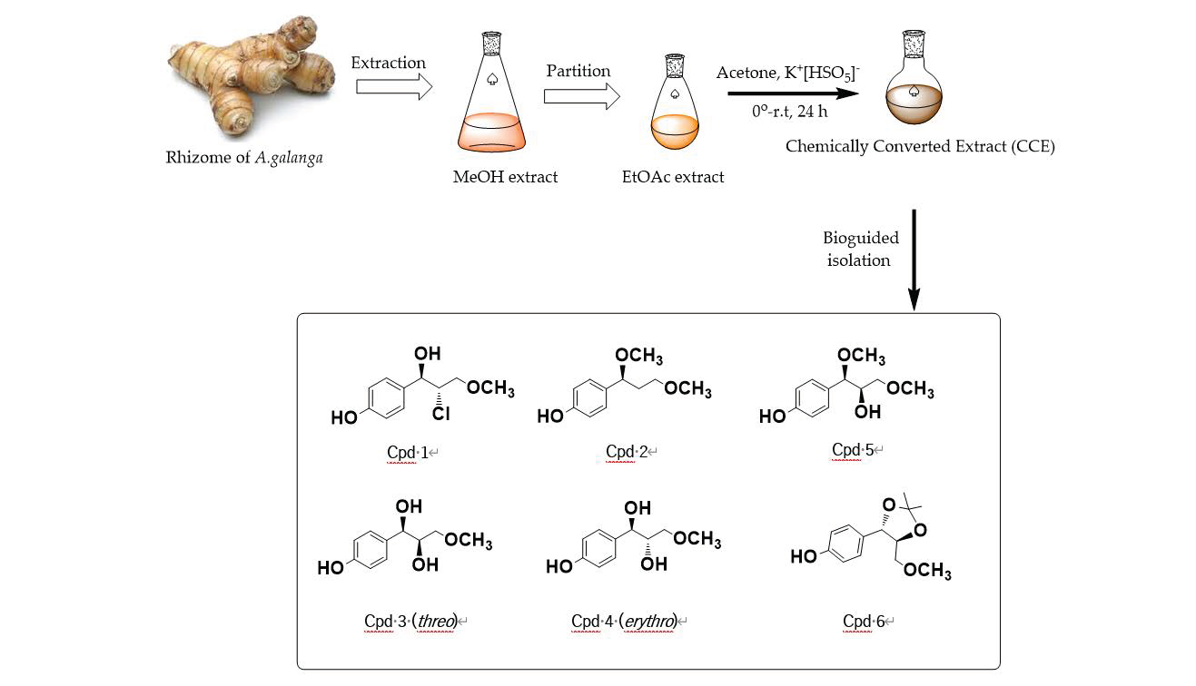
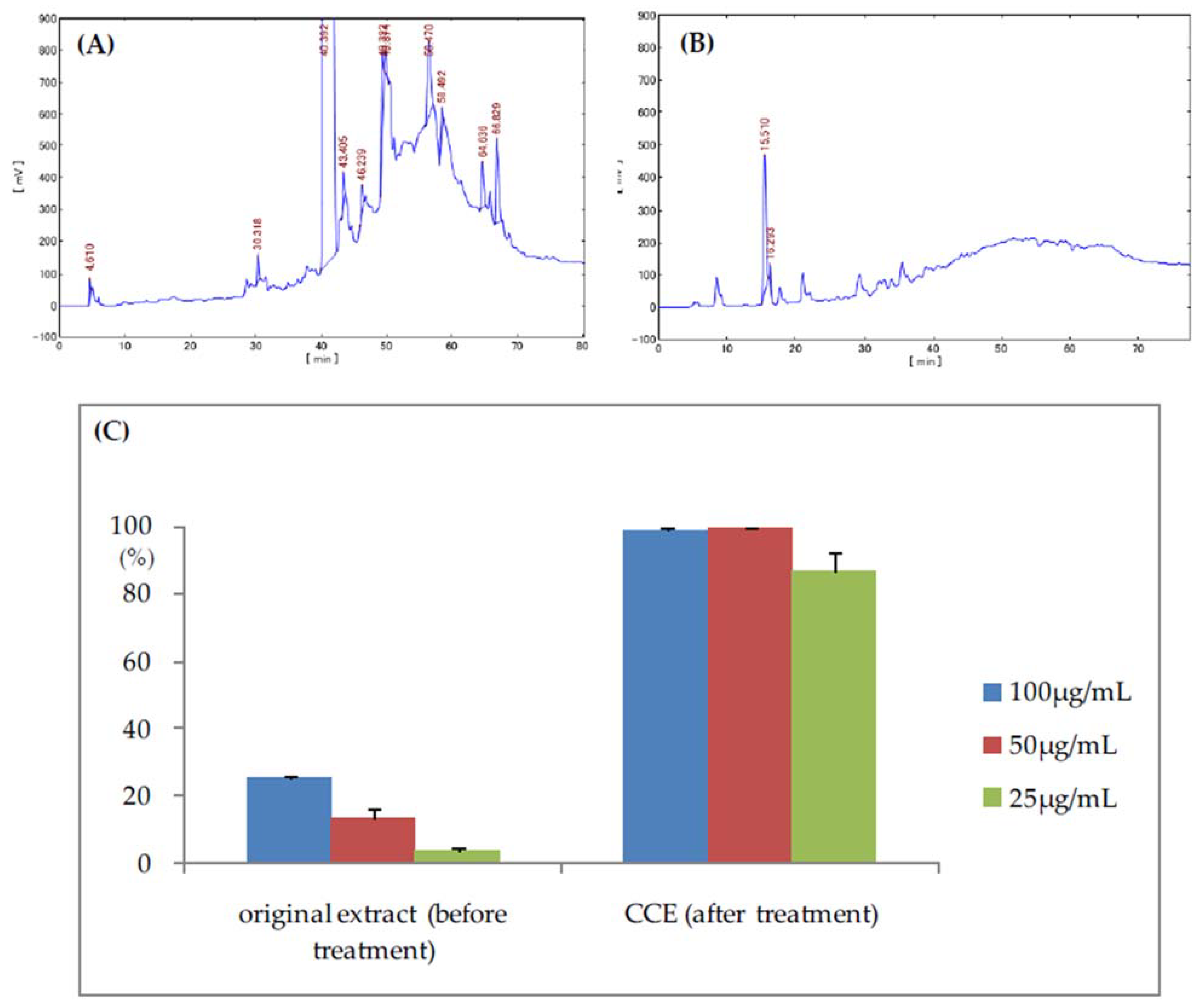
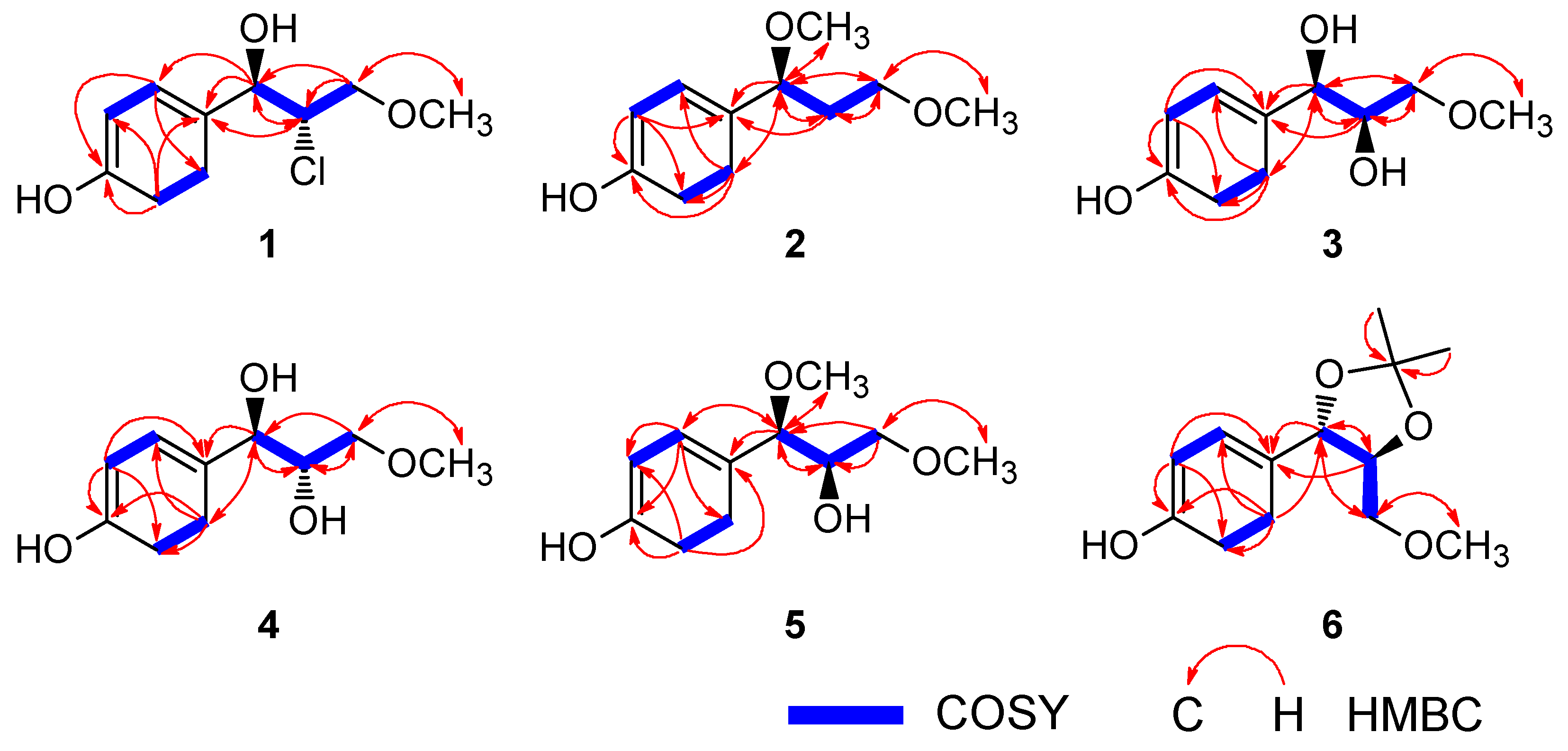
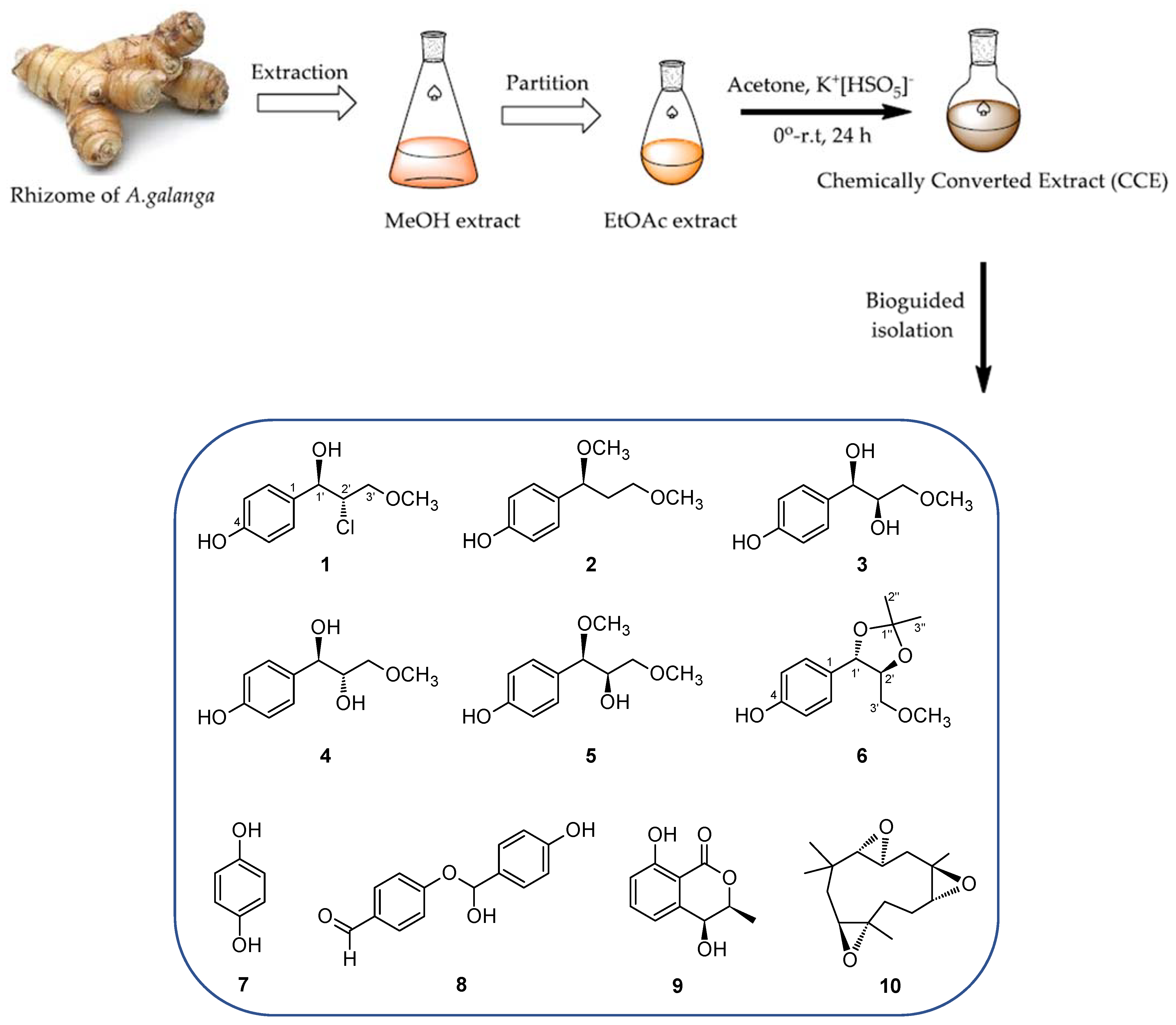
2.1. Isolation of Chemical Components of Chemically Converted Extract of Alpinia Galanga

The dried rhizomes of A. galanga (2.0 kg) were extracted with MeOH (3 × 10 L) at room temperature to give MeOH extract. Afterward, this extract was partitioned by solvent fractionation with ethyl acetate (EtOAc)/H2O to give EtOAc extract (E). The EtOAc extract was treated with dioxirane to produce the chemically converted extract (CCE) (Scheme 1). Dioxirane is a powerful reagent for epoxidation, which is generated in situ by the reaction of potassium peroxomonosulfate with acetone [30]. After being quenched and evaporated, HPLC analyses and biological assays of CCE and E were carried out. The anti-Leishmania activity of E and CCE against promastigotes of L. major was conducted by the colorimetric MTT (3-(4,5-dimethylthiazol-2-yl)-2,5-diphenyl-tetrazolium bromide) method [31]. The HPLC profile showed that major peaks in the original extract have vanished, and some new peaks appeared in the CCE (Figure 1). This result exhibited that new chemical components were produced by the chemical treatment. Furthermore, the inhibitory activity of CCE was significantly increased after treating with dioxirane. The CCE demonstrated 82.6 ± 6.2 % of inhibition against L. major, while the original EtOAc extract did not inhibit (2.7 ± 0.8 %) at 25 μg/mL (Figure 1).
Subsequently, by using the bioassay-guided isolation procedure, the CCE was further separated by various column chromatographies and HPLC to provide new compounds 16 (Scheme 1). In addition, four known compounds were identified such as hydroquinone (7) [32], 4-hydroxy(4-hydroxyphenyl)methoxy)benzaldehyde (8) [33], isocoumarin cis 4-hydroxymelein (9) [34], and (2S,3S,6R,7R,9S,10S)-humulene triepoxide (10) [35] by comparing with the spectral data (see Supplementary Materials Figures S4–S60, Tables S1–S2).
Compound 1 was obtained as a colorless liquid having molecular formula C10H13O3Cl from an [M + Na]+ ion at m/z 239.0446 with characteristic 3: 1 pattern of isotopic 35Cl and 37Cl in the high resolution electrospray ionization (HR-ESI) MS. The IR data exhibited absorption bands for hydroxy groups (3351 cm−1) and the C-Cl stretching bond (850 cm−1). The 1H NMR data (Table 1) showed the presence of one methoxy group at 3.36 ppm and a para-disubstituted aromatic ring with ortho-coupled protons at 7.21 and 6.76 ppm as doublets (J = 8.6 Hz) each in 1. The 1H and 13C-NMR data were closely similar to those of the analogous compound, erythro-2-chloro-1-(4-hydroxyphenyl)propane-1,3-diol. On the basis of the coupling constant of H-1′ and H-2′ (J = 5.9 Hz), 1 was assigned as erythro in accordance with a reference (J = 5.9 Hz) [36]. Therefore, the structure of 1 was determined to be 4-[threo-2-chloro-1-hydroxy-3-methoxypropyl]phenol.
Compound 2 was obtained as a colorless liquid. Its molecular formula showed to be C11H16O3 from a pseudo molecular ion at m/z 219.0991 ([M + Na]+) in the HRESIMS. The IR data showed bands for a hydroxyl group (3379 cm−1) and an aromatic ring (1604, 1510, and 1442 cm−1). The 1H-NMR data (Table 1) showed two methoxy groups at 3.14 and 3.30 ppm, and a para-substituted aromatic ring at 7.10 and 6.77 ppm as doublets (J = 8.5 Hz). According to the correlation spectroscopy (COSY) and heteronuclear multiple bond connectivity correlations (HMBC) (Figure 2), 2 was assigned as 4-(1,3-dimethoxypropyl)phenol.
Compounds 3 and 4 were isolated as different peaks by HPLC. Compounds 3 and 4 were colorless liquids with the same molecular formula C10H14O4 by HRESIMS at m/z 221.0781 ([M + Na]+). The IR data indicated the presence of hydroxy groups (3: 3384, and 4: 3394 cm−1) and an aromatic ring (3: 1606, 1514, and 1455, and 4: 1616, 1514, and 1455 cm−1). The 1H NMR data (Table 1) displayed signals caused by the presence of one methoxy group (3: 3.26, and 4: 3.34 ppm), a para-substituted benzene ring at δH 6.74 (2H, d, J = 8.5 Hz) and 7.16 (2H, d, J = 8.5 Hz) for 3, and δH 6.76 ppm (2H, d, J = 8.6 Hz) and 7.21 (2H, d, J = 8.6 Hz) for 4. The 13C-NMR data (Table 2) exhibited eight carbon peaks that were classified by DEPT and heteronuclear single quantum coherence (HSQC) spectrum as one methoxy carbon group (3: δC 59.2, and 4: δC 59.3), two sp2 methine carbons (3: δC 116.0 (2C), 129.1(2C), and 4: δC 115.8 (2C), 129.4(2C)), two sp2quaternary carbons (3: δC 133.9 and 158.0, and 4: δC 133.9 and 157.9) and three oxygenated carbons (3: δC 74.9, 75.5, and 76.0, and 4: δC 75.1, 75.0, and 75.6). These results indicated that compounds 3 and 4 have the same planar structure as shown in Scheme 1. Yang et al. and Li et al. reported slight but practical differences between erythro- and threo- forms of the related compounds, 1-(4-hydroxyphenyl)-1-methoxy-2,3-propanediol: that is, the erythro- and threo- forms had different coupling constants of H-1′ (6.6 Hz and 6.3 Hz, respectively), and the erythro-form showed a 0.9 ppm higher-field chemical shift value at C-2′ compared to that of threo-form. In the 1H and 13C-NMR spectrum of 3 and 4, the coupling constants of H-1′ were 6.6 and 6.3 Hz, respectively, and the relatively lower-field chemical shift value at C-2′ of 3C 76.0) compared to C-2′ of 4C 75.0) suggested threo- and erythro-form for 3 and 4, respectively, as shown in Scheme 1 [37,38].
Compound 5 was yielded as a colorless liquid with molecular formula C11H16O4 determined by HRESIMS from the peak at m/z 235.0939 ([M + Na]+). The IR data indicated bands for a hydroxy group (3379 cm−1) and an aromatic ring (1604, 1509, and 1455 cm−1). The 1H NMR data (Table 1) showed peaks caused by two methoxy groups at 3.19 and 3.24 ppm, and a para-substituted coupling system at δH 6.78 (2H, d, J= 8.5 Hz) and 7.13 (2H, d, J= 8.5 Hz). The 13C-NMR data (Table 2) exhibited nine carbon peaks that were classified by chemical shift values, DEPT and HSQC spectrum as two methoxy carbons (δC 56.8 and 59.2), two sp2 methine carbons (δC 116.2 (2C), 129.9 (2C)), two sp2 quaternary carbons (δC 130.6 and 158.5) and three oxygenated carbons (δC 74.6, 75.6, and 85.7). Chakraborty et al. recently stereoselectively synthesized the related compounds, threo- and erythro-1-(4-hydroxyphenyl)-1-methoxy-2,3-propanediol, and the coupling constants of H-1′ of threo- and erythro- forms were 7.0 and 6.4 Hz, respectively. According to this report, compound 5 showing the coupling constant of H-1′ (J = 7.3 Hz) can be concluded as a threo isomer, 4-[threo-2-hydroxy-1,3-dimethoxypropyl]phenol [39].
Compound 6 was isolated as a colorless liquid with molecular formula C13H18O4 as measured by HRESIMS at m/z 261.1099 ([M + Na]+). The IR data indicated absorption bands for a hydroxyl group (3368 cm−1) and the presence of an aromatic ring (1607 and 1512 cm−1). The 1H NMR data (Table 1) showed peaks caused by one methoxy group at 3.35 ppm, two methyl group at 1.46 and 1.51 ppm, and a para-disubstituted coupling system at δH 6.79 ppm (2H, d, J= 8.6 Hz) and 7.21 (2H, d, J= 8.6 Hz). The 13C-NMR data (Table 2) exhibited 11 carbon peaks that were categorized as two methyl carbons group (δC 27.3 and 27.4), one methoxy group (δC 59.6), two sp2 methine carbons (δC 116.4 (2C) and 129.3(2C)), two sp2 quaternary carbons (δC 129.5 and 158.8), and three oxygenated carbons (δC 72.6, 80.9, and 83.8). These results suggested a planar structure of 6 as shown in Scheme 1. The related acetonide, 4-(hydroxymethyl)-5-[3‘,5‘-dihydroxyphenyl]-2,2-dimethyl-1,3-dioxolane, a threo isomer, has coupling constant J= 8.1 Hz for H-1′ and H-2′ [40]. Morikawa et al. explained that threo- and erythro- forms of acetonides have similar chemical shift values except for the coupling constants of H-1′ and H-2′, that is, erythro- form (J = 2.8 Hz) and threo-form (J = 10.1 Hz) [41]. Therefore, based on the coupling constants of H-1′ and H-2′ (J = 8.7 Hz), compound 6 can be assigned as threo isomer as 4-[threo-5-(methoxymethyl)-2,2-dimethyl-1,3-dioxolan-4-yl]phenol.
All of the isolated compounds did not show significant optical rotation values, probably because of the formation of the racemic mixture through the chemical derivatization.

2.2. Antiparasitic Activity Examinations of the Isolated Compounds

All the isolated compounds were investigated for their biological activity toward selected protozoan parasites. Table 3 shows IC50 values of isolated compounds against L. major, T. b. gambisense, T. b. rhodesiense, and P. falciparum. Among isolated compounds, hydroquinone (7) had the highest activity against parasites belonging to the family Trypanosomatidae. The leishmanicidal and trypanocidal activities of hydroquinone derivatives had been reported [41,42]. Our result of 7 further supports the importance of the hydroquinone motif as an important pharmacophore. Compounds 2, 8 and 9 possessed significant activities against both L. major and Trypanosoma parasites. On the other hand, compound 1 had a strong activity on Trypanosoma parasites only. However, all isolated compounds showed no activity against P. falciparum (Table 3), probably because of the biological difference at the phylum level; Sarcomastigophora (L. major, T. b. gambiense, and T. b. rhodesiense) and Apicomplexa (P. falciparum). Alternatively, another possible reason could be the absence of a peroxide ring system which is important for the antimalarial activity of artemisinin [43]. Therefore, a chemical modification to introduce peroxide functions may be a promising method to produce novel antimalarial agents in the future.
To our understanding, there are no reports on the biological activity of 4-hydroxyphenylpropanols against L. major, P. falciparum, T. brucei gambisense, and T. brucei rhodeisense. Therefore, our study is the first report on the activities of 4-hydroxyphenylpropanol and related compounds against those parasites. Ropes et al. reported the derivatives of p-coumaric acid alkyl esters and their inhibitory activities against Leishmania and Plasmodium [44]. The presence of alkyl function on C-3′ (-OMe) and the relatively non-polar nature of 1 and 2 may be related to the higher activity among 16.

3. Materials and Methods

3.1. General Methods

Optical rotations were determined on a P-1030 digital polarimeter (JASCO, Tokyo, Japan). IR spectra were measured on a FT-710 Fourier transform infrared spectrophotometer (Horiba, Kyoto, Japan), while UV spectra were taken on a V-520 UV/Vis spectrophotometer (JASCO, Tokyo, Japan). NMR data were obtained by an Ultrashield 600 spectrometer (Bruker, Ettlingen, Germany) with tetramethylsilane (TMS) as an internal standard. Positive ion HR-ESI-MS measurement was performed with the LTQ Orbitrap XL mass spectrometer (Thermo Fisher Scientific, Waltham, MA, USA). Column chromatography (CC) was carried out on silica gel 60 (E. Merck, Darmstadt, Germany), and octadecyl silica (ODS) gel (Cosmosil 75C18-OPN (NacalaiTesque, Kyoto, Japan; Φ = 35 mm, L = 350 mm). HPLC was performed on ODS gel (Inertsil ODS-3, GL-science, Φ = 6 mm, 250 mm, flow rate = 1.6 mL/min, GL Sciences, Tokyo, Japan), and the eluate was observed by refractive index detector with the RI-930 intelligent detector (JASCO, Tokyo, Japan) and a PU-1580 intelligent pump (JASCO, Tokyo, Japan). All chemicals were purchased from Wako Pure Chemical Industries, Ltd. (Osaka, Japan) and TCI (Tokyo, Japan).

3.2. Plant Material

Rhizome of A. galanga (L.) was purchased from Shinwa-bussan Co., Osaka, Japan, in 2014. A voucher specimen (Ay-AG2014-Shinwa) was deposited in the herbarium of the Department of Pharmacognosy, Graduate School of Biomedical and Health Sciences, Hiroshima University, Japan.

3.3. Preparation of Chemically Converted Extract of A. galanga and Isolation of Compounds 110

Rhizome of A. galanga (2.0 kg) was cut into small pieces and extracted by maceration at room temperature with MeOH (10 L) three times and then evaporated to give an MeOH extract (303 g). This residue was suspended in 1 L of saturated sodium carbonate solution and partitioned with EtOAc (1 L) three times to produce an EtOAc extract (68 g).
The EtOAc extract (14 g) was dissolved in the mixture of acetonitrile (360 mL) and water (360 mL). Then, acetone (10.35 mL), sodium bicarbonate (19.6 g) and potassium peroxosulphate (28.2 g) were added sequentially to the solution at 0 °C. The mixture was stirred for 3 h at 0 °C and then potassium peroxosulphate (28.2 g) was added again to the solution with stirring for another 3 h at 0 °C. The mixture was then allowed to rise naturally to room temperature with stirring for 24 h. The obtained reaction mixture was poured into water and extracted with ethyl acetate three times. The combined EOAc layer was washed with water, brine, dried over sodium sulfate, and evaporated to yield a residue, designated as CCE of A. galanga (10.5 g).
The CCE was separated on a silica gel (50 g) CC and eluted with an increasing amount of MeOH in CHCl3 (20:1, 10:1, 7:1, 5:1, 3:1, 1:1, and 100% MeOH, 500 mL each), generating nine fractions. Fraction CCE2 (1.1 g), CCE3 (1.4 g) and CCE4 (4.0 g) were subjected to ODS CC with a step gradient from 10% aq. MeOH to 100% MeOH, (400 mL each), leading to eight fractions. The residue of fraction CCE 2.1 (226 mg) was purified by HPLC using 20% aq. acetone to give 1 (40.1 mg), 7 (hydroquinone, 102 mg), and 8 (4-hydroxy(4-hydroxyphenyl)methoxy)benzaldehyde, 20.2 mg). The residue of fraction CCE 2.2 (79.5 mg) was purified by HPLC using 30% aq. MeOH to give 1 (1.9 mg), 9 (isocoumarin cis-4-hydroxymelein, 2.2 mg), 2 (2.4 mg), 6 (5.8 mg), and 10 ((2S, 3S, 6R, 7R, 9S, 10S)-humulene triepoxide, 2.8 mg). The residue of fraction CCE 3.1 (297.5 mg) was purified by HPLC using 20% aq. acetone to give 7 (70.1 mg), 3 (20.1 mg), 4 (6.0 mg), and 5 (12.5 mg) (Supplementary Materials Figures S1–S3).
4-[erytho-2-chloro-1-hydroxy-3-methoxypropyl]phenol (1)
Colorless oil, IR υmax (film) cm−1: 3351, 2935, 1668, 1607, 1454, 1377, 1236, 1118, 1038, 850; UV λmax (MeOH) nm (log ε): 276 (2.58); 1H NMR (CD3OD, 600 MHz), see Table 1; 13C-NMR (CD3OD, 150 MHz), see Table 2. HRESIMS (positive ion) m/z: 239.0446 [M + Na]+ (calcd for C10H13O3ClNa: 239.0445).
4-(1,3-dimethoxypropyl)phenol (2)
Colorless oil, IR υmax (film) cm−1: 3379, 2930, 2854, 1718, 1604, 1510, 1442, 1233, 1104; UV λmax (MeOH) nm (log ε): 259 (2.57); 1H NMR (CD3OD, 600 MHz), see Table 1; 13C-NMR (CD3OD, 150 MHz), see Table 2. HRESIMS (positive ion) m/z: 219.0991 [M + Na]+ (calcd for C11H16O3Na: 219.0992).
threo-1-(4-hydroxyphenyl)-3-methoxypropane-1,2-diol (3)
Colorless oil, IR υmax (film) cm−1: 3384, 2987, 2831, 1677, 1606, 1514, 1455, 1240, 1119; UV λmax (MeOH) nm (log ε): 276 (2.96); 1H-NMR (CD3OD, 600 MHz), see Table 1; 13C-NMR (CD3OD, 150 MHz), see Table 2. HRESIMS (positive ion) m/z: 221.0781 [M + Na]+ (calcd for C10H14O4Na: 221.0784).
erythro-1-(4-hydroxyphenyl)-3-methoxypropane-1,2-diol (4)
Colorless oil, IR υmax (film) cm−1: 3394, 2930, 2360, 1679, 1616, 1514, 1455, 1240; UV λmax (MeOH) nm (log ε): 276 (2.33); 1H NMR (CD3OD, 600 MHz), see Table 1; 13C-NMR (CD3OD, 150 MHz), see Table 2. HRESIMS (positive ion) m/z: 221.0781 [M + Na]+ (calcd for C10H14O4Na: 221.0784).
4-(threo-2-hydroxy-1,3-dimethoxypropyl)phenol (5)
Colorless oil, IR υmax (film) cm−1: 3379, 2930, 2360, 1604, 1509, 1455, 1375; UV λmax (MeOH) nm (log ε): 279 (3.26); 1H NMR (CD3OD, 600 MHz), see Table 1; 13C-NMR (CD3OD, 150 MHz), see Table 2. HRESIMS (positive ion) m/z: 235.0939 [M + Na]+ (calcd for C11H16O4Na: 235.0941).
4-(threo-5-(methoxymethyl)-2,2-dimethyl-1,3-dioxolan-4-yl)phenol (6)
Colorless oil, IR υmax (film) cm−1: 3368, 2986, 1732, 1607, 1512, 1375, 1230, 1165, 1078; UV λmax (MeOH) nm (log ε): 276 (2.95); 1H NMR (CD3OD, 600 MHz), see Table 1; 13C-NMR (CD3OD, 150 MHz), see Table 2. HRESIMS (positive ion) m/z: 261.1099 [M + Na]+ (calcd for C13H18O4Na: 261.1097).

3.4. Biological Assay of Compounds 110

The leishmanicidal activities of the isolated compounds were evaluated using MTT colorimetric assay, as described by Asaumi et al. [31] MTT, 3-(4,5-dimethylthiazol-2-yl)-2,5-diphenyltetrazolium bromide, was reduced to form an insoluble purple precipitate, formazan, by cell metabolic activity, which is known to correlate to the viable cell number. In a 96-well plate, 1 µL of sample solutions in dimethylsulfoxide (DMSO) and L. major (1 × 10⁵ parasite/well) in 99 µL of medium were added to each well. It was then incubated for 72 h in a CO2 incubator at 25 °C. M-199 medium, supplemented with 10% heat-inactivated fetal bovine serum and 100 µg/mL of kanamycin, was used. After incubation, 100 µL of MTT solution was added to each well and the plates were incubated for another 8 h. The absorbance of the formazan solution in DMSO was recorded using a microplate reader at λ540 nm.
The trypanocidal activities of the isolated compounds were performed in 96-well plates as previously explained with slight modifications [45]. The amount of ATP was proportional to the number of metabolically active cells. This method utilized the enzymatic reaction of luciferase and luciferin that requires ATP to generate luminescence. In brief, each well contained 100 µL of parasite culture (1 × 10⁴ parasites/well) with serial dilutions of compounds. After incubation for 72 h at 37 °C under 5% CO2, 25 µL of CellTiter-GloTM Luminescent Cell Viability Assay reagent (Promega Japan, Tokyo, Japan) was added to evaluate intracellular ATP concentration according to the instruction.
Antimalarial activity of isolated compounds was evaluated according to the previous report [46]. The SYBR green assay is widely used for the antimalarial assay. This dye binds to parasite DNA and emits fluorescence reflecting the number of parasites in the red blood cells. In brief, 100 µL of P. falciparum parasite culture [47] was plated in a 96-well plate with various concentrations of the compounds. After incubation for 72 h at 37 °C in a humidified chamber under a gas mixture of 90%N2, 5%O2 and 5%CO2, the parasitemia was determined by SYBR Green I assay (Lonza Ltd., Basel, Switzerland) with a microplate reader at 485 and 530 nm. Dihydroartmesinin was used as the positive control and DMSO as a negative control. Human erythrocytes and plasma were obtained from the Nagasaki Red Cross Blood Center, and their usage was approved by the ethical committee of the Institute of Tropical Medicine, Nagasaki University.
The biological assays were performed three times for each assay and the IC50 values were calculated by linear regression using Microsoft Excel software.

4. Conclusions

The ethyl acetate extract of A. galanga was originally inactive against L. major. However, the chemical conversion by epoxidation generated significant activity to this parasite. The HPLC profile clearly indicated the constituent difference between the original and chemically converted extract (CCE) of A. galanga. Through the intensive fractionation and purification of CCE, six new phenylpropanoids (16) and four known compounds were obtained as active principles. The most active compound was hydroquinone (7), which further supports the importance of this chemical motif as shown in our previous report on the discovery of new anti-Leishmanial benzoquinones, named ardisiaquinones A–H [31]. In addition, the new phenylpropanoid 1 was revealed to inhibit two African Trypanosoma parasites effectively with high selective manner compared to L. major. Phenylpropanoids are an important constituent of plant essential oils. Several plant oils have been reported to show antiparasitic activity by inhibiting proteinase, dehydrogenase, and lipid trafficking [48]. The phenylpropanoids 16 isolated in this study may have a similar mode of action. The chemical synthesis of analogous compounds of 1 and the mechanism analysis are under investigation.

Supplementary Materials

The following are available online, Figure S1: HPLC chromatogram for the isolation of 1, 7 and 8, Figure S2: HPLC chromatogram for the isolation of 1, 2, 6, 9 and 10, Figure S3: HPLC chromatogram for the isolation of 3, 4, 5 and 7, Figure S4: HR-ESI-MS of 1, Figure S5: 1H NMR spectrum of 1, Figure S6: 13C NMR spectrum of 1, Figure S7: DEPT 135 spectrum of 1, Figure S8: COSY spectrum of 1, Figure S9: HSQC spectrum of 1, Figure S10: HMBC spectrum of 1,Figure S11: IR spectrum of 1, Figure S12: HR-ESI-MS of 2, Figure S13: 1H NMR spectrum of 2, Figure S14: 13C NMR spectrum of 2, Figure S15: DEPT 135 spectrum of 2, Figure S16: COSY spectrum of 2, Figure S17: HSQC spectrum of 2, Figure S18: HMBC spectrum of 2, Figure S19: IR spectrum of 2, Figure S20: HR-ESI-MS of 3, Figure S21: 1H NMR spectrum of 3, Figure S22: 13C NMR spectrum of 3, Figure S23: DEPT 135 spectrum of 3, Figure S24: COSY spectrum of 3, Figure S25: HSQC spectrum of 3, Figure S26: HMBC spectrum of 3, Figure S27: IR spectrum of 3, Figure S28: HR-ESI-MS of 4, Figure S29: 1H NMR spectrum of 4, Figure S30: 13C NMR spectrum of 4, Figure S31: DEPT 135 spectrum of 4, Figure S32: COSY spectrum of 4, Figure S33: HSQC spectrum of 4, Figure S34: HMBC spectrum of 4, Figure S35: IR spectrum of 4, Figure S36: HR-ESI-MS of 5, Figure S37: 1H NMR spectrum of 5, Figure S38: 13C NMR spectrum of 5, Figure S39: DEPT 135 spectrum of 5, Figure S40: COSY spectrum of 5, Figure S41: HSQC spectrum of 5, Figure S42: HMBC spectrum of 5, Figure S43: IR spectrum of 5, Figure S44: HR-ESI-MS of 6, Figure S45: 1H NMR spectrum of 6, Figure S46: 13C NMR spectrum of 6, Figure S47: DEPT 135 spectrum of 6, Figure S48: COSY spectrum of 6, Figure S49: HSQC spectrum of 6, Figure S50: HMBC spectrum of 6, Figure S51: IR spectrum of 6, Figure S52: 1H NMR spectrum of 7, Figure S53: 13C NMR spectrum of 7, Figure S54: 1H NMR spectrum of 8, Figure S55: 13C NMR spectrum of 8, Figure S56: 1H NMR spectrum of 9, Figure S57: 13C NMR spectrum of 9, Figure S58: 1H NMR spectrum of 10, Figure S59: 13C NMR spectrum of 10, Figure S60: Structures of positive control for the bioassay, Table S1: COSY correlations of compounds 16, Table S2: HMBC correlations of compounds 16.

Author Contributions

Conceptualization, K.M. and M.I.S.; synthesis and isolation, M.I.S. and N.H.U.; manuscript preparation, K.M., M.I.S., O.K. and K.S.; facilitated the research and supplied the materials, S.S. and Y.Y.; Anti-plasmodium assay, O.K, K.Y. and B.-Y.A.C.; Anti-trypanosoma assay, K.S.; spectrometric measurement, S.K. and H.O.; anti-Leishmania assay, K.M. and M.I.S. All authors have read and agreed to the published version of the manuscript.

Funding

This work was funded in part by Grants-in-Aid from the Ministry of Education, Culture, Sports, Science and Technology of Japan, and the Japan Society for the Promotion of Science (Nos. 18K06740, and 17K08336, 17K15465, and 16K18793). This work was also partly supported by the Joint Usage/Research Center on Tropical Disease, Institute of Tropical Medicine, Nagasaki University (2019-Ippan-1, 2020-Ippan-18) and Ohyama Health Foundation Inc.

Data Availability Statement

Data is contained within the article or supplementary material.

Acknowledgments

The authors are indebted for superconducting spectroscopic instruments at the Natural Science Center for Basic Research and Development (N-BARD), Hiroshima University. The authors are grateful to the Indonesia Endowment Fund for Education (LPDP), Ministry of Finance, the Republic of Indonesia for doctoral scholarships.

Conflicts of Interest

The authors declare no conflict of interest.

Sample Availability

Samples of the compounds 16 are available from the authors.

References

  1. Neglected Tropical Diseases: Fact Sheets Relating to NTDs. Available online: https://www.who.int/neglected_diseases/mediacentre/factsheet/en/ (accessed on 17 February 2021).
  2. Malaria: Fact Sheets Relating to Malaria. Available online: https://www.who.int/en/news-room/fact-sheets/detail/malaria (accessed on 17 February 2021).
  3. Trypanosomiasis, Human African (Sleeping Sickness): Fact Sheets. Available online: https://www.who.int/news-room/fact-sheets/detail/trypanosomiasis-human-african-(sleeping-sickness) (accessed on 17 February 2021).
  4. Jesse, W.-H.L.; Vederas, J.C. Drug Discovery and Natural Products: End of an Era or an Endless Frontier? Science 2009, 325, 161–165. [Google Scholar]
  5. Newman, D.J.; Cragg, G.M. Natural Products as Sources of New Drugs from 1981 to 2014. J. Nat. Prod. 2016, 79, 629–661. [Google Scholar] [CrossRef] [Green Version]
  6. The Dictionary of Natural Products. CRC Press. Available online: https://www.routledge.com/go/the_dictionary_of_natural_products (accessed on 17 February 2021).
  7. Salazar, M.O.; Ramallo, I.A.; Micheloni, O.; Sierra, M.G.; Furlan, R.L. Chemically engineered extracts: Bioactivity alteration through sulfonylation. Bioorganic Med. Chem. Lett. 2009, 19, 5067–5070. [Google Scholar] [CrossRef] [PubMed]
  8. Ramallo, I.A.; Salazar, M.O.; Méndez, L.; Furlán, R.L.E. Chemically Engineered Extracts: Source of Bioactive Compounds. Accounts Chem. Res. 2011, 44, 241–250. [Google Scholar] [CrossRef] [PubMed]
  9. López, S.N.; Ramallo, I.A.; Sierra, M.G.; Zacchino, S.A.; Furlan, R.L.E. Chemically engineered extracts as an alternative source of bioactive natural product-like compounds. Proc. Natl. Acad. Sci. USA 2006, 104, 441–444. [Google Scholar] [CrossRef] [Green Version]
  10. Wu, T.; Jiang, C.; Wang, L.; Morris-Natschke, S.L.; Miao, H.; Gu, L.; Xu, J.; Lee, K.H.; Gu, Q. 3,5-Diarylpryrazole Derivatives Obtained by Ammonolysis of the Total Flavonoids from Chrysanthemum indicum extract Show Potential for the Treatment of Alzheimer’s Disease. J. Nat. Prod. 2015, 78, 1593–1599. [Google Scholar] [CrossRef] [PubMed]
  11. Mendez, L.; Salazar, M.O.; Ramallo, I.A.; Furlán, R.L.E. Brominated Extracts as Source of Bioactive Compounds. ACS Comb. Sci. 2010, 13, 200–204. [Google Scholar] [CrossRef] [PubMed]
  12. Kawamura, T.; Matsubara, K.; Otaka, H.; Tashiro, E.; Shindo, K.; Yanagita, R.C.; Irie, K.; Imoto, M. Generation of ‘Unnatural Natural Product’ library and identification of a small molecule inhibitor of XIAP. Bioorganic Med. Chem. 2011, 19, 4377–4385. [Google Scholar] [CrossRef] [PubMed]
  13. Kikuchi, H.; Sakurai, K.; Oshima, Y. Development of Diversity-Enhanced Extracts of Curcuma zedoria and Their New Ses-quiterpene-like Compounds. Org. Lett. 2014, 16, 1916–1919. [Google Scholar] [CrossRef] [PubMed]
  14. Ramallo, I.A.; Sierra, M.G.; Furlan, R.L.E. Discovery of β-Glucosidase Inhibitors from a Chemically Engineered Extract Prepared through Ethanolysis. Med. Chem. 2012, 8, 112–117. [Google Scholar] [CrossRef]
  15. Padalia, R.C.; Verma, R.S.; Sundaresan, V.; Chanotiya, C.S. Chemical Diversity in the Genus Alpinia (Zingiberaceae): Com-parative Composition of Four Alpinia Species Grown in Northern India. Chem. Biodivers. 2010, 7, 2076–2087. [Google Scholar] [CrossRef]
  16. Zhu, X.-L.; Yang, M.-H.; Luo, J.-G.; Huang, X.-F.; Kong, L.-Y. A New Phenylpropanoid from Alpiniagalanga. Chin. J. Nat. Med. 2009, 7, 19–20. [Google Scholar] [CrossRef]
  17. Matsuda, H.; Morikawa, T.; Managi, H.; Yoshikawa, M. Antiallergic Principles from Alpinia galangal: Structural Requirements of Phenylpropanoids for Inhibition of Degranulation and Release of TNF-α and IL-4 in RBL-2H3 Cells. Bioorg. Med. Chem. 2003, 13, 3197–3202. [Google Scholar] [CrossRef]
  18. Zhao, L.; Chen, L.-Y.; Liang, J.-Y. Two new phenylpropanoids isolated from the rhizomes of Alpiniagalanga. Chin. J. Nat. Med. 2012, 10, 370–373. [Google Scholar]
  19. Chourasiya, S.S.; Sreedhar, E.; Babu, K.S.; Shankaraia, N.; Nayak, L.; Ramakrishna, S.; Sravani, S.; Rao, B.M.V. Isolation, Synthesis and Biological Evaluation of Phenylpropanoids from the Rhizomes of Alpiniagalanga. Nat. Prod. Commun. 2013, 8, 1741–1746. [Google Scholar]
  20. Laksmi, S.; Sandra, S.; Rahul, B.S.; Saikant, B.; Vani, M.; Manoj, G.; Padmaja, G.; Remani, P. In vitro and in vivo studies of 5,7-dihydroxy flavones isolated from Alpinia galangal (L.) against human lung cancer and ascetic lymphoma. Med. Chem. Res. 2019, 28, 39–51. [Google Scholar] [CrossRef]
  21. Morikawa, T.; Ando, S.; Matsuda, H.; Kataoka, S.; Muraoka, O.; Yoshikawa, M. Inhibitors of Nitric Oxide Production from the Rhizomes of Alpiniagalangal: Structures of New 8-9′ Linked Neolignans and Sesquineolignan. Chem. Pharm. Bull. 2005, 53, 625–630. [Google Scholar] [CrossRef] [Green Version]
  22. Oonmetta-aree, J.; Suzuki, T.; Gasaluck, P.; Eumkeb, G. Antimicrobial properties and action of galangal (Alpiniagalanga Linn.) on Staphylococccus aureus. LWT 2006, 39, 1214–1220. [Google Scholar] [CrossRef]
  23. Gupta, P.; Bhatter, P.; D’souza, D.; Tolani, M.; Daswani, P.; Tetali, P.; Birdi, T. Evaluating the anti Mycobacterium tuberculosis activity of Alpiniagalanga (L.) Willd. Axenically under reducing oxygen conditions and in intracellular assays. BMC Complement. Altern. Med. 2014, 14, 84–92. [Google Scholar] [CrossRef]
  24. Tang, X.; Xu, C.; Yagiz, Y.; Simonne, A.; Marshall, M.R. Phytochemical profiles, and antimicrobial and antioxidant activities of greater galangal [Aplinia galangal (Linn.) Swartz.] flowers. Food Chem. 2018, 255, 300–308. [Google Scholar] [CrossRef]
  25. Chansriniyom, C.; Bunwatcharaphansakun, P.; Eaknai, W.; Nalinratana, N.; Ratanawong, A.; Khongkow, M.; Leuchapudi-porn, R. A synergistic combination of Phyllanthus emblica and Alpiniagalanga against H2O2-induced oxidative stress and lipid peroxidation in human ECV304 cells. J. Funct. Foods 2018, 43, 44–54. [Google Scholar] [CrossRef]
  26. Singh, J.C.H.; Alagarsamy, V.; Diwan, P.V.; Kumar, S.S.; Nisha, J.C.; Reddy, Y.N. Neuroprotective effect of Alpiniagalanga (L) fractions on Aβ(25-35) induced amnesia in mice. J. Ethnopharmacol. 2011, 138, 85–91. [Google Scholar] [CrossRef]
  27. Seo, J.-W.; Cho, S.-C.; Park, S.-J.; Lee, E.-J.; Lee, J.-H.; Han, S.-S.; Pyo, B.S.; Park, D.-H.; Kim, B.-H. 1′-Acetoxychavicol Acetate Isolated from Alpiniagalangal Ameliorates Ovalbumin-Induced Asthma in Mice. PLoS ONE 2013, 8, e56447. [Google Scholar] [CrossRef] [PubMed]
  28. Yoshiaki, M.; Ninomiya, K.; Nishi, R.; Kamei, I.; Katsuyama, Y.; Imagawa, T.; Chaipech, S.; Muraoka, O.; Morikawa, T. Mel-anogenesis inhibitory activity of a 7-O-9′-linked neolignan from Alpiniagalangafruit. Bioorg. Med. Chem. 2016, 24, 6215–6224. [Google Scholar]
  29. Thanh, B.L.; Claire, B.; Duc Trong, N.; Marie-Paule, M.; Joelle, Q.L. In vitro Anti-Leishmanial Activity of Essential Oils Ex-tracted from Vietnamese Plants. Molecules 2017, 22, 1071–1083. [Google Scholar]
  30. Yang, D.; Wong, M.-K.; Yip, Y.-C. Epoxidation of Olefins Using Methyl(trifluoromethyl)dioxirane Generated in Situ. J. Org. Chem. 1995, 60, 3887–3889. [Google Scholar] [CrossRef]
  31. Asaumi, S.; Kawakami, S.; Sugimoto, S.; Matsunami, K.; Otsuka, H.; Shinzato, T. Alkylated Benzoquinones: Ardisiaquinones A–H from the Leaves of Ardisiaquinquegona and Their Anti-Leishmania Activity. Chem. Pharm. Bull. 2018, 66, 757–763. [Google Scholar] [CrossRef] [Green Version]
  32. Bernini, R.; Coratti, A.; Provenzano, G.; Fabrizi, G.; Tofani, D. Oxidation of aromatic aldehydes and ketones by H2O2/CH3ReO3 in ionic liquids: A catalytic efficient reaction to achieve dihydric phenols. Tetrahedron 2005, 61, 1821–1825. [Google Scholar] [CrossRef]
  33. Tao, P.; Meng, Q.-H.; Zhang, H.-F.; Zhang, C.-H.; Fang, N.-H. Analysis of impurities in 4-hydrobenzaldehyde by HPLC, combined with mass spectroscopy. Ranliao Yu Ranse 2005, 42, 59–60. [Google Scholar]
  34. Rivera-Chavez, J.; Figueroa, M.; Gonzalez, M.C.; Glenn, A.E.; Mata, R. α-Glucosidase Inhibitors from a Xylariafeejeensis Associated with Hintonialatiflora. J. Nat. Prod. 2015, 78, 730–735. [Google Scholar] [CrossRef]
  35. Hayano, K.; Mochizu, K. First Isolation and Determination of Three Humulene Triepoxides. Heterocycles 1997, 45, 1573–1577. [Google Scholar] [CrossRef]
  36. Swamy, P.; Kumar, M.A.; Reddy, M.M.; Naresh, M.; Srujana, K.; Narender, N. The vicinal functionalization of olefins: A facile route to the direct synthesis of β-chlorohydrins and β-chloroethers. RCS Adv. 2014, 4, 26288–26294. [Google Scholar] [CrossRef]
  37. Yang, X.-W.; Li, S.-M.; Li, Y.-L.; Feng, L.; Shen, Y.-H.; Lin, S.; Tian, J.-M.; Zeng, H.-W.; Wang, N.; Steinmetz, A.; et al. Chemical constituents of Abies delavayi. Phytochemistry 2014, 105, 164–170. [Google Scholar] [CrossRef]
  38. Li, Y.-L.; Gao, Y.-X.; Jin, H.-Z.; Shan, L.; Chang, W.-L.; Yang, X.-W.; Zeng, H.-W.; Wang, N.; Steinmetz, A.; Zhang, W.-D. Chemical constituents of Abies fabri. Phytochemistry 2015, 117, 135–143. [Google Scholar] [CrossRef] [PubMed]
  39. Chakraborty, I.; Chatterjee, S.; Manna, A.; Bhaumik, T. Confirmation of the stereochemistry of two naturally occurring epimeric phenylpropanoids via synthesis: Elucidation of hitherto unknown full stereostructures. Tetrahedron Lett. 2019, 60, 1597–1599. [Google Scholar] [CrossRef]
  40. Balamurugan, R.; Kothapalli, R.B.; Thota, G.K. Gold-Catalysed Activation of Epoxides: Application in the Synthesis of Bicyclic Ketals. Eur. J. Org. Chem. 2011, 2011, 1557–1569. [Google Scholar] [CrossRef]
  41. Shukla, A.K.; Patra, S.; Dubey, V.K. Evaluation of selected antitumor agents as subversive substrate and potential inhibitor of trypanothione reductase: An alternative approach for chemotherapy of Leishmaniasis. Mol. Cell. Biochem. 2011, 352, 261–270. [Google Scholar] [CrossRef]
  42. Aldunate, J.; Traipe, L.; Spencer, P.; Morelló, A.; Repetto, Y. Effects of hydroquinones on intact Trypanosoma cruzi epimastigotes. Comp. Biochem. Physiol. Part C Comp. Pharmacol. 1992, 103, 97–100. [Google Scholar] [CrossRef]
  43. Grigorov, M.G.; Weber, J.; Tronchet, J.M.J.; Jefford, C.W.; Jefford, C.W.; Jefford, C.W.; Milhous, W.K.; Maric, D.M. A QSAR Study of the Antimalarial Activity of Some Synthetic 1,2,4-Trioxanes£. J. Chem. Inf. Comput. Sci. 1997, 37, 124–130. [Google Scholar] [CrossRef]
  44. Lopes, S.P.; Yepes, L.M.; Pérez-Castillo, Y.; Robledo, S.M.; De Sousa, D.P. Alkyl and Aryl Derivatives Based on p-Coumaric Acid Modification and Inhibitory Action against Leishmania braziliensis and Plasmodium falciparum. Molecules 2020, 25, 3178. [Google Scholar] [CrossRef]
  45. Suganuma, K.; Allamanda, P.; Hakimi, H.; Zhou, M.; Angeles, J.M.; Kawazu, S.-I.; Inoue, N. Establishment of ATP-Based Luciferase Viability Assay in 96-Well Plate for Trypanosoma congolense. J. Veter. Med. Sci. 2014, 76, 1437–1441. [Google Scholar] [CrossRef] [PubMed] [Green Version]
  46. Ishikawa, T.; Mizuta, S.; Kaneko, O.; Yahata, K. Fragment Molecular Orbital Study of the Interaction between Sar-co/Endoplasmic Reticulum Ca2+-ATPase and its Inhibitor Thapsigargin toward Anti-Malarial Development. J. Phys. Chem. B. 2018, 122, 7970–7977. [Google Scholar] [CrossRef] [PubMed]
  47. Walliker, D.; A Quakyi, I.; E Wellems, T.; McCutchan, T.F.; Szarfman, A.; London, W.T.; Corcoran, L.M.; Burkot, T.R.; Carter, R. Genetic analysis of the human malaria parasite Plasmodium falciparum. Science 1987, 236, 1661–1666. [Google Scholar] [CrossRef] [PubMed]
  48. Anthony, J.-P.; Fyfe, L.; Smith, H. Plant active components—A resource for antiparasitic agents? Trends Parasitol. 2005, 21, 462–468. [Google Scholar] [CrossRef] [PubMed]
Scheme 1. Isolation of compounds from the chemically converted extract of A. galanga.
Molecules 26 01756 sch001
Figure 1. UV-HPLC profiles and anti-Leishmania major activity of A. galanga extract; (A) before (B) after treatment with dioxirane. HPLC: ODS, UV: 215 nm; (C) the percentage of inhibition against L. major before and after chemical treatment. The error bar represents SE, n = 3.
Molecules 26 01756 g001
Figure 2. H-H COSY and HMBC correlations of compounds 16.
Molecules 26 01756 g002
Table 1. 1H-NMR spectroscopic data for compounds 16.
Position123456
2,67.21 d (8.6)7.10 d (8.5)7.16 d (8.5)7.21 d (8.6)7.13 d (8.5)7.21 d (8.6)
3,56.76 d (8.6)6.77 d (8.5)6.74 d (8.5)6.76 d (8.6)6.78 d (8.5)6.79 d (8.6)
1′4.80 d (5.9)4.19 dd (7.8, 6.1)4.49 d (6.6)4.52 d (6.3)4.08 d (7.3)4.69 d (8.7)
1′-OCH3 3.14 s 3.19 s
2′4.22 dt-like
(5.9, 5.2)
2.02 m
1.78 m
3.73 td-like
(6.6, 3.4)
3.84 td-like
(6.5, 3.4)
3.74 ddd
(7.3, 5.8, 3.2)
3.89 ddd
(8.7, 5.6, 2.8)
3′3.61 m (2H)3.44 dt-like
(9.4, 6.5)
3.14 dd
(10.1, 6.2)
3.44 dd
(10.1, 6.7)
3.08 dd
(10.1, 5.8)
3.52 dd
(11.0, 2.8)
3.28 m3.29 m3.49 dd
(10.1, 3.4)
3.26 br t (8.4)3.47 dd
(11.0, 5.6)
3′-OCH33.36 s3.30 s3.26 s3.34 s3.24 s3.35 s
2” 1.46 s
3” 1.51 s
Measured in CD3OD at 600 MHz (δ in ppm, J in Hz); m = multiplet or overlapped.
Table 2. 13C-NMR spectroscopic data for compounds 16.
Position123456
1132.8133.6133.9133.9130.6129.5
2,6129.5129.0129.1129.4129.9129.3
3,5115.8116.2116.0115.8116.2116.4
4158.2158.2158.0157.9158.5158.8
1′75.281.875.575.685.780.9
1′-OCH3 56.5 56.8
2′65.038.976.075.075.683.8
3′74.670.474.975.174.672.6
3′-OCH359.258.859.259.359.259.6
1” 110.2
2” 27.3
3” 27.4
Measured in CD3OD at 150 MHz (δ in ppm).
Table 3. IC50 of the isolated compounds against parasites (Mean ± SE, μg/mL, n = 3).
CompoundsL. majorT. brucei gambisenseT. brucei rhodeisenseP. falciparum
1>100 4.20 ± 0.785.62 ± 1.46>100
227.83 ± 0.3423.66 ± 0.8726.85 ± 2.20>100
3>10079.59 ± 0.9077.80 ± 11.01>100
4>10071.54 ± 0.5273.57 ± 1.67>100
570.33 ± 0.1084.54 ± 1.2380.44 ± 4.91>100
687.60 ± 1.5038.84 ± 0.9240.47 ± 3.36>100
70. 37 ± 1.373.18 ± 0.093.27 ± 0.33>100
851.10 ± 1.1915.08 ± 0.3918.01 ± 1.27>100
940.55 ± 0.9528.37 ± 0.5630.94 ± 2.28>100
10>100>100>100>100
Positive control7.47 ± 0.30
(Miltefosine)
1.32 ± 0.68
(Niflutimox)
1.25 ± 0.46 (Niflutimox)2.64 ± 0.29 (Dihydroartemisinin)
Publisher’s Note: MDPI stays neutral with regard to jurisdictional claims in published maps and institutional affiliations.

