Article

Comparison of the Phytochemical Properties, Antioxidant Activity and Cytotoxic Effect on HepG2 Cells in Mongolian and Taiwanese Rhubarb Species

1
International MS/PhD Program in Medicine, College of Medicine, Taipei Medical University, Taipei 11031, Taiwan
2
Liver Center, Ulaanbaatar 14230, Mongolia
3
Department of Microbiology, Immunology and Biopharmaceuticals, College of Life Sciences, National Chiayi University, Chiayi 60004, Taiwan
4
Graduate Institute of Medical Sciences, College of Medicine, Taipei Medical University, Taipei 11031, Taiwan
5
Cancer Research Center and Orthopedics Research Center, Taipei Medical University, Taipei 11031, Taiwan
6
Cell Physiology and Molecular Image Research Center, Wan Fang Hospital, Taipei Medical University, Taipei 11031, Taiwan
7
School of Pharmacy, College of Pharmacy, Taipei Medical University, Taipei 11031, Taiwan
8
American College of Acupuncture and Oriental Medicine, Houston, TX 77063, USA
9
Division of Gastroenterology, Department of Internal Medicine, Wan Fang Hospital, Taipei Medical University, Taipei 11031, Taiwan
10
Division of Gastroenterology and Hepatology, Department of Internal Medicine, School of Medicine, College of Medicine, Taipei Medical University, Taipei 11031, Taiwan
11
Integrative Therapy Center for Gastroenterological Cancers, Wan Fang Hospital, Taipei Medical University, Taipei 11031, Taiwan
*
Author to whom correspondence should be addressed.
Molecules 2021, 26(5), 1217; https://doi.org/10.3390/molecules26051217
Submission received: 11 February 2021 / Revised: 21 February 2021 / Accepted: 22 February 2021 / Published: 25 February 2021

Abstract

:
The Mongolian rhubarb—Rheum undulatum L. (RU)—and Rumex crispus L. (RC)—a Taiwanese local rhubarb belonging to the family of Polygonaceae—are principal therapeutic materials in integrative medicine due to their rich quantities of bioactive compounds; however, their phytochemical and antioxidant properties, and anti-cancer activity is poorly investigated. Furthermore, the phytochemical characteristics of both species may be affected by their different geographical distribution and climatic variance. The current study aimed to compare RU with RC extracts in different polarity solvents (n-hexane, ethyl acetate, acetone, ethanol, and water) for their phytochemical contents including the total phenolic content (TPC), total anthraquinone content (TAC), total flavonoid content (TFC), antioxidant and free radical scavenging capacities, and anticancer ability on the HepG2 cell. Except for the n-hexane extract, all of the RU extracts had considerably higher TPCs than RC extracts, ranging from 8.39 to 11.16 mg gallic acid equivalent (GAE) per gram of dry weight, and the TPCs of each extract were also significantly correlated with their antioxidant capacities by ABTS, DPPH, and FRAP assays (p < 0.05). Moreover, there was no remarkable association between the antioxidant capacities and either TACs or TFCs in both the RU and RC extracts. Besides, high-performance liquid chromatography (HPLC) analysis revealed that both the RU and RC extracts contained chrysophanol, emodin, and physcion, and those bioactive compounds were relatively higher in the n-hexane solvent extracts. Additionally, we observed different levels of dose-dependent cytotoxic effects in all the extracts by cell viability assay. Notably, the ethanol extract of RU had a compelling cytotoxic effect with the lowest half-maximum inhibition concentration (IC50-171.94 ± 6.56 µg/mL at 48 h) among the RU extracts than the ethanol extract of RC. Interestingly, the ethanol extract of RU but not RC significantly induced apoptosis in the human liver cancer cell line, HepG2, with a distinct pattern in caspase-3 activation, resulting in increased PARP cleavage and DNA damage. In summary, Mongolian Rhubarb, RU, showed more phytochemical contents, as well as a higher antioxidant capacity and apoptotic effect to HepG2 than RC; thus, it can be exploited for the proper source of natural antioxidants and liver cancer treatment in further investigation.

1. Introduction

The Rhubarb (da huang) is a well-known herb that has been used as a vegetable as well as an essential constituent in traditional medicine for more than 2000 years. Researchers have broadly studied Rheum palmatum, Rheum officinale, and Rheum tanguticum, called official rhubarb, rather than the unofficial rhubarb species such as Rheum undulatum L. (RU) and Rumex crispus L. (RC) [1,2]. RU, cultivated in some countries of East Asia and Eastern Europe such as Mongolia, Korea, and Russia, has been used in integrative medicine due to its rich source of bioactive contents [3]. RU extract has been studied for its therapeutic effects in the treatment of various diseases, and for its identified active compounds, namely emodin, aloe-emodin, chrysophanol, rhein, and stilbene [4,5,6,7], but RU from Mongolia has not been studied. RC, locally known in Taiwan as da huang, is an endemic plant in many countries of Eurasia that particularly tends to grow in humid weather [8]. In ancient times, RC was effectively used in the treatment of gastrointestinal disorders, skin diseases, and urinary tract infections [9]. Studies from Russia and Korea have thoroughly explored the phytochemical contents and antioxidant, anti-inflammatory, and anticancer capacities of RC extract in different solvents, and its active compounds such as chrysophanol, emodin, and physcion [10,11].
Hepatocellular carcinoma (HCC), accounting for >80% of primary liver cancers, is a leading cause of cancer death in many parts of the world due to its late-stage diagnosis, a lack of effective treatment, and serious adverse events of approved drugs [12]. Mongolia has the highest prevalence of HCC in the world, with a prevalence that is 10 times higher than the world average, and HCC has been the second most common cause of mortality after cardiovascular diseases in Mongolia since 2002 [13,14]. In contrary, Taiwan has the highest survival rate of HCC in the world, but HCC is still the leading reason of cancer-caused death in Taiwan [15]. As long as new curative treatment is compulsory for HCC, developing drugs from herbal medicine could find optimal treatment options. Natural products such as medicinal herbs and their phytochemicals such as phenolic, flavonoid, and anthraquinone have been well-studied for their therapeutic and preventive effect on liver diseases such as liver cancer due to their readily available source, less adverse effects, successful treatment outcome, and cost-effectiveness [16].
Moreover, numerous studies have demonstrated that differences in the geographical area and climatic change significantly affect plant constituents, which means the same plant species in different regions may not yield identical compounds or the same amounts of constituents [17,18]. Thus, this study was conducted to estimate the phytochemical contents and antioxidant properties of Mongolian rhubarb extracts, and to study its anti-liver cancer ability in comparison with Taiwanese rhubarb extracts.

2. Results

2.1. Total Phenolic, Anthraquinone, and Flavonoid Contents

We screened for the presence of phytochemical contents by performing qualitative analyses and then estimated the total phenolic content (TPC), total flavonoid content (TFC), and total anthraquinone content (TAC) in both RU and RC extracts according to the specified protocols (Table 1). As shown in Table 1, except for the n-hexane (n-Hex) extract of RU (2.44 ± 0.69 mg GAE/g DW), all RU extracts had considerably higher TPCs than those of RC extracts (p < 0.001). Among the RU extracts, ethanol (EtOH) and ethyl acetate (EtOAc) extracts exhibited comparatively higher phenolic contents than other solvents. By contrast, no significant difference was observed in the TPCs of RC extracts in different solvents (p > 0.05), while, the n-Hex extracts of both RU and RC species exhibited the highest concentrations of TAC compared with other samples (p < 0.0001). Furthermore, the TAC of the n-Hex extract of RU was higher than that of RC (p < 0.05). The TFCs of the n-Hex extract followed by the acetone (Ac) and EtOAc extracts of RC were higher than those of the same solvent extracts of RU (p < 0.05). Solvent-based differences in TFC were not observed in RU extracts (p > 0.05).

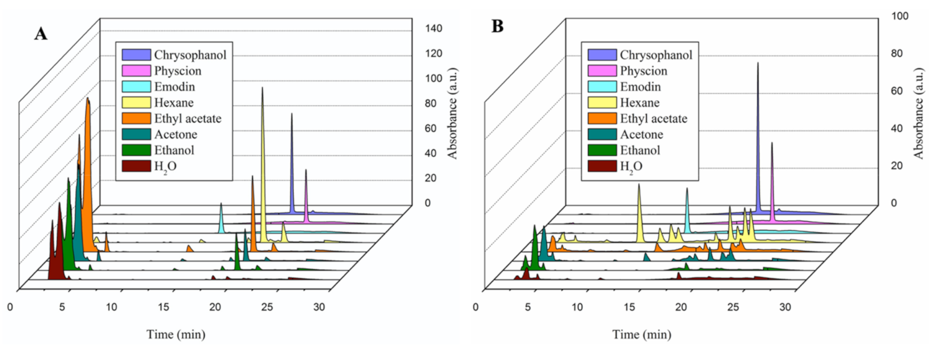
2.2. Determination and Quantification of Bioactive Constituents of Each Extract through High-Performance Liquid Chromatography (HPLC) Assay

The bioactive compounds in RU and RC extracts were identified using HPLC analysis. Emodin, physcion, and chrysophanol were used as reference compounds, and the results of these extracts are displayed in Figure 1. HPLC chromatograms revealed the presence of the aforementioned three compounds in all of the RU and RC extracts (Figure 1A,B).
The concentrations of the three bioactive compounds, which differed among the extracts, are summarized in Table 2. Reference compounds had higher concentrations in RU extracts than in RC extracts. Water extracts of both RU and RC exhibited the lowest value of the reference compounds. For both RU and RC, n-Hex extracts exhibited the highest chrysophanol and physcion contents compared with other extracts. A higher quantity of emodin was noted in the EtOAc and n-Hex extracts of RU and RC than in any other extracts.

2.3. Antioxidant Activities of Each Extract

We evaluated the antioxidant and free radical scavenging activities of RU and RC extracts by using ABTS, DPPH, and FRAP assays. For the ABTS and DPPH assays, the positive controls were gallic acid (GA) and Trolox. The results of ABTS and DPPH scavenging assays were expressed as the half-maximum inhibition concentration to reduce free radicals (Table 3). The FRAP assay was used to measure the reduction of Fe3+ to Fe2+, with a high value for Fe2+ indicating superior antioxidant ability. Antioxidant properties were observed in all extracts to different extents. Of all the extracts, the EtOAc, Ac, EtOH, and water extracts of RU, which had high TPCs, exhibited high antioxidant activity with the lowest inhibition concentration compared with the same extracts of RC (p < 0.0001). All of the free radical scavenging assays showed that the n-Hex extract of RC had a greater inhibition ability than that of RU extract in n-Hex.

2.4. Cytotoxicity of RU and RC Extracts on Liver Cancer Cell Lines

To estimate the anti-liver cancer effects of the extracts, an MTT assay was performed on HepG2 cell lines treated with various concentrations of extracts at three time points (24, 48, and 72 h) (Table 4). Table 4 shows that the cytotoxic effects of the RU extracts were significantly higher than those of the RC extracts at all time points. Owing to the excessive concentrations of RC extracts used to inhibit HepG2 cell growth, we could not obtain the cytotoxic doses of RC in n-Hex, EtOAc, and Ac solvents at the 24-h time point. The time-dependent cytotoxic effects of RU extracts were significantly different at the 24-h time period compared with at 48 and 72 h (24 h vs. 48 h; 24 h vs. 72 h, p < 0.05). In the case of RC extracts, the n-Hex and EtOAc extracts exhibited strong time-dependent cytotoxic effects (48 h vs. 72 h; p < 0.05). The time point at which the cell growth of all extracts was significantly inhibited was 48 h. Notably, the EtOH extract of RU (RU-EtOH) had compelling cytotoxic effects, with the lowest IC50 values at all time points (262.28 ± 13.99; 171.94 ± 6.56; and 167.09 ± 8.99 µg/mL; p < 0.05).

2.5. Elucidation of Liver Cancer Cell Death

After the RU-EtOH was found to have a more potent cytotoxic effect on HepG2 cells than that of other extracts, further experiments were conducted using the EtOH extracts of RU and RC. The constituents of RU and RC extracts identified through HPLC analysis in this study are known to induce cell death through different pathways [19,20,21]. Therefore, we attempted to determine which cell-death type is affected by the EtOH extracts of RU and RC. We performed a DNA fragmentation assay to detect the apoptotic effect of the extracts. The results are displayed in Figure 2. Compared with the EtOH extract of RC (RC-EtOH), the EtOH extract of RU (RU-EtOH) had a stronger stimulating effect on the loss of DNA integrity and DNA ladder formation, and this effect was dose dependent as well (Figure 2A,B). DNA ladders were observed at both 48 and 72 h after treatment incubation of RU-EtOH (Figure 2A). However, there was no significant difference between the results at 48 and 72 h.
Then, the HepG2 cells were treated with the same concentrations of RU-EtOH and RC-EtOH extracts for 48 h, and the percentage of apoptotic cells was measured through double staining (annexin V-FITC) and flowcytometry (CytoFlex, Beckman Coulter, Indianapolis, Indiana, USA). The results shown in Figure 2C indicate that that the cell distribution pattern was different in RU-EtOH and RC-EtOH exposed HepG2 cells. The apoptotic cells were observed more when RU-EtOH was used for treatment than when RC-EtOH was used (21.02% vs. 0.15%; Figure 2C).
PARP and caspase-3 are responsible proteins in the apoptotic program. HepG2 cells were exposed to different doses of RU and RC extracts in EtOH for 48 h, and the expressions of PARP, cleaved PARP, and caspase-3 were evaluated through western blotting (Figure 2D). RU-EtOH diminished the expression of PARP and caspase-3 proteins and increased the level of cleaved PARP compared with those of RC-EtOH, and these effects were dose dependent. These data, which agree with other cell experiment results, confirm that RU-EtOH induces more apoptosis than RC-EtOH does in HepG2 cells, with a characteristic pattern in annexin V-FITC, activation of caspase-3, and cleavage of PARP.
All the results suggest that some of the anticancer mechanisms in the ethanol extract of RU are attributable to apoptosis in the HepG2 cells.

3. Discussion

To the best of our knowledge, this is the first study to describe the phytochemical content, antioxidant free radical scavenging activity, and anti–liver cancer effects of RU from Mongolia. In addition, we compared two unofficial rhubarb species (da huang) cultivated from two different areas—Mongolia and Taiwan—and extracted them in five different polarity solvents (n-Hex, EtOAc, Ac, EtOH, and water). The results of this study revealed that RU extracts (Mongolian rhubarb) had higher TPCs, TFCs, and TACs, superior antioxidant abilities, and greater induced apoptosis of liver cancer cells than did RC extracts (Taiwanese rhubarb). The TPCs of the RU and RC extracts were significantly associated with antioxidant capabilities measured using ABTS, DPPH, and FRAP assays (p < 0.05), which supports previous findings [22]. However, the antioxidant capabilities of the RU and RC extracts were not associated with their flavonoid and anthraquinone contents (p > 0.05).
Trinh stated that aloe-emodin, chrysophanol, rhapontigenin, and rhaponticin were isolated from the RU extracts and their fractions [23]. Nevertheless, our study identified chrysophanol, emodin, and physcion to be the major compounds in the RU extracts. The concentrations of these compounds were particularly high in n-Hex, EtOAc, Ac, and EtOH solvents. Similar to previous studies [10,24], our study found chrysophanol, emodin, and physcion in the RC extracts. Furthermore, a comparable trend has documented that the water extract of RC showed the lowest value of pure compounds [24]. From these results, which are consistent with those of a study by Lee [25], it is clear that pure compounds are extracted mostly in nonpolar and moderately polar solvents, such as n-Hex, EtOAc, and EtOH.
As we mentioned above, antioxidant activities of RU and RC extracts significantly correlated with phenolic contents, but not with flavonoid and anthraquinone contents. Moreover, there was no correlation between pure compound concentrations and antioxidant activities. It can be explained that there might be some synergistic and antagonist effects between bioactive compounds in RU and RC extracts according to the previous report [26,27].
Previous studies have investigated official rhubarb species, such as R.palmatum, and mostly studied their active compounds and their anti-liver cancer activities [2,28,29]. Moreover, although RU (Mongolian rhubarb) has long been used empirically for the treatment of hepatitis and liver cancer, it has not been studied specifically with respect to its anti-liver cancer activity. Our findings confirm that a lower dose of RU-EtOH is required to inhibit HepG2 cell growth than the doses required of the five other solvents investigated in this study. In agreement with the findings reported by Eot [24], RC-EtOH did not exhibit a cytotoxic effect on HepG2 cells at an equal dose to that of RU extract in this study.
Apoptosis, a programmed cell death that does not affect bordering cells, is considered as a good target for anticancer treatment [30]. We found that the RU-EtOH dose-dependently induced HepG2 cell apoptosis through the activation of caspase-3 and cleavage of PARP, resulting in DNA fragmentation. Moreover, the RU-EtOH showed the highest ability to chelate iron (data not shown in this study); thus, the apoptotic effect of RU-EtOH might be closely related to suppressing iron-dependent enzymes such as topoisomerase and iron-containing ribonucleotide reductase, which are important for DNA synthesis, rather than to increasing reactive oxygen species (ROS) in cancer cells [31,32]. However, the correlation between iron chelators, antioxidants, and ROS is still not completely understood in cancer progression, and the exact mechanism of antioxidants in cancer treatment must be further explored.
Each compound, namely chrysophanol, physcion, and emodin, which were found in the RU and RC extracts in this study, are already known to have antitumor activity in various cancers as well as a synergistic effect on approved drugs for liver cancer [20,33]. The content of chrysophanol, which exhibits a necrotic effect on liver cancer [20,34], was higher in RU and RC extracts than the contents of the other two compounds. Notably, the results of the current study confirm that RU-EtOH induces apoptosis in liver cancer cells regardless of the chrysophanol concentration. Moreover, the concentration of these three compounds was not associated with their cytotoxic effects. Thus, compared with a single compound, an optimal ratio of pure compounds may exert greater effects on cancer cells. However, the pure compounds in the RU and RC extracts in this study were not comprehensively defined. Additionally, evidence is lacking to prove that a combination of pure compounds is more effective than a mixture of herbal extracts for the cancer treatment.
In conclusion, this study provides the evidence to support that Mongolian rhubarb is superior to Taiwanese rhubarb in terms of its phytochemical contents and antioxidant activity. However, the phytochemical contents, antioxidant ability, and pure compound concentrations depend on solvent polarity. Moreover, an in vitro study confirmed that RU-EtOH exerts an apoptotic effect on HepG2, human liver cancer cell line. Thus, Mongolian rhubarb could be an appropriate source of natural antioxidants and potential therapeutic agent for HCC.

4. Materials and Methods

4.1. Plant Material Collection

The plant materials used in this study were Rheum undulatum L. and Rumex crispus L. Dried roots of RU were purchased from the traditional herb company “Knight and forest friendship”, Ulaanbaatar, Mongolia, and RC roots were collected from a mountain in HshinChu, Taiwan. Both species were identified by professor Ling-Ling Yang, an expert of botany in Taiwan, and the Natural Compound Analysis Core in Taipei Medical University, Taipei, Taiwan. The plant material was dried for 15 days at ambient temperature in a dark and well-ventilated place, and kept in an air-tight, amber glass jar to protect from the light. The voucher specimens had been deposited under the numbers of AF303435, and AF303439 in the Department of Pharmacognosy, School of Pharmacy, College of Pharmacy, and College of Medicine, Taipei Medical University, Taipei, Taiwan.

4.2. Plant Extraction

The dried roots of RU and RC were finely chopped and then soaked in five solvents with different polarities n-Hex, EtOAc, Ac, EtOH, and Water (H2O). They were then extracted in Soxhlet apparatus for approximately 8 h. All the extracts were filtered and then the solvents were removed using a vacuum freeze dryer (Biobase Biodustry Co. Ltd. Qindao, China). The concentrated extracts of both RU and RC were dissolved in dimethyl sulfoxide (DMSO), and 50mg/mL stock solution was prepared and stored at −20 °C. In further experiments, the stock solution was diluted with either culture medium or methanol.

4.3. Quantitative Phytochemical Analysis of Extracts

4.3.1. Total Phenolic Content

A Folin–Ciocalteau assay [35] was used to estimate the total phenolic content (TPC). To make a calibration curve (y = 2.6819x + 0.0814; R2 = 0.9949), gallic acid (GA) was used with several dilutions (62.5–1000 µg/mL). In each well, 20 µL of the extracts, 80 µL of 7.5% sodium bicarbonate (NaHCO3), and 100 µL of the assay solution were added and then incubated for 5 min at 50 °C. The absorbance was recorded at 600 nm by using enzyme-linked immunosorbent assay reader. The TPC was calculated using the formula obtained from the calibration curve of GA. TPCs of extracts were presented in milligrams of GA equivalents (GAE) per gram of extracts.

4.3.2. Total Anthraquinone Content

The protocol of Gritsanapan [36] with slight modification was followed to determine the total anthraquinone content (TAC). The calibration curve of emodin (y = 9.6454x + 0.0188; R2 = 0.9998), which served as a reference, was created using five sequential concentrations (6.25–100 µg/mL). Next, 100 µL of test solution mixed with 100 µL of 0.5% magnesium acetate. The reaction absorbance was immediately measured at 515 nm. Finally, TAC was calculated using the calibration curve equation and expressed in milligrams of emodin equivalents (EE) per gram of extracts.

4.3.3. Total Flavonoid Content

An aluminium chloride colorimetric assay was used to estimate the total flavonoid content (TFC) according to the method of Gosh [37]. Each 100 µL of test solution was incubated with 100 µL of 2% aluminum chloride solution at ambient temperature for 10 min. The absorbance of all the extracts was then measured at 368 nm. TFCs were quantified from the standard quercetin curve (y = 3.9886x + 0.0267; R2 = 0.9999). The results were expressed in milligrams of quercetin equivalent (QE) per gram of extracts.

4.4. Standardization and Quantitative Analysis of Anthraquinone Derivatives from Extract of RU and RC through the Use of High-Performance Liquid Chromatography (HPLC)

HPLC analysis, a method of quality control of extracts, was performed to detect the major compounds of RU and RC extracts. Chrysophanol, emodin, and physcion were reference compounds, and were purchased from Sigma-Aldrich (St. Louis, MO, USA). The 1 mg/mL compounds or extracts were dissolved in the methanol solution to prepare stock solution, and then a 0.45-μm Millipore filter was used to filter the solution. Then, stock solutions were diluted to obtain 500 μg/mL of extracts and 25 μg/mL of compounds for the HPLC analysis. Testing compounds were analyzed in an HPLC system (Hitachi High-Technologies, Tokyo, Japan) equipped with a 250-nm detector or fluorescence detector (Ex: 430 nm, Em: 525 nm) and LiChroCART RP-C18 column (4.6 mm i.d. × 250 mm, 5 μm, Merck, Darmstadt, Germany). The mobile phase was constituted of MeCN-H2O (0.05% TFA): 0 min, 55/45; 10 min, 60/40, 15 min, 85/15, 20 min, 85/15; 25 min, 55/45; 30 min, 55/45 and the flow rate was 1.0 mL/min.

4.5. Analysis of Antioxidant and Free Radical Scavenging Activities

The percentage of free radical scavenging activity was calculated using the following formula with 2,2′-azinobis-(3-ethylbenzothiazoline-6-sulfonic acid) (ABTS) and 2,2-diphenyl-1-picrylhydrazyl (DPPH) assays. All the results of scavenging activities were presented as the half-maximum inhibition concentration. Trolox and GA were used as a positive control.
Scavenging   % = Abs control Abs sample Abs control × 100 %

4.5.1. ABTS Scavenging Activity

To prepare the stock solution for ABTS assays [38], the stock solution of ABTS was prepared according to specific protocol, and incubated overnight before every experiment. The ABTS stock solution was then diluted several times to prepare a testing solution with an absorbance of 0.7 to 1.0 for the experiment. GA and Trolox were used as a standard solution and methanol was used as a blank solution. In each well, 100 µL of ABTS testing solution was added and followed by 100 µL of extracts with different concentrations. The absorbance was recorded at 734 nm after 10 min of incubation. The scavenging activity of each extract was calculated using the aforementioned formula.

4.5.2. DPPH Scavenging Activity

DPPH scavenging was carried out using the method developed by Om P.Sharma [39] with slight modification. The stock of extracts was added to 100 µL of 200 µM DPPH and incubated for 30 min in a dark environment at ambient temperature. GA and Trolox were reference compounds. The absorbance was detected at 517 nm through spectrophotometry, and the IC50 dose of extracts was computed in a similar manner to the computation for the ABTS scavenging assay.

4.5.3. Ferric Reducing Antioxidant Power Assay (FRAP)

A FRAP assay was done using the method proposed by Faria, A [40]. Each extract (20 µL) was mixed with 200 µL of FRAP reagent and incubated at ambient temperature for 8 min; the absorbance of the mixture was then recorded at 595 nm. A calibration curve was organized with ferrous sulfate (y = 0.7266x − 0.005; R2 = 0.9993), and the results were expressed in millimole ferrous ion equivalents per gram dry weight of the sample (mmol Fe2+/g DW).

4.6. In Vitro Assays

4.6.1. Cell Culture

HepG2 cell lines were provided by The Cancer Research Laboratory of Wan Fang Hospital, affiliated by Taipei Medical University, Taipei, Taiwan. The cells were grown in a 10-cm dish with Dulbecco’s modified Eagle’s medium supplemented with 10% heat-inactivated fetal bovine serum, 5% penicillin/streptomycin (1000 µg/mL). The cells were passaged every two days.

4.6.2. Cell Viability Assay: (3-(4,5-dimethylthiazol-2-yl)-2,5-diphenyltetrazolium Bromide Assay (MTT Assay)

HepG2 cells were seeded on 96-well plates with 1 × 10⁵cell/well density. Next, the cells were treated with different doses of extracts and incubated for the following time periods: 24, 48, and 72 h. Thereafter, the medium was replaced with 10 µL of a 12 mM MTT solution in 100 µL of medium and kept at 37 °C for 4 h. After incubation, the medium was substituted with 100 µL of DMSO to dissolve the MTT crystals, and then absorbance was measured at 570 nm through spectrophotometry. The cell viability percentage was estimated from a comparison of the absorbances of treated and untreated cells. The results were expressed as IC50 doses.

4.7. DNA Fragmentation Assay

HepG2 (8 × 10⁵) were seeded in each well of a six-well plate and incubated overnight. Then, the cells were treated with EtOH extracts of RU and RC and incubated for 24, 48, and 72 h. Next, scrapping and centrifuging at 2000 rpm for 5 min were performed to collect the cells from each well. After removal of the medium, the samples were washed with phosphate-buffered saline (PBS) once. To each sample, 100 µL of DNA lysis buffer was added and incubated overnight at 61 °C. The next day, 1 µL of RNAse A (10 mg/mL) was added to each well and incubated for 1 h under the previous condition. Subsequently, 100 µL of phenol–chloroform was added and centrifuged at 12,000 rpm for 30 min to isolate and then collect DNA samples.

4.7.1. Gel Preparation

Agarose gel was prepared with a 2% concentration. A microwave was used to dissolve agarose powder in TBE buffer. Agarose solution (20 mL) with 2 µL of nucleic acid staining was placed in a gel tank and kept at ambient temperature for 30 min to solidify.

4.7.2. DNA Loading Procedure

DNA samples (20 µL) were mixed with 3 µL of DNA loading dye. The mixture was then added to the agarose gel placed in the running tank. Samples were run at a voltage of 50 mAh for 40 min. Pictures were captured under ultraviolet light.

4.8. Western Blot

HepG2 cells were treated with EtOH extracts of RU and RC for different time durations and then harvested. Treated cells washed with PBS were lysed in RIPA (Sigma-Aldrich) overnight, and protein samples were collected through centrifugation at 12,000 rpm for 30 min at 4 °C. A calibration curve was made with 1 mg/mL bovine serum albumin (BSA) to estimate the protein concentrations of the samples. The protein samples were mixed with the loading dye in equal proportions and denaturized at 95 °C for 5 min. The prepared proteins were separated using polyacrylamide gels with sodium dodecyl sulfate and transferred to immobilon polyvinylidene difluoride membranes. The membranes were incubated in 1% BSA for 1 h and kept with indicated antibodies at 4 °C overnight. Next, the membranes were added to alkaline phosphatase-conjugated immunoglobulin G antibody for 1 h. Proteins were evaluated using colorimetric substrates of nitro blue tetrazolium and 5-btomo-4-chloro-3-indlyl phosphate.

4.9. Annexin V-Fluorescein Isothiocyanate/Propidium Iodide Analysis

To detect apoptosis induced by the EtOH extracts of RU and RC, an Annexin V-fluorescein isothiocyanate (FITC) apoptosis detection kit (Strong Biotech Corporation, Taipei, Taiwan) was used. Cells were plated on six-well plates with a density of 8 × 10⁵ cells/well. The cells were treated with different doses of EtOH extracts from RU and RC. Supernatants and cells were collected together and centrifuged at 2000 rpm for 5 min. Samples were washed with PBS twice and then stained with FITC-conjugated Annexin V and propidium iodide for 15 min in a dark environment. The results were evaluated using CytoFlex (Beckman Coulter, Brea, CA, USA).

4.10. Statistical Analysis

The quantitative results were expressed as the mean ± standard error of the mean based on triplicate experiments and analyzed by one-way and two-way ANOVA. A p-value of < 0.05 was considered statistically significant. GraphPad Prism 8 software (GraphPad, Inc., San Diego, CA, USA) was used to analyze and express the data.

Author Contributions

Conceptualization, M.-S.W., L.-L.Y., Y.-C.C. and G.J.; data curation G.J.; formal analysis, G.J., J.-Y.W. and Y.-C.C.; investigation, G.J., J.-Y.W.; funding acquisition, M.-S.W.; methodology, M.-S.W., L.-L.Y., Y.-C.C.; project administration, M.-S.W., L.-L.Y., Y.-C.C.; resources, M.-S.W., L.-L.Y., Y.-C.C.; software, G.J. and J.-Y.W.; supervision, M.-S.W., L.-L.Y., Y.-C.C.; validation, G.J., J.-Y.W. and Y.-C.C.; visualization, G.J., J.-Y.W. and L.-L.Y.; writing original draft, G.J., writing—review & editing, J.-Y.W., M.-S.W., L.-L.Y., Y.-C.C. All authors have read and agreed to the published version of the manuscript.

Funding

This research was funded by Taipei Medical University-Wan Fang Hospital Research Program, grant number 109 TMU-WFH-16.

Institutional Review Board Statement

Not applicable.

Informed Consent Statement

Not applicable.

Data Availability Statement

The data are available in this article and raw data is available upon request to the author G.J.

Acknowledgments

The authors would like to thank to our colleague Sheng-Jie Shiue, PhD in Division of Gastroenterology, Department of Internal Medicine, Wan Fang Hospital, Taipei Medical University, Taipei, Taiwan for editorial and experimental support in this study.

Conflicts of Interest

The authors declare no conflict of interest.

Sample Availability

Not available.

References

  1. Zhuang, T.; Gu, X.; Zhou, N.; Ding, L.; Yang, L.; Zhou, M. Hepatoprotection and hepatotoxicity of Chinese herb Rhubarb (Dahuang): How to properly control the “General (Jiang Jun)” in Chinese medical herb. Biomed. Pharmacother. 2020, 127, 110224. [Google Scholar] [CrossRef] [PubMed]
  2. Tan, Z.-B.; Fan, H.-J.; Wu, Y.-T.; Xie, L.-P.; Bi, Y.-M.; Xu, H.-L.; Chen, H.-M.; Li, J.; Liu, B.; Zhou, Y.-C. Rheum palmatum extract exerts anti-hepatocellular carcinoma effects by inhibiting signal transducer and activator of transcription 3 signaling. J. Ethnopharmacol. 2019, 232, 62–72. [Google Scholar] [CrossRef] [PubMed]
  3. Lee, S.W.; Hwang, B.S.; Kim, M.-H.; Park, C.-S.; Lee, W.S.; Oh, H.-M.; Rho, M.-C. Inhibition of LFA-1/ICAM-1-mediated cell adhesion by stilbene derivatives from Rheum undulatum. Arch. Pharmacal Res. 2012, 35, 1763–1770. [Google Scholar] [CrossRef]
  4. Choi, E.-S.; Cho, S.-D.; Jeon, J.-G.; Cho, N.-P. The Apoptotic Effect of the Hexane Extract of Rheum undulatum L. in Oral Cancer Cells through the Down-regulation of Specificity Protein 1 and Survivin. Lab. Anim. Res. 2011, 27, 19–24. [Google Scholar] [CrossRef] [PubMed] [Green Version]
  5. Kim, B.J.; Hong, N.R.; Park, H.S.; Ahn, T.S.; Jung, M.H. Association of a Methanol Extract of Rheum undulatum L. Mediated Cell Death in AGS Cells with an Intrinsic Apoptotic Pathway. J. Pharmacopunct. 2015, 18, 26–32. [Google Scholar] [CrossRef] [PubMed]
  6. Lee, W.-J.; Yoon, G.; Hwang, Y.R.; Kim, Y.K.; Kim, S.-N. Anti-obesity and hypolipidemic effects of Rheum undulatum in high-fat diet-fed C57BL/6 mice through protein tyrosine phosphatase 1B inhibition. BMB Rep. 2012, 45, 141–146. [Google Scholar] [CrossRef] [PubMed] [Green Version]
  7. Moon, M.K.; Kang, D.G.; Lee, J.K.; Kim, J.S.; Lee, H.S. Vasodilatory and anti-inflammatory effects of the aqueous extract of rhubarb via a NO-cGMP pathway. Life Sci. 2006, 78, 1550–1557. [Google Scholar] [CrossRef] [PubMed]
  8. Cavers, P.B.; Harper, J.L. Rumex obtusifolius L. and R. crispus L. J. Ecol. 1964, 52, 737–766. [Google Scholar] [CrossRef]
  9. Vasas, A.; Orbán-Gyapai, O.; Hohmann, J. The Genus Rumex: Review of traditional uses, phytochemistry and pharmacology. J. Ethnopharmacol. 2015, 175, 198–228. [Google Scholar] [CrossRef] [PubMed]
  10. Shim, K.-S.; Lee, B.; Ma, J.Y. Water extract of Rumex crispus prevents bone loss by inhibiting osteoclastogenesis and inducing osteoblast mineralization. BMC Complement. Altern. Med. 2017, 17, 483. [Google Scholar] [CrossRef] [PubMed]
  11. Feduraev, P.; Chupakhina, G.; Maslennikov, P.; Tacenko, N.; Skrypnik, L. Variation in Phenolic Compounds Content and Anti-oxidant Activity of Different Plant Organs from Rumex crispus L. and Rumex obtusifolius L. at Different Growth Stages. Antioxidants 2019, 8, 237. [Google Scholar]
  12. El–Serag, H.B.; Rudolph, K.L. Hepatocellular Carcinoma: Epidemiology and Molecular Carcinogenesis. Gastroenterology 2007, 132, 2557–2576. [Google Scholar] [CrossRef]
  13. Baatarkhuu, O.; Gerelchimeg, T.; Munkh-Orshikh, D.; Batsukh, B.; Sarangua, G.; Amarsanaa, J. Epide-miology, Genotype Distribution, Prognosis, Control and Management of Viral Hepatitis B, C, D, and Hepatocellular Carcinoma in Mongolia. Euroasian J. Hepato-Gastroenterol. 2018, 8, 57. [Google Scholar] [CrossRef]
  14. Ferlay, J.; Ervik, M.; Lam, F.; Colombet, M.; Mery, L.; Pineros, M.; Znaor, A.; Soerjotaram, I.; Bray, F. Global Cancer Observatory: Cancer Today. International Agency for Research on Cancer Lyon. 2018. Available online: https://gco.iarc.fr/today (accessed on 20 February 2021).
  15. Park, J.W.; Chen, M.; Colombo, M.; Roberts, L.R.; Schwartz, M.; Chen, P.J.; Sherman, M. Global patterns of hepatocellular carcinoma man-agement from diagnosis to death: The BRIDGE Study. Liver Int. 2015, 35, 2155–2166. [Google Scholar] [CrossRef] [PubMed] [Green Version]
  16. Abdel-Hamid, N.M.; Abass, S.A.; Mohamed, A.A.; Hamid, D.M. Herbal management of hepatocellular carcinoma through cutting the pathways of the common risk factors. Biomed. Pharmacother. 2018, 107, 1246–1258. [Google Scholar] [CrossRef]
  17. Muraina, I.; Adaudi, A.; Mamman, M.; Kazeem, H.; Eloff, J. Effects of geographical location on the yield and bioactivity of Anoigeissus leiocarpus. J. Pharm. Bioresour. 2010, 5, 68–72. [Google Scholar] [CrossRef]
  18. Zhao, B.; Li, J.; Yuan, R.; Mao, S. Adaptive evolution of the rbcL gene in the genus Rheum (Polygonaceae). Biotechnol. Biotechnol. Equip. 2017, 31, 493–498. [Google Scholar] [CrossRef] [Green Version]
  19. Zhang, L.; He, D.; Li, K.; Liu, H.; Wang, B.; Zheng, L.; Li, J. Emodin targets mitochondrial cyclophilin D to induce apoptosis in HepG2 cells. Biomed. Pharmacother. 2017, 90, 222–228. [Google Scholar] [CrossRef] [PubMed]
  20. Ni, C.H.; Chen, P.Y.; Lu, H.F.; Yang, J.S.; Huang, H.Y.; Wu, S.H.; Chung, J.G. Chrysophanol-induced necrotic-like cell death through an im-paired mitochondrial ATP synthesis in Hep3B human liver cancer cells. Arch. Pharm. Res. 2012, 35, 887–895. [Google Scholar] [CrossRef] [PubMed]
  21. Pan, X.P.; Wang, C.; Li, Y.; Huang, L.H. Physcion induces apoptosis through triggering endoplasmic reticulum stress in hepato-cellular carcinoma. Biomed. Pharmacother. 2018, 99, 894–903. [Google Scholar] [CrossRef]
  22. Cheung, Y.; Meenu, M.; Yu, X.; Xu, B. Phenolic acids and flavonoids profiles of commercial honey from different floral sources and geographic sources. Int. J. Food Prop. 2019, 22, 290–308. [Google Scholar] [CrossRef]
  23. Trinh, T.A.; Lee, D.; Park, S.; Kim, S.H.; Park, J.G.; Kim, J.H.; Kang, K.S. Stilbenes contribute to the anticancer effects of Rheum undulatum L. through activation of apoptosis. Oncol. Lett. 2019, 17, 2953–2959. [Google Scholar] [CrossRef] [Green Version]
  24. Eom, T.; Kim, E.; Kim, J.S. In Vitro Antioxidant, Antiinflammation, and Anticancer Activities and Anthraquinone Content from Rumex crispus Root Extract and Fractions. Antioxidants 2020, 9, 726. [Google Scholar] [CrossRef] [PubMed]
  25. Lee, N.-J.; Choi, J.-H.; Koo, B.-S.; Ryu, S.-Y.; Han, Y.-H.; Lee, S.-I.; Lee, D.-U. Antimutagenicity and Cytotoxicity of the Constituents from the Aerial Parts of Rumex acetosa. Biol. Pharm. Bull. 2005, 28, 2158–2161. [Google Scholar] [CrossRef] [PubMed] [Green Version]
  26. Freeman, B.L.; Eggett, D.L.; Parker, T.L. Synergistic and Antagonistic Interactions of Phenolic Compounds Found in Navel Oranges. J. Food Sci. 2010, 75, C570–C576. [Google Scholar] [CrossRef] [PubMed]
  27. Mercado-Mercado, G.; De La Rosa, L.A.; Alvarez-Parrilla, E.; De La Rosa-Carrillo, L.A. Effect of pectin on the interactions among phenolic compounds determined by antioxidant capacity. J. Mol. Struct. 2020, 1199, 126967. [Google Scholar] [CrossRef]
  28. Hsu, C.-M.; Hsu, Y.-A.; Tsai, Y.; Shieh, F.-K.; Huang, S.-H.; Wan, L.; Tsai, F.-J. Emodin inhibits the growth of hepatoma cells: Finding the common anti-cancer pathway using Huh7, Hep3B, and HepG2 cells. Biochem. Biophys. Res. Commun. 2010, 392, 473–478. [Google Scholar] [CrossRef]
  29. Lu, G.D.; Shen, H.-M.; Ong, C.N.; Chung, M.C.M. Anticancer effects of aloe-emodin on HepG2 cells: Cellular and proteomic studies. Proteom. Clin. Appl. 2007, 1, 410–419. [Google Scholar] [CrossRef] [PubMed]
  30. Pfeffer, C.M.; Singh, A.T.K. Apoptosis: A Target for Anticancer Therapy. Int. J. Mol. Sci. 2018, 19, 448. [Google Scholar] [CrossRef] [Green Version]
  31. Gaur, K.; Vázquez-Salgado, A.M.; Duran-Camacho, G.; Dominguez-Martinez, I.; Benjamín-Rivera, J.A.; Fernández-Vega, L.; Sarabia, L.C.; García, A.C.; Pérez-Deliz, F.; Román, J.A.M.; et al. Iron and Copper Intracellular Chelation as an Anticancer Drug Strategy. Inorganics 2018, 6, 126. [Google Scholar] [CrossRef] [Green Version]
  32. Kontoghiorghes, G.J. Advances on Chelation and Chelator Metal Complexes in Medicine. Int. J. Mol. Sci. 2020, 21, 2499. [Google Scholar] [CrossRef] [PubMed] [Green Version]
  33. Pan, X.; Wang, C.; Zhang, T. Retracted: Physcion Synergistically Enhances the Cytotoxicity of Sorafenib in Hepatocellular Carcinoma. Anat. Rec. Adv. Integr. Anat. Evol. Biol. 2019, 302, 2171–2177. [Google Scholar] [CrossRef] [PubMed]
  34. Lu, C.-C.; Yang, J.-S.; Huang, A.-C.; Hsia, T.-C.; Chou, S.-T.; Kuo, C.-L.; Lu, H.-F.; Lee, T.-H.; Wood, W.G.; Chung, J.-G. Chrysophanol induces necrosis through the production of ROS and alteration of ATP levels in J5 human liver cancer cells. Mol. Nutr. Food Res. 2010, 54, 967–976. [Google Scholar] [CrossRef] [PubMed] [Green Version]
  35. Singleton, V.L.; Orthofer, R.; Lamuela-Raventós, R.M. Analysis of total phenols and other oxidation substrates and antiox-idants by means of folin-ciocalteu reagent. Methods Enzymol. 1999, 299, 152–178. [Google Scholar]
  36. Sakulpanich, A.; Gritsanapan, W. Extraction method for high content of anthraquinones from cassia fistula pods. J. Health Res. 2008, 22, 167–172. [Google Scholar]
  37. Ghosh, S.; Derle, A.; Ahire, M.; More, P.; Jagtap, S.; Phadatare, S.D.; Patil, A.B.; Jabgunde, A.M.; Sharma, G.K.; Shinde, V.S.; et al. Phytochemical Analysis and Free Radical Scavenging Activity of Medicinal Plants Gnidia glauca and Dioscorea bulbifera. PLoS ONE 2013, 8, e82529. [Google Scholar] [CrossRef] [Green Version]
  38. Zheleva-Dimitrova, D.; Nedialkov, P.; Kitanov, G. Radical scavenging and antioxidant activities of methanolic extracts from Hypericum species growing in Bulgaria. Pharmacogn Mag. 2010, 6, 74–78. [Google Scholar] [CrossRef] [PubMed] [Green Version]
  39. Sharma, O.P.; Bhat, T.K. DPPH antioxidant assay revisited. Food Chem. 2009, 113, 1202–1205. [Google Scholar] [CrossRef]
  40. Faria, A.; Oliveira, J.; Neves, P.; Gameiro, P.; Santos-Buelga, C.; De Freitas, V.; Mateus, N. Antioxidant Properties of Prepared Blueberry (Vaccinium myrtillus) Extracts. J. Agric. Food Chem. 2005, 53, 6896–6902. [Google Scholar] [CrossRef]
Figure 1. Identification and quantification of the bioactive constituents of Rheum undulatum L. and Rumex crispus L. through high-performance liquid chromatography. The HPLC spectrum of the bioactive constituents of Rheum undulatum L. (RU) (A) and Rumex crispus L. (RC) (B) extracts in different solvents. Chrysophanol (purple), physcion (pink), and emodin (light blue) were reference compounds. Each color represents different extract of RU and RC.
Molecules 26 01217 g001
Figure 2. RU, not RC, induces significant apoptosis responses in human hepatocellular carcinoma cell line HepG2. DNA fragmentation using agarose gel electrophoresis of DNA isolated from HepG2 cells treated with either RU-EtOH or RC-EtOH (A,B). Annexin V-FITC/PI flow cytometric shape of HepG2 cells, 48 h post-treatment of the ethanol extracts of RU (C) and RC (D). Compared with negative control (DMSO), apoptotic percentage of the total cell population was presented and given as mean ± SD (n = 3) *** p < 0.0001; ns: not significant (E). Effects of different concentrations of RU-EtOH and RC-EtOH on the protein expression responsible for apoptosis in HepG2 cells. GAPDH was used as an internal control (F).
Molecules 26 01217 g002
Table 1. Phytochemical contents of RU and RC extracts in different solvents.
Total Phenols
(mg GAE/g DW)
Total Anthraquinones
(mg EE/g DW)
Total Flavonoids
(mg QE/g DW)
RURCRURCRURC
n-Hex2.44 ± 0.694.23 ± 0.36 ns94.10 ± 4.41 b73.65 ± 11.3047.47 ± 1.64192.60 ± 0.47 a
EtOAc11.16 ± 1.41 a2.89 ± 0.2933.68 ± 2.2834.14 ± 5.47 ns50.98 ± 1.7970.99 ± 3.72 b
Ac8.45 ± 0.61 b5.01 ± 0.0815.14 ± 2.4743.54 ± 3.82 a37.92 ± 12.44121.63 ± 10.87 a
EtOH9.57 ± 1.03 a2.96 ± 0.1725.15 ± 2.69 b11.99 ± 2.9050.70 ± 2.96 ns42.66 ± 1.21
H2O8.39 ± 1.83 a3.5 ± 0.449.43 ± 3.3112.73 ± 2.27 ns49.63 ± 10.53 ns39.51 ± 4.96
RU: Rheum undulatum, RC: Rumex crispus, GAE: gallic acid equivalent, EE: emodin equivalent, QE: quercetin equivalent, DW: dry weight, n-Hex: n-hexane, EtOAc: Ethyl acetate, Ac: Acetone, EtOH: Ethanol, H2O: Water. Value represent mean ± standard error of the mean. Different letters between RU and RC in each phytochemical content represent significant difference: a p < 0.0001; b p < 0.05; ns: not significant.
Table 2. Quantitative HPLC analysis of the RU and RC extracts.
RURC
ChrysophanolPhyscionEmodinChrysophanolPhyscionEmodin
n-Hex120.06 ± 13.82 a19.02 ± 2.58 ns3.33 ± 0.5417.25 ± 5.2320.34 ± 3.6910.59 ± 2.47 a
EtOAc58.83 ± 6.29 a8.07 ± 0.82 ns6.51 ± 1.67 ns5.01 ± 0.276.48 ± 1.025.19 ± 0.89
Ac24.15 ± 2.69 a3.5 ± 0.583.12 ± 0.216.47 ± 0.597.05 ± 1.98 a5.42 ± 1.02 a
EtOH28.81 ± 2.08 a4.53 ± 0.34 a2.39 ± 0.27 a0.09 ± 0.020.98 ± 0.190.74 ± 0.08
H2O1.29 ± 0.2 a0.79 ± 0.14 a0.26 ± 0.08 a0 ± 0.000.40 ± 0.060 ± 0.01
(Concentrations = mg compound/g extract) HPLC: high-performance liquid chromatography, RU: Rheum undulatum, RC: Rumex crispus, n-Hex: n-hexane, EtOAc: Ethyl acetate, Ac: Acetone, EtOH: Ethanol, H2O: Water. Data represent mean ± standard error of the mean. Different letters between RU and RC in each solvent extract represent significant difference: a p < 0.05; ns: not significant.
Table 3. Antioxidant and free radical scavenging activities of RU and RC extracts.
IC50 (µg/mL)FRAP (mMFe2+/g)
ABTSDPPH
GA3.16 ± 0.151.25 ± 0.20nt
Trolox13.53 ± 0.406.28 ± 1.58nt
RURCRURCRURC
n-Hex93.23 ± 4.1875.12 ± 8.98 a1800.87 ± 151.0374.39 ± 0.01 a0.99 ± 0.301.79 ± 0.13 b
EtOAc5.67 ± 0.23 a207.17 ± 2.5041.37 ± 1.98 b246.76 ± 20.026.42 ± 0.24 a1.26 ± 0.15
Ac10.25 ± 0.51 a52.05 ± 2.6453.49 ± 4.93 ns70.01 ± 0.015.17 ± 0.19 a2.65 ± 0.13
EtOH14.08 ± 0.13 a77.95 ± 3.3152.12 ± 3.29 ns146.72 ± 1.464.66 ± 0.04 a1.98 ± 0.17
H2O14.09 ± 1.88 a70.55 ± 1.8146.15 ± 4.66 ns137.40 ± 1.824.86 ± 0.26 a2.34 ± 0.22
RU: Rheum undulatum, RC: Rumex crispus, ABTS: 2,2′-azinobis-(3-ethylbenzothiazoline-6-sulfonic acid), DPPH: 2,2-diphenyl-1-picrylhydrazyl, FRAP: ferric reducing antioxidant power, n-Hex: n-hexane, EtOAc: ethyl acetate, Ac: acetone, EtOH: ethanol, H2O: water, nt: not tested. Data represent mean ± standard error of the mean. Different letters between RU and RC in each assay represent significant difference: a p < 0.0001; b p < 0.05; ns: not significant.
Table 4. IC50 values after 24, 48, and 72 h of treatment incubation of RU and RC extracts.
SolventsRU (IC50 µg/mL)RC (IC50 µg/mL)
24 h48 h72 h24 h48 h72 h
n-Hex625.22 ± 30.73419.77 ± 13.20427.55 ± 21.54nt1973.14 ± 257.15146.09 ± 24.99
EtOAc449.82 ± 51.68208.46 ± 30.63191.63 ± 26.31nt925.30 ± 126.58468.95 ± 7.59
Ac403.69 ± 52.45187.18 ± 20.59170.16 ± 28.03nt606.56 ± 41.88456.74 ± 20.10
EtOH262.28 ± 13.99171.94 ± 6.56167.09 ± 8.992792.33 ± 396.281753.02 ± 77.841617.32 ± 31.49
H2O880.33 ± 64.16555.84 ± 2.09635.09 ± 22.582465.74 ± 317.401397.90 ± 30.761176.79 ± 16.36
RU: Rheum undulatum, RC: Rumex crispus, n-Hex: n-hexane, EtOAc: ethyl acetate, Ac: acetone, EtOH: ethanol, H2O: water, nt: not tested. Data represent mean ± standard error of the mean.
Publisher’s Note: MDPI stays neutral with regard to jurisdictional claims in published maps and institutional affiliations.

