Essential Oils and Mono/bi/tri-Metallic Nanocomposites as Alternative Sources of Antimicrobial Agents to Combat Multidrug-Resistant Pathogenic Microorganisms: An Overview


Abstract
1. Introduction
2. Methods of Extraction of Essential Oils
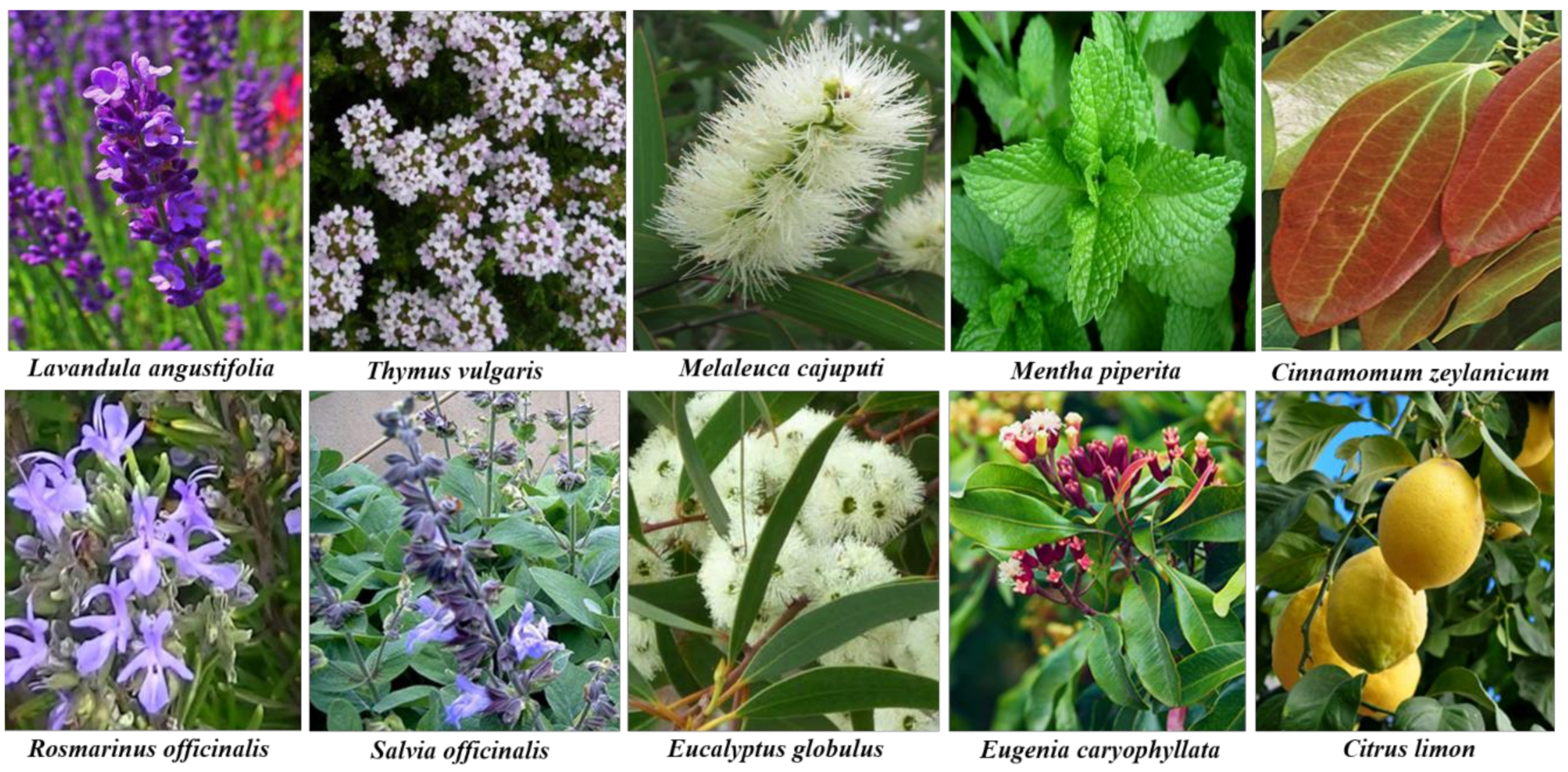
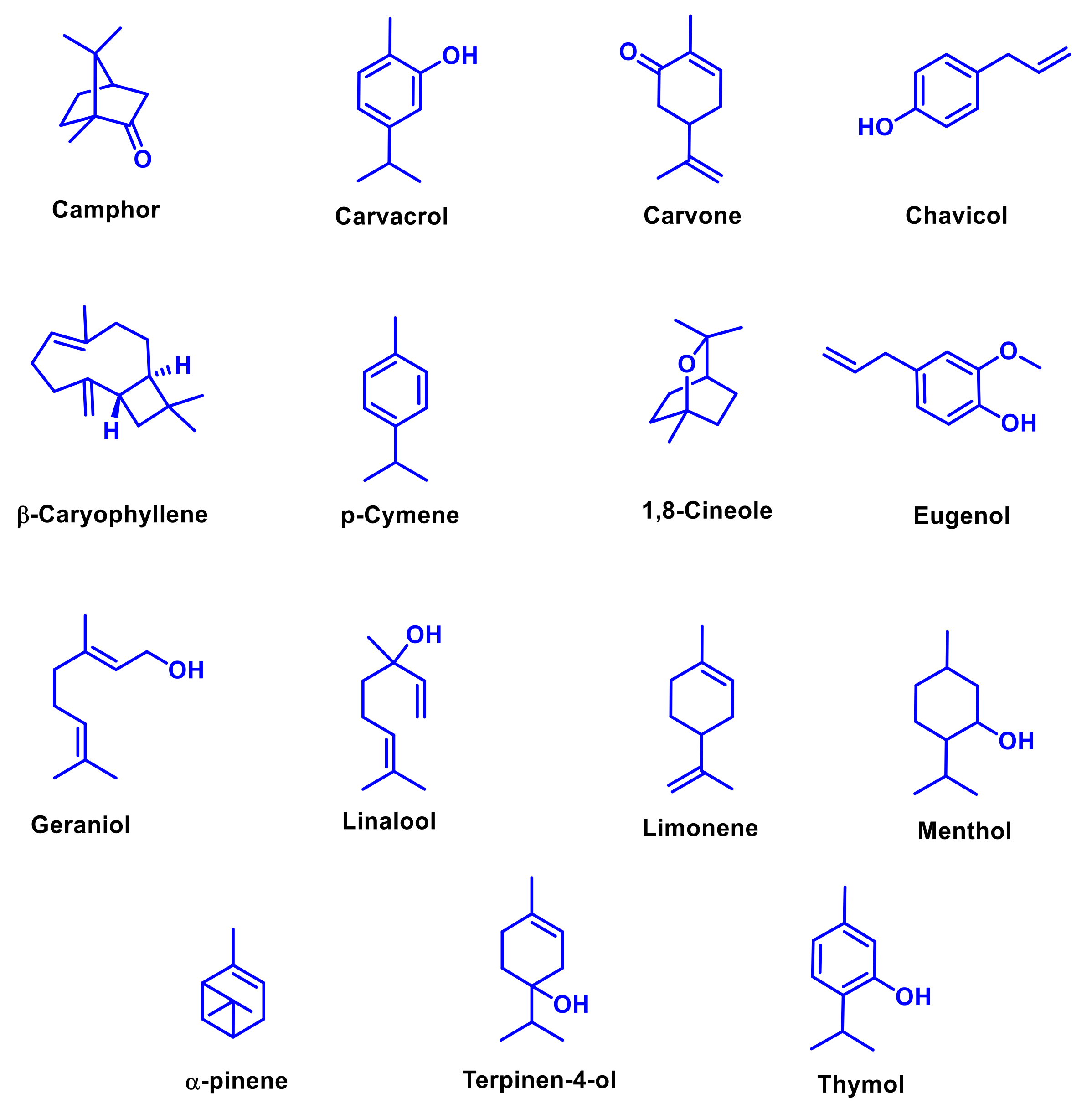
3. Chemical Composition of Essential Oils
4. Antimicrobial Effects of Essential Oils
5. Antioxidant Activity of Essential Oils
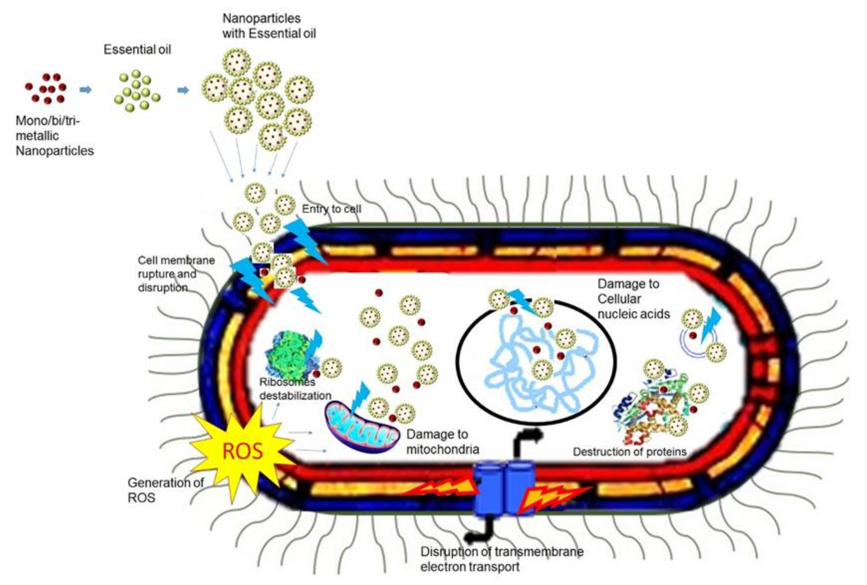
6. Potential Impacts of Bi-Metallic and Tri-Metallic Nanoparticles
7. Antimicrobial Activities of Metallic Nanoparticles
8. Efficiency of Nano-Encapsulated Essential Oils
9. Interaction of Essential Oils and Metallic Nanoparticles
10. Synergistic Antimicrobial Activity of Essential Oils
11. Challenges and Future Directions
12. Conclusions
Author Contributions
Funding
Acknowledgments
Conflicts of Interest
References
- Gyles, C. The growing problem of antimicrobial resistance. Can. Veter. J. Rev. Veter. Can. 2011, 52, 817–820. [Google Scholar]
- Livermore, D.M. Has the era of untreatable infections arrived? J. Antimicrob. Chemother. 2009, 64, i29–i36. [Google Scholar] [CrossRef]
- Mahady, G. Medicinal plants for the prevention and treatment of bacterial infections. Curr. Pharm. Des. 2005, 11, 2405–2427. [Google Scholar] [CrossRef]
- Kon, K.; Rai, M. Combining Essential Oils with Antibiotics and other Antimicrobial Agents to Overcome Multidrug-Resistant Bacteria; Elsevier BV: London, UK, 2013; pp. 149–164. [Google Scholar]
- Abad, M.J.; Bedoya, L.M.; Bermejo, P. Essential Oils from the Asteraceae Family Active against Multidrug-Resistant Bacteria; Elsevier BV: London, UK, 2013. [Google Scholar]
- Holmes, C.; Hopkins, V.; Hensford, C.; MacLaughlin, V.; Wilkinson, D.; Rosenvinge, H. Lavender oil as a treatment for agitated behaviour in severe dementia: a placebo controlled study. Int. J. Geriatr. Psychiatry 2002, 17, 305–308. [Google Scholar] [CrossRef]
- Auddy, B.; Ferreira, M.; Blasina, F.; Lafon, L.; Arredondo, F.; Dajas, F.; Tripathi, P.C.; Seal, T.; Mukherjee, B. Screening of antioxidant activity of three Indian medicinal plants, traditionally used for the management of neurodegenerative diseases. J. Ethnopharmacol. 2003, 84, 131–138. [Google Scholar] [CrossRef]
- Prabuseenivasan, S.; Jayakumar, M.; Ignacimuthu, S. In vitro antibacterial activity of some plant essential oils. BMC Complement. Altern. Med. 2006, 6, 39. [Google Scholar] [CrossRef]
- Lee, J.; Jang, M.; Seo, J.; Kim, G.-H. Evaluation For Antibacterial Effects of Volatile Flavors from Chrysanthemum indicum Against Food-borne Pathogens and Food Spoilage Bacteria. J. Food Saf. 2011, 31, 140–148. [Google Scholar] [CrossRef]
- Pandey, A.K.; Kumar, P.; Singh, P.; Tripathi, N.N.; Bajpai, V.K. Essential Oils: Sources of Antimicrobials and Food Preservatives. Front. Microbiol. 2017, 7, 45. [Google Scholar] [CrossRef]
- Burt, S. Essential oils: their antibacterial properties and potential applications in foods—a review. Int. J. Food Microbiol. 2004, 94, 223–253. [Google Scholar] [CrossRef]
- Chouhan, S.; Sharma, K.; Guleria, S. Antimicrobial Activity of Some Essential Oils—Present Status and Future Perspectives. Medicines 2017, 4, 58. [Google Scholar] [CrossRef]
- Barra, A. Factors affecting chemical variability of essential oils: a review of recent developments. Nat. Prod. Commun. 2009, 4, 1147–1154. [Google Scholar] [CrossRef]
- Edwards-Jones, V. Alternative Antimicrobial Approaches to Fighting Multidrug-Resistant Infections; Elsevier BV: Oxford, UK, 2013; pp. 1–9. [Google Scholar]
- Bassole, I.H.N.; Lamien-Meda, A.; Bayala, B.; Tirogo, S.; Franz, C.; Novak, J.; Nebié, R.C.; Dicko, M.H. Composition and Antimicrobial Activities of Lippia multiflora Moldenke, Mentha piperita L. and Ocimum basilicum L. Essential Oils and Their Major Monoterpene Alcohols Alone and in Combination. Molecules 2010, 15, 7825–7839. [Google Scholar] [CrossRef]
- Rakholiya, K.D.; Kaneria, M.J.; Chanda, S.V. Medicinal Plants as Alternative Sources of Therapeutics against Multidrug-Resistant Pathogenic Microorganisms Based on Their Antimicrobial Potential and Synergistic Properties; Elsevier BV: Oxford, UK, 2013; pp. 165–179. [Google Scholar]
- Sharma, G.; Kumar, A.; Sharma, S.; Naushad, M.; Dwivedi, R.P.; Alothman, Z.A.; Mola, G.T. Novel development of nanoparticles to bimetallic nanoparticles and their composites: A review. J. King Saud Univ. Sci. 2019, 31, 257–269. [Google Scholar] [CrossRef]
- Srinoi, P.; Chen, Y.-T.; Vittur, V.; Marquez, M.D.; Lee, T.R. Bimetallic Nanoparticles: Enhanced Magnetic and Optical Properties for Emerging Biological Applications. Appl. Sci. 2018, 8, 1106. [Google Scholar] [CrossRef]
- Mishra, K.; Basavegowda, N.; Lee, Y.R. Biosynthesis of Fe, Pd, and Fe–Pd bimetallic nanoparticles and their application as recyclable catalysts for [3 + 2] cycloaddition reaction: a comparative approach. Catal. Sci. Technol. 2015, 5, 2612–2621. [Google Scholar] [CrossRef]
- Yu, W.; Porosoff, M.; Chen, J.G. Review of Pt-Based Bimetallic Catalysis: From Model Surfaces to Supported Catalysts. Chem. Rev. 2012, 112, 5780–5817. [Google Scholar] [CrossRef]
- Yao, T.; Cui, T.; Wang, H.; Xu, L.; Cui, F.; Wu, J. A simple way to prepare Au@polypyrrole/Fe3O4 hollow capsules with high stability and their application in catalytic reduction of methylene blue dye. Nanoscale 2014, 6, 7666–7674. [Google Scholar] [CrossRef]
- Fang, Y.; Wang, E. Simple and direct synthesis of oxygenous carbon supported palladium nanoparticles with high catalytic activity. Nanoscale 2013, 5, 1843. [Google Scholar] [CrossRef]
- Basavegowda, N.; Mishra, K.; Lee, Y.R. Trimetallic FeAgPt alloy as a nanocatalyst for the reduction of 4-nitroaniline and decolorization of rhodamine B: A comparative study. J. Alloy. Compd. 2017, 701, 456–464. [Google Scholar] [CrossRef]
- Guisbiers, G.; Khanal, S.; Ruiz-Zepeda, F.; De La Puente, J.R.; Yacaman, M.J. Cu–Ni nano-alloy: mixed, core–shell or Janus nano-particle? Nanoscale 2014, 6, 14630–14635. [Google Scholar] [CrossRef]
- Zhang, L.F.; Zhong, S.; Wang, Y. Highly Branched Concave Au/Pd Bimetallic Nanocrystals with Superior Electrocatalytic Activity and Highly Efficient SERS Enhancement. Angew. Chem. Int. Ed. 2012, 52, 645–649. [Google Scholar] [CrossRef]
- Lim, B.; Lu, X.; Jiang, M.; Camargo, P.; Cho, E.C.; Lee, E.P.; Xia, Y. Facile Synthesis of Highly Faceted Multioctahedral Pt Nanocrystals through Controlled Overgrowth. Nano Lett. 2008, 8, 4043–4047. [Google Scholar] [CrossRef]
- Boucher, M.B.; Zugic, B.; Cladaras, G.; Kammert, J.; Marcinkowski, M.D.; Lawton, T.J.; Sykes, E.C.H.; Flytzani-Stephanopoulos, M. Single atom alloy surface analogs in Pd0.18Cu15 nanoparticles for selective hydrogenation reactions. Phys. Chem. Chem. Phys. 2013, 15, 12187. [Google Scholar] [CrossRef] [PubMed]
- Gu, H.; Yang, Z.; Gao, J.; Chang, C.; Xu, B. Heterodimers of Nanoparticles: Formation at a Liquid−Liquid Interface and Particle-Specific Surface Modification by Functional Molecules. J. Am. Chem. Soc. 2005, 127, 34–35. [Google Scholar] [CrossRef]
- Liu, X.; Liu, X. Bimetallic nanoparticles: kinetic control matters. Angew. Chem. Int. Ed. 2012, 51, 3311–3313. [Google Scholar] [CrossRef] [PubMed]
- Rubiolo, P.; Sgorbini, B.; Liberto, E.; Cordero, C.; Bicchi, C. Essential oils and volatiles: sample preparation and analysis. A review. Flavour Fragr. J. 2010, 25, 282–290. [Google Scholar] [CrossRef]
- Schmidt, E. Production of Essential Oils. In Handbook of Essential Oils: Science, Technology and Applications, 2nd ed.; Baser, K.H.C., Buchbauer, G., Eds.; CRC Press: Boca Raton, FL, USA, 2015. [Google Scholar]
- Cowan, M.M. Plant Products as Antimicrobial Agents. Clin. Microbiol. Rev. 1999, 12, 564–582. [Google Scholar] [CrossRef]
- Bakkali, F.; Averbeck, S.; Averbeck, D.; Idaomar, M. Biological effects of essential oils – A review. Food Chem. Toxicol. 2008, 46, 446–475. [Google Scholar] [CrossRef]
- Masotti, V.; Juteau, F.; Bessiere, J.M.; Viano, J. Seasonal and Phenological Variations of the Essential Oil from the Narrow Endemic Species Artemisia molinieri and Its Biological Activities. J. Agric. Food Chem. 2003, 51, 7115–7121. [Google Scholar] [CrossRef]
- Angioni, A.; Barra, A.; Coroneo, V.; Dessi, S.; Cabras, P. Chemical composition, seasonal variability, and antifungal activity of Lavandula stoechas L. ssp. stoechas essential oils from stem/leaves and flowers. J. Agric. Food Chem. 2006, 54, 4364–4370. [Google Scholar] [CrossRef]
- Eloff, J.N. Which extractant should be used for the screening and isolation of antimicrobial components from plants? J. Ethnopharmacol. 1998, 60, 1–8. [Google Scholar] [CrossRef]
- Parekh, J.; Jadeja, D.; Chanda, S. Efficacy of aqueous and methanol extracts of some medicinal plants for potential antibacterial activity. Turk. J. Biol. 2005, 29, 203–210. [Google Scholar]
- National Cancer Institute. Aromatherapy and Essential Oils (PDQ®) - General Information. Available online: http://www.cancer.gov/cancertopics/pdq/cam/aromatherapy/Health%20Professional/page2 (accessed on 14 July 2012).
- Voon, H.C.; Bhat, R.; Rusul, G. Flower Extracts and Their Essential Oils as Potential Antimicrobial Agents for Food Uses and Pharmaceutical Applications. Compr. Rev. Food Sci. Food Saf. 2011, 11, 34–55. [Google Scholar] [CrossRef]
- Wijekoon, M.J.O.; Bhat, R.; Karim, A.A. Effect of extraction solvents on the phenolic compounds and antioxidant activities of bunga kantan (Etlingera elatior Jack.) inflorescence. J. Food Compos. Anal. 2011, 24, 615–619. [Google Scholar] [CrossRef]
- Dilworth, L.L.; Riley, C.K.; Stennett, D.K. Plant. Constituents; Elsevier BV: Oxford, UK, 2017; pp. 61–80. [Google Scholar]
- Silva, L.; Nelson, D.; Drummond, M.; Dufossé, L.; Glória, M. Comparison of hydrodistillation methods for the deodorization of turmeric. Food Res. Int. 2005, 38, 1087–1096. [Google Scholar] [CrossRef]
- Mahadagde, P. Techniques Available for the Extraction of Essential Oils from Plants: A Review. Int. J. Res. Appl. Sci. Eng. Technol. 2018, 6, 2931–2935. [Google Scholar] [CrossRef]
- Rozzi, N.; Phippen, W.; Simon, J.; Singh, R. Supercritical Fluid Extraction of Essential Oil Components from Lemon-Scented Botanicals. LWT 2002, 35, 319–324. [Google Scholar] [CrossRef]
- Pourmortazavi, S.M.; Saghafi, Z.; Ehsani, A.; Yousefi, M. Application of supercritical fluids in cholesterol extraction from foodstuffs: A review. J. Food Sci. Technol. 2018, 55, 2813–2823. [Google Scholar] [CrossRef]
- Alexandru, L.; Cravotto, G.; Giordana, L.; Binello, A.; Chemat, F. Ultrasound-assisted extraction of clove buds using batch- and flow-reactors: A comparative study on a pilot scale. Innov. Food Sci. Emerg. Technol. 2013, 20, 167–172. [Google Scholar] [CrossRef]
- Roldán-Gutiérrez, J.M.; Jimenez, J.R.; Luquedecastro, M.; De Castro, M.L. Ultrasound-assisted dynamic extraction of valuable compounds from aromatic plants and flowers as compared with steam distillation and superheated liquid extraction. Talanta 2008, 75, 1369–1375. [Google Scholar] [CrossRef]
- Leonelli, C.; Veronesi, P.; Cravotto, G. Microwave-assisted Extraction for Bioactive Compounds; Springer Science and Business Media: New York, NY, USA, 2013; p. 238. [Google Scholar]
- Kahriman, N.; Yayli, B.; Yücel, M.; Karaoglu, S.A.; Yayli, N. Chemical constituents and antimicrobial activity of the essential oil from Vicia dadianorum extracted by hydro and microwave distillations. Rec. Nat. Prod. 2012, 6, 49–56. [Google Scholar]
- Vian, M.A.; Fernandez, X.; Visinoni, F.; Chemat, F. Microwave hydrodiffusion and gravity, a new technique for extraction of essential oils. J. Chromatogr. A 2008, 1190, 14–17. [Google Scholar] [CrossRef]
- Handa, S.S.; Khanuja, S.P.S.; Longo, G.; Rakesh, D.D. Extraction technologies for medicinal and aromatic plants; ICS-UNIDO: Trieste, Italy, 2008; pp. 93–106. [Google Scholar]
- Elyemni, M.; Louaste, B.; Nechad, I.; Elkamli, T.; Bouia, A.; Taleb, M.; Chaouch, M.; Eloutassi, N. Extraction of Essential Oils of Rosmarinus officinalis L. by Two Different Methods: Hydrodistillation and Microwave Assisted Hydrodistillation. Sci. World J. 2019, 3659432–3659436. [Google Scholar] [CrossRef]
- Gao, X.; Lv, S.; Wu, Y.; Li, J.; Zhang, W.; Meng, W.; Wang, C.; Meng, Q. Volatile components of essential oils extracted from Pu-erh ripe tea by different extraction methods. Int. J. Food Prop. 2017, 20, S240–S253. [Google Scholar] [CrossRef]
- Ćujić, N.; Savikin, K.; Janković, T.; Pljevljakusic, D.; Zdunić, G.; Ibrić, S. Optimization of polyphenols extraction from dried chokeberry using maceration as traditional technique. Food Chem. 2016, 194, 135–142. [Google Scholar] [CrossRef]
- Ferhat, M.A.; Boukhatem, M.N.; Hazzit, M.; Meklati, B.Y.; Chemat, F. Cold pressing hydrodistillation and microwave dry distillation of citrus essential oil from Algeria: A comparative study. Electron. J. Biol. 2016, S1, 30–41. [Google Scholar]
- Nematollahi, A.; Kamali, H.; Aminimoghadamfarouj, N.; Golmakani, E. The optimization of essential oils supercritical CO2extraction from Lavandula hybrida through static-dynamic steps procedure and semi-continuous technique using response surface method. Pharmacogn. Res. 2015, 7, 57–65. [Google Scholar] [CrossRef]
- Chen, P.; Liu, B.; Liu, X.; Fu, J. Ultrasound-assisted extraction and dispersive liquid–liquid microextraction coupled with gas chromatography-mass spectrometry for the sensitive determination of essential oil components in lavender. Anal. Methods 2019, 11, 1541–1550. [Google Scholar] [CrossRef]
- Abedi, A.-S.; Rismanchi, M.; Shahdoostkhany, M.; Mohammadi, A.; Mortazavian, A.M. Microwave-assisted extraction of Nigella sativa L. essential oil and evaluation of its antioxidant activity. J. Food Sci. Technol. 2017, 54, 3779–3790. [Google Scholar] [CrossRef]
- Martins, N.; Barros, L.; Santos-Buelga, C.; Henriques, M.; Silva, S.; Ferreira, I.C. Decoction, infusion and hydroalcoholic extract of Origanum vulgare L.: Different performances regarding bioactivity and phenolic compounds. Food Chem. 2014, 158, 73–80. [Google Scholar] [CrossRef]
- Cazella, L.N.; Glamoclija, J.; Soković, M.; Gonçalves, J.E.; Linde, G.A.; Colauto, N.B.; Gazim, Z.C. Antimicrobial Activity of Essential Oil of Baccharis dracunculifolia DC (Asteraceae) Aerial Parts at Flowering Period. Front. Plant. Sci. 2019, 10, 27. [Google Scholar] [CrossRef] [PubMed]
- Dhifi, W.; Bellili, S.; Jazi, S.; Bahloul, N.; Mnif, W. Essential Oils’ Chemical Characterization and Investigation of Some Biological Activities: A Critical Review. Medicines 2016, 3, 25. [Google Scholar] [CrossRef] [PubMed]
- Regnault-Roger, C.; Vincent, C.; Arnason, J. Essential Oils in Insect Control: Low-Risk Products in a High-Stakes World. Annu. Rev. Èntomol. 2012, 57, 405–424. [Google Scholar] [CrossRef] [PubMed]
- Lalit, M. Errata for Plant Growth and Development. Plant. Growth Dev. 2002, ibc1–ibc2. [Google Scholar]
- Eslahi, H.; Fahimi, N.; Sardarian, A.R. Chemical composition of essential oils. In Essential oils in food processing: Chemistry, safety and applications, 1st ed.; Hashemi, S.M.B., Khaneghah, A.M., de Souza Sant’ Ana, A., Eds.; IFT Press, Wiley Blackwell: Hoboken, NJ, USA, 2018. [Google Scholar]
- Swamy, M.K.; Akhtar, M.S.; Sinniah, U.R. Antimicrobial Properties of Plant Essential Oils against Human Pathogens and Their Mode of Action: An Updated Review. Evidence Based Complement. Altern. Med. 2016, 2016, 1–21. [Google Scholar] [CrossRef]
- Bilia, A.R.; Guccione, C.; Isacchi, B.; Righeschi, C.; Firenzuoli, F.; Bergonzi, M.C. Essential Oils Loaded in Nanosystems: A Developing Strategy for a Successful Therapeutic Approach. Evidence Based Complement. Altern. Med. 2014, 2014, 1–14. [Google Scholar] [CrossRef]
- Manayi, A.; Nabavi, S.M.; Daglia, M.; Jafari, S. Natural terpenoids as a promising source for modulation of GABAergic system and treatment of neurological diseases. Pharmacol. Rep. 2016, 68, 671–679. [Google Scholar] [CrossRef]
- Silva, L.N.; Zimmer, K.R.; Macedo, A.J.; Trentin, D. Plant Natural Products Targeting Bacterial Virulence Factors. Chem. Rev. 2016, 116, 9162–9236. [Google Scholar] [CrossRef]
- Salini, R.; Pandian, S.K. Interference of quorum sensing in urinary pathogen Serratia marcescens by Anethum graveolens. Pathog. Dis. 2015, 73. [Google Scholar] [CrossRef]
- Martín-Rodríguez, A.J.; Ticona, J.C.; Jiménez, I.A.; Flores, N.; Fernandez, J.J.; Bazzocchi, I.L. Flavonoids from Piper delineatum modulate quorum-sensing-regulated phenotypes in Vibrio harveyi. Phytochemistry 2015, 117, 98–106. [Google Scholar] [CrossRef]
- Grayson, D.H. Monoterpenoids (mid-1997 to mid-1999). Nat. Prod. Rep. 2000, 17, 385–419. [Google Scholar] [CrossRef] [PubMed]
- Shireman, R. Essential Fatty Acids; Elsevier BV: London, UK, 2003; pp. 2169–2176. [Google Scholar]
- Iranshahi, M. A review of volatile sulfur-containing compounds from terrestrial plants: biosynthesis, distribution and analytical methods. J. Essent. Oil Res. 2012, 24, 393–434. [Google Scholar] [CrossRef]
- Morsy, N. Chemical Structure, Quality Indices and Bioactivity of Essential Oil Constituents. Act. Ingred. Aromat. Med. Plants 2017, 175–206. [Google Scholar]
- Safaei-Ghomi, J.; Ahd, A.A. Antimicrobial and antifungal properties of the essential oil and methanol extracts of Eucalyptus largiflorens and Eucalyptus intertexta. Pharmacogn. Mag. 2010, 6, 172–175. [Google Scholar] [CrossRef]
- Astani, A.; Reichling, J.; Schnitzler, P. Comparative study on the antiviral activity of selected monoterpenes derived from essential oils. Phytotherapy Res. 2009, 24, 673–679. [Google Scholar] [CrossRef]
- Teixeira, B.; Marques, A.; Ramos, C.; Neng, N.R.; Nogueira, J.; Saraiva, J.A.; Nunes, M.L. Chemical composition and antibacterial and antioxidant properties of commercial essential oils. Ind. Crop. Prod. 2013, 43, 587–595. [Google Scholar] [CrossRef]
- Cosentino, S.; Tuberoso, C.; Pisano, M.B.; Satta, M.; Mascia, V.; Arzedi, E.; Palmas, F. In-vitro antimicrobial activity and chemical composition of Sardinian Thymus essential oils. Lett. Appl. Microbiol. 1999, 29, 130–135. [Google Scholar] [CrossRef]
- Edris, A. Pharmaceutical and therapeutic Potentials of essential oils and their individual volatile constituents: A review. Phytotherapy Res. 2007, 21, 308–323. [Google Scholar] [CrossRef]
- Oosterhaven, K.; Poolman, B.; Smid, E. S-carvone as a natural potato sprout inhibiting, fungistatic and bacteristatic compound. Ind. Crop. Prod. 1995, 4, 23–31. [Google Scholar] [CrossRef]
- Cox, S.D.; Mann, C.M.; Markham, J.L.; Bell, H.C.; Gustafson, J.E.; Warmington, J.R.; Wyllie, S.G. The mode of antimicrobial action of the essential oil of Melaleuca alternifolia (tea tree oil). J. Appl. Microbiol. 2001, 88, 170–175. [Google Scholar] [CrossRef]
- Balouiri, M.; Sadiki, M.; Ibnsouda, S.K. Methods for in vitro evaluating antimicrobial activity: A review. J. Pharm. Anal. 2015, 6, 71–79. [Google Scholar] [CrossRef]
- Weinstein, M.P.; Patel, J.B.; Burnham, C.A.; Campeau, S.; Conville, P.S.; Limbago, B.; Mathers, A.; Mazzulli, T.; Munro, S.; Eliopoulos, G.M.; et al. Methods for dilution antimicrobial susceptibility test for bacteria that grow aerobically: Approved Standard—8th Edition; CLSI Document M07-A8; Clinical and Laboratory Standards Institute: Wayne, PA, USA, 2009.
- Canillac, N.; Mourey, A. Antibacterial activity of the essential oil of Picea excelsa on Listeria, Staphylococcus aureus and coliform bacteria. Food Microbiol. 2001, 18, 261–268. [Google Scholar] [CrossRef]
- Zhang, J.; Ye, K.-P.; Zhang, X.; Pan, D.-D.; Sun, Y.-Y.; Cao, J.-X. Antibacterial Activity and Mechanism of Action of Black Pepper Essential Oil on Meat-Borne Escherichia coli. Front. Microbiol. 2017, 7, 4168. [Google Scholar] [CrossRef]
- Zhang, Y.; Liu, X.; Wang, Y.; Jiang, P.; Quek, S.Y. Antibacterial activity and mechanism of cinnamon essential oil against Escherichia coli and Staphylococcus aureus. Food Control. 2016, 59, 282–289. [Google Scholar] [CrossRef]
- Díez, J.G.; Alheiro, J.; Falco, V.; Fraqueza, M.J.; Patarata, L.A.D.S.C. Chemical characterization and antimicrobial properties of herbs and spices essential oils against pathogens and spoilage bacteria associated to dry-cured meat products. J. Essent. Oil Res. 2016, 29, 117–125. [Google Scholar] [CrossRef]
- Ibrahim, N.A.; El-Hawary, S.S.; Mohammed, M.M.; Farid, M.A.; Abdel-Wahed, N.A.; Ali, M.A.; El-Abd, E.A. Chemical Composition, Antiviral against avian Influenza (H5N1) Virus and Antimicrobial activities of the Essential Oils of the Leaves and Fruits of Fortunella margarita, Lour Swingle, Growing in Egypt. J. Appl. Pharm. Sci. 2015, 5, 6–12. [Google Scholar]
- Mitropoulou, G.; Bardouki, H.; Vamvakias, M.; Panas, P.; Paraskeuas, P.; Rangou, A.; Kourkoutas, I. Antimicrobial activity of Pistacia lentiscus and Fortunella margarita essential oils against Saccharomyces cerevisiae and Aspergillus niger in fruit juices. J. Biotechnol. 2018, 280, S62–S63. [Google Scholar] [CrossRef]
- Dos Santos, N.O.; Mariane, B.; Lago, J.H.G.; Sartorelli, P.; Rosa, W.; Soares, M.G.; Silva, A.; Lorenzi, H.; Vallim, M.A.; Pascon, R. Assessing the Chemical Composition and Antimicrobial Activity of Essential Oils from Brazilian Plants—Eremanthus erythropappus (Asteraceae), Plectrantuns barbatus, and P. amboinicus (Lamiaceae). Molecules 2015, 20, 8440–8452. [Google Scholar] [CrossRef]
- Novy, P.; Davidova, H.; Serrano-Rojero, C.S.; Rondevaldova, J.; Pulkrabek, J.; Kokoska, L. Composition and Antimicrobial Activity of Euphrasia rostkoviana Hayne Essential Oil. Evidence Based Complement. Altern. Med. 2015, 2015, 1–5. [Google Scholar] [CrossRef]
- Yu, X.D.; Xie, J.H.; Wang, Y.H.; Li, Y.C.; Mo, Z.Z.; Zheng, Y.; Su, J.Y.; Liang, Y.E.; Liang, J.Z.; Su, Z.R.; et al. Selective Antibacterial Activity of Patchouli Alcohol Against Helicobacter pylori Based on Inhibition of Urease. Phytotherapy Res. 2014, 29, 67–72. [Google Scholar] [CrossRef]
- Das, P.; Dutta, S.; Begum, J.; Anwar, N. Antibacterial and Antifungal Activity Analysis of Essential Oil of Pogostemon cablin (Blanco) Benth. Bangladesh J. Microbiol. 2016, 30, 7–10. [Google Scholar] [CrossRef]
- Crevelin, E.J.; Caixeta, S.C.; Dias, H.J.; Groppo, M.; Cunha, W.; Martins, C.H.G.; Crotti, A.E. Antimicrobial Activity of the Essential Oil of Plectranthus neochilus against Cariogenic Bacteria. Evidence Based Complement. Altern. Med. 2015, 2015, 1–6. [Google Scholar] [CrossRef] [PubMed]
- Crevelin, E.J.; Aguiar, G.P.; Lima, K.E.; Severiano, M.; Groppo, M.; Ambrósio, S.R. Antifungal activity of the essential oils of Plectranthus neochilus (Lamiaceae) and Tagetes erecta (Asteraceae) cultivated in brazil. Int. J. Complement. Altern. Med. 2018, 11, 1. [Google Scholar] [CrossRef][Green Version]
- Beatovic, D.; Krstic-Milosevic, D.; Trifunovic, S.; Šiljegović, J.; Glamočlija, J.; Ristić, M.; Jelačić, S. Chemical composition, antioxidant and antimicrobial activities of the essential oils of twelve Ocimum basilicum L. cultivars grown in Serbia. Rec. Nat. Prod. 2015, 9, 62–75. [Google Scholar]
- Helal, I.; El-Bessoumy, A.; Al-Bataineh, E.; Joseph, M.R.P.; Rajagopalan, P.; Chandramoorthy, H.C.; Ahmed, S.B.H. Antimicrobial Efficiency of Essential Oils from Traditional Medicinal Plants of Asir Region, Saudi Arabia, over Drug Resistant Isolates. BioMed Res. Int. 2019, 2019, 8928306–8928309. [Google Scholar] [CrossRef]
- Cui, H.; Zhang, X.; Zhou, H.; Zhao, C.; Lin, L. Antimicrobial activity and mechanisms of Salvia sclarea essential oil. Bot. Stud. 2015, 56, 16. [Google Scholar] [CrossRef]
- Antimicrobial activity of the essential oil of Thymus kotschyanus grown wild in Iran. Int. J. Biosci. IJB 2015, 6, 239–248.
- Mahboubi, M.; HeidaryTabar, R.; Mahdizadeh, E.; Hosseini, H. Antimicrobial activity and chemical composition of Thymus species and Zataria multiflora essential oils. Agric. Nat. Resour. 2017, 51, 395–401. [Google Scholar] [CrossRef]
- Venturi, C.R.; Danielli, L.J.; Klein, F.; Apel, M.A.; Montanha, J.A.; Bordignon, S.A.L.; Roehe, P.; Fuentefria, A.; Henriques, A.T. Chemical analysis and in vitro antiviral and antifungal activities of essential oils from Glechon spathulata and Glechon marifolia. Pharm. Biol. 2014, 53, 682–688. [Google Scholar] [CrossRef]
- Bouzabata, A.; Cabral, C.; Gonçalves, M.J.; Cruz, M.T.; Bighelli, A.; Cavaleiro, C.; Casanova, J.; Tomi, F.; Salgueiro, L. Myrtus communis L. as source of a bioactive and safe essential oil. Food Chem. Toxicol. 2015, 75, 166–172. [Google Scholar] [CrossRef]
- Abbaszadegan, A.; Nabavizadeh, M.; Gholami, A.; Sheikhiani, R.; Shokouhi, M.M.; Shams, M.S.; Ghasemi, Y. Chemical constituent and antimicrobial effect of essential oil from Myrtus communis leaves on microorganisms involved in persistent endodontic infection compared to two common endodontic irrigants: An in vitro study. J. Conserv. Dent. 2014, 17, 449–453. [Google Scholar] [CrossRef]
- Papajani, V.; Haloci, E.; Goci, E.; Shkreli, R.; Manfredini, S. Evaluation of antifungal activity of Origanum vulgare and Rosmarinus officinalis essential oil before and after inclusion in 𝛽-cyclodextrine. Int. J. Pharm. Pharm. Sci. 2015, 7, 270–273. [Google Scholar]
- Puškárová, A.; Bučková, M.; Kraková, L.; Pangallo, D.; Kozics, K. The antibacterial and antifungal activity of six essential oils and their cyto/genotoxicity to human HEL 12469 cells. Sci. Rep. 2017, 7, 8211. [Google Scholar] [CrossRef] [PubMed]
- Latifah-Munirah, B.; Himratul-Aznita, W.H.; Zain, N.M. Eugenol, an essential oil of clove, causes disruption to the cell wall of Candida albicans (ATCC 14053). Front. Life Sci. 2015, 8, 231–240. [Google Scholar] [CrossRef]
- Radünz, M.; Da Trindade, M.L.M.; Camargo, T.M.; Radünz, A.L.; Borges, C.D.; Gandra, E.A.; Helbig, E. Antimicrobial and antioxidant activity of unencapsulated and encapsulated clove (Syzygium aromaticum, L.) essential oil. Food Chem. 2019, 276, 180–186. [Google Scholar] [CrossRef] [PubMed]
- Souza, C.M.C.; Junior, S.A.P.; Moraes, T.D.S.; Damasceno, J.L.; Mendes, S.A.; Dias, H.J.; Stefani, R.; Tavares, D.C.; Martins, C.H.G.; Crotti, A.E.; et al. Antifungal activity of plant-derived essential oils on Candida tropicalis planktonic and biofilms cells. Med. Mycol. 2016, 54, 515–523. [Google Scholar] [CrossRef] [PubMed]
- Boukhatem, M.N.; Kameli, A.; Saidi, F. Essential oil of Algerian rose-scented geranium (Pelargonium graveolens): Chemical composition and antimicrobial activity against food spoilage pathogens. Food Control. 2013, 34, 208–213. [Google Scholar] [CrossRef]
- Roy, S.; Chaurvedi, P.; Chowdhary, A. Evaluation of antiviral activity of essential oil of Trachyspermum ammi against Japanese encephalitis virus. Pharmacogn. Res. 2015, 7, 263–267. [Google Scholar] [CrossRef]
- Vitali, L.A.; Beghelli, D.; Nya, P.C.B.; Bistoni, O.; Cappellacci, L.; Damiano, S.; Lupidi, G.; Maggi, F.; Orsomando, G.; Papa, F.; et al. Diverse biological effects of the essential oil from Iranian Trachyspermum ammi. Arab. J. Chem. 2016, 9, 775–786. [Google Scholar] [CrossRef]
- Brand, Y.M.; Roa-Linares, V.C.; Betancur-Galvis, L.A.; Durán-García, D.C.; Stashenko, E. Antiviral activity of Colombian Labiatae and Verbenaceae family essential oils and monoterpenes on Human Herpes viruses. J. Essent. Oil Res. 2015, 28, 1–8. [Google Scholar] [CrossRef]
- Tardugno, R.; Serio, A.; Pellati, F.; D’Amato, S.; Lopez, C.C.; Bellardi, M.G.; Di Vito, M.; Savini, V.; Paparella, A.; Benvenuti, S. Lavandula x intermedia and Lavandula angustifolia essential oils: phytochemical composition and antimicrobial activity against foodborne pathogens. Nat. Prod. Res. 2018, 33, 3330–3335. [Google Scholar] [CrossRef] [PubMed]
- Garzoli, S.; Petralito, S.; Ovidi, E.; Turchetti, G.; Masci, V.L.; Tiezzi, A.; Trilli, J.; Cesa, S.; Casadei, M.A.; Giacomello, P.; et al. Lavandula x intermedia essential oil and hydrolate: Evaluation of chemical composition and antibacterial activity before and after formulation in nanoemulsion. Ind. Crop. Prod. 2020, 145, 112068. [Google Scholar] [CrossRef]
- Vinciguerra, V.; Rojas, F.; Tedesco, V.; Giusiano, G.; Angiolella, L. Chemical characterization and antifungal activity of Origanum vulgare, Thymus vulgaris essential oils and carvacrol against Malassezia furfur. Nat. Prod. Res. 2018, 33, 3273–3277. [Google Scholar] [CrossRef] [PubMed]
- Almeida, E.T.D.C.; De Souza, E.L.; Guedes, J.P.D.S.; Barbosa, I.M.; De Sousa, C.P.; Castellano, L.R.C.; Magnani, M.; De Souza, E.L. Mentha piperita L. essential oil inactivates spoilage yeasts in fruit juices through the perturbation of different physiological functions in yeast cells. Food Microbiol. 2019, 82, 20–29. [Google Scholar] [CrossRef]
- Saharkhiz, M.J.; Motamedi, M.; Zomorodian, K.; Pakshir, K.; Miri, R.; Hemyari, K. Chemical Composition, Antifungal and Antibiofilm Activities of the Essential Oil of Mentha piperita L. ISRN Pharm. 2012, 2012, 1–6. [Google Scholar] [CrossRef] [PubMed]
- Bharat, C.S.; Praveen, D. Evaluation of in vitro antimicrobial potential and phytochemical analysis of spruce, cajeput and jamrosa essential oil against clinical isolates. Int. J. Green Pharm. 2016, 10, 27–42. [Google Scholar]
- Feng, J.; Zhang, S.; Shi, W.; Zubcevik, N.; Miklossy, J.; Zhang, Y. Selective Essential Oils from Spice or Culinary Herbs Have High Activity against Stationary Phase and Biofilm Borrelia burgdorferi. Front. Med. 2017, 4, 169. [Google Scholar] [CrossRef]
- Condò, C.; Anacarso, I.; Sabia, C.; Iseppi, R.; Anfelli, I.; Forti, L.; De Niederhäusern, S.; Bondi, M.; Messi, P. Antimicrobial activity of spices essential oils and its effectiveness on mature biofilms of human pathogens. Nat. Prod. Res. 2018, 34, 567–574. [Google Scholar] [CrossRef]
- Mahboubi, M.; Mahboubi, M. Chemical Composition, Antimicrobial and Antioxidant Activities of Eugenia caryophyllata Essential Oil. J. Essent. Oil Bear. Plants 2015, 18, 967–975. [Google Scholar] [CrossRef]
- Aldoghaim, F.S.; Flematti, G.; Hammer, K. Antimicrobial Activity of Several Cineole-Rich Western Australian Eucalyptus Essential Oils. Microorganisms 2018, 6, 122. [Google Scholar] [CrossRef]
- Wei, Z.-F.; Zhao, R.-N.; Dong, L.-J.; Zhao, X.; Su, J.-X.; Zhao, M.; Li, L.; Bian, Y.-J.; Zhang, L.-J. Dual-cooled solvent-free microwave extraction of Salvia officinalis L. essential oil and evaluation of its antimicrobial activity. Ind. Crop. Prod. 2018, 120, 71–76. [Google Scholar] [CrossRef]
- Damjanovic-Vratnica, B.; Ðakov, T.; Sukovic, D.; Damjanovic, J. Chemical Composition and Antimicrobial Activity of Essential Oil of Wild-Growing Salvia officinalis L. from Montenegro. J. Essent. Oil Bear. Plants 2008, 11, 79–89. [Google Scholar] [CrossRef]
- Mertas, A.; Garbusińska, A.; Szliszka, E.; Jureczko, A.; Kowalska, M.; Krol, W. The Influence of Tea Tree Oil (Melaleuca alternifolia) on Fluconazole Activity against Fluconazole-Resistant Candida albicans Strains. BioMed Res. Int. 2015, 2015, 590470–590479. [Google Scholar] [CrossRef] [PubMed]
- Brun, P.; Bernabè, G.; Filippini, R.; Piovan, A. In Vitro Antimicrobial Activities of Commercially Available Tea Tree (Melaleuca alternifolia) Essential Oils. Curr. Microbiol. 2018, 76, 108–116. [Google Scholar] [CrossRef] [PubMed]
- Khalil, N.; Mohamed, L.A.; Fikry, S.; Singab, A.N.; Salama, O. Chemical composition and antimicrobial activity of the essential oils of selected Apiaceous fruits. Futur. J. Pharm. Sci. 2018, 4, 88–92. [Google Scholar] [CrossRef]
- Yildiz, H. Chemical Composition, Antimicrobial, and Antioxidant Activities of Essential Oil and Ethanol Extract of Coriandrum sativum L. Leaves from Turkey. Int. J. Food Prop. 2015, 19, 1593–1603. [Google Scholar] [CrossRef]
- Radaelli, M.; Da Silva, B.P.; Weidlich, L.; Hoehne, L.; Flach, A.; Da Costa, L.A.M.A.; Ethur, E.M. Antimicrobial activities of six essential oils commonly used as condiments in Brazil against Clostridium perfringens. Braz. J. Microbiol. 2016, 47, 424–430. [Google Scholar] [CrossRef]
- Lorenzo-Leal, A.C.; Palou, E.; López-Malo, A.; Bach, H. Antimicrobial, Cytotoxic, and Anti-Inflammatory Activities of Pimenta dioica and Rosmarinus officinalis Essential Oils. BioMed Res. Int. 2019, 2019, 1639726. [Google Scholar] [CrossRef]
- Bajer, T.; Šilha, D.; Ventura, K.; Bajerová, P. Composition and antimicrobial activity of the essential oil, distilled aromatic water and herbal infusion from Epilobium parviflorum Schreb. Ind. Crop. Prod. 2017, 100, 95–105. [Google Scholar] [CrossRef]
- Guleria, S.; Tiku, A.K.; Koul, A.; Gupta, S.; Singh, G.; Razdan, V.K. Antioxidant and Antimicrobial Properties of the Essential Oil and Extracts of Zanthoxylum alatum Grown in North-Western Himalaya. Sci. World J. 2013, 2013, 1–9. [Google Scholar] [CrossRef]
- Manssouri, M.; Znini, M.; Majidi, L. Studies on the antioxidant activity of essential oil and various extracts of Ammodaucus leucotrichus Coss. & Dur. Fruits from Morocco. J. Taibah Univ. Sci. 2020, 14, 124–130. [Google Scholar]
- Sarikurkcu, C.; Tepe, B.; Daferera, D.; Polissiou, M.; Harmandar, M. Studies on the antioxidant activity of the essential oil and methanol extract of Marrubium globosum subsp. globosum (lamiaceae) by three different chemical assays. Bioresour. Technol. 2008, 99, 4239–4246. [Google Scholar] [CrossRef] [PubMed]
- Toscano-Garibay, J.D.; Arriaga-Alba, M.; Sánchez-Navarrete, J.; Mendoza-García, M.; Flores-Estrada, J.J.; Moreno-Eutimio, M.A.; Espinosa-Aguirre, J.J.; González-Ávila, M.; Ruiz-Pérez, N.J. Antimutagenic and antioxidant activity of the essential oils of Citrus sinensis and Citrus latifolia. Sci. Rep. 2017, 7, 11479. [Google Scholar] [CrossRef] [PubMed]
- Elaguel, A.; Kallel, I.; Gargouri, B.; Ben Amor, I.; Hadrich, B.; Ben Messaoud, E.; Gdoura, R.; Lassoued, S.; Gargouri, A. Lawsonia inermis essential oil: extraction optimization by RSM, antioxidant activity, lipid peroxydation and antiproliferative effects. Lipids Heal. Dis. 2019, 18, 1–11. [Google Scholar] [CrossRef] [PubMed]
- Benyoucef, F.; Dib, M.E.A.; Arrar, Z.; Costa, J.; Muselli, A. Synergistic Antioxidant Activity and Chemical Composition of Essential Oils From Thymus fontanesii, Artemisia herba-alba and Rosmarinus officinalis. J. Appl. Biotechnol. Rep. 2018, 5, 151–156. [Google Scholar] [CrossRef]
- Khanal, S.; Bhattarai, N.; McMaster, D.; Bahena, D.; Velázquez-Salazar, J.J.; Yacaman, M.J. Highly monodisperse multiple twinned AuCu-Pt trimetallic nanoparticles with high index surfaces. Phys. Chem. Chem. Phys. 2014, 16, 16278–16283. [Google Scholar] [CrossRef]
- Toshima, N.; Ito, R.; Matsushita, T.; Shiraishi, Y. Trimetallic nanoparticles having a Au-core structure. Catal. Today 2007, 122, 239–244. [Google Scholar] [CrossRef]
- Tsai, S.-H.; Liu, Y.-H.; Wu, P.-L.; Yeh, C.-S. Preparation of Au–Ag–Pd trimetallic nanoparticles and their application as catalysts Electronic supplementary information (ESI) available: UV-vis spectra and EDX analysis of Au–Ag–Pd colloidal suspensions. J. Mater. Chem. 2003, 13, 978–980. Available online: http://www.rsc.org/suppdata/jm/b3/b300952a/ (accessed on 24 February 2020). [CrossRef]
- Tang, S.; Vongehr, S.; Wang, Y.; Cui, J.; Wang, X.; Meng, X. Versatile synthesis of high surface area multi-metallic nanosponges allowing control over nanostructure and alloying for catalysis and SERS detection. J. Mater. Chem. A 2014, 2, 3648–3660. [Google Scholar] [CrossRef]
- Li, T.; Vongehr, S.; Tang, S.; Dai, Y.; Huang, X.; Meng, X. Scalable Synthesis of Ag Networks with Optimized Sub-monolayer Au-Pd Nanoparticle Covering for Highly Enhanced SERS Detection and Catalysis. Sci. Rep. 2016, 6, 37092. [Google Scholar] [CrossRef]
- Zhang, X.; Li, F.; Zhang, Y.; Bond, A.M.; Zhang, J. Stannate derived bimetallic nanoparticles for electrocatalytic CO2 reduction. J. Mater. Chem. A 2018, 6, 7851–7858. [Google Scholar] [CrossRef]
- Guo, H.; Li, H.; Jarvis, K.; Wan, H.; Kunal, P.; Dunning, S.G.; Liu, Y.; Henkelman, G.; He, S. Microwave-Assisted Synthesis of Classically Immiscible Ag–Ir Alloy Nanoparticle Catalysts. ACS Catal. 2018, 8, 11386–11397. [Google Scholar] [CrossRef]
- Wu, M.-L.; Lai, L.-B. Synthesis of Pt/Ag bimetallic nanoparticles in water-in-oil microemulsions. Colloids Surfaces A Physicochem. Eng. Asp. 2004, 244, 149–157. [Google Scholar] [CrossRef]
- Allaedini, G.; Tasirin, S.M.; Aminayi, P. Synthesis of Fe–Ni–Ce trimetallic catalyst nanoparticles via impregnation and co-precipitation and their application to dye degradation. Chem. Pap. 2016, 70, 231–242. [Google Scholar] [CrossRef]
- Ding, K.; Zhao, Y.; Liu, L.; Li, Y.; Zhao, J.; Chen, Y.; Niu, J.; Wang, Q. Preparation of trimetallic composite nanoparticles PdxNiyFez by a pyrolysis method in the solvent of ionic liquids for ethanol oxidation reaction. Int. J. Electrochem. Sci. 2015, 10, 6768–6779. [Google Scholar]
- Wu, Y.; Wang, Z.; Chen, S.; Guo, X.; Liu, Z. One-step hydrothermal synthesis of silver nanoparticles loaded on N-doped carbon and application for catalytic reduction of 4-nitrophenol. RSC Adv. 2015, 5, 87151–87156. [Google Scholar] [CrossRef]
- Li, P.; Xin, Y.; Li, Q.; Wang, Z.; Zhang, Z.; Zheng, L. Ce–Ti Amorphous Oxides for Selective Catalytic Reduction of NO with NH3: Confirmation of Ce–O–Ti Active Sites. Environ. Sci. Technol. 2012, 46, 9600–9605. [Google Scholar] [CrossRef]
- Khodadadi, B. TiO2/SiO2 prepared via facile sol-gel method as an ideal support for green synthesis of Ag nanoparticles using Oenothera biennis extract and their excellent catalytic performance in the reduction of 4-nitrophenol. Nanochem Res. 2017, 2, 140–150. [Google Scholar]
- Bhunia, K.; Khilari, S.; Pradhan, D. Trimetallic PtAuNi alloy nanoparticles as an efficient electrocatalyst for the methanol electrooxidation reaction. Dalton Trans. 2017, 46, 15558–15566. [Google Scholar] [CrossRef]
- Ashok, A.; Kumar, A.; Tarlochan, F. Surface Alloying in Silver-Cobalt through a Second Wave Solution Combustion Synthesis Technique. Nanomaterials 2018, 8, 604. [Google Scholar] [CrossRef]
- Li, H.; Chen, Q.; Zhao, J.; Urmila, K. Enhancing the antimicrobial activity of natural extraction using the synthetic ultrasmall metal nanoparticles. Sci. Rep. 2015, 5, 11033. [Google Scholar] [CrossRef]
- Armentano, I.; Arciola, C.R.; Fortunati, E.; Ferrari, D.; Mattioli, S.; Amoroso, C.F.; Rizzo, J.; Kenny, J.M.; Imbriani, M.; Visai, L. The Interaction of Bacteria with Engineered Nanostructured Polymeric Materials: A Review. Sci. World J. 2014, 2014, 1–18. [Google Scholar] [CrossRef]
- Luan, B.; Huynh, T.; Zhou, R. Complete wetting of graphene by biological lipids. Nanoscale 2016, 8, 5750–5754. [Google Scholar] [CrossRef]
- Prabhu, S.; Poulose, E.K. Silver nanoparticles: Mechanism of antimicrobial action, synthesis, medical applications, and toxicity effects. Int. Nano Lett. 2012, 2, 32. [Google Scholar] [CrossRef]
- Dakal, T.C.; Kumar, A.; Majumdar, R.S.; Yadav, V. Mechanistic Basis of Antimicrobial Actions of Silver Nanoparticles. Front. Microbiol. 2016, 7, 720654. [Google Scholar] [CrossRef]
- Raza, M.A.; Kanwal, Z.; Rauf, A.; Sabri, A.N.; Riaz, S.; Naseem, S. Size- and Shape-Dependent Antibacterial Studies of Silver Nanoparticles Synthesized by Wet Chemical Routes. Nanomaterials 2016, 6, 74. [Google Scholar] [CrossRef]
- McShan, D.; Zhang, Y.; Deng, H.; Ray, P.C.; Yu, H. Synergistic Antibacterial Effect of Silver Nanoparticles Combined with Ineffective Antibiotics on Drug Resistant Salmonella typhimurium DT104. J. Environ. Sci. Heal. Part C 2015, 33, 369–384. [Google Scholar] [CrossRef]
- D’Agostino, A.; Taglietti, A.; Desando, R.; Bini, M.; Patrini, M.; Dacarro, G.; Cucca, L.; Pallavicini, P.; Grisoli, P. Bulk Surfaces Coated with Triangular Silver Nanoplates: Antibacterial Action Based on Silver Release and Photo-Thermal Effect. Nanomaterials 2017, 7, 7. [Google Scholar] [CrossRef]
- Ramalingam, B.; Parandhaman, T.; Das, S.K. Antibacterial Effects of Biosynthesized Silver Nanoparticles on Surface Ultrastructure and Nanomechanical Properties of Gram-Negative Bacteria viz. Escherichia coli and Pseudomonas aeruginosa. ACS Appl. Mater. Interfaces 2016, 8, 4963–4976. [Google Scholar] [CrossRef]
- Aazam, E.S.; Zaheer, Z. Growth of Ag-nanoparticles in an aqueous solution and their antimicrobial activities against Gram positive, Gram negative bacterial strains and Candida fungus. Bioprocess. Biosyst. Eng. 2016, 39, 575–584. [Google Scholar] [CrossRef]
- Katas, H.; Lim, C.S.; Azlan, A.Y.H.N.; Buang, F.; Busra, M.F.M. Antibacterial activity of biosynthesized gold nanoparticles using biomolecules from Lignosus rhinocerotis and chitosan. Saudi Pharm. J. 2018, 27, 283–292. [Google Scholar] [CrossRef]
- Folorunso, A.; Akintelu, S.; Oyebamiji, A.K.; Ajayi, S.; Abiola, B.; Abdusalam, I.; Morakinyo, A. Biosynthesis, characterization and antimicrobial activity of gold nanoparticles from leaf extracts of Annona muricata. J. Nanostructure Chem. 2019, 9, 111–117. [Google Scholar] [CrossRef]
- Zheng, K.; Setyawati, M.I.; Leong, D.T.; Xie, J. Antimicrobial Gold Nanoclusters. ACS Nano 2017, 11, 6904–6910. [Google Scholar] [CrossRef]
- Jahnke, J.P.; Cornejo, J.A.; Sumner, J.J.; Schuler, A.J.; Atanassov, P.; Ista, L.K. Conjugated gold nanoparticles as a tool for probing the bacterial cell envelope: The case of Shewanella oneidensis MR-1. Biointerphases 2016, 11, 011003. [Google Scholar] [CrossRef]
- Narayanasamy, P.; Switzer, B.L.; Britigan, B.E. Prolonged-acting, Multi-targeting Gallium Nanoparticles Potently Inhibit Growth of Both HIV and Mycobacteria in Co-Infected Human Macrophages. Sci. Rep. 2015, 5, 8824. [Google Scholar] [CrossRef]
- Syed, B.; Karthik, N.; Bhat, P.; Bisht, N.; Prasad, A.; Satish, S.; Prasad, M.N. Phyto-biologic bimetallic nanoparticles bearing antibacterial activity against human pathogens. J. King Saud Univ. Sci. 2019, 31, 798–803. [Google Scholar] [CrossRef]
- Dobrucka, R.; Dlugaszewska, J. Antimicrobial activity of the biogenically synthesized core–shell Cu@Pt nanoparticles. Saudi Pharm. J. 2018, 26, 643–650. [Google Scholar] [CrossRef]
- Aleksandr, L.; Alexander, P.; Bakina, O.V.; Sergey, K.; Irena, G. Synthesis of antimicrobial AlOOH–Ag composite nanostructures by water oxidation of bimetallic Al–Ag nanoparticles. RSC Adv. 2018, 8, 36239–36244. [Google Scholar] [CrossRef]
- Lozhkomoev, A.; Lerner, M.I.; Pervikov, A.; Kazantsev, S.O.; Fomenko, A.N. Development of Fe/Cu and Fe/Ag Bimetallic Nanoparticles for Promising Biodegradable Materials with Antimicrobial Effect. Nanotechnol. Russ. 2018, 13, 18–25. [Google Scholar] [CrossRef]
- Vaseghi, Z.; Tavakoli, O.; Nematollahzadeh, A. Rapid biosynthesis of novel Cu/Cr/Ni trimetallic oxide nanoparticles with antimicrobial activity. J. Environ. Chem. Eng. 2018, 6, 1898–1911. [Google Scholar] [CrossRef]
- Alzahrani, K.E.; Niazy, A.; Alswieleh, A.M.; Wahab, R.; El-Toni, A.M.; Alghamdi, H. Antibacterial activity of trimetal (CuZnFe) oxide nanoparticles. Int. J. Nanomed. 2017, 13, 77–87. [Google Scholar] [CrossRef]
- Yadav, N.; Jaiswal, A.K.; Dey, K.K.; Yadav, V.B.; Nath, G.; Srivastava, A.K.; Yadav, R.R. Trimetallic Au/Pt/Ag based nanofluid for enhanced antibacterial response. Mater. Chem. Phys. 2018, 218, 10–17. [Google Scholar] [CrossRef]
- Akbar, A.; Sadiq, M.B.; Ali, I.; Muhammad, N.; Rehman, Z.; Khan, M.N.; Muhammad, J.; Khan, S.A.; Rehman, F.U.; Anal, A.K. Synthesis and antimicrobial activity of zinc oxide nanoparticles against foodborne pathogens Salmonella typhimurium and Staphylococcus aureus. Biocatal. Agric. Biotechnol. 2019, 17, 36–42. [Google Scholar] [CrossRef]
- Nabila, M.I.; Kannabiran, K. Biosynthesis, characterization and antibacterial activity of copper oxide nanoparticles (CuO NPs) from actinomycetes. Biocatal. Agric. Biotechnol. 2018, 15, 56–62. [Google Scholar] [CrossRef]
- Nguyen, N.-Y.T.; Grelling, N.; Wetteland, C.L.; Rosario, R.; Liu, H. Antimicrobial Activities and Mechanisms of Magnesium Oxide Nanoparticles (nMgO) against Pathogenic Bacteria, Yeasts, and Biofilms. Sci. Rep. 2018, 8, 16260. [Google Scholar] [CrossRef]
- Azizi-Lalabadi, M.; Ehsani, A.; Divband, B.; Alizadeh-Sani, M. Antimicrobial activity of Titanium dioxide and Zinc oxide nanoparticles supported in 4A zeolite and evaluation the morphological characteristic. Sci. Rep. 2019, 9, 1–10. [Google Scholar] [CrossRef]
- Artiga-Artigas, M.; Guerra-Rosas, M.; Morales-Castro, J.; Salvia-Trujillo, L.; Martin-Belloso, O. Influence of essential oils and pectin on nanoemulsion formulation: A ternary phase experimental approach. Food Hydrocoll. 2018, 81, 209–219. [Google Scholar] [CrossRef]
- Sotelo-Boyás, M.; Correa-Pacheco, Z.; Bautista-Baños, S.; Corona-Rangel, M. Physicochemical characterization of chitosan nanoparticles and nanocapsules incorporated with lime essential oil and their antibacterial activity against food-borne pathogens. LWT 2017, 77, 15–20. [Google Scholar] [CrossRef]
- Ben Jemaa, M.; Falleh, H.; Serairi, R.; Neves, M.A.; Snoussi, M.; Isoda, H.; Nakajima, M.; Ksouri, R. Nanoencapsulated Thymus capitatus essential oil as natural preservative. Innov. Food Sci. Emerg. Technol. 2018, 45, 92–97. [Google Scholar] [CrossRef]
- Alcaraz, A.G.L.; Cortez-Rocha, M.O.; Velá Zquez-Contreras, C.A.; Acosta-Silva, A.L.; Santacruz-Ortega, H.D.C.; Burgos-Hernandez, A.; Argü Argüelles-Monal, W.M. Enhanced Antifungal Effect of Chitosan/Pepper Tree (Schinus molle) Essential Oil Bionanocomposites on the Viability of Aspergillus parasiticus Spores. J. Nanomater. 2016, 2016, 1–10. [Google Scholar] [CrossRef]
- Nasseri, M.; Golmohammadzadeh, S.; Arouiee, H.; Jaafari, M.R.; Neamati, H. Antifungal activity of Zataria multiflora essential oil-loaded solid lipid nanoparticles in-vitro condition. Iran. J. Basic Med. Sci. 2016, 19, 1231. [Google Scholar]
- Kujur, A.; Kiran, S.; Dubey, N.; Prakash, B. Microencapsulation of Gaultheria procumbens essential oil using chitosan-cinnamic acid microgel: Improvement of antimicrobial activity, stability and mode of action. LWT 2017, 86, 132–138. [Google Scholar] [CrossRef]
- Benjemaa, M.; Neves, M.A.; Falleh, H.; Isoda, H.; Ksouri, R.; Nakajima, M. Nanoencapsulation of Thymus capitatus essential oil: Formulation process, physical stability characterization and antibacterial efficiency monitoring. Ind. Crop. Prod. 2018, 113, 414–421. [Google Scholar] [CrossRef]
- Jamil, B.; Abbasi, R.; Abbasi, S.; Khan, S.U.; Ihsan, A.; Javed, S.; Bokhari, H.; Imran, M. Encapsulation of Cardamom Essential Oil in Chitosan Nano-composites: In-vitro Efficacy on Antibiotic-Resistant Bacterial Pathogens and Cytotoxicity Studies. Front. Microbiol. 2016, 7, 1277. [Google Scholar] [CrossRef]
- Ferreira, T.P.; Haddi, K.; Corrêa, R.F.T.; Zapata, V.L.B.; Piau, T.B.; Souza, L.F.N.; Santos, S.-M.G.; Oliveira, E.E.; Jumbo, L.O.V.; Ribeiro, B.; et al. Prolonged mosquitocidal activity of Siparuna guianensis essential oil encapsulated in chitosan nanoparticles. PLoS Neglected Trop. Dis. 2019, 13, e0007624. [Google Scholar] [CrossRef]
- Rai, M.; Paralikar, P.; Jogee, P.; Agarkar, G.; Ingle, A.P.; Derita, M.; Zacchino, S. Synergistic antimicrobial potential of essential oils in combination with nanoparticles: Emerging trends and future perspectives. Int. J. Pharm. 2017, 519, 67–78. [Google Scholar] [CrossRef]
- Ferreira, C.D.; Nunes, I.L. Oil nanoencapsulation: Development, application, and incorporation into the food market. Nanoscale Res. Lett. 2019, 14, 9. [Google Scholar] [CrossRef]
- Tamayo, L.; Zapata, P.; Vejar, N.; Azocar, M.; Gulppi, M.; Zhou, X.; Thompson, G.; Rabagliati, F.; Páez, M.A. Release of silver and copper nanoparticles from polyethylene nanocomposites and their penetration into Listeria monocytogenes. Mater. Sci. Eng. C 2014, 40, 24–31. [Google Scholar] [CrossRef]
- Wu, J.; Shu, Q.; Niu, Y.; Jiao, Y.; Chen, Q. Preparation, Characterization, and Antibacterial Effects of Chitosan Nanoparticles Embedded with Essential Oils Synthesized in an Ionic Liquid Containing System. J. Agric. Food Chem. 2018, 66, 7006–7014. [Google Scholar] [CrossRef]
- Oroojalian, F.; Orafaee, H.; Azizi, M. Synergistic antibaterial activity of medicinal plants essential oils with biogenic silver nanoparticles. Nanomed. J. 2017, 4, 237–244. [Google Scholar]
- Kalagatur, N.K.; Ghosh, O.S.N.; Sundararaj, N.; Mudili, V. Antifungal Activity of Chitosan Nanoparticles Encapsulated With Cymbopogon martinii Essential Oil on Plant Pathogenic Fungi Fusarium graminearum. Front. Pharmacol. 2018, 9, 610. [Google Scholar] [CrossRef]
- Heydari, M.A.; Mobini, M.; Salehi, M. The Synergic Activity of Eucalyptus Leaf Oil and Silver Nanoparticles Against Some Pathogenic Bacteria. Arch. Pediatr. Infect. Dis. 2017, 5, 61654. [Google Scholar]
- Manju, S.; Malaikozhundan, B.; Sekar, V.; Shanthi, S.; Jaishabanu, A.; Ekambaram, P.; Vaseeharan, B. Antibacterial, antibiofilm and cytotoxic effects of Nigella sativa essential oil coated gold nanoparticles. Microb. Pathog. 2016, 91, 129–135. [Google Scholar] [CrossRef]
- Morsy, M.; Khalaf, H.H.; Sharoba, A.M.; Cutter, C.N.; El-Tanahi, H.H. Incorporation of Essential Oils and Nanoparticles in Pullulan Films to Control Foodborne Pathogens on Meat and Poultry Products. J. Food Sci. 2014, 79, M675–M684. [Google Scholar] [CrossRef]
- Hossain, F.; Follett, P.; Vu, K.D.; Harich, M.; Salmieri, S.; Lacroix, M. Evidence for synergistic activity of plant-derived essential oils against fungal pathogens of food. Food Microbiol. 2016, 53, 24–30. [Google Scholar] [CrossRef]
- Knezevic, P.; Aleksic, V.; Simin, N.; Svirčev, E.; Petrović, A.; Mimica-Dukic, N. Antimicrobial activity of Eucalyptus camaldulensis essential oils and their interactions with conventional antimicrobial agents against multi-drug resistant Acinetobacter baumannii. J. Ethnopharmacol. 2016, 178, 125–136. [Google Scholar] [CrossRef]
- Biasi-Garbin, R.P.; Otaguiri, E.S.; Morey, A.T.; Da Silva, M.F.; Morguette, A.E.B.; Lancheros, C.A.C.; Kian, D.; Perugini, M.R.E.; Nakazato, G.; Durán, N.; et al. Effect of Eugenol against Streptococcus agalactiae and Synergistic Interaction with Biologically Produced Silver Nanoparticles. Evidence Based Complement. Altern. Med. 2015, 2015, 1–8. [Google Scholar] [CrossRef]
- Theophel, K.; Schacht, V.J.; Schluter, M.; Schnell, S.; Stingu, C.-S.; Schaumann, R.; Bunge, M. The importance of growth kinetic analysis in determining bacterial susceptibility against antibiotics and silver nanoparticles. Front. Microbiol. 2014, 5, 544. [Google Scholar] [CrossRef]
- Scandorieiro, S.; De Camargo, L.C.; Lancheros, C.A.C.; Yamada-Ogatta, S.F.; Nakamura, C.V.; De Oliveira, A.G.; Andrade, C.G.T.J.; Durán, N.; Nakazato, G.; Kobayashi, R. Synergistic and Additive Effect of Oregano Essential Oil and Biological Silver Nanoparticles against Multidrug-Resistant Bacterial Strains. Front. Microbiol. 2016, 7, 3974. [Google Scholar] [CrossRef]
- Badea, M.L.; Iconaru, S.; Groza, A.; Chifiriuc, M.C.; Beuran, M.; Predoi, D. Peppermint Essential Oil-Doped Hydroxyapatite Nanoparticles with Antimicrobial Properties. Molecules 2019, 24, 2169. [Google Scholar] [CrossRef]
- Saporito, F.; Sandri, G.; Bonferoni, M.C.; Rossi, S.; Boselli, C.; Cornaglia, A.I.; Mannucci, B.; Grisoli, P.; Vigani, B.; Ferrari, F. Essential oil-loaded lipid nanoparticles for wound healing. Int. J. Nanomed. 2017, 13, 175–186. [Google Scholar] [CrossRef]
- Vahedikia, N.; Garavand, F.; Tajeddin, B.; Cacciotti, I.; Jafari, S.M.; Omidi, T.; Zahedi, Z. Biodegradable zein film composites reinforced with chitosan nanoparticles and cinnamon essential oil: Physical, mechanical, structural and antimicrobial attributes. Colloids Surf. B Biointerfaces 2019, 177, 25–32. [Google Scholar] [CrossRef]
- Ferrando, R.; Jellinek, J.; Johnston, R. Nanoalloys: From Theory to Applications of Alloy Clusters and Nanoparticles. Chem. Rev. 2008, 108, 845–910. [Google Scholar] [CrossRef]
- Burda, C.; Chen, X.; Narayanan, R.; El-Sayed, M.A. Chemistry and Properties of Nanocrystals of Different Shapes. Chem. Rev. 2005, 105, 1025–1102. [Google Scholar] [CrossRef]
- Mishra, K.; Basavegowda, N.; Lee, Y.R. AuFeAg hybrid nanoparticles as an efficient recyclable catalyst for the synthesis of α,β- and β,β-dichloroenones. Appl. Catal. A Gen. 2015, 506, 180–187. [Google Scholar] [CrossRef]


| Common Name | Scientific Name | Plant Parts | Extraction Method | References |
|---|---|---|---|---|
| Rosemary | Rosmarinus officinalis | Leaves | Hydrodistillation | [52] |
| Pu-erh ripe tea | Camellia sinensis | Leaves | Soxhlet extraction | [53] |
| Chokeberry | Aronia melanocarpa | Fruits | Maceration | [54] |
| Lemon | Citrus limon | Fruits | Cold pressing | [55] |
| Lavandin | Lavandula angustifolia | Flowers | Supercritical fluid | [56] |
| Lavender | Lavandula angustifolia | Flowers | Ultrasound-assisted | [57] |
| Black cumin | Nigella sativa | Seeds | Microwave-assisted | [58] |
| Oregano | Origanum vulgare | Leaves | Infusion and decoction | [59] |
| Plant Source | Plant Part | Major Chemical Compounds | Microorganisms | References |
|---|---|---|---|---|
| Fortunella margarita | Leaves | Gurjunene, eudesmol, muurolene | B. subtilis, S. aureus, Sarcina luta, S. faecalis, E. coli, K. pneumonia, P. aeruginosa | [88,89] |
| Eremanthus erythropapps | Leaves | Germacrene D, p-cymene, 𝛾-terpinene | S. epidermidis | [90] |
| Euphrasia rostkoviana | Commercial EOs | n-Hexadecanoic acid, thymol, myristic acid, linalool | E. faecalis, E. coli, K. pneumoniae, S. aureus, S. epidermidis, P. aeruginosa | [91] |
| Pogostemon cablin | Leaves | Patchoulol | H. pylori | [92,93] |
| Plectranthus neochilus | Leaves | 𝛼-Pinene, trans-caryophyllene | S. mutans | [94,95] |
| Ocimum basilicum | Arial parts | Linalool, methyl chavicol | M. flavus | [96,97] |
| Salvia sclarea | Arial parts | Linalool, linalyl acetate | E. coli, S. aureus, B. subtilis, S. typhimurium, K. pneumonia, P. Aeruginosa, B. pumilus | [98] |
| Thymus kotschyanus | Arial part | Carvacrol, 1,8 cineole, thymol, borneol | S. aureus, S. epidermidis, B. cereus, E. coli | [99,100] |
| Glechon marifolia | Leaves | 𝛽-Caryophyllene, bicyclogermacrene | herpes simplex virus type 1 | [101] |
| Myrtus communis | Leaves | α-Pinene, 1,8-cineole | C. albicans, A. flavus | [102,103] |
| Origanum vulgare | Leaves | Carvacrol | T. tonsurans, T. violaceum, T. floccosum, T. mentagrophytes | [104,105] |
| Syzygium aromaticum | Leaves | Eugenol, eugenylacetate | C. albicans, Candida spp. | [106,107] |
| Pelargonium graveolens | Leaves | Citronellol, geraniol | C. tropicalis | [108,109] |
| Trachyspermum ammi | Leaves | Thymol, 𝛼-pinene, | Japanese encephalitis virus | [110,111] |
| Lepechinia salviifolia | Leaves | Germacrene D | Herpes simplex virus type 1 | [112] |
| Lavandula x intermedia | EOs | Linalool, camphor and 1,8-cineole | L. monocytogenes | [113,114] |
| Thymus vulgaris | Leaves | Carvacrol | M. furfur | [115] |
| Mentha piperita L. | Leaves | Menthol | C. albicans, C. tropicalis, P. anomala and S. cerevisiae | [116,117] |
| Melaleuca cajuputi | Leaves | 1,8-Cineole, linalool, terpinen-4-ol | Aspergillus spp. A. niger | [118] |
| Cinnamomum zeylanicum | Bark | Carvacrol | Borrelia Burgdorferi | [119,120] |
| Eugenia caryophyllata | Clove buds | Eugenol, 𝛽-caryophyllene | S. aureus. | [121] |
| E. loxophleba | Leaves | 1,8 Cineole | S. aureus and E. coli | [122] |
| Salvia officinalis L. | Leaves | 1,8-Cineole, α-thujone, camphor | B. subtillis and S. epidermidis | [123,124] |
| Melaleuca alternifolia | Leaves | Terpinen-4-ol | C. albicans | [125,126] |
| Coriandrum sativum L. | Fruits | Linalool | E. coli B. bronchiseptica | [127,128] |
| B. dracunculifolia | Leaves, flowers | Spathulenol, nerolidol, | S. aureus, B. cereus, and P. aeruginosa. | [60] |
| Ocimum basilicum | Arial parts | Linalool | C. albicans, S. aureus | [129] |
| Rosmarinus officinalis | Leaves | 1,8-Cineole, camphor | C. perfringens | [130] |
| Epilobium parviflorum | Arial parts | Oenothein B, myricitrin | E. fecalis, S.aureus | [131] |
| NPs | Size and Shape | Bacteria Pathogens | Mode of Action | Ref. |
|---|---|---|---|---|
| Ag | 15 nm, triangular | P. aeruginosa and E. coli | Deactivation of enzymes and cellular proteins | [158] |
| 23 nm, | S. typhimurium | Interaction of NPs with membrane proteins | [159] | |
| 20 nm, triangular | E. coli and S. aureus | Destruction of outer and inner membrane | [160] | |
| 7.1 nm, spherical | E. coli and P. aeruginosa | Permeabilized membrane | [161] | |
| 25 nm, spherical | S. aureus, and E. coli | Structural changes in the cell wall and nuclear membrane | [162] | |
| Au | 10 nm, spherical | S. aureus and P. aeruginosa | Disruption of cell membrane | [163] |
| 20 nm, spherical | S. pneumoniae | Disruption of cell membrane | [164] | |
| 1-3 nm, spherical | E. coli, P. aeruginosa, S. epidermidis and B. subtilis | Interaction between NPs and bacteria could induce a metabolic imbalance | [165] | |
| 50 nm, spherical | S. oneidensis | Interaction of NPs with membrane proteins | [166] | |
| Ga | 305 nm, rod | M. smegmatis and HIV | Disruption of cell membrane | [167] |
| Ag/Au | 30 nm, triangular, | B. subtilis, E. coli, S. typhi, and S. aureus | Interaction between NPs and vital components leads to enzyme inactivation | [168] |
| Cu/Pt | 30 nm, spherical | E. coli, S. aureus, P. aeruginosa, and C. albicans | Permeabilized membrane | [169] |
| Al/Ag | 200 nm, spherical | E. coli, and S. aureus | Adsorption and inactivation of bacterial strains | [170] |
| Fe/Cu | 68 and 82 nm, spherical | S. aureus, and P. aeruginosa | Structural changes in the cell wall and nuclear membrane | [171] |
| Cu/Cr/Ni | 100 and 200 nm, plate | E. coli and S. aureus | Rupture of the membrane and denaturation of bacterial proteins | [172] |
| Cu/Zn/Fe | 42 nm, spherical | E. coli and E. faecalis. | Disruption of cell membrane | [173] |
| Au/Pt/Ag | 20-40 nm, spherical, triangle, ellipsoidal | E. coli, S. typhi, Klebsiella, E. coli and, and E. faecalis | Interaction with the cell components such as DNA and enzymes | [174] |
| ZnO | 20 nm, spherical | S. typhimurium, and S. aureus | Cell wall damage | [175] |
| CuO | 198 nm, | B. cereus, P. mirabilis and A. caviae | Loss of membrane integrity and increased permeability | [176] |
| MgO | 24 nm, | S. epidermidis | Disruption of cell membrane | [177] |
| TiO2 | 50 nm | P. fluorescens and E. coli | Destruction of membrane, DNA and proteins | [178] |
© 2020 by the authors. Licensee MDPI, Basel, Switzerland. This article is an open access article distributed under the terms and conditions of the Creative Commons Attribution (CC BY) license (http://creativecommons.org/licenses/by/4.0/).
Share and Cite
Basavegowda, N.; Patra, J.K.; Baek, K.-H. Essential Oils and Mono/bi/tri-Metallic Nanocomposites as Alternative Sources of Antimicrobial Agents to Combat Multidrug-Resistant Pathogenic Microorganisms: An Overview. Molecules 2020, 25, 1058. https://doi.org/10.3390/molecules25051058
Basavegowda N, Patra JK, Baek K-H. Essential Oils and Mono/bi/tri-Metallic Nanocomposites as Alternative Sources of Antimicrobial Agents to Combat Multidrug-Resistant Pathogenic Microorganisms: An Overview. Molecules. 2020; 25(5):1058. https://doi.org/10.3390/molecules25051058
Chicago/Turabian StyleBasavegowda, Nagaraj, Jayanta Kumar Patra, and Kwang-Hyun Baek. 2020. "Essential Oils and Mono/bi/tri-Metallic Nanocomposites as Alternative Sources of Antimicrobial Agents to Combat Multidrug-Resistant Pathogenic Microorganisms: An Overview" Molecules 25, no. 5: 1058. https://doi.org/10.3390/molecules25051058
APA StyleBasavegowda, N., Patra, J. K., & Baek, K.-H. (2020). Essential Oils and Mono/bi/tri-Metallic Nanocomposites as Alternative Sources of Antimicrobial Agents to Combat Multidrug-Resistant Pathogenic Microorganisms: An Overview. Molecules, 25(5), 1058. https://doi.org/10.3390/molecules25051058