Article

Interaction with Soil Bacteria Affects the Growth and Amino Acid Content of Piriformospora indica

by
Jorge A. Leyva-Rojas
1,2,*,
Ericsson Coy-Barrera
3,* and
Rüdiger Hampp
2
1
Faculty of Basic and Biomedical Science, Universidad Simón Bolivar, Barranquilla 080002, Colombia
2
Institute of Microbiology and Infection Biology (IMIT), University of Tübingen, Auf der Morgenstelle 5, 72076 Tübingen, Germany
3
Faculty of Basic and Applied Science, Universidad Militar Nueva Granada, Cajica 250247, Colombia
*
Authors to whom correspondence should be addressed.
Molecules 2020, 25(3), 572; https://doi.org/10.3390/molecules25030572
Submission received: 13 December 2019 / Revised: 25 January 2020 / Accepted: 27 January 2020 / Published: 28 January 2020
(This article belongs to the Special Issue Microbial Natural Products)

Abstract

:
Exploration of the effect of soil bacteria on growth and metabolism of beneficial root endophytic fungi is relevant to promote favorable associations between microorganisms of the plant rhizosphere. Hence, the interaction between the plant-growth-promoting fungus Piriformospora indica and different soil bacteria was investigated. The parameters studied were fungal growth and its amino acid composition during the interaction. Fungus and bacteria were confronted in dual cultures in Petri dishes, either through agar or separated by a Perspex wall that only allowed the bacterial volatiles to be effective. Fungal growth was stimulated by Azotobacter chroococcum, whereas Streptomyces anulatus AcH 1003 inhibited it and Streptomyces sp. Nov AcH 505 had no effect. To analyze amino acid concentration data, targeted metabolomics was implemented under supervised analysis according to fungal-bacteria interaction and time. Orthogonal partial least squares-discriminant analysis (OPLS-DA) model clearly discriminated P. indicaA. chroococcum and P. indicaS. anulatus interactions, according to the respective score plot in comparison to the control. The most observable responses were in the glutamine and alanine size groups: While Streptomyces AcH 1003 increased the amount of glutamine, A. chroococcum decreased it. The fungal growth and the increase of alanine content might be associated with the assimilation of nitrogen in the presence of glucose as a carbon source. The N-fixing bacterium A. chroococcum should stimulate fungal amino acid metabolism via glutamine synthetase-glutamate synthase (GS-GOGAT). The data pointed to a stimulated glycolytic activity in the fungus observed by the accumulation of alanine, possibly via alanine aminotransferase. The responses toward the growth-inhibiting Streptomyces AcH 1003 suggest an (oxidative) stress response of the fungus.

Graphical Abstract

1. Introduction

The establishment of the symbiosis between beneficial root endophytic and mycorrhizal fungi and plant roots can be affected by other microorganisms in the rhizosphere, especially by bacteria, so-called “mycorrhiza helper bacteria” (MHB) [1]. In the last two decades, a range of bacteria has been reported to be associated with the mycorrhizosphere or surface of the mycorrhizal structure of different host plants [2]. Studies by other authors demonstrated that the rhizosphere microbiome could have a positive, negative, or no impact on the mycorrhizal symbiosis, depending on the bacterial isolates [3,4,5,6]. Garbaye proposed five potential mechanisms underlying the MHB effect: (i) Enhancement of plant susceptibility to mycorrhizal colonization, (ii) enhancement of the root-fungus recognition process, (iii) nutritional improvement of fungal growth, (iv) a beneficial change in the composition of the rhizosphere soil, and (v) stimulation of fungal propagules germination [7]. It has been proposed that active bacterial attachment and colonization of the mycorrhizal surfaces are required for MHB to influence mycorrhizal plant growth and health [8,9]. Streptomycetes studies demonstrated that the exudation of bacterial compounds can both inhibit pathogenic fungi growth and support symbiotic fungi growth [10,11]. They can, however, also improve infection by plant-pathogenic fungi such as Heterobasidion abietinum [12].
Piriformospora indica is a root endophytic fungus, originally isolated from the Thar Desert in India [13]. It interacts beneficially with a broad spectrum of plants and is known to enhance plant growth, biomass production, drought tolerance, salt tolerance, phosphorus acquisition, and resistance to pathogens [14,15]. In fact, P. indica is functionally similar to arbuscular mycorrhiza fungi (AMF) except that it can grow axenically. The effect of MHB has also been observed with this plant-growth-promoting fungus in dual culture with rhizobacteria. It was found that Azotobacter chroococcum, Azospirillum spec. and Bradyrhizobium spec. promoted fungal growth. In contrast, Pseudomonas fluorescens and Pseudomonas putida completely blocked fungal growth [16].
There are some important impacts on fungus of such associations with soil bacteria, such as on the fungal growth but also on the fungal amino acid metabolism [17]. In this regard, during the symbiosis, the plant enters mainly to provide glucose to the fungus, and it will provide amino acids and other nutrients (e.g., phosphate) to the plant within the course of carbon flux. Therefore, describing the influence of soil bacteria on fungal amino acid metabolism is particularly important to define a parameter of functional bacteria–fungus association for plant nutrition [18].
In this study, we focused our investigation on the potential effect of soil bacteria on fungal growth and amino acid metabolism. For this purpose, co-cultures of P. indica with a range of bacterial isolates (Azotobacter chroococcum, Pseudomonas fluorescens, Pseudomonas putida, and the strains of Streptomyces AcH 1003, AcH 2009, and AcH 505) were established on Petri dishes. Here, we report that two soil bacteria affected both fungal development and amino acid metabolism.

2. Results

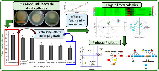
2.1. Dual Culture Assay of P. indica with Soil Microorganisms

The influence of soil microorganisms on the growth of Piriformospora indica was initially determined in dual culture (DC) with the bacteria Azotobacter chroococcum, Streptomyces sp. nov 505 (AcH505), Streptomyces 2009 (AcH2009), Streptomyces anulatus 1003 (AcH1003), Pseudomonas putida and Pseudomonas fluorescens. After twelve days significant growth stimulation (p < 0.05) was observed in the mycelia of fungi interacting with A. chroococcum. In another case, growth inhibition was observed in the interactions with the two Pseudomonas-strains and the two Streptomyces-strains AcH 2009 and AcH 1003. The assay with Streptomyces sp. nov AcH 505 (AcH 505) showed no significant influence on fungal growth (Figure 1).

2.2. P. indica in Dual Culture with A. chroococcum and S. anulatus 1003

The soil bacteria A. chroococcum and S. anulatus 1003 (AcH 1003) were used to determine the effect of the interaction (in dual culture) on P. indica’s growth rate. Due to the non-isodiametric development of the colony in response to the bacteria, only the area of the colony facing the bacterial inoculum (Figure 2a) was measured. The increase of fungal growth in dual cultures was recorded from day 4 in two-day-intervals (Figure 2b). A. chroococcum showed a growth-promoting effect on the fungus. This effect was visible on day 6 and exhibited an important increase on day 8. On the other hand, a significant inhibitory growth effect (p < 0.05) of S. anulatus 1003 was observed from the 4th day onwards in comparison to P. indica control and the dual culture with A. chroococcum.
Parallel to the time course of growth experiments, the effect of volatile compounds produced by S. anulatus 1003 was also tested. For that, split Petri-dishes containing bacteria and fungi were cultivated under the same conditions as described for the time course. In this case, no effect of bacterial volatiles on fungal growth was apparently observed after eight days of co-culture (Figure 2c).

Morphology of P. indica Hyphae during Dual Culture with Soil Bacteria

Alterations in the hyphal thickness and mycelium morphology of P. indica in interaction with the soil bacteria were observed. In general, significant changes (p < 0.05) in the hyphal thickness were then observed from the 10th day during the interaction with S. anulatus AcH 1003, whereas no substantial changes were registered in the DC with A. chrococcum. In this regard, the hyphae were found to be 3%–5% thinner to than the control during interaction with A. chroococcum, whereas DC with S. anulatus AcH 1003 generated an increase above 8% in comparison to that of control (Figure 2d). In addition, changes in the mycelium morphology from the sixth day were also observed in the DC with S. anulatus AcH 1003, accompanied by increased hyphal density and sporulation. In contrast, the hyphae presented a slightly increased hyphal extension and reduced and delayed sporulation during interaction with A. chroococcum (Figure 2e).

2.3. Free Amino Acid Content in P. indica

During the time course, the most abundant amino acids (AA) in extracts of P. indica (among 26 quantified AA, see Table A1, Figure 3a) were Gln, followed by Ala, Arg, and Glu. However, interactions with soil bacteria caused some variations in the fungal free AA contents (see Figure 3a). Co-culture with A. chroococcum caused an increase of Ala, whereas the amounts of Gln and Glu were considerably reduced in comparison to the control. In contrast, Pro, Ile, Val, and Leu showed an important increase, which was up to 4.0, 4.7, 4.2, and 4.6-fold, respectively, when compared to the control. Other AA showed no substantial changes (Figure 3b). Co-culture of P. indica with Streptomyces AcH 1003 affected the fungal AA pattern in a different way. In contrast to A. chroococcum and the control, glutamine was notably increased by AcH 1003. In addition, Gly and Ser were increased drastically. Their accumulation was up to 2.0 and 2.2-fold compared to the control, whereas Cys and cystathionine/methionine showed a meaningful reduction. The other amino acids presented no important variations (Figure 3b).
The orthogonal partial least squares discriminant analysis (OPLS-DA) model (performed on AA content data using interaction type as supervision variable) clearly discriminated P. indicaA. chroococcum and P. indicaS. anulatus interactions, according to the respective score plot (Figure 3c) compared to the control. This model involved a well-explained variance (R2(cum) = 0.995) using a 2+2+0 component system. This indicates that the observed differences among interactions are due to a remarkable change in AA contents. The corresponding biplot (Figure 3d) revealed the most-influencing AA for the resulting discrimination. Thus, A. chroococcum was related to the changes to Ala, GABA, Pro, Lys, and others, while interaction with S. anulatus induced the Gln and Ser accumulation in P. indica. Some amino acids (such as Thr, Cys/Met, among others) were related to the control in the OPLS-DA model because these had higher concentrations/contents in control, but no substantial changes in concentrations were observed on interaction experiments. For this model, major influences were consequently found to be for Gln and Ala, followed by GABA, Asn, and Leu, according to the variable influence on projection (VIP) values (Figure 3e).
OPLS-DA models for dual comparisons were also tested. The resulting biplots (Figure 4a) showed good separations along the first principal component (PC1), but interaction time discriminated some observations along the first orthogonal component (OC1). In this regard, the AA content evolved particularly different for the fungus-A. chroococcum interaction compared to that with S. anulatus. Therefore, AA content was particularly altered depending on the interaction, but the most-affected AA content was also different. Respective S-plots (Figure 4b) specified the most affected AA for each comparison. S. anulatus interaction led to a Gln accumulation, whereas Ala was induced by the interaction with A. chroococcum (Figure 4a-3). The control also exhibited the accumulation of Gln, but the respective concentration of Gln in experiments testing the fungus–bacteria interaction was found to be altered.
OPLS-DA models using interaction time as supervision variable allowed identifying that time-dependent evolution of these contents was also found to be an important factor for the AA metabolism since interactions during three different periods of incubation were statistically discriminated. In the case of A. chroococcum (Figure 5a), the most important AA changes occurred between 4 and 12 days of interaction, and the levels of Aaa caused a different clustering along PC2 at eight days, although also influenced the 12-days cluster along PC1. In addition, the change in Gln for A. chroococcrum was more related to four days in combination with Arg and Asn (and other 10 AA), while Ala, GABA, and Leu (and other 6 AA) were accumulated in higher amounts after 12 days. In contrast, S. anulatus caused an accumulation of Gln (as well as His, Gly, Am, Pro, and also Asp) at 12 days, Ala and GABA were more affected after eight days, and Leu and Val (and other 9 AA) after four days. In both interactions with A. chroococcum and S. anulatus 1003, Cys/Met and Phe levels influenced the discrimination at four days (Figure 5b). Cys, Arg, and Glu influenced differentially the discrimination of four and eight days, since four-days cluster was influenced by these AA along PC2, whereas eight-days cluster was marked along PC1, being this influence more important due to the explained variance of PC1 (47.7%). The controls showed a fluctuation in relation to incubation time. In the control, 8 AA (such as Tyr, Cys/Met, Ser, His, Gln, Asn, Thr, and Am) are mainly accumulated at the beginning (0 day) whereas Ala, Gly, Aba, and GABA are mostly produced/accumulated after eight days of incubation (Figure 3c).
In order to rationalize alterations of the AA content of P. indica in DC with soil bacteria, a pathway enrichment analysis (PEA) and a pathway topology analysis (PTA) were also performed using the model KEGG library associated to the Pathway Analysis module within Metaboanalyst 3.0. The overview of this pathway analysis (Figure 6) revealed the impact of the P. indica-bacterial interactions in some fungal AA metabolism. The two interactions showed different pathway impact profiles, but they exhibited nitrogen and Ala-Asp-Glu metabolism as the most importantly impacted pathway nodes. However, these nodes were found to be opposing in the Pathway Analysis according to their p values. Thus, A. chroococcum seems to affect: (1) The nitrogen metabolism in P. indica in general (Figure 6a-1), or (2) the Ala, Asp, and Glu metabolism (Figure 6a-2) specifically by altering the stored amounts of Asp and Gln as products or precursors, respectively, of these pathways. On the other hand, S. anulatus AcH 1003 induces accumulation of Asn and Glu within nitrogen metabolism in general (Figure 6b-1), and/or Ala, Asn, and GABA for the specific pathway (Figure 6b-2).

3. Discussion

3.1. Effect of the Soil Bacteria on the Growth of P. indica

The establishment of the symbiosis between beneficial endophytic root fungi and plant roots is affected by other microorganisms in the rhizosphere, especially by bacteria [7]. Interactions between soil bacteria and symbiotic fungi can have a positive, negative, or no effect on fungal growth [19]. In the present study, the effect of different soil bacteria on the growth of P. indica was analyzed using a fungus–bacterium DC system that has been used before for in vitro studies to elucidate the effect of mycorrhiza helper bacteria on the growth of mycorrhizal fungi and mycorrhiza formation [10]. Results obtained support former studies by Pham et al. [16] which revealed the stimulation of the growth of the fungus P. indica by the rhizobacterium Azotobacter chroococcum and showed the inhibition of fungal growth through the Pseudomonas strains P. putida and P. fluorescens. In addition, in the present work, the effect of different streptomycete strains isolated by Maier et al. [10] on the growth of P. indica was analyzed. Here, AcH 1003 and 2009 inhibited fungal growth, whereas AcH 505 had no effect. The interaction between the bacteria and P. indica resulted in non-isodiametric fungal colonies, which indicates that the bacterial exudates influencing the hyphal growth had a slow diffusion rate in the culture media.
Two bacterial strains with a contrasting influence on the development of P. indica were selected for biochemical analysis. Interaction between the fungus and A. chroococcum resulted in a considerable increase in fungal growth rate (as presented in Figure 2). This positive impact on P. indica growth was accompanied by an enhancing of the hyphal extension (Figure 2e) with delayed sporulation, but no effect on hyphal diameter was observed (Figure 2d). Similar results were reported recently by Bhuyan et al. [20] for the co-culture of the P. indica with the Azotobacter strain WR5 on the same Kaefer agar medium, showing a highly significant growth stimulation in comparison with other Azotobacter strains. A. chroococcum is a well-known free-living, N-fixing rhizobacterium, which is capable of improving plant growth through nitrogen fixation or by the production of growth-promoting substances [21]. The ability to fix nitrogen has also drawn interest to the use of A. chroococcum as bioinoculant to increase crop yield and productivity. Recently, the synergistic effect of dual inoculation of A. chroococcum with P. indica or arbuscular mycorrhizal fungi (AMF) on Panicum miliaceum [22], Stevia rebaudiana [23], Apple [24], and Artemisia annua [25] has been studied. The beneficial effects of MHB are attributed to the production of diverse metabolites. There are several reports stating that Azotobacter produces siderophores and indole acetic acid (IAA). These compounds may exert pronounced effects on plant growth by acting as mycorrhizal growth-promotors, as well as facilitators of phosphate solubilization and uptake [26].
In contrast, a fungistatic effect of AcH 1003 on the growth rate of P. indica during dual culture was observed. In combination with a reduction of the colony growth of P. indica (as observed in Figure 2a), this Streptomyces strain also had a notable effect on the fungal development by affecting the colony morphology (Figure 2e) and increasing the hyphal diameter (Figure 2d), but only when the bacteria and fungus grow in a continuous medium without direct contact. The changes in the hyphal diameter of P. indica could be associated with the reorganization of the fungal actin cytoskeleton. The ectomycorrhiza-forming fungus Amanita muscaria responded to the streptomycete strain AcH 505 in hyphae with a reduced diameter, which might have been an effect of bacterial substances released into the culture medium [27]. In filamentous fungi, several mutants inducing changes in hyphal morphology have been linked to the reorganization of the actin cytoskeleton [28].
Streptomycetes are known to produce pyrazines, i.e., volatile organic compounds well-known for their antimicrobial activities. These compounds are used as competition tools against other microorganisms that do not reside directly adjacent to each other [29,30]. However, volatiles from AcH 1003 did not show any effects on the mycelial extension rate of the P. indica. This suggests that the fungal response to AcH 1003 is generated necessarily by diffusion of bacterial metabolites within the medium, excluding volatile compounds.
Contrary to P. indica’s effects, prominent stimulatory effects of streptomycetes AcH 1003 and AcH 505 on A. muscaria mycelial growth rates have been reported [10,31]. The shape of fungal colonies remained isodiametric in all cultures, indicating that the growth-promoting compounds excreted by the bacteria were homogenously spread within the medium, and more rapidly diffusing than the mycelial growth rate of the responsive fungus. Such results have been shown for the ectomycorrhizal fungus Agaricus bisporus during DC with Pseudomonas putida, resulting in an isodiametric colony extension of this fungus [32]. Studies carried out with diverse mycorrhiza fungi showed the ability of Streptomyces to improve mycorrhization and mycorrhizal growth [5]. The streptomycetes used in this study also promoted the Suillus bovinus growth, whereas a suppressing effect on the hyphal extension of Hebeloma cylindrosporum was observed [33]. On the other hand, experiments carried out with streptomycete strains A and H showed growth inhibition of the plant pathogenic fungus Armillaria sp. [34]. Similarly, it was demonstrated that AcH 505 suppresses the growth of plant pathogens such as Heterobasidion annosum and Amillaria abietina [10].
It has been observed that Streptomycetes produce a variety of compounds with a stimulatory or inhibitory activity towards soil microorganisms, Riedlinger et al. [11] isolated and characterized three dominant secondary metabolites from Streptomyces AcH 505. They demonstrated that the antibiotics WS-5995 B and C suppressed the growth of A. muscaria, whereas the third substance, auxofuran, promoted the hyphal extension significantly. Furthermore, AcH 505 can also trigger the production of effectors in plant-pathogenic fungi [12].

3.2. P. indica Amino Acid Content During DC with A. chroococcum and AcH 1003

Free AA represent an important parameter to evaluate the absorption and assimilation of carbon and nitrogen of mycorrhizal fungi [35]. In the present study, substantial changes in the content of some free AA in the fungal mycelia of P. indica in DC with the soil bacteria were observed. According to the discriminant analysis in combination with PEA and PTA, the most-impacted pathway nodes were linked to nitrogen and Ala-Asp-Glu metabolism based on the variations of the AA contents (see Figure 6), therefore, it was deduced that bacterial interaction influenced nitrogen-related AA. Thus, during the interaction with A. chroococcum, Ala was the most abundant AA whereas glutamine and glutamic acid were considerably reduced (Figure 3 and Figure 4b-2). As A. chroococcum can assimilate atmospheric nitrogen and thus produces ammonia, we would have expected an increase of the fungal pool sizes of glutamate and glutamine through glutamine synthetase-glutamate synthase pathway (GS-GOGAT) involved in ammonium assimilation in ectomycorrhizal and endomycorrhizal fungi [36,37]. However, we found a considerable reduction in the concentration of these two amino acids compared to control. Reduction in the concentration of Glu and Gln could be related to the increment in Ala content presumably via alanine aminotransferase (EC 2.6.1.2). Alanine synthesis is an important end-product for the glucose-carbon and a convenient reservoir of both amino groups and pyruvate in mycelia during periods of sufficient nitrogen and carbon supply [35,38]. Ala-derived nitrogen can be converted to glutamate nitrogen with the release of pyruvate, as observed in the ectomycorrhizal ascomycete fungus Cenococcum geophilum under carbon starvation [39]. In Tuber borchii an increase in the pool size of Ala was also associated with a period of active growth [40]. In a study on differentially expressed proteins of P. indica co-cultured with A. chroococcum was reported the up-regulation of ENO1 (2-phosphoglycerate dehydratase) and Ure D fungi genes, suggesting that the bacterium could trigger the uptake of hexose sugar by the activation of several glucose transporters. ENO1 is one of the key regulatory enzymes of glycolysis [20]. Ure D is one of the accessory proteins of the apoprotein UreABC which is a nickel-dependent regulatory enzyme involved in recycling of urea [41]. The increase of the Ala content could thus be associated with the growth promotion of P. indica during the interaction with A. chroococcum.
The decrease in Gln and Glu could be due to either reduced activities of glutamine synthetase (GS) and glutamate dehydrogenase (GDH), or to rapid metabolism of these AA, also explaining the four-fold increase in proline as a typical result of this pathway for cellular homeostasis in fungi [42]. Reduction in the activity of these enzymes in Laccaria bicolor has been related to the presence of organic N sources such as alanine or arginine in the culture medium [43]. The excretion of AA into the culture medium has been reported for A. chrooccocum [44] and Beijerinckia derxii [45], but detailed information on the specific AA released is lacking. Additionally, the ca. four-fold increases of the branched-chain amino acids family (i.e., leucine, isoleucine, and valine) can be then rationalized as a consequence of the competitive carbon flux during interaction.
In contrast, the DC of P. indica with S. anulatus AcH 1003 caused an increase in the amount of Glu, Gly and Ser in the fungal mycelia (Figure 3 and Figure 4a-2). At the same time, a substantial reduction of P. indica growth was observed. Gln, Cys, and Gly are important for the synthesis of the antioxidant, glutathione (GSH; γ-l-glutamyl-l-cysteinyl-glycine), which detoxifies non-enzymatically a series of reactive oxygen species, and is generally present in a high concentration of up to 10 mM in yeasts and filamentous fungi [46]. In yeast, serine O-acetyltransferase and O-acetylserine sulfhydrylase constitute a pathway for synthesis of cysteine from serine [47]. Subsequently, Cys is converted to methionine via cystathionine and homocysteine [48]. This pathway could explain the increase of Ser as a precursor of Cys and the considerable decrease of methionine and cystathionine by the consumption of Cys for the synthesis of GSH. AcH 1003 produces at least three secondary metabolites, one of which belongs to the group of polyene macrolide antibiotics [49]. These are known to act as fungicides and, together with polyol macrolide antibiotics, have been shown to induce oxidative stress [50]. Some substances produced by AcH 1003 were able to generate oxidative stress which, in turn, results in enhanced production of AA, which are important for GSH generation.

4. Materials and Methods

4.1. Dual Culture of Piriformospora indica with Soil Microorganisms and Sample Collection

To determine the influence of the bacteria on fungal growth, the mycelium of Piriformospora indica was cultivated in 9-cm-Petri dishes on Kaefer [51] agar medium in a DC system, together with the respective soil bacteria. A fungal inoculum with a diameter of 4 mm was excised from actively growing rims of five-day-old fungal colonies, using the wide end of a Pasteur pipette. This was placed onto a plate on a sterile cellophane-sheet (exclusion limit 10 kDa; Folia Wendelstein, Germany). Thus, the fungal inoculum always had the same size and the hyphae could be collected easily at the end of the experiment for further analysis. In the same way, 10 μL of soil bacteria from a liquid culture (OD600nm: 0.6–0.7) were inoculated as a line of about 4 cm long directly on the media (outside the cellophane sheet) at 4 cm from the fungal colony. The Petri dishes were incubated in darkness at 25 ± 2 °C. In order to select two antagonistic bacterial strains, one that inhibited and one that promoted the fungal growth, dual cultures of P. indica with Azotobacter chroococcum, Pseudomonas fluorescens, and Pseudomonas putida were carried out. The latter were isolated and kindly provided by Prof. A. Varma, School Life Science, New Delhi, India. In addition, we investigated the effects of Streptomyces anulatus (Beijerinck) Waksman 1003 (AcH 1003), Streptomyces sp. nov. 505 (AcH 505), and Streptomyces 2009 (AcH 2009). These bacteria were isolated from the hyphosphere of Picea abies [10]. In this experiment, ten replicates were used. The effect of the fungus-bacteria interaction on fungal growth was evaluated after twelve days as the radial growth of the colony portion that was oriented towards the bacteria. From this assay, two bacterial strains were selected, A. chroococcum as growth-promoting bacterium and Streptomyces AcH 1003 as a growth-inhibiting bacterium. Cultures of P. indica with A. chroococcum, AcH 1003, and fungus alone as control were carried out using the same DC system. Samples of mycelia from the first centimeter of the proximal part of the fungal colony were collected after 4, 8, and 12 days, rapidly homogenized in liquid nitrogen and freeze-dried for three weeks at below −20 °C. Mycelium from five plates was collected as one replicate, four replicas were obtained for each time point. These samples were used to determine the amino acid content.

4.2. Influence of Soil Bacteria on Fungal Growth

The bacterial effect on the growth of P. indica in the dual cultures was evaluated every two days. Data were registered as colony area of the part oriented towards the bacteria. At least 10 replicates were used for the growth test.

4.3. Morphology of Piriformospora indica Hyphae

The influence of the fungus-bacteria interaction on the thickness of P. indica hyphae was determined after 4, 6, 8, 10, and 12 days using a Leica DM500 binocular light microscope (Leica Microsystems GmbH, Wetzlar, Germany). The hyphae thickness was measured approximately 10 μm from the hyphal apex using the program Image J 1.62 (National Institute of Health, Bethesda, MD, USA). At least 100 replicates per time point from independent colonies were used. Additionally, the mycelium morphology after 12 days of interaction was registered by optical micrographs at 100× using a Leica S9 D stereo microscope (Leica Microsystems GmbH, Wetzlar, Germany).

4.4. Soil Bacteria Culture

Twenty mL of each soil bacteria strain were cultivated in liquid Kaefer media (1977) in a 100 mL-Erlenmeyer flask, containing one metal spiral. A. chroococcum, P. putida, and P. fluorescens were initially incubated for two days, whereas Streptomyces-strains were incubated for four days. The cultures were grown at 28 °C on a rotary shaker at 180 rpm. New cultures were started inoculating 2–4 mL of the cultures incubated for four days in 20 mL of liquid Kaefer media until OD600nm: 0.6–0.7 was reached. These cultures were used to inoculate the plates of the dual cultures. The stocks of bacteria were grown at 28 °C in solid Kaefer medium and maintained at 4 °C in the cold room. The stock cultures were replicated every six weeks.

4.5. Determination of Amino Acid Content

The determination of the content of 26 different amino acids (Table A1) in mycelia of P. indica was carried out as follows: 7 to 10 mg of freeze-dried homogenized mycelia were re-suspended in 500 μL of 80% (v/v) methanol and heated in a water bath for 3 min. After centrifugation 10 min at 16,069 g, the supernatant was placed on ice and the pellet was resuspended with 500 μL of methanol 20% (v/v) and heated for 3 min in a water bath. The cell debris was removed by centrifugation at 16,069 g, and the supernatant was transferred into an Eppendorf tube, together with the first extract. The extract was dried in a speed vac centrifuge and the pellet resuspended in a lithium buffer (Li 220 Pickering Laboratories, Mountain View, CA, USA). Analysis was by high performance liquid chromatography (HPLC) on a cation-exchange column (high efficiency fluid column, 3 × 150 mm; Pickering Laboratories, Mountain View, CA, USA) using lithium buffer as eluent. Amino acids were derivatized with ninhydrine before photometric detection as described by Pilot et al. [52]. Three replicates were measured at each time point. The results were reported as nmol amino acid/mg dry weight (DW).

4.6. Data Analysis

Differences in the amino acid content between treatments in response to the effect of the fungal bacteria interaction were analyzed with a one-factor ANOVA followed by Tukey’s studentized range (HSD) test using the SAS 8.0 (SAS Institute, Cary, NC, USA).
Targeted metabolomics was also implemented to analyze the amino acid concentrations data under supervised analysis according to fungal bacteria interaction and time. Thus, the resulting two-way array raw data were organized, and a 20 observations (treatments) × 26 variables (amino acid contents in nmol/mg DW) matrix was built. OPLS-DA models were then constructed for classification according to the holistic variation of amino acid content between treatments. OPLS-DA models were created using SIMCA 14 software (Umetrics Inc., Umeå, Sweden) on a PC (Core i5 processor; 8 GB RAM; on Microsoft Windows 7). Integrating enrichment and pathway topology analyses were performed according to the comprehensive, Web-based tool MetaboAnalyst 4.0 (McGill University, Montreal, QC, Canada).

5. Conclusions

Fungal growth and its amino acid composition during the interaction of P. indica and soil bacteria were studied. Fungal growth was stimulated by Azotobacter chroococcum, whereas Streptomyces anulatus AcH 1003 inhibited it and Streptomyces sp. Nov AcH 505 had no effect. OPLS-DA model clearly discriminated P. indica-A. chroococcum and P. indica-S. anulatus interactions. The most-observable responses were found to be in the cases of glutamine and alanine size groups. In this sense, while Streptomyces AcH 1003 increased the amount of glutamine, A. chroococcum decreased it. The fungal growth and the increase of alanine content might be associated with the assimilation of nitrogen in the presence of glucose as a carbon source. Hence, the N-fixing bacterium A. chroococcum should stimulate fungal amino acid metabolism via glutamine synthetase-glutamate synthase (GS-GOGAT). The data pointed to a stimulated glycolytic activity in the fungus observed by the accumulation of alanine, possibly via alanine aminotransferase. Responses toward the growth-inhibiting Streptomyces AcH 1003 suggested an oxidative-mediated stress response of the fungus. However, additional evidence should be compiled to support it, for instance, by means of the determination of ROS or lipids peroxidation products in fungal cells. Additionally, an important limitation of this AA metabolism study, as a phenotypic response of P. indica facing the contrasting bacterial interactions, is related to the direct assignment of the role of each AA. In this regard, these results can be combined with gene expression in order to explain such roles at the biochemical level. However, the present results defined an important parameter to describe a functional bacteria–fungus association to be used properly in symbiotic-mediated plant nutrition.

Author Contributions

All authors have read and agree to the published version of the manuscript. Conceptualization, J.A.L.-R. and R.H.; methodology, J.A.L.-R. and R.H.; software, E.C.-B.; validation, J.A.L.-R.; formal analysis, J.A.L.-R. and E.C.-B.; investigation, J.A.L.-R., E.C.-B., and R.H.; data curation, J.A.L.-R. and E.C.-B.; writing—original draft preparation, J.A.L.-R.; writing—review and editing, J.A.L.-R., E.C.-B., and R.H.; supervision, R.H.; funding acquisition, R.H.

Funding

This research was funded by University of Tuebingen, German Academic Exchange Service (DAAD) and Universidad Militar Nueva Granada (UMNG, grant code IMP-CIAS-2293).

Acknowledgments

The authors would like to thank Dr. Harald Stransky and Bettina Stadelhofer of the Center for Plant Molecular Biology, University of Tuebingen, Germany, for the amino acid analysis and Dr. Ernesto Vargas to read and discussed the article. JA Leyva-Rojas was supported by DAAD. E. Coy-Barrera thank UMNG for the financial support (project IMP-CIAS-2293).

Conflicts of Interest

The authors declare no conflict of interest. The funders had no role in the design of the study; in the collection, analyses, or interpretation of data; in the writing of the manuscript, or in the decision to publish the results.

Appendix A

Table A1. Name and abbreviations of quantified amino acids for P. indica in control and dual cultures (bacterial interactions).
NoNameAbbreviationNoNameAbbreviation
1glycineGly14hydroxylysineHlys
2serineSer15glutamic acidGlu
3cysteineCys16glutamineGln
4ornithineOrn17alanineAla
5histidineHis18arginineArg
6tyrosineTyr19asparagineAsn
7isoleucineIle20prolinePro
8threonineThr21valineVal
9α-aminobutyric acidAba22leucineLeu
10cystathionine/methionine aCys/Met23lysineLys
11PhenylalaninePhe24ammoniaAm
12aminoapidic acidAaa25aspartic acidAsp
13TryptophanTrp26γ-aminobutyric acidGaba
a cystathionine and methionine could not be separated since they were part of the same chromatographic signal. Hence, the amounts could not be individually determined.

References

  1. Deveau, A.; Bonito, G.; Uehling, J.; Paoletti, M.; Becker, M.; Bindschedler, S.; Hacquard, S.; Hervé, V.; Labbé, J.; Lastovetsky, O.A.; et al. Bacterial-fungal interactions: Ecology, mechanisms and challenges. FEMS Microbiol. Rev. 2018, 42, 335–352. [Google Scholar] [CrossRef] [Green Version]
  2. Churchland, C.; Grayston, S.J. Specificity of plant-microbe interactions in the tree mycorrhizosphere biome and consequences for soil cycling. Front. Microbiol. 2014, 5, 1–20. [Google Scholar] [CrossRef] [Green Version]
  3. Bonfante, P.; Anca, I.-A. Plants, mycorrhizal fungi, and bacteria: a network of interactions. Annu. Rev. Microbiol. 2009, 63, 363–383. [Google Scholar] [CrossRef] [Green Version]
  4. Kim, M.; Yoon, H.; Kim, Y.E.; Kim, Y.J.; Kong, W.S.; Kim, J.G. Comparative analysis of bacterial diversity and communities inhabiting the fairy ring of Tricholoma matsutake by barcoded pyrosequencing. J. Appl. Microbiol. 2014, 117, 699–710. [Google Scholar] [CrossRef]
  5. Schrey, S.D.; Tarkka, M.T. Friends and foes: Streptomycetes as modulators of plant disease and symbiosis. Antonie van Leeuwenhoek, Int. J. Gen. Mol. Microbiol. 2008, 94, 11–19. [Google Scholar] [CrossRef] [PubMed]
  6. Xing, R.; Yan, H.Y.; Gao, Q.B.; Zhang, F.Q.; Wang, J.L.; Chen, S.L. Microbial communities inhabiting the fairy ring of Floccularia luteovirens and isolation of potential mycorrhiza helper bacteria. J. Basic Microbiol. 2018, 58, 554–563. [Google Scholar] [CrossRef] [PubMed]
  7. Garbaye, J. Helper bacteria: A new dimension to the mycorrhizal symbiosis. New Phytol. 1994, 128, 197–210. [Google Scholar] [CrossRef]
  8. Takács, T.; Cseresnyés, I.; Kovács, R.; Parádi, I.; Kelemen, B.; Szili-Kovács, T.; Füzy, A. Symbiotic effectivity of dual and tripartite associations on soybean (Glycine max L. Merr.) cultivars inoculated with Bradyrhizobium japonicum and AM Fungi. Front. Plant Sci. 2018, 9, 1–14. [Google Scholar] [CrossRef]
  9. Turrini, A.; Avio, L.; Giovannetti, M.; Agnolucci, M. Functional complementarity of arbuscular mycorrhizal fungi and associated microbiota: The challenge of translational research. Front. Plant Sci. 2018, 9, 10–13. [Google Scholar] [CrossRef]
  10. Maier, A.; Riedlinger, J.; Fiedler, H.-P.; Hampp, R. Actinomycetales bacteria from a spruce stand: Characterization and effects on growth of root symbiotic and plant parasitic soil fungi in dual culture. Mycol. Prog. 2004, 3, 129–136. [Google Scholar] [CrossRef]
  11. Riedlinger, J.; Schrey, S.D.; Tarkka, M.T.; Hampp, R.; Kapur, M.; Fiedler, H.-P. Auxofuran, a novel metabolite that stimulates the growth of fly agaric, is produced by the mycorrhiza helper bacterium Streptomyces strain AcH 505. Appl. Environ. Microbiol. 2006, 72, 3550–3557. [Google Scholar] [CrossRef] [PubMed] [Green Version]
  12. Keilhofer, N.; Nachtigall, J.; Kulik, A.; Ecke, M.; Hampp, R.; Süssmuth, R.D.; Fiedler, H.P.; Schrey, S.D. Streptomyces AcH 505 triggers production of a salicylic acid analogue in the fungal pathogen Heterobasidion abietinum that enhances infection of Norway spruce seedlings. Antonie van Leeuwenhoek Int. J. Gen. Mol. Microbiol. 2018, 111, 691–704. [Google Scholar] [CrossRef] [PubMed]
  13. Varma, A.; Verma, S.; Sudha; Sahay, N.; Bütehorn, B.; Franken, P. Piriformospora indica, a cultivable plant-growth-promoting root endophyte. Appl. Environ. Microbiol. 1999, 65, 2741–2744. [Google Scholar] [CrossRef] [Green Version]
  14. Franken, P. The plant strengthening root endophyte Piriformospora indica: Potential application and the biology behind. Appl. Microbiol. Biotechnol. 2012, 96, 1455–1464. [Google Scholar] [CrossRef] [PubMed] [Green Version]
  15. Gill, S.S.; Gill, R.; Trivedi, D.K.; Anjum, N.A.; Sharma, K.K.; Ansari, M.W.; Ansari, A.A.; Johri, A.K.; Prasad, R.; Pereira, E.; et al. Piriformospora indica: Potential and significance in plant stress tolerance. Front. Microbiol. 2016, 7, 1–20. [Google Scholar] [CrossRef] [Green Version]
  16. Pham, G.H.; Singh, A.; Malla, R.; Kumari, R.; Prasad, R.; Sachdev, M.; Rexer, K.H.; Kost, G.; Luis, P.; Kaldorf, M.; et al. Interaction of Piriformospora indica with diverse microorganisms and plants. In Plant Surface Microbiology; Springer: Berlin/Heidelberg, Germany, 2008; pp. 237–265. ISBN 9783540740506. [Google Scholar]
  17. Bonfante, P.; Genre, A. Mechanisms underlying beneficial plant–fungus interactions in mycorrhizal symbiosis. Nat. Commun. 2010, 1, 48. [Google Scholar] [CrossRef] [Green Version]
  18. Wang, W.; Shi, J.; Xie, Q.; Jiang, Y.; Yu, N.; Wang, E. Nutrient exchange and regulation in arbuscular mycorrhizal symbiosis. Mol. Plant 2017, 10, 1147–1158. [Google Scholar] [CrossRef] [Green Version]
  19. Johansson, J.F.; Paul, L.R.; Finlay, R.D. Microbial interactions in the mycorrhizosphere and their significance for sustainable agriculture. FEMS Microbiol. Ecol. 2004, 48, 1–13. [Google Scholar] [CrossRef]
  20. Kumar Bhuyan, S.; Bandyopadhyay, P.; Kumar, P.; Kumar Mishra, D.; Prasad, R.; Kumari, A.; Chandra Upadhyaya, K.; Varma, A.; Kumar Yadava, P. Interaction of Piriformospora indica with Azotobacter chroococcum. Sci. Rep. 2015, 5, 1–13. [Google Scholar] [CrossRef] [Green Version]
  21. Bhattacharyya, P.N.; Jha, D.K. Plant growth-promoting rhizobacteria (PGPR): Emergence in agriculture. World J. Microbiol. Biotechnol. 2012, 28, 1327–1350. [Google Scholar] [CrossRef]
  22. Shishehbor, M.; Madani, H.; Ardakani, M.R. Effect of vermicompost and biofertilizers on yield and yield components of common millet (Panicum miliaceum). Ann. Biol. Res. 2013, 4, 174–180. [Google Scholar]
  23. Vafadar, F.; Amooaghaie, R.; Otroshy, M. Effects of plant-growth-promoting rhizobacteria and arbuscular mycorrhizal fungus on plant growth, stevioside, NPK, and chlorophyll content of Stevia rebaudiana. J. Plant Interact. 2014, 9, 128–136. [Google Scholar] [CrossRef]
  24. Sharma, S.D.; Sharma, N.C.; Sharma, C.L.; Kumar, P.; Chandel, A. Glomus-Azotobacter symbiosis in apple under reduced inorganic nutrient fertilization for sustainable and economic orcharding enterprise. Sci. Hortic. (Amsterdam) 2012, 146, 175–181. [Google Scholar] [CrossRef]
  25. Arora, M.; Saxena, P.; Choudhary, D.K.; Abdin, M.Z.; Varma, A. Dual symbiosis between Piriformospora indica and Azotobacter chroococcum enhances the artemisinin content in Artemisia annua L. World J. Microbiol. Biotechnol. 2016, 32, 19. [Google Scholar] [CrossRef]
  26. Maheshwari, D.K.; Dubey, R.C.; Aeron, A.; Kumar, B.; Kumar, S.; Tewari, S.; Arora, N.K. Integrated approach for disease management and growth enhancement of Sesamum indicum L. utilizing Azotobacter chroococcum TRA2 and chemical fertilizer. World J. Microbiol. Biotechnol. 2012, 28, 3015–3024. [Google Scholar] [CrossRef]
  27. Schrey, S.D.; Salo, V.; Raudaskoski, M.; Hampp, R.; Nehls, U.; Tarkka, M.T. Interaction with mycorrhiza helper bacterium Streptomyces sp. AcH 505 modifies organisation of actin cytoskeleton in the ectomycorrhizal fungus Amanita muscaria (fly agaric). Curr. Genet. 2007, 52, 77–85. [Google Scholar] [CrossRef]
  28. Walther, A.; Wendland, J. Apical localization of actin patches and vacuolar dynamics in Ashbya gossypii depend on the WASP homolog Wal1p. J. Cell Sci. 2004, 117, 4947–4958. [Google Scholar] [CrossRef] [Green Version]
  29. Jones, S.E.; Elliot, M.A. Streptomyces Exploration: Competition, volatile communication and new bacterial behaviours. Trends Microbiol. 2017, 25, 522–531. [Google Scholar] [CrossRef]
  30. Tyc, O.; Song, C.; Dickschat, J.S.; Vos, M.; Garbeva, P. The ecological role of volatile and soluble secondary metabolites produced by soil bacteria. Trends Microbiol. 2017, 25, 280–292. [Google Scholar] [CrossRef]
  31. Adams, A.S.; Currie, C.R.; Cardoza, Y.; Klepzig, K.D.; Raffa, K.F. Effects of symbiotic bacteria and tree chemistry on the growth and reproduction of bark beetle fungal symbionts. Can. J. For. Res. 2009, 39, 1133–1147. [Google Scholar] [CrossRef]
  32. Rainey, P.B. Effect of Pseudomonas putida on hyphal growth of Agaricus bisporus. Mycol. Res. 1991, 95, 699–704. [Google Scholar] [CrossRef]
  33. Schrey, S.D.; Schellhammer, M.; Ecke, M.; Hampp, R.; Tarkka, M.T. Mycorrhiza helper bacterium Streptomyces AcH 505 induces differential gene expression in the ectomycorrhizal fungus Amanita muscaria. New Phytol. 2005, 168, 205–216. [Google Scholar] [CrossRef] [PubMed]
  34. Becker, D.M.; Bagley, S.T.; Podila, G.K. Effects of mycorrhizal-associated Streptomycetes on growth of Laccaria bicolor, Cenococcum geophilum, and Armillaria species and on gene expression in Laccaria bicolor. Mycologia 1999, 91, 33. [Google Scholar] [CrossRef]
  35. Martin, F.; Ramstedt, M.; Söderhäll, K.; Canet, D. Carbohydrate and amino acid metabolism in the ectomycorrhizal ascomycete Sphaerosporella brunnea during glucose utilization: A 13C NMR Study. Plant Physiol. 1988, 86, 935–940. [Google Scholar] [CrossRef] [Green Version]
  36. Breuninger, M.; Trujillo, C.G.; Serrano, E.; Fischer, R.; Requena, N. Different nitrogen sources modulate activity but not expression of glutamine synthetase in arbuscular mycorrhizal fungi. Fungal Genet. Biol. 2004, 41, 542–552. [Google Scholar] [CrossRef]
  37. Márquez, A.J.; Betti, M.; Ottonello, S.; Bonfante, P.; Montanini, B.; Balestrini, R. Distinctive properties and expression profiles of glutamine synthetase from a plant symbiotic fungus. Biochem. J. 2003, 373, 357–368. [Google Scholar]
  38. Botton, B.; Chalot, M.; Garnier, A.; Martin, F. L’assimilation de l’azote minéral chez les ectomycorhizes. Acta Bot. Gall. 1994, 141, 469–481. [Google Scholar] [CrossRef]
  39. Martin, F.; Canet, D. Biosynthesis of amino acids during [13C]glucose utilization by the ectomycorrhizal ascomycete Cenococciiin geophiluin monitored by 13C nuclear magnetic resonance. Physiol Vég 1986, 24, 209–218. [Google Scholar]
  40. Ceccaroli, P.; Saltarelli, R.; Cesari, P.; Pierleoni, R.; Sacconi, C.; Vallorani, L.; Rubini, P.; Stocchi, V.; Martin, F. Carbohydrate and amino acid metabolism in Tuber borchii mycelium during glucose utilization: A 13C NMR study. Fungal Genet. Biol. 2003, 39, 168–175. [Google Scholar] [CrossRef]
  41. Kim, J.K.; Mulrooney, S.B.; Hausinger, R.P. The UreEF fusion protein provides a soluble and functional form of the UreF urease accessory protein. J. Bacteriol. 2006, 188, 8413–8420. [Google Scholar] [CrossRef] [Green Version]
  42. Fichman, Y.; Gerdes, S.Y.; Kovács, H.; Szabados, L.; Zilberstein, A.; Csonka, L.N. Evolution of proline biosynthesis: Enzymology, bioinformatics, genetics, and transcriptional regulation. Biol. Rev. 2015, 90, 1065–1099. [Google Scholar] [CrossRef] [PubMed]
  43. Ahmad, I.; Carleton, T.; Malloch, D.; Hellebust, J. Nitrogen metabolism in the ectomycorrhizal fungus Laccaria bicolor (R. Mre.) Orton. New Phytol. 1990, 116, 431–441. [Google Scholar] [CrossRef]
  44. Pati, B.R.; Sengupta, S.; Chandra, A.K. Studies on the amino acids released by phyllosphere diazotrophic bacteria. Microbiol. Res. 1994, 149, 287–290. [Google Scholar] [CrossRef]
  45. Thuler, D.S.; Floh, E.I.S.; Handro, W.; Barbosa, H.R. Beijerinckia derxii releases plant growth regulators and amino acids in synthetic media independent of nitrogenase activity. J. Appl. Microbiol. 2003, 95, 799–806. [Google Scholar] [CrossRef] [Green Version]
  46. Pócsi, I.; Prade, R.A.; Penninckx, M.J. Glutathione, altruistic metabolite in fungi. In Advances in Microbial Physiology; Academic Press: Cambridge, MA, USA, 2004; Volume 49, pp. 1–76. ISBN 0120277492. [Google Scholar]
  47. Ono, B.; Shirahige, Y.; Nanjoh, A.; Andou, N.; Ohue, H.; Ishino-Arao, Y. Cysteine biosynthesis in Saccharomyces cerevisiae: Mutation that confers cystathionine beta-synthase deficiency. J. Bacteriol. 1988, 170, 5883–5889. [Google Scholar] [CrossRef] [Green Version]
  48. Savin, M.A.; Flavin, M. Cystationine synthesis in yeast: An alternative pathway for homocysteine biosynthesis. J. Bacteriol. 1972, 112, 299–303. [Google Scholar] [CrossRef] [Green Version]
  49. Riedlinger, J. Die stofflichen Grundlagen der Modulation des Myzelwachstums von symbiotischen und pathogenen Pilzen durch Streptomyceten; Eberhard-Karls University of Tübingen: Tübingen, Germany, 2006. [Google Scholar]
  50. Ogita, A.; Matsumoto, K.; Fujita, K.; Usuki, Y.; Hatanaka, Y.; Tanaka, T. Synergistic fungicidal activities of amphotericin B and N-methyl-N”-dodecylguanidine: A constituent of polyol macrolide antibiotic niphimycin. J. Antibiot. (Tokyo) 2007, 60, 27–35. [Google Scholar] [CrossRef] [Green Version]
  51. Käfer, E. Meiotic and mitotic recombination in Aspergillus and its chromosomal aberrations. Adv. Genet. 1977, 19, 33–131. [Google Scholar]
  52. Pilot, G.; Stransky, H.; Bushey, D.F.; Pratelli, R.; Ludewig, U.; Wingate, V.P.; Frommer, W.B. Overexpression of GLUTAMINE DUMPER1 leads to hypersecretion of glutamine from hydathodes of Arabidopsis leaves. Plant Cell 2004, 16, 1827–1840. [Google Scholar] [CrossRef] [Green Version]
Sample Availability: Samples of the compounds are available from the authors.
Figure 1. Effect of the soil microorganisms on the growth of Piriformospora indica after 12 days of dual culture. Azotobacter chroococcum, Streptomyces strains (AcH 505, AcH 2009, and AcH 1003 (S. anulatus)), Pseudomonas putida and P. fluorescens. Control corresponds to P. indica growing alone. Means with the same letters were not significantly different according to one-way ANOVA and the Tukey test (p < 0.05).
Molecules 25 00572 g001
Figure 2. (a) Effect of the soil bacteria on the growth of P. indica after 12 days of dual culture. (b) Time course of P. indica (control) in dual cultures (DC) with A. chroococcum and S. anulatus AcH 1003. Means with the same letter were not significantly different according to one-way ANOVA and the Tukey test (p < 0.05). These tests were performed on growth data per recording day in order to observe differences between control and DC. (c) Results of the experiment to observe the plausible effect of volatile substances produced by AcH 1003 on the growth of P. indica after eight days of culture in split Petri-dishes. (d) Determination of P. indica hyphal thickness in DC with A. chroococcum and S. anulatus AcH 1003. Means with the same letter were not significantly different according to one-way ANOVA and the Tukey test (p = 0.05). (e) Impact of soil bacteria on P. indica mycelium morphology after 12 days of interaction (optical micrograph at 100×).
Molecules 25 00572 g002
Figure 3. (a) Heatmap using ward algorithm for the variations of AA contents (expressed as nmol/mg dry weight (DW)) among interaction type (C0 = P. indica control at t = 0 day, C = P. indica control, A = P. indica+A.chroococcum, S = P. indica + S. anulatus 1003 (AcH 1003)) and time (in days) of interaction (4 = 4 days, 8 = 8 days, 12 = 12 days). (b) Boxplot for AA distribution according to time and interaction type. Names and abbreviations of 26 quantified AA are presented in Table A1. (c) Score plot on AA content data from a 2+2+0 OPLS-DA model (R2(cum) = 0.995, Q2(cum) = 0.967) using interaction type as categorical variable. (d) OPLS-DA-derived biplot on AA content data using interaction type as categorical variable. (e) VIP values from previous OPLS-DA model.
Molecules 25 00572 g003
Figure 4. (a) OPLS-DA-derived biplot on AA content data using interaction type as categorical variable per each dual comparison: (a-1) Control vs. S. anulatus 1003, (a-2) Control vs. A. chroococcum, (a-3) S. anulatus 1003 vs. A. chroococcum, (b) OPLS-DA-derived S-plot from previous OPLS-DA model: (b-1) Control vs. S. anulatus 1003, (b-2) Control vs. A. chroococcum, (b-3) S. anulatus 1003 vs. A. chroococcum.
Molecules 25 00572 g004
Figure 5. OPLS-DA-derived biplots on evolution of AA content through interaction time in days (as categorical variable) per interaction: (a) A. chroococcum, (b) S. anulatus, (c) control.
Molecules 25 00572 g005
Figure 6. Pathway nodes for (a) P. indica-A. chroococcum and (b) P. indica-S. anulatus interactions. Matched pathways were obtained according to p and pathway impact values from PEA and PTA algorithms, respectively, using a pathway library of a model microorganism. (1) Nitrogen metabolic pathway and (2) Ala, Asp, and Glu metabolism as the most important matched pathways, according to the pathway library within the KEGG database. Compound colors within the pathway: Light blue (metabolite not included in current analysis but used as background for PEA), from yellow to red (metabolites included in current analysis with different significance levels, yellow least, red most significant.
Molecules 25 00572 g006

