Article

Cycloartane and Oleanane Glycosides from the Tubers of Eranthis cilicica

1
Faculty of Pharmacy, Takasaki University of Health and Welfare, 37-1, Nakaorui, Takasaki, Gunma 370-0033, Japan
2
Department of Medicinal Pharmacognosy, School of Pharmacy, Tokyo University of Phramacy and Life Sciences, 1432-1, Horinouchi, Hachioji, Tokyo 192-0392, Japan
*
Author to whom correspondence should be addressed.
Molecules 2019, 24(1), 69; https://doi.org/10.3390/molecules24010069
Submission received: 22 November 2018 / Revised: 14 December 2018 / Accepted: 20 December 2018 / Published: 25 December 2018
(This article belongs to the Special Issue Natural Product Isolation, Identification and Biological Activity)

Abstract

:
Phytochemical analysis of the tubers of Eranthis cilicica was performed as part of our continuous study on the plants of the family Ranunculaceae, which resulted in the isolation of eleven new cycloartane glycosides (111) and one new oleanane glycoside (13), together with one known oleanane glycoside (12). The structures of the new compounds were determined by extensive spectroscopic analysis, including two-dimensional (2D) NMR, and enzymatic hydrolysis followed by either X-ray crystallographic or chromatographic analysis. The aglycone (1a) of 2 and its C-23 epimer (8a), and the oleanane glycosides (12 and 13) showed cytotoxic activity against HL-60 leukemia cells with IC50 values ranging from 10.6 μM to 101.6 μM. HL-60 cells were much more sensitive to 8a (IC50 14.8 μM) than 1a (IC50 101.1 μM), indicating that the C-23 configuration is associated with the cytotoxicity of these cycloartane derivatives. Compound 12 was revealed so as to partially induce apoptotic cell death in HL-60 cells, as was evident from morphology of HL-60 cells treated with 12.

Graphical Abstract

1. Introduction

We carried out systematic phytochemical screenings of the plants belonging to the family Ranunculaceae, such as the Adonis [1,2,3,4], Anemone [5,6], Cimicifuga [7,8], Clematis [9], Helleborus [10,11,12,13,14], and Pulsatilla species [15,16], and isolated various triterpene and steroidal glycosides, including cardiac and pregnane glycosides. Among them, cycloartane glycosides had a unique structure and showed relationships between chemical structure and cytotoxic activity [8]. The genus Eranthis also belongs to the family Ranunculaceae and is taxonomically related to the genus Helleborus [17]. Previously, we have isolated two new oleanane bisdesmosides, eranthisaponins A and B [18], and eight new chromone derivatives from the tubers of Eranthis cilicica [19]. Further phytochemical examination of the E. cilicica tubers resulted in the isolation of eleven new cycloartane glycosides and one new oleanane glycoside, together with one known oleanane glycoside. We report herein the structural determination of the new compounds by extensive spectroscopic analysis, including two-dimensional (2D) NMR, and enzymatic hydrolysis, followed by either X-ray crystallographic or chromatographic analysis. As part of our ongoing phytochemical study of Ranunculaceae plants, the cytotoxic activity of the cycloartane-type glycosides 2 and 8, the aglycone 1a and its C-23 epimer 8a, and the oleanane-type triterpene glycosides 12 and 13 against HL-60 human promyelocytic leukemia cells is evaluated and briefly discussed.

2. Results and Discussion

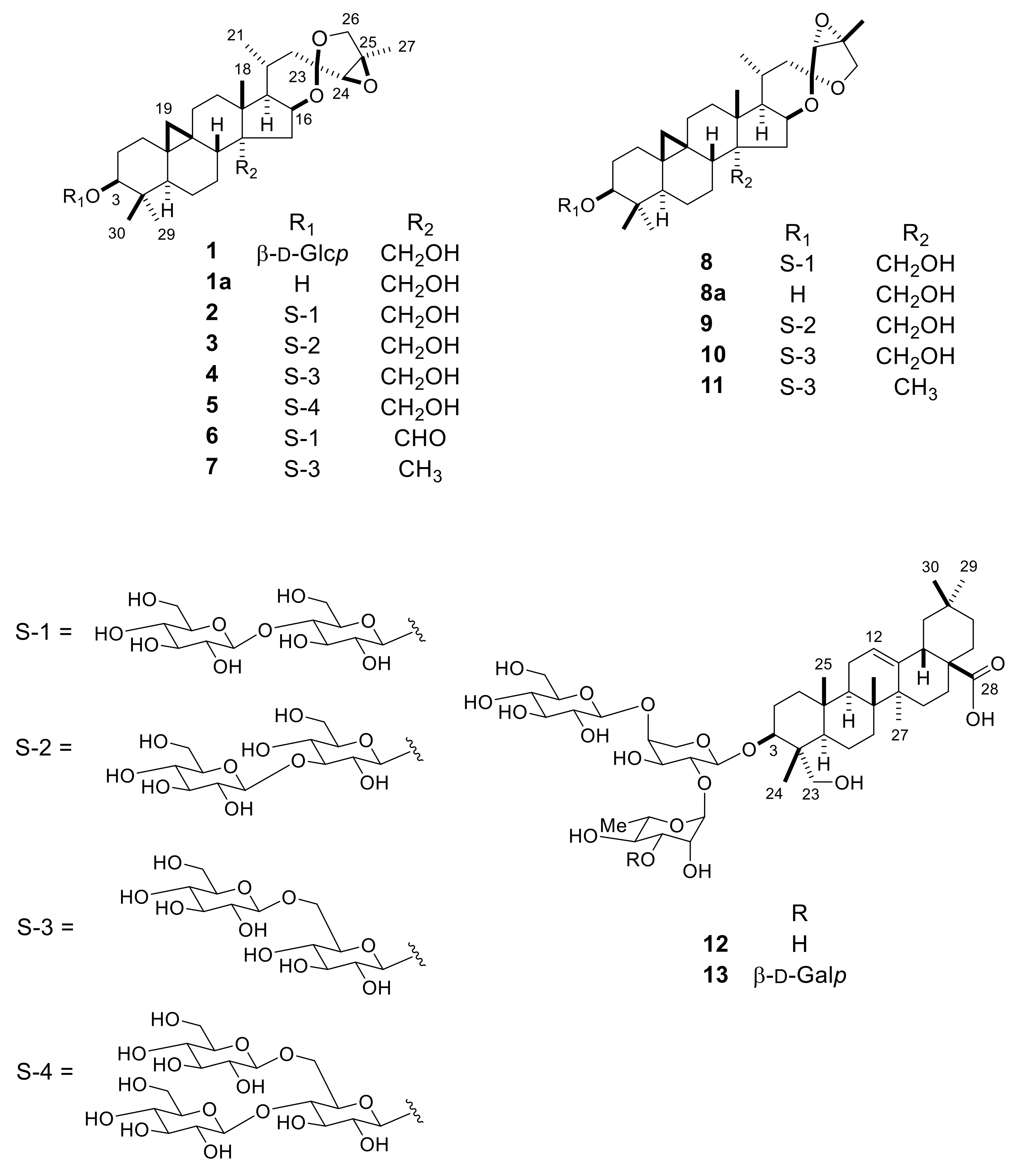
2.1. Isolation and Structure Elucidation of 1–13

The MeOH extract of the E. cilicica tubers was allowed to pass through the porous-polymer polystyrene resin (Diaion HP-20) column, and a series of chromatographic separations of the glycoside-enriched fraction using column chromatography (CC) on silica gel and octadecylsilanized (ODS) silica gel were performed to obtain compounds 113 (Figure 1). The known compound 12 was identified as 3β-[(O-β-d-glucopyranosyl-(1→4)-O-[α-l-rhamnopyranosyl-(1→2)]-α-l-arabinopyranosyl)oxy]-23-hydroxyolean-12-en-28-oic acid by direct comparison with an authentic sample isolated from Anemone coronaria [6].
Compound 1 was obtained as an amorphous solid, and its molecular formula was determined to be C36H56O10, based on high-resolution (HR)-electrospray ionization (ESI)-time-of-flight (TOF)-MS (m/z 671.3794 [M + Na]+) data and 13C-NMR spectrum with 36 carbon signals (Table 1). The IR spectrum of 1 showed an absorption band attributed to hydroxy groups at 3388 cm−1. The 1H-NMR spectrum of 1 was typical of a triterpene glycoside based on a cycloartane derivative, showing signals for a cyclopropane methylene group at δH 0.48 and 0.20 (each d, J = 4.0 Hz); four tertiary methyl groups at δH 1.42, 1.36, 1.29, and 1.03 (each s); a secondary methyl group at δH 0.97 (d, J = 6.5 Hz); and an anomeric proton δH 4.94 (d, J = 7.8 Hz). Enzymatic hydrolysis of 1 using naringinase gave a new triterpene aglycone (1a: C30H46O5) as colorless needles and d-glucose as a carbohydrate moiety. Based on X-ray crystallographic analysis, the structure of 1a was unambiguously established as (23R,24R,25R)-16β,23:23,26:24,25-triepoxy-9,19-cycloartane-3β,28-diol (Figure 2). Linkage of a β-d-glucopyranosyl group to the C-3 hydroxy group of the aglycone in 1 was ascertained by long-range correlations between the anomeric proton (H-1′) at δH 4.94 and the C-3 carbon of the aglycone at δC 88.6 in the 1H-detected heteronuclear multiple-bond connectivity (HMBC) spectrum of 1. Thus, 1 was established as (23R,24R,25R)-16β,23:23,26:24,25-triepoxy-28-hydroxy-9,19-cycloartan-3β-yl β-d-glucopyranoside.
Compound 2 had the molecular formula C42H66O15, as determined by HR-ESI-TOF-MS (m/z 833.4297 [M + Na]+) and 13C-NMR (42 carbon signals) data. The molecular formula was larger than that of 1 by C6H10O5, which corresponded to one hexosyl unit. The 1H-NMR spectrum of 2 showed signals due to two anomeric protons at δH 5.20 (d, J = 7.9 Hz) and 4.87 (d, J = 7.8 Hz), as well as a cyclopropane methylene group at δH 0.47 and 0.20 (each d, J = 4.0 Hz), four tertiary methyl groups at δH 1.42, 1.35, 1.27, and 1.02 (each s), a secondary methyl group at δH 0.97 (d, J = 6.6 Hz). Enzymatic hydrolysis of 2 with naringinase gave 1a and d-glucose. The 1H- and 13C-NMR signals of the diglycoside moiety, which were assigned based on 1H-1H correlation spectroscopy (COSY) and 1H-detected heteronuclear multiple coherence (HMQC) spectra, indicated the presence of a terminal β-d-glucopyranosyl unit [δH 5.20 (d, J = 7.9 Hz, H-1′′ of Glc (II)); δC 104.9 (CH), 74.8 (CH), 78.2 (CH), 71.5 (CH), 78.4 (CH), 62.3 (CH2)] and a C-4 glycosylated β-d-glucopyranosyl unit [δH 4.87 (d, J = 7.8 Hz, H-1′ of Glc (I)); δC 106.4 (CH), 75.2 (CH), 76.9 (CH), 81.6 (CH), 76.2 (CH), 62.3 (CH2)] in the molecule of 2 [20]. In the HMBC spectrum of 2, long-range correlations were observed between H-1′′ of Glc (II) and C-3′ of Glc (I), and between H-1′ of Glc (I) and C-3 of the aglycone moiety at δC 88.7. The molecular formula of 2 was assigned as (23R,24R,25R)-16β,23:23,26:24,25-triepoxy-28-hydroxy-9,19-cylcoartan-3β-yl O-β-d-glucopyranosyl-(1→4)-β-d-glucopyranoside.
Compounds 3 and 4, which had the same molecular formula as 2 (C42H66O15), gave 1a and d-glucose upon enzymatic hydrolysis. The 1H- and 13C-NMR spectra of 3 and 4 suggested that they were constitutional isomers of 2 based on the linkage position of the terminal d-glucosyl unit (Glc (II)) at the inner d-glucosyl unit (Glc (I)) in the diglucosyl residue. In the HMBC spectrum of 3, H-1′′ of Glc (II) at δH 5.28 (d, J = 7.9 Hz) showed a long-range correlation with C-3′ of Glc (I) at δC 88.7, and H-1′ of Glc (I) at δH 4.86 (d, J = 7.8 Hz), in turn, showed a long-range correlation with C-3 of the aglycone at δC 88.6. On the other hand, HMBC correlations were observed between H-1′′ of Glc (II) at δH 5.28 (d, J = 7.8 Hz) and C-6′ of Glc (I) at δC 70.2, and between H-1′ of Glc (I) at δH 4.86 (d, J = 7.7 Hz) and C-3 of the aglycone at δC 88.4 in 4. Compounds 3 and 4 were established as O-β-d-glucopyranosyl-(13)-β-d-glucopyranoside and O-β-d-glucopyranosyl-(16)-β-d-glucopyranoside of (23R,24R,25R)-16β,23:23,26:24,25-triepoxy-28-hydroxy-9,19-cycloartan-3β-yl, respectively.
Compound 5 (C48H76O20) also yielded 1a and d-glucose upon enzymatic hydrolysis. The 1H- and 13C-NMR spectra of 5 showed signals for two terminal β-d-glucopyranosyl units [δH 5.45 (d, J = 7.9 Hz, H-1′′ of Glc (II)); δC 104.7 (CH), 75.1 (CH), 78.2 (CH), 71.6 (CH), 78.1 (CH), 62.3 (CH2); and δH 5.34 (d, J = 7.8 Hz, H-1′′′ of Glc (III)); δC 105.0 (CH), 75.1 (CH), 78.2 (CH), 71.5 (CH), 78.1 (CH), 62.5 (CH2)] and a C-4 and C-6 diglycosylated β-d-glucopyranosyl unit [δH 4.80 (d, J = 7.5 Hz, H-1′ of Glc (I)); δC 106.3 (CH), 75.0 (CH), 76.5 (CH), 81.1 (CH), 74.9 (CH), 68.7 (CH2)] [21]. In the HMBC spectrum of 5, long-range correlations were observed between H-1′′ of Glc (II) and C-4′ of Glc (I), H-1′′′ of Glc (III) and C-6′ of Glc (I), and between H-1′ of Glc (II) and C-3 of the aglycone at δC 88.7. Thus, 5 was deduced to be (23R,24R,25R)-16β,23:23,26:24,25-triepoxy-28-hydroxy-9,19-cylcoartan-3β-yl O-β-d-glucopyranosyl-(14)-O-[β-d-glucopyranosyl(16)]-β-d-glucopyranoside.
Compounds 6 and 7 had the molecular formula C42H64O15 and C42H66O14, respectively, based on HR-ESI-TOF-MS and 13C-NMR data. The 1H- and 13C-NMR spectral properties of 6 were essentially analogous to those of 2; however, the hydroxymethyl signals at δH 4.02 and 3.94 (each d, J = 11.5 Hz) and δC 63.4 assignable to H2-28 and C-28 in 2 were replaced by those due to an aldehyde group at δH 10.14 (s) and δC 210.2 in 6. Treatment of 6 with NaBH4 in EtOH afforded 2. On the other hand, comparison of the 1H- and 13C-NMR spectra of 7 with those 4 revealed that the H2-28/C-28 hydroxymethyl proton and carbon signals observed for 4 disappeared in the case of 7. Instead, the signals from a tertiary methyl group were detected at δH 0.80 (s) and δC 19.7 in the spectrum of 7. The methyl proton signal showed HMBC correlations with the C-8 (δC 47.5), C-13 (δC 46.3), C-14 (δC 44.7) and C-15 (δC 44.4) carbon signals. Compounds 6 and 7 were determined to be (23R,24R,25R)-16β,23:23,26:24,25-triepoxy-28-oxo-9,19-cycloartan-3β-yl O-β-d-glucopyranosyl-(14)-β-d-glucopyranoside and (23R,24R,25R)-16β,23:23,26:24,25-triepoxy-9,19-cycloartan-3β-yl O-β-d-glucopyranosyl-(16)-β-d-glucopyranoside.
Compound 8 had the same molecular formula as 2 (C42H66O15), and its 1H- and 13C-NMR spectral features were closely related to those of 2, except for the signals from the ring E and F parts. Enzymatic hydrolysis of 8 gave an aglycone (8a; C30H46O5) and d-glucose. The phase-sensitive NOE correlation spectroscopy (PHNOESY) spectrum of 8a showed NOE correlations between H-16 (δH 4.80) and H-22 α (δH 1.56)/H-26a (δH 3.82), H-20 (δH 1.85) and H-24 (δH 3.69), and between H-22β(δH 2.16) and H-24, indicating that 8a was a new aglycone, the C-23 epimer of 1a. Compound 8 was assigned as (23S,24R,25R)-16β,23:23,26:24,25-triepoxy-28-hydroxy-9,19-cycloartan-3β-yl O-β-d-glucopyranosyl-(14)-β-d-glucopyranoside.
Compounds 9 and 10 had the same molecular formula C42H66O15, and their 1H- and 13C-NMR spectra were suggestive of cycloartane diglucosides closely related to 8. Indeed, enzymatic hydrolysis of 9 and 10 gave 8a and d-glucose. Assignments of the 1H- and 13C-NMR signals from the sugar moieties of 9 and 10, which were established by 1H-1H COSY and HMQC spectral analysis, implied that the diglucoside sequences of 9 and 10 corresponded to those of 3 and 4, respectively. Furthermore, HMBC correlations were observed from H-1′′ of Glc (II) at δH 5.31 (d, J = 7.9 Hz) to C-3′ of Glc (I) at δC 88.9, and from H-1′ at δH 4.89 (d, J = 7.8 Hz) to C-3 of the aglycone at δC 88.7 in 9, and from H-1′′ of Glc (II) at δH 5.16 (d, J = 7.8 Hz) to C-6′ of Glc (I) at δC 70.3, and from H-1′ at δH 4.89 (d, J = 7.7 Hz) to C-3 of the aglycone at δC 88.4 in 10. Compounds 9 and 10 were formulated as O-β-d-glucopyranosyl-(13)-β-d-glucopyranoside and O-β-d-glucopyranosyl-(16)-β-d-glucopyranoside of (23S,24R,25R)-16β,23:23,26:24,25-triepoxy-28-hydroxy-9,19-cylcoartan-3β-yl, respectively.
Compound 11 (C42H66O14) bore close similarity to 10 in terms of the 1H- and 13C-NMR spectral features. However, the hydroxymethyl signals at δH 3.98 and 3.89 (each d, J = 11.5 Hz) and δC 63.2 assignable to H2-28 and C-28 in 10 were replaced by the signals due to a methyl group at δH 0.79 (s) and δC 19.6 in 11. In addition, the methyl proton signal showed HMBC correlations with the C-8 (δC 47.6), C-13 (δC 46.3), C-14 (δC 44.5), and C-15 (δC 43.8) carbon signals. Compound 11 was established to be (23S,24R,25R)-16β,23:23,26:24,25-triepoxy-9,19-cycloartan-3β-yl O-β-d-glucopyranosyl-(16)-β-d-glucopyranoside.
Compound 13 had the molecular formula C53H86O22, as revealed by HR-ESI-TOF-MS (m/z 1075.5710 [M + H]+) and 13C-NMR (53 carbon signals) data. The molecular formula was larger than that of 12 by C6H10O5, which corresponded to one hexosyl unit. The 1H-NMR spectrum of 13 displayed signals for four anomeric protons at δH 6.02 (br s), 5.29 (d, J = 7.8 Hz), 5.06 (d, J = 7.9 Hz) and 4.92 (d, J = 6.9 Hz), as well as the signals for six tertiary methyl groups at δH 1.25, 1.05, 1.02, 1.00, 0.94 and 0.92 (each s), and an olefinic proton at δH 5.46 (t-like, J = 3.0 Hz), suggesting that this compound was a tetraglycoside of an oleanoic acid derivative. Acid hydrolysis of 13 with 1M HCl in dioxane-H2O (1:1) gave 23-hydroxyolean-12-en-28-oic acid (hederagenin) as the aglycone [22], and l-arabinose, d-galactose, d-glucose and l-rhamnose as the carbohydrate moieties. When the 13C-NMR spectrum of 13 was compared with that of 12 [6], six signals assignable to a terminal β-d-galactopyraosyl group (Gal) were observed in addition to signals for a 2,4-disubstituted α-l-arabinopyranosyl group (Ara), C-3 substituted α-l-rhamnopyranosyl group (Rha), and terminal β-d-glucopyranosyl group (Glc). The anomeric configurations of Ara, Gal and Glc were confirmed to be α, β, and β, respectively, based on the relatively large 3JH-1,H-2 values (6.9–7.9 Hz). In the case of the Rha moiety, the large 1JC-1,H-1 (172 Hz) was indicative of the α-anomeric configuration [6]. In the HMBC spectrum of 13, long-range couplings were observed between H-1′′′ of Glc (δH 5.29) and C-4′ of Ara at δC 80.0, H-1′′′′ of Gal (δH 5.06) and C-3′′ of Rha at δC 83.2, H-1′′ of Rha (δH 6.02) and C-2′ of Ara at δC 76.5, and between H-1′ of Ara (δH 4.92) and C-3 of the aglycone at δC 81.3. Compound 13 was determined to be 3 β-[(O-β-d-galactopyranosyl-(13)-O-α-l-rhamnopyranosyl-(12)-O-[β-d-glucopyranosyl-(14)]-α-l-arabinopyranosyl)oxy]-23-hydroxyolean-12-en-28-oic acid.

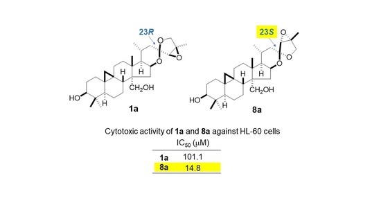
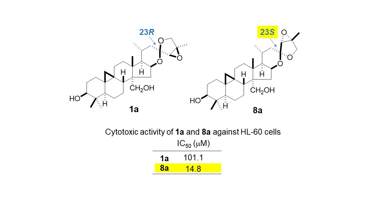
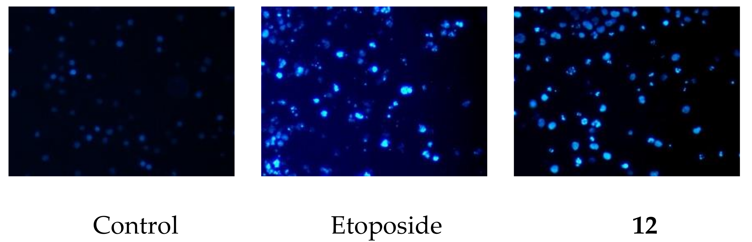
2.2. Cytotoxic Activity of 1a, 2, 8, 8a, 12, and 13

We previously reported that the slight differences in the structure of cycloartane glycosides effected on cytotoxic activity [8]. In this study, some selected compounds, the new cycloartane glycosides (2 and 8), and the aglycone (1a) of 2 and its C-23 epimer (8a), as well as oleanane glycosides (12 and 13) were evaluated for their cytotoxic activity against HL-60 cells using a modified 3-(4,5-dimethylthiazol-2-yl)-2,5-diphenyl-2H-tetrazolium bromide (MTT) assay (Table 2). Etoposide was used as a positive control, and it gave an IC50 value of 0.32 ± 0.01 μM. The cytotoxic activity of 8a against HL-60 cells (IC50 14.8 ± 1.00 μM) was much more potent than that of 1a (IC50 101.1 ± 0.44 μM), indicating that the C-23 configuration is associated with the cytotoxicity of these cycloartane derivatives. On the other hand, 2 and 8 were not cytotoxic to HL-60 cells at sample concentrations up to 200 μM. Compounds 12 and 13 were cytotoxic to HL-60 cells, with IC50 values of 10.6 ± 0.40 μM and 10.8 ± 0.53 μM, respectively. After HL-60 cells were exposed to 12 at a sample concentration of 20 μM for 72 h, they were stained with 4′,6-diamidino-2-phenylindole dihydrochloride (DAPI) and observed under a fluorescence microscope. The cells showed nuclear chromatin condensation and nuclear disassembly, illustrated in Figure 3. Therefore, 12 partially induced apoptotic cell death in HL-60 cells.

3. Materials and Methods

3.1. General Experimental Procedures and Plant Material

The instruments, experimental conditions, and plant material used (except for those mentioned below) were the same as those described in previous papers [18,19]. Melting point was determined on an MP-3 melting point apparatus (Yanaco, Kyoto, Japan). X-ray diffraction experiments were carried out on a DIP image plate diffractometer (Bruker AXS, Karlsruhe, Germany).

3.2. Extraction and Isolation

The MeOH extract (135 g) of Eranthis cilicica tubers (1.3 kg) was subjected to Diaion HP-20 CC [19]. The 80% MeOH eluted portion (30 g) was chromatographed on silica gel eluted with gradient mixtures of CHCl3-MeOH (20:1; 9:1, 4:1, 2:1) and finally with MeOH to give 9 subfractions (Frs. A–I). Fr. E was subjected silica gel CC eluted with CHCl3-MeOH (9:1) and ODS silica gel CC eluted with MeCN-H2O (1:2) to yield 1 (17.0 mg) and 7 (13.4 mg). Fr. G was repeatedly subjected to silica gel CC eluted with CHCl3-MeOH-H2O (60:10:1, 50:10:1, 40:10:1) and ODS silica gel CC eluted with MeCN-H2O (5:8, 5:9, 1:2) to yield 2 (121 mg), 3 (7.8 mg), 8 (100 mg), 9 (4.1 mg), 10 (35.0 mg), and 11 (5.1 mg). Fr. H was subjected to CC on silica gel eluted with CHCl3-MeOH-H2O (30:10:1) and ODS silica gel eluted with MeCN-H2O (1:2) and MeOH-H2O (7:5) to yield 13 (15.2 mg). Fr. I was subjected to CC on silica gel eluted with CHCl3-MeOH-H2O (14:8:1; 10:10:1) and ODS silica gel eluted with MeCN-H2O (5:8, 2:5, 4:11, 1:3) to yield 12 (13.7 mg). The 50% MeOH eluted portion (35 g) was chromatographed on silica gel eluted with CHCl3-MeOH-H2O (40:10:1) to give 6 subfractions (Frs. a–f). Fr. b was subjected to silica gel column eluted with CHCl3-MeOH (20:1) and CHCl3-MeOH-H2O (40:10:1) and ODS silica gel column eluted with MeCN-H2O (1:2, 2:5, 1:3) to yield 4 (15.2 mg) and 6 (10.0 mg). Fr. c was subjected to silica gel CC eluted with CHCl3-MeOH (10:1) and CHCl3-MeOH-H2O (40:10:1) and ODS silica gel CC eluted with MeCN-H2O (1:2) to yield 5 (33.8 mg).

3.3. Structural Characterization

Compound 1: Amorphous solid. [α]D25 −46.0 (c 0.10, MeOH). HR-ESI-TOF-MS m/z: 671.3794 [M + Na]+ (Calcd for C36H56NaO10, 671.3771). IR νmax (film) cm−1: 3388 (OH), 2928 and 2870 (CH). 1H-NMR spectral data for 1 are provided in the Supplementary Materials. 13C-NMR, see Table 1.
Enzymatic Hydrolysis of 1 Compound 1 (4.5 mg) was treated with naringinase (EC 232-96-4, Sigma; 22.5 mg) in a mixture of HOAc/KOAc buffer (pH 4.3, 5 mL) and EtOH (2 mL) at room temperature for 72 h. The reaction mixture was purified by silica gel CC eluted with CHCl3-MeOH (22:1) followed by MeOH to give 1a (2.4 mg) and a sugar fraction (0.6 mg). The sugar fraction was analyzed by HPLC under the following conditions: Capcell Pak NH2 UG80 column (4.6 mm i.d. × 250 mm, 5 μm, Shiseido, Tokyo, Japan); mobile phase of MeCN-H2O (7:3); detection by refractive index and optical rotation, and at a flow rate of 1.0 mL/min. d-glucose was identified by comparing its retention time and optical rotation with those of an authentic sample; tR 11.62 min (d-glucose, positive optical rotation).
Compound 1a: Colorless needles from MeOH-MeCN (1:1). mp 281–285 °C. [α]D24 −48.0 (c 0.10, MeOH). HR-ESI-TOF-MS m/z: 509.3255 [M + Na]+ (Calcd for C30H46NaO5, 509.3243). IR νmax (film) cm−1: 3476 (OH), 2953, 2925 and 2868 (CH). 1H-NMR spectral data for 1a are provided in the Supplementary Materials. 13C-NMR, see Table 1.
X-Ray Crystallography of 1a Monoclinic, space group C2, unit cell dimension a = 32.945(2) Å, b = 6.9250(2) Å, c = 13.5690(6), β = 111.270(2)°, V = 2884.8(2) Å3, Z = 4; T = 296 K, dcalc = 1.204 Mg/m3; μ (Mo Kα, λ = 0.71073 Å) = 0.084 mm−1; R [I > 2σ(I)] = 0.0370, wR [I > 2σ(I)] = 0.1054, R [for all data] = 0.0409, wR [for all data] = 0.1081. Crystallographic data of 1a have been deposited with the Cambridge Crystallographic Data Centre (CCDC) as supplementary publication no. CCDC 1877743. Copies of the data can be obtained free of charge from the CCDC via http://beta-www.ccdc.cam.ac.uk.
Compound 2: Amorphous solid. [α]D26 −34.0 (c 0.10, MeOH). HR-ESI-TOF-MS m/z: 833.4297 [M + Na]+ (Calcd for C42H66NaO15, 833.4299). IR νmax (film) cm−1: 3397 (OH), 2930 and 2870 (CH). 1H-NMR spectral data for 2 are provided in the Supplementary Materials. 13C-NMR, see Table 1.
Compound 3: Amorphous solid. [α]D24 −48.0 (c 0.10, MeOH). HR-ESI-TOF-MS m/z: 833.4359 [M + Na]+ (Calcd for C42H66NaO15, 833.4299). IR νmax (film) cm−1: 3450 (OH), 2932 and 2872 (CH). 1H-NMR spectral data for 3 are provided in the Supplementary Materials. 13C-NMR, see Table 1.
Compound 4: Amorphous solid. [α]D26 −44.0 (c 0.10, MeOH). HR-ESI-TOF-MS m/z: 833.4340 [M + Na]+ (Calcd for C42H66NaO15, 833.4299). IR νmax (film) cm−1: 3408 (OH), 2933 and 2871 (CH). 1H-NMR spectral data for 4 are provided in the Supplementary Materials. 13C-NMR, see Table 1.
Compound 5: Amorphous solid. [α]D28 −16.0 (c 0.10, MeOH). HR-ESI-TOF-MS m/z: 973.5001 [M + H]+ (Calcd for C48H77O20, 973.5008). IR νmax (film) cm−1: 3363 (OH), 2932 and 2872 (CH). 1H-NMR spectral data for 5 are provided in the Supplementary Materials. 13C-NMR, see Table 1.
Enzymatic Hydrolysis of 25 Compounds 2 (54.1 mg), 3 (2.5 mg), 4 (5.1 mg), and 5 (4.8 mg) were independently subjected to enzymatic hydrolysis with naringinase as described for 1 to give 1a (25.2 mg from 2; 1.3 mg from 3; 2.5 mg from 4; 2.6 mg from 5) and a sugar fraction (5.9 mg from 2; 0.4 mg from 3; 0.8 mg from 4; 1.2 mg from 5). HPLC analysis of the sugar fraction under the same conditions as in the case of that of 1 showed the presence of d-glucose.
Compound 6: Amorphous solid. [α]D24 −84.0 (c 0.10, MeOH). HR-ESI-TOF-MS m/z: 831.4100 [M + Na]+ (Calcd for C42H64NaO15, 831.4143). IR νmax (film) cm−1: 3387 (OH), 2925 and 2870 (CH), 1712 (C=O). 1H-NMR spectral data for 6 are provided in the Supplementary Materials. 13C-NMR, see Table 1.
Preparation of 2 from 6 Compound 6 (1.7 mg) was dissolved in NaBH4 (2.2 mg) ethanolic solution (1 mL) and it was stirred at room temperature for 5 h. After Me2CO was added to the reaction mixture, the solvent was removed under reduced pressure. The residue was suspended with H2O (3 mL) and extracted with EtOAc (3 mL × 3). The EtOAc extract was chromatographed on silica gel eluted with CHCl3-MeOH-H2O (30:10:1) to yield 2 (1.2 mg).
Compound 7: Amorphous solid. [α]D25 −70.0 (c 0.10, MeOH). HR-ESI-TOF-MS m/z: 817.4393 [M + Na]+ (Calcd for C42H66NaO14, 817.4350). IR νmax (film) cm−1: 3418 (OH), 2931 and 2868 (CH). 1H-NMR spectral data for 7 are provided in the Supplementary Materials. 13C-NMR, see Table 1.
Compound 8: Amorphous solid. [α]D26 −50.0 (c 0.10, C5H5N). HR-ESI-TOF-MS m/z: 833.4359 [M + Na]+ (Calcd for C42H66NaO15, 833.4299). IR νmax (film) cm−1: 3348 (OH), 2926 and 2867 (CH). 1H-NMR spectral data for 8 are provided in the Supplementary Materials. 13C-NMR, see Table 1.
Compound 8a: Amorphous solid. [α]D24 −58.0 (c 0.10, MeOH). HR-ESI-TOF-MS m/z: 487.3423 [M + H]+ (Calcd for C30H47O5, 487.3424). IR νmax (film) cm−1: 3442 (OH), 2955, 2927 and 2870 (CH). 1H-NMR spectral data for 8a are provided in the Supplementary Materials. 13C-NMR, see Table 1.
Compound 9: Amorphous solid. [α]D26 −36.0 (c 0.10, C5H5N). HR-ESI-TOF-MS m/z: 833.4276 [M + Na]+ (Calcd for C42H66NaO15, 833.4299). IR νmax (film) cm−1: 3347 (OH), 2919 and 2871 (CH). 1H-NMR spectral data for 9 are provided in the Supplementary Materials. 13C-NMR, see Table 1.
Compound 10: Amorphous solid. [α]D26 −72.0 (c 0.10, MeOH). HR-ESI-TOF-MS m/z: 833.4313 [M + Na]+ (Calcd for C42H66NaO15, 833.4299). IR νmax (film) cm−1: 3388 (OH), 2933 and 2872 (CH). 1H-NMR spectral data for 10 are provided in the Supplementary Materials. 13C-NMR, see Table 1.
Enzymatic Hydrolysis of 810 Compounds 8 (31.3 mg), 9 (1.9 mg), and 10 (5.3 mg) were independently subjected to enzymatic hydrolysis with naringinase as described for 1 to give 8a (19.4 mg from 8; 0.9 mg from 9; 2.8 mg from 10) and a sugar fraction (3.2 mg from 8; 0.3 mg from 9; 1.1 mg from 10). HPLC analysis of the sugar fraction under the same conditions as in the case of that of 1 showed the presence of d-glucose.
Compound 11: Amorphous solid. [α]D26 −62.0 (c 0.10, MeOH). HR-ESI-TOF-MS m/z: 795.4583 [M + H]+ (Calcd for C42H67O14, 795.4531). IR νmax (film) cm−1: 3387 (OH), 2927 and 2871 (CH). 1H-NMR spectral data for 11 are provided in the Supplementary Materials. 13C-NMR, see Table 1.
Compound 13: Amorphous solid. [α]D27 +6.0 (c 0.10, MeOH). HR-ESI-TOF-MS m/z: 1075.5710 [M + H]+ (Calcd for C53H87O22, 1075.5689). IR νmax (film) cm−1: 3376 (OH), 2926 and 2858 (CH), 1696 (C=O). 1H-NMR (C5D5N) δH 6.02 (1H, br s, H-1′′), 5.46 (1H, t-like, J = 3.0 Hz, H-12), 5.29 (1H, d, J = 7.8 Hz, H-1′′′), 5.06 (1H, d, J = 7.9 Hz, H-1′′′′), 4.92 (1H, d, J = 6.9 Hz, H-1′), 4.48 (1H, dd, J = 9.3, 7.8 Hz, H-2′′′), 4.46 (1H, br s, H-4′′′), 4.36 (1H, m, H-6a′′′), 4.34 (1H, m, H-6b′′′), 4.24 (1H, d, J = 11.0 Hz, H-23a), 4.18 (1H, dd, J = 10.5, 4.6 Hz, H-3), 4.10 (1H, dd, J = 9.3, 3.3 Hz, H-3′′′), 4.07 (1H, m, H-5′′′), 3.86 (1H, d, J = 11.0 Hz, H-23b), 3.26 (1H, dd, J = 13.8, 3.9 Hz, H-18), 1.54 (3H, d, J = 6.1 Hz, Me-6′′), 1.25 (3H, s, Me-27), 1.05 (3H, s, Me-24), 1.02 (3H, s, Me-26), 1.00 (3H, s, Me-30), 0.94 (3H, s, Me-25), 0.92 (3H, s, Me-29). 13C-NMR (C5D5N) δC: 39.0, 26.3, 81.3, 43.5, 47.6, 18.2, 32.9, 39.8, 48.2, 36.9, 23.8, 122.6, 144.8, 42.2, 28.3, 23.7, 46.7, 42.0, 46.5, 30.9, 34.2, 32.0, 64.0, 13.9, 16.0, 17.5, 26.1, 180.1, 33.2, 23.8 (aglycone C-1–C-30), 104.7, 76.5, 74.2, 80.0, 65.5 (Ara C-1–C-5), 101.6, 71.2, 83.2, 72.6, 69.9, 18.5 (Rha C-1–C-6), 107.1, 73.3, 75.2, 70.0, 76.8, 62.1 (Gal C-1–C-6), 106.5, 75.5, 78.3, 71.4, 78.5, 62.5 (Glc C-1–C-6).
Acid Hydrolysis of 13 A solution of 13 (5.0 mg) in 1 M HCl (dioxane-H2O, 1:1, 3 mL) was heated at 95 °C for 1 h under an Ar atmosphere. After the reaction mixture was diluted with H2O (2 mL), it was extracted with Et2O (5 mL × 2). The Et2O extract was chromatographed on silica gel eluted with CHCl3-MeOH (19:1) to give hederagenin (2.6 mg). The H2O residue was neutralized using an Amberlite IRA-93ZU (Organo, Tokyo, Japan) column and passed through a Sep-Pak C18 cartridge (Waters, Milford, MA) eluted with H2O-MeOH (3:2) to give a sugar fraction (1.8 mg). HPLC analysis of the sugar fraction under the same conditions as in the case of that of 1 (flow rate, 0.9 mL/min) showed the presence of l-arabinose, d-galactose, d-glucose, and l-rhamnose. tR (min): 7.08 (negative optical rotation, l-rhamnose), 8.14 (positive optical rotation, l-arabinose), 12.86 (positive optical rotation, d-galactose), and 13.17 (positive optical rotation, d-glucose).

3.4. Cytotoxic Activity

HL-60 cells were maintained in an RPMI-1640 medium. The cell media contained heat-inactivated 10% (v/v) FBS supplemented with l-glutamine, penicillin G sodium salt (100 units/mL), and streptomycin sulfate (100 μg/mL). HL-60 (4 × 10⁴ cells/mL) cells were continuously treated with each compound for 72 h, and cell growth was measured using an MTT reduction assay as previously described [23]. Data represented as mean ± S.E.M. of three experiments performed in triplicate. The concentration, resulting in a 50% inhibition value (IC50), was calculated from the dose response curve.

3.5. DAPI Staininig

The cells (1 × 10⁵ cells/mL) were plated on coverslips in 96-well plates. After 24 h, HL-60 cells were treated with either 20 μM of 12 or 17 μM of etoposide for 72 h. The cells were fixed with 1% glutaraldehyde for 30 min at room temperature before staining with DAPI (0.5 μg/mL in H2O). They were observed under a CKX41 fluoroscence microscope (Olympus, Tokyo, Japan).

4. Conclusions

Further phytochemical examination of E. cilicica tubers gave eleven new cycloartane glycosides (111) and one new oleanane glycoside (13), together with one known oleanane glycosides (12). The structures of the new compounds were established by extensive spectroscopic analysis, including 2D NMR, and enzymatic hydrolysis, followed by either X-ray crystallographic or chromatographic analysis. The new cycloartane glycosides 2 and 8, the aglycone 1a and its C-23 epimer 8a, and oleanane-type triterpene glycosides 12 and 13 were evaluated for their cytotoxic activity against HL-60 leukemia cells. The cytotoxic activity of 8a was much more potent than that of 1a, indicating that the C-23 configuration is associated with the cytotoxicity of these cycloartane derivatives. This is the first report of structure–activity relationships of cycloartane-type triterpenoids on C-23 epimer. Compounds 12 and 13 were moderately cytotoxic to HL-60 cells, and 12 partially induced apoptotic cell death in HL-60 cells.

Supplementary Materials

All NMR spectra of 1, 1a, 28, 8a, and 913 are available online.

Author Contributions

K.W. and Y.M. (Yoshihiro Mimaki) conceived and designed the experiments; K.W., Y.M. (Yoshihiro Mimaki), H.F., and Y.M. (Yukiko Matsuo) performed the experiments and analyzed the data. Y.M. (Yoshihiro Mimaki) and Y.M. (Yukiko Matsuo) wrote the paper. All authors have read and approved the manuscript.

Funding

This research received no external funding.

Acknowledgments

We thank Chisato Hashimoto for technical advice and discussion.

Conflicts of Interest

The authors declare no conflict of interest.

References

  1. Kuroda, M.; Kubo, S.; Masatani, D.; Matsuo, Y.; Sakagami, H.; Mimaki, Y. Aestivalosides A–L, twelve pregnane glycosides from the seeds of Adonis aestivalis. Phytochemistry 2018, 150, 75–84. [Google Scholar] [CrossRef] [PubMed]
  2. Kubo, S.; Kuroda, M.; Yokosuka, A.; Sakagami, H.; Mimaki, Y. Amurensiosides L–P, five new cardenolide glycosides from the roots of Adonis amurensis. Nat. Prod. Commun. 2015, 10, 27–32. [Google Scholar] [PubMed]
  3. Kubo, S.; Kuroda, M.; Matsuo, Y.; Masatani, D.; Sakagami, H.; Mimaki, Y. New cardenolides from the seeds of Adonis aestivalis. Chem. Pharm. Bull. 2012, 60, 1275–1282. [Google Scholar] [CrossRef] [PubMed]
  4. Kuroda, M.; Kubo, S.; Uchida, S.; Sakagami, H.; Mimaki, Y. Amurensiosides A–K, 11 new pregnane glycosides from the roots of Adonis amurensis. Steroids 2010, 75, 83–94. [Google Scholar] [CrossRef] [PubMed]
  5. Yokosuka, A.; Sano, T.; Hashimoto, K.; Sakagami, H.; Mimaki, Y. Triterpene glycosides from the whole plant of Anemone hupehensis var. japonica and their cytotoxic activity. Chem. Pharm. Bull. 2009, 57, 1425–1430. [Google Scholar] [CrossRef]
  6. Mimaki, Y.; Watanabe, K.; Matsuo, Y.; Sakagami, H. Triterpene glycosides from the tubers of Anemone coronaria. Chem. Pharm. Bull. 2009, 57, 724–729. [Google Scholar] [CrossRef]
  7. Mimaki, Y.; Nadaoka, I.; Yasue, M.; Ohtake, Y.; Ikeda, M.; Watanabe, K.; Sashida, Y. Neocimicigenosides A and B, cycloartane glycosides from the rhizomes of Cimicifuga racemosa and their effects on CRF-stimulated ACTH secretion from AtT-20 cells. J. Nat. Prod. 2006, 69, 829–832. [Google Scholar] [CrossRef]
  8. Watanabe, K.; Mimaki, Y.; Sakagami, H.; Sashida, Y. Cycloartane glycosides from the rhizomes of Cimicifuga racemosa and their cytotoxic activities. Chem. Pharm. Bull. 2002, 50, 121–125. [Google Scholar] [CrossRef]
  9. Mimaki, Y.; Yokosuka, A.; Hamanaka, M.; Sakuma, C.; Yamori, T.; Sashida, Y. Triterpene saponins from the roots of Clematis chinensis. J. Nat. Prod. 2004, 67, 1511–1516. [Google Scholar] [CrossRef]
  10. Yokosuka, A.; Iguchi, T.; Kawahata, R.; Mimaki, Y. Cytotoxic bufadienolides from the whole plants of Helleborus foetidus. Phytochem. Lett. 2018, 23, 94–99. [Google Scholar] [CrossRef]
  11. Mimaki, Y.; Matsuo, Y.; Watanabe, K.; Sakagami, H. Furostanol glycosides from the rhizomes of Helleborus orientalis. J. Nat. Med. 2010, 64, 452–459. [Google Scholar] [CrossRef] [PubMed]
  12. Watanabe, K.; Sakagami, H.; Mimaki, Y. Four new steroidal saponins from the rhizomes of Helleborus orientalis. Heterocycles 2005, 65, 775–785. [Google Scholar]
  13. Mimaki, Y.; Watanabe, K.; Sakuma, C.; Sakagami, H.; Sashida, Y. Novel polyoxygenated spirostanol glycosides from the rhizomes of Helleborus orientalis. Helv. Chim. Acta 2003, 86, 398–407. [Google Scholar] [CrossRef]
  14. Watanabe, K.; Mimaki, Y.; Sakagami, H.; Sashida, Y. Bufadienolide and spirostanol glycosides from the rhizomes of Helleborus orientalis. J. Nat. Prod. 2003, 66, 236–241. [Google Scholar] [CrossRef] [PubMed]
  15. Mimaki, Y.; Yokosuka, A.; Kuroda, M.; Hamanaka, M.; Sakuma, C.; Sashida, Y. New bisdesmosidic triterpene saponins from the roots of Pulsatilla chinensis. J. Nat. Prod. 2001, 64, 1226–1229. [Google Scholar] [CrossRef] [PubMed]
  16. Mimaki, Y.; Kuroda, M.; Asano, T.; Sashida, Y. Triterpene saponins and lignans from the roots of Pulsatilla chinensis and their cytotoxic activity against HL-60 cells. J. Nat. Prod. 1999, 62, 1279–1283. [Google Scholar] [CrossRef]
  17. Tsukamoto, Y. The Grand Dictionary of Horticulture, Exec. ed.; Shogakukan: Tokyo, Japan, 1988; Vol. 2, pp. 89–92. ISBN 4093051119. [Google Scholar]
  18. Watanabe, K.; Mimaki, Y.; Sakuma, C.; Sashida, Y. Eranthisaponins A and B, two new bisdesmosidic triterpene saponins from the tubers of Eranthis cilicica. J. Nat. Prod. 2003, 66, 879–882. [Google Scholar] [CrossRef]
  19. Kuroda, M.; Uchida, S.; Watanabe, K.; Mimaki, Y. Chromones from the tubers of Eranthis cilicica and their antioxidant activity. Phytochemistry 2009, 70, 288–293. [Google Scholar] [CrossRef]
  20. Malinow, M.R.; Gardner, J.O.; Nelson, J.T.; McLaughlin, P.; Upson, B.; Rosemarie, A.H. Effects of α-and β-tigogenin cellobiosides on cholesterol absorption. Steroids 1986, 48, 197–211. [Google Scholar] [CrossRef]
  21. Sautour, M.; Miyamoto, T.; Lacaille-Dubois, M.A. Steroidal saponins from Smilax medica and their antifungal activity. J. Nat. Prod. 2005, 68, 1489–1493. [Google Scholar] [CrossRef]
  22. Matsuo, Y.; Watanabe, K.; Mimaki, Y. Triterpene glycosides from the underground parts of Caulophyllum thalictroides. J. Nat. Prod. 2009, 72, 1155–1160. [Google Scholar] [CrossRef] [PubMed]
  23. Matsuo, Y.; Mimaki, Y. Lignans from Santalum album and their cytotoxic activities. Chem. Pharm. Bull. 2010, 58, 587–590. [Google Scholar] [CrossRef] [PubMed]
Sample Availability: Not available.
Figure 1. Structures of 1, 1a, 28, 8a, and 913.
Molecules 24 00069 g001
Figure 2. Perspective drawing of 1a.
Molecules 24 00069 g002
Figure 3. Morphology of HL-60 cells treated with either etoposide or 12. HL-60 cells were incubated with either etoposide (17 μM) or 12 (20 μM) for 72 h, stained with DAPI and observed under a fluorescence microscope (magnification, 200×).
Molecules 24 00069 g003
Table 1. 13C-NMR spectral data for 1, 1a, 28, 8a, and 911 in C5D5N.
C11a23456788a91011
132.1 32.5 32.1 32.0 32.1 32.0 31.3 32.1 32.1 32.4 32.1 32.1 32.0
229.8 31.2 29.7 29.6 29.9 29.7 29.5 30.2 29.8 31.1 29.8 30.0 29.9
388.6 77.9 88.7 88.6 88.4 88.7 88.3 88.5 88.7 77.8 88.7 88.4 88.5
441.2 41.0 41.1 41.0 41.1 41.1 41.0 41.2 41.2 41.0 41.2 41.2 41.1
547.5 47.5 47.5 47.4 47.3 47.4 46.4 47.3 47.5 47.4 47.5 47.4 47.2
621.1 21.5 21.1 21.0 21.1 21.0 20.0 20.8 21.2 21.5 21.2 21.2 20.8
727.4 27.6 27.4 27.3 27.4 27.4 26.7 26.2 27.5 27.6 27.5 27.5 26.2
848.3 48.5 48.3 48.3 48.4 48.3 41.9 47.5 48.5 48.7 48.5 48.5 47.6
919.7 19.8 19.8 19.7 19.7 19.7 19.4 19.6 19.8 19.8 19.8 19.8 19.5
1026.6 27.0 26.6 26.5 26.5 26.5 27.6 26.5 26.7 26.9 26.6 26.7 26.5
1127.3 27.4 27.2 27.2 27.1 27.1 27.6 26.3 27.2 27.2 27.2 27.1 26.1
1232.6 32.7 32.6 32.5 32.5 32.5 33.6 33.2 32.6 32.6 32.6 32.6 33.1
1345.5 45.5 45.4 45.4 45.4 45.4 46.5 46.3 45.3 45.3 45.3 45.3 46.3
1451.5 51.6 51.5 51.5 51.5 51.5 64.0 44.7 51.6 51.6 51.6 51.6 44.5
1538.2 38.3 38.2 38.1 38.1 38.1 36.1 44.4 37.6 37.6 37.6 37.6 43.8
1675.3 75.4 75.3 75.2 75.3 75.3 74.2 75.2 73.7 73.7 73.7 73.7 73.1
1756.9 57.0 56.9 56.7 56.8 56.8 57.1 56.6 57.1 57.1 57.1 57.1 56.7
1822.0 22.1 22.0 21.9 22.0 22.0 20.5 20.6 22.0 22.0 22.0 22.0 20.5
1930.6 30.9 30.6 30.5 30.6 30.5 28.1 30.0 30.6 30.8 30.6 30.7 30.1
2023.9 23.9 23.9 23.8 23.8 23.8 23.1 23.7 26.5 26.4 26.5 26.4 26.2
2120.7 20.7 20.7 20.6 20.6 20.6 20.9 20.7 20.5 20.4 20.5 20.4 20.3
2237.7 37.8 37.7 37.6 37.6 37.6 36.8 37.6 36.8 36.7 36.8 36.7 36.5
23106.1 106.1 106.1 106.0 106.1 106.1 106.2 106.1 106.1 106.1 106.1 106.1 106.0
2462.0 62.1 62.0 62.0 62.0 62.0 62.1 62.0 63.4 63.4 63.4 63.4 63.3
2562.4 62.4 62.4 62.4 62.4 62.4 62.4 62.5 63.1 63.1 63.1 63.1 63.1
2667.9 68.0 67.9 67.9 67.9 67.9 68.1 68.0 68.7 68.6 68.7 68.6 68.6
2714.2 14.2 14.2 14.1 14.2 14.1 14.2 14.2 13.7 13.7 13.7 13.7 13.7
2863.5 63.6 63.4 63.3 63.4 63.3 210.2 19.7 63.2 63.2 63.2 63.2 19.6
2925.7 26.1 25.6 25.5 25.5 25.5 25.6 25.7 25.7 26.0 25.6 25.6 25.6
3015.4 14.8 15.4 15.3 15.3 15.3 15.1 15.4 15.4 14.8 15.4 15.4 15.3
1′106.8 106.4 106.1 106.6 106.3 106.4 106.8 106.4 106.2 106.7 106.6
2′75.8 75.2 74.4 75.5 75.0 75.3 75.6 75.3 74.4 75.6 75.5
3′78.7 76.9 88.7 78.2 76.5 77.0 78.5 76.9 88.9 78.5 78.5
4′71.8 81.6 69.7 71.6 81.1 81.6 71.7 81.6 69.8 71.6 71.6
5′78.3 76.2 77.7 77.0 74.9 76.3 77.1 76.2 77.9 77.1 77.0
6′63.0 62.3 62.4 70.2 68.7 62.4 70.3 62.4 62.5 70.3 70.1
1′′ 104.9 105.7 105.2 104.7 105.0 105.4 105.0 105.9 105.3 105.2
2′′ 74.8 75.4 75.1 75.1 74.8 74.7 74.8 75.5 75.2 75.1
3′′ 78.2 78.1 78.4 78.2 78.2 78.4 78.2 78.2 78.3 78.2
4′′ 71.5 71.4 71.6 71.6 71.5 71.7 71.5 71.6 71.7 71.5
5′′ 78.4 78.6 78.3 78.1 78.5 78.4 78.4 78.7 78.4 78.3
6′′ 62.3 62.3 62.5 62.3 62.5 62.7 62.4 62.5 62.6 62.5
1′′′ 105.0
2′′′ 75.1
3′′′ 78.2
4′′′ 71.5
5′′′ 78.1
6′′′ 62.5
Table 2. Cytotoxic activity of 1a, 2, 8, 8a, 12, 13, and etoposide against HL-60 cells a.
CompoundIC50 (µM)
1a101.1 ± 0.44
2>200
8>200
8a14.8 ± 1.00
1210.6 ± 0.40
1310.8 ± 0.53
etoposide0.32 ± 0.01
a Data represent the mean value ± standard error of the mean (SEM) of three experiments performed in triplicate.

