Article

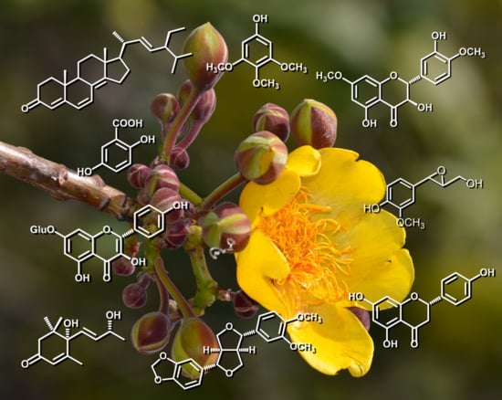
Flavonoids, Sterols and Lignans from Cochlospermum vitifolium and Their Relationship with Its Liver Activity

by
A. Berenice Aguilar-Guadarrama
and
María Yolanda Rios
*
Centro de Investigaciones Químicas, IICBA, Universidad Autónoma del Estado de Morelos, Avenida Universidad 1001, Col. Chamilpa, 62209 Cuernavaca, Morelos, Mexico
*
Author to whom correspondence should be addressed.
Molecules 2018, 23(8), 1952; https://doi.org/10.3390/molecules23081952
Submission received: 13 July 2018 / Revised: 2 August 2018 / Accepted: 3 August 2018 / Published: 5 August 2018

Abstract

:
The sterols β-sitostenone (1), stigmast-4,6,8(14),22-tetraen-3-one (2), β-sitosterol (3) and stigmasterol (4), the aromatic derivatives antiarol (5) and gentisic acid (6), the phenylpropanes coniferyl alcohol (7), epoxyconiferyl alcohol (8) and ferulic acid (9), the apocarotenoid vomifoliol (10), the flavonoids naringenin (11), 7,4′-dimethoxytaxifolin (7,4′-dimethoxydihydroquercetin, 12), aromadendrin (13), kaempferol (14), taxifolin (dihydroquercetin, 15), prunin (naringenin-7-O-β-d-glucoside, 16), populnin (kaempferol-7-O-β-d-glucoside, 17) and senecin (aromadendrin-7-O-β-d-glucoside, 18) and the lignans kobusin (19) and pinoresinol (20), were isolated from the dried bark of Cochlospermum vitifolium Spreng (Cochlospermaceae), a Mexican medicinal plant used to treat jaundice, liver ailments and hepatitis C. Fourteen of these compounds were isolated for the first time from this plant and from the Cochlospermum genus. Compounds 34, 67, 911, 1317 and 20 have previously exhibited diverse beneficial liver activities. The presence of these compounds in C. vitifolium correlates with the use of this Mexican medicinal plant.

Graphical Abstract

1. Introduction

The Cochlospermaceae family comprises seven genera: Amoreuxia, Azeredia, Cochlospermum, Euryanthe, Lachnocistus, Maximilianea and Wittelsbachia. In turn, the genus Cochlospermum (syn. Maximilianea and Bixaceae) is composed of 13 species of tropical trees ranging in height from 3 to 15 m, distributed in deciduous forests worldwide [1]. The bark and roots from the Cochlospermum species have been the most studied parts of these plants and only six of the 13 species of Cochlospermum have been chemically analyzed. Within Cochlospermum gillivraei the flavonoids apigenin, naringenin and afzelequin were found [2]; from Cochlospermum gossypium only carbohydrates were identified [3,4]; Cochlospermum planchonii biosynthesized the flavonoids miricetin, quercetin, aromadendrin and cianidin [5] and gallic acid, saponins, tannins, glycosides and carbohydrates [6]. From Cochlospermum regium, the gallic and ellagic acids, the flavonoid dihidrokaempferol-3-O-β-(6″-galloyl)-glucopyranoside and the lignans pinoresinol and excelsin have been isolated [7]. From Cochlospermum tinctorium, the triterpene arjunolic acid, along with tannins and carotenoids, β-bisabolene, 1-dodecanol, 1-hydroxy-3-octadecanone, 2-pentadecanone [8,9], alphitolic acid, cochloxantin and dihydrocochloxantin were identified [10]. Finally, the composition of the essential oils obtained from the leaves, root bark and root wood of Cochlospermum vitifolium has been established by GC/MS. The leaves’ essential oil consist of four major components: β-caryophyllene (46.5%), α-humulene (26.0%), β-pinene (10.6%) and α-pinene (4.8%), which, together, make up 87.9% of the total oil. The essential oil derived from the root’s bark is predominantly made up of β-bisabolene (29.3%), 1-hydroxy-3-hexadecanone (19.5%) and β-caryophyllene (8.2%), which corresponds to 57.0%. Furthermore, the root wood’s essential oil is composed of γ-muurolene (28.4%), 1-hydroxy-3-hexadecanone (16.2%), β-caryophyllene (11.6%), β-bisabolene (11.5%) and 2-dodecanone (6.3%), which represent 74.0% of the total essential oil [11]. The ethanol extracts from the root’s bark and wood are composed of gallic acid, the lignans excelsin and pinoresinol, the flavonoids naringenin and aromadendrin, and the sterols β-sitosterol, stigmasterol, 3-O-β-glycopyranosyl-β-sitosterol and 3-O-β-glycopyranosylstigmasterol while the root’s wood also contains 1-dodecanoyl-3,5-di(tetradecanoyl)benzene [11]. A second chemical analysis yielded the apocarotenoids cochloxanthin, dihydrocochloxanthin, vitixanthin and dihydrovitixanthin [12]. On the other hand, the plant’s flowers contain the flavonoids apigenin, naringenin and dihydroquercetin, and the carotenoids β-carotene, γ-carotene, lycopene, capsanthin, and zeaxanthin [13]. Finally, the stems contain naringenin and dihydroquercetin [14]. All of these studies indicate that the Cochlospermum species are characterized by the presence of sterols, flavonoids, carotenoids, apocarotenoids and lignans.
Cochlospermum vitifolium (common name panicua, yellow rose or pongolote) is a medicinal tree which grows up to 5 m in height and is found from México to South America. It is characterized by its attractive yellow flowers and seed pods. This plant has been used in several countries due to its medicinal properties. In Cuba, for instance, a decoction of its leaves is used in the treatment of ulcers. In Costa Rica, the sap of the leaves is used to treat jaundice, and in Guatemala it is used due to its emmenagogue effects [15]. In some Mexican states, such as Morelos, Oaxaca, Puebla, and Veracruz, a decoction of its wood and leaves is drunk as an alternative treatment for liver and kidney ailments [16]. For example, in the state of Morelos, an infusion, prepared by boiling 10 g of its dried bark in 1 L of water, is used to treat hepatitis C, jaundice, liver diseases, diabetes, metabolic syndrome, and high blood pressure [17,18].
According to different studies, Cochlospermum vitifolium’s lethal dose 50 (LD50) in its lyophilized aqueous phase, obtained from the partitioned methanol extract, first with chloroform and then with ethyl acetate, was greater than 2000 mg/kg when administered intraperitoneally in mice [15]. The pharmacological analysis of the methanol extract from its dried bark demonstrated a decrease in noradrenaline induced vasoconstriction in rat aortic rings in a concentration and endothelium dependent manner (NO-cGMP system) [19]. The extract showed in vivo antihypertensive effects on spontaneously hypertensive rats [20] by inhibiting the [3H]-AT-II binding (angiotensin II AT1 receptor) by more than 50% [21]. In addition, hypoglycemic and antidiabetic effects were also seen in normoglycemic and STZ-nicotinamide-induced diabetic rats, both, in acute and subchronic models [22]. Additionally, the ethanol extract from the same part of the plant exhibited anti-inflammatory and immunomodulatory properties [23]. Finally, the dichloromethane extract was evaluated ex vivo using rat trachea rings to determine its relaxant activity against contractions induced by carbachol, showing a maximum effect at Emax = 106.58 ± 2.42% and an EC50 = 219.54 ± 7.61 μg/mL [24].

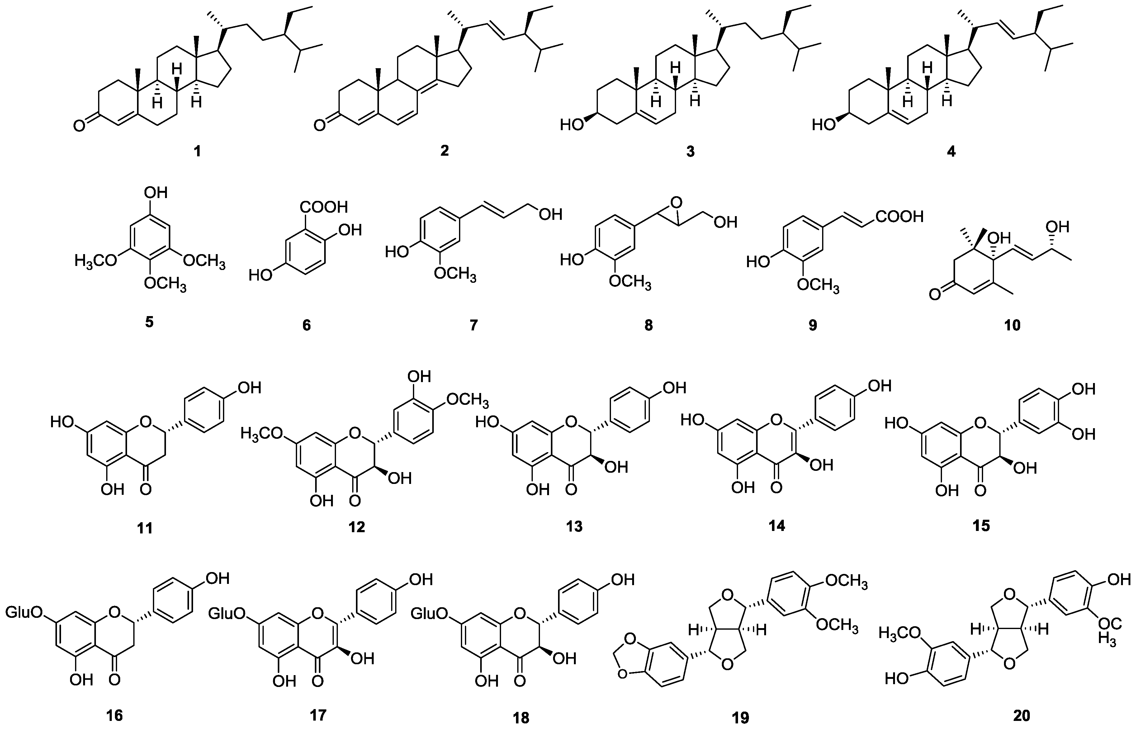
2. Results and Discussion

During our ongoing phytochemical research from the dichloromethane extract of the dried bark of Cochlospermum vitifolium, the following metabolites were characterized: the sterols β-sitostenone (1), stigmast-4,6,8(14),22-tetraen-3-one (2), β-sitosterol (3) and stigmasterol (4), the aromatic derivatives antiarol (5), gentisic acid (6), coniferyl alcohol (7), epoxyconiferyl alcohol (8) and ferulic acid (9), the apocarotenoid vomifoliol (10), and the flavonoid naringenin (11). On the other hand, from the methanol extract two types of metabolites were identified: the flavonoids naringenin (11), 7,4′-dimethoxytaxifolin (7,4′-dimethoxydihydroquercetin, 12), aromadendrin (13), kaempferol (14), taxifolin (dihydroquercetin, 15), prunin (naringenin-7-O-β-d-glucoside, 16), populnin (kaempferol-7-O-β-d-glucoside, 17) and senecin (aromadendrin-7-O-β-d-glucoside, 18); along with the lignans kobusin (19) and pinoresinol (20) (Figure 1). Vomifoliol (10) and naringenin (11) were the major constituents of the dichloromethane extract, while the flavonoids naringenin (11) and senecin (18) were isolated as the major components from the methanol extract. The structures for compounds 120 were established by the analysis of their 1H and 13C NMR (1D and 2D experiments) and MS spectra parameters and its comparison with those reported in the literature. The unequivocal 1H- and 13C- NMR assignments for compound 2 are reported here for the first time.
Six of these 20 compounds (3, 4, 11, 13, 15 and 20) had been previously isolated from Cochlospermum vitifolium, however all other compounds were isolated here for the first time from this plant and from this genus. Compounds 120 belong to either the sterols; C6, C6–C1 and C6–C3 aromatic compounds; apocarotenoids; flavonoids and lignans groups, which are the most frequently identified compounds in this genus.
Previous studies have been conducted to demonstrate the antihypertensive [19,20,21,23], hypoglycemic and antidiabetic [22], immunomodulatory [23] and anti-inflammatory [24] effects of Cochlospermum vitifolium extracts. An exhaustive revision of the existing literature indicated that several of the compounds isolated in this research (Figure 1) can be associated to its popular use as different liver treatments. Due to the fact that this plant is broadly used as a treatment for jaundice, hepatitis C and other liver ailments in Mexican traditional medicine, its methanol extract was administrated at a dose of 100 mg/kg to bile duct-obstructed rats, to determine its hepatoprotective activity, showing a statistically significant decrease of serum glutamic-pyruvic transaminase (PGT, 45%) and alkaline phosphatase (APh, 15%) [19].
The importance of such activities lie in the fact that hepatic diseases (which comprise several conditions, such as: cirrhosis, hepatitis, alcoholic liver disease, non-alcoholic fatty liver disease, cholestatic and drug-induced liver diseases and liver cancer) are extremely high-priced in terms of human suffering, loss of productivity, and medical or hospital consultations. In fact, chronic liver diseases are the major cause of mortality worldwide. In 2013, 29 million people in Europe suffered from a chronic liver condition [25] and more than 30 million Americans had hepatic disease [26]. In China, liver diseases, viral hepatitis (predominantly hepatitis B virus), non-alcoholic fatty liver and alcoholic liver disease affect approximately 300 million people [27]. From a physiopathological perspective, most chronic liver diseases begin as an inflammatory process which evolves into focal fibrosis, and afterwards, to complete fibrosis of the gland (hepatic cirrhosis), which increases the risk of liver cancer. This leads to severe hepatic injury and ultimately to liver failure and other complications.
Thirteen of the twenty compounds isolated from Cochlospermum vitifolium have previously exhibited in vitro and in vivo beneficial liver effects. β-Sitosterol (3) decreased hepatofibrosis [28], protecting against CCl4-induced hepatotoxicity [29] in animal models. Stigmasterol (4) induced apoptosis in hepatocarcimona (HepG2) cells being a potential antineoplastic therapeutic agent [30]. Gentisic acid (6) showed anti-inflammatory and antimutagenic properties, demonstrating protective effects against induced genotoxicity and hepatotoxicity [31]. On the other hand, coniferyl alcohol (7) had a moderated anti-hepatitis B virus (HBV) activity [32]. Ferulic acid (9) had an in vivo hepato-protective effect against the CCl4- and formaldehyde-induced hepatotoxicity [33] and also a capacity to inhibit the development of hepatic fibrosis by activation of Hepatic Stellate Cells (HSCs) in the presence of liver damage [34]. Vomifoliol (10) showed moderate activity against human hepatocarcinoma Hep3B cells [35]. Naringenin (11) had a potent lipid-lowering effect reducing the hepatic lipogenesis in rats and acting as an insulin sensitizer in vivo [36], thus, preventing rat liver damage caused by lead acetate, arsenic and high glucose. Furthermore, this same compound suppresses the metastatic potential of hepatocellular carcinoma [37]. Additionally, aromadendrin (13) possessed radical scavenging and activity against inflammatory, tumor and diabetic processes [38]. Kaempferol (14) had hepatoprotective effects in CCl4-, drug- and alcoholic-induced liver injury, constituting a promising therapeutic option for patients with atherosclerotic disease [39]. Taxifolin (15) had antioxidant and cytoprotective effects that prevent and help treat fulminant hepatitis and hepatitis caused CCl4 [40]. This natural flavonoid is licensed as Silymarin (Legalon®), a drug used for the treatment of toxic liver damage, chronic inflammatory liver disease and liver cirrhosis. Prunin (16) showed activity against the hepatitis B (HBV) virus with an IC50 of 41.59 μM [41]. Administered at a dose of 25 mg/kg, populnin (17) exhibited in vivo hepatoprotective effects against CCl4- and d-GalN-induced hepatotoxicity, preventing the development of hepatic lesions [42]. Finally, at 50 and 100 mg/kg, the lignane pinoresinol (20) showed hepatoprotective effects improving CCl4-induced liver injury [43]. All these liver beneficial effects from the compounds isolated from Cochlospermum vitifolium directly correlate with its traditional use in Mexican medicine.

3. Materials and Methods

3.1. General Procedures

Compounds 120 were purified by successive open column chromatography (CC) using silica gel (70–230 and 230–400 mesh, Sigma-Aldrich, Toluca, México). The isolation procedures and purity of compounds were monitored by thin layer chromatography (TLC) using precoated silica gel 60 F254 aluminium sheets, visualizing with UV-light and subsequently spraying the plates with (NH4)4Ce(SO4)4 in 2 N H2SO4 (Sigma-Aldrich, Toluca, México). All 1H-, 13C- and 2-D NMR experiments were performed in CDCl3 on a Varian Unity 400 spectrometer (Varian, Inc., Palo Alto, CA, USA) equipped with a 5 mm inverse detection pulse field gradient probe at 25 °C, at 400 MHz for 1H-NMR and 100 MHz for 13C-NMR. Chemical shifts were referenced to tetramethylsilane as an internal standard.

3.2. Plant Material

The wood of Cochlospermum vitifolium was collected from “Sierra de Huautla” (20°26′10″ N, 99°05′42″ W, 1915 m above sea level), Morelos, México, in October 2011, and identified by Dr. Rolando Ramírez Rodríguez, Centro de Investigación en Biodiversidad y Conservación-UAEM. A voucher specimen (number 14628) was deposited at HUMO Herbarium from the Universidad Autónoma del Estado de Morelos, México.

3.3. Extraction and Isolation

Throughout three months the wood of this plant was dried at room temperature. The dried and ground wood (1.65 kg) was extracted with CH2Cl2 and MeOH. These extracts were dried under a vacuum to render 11.9 g (0.72% yield) and 25.8 g (1.56% yield) of residue, respectively.
Fractionation of the CH2Cl2 extract by open CC (silica gel, 70–230 mesh; 5 cm i.d. × 20 cm) was performed with a step gradient of n-hexane-acetone 100:0 to 0:100, collecting 330 fractions of 50 mL each. Based on TLC analysis, these fractions were pooled into nine groups, namely G-1 (fractions 1–70, n-hexane 100%), G-2 (fractions 71–76, 722 mg, n-hexane:acetone 95:5), G-3 (fractions 77–81, n-hexane:acetone 95:5), G-4 (fractions 82–89, 131 mg, n-hexane:acetone 95:5), G-5 (fractions 90–166, 780 mg, n-hexane:acetone 95:5), G-6 (fractions 167–188, 620 mg, n-hexane:acetone 9:1), G-7 (fractions 189–265, 639 mg, n-hexane:acetone 8:2), G-8 (fractions 266–324, 540 mg, n-hexane:acetone 1:1) and G-9 (fractions 325–335, acetone 100%). G-1 was made up of fatty acids, G-3 of triglycerides and G-9 of resins. The rest of the groups were subjected to column chromatography using silica gel 70–230 mesh. G-2 (2.0 cm i.d. × 30 cm, eluent n-hexane 100% to n-hexane:acetone 8:2) yielded 45 fractions of 40 mL. Fractions 23–33 (255 mg) were subjected to a second column chromatography (1.0 cm i.d. × 30 cm, eluent n-hexane:AcOEt 99:1) obtaining 91 fractions of 20 mL to yield β-sitosterone (1, 58 mg, 0.0035% with respects dry weigh of plant material). G-4 (1.5 cm i.d. × 15 cm, eluent n-hexane:AcOEt 99:1 to n-hexane:AcOEt 97:3) yielded 132 fractions of 50 mL which rendered stigmast-4,6,8(14),22-tetraen-3-one (2, 71 mg, 0.0043%), coniferyl alcohol (7, 21 mg, 0.0013%) and ferulic acid (9, 27 mg, 0.0016%). G-5 (2.0 cm i.d. × 30 cm, eluent n-hexane:AcOEt 97:3 to n-hexane:AcOEt 9:1) yielded 180 fractions of 50 mL resulting in the isolation of a 7:3 mixture of β-sitosterol (3) and stigmasterol (4, 280 mg, 0.0109%). G-6 (2.0 cm i.d. × 30 cm, eluent n-hexane:AcOEt 9:1 to n-hexane:AcOEt 7:3) yielded 77 fractions of 50 mL resulting in vomifoliol (10, 94 mg, 0.0056%). G-7 (2.0 cm i.d. × 30 cm, eluent n-hexane:AcOEt 9:1) yielded 50 fractions of 50 mL obtaining antiarol (5, 31 mg, 0.0018%) and gentisic acid (6, 29 mg, 0.0017%). Finally, G-8 (2.0 cm i.d. × 30 cm, n-hexane:acetone 8:2) yielded 187 fractions of 50 mL. Fractions 104–167 (280 mg) were subjected to a second column chromatography (1.0 cm i.d. × 30 cm, eluent n-hexane:AcOEt 6:4) resulting in 248 fractions of 20 mL to yield naringenin (11, 102 mg, 0.0061%) and epoxy-coniferyl alcohol (8, 36 mg, 0.0022%).
Fractionation of the MeOH extract by open CC (silica gel, 100–230 mesh; 5 cm i.d. × 30 cm) was performed with a step gradient of n-hexane-acetone 100:0 to 0:100, collecting 268 fractions of 50 mL each. These fractions were pooled into three groups: MG-1 (fractions 1–93, n-hexane to n-hexane:acetone 6:4), MG-2 (fractions 94–135, 2.2 g, n-hexane:acetone 1:1) and MG-3 (fractions 136–268, 3.29 g, n-hexane:acetone 1:1 to acetone). MG-1 was a complex mixture including compounds 1, 3, 4, 11 and aromadendrin (13); MG-2 (3.0 cm i.d. × 30 cm, eluent n-hexane:AcOEt 85:15 to n-hexane:AcOEt 75:25) yielded 250 fractions of 50 mL which resulted in the purification of naringenin (11, 214 mg, 0.0129%); and MG-3 (4.0 cm i.d. × 30 cm, eluent n-hexane:AcOEt 7:3 to acetone) yielded 320 fractions of 50 mL. Fractions 33–51 (181 mg) were subjected to a second column chromatography (1.5 cm i.d. × 20 cm, eluent with n-hexane:AcOEt 7:3) collecting 56 fractions of 20 mL to yield naringenin (11, 41 mg, 0.0024%) and 7,4′-dimethoxy-taxifolin (12, 21 mg, 0.0013%). Fractions 77–83 (52 mg) were subjected to a preparative TLC (2 mm × 20 cm, eluent n-hexane:AcOEt 7:3 twice) to yield 30 mg (0.0018%) of a 6:4 mixture of 13 and kaempferol (14). Fractions 100–111 (49 mg) were subjected to a preparative TLC (2 mm × 20 cm, eluent n-hexane:AcOEt 7:3 twice) to yield kobusin (19, 19 mg, 0.0011%) and pinoresinol (20, 17 mg, 0.0010%). Fractions 145–182 (93 mg) were subjected to a preparative TLC (2 mm × 20 cm, eluent with CH2Cl2:MeOH 93:7) to yield taxifolin (15, 26 mg, 0.0016%). Finally, fractions 289–309 (691 mg) were subjected to a second column chromatography (2.0 cm i.d. × 30 cm, eluent with CH2Cl2:MeOH 9:1) collecting 140 fractions of 20 mL to yield prunin (16, 32 mg, 0.0019%), and 59 mg (0.0035%) of a 45:55 mixture of 16, populnin (17), and senecin (18, 47 mg, 0.0028%).
Stigmast-4,6,8(14),22-tetraen-3-one (2). 1H-NMR (CDCl3): δ 6.61 (1H, d, J = 7.2, H-7), 6.04 (1H, d, J = 7.2, H-6), 5.74 (1H, s, H-4), 5.27 (1H, dd, J = 15.2, 6.8, H-22), 5.20 (1H, dd, J = 15.2, 7.6, H-23), 2.49 (1H, m, H-2a), 2.57 (1H, m, H-2b), 2.51 (1H, m, H-15b), 2.47 (1H, m, H-15a), 2.16 (1H, m, H-20), 2.15 (1H, m, H-9), 2.10 (1H, m, H-12a), 2.03 (1H, m, H-1a), 2.54 (1H, m, H-1b), 1.88 (1H, m, H-24), 1.83 (1H, m, H-16b), 1.53 (1H, m, H-16a), 1.51 (1H, m, H-25), 1.32 (1H, m, H-12b), 1.30 (1H, m, H-17), 1.06 (3H, d, J = 6.4, H-21), 1.00 (3H, s, H-19), 0.96 (3H, s, H-18), 0.93 (3H, d, J = 6.8, H-26), 0.85 (3H, d, J = 6.8, H-27), 0.84 (3H, t, J = 6.8, H-29), 0.78 (2H, m, H-28). 13C-NMR (CDCl3): δ 199.76 (s, C-3), 164.67 (s, C-5), 156.35 (s, C-14), 135.25 (d, C-22), 134.28 (d, C-7), 132.78 (d, C-23), 124.71 (d, C-6), 124.66 (s, C-8), 123.23 (d, C-4), 55.95 (d, C-17), 44.57 (d, C-9), 44.24 (s, C-13), 43.13 (d, C-24), 39.55 (d, C-20), 37.02 (s, C-10), 35.84 (t, C-12), 34.37 (t, C-1, C-2), 33.34 d, C-25), 27.98 (t, C-16), 25.63 (t, C-15), 22.98 (t, C-11), 21.48 (q, C-21), 20.25 (q, C-27), 19.92 (q, C-18), 19.21 (q, C-28), 17.90 (q, C-26), 16.91 (q, C-19), 14.38 (q, C-29).

4. Conclusions

Cochlospermum vitifolium biosynthesizes among other compounds the sterols 3 and 4, the aromatic compounds 6, 7 and 9, the apocatrotenoid 10, the flavonoids 11 and 1317, and the lignan 20, which have demonstrated beneficial activity to alleviate different liver diseases. The presence of these compounds in the plant agrees with its traditional use in Mexican medicine and some are even included in commercial pharmaceutical formulations used in the treatment of hepatopathies. Their presence within Cochlospermum vitifolium extracts indicates that this plant could be an active hepatoprotective agent. However, the human consumption of this plant must be subjected to toxicity, pharmacodynamic and pharmacokinetic studies to determine how it can be a health contributor.
The isolated metabolites in this study and the chemical composition previously reported for Cochlospermum vitifolium agree with the metabolic content within other Cochlospermum species. The flavonoids, sterols, carotenoids, apocarotenoids and lignans isolated here have chemotaxonomic significance within this genus. This is the first report of compounds 12, 510, 12, 14, 1619 from Cochlospermum vitifolium.

Author Contributions

A.B.A.-G. prepared the extracts, isolated, and identified the secondary metabolites. M.Y.R. designed overall research and identified the secondary metabolites.

Funding

This research was funded by CONACYT-México grant number CB-2015-241044.

Acknowledgments

All spectroscopic and spectrometric analyses were obtained from LANEM (grant numbers 279905).

Conflicts of Interest

The authors declare no conflict of interest.

References

  1. Lamien-Meda, A.; Kiendrebeogo, M.; Compaoré, M.; Meda, R.N.T.; Bacher, M.; Koenig, K.; Pacher, T.; Fuehrer, H.-P.; Noedl, H.; Willcox, M.; et al. Quality assessment and antiplasmodial activity of West African Cochlospermum species. Phytochemistry 2015, 119, 51–61. [Google Scholar] [CrossRef] [PubMed]
  2. Cook, I.F.; Knox, J.R. Flavonoids from Cochlospermum gillivraei. Phytochemistry 1975, 14, 2510–2511. [Google Scholar] [CrossRef]
  3. Vinod, V.T.P.; Sashidhar, R.B.; Suresh, K.I.; Rama Rao, B.; Vijaya Saradhi, U.V.R.; Prabhakar Rao, T. Morphological, physico-chemical and structural characterization of gum kondagogu (Cochlospermum gossypium): A tree gum from India. Food Hydrocoll. 2008, 22, 899–915. [Google Scholar] [CrossRef]
  4. Hongsing, P.; Palanuvej, C.; Ruangrungsi, N. Chemical compositions and biological activities of selected exudate gums. J. Chem. Pharm. Res. 2012, 4, 4174–4180. [Google Scholar]
  5. Bate-Smith, E.C. Chemotaxonomie der Pflanzen. Bull. Soc. Bot. Mem. 1964, 435. [Google Scholar] [CrossRef]
  6. Anaga, A.O.; Oparah, N. Investigation of the methanol root extract of Cochlospermum planchonii for pharmacological activities in vitro and in vivo. Pharm. Biol. 2009, 47, 1027–1034. [Google Scholar] [CrossRef]
  7. Solon, S.; Carollo, C.A.; Brandão, L.F.G.; Macedo, C.; Klein, A.; Dias-Junior, C.A.; Siqueira, J.M. Phenolic derivatives and other chemical compounds from Cochlospermum regium. Quim. Nova 2012, 35, 1169–1172. [Google Scholar] [CrossRef]
  8. Diallo, B.; Vanhaelen, M.; Vanhaelen-Fastré, R.; Konoshima, T.; Kozuka, M.; Tokuda, H. Studies on inhibitors of skin-tumor promotion. Inhibitory effects of triterpenes from Cochlospermum tinctorium on Epstein-Barr Virus Activation. J. Nat. Prod. 1989, 52, 879–881. [Google Scholar] [CrossRef] [PubMed]
  9. Diallo, B.; Vanhaelen-Fastré, R.; Vanhaelen, M. Triacylbenzenes and long-chain volatile ketones from Cochlospermum tinctorium rhizome. Phytochemistry 1991, 30, 4153–4156. [Google Scholar] [CrossRef]
  10. Ballin, N.Z.; Traore, M.; Tinto, H.; Sittie, A.; Mølgaard, P.; Olsen, C.E.; Kharazmi, A.; Christensen, S.B. Antiplasmodial compounds from Cochlospermum tinctorium. J. Nat. Prod. 2002, 65, 1325–1327. [Google Scholar] [CrossRef] [PubMed]
  11. Xenofonte de Almeida, S.C.; Gomes de Lemos, L.T.; Rocha Silveira, E.; Loiola Pessoa, O.D. Constituintes químicos voláteis e não-voláteis de Cochlospermum vitifolium (Willdenow) Sprengel. Quim. Nov. 2005, 28, 57–60. [Google Scholar] [CrossRef]
  12. Achenbach, H.; Blümm, E.; Waibel, R. Vitixanthin and dihydrovitixanthin - new unusual 7′-apocarotenoic acids from. Tetrahedron Lett. 1989, 30, 3059–3060. [Google Scholar] [CrossRef]
  13. Dixit, B.S.; Srivastava, S.N. Flavonoids and carotenoids of Cochlospermum vitifolium flowers. Fitoterapia 1992, 63, 270. [Google Scholar]
  14. López, J.A. Flavonoids in Cochlospermum vitifolium Willd (Cochlospermaceae). Ing. Cienc. Quim. 1981, 5, 101–102. [Google Scholar]
  15. Esposito-avella, M.; Brown, P.; Tejeira, I.; Buitrago, R.; Barrios, L.; Sanchez, C.; Gupta, M.P.; Cedeño, J. Pharmacological screening of Panamanian medicinal plants. Part 1. Int. J. Crude Drug Res. 1985, 23, 17–25. [Google Scholar] [CrossRef]
  16. Zamora-Martinez, M.C.; de Pascual Pola, C.N. Medicinal plants used in some rural populations of Oaxaca, Puebla and Veracruz, Mexico. J. Ethnopharmacol. 1992, 35, 229–257. [Google Scholar] [CrossRef]
  17. Monroy-Ortiz, C.; Castillo-España, P. Plantas Medicinales Utilizadas en el Estado de Morelos, 2nd ed.; Universidad Autónoma del Estado de Morelos: Cuernavaca, México, 2007; ISBN 968-878-277-7. [Google Scholar]
  18. Banos, G.; Perez-Torres, I.; El Hafidi, M. Medicinal agents in the metabolic syndrome. Cardiovasc. Hematol. Agents Med. Chem. 2008, 6, 237–252. [Google Scholar] [CrossRef] [PubMed]
  19. Sánchez-Salgado, J.C.; Ortiz-Andrade, R.R.; Aguirre-Crespo, F.; Vergara-Galicia, J.; León-Rivera, I.; Montes, S.; Villalobos-Molina, R.; Estrada-Soto, S. Hypoglycemic, vasorelaxant and hepatoprotective effects of Cochlospermum vitifolium (Willd.) Sprengel: A potential agent for the treatment of metabolic syndrome. J. Ethnopharmacol. 2007, 109, 400–405. [Google Scholar] [CrossRef] [PubMed]
  20. Sánchez-Salgado, J.C.; Castillo-España, P.; Ibarra-Barajas, M.; Villalobos-Molina, R.; Estrada-Soto, S. Cochlospermum vitifolium induces vasorelaxant and antihypertensive effects mainly by activation of NO/cGMP signaling pathway. J. Ethnopharmacol. 2010, 130, 477–484. [Google Scholar] [CrossRef] [PubMed]
  21. Caballero-George, C.; Vanderheyden, P.M.L.; Solis, P.N.; Pieters, L.; Shahat, A.A.; Gupta, M.P.; Vauquelin, G.; Vlietinck, A.J. Biological screening of selected medicinal Panamanian plants by radioligand-binding techniques. Phytomedicine 2001, 8, 59–70. [Google Scholar] [CrossRef] [PubMed]
  22. Ortíz-Andrade, R.; Torres-Piedra, M.; Sánchez-Salgado, J.C.; García-Jiménez, S.; Villalobos-Molina, R.; Ibarra-Barajas, M.; Gallardo-Ortíz, I.; Estrada-Soto, S. Acute and sub-chronic effects of Cochlospermum vitifolium in blood glucose levels in normoglycemic and STZ-nicotinamide-induced diabetic rats. Rev. Latinoamer. Quím. 2009, 37, 122–132. [Google Scholar]
  23. Deharo, E.; Baelmans, R.; Gimenez, A.; Quenevo, C.; Bourdy, G. In vitro immunomodulatory activity of plants used by the Tacana ethnic group in Bolivia. Phytomedicine 2004, 11, 516–522. [Google Scholar] [CrossRef] [PubMed]
  24. Sánchez-Recillas, A.; Mantecón-Reyes, P.; Castillo-España, P.; Villalobos-Molina, R.; Ibarra-Barajas, M.; Estrada-Soto, S. Tracheal relaxation of five medicinal plants used in Mexico for the treatment of several diseases. Asian Pac. J. Trop. Med. 2014, 7, 179–183. [Google Scholar] [CrossRef]
  25. HEPAMAP A Roadmap for Hepatology Research in Europe: An Overview for Policy Makers. Available online: www.easl.eu/medias/EASLimg/News/3f9dd90221ef292_file.pdf (accessed on 10 July 2018).
  26. American Liver Foundation The Liver Lowdown–Liver Disease: The Big Picture. Available online: https://liverfoundation.org/for-patients/resources/liver-lowdown/ (accessed on 10 July 2018).
  27. Wang, F.-S.; Fan, J.-G.; Zhang, Z.; Gao, B.; Wang, H.-Y. The global burden of liver disease: The major impact of China. Hepatology 2014, 60, 2099–2108. [Google Scholar] [CrossRef] [PubMed] [Green Version]
  28. Kim, K.-S.; Yang, H.J.; Lee, J.-Y.; Na, Y.-C.; Kwon, S.-Y.; Kim, Y.-C.; Lee, J.-H.; Jang, H.-J. Effects of β-sitosterol derived from Artemisia capillaris on the activated human hepatic stellate cells and dimethylnitrosamine-induced mouse liver fibrosis. BMC Complement. Altern. Med. 2014, 14, 363. [Google Scholar] [CrossRef] [PubMed]
  29. Wong, H.-S.; Chen, J.-H.; Leong, P.-K.; Leung, H.-Y.; Chan, W.-M.; Ko, K.-M. β-Sitosterol protects against carbon tetrachloride hepatotoxicity but not Gentamicin nephrotoxicity in rats via the induction of mitochondrial glutathione redox cycling. Molecules 2014, 19, 17649–17662. [Google Scholar] [CrossRef] [PubMed]
  30. Kim, Y.-S.; Li, X.-F.; Kang, K.-H.; Ryu, B.; Kim, S.K. Stigmasterol isolated from marine microalgae Navicula incerta induces apoptosis in human hepatoma HepG2 cells. BMB Rep. 2014, 47, 433–438. [Google Scholar] [CrossRef] [PubMed]
  31. Nafees, S.; Ahmad, S.T.; Arjumand, W.; Rashid, S.; Ali, N.; Sultana, S. Modulatory effects of gentisic acid against genotoxicity and hepatotoxicity induced by cyclophosphamide in Swiss albino mice. J. Pharm. Pharmacol. 2012, 64, 259–267. [Google Scholar] [CrossRef] [PubMed]
  32. Wang, H.-L.; Geng, C.-A.; Ma, Y.-B.; Zhang, X.-M.; Chen, J.-J. Three new secoiridoids, swermacrolactones A–C and anti-hepatitis B virus activity from Swertia macrosperma. Fitoterapia 2013, 89, 183–187. [Google Scholar] [CrossRef] [PubMed]
  33. Gerin, F.; Erman, H.; Erboga, M.; Sener, U.; Yilmaz, A.; Seyhan, H.; Gurel, A. The effects of ferulic acid against oxidative stress and inflammation in formaldehyde-induced hepatotoxicity. Inflammation 2016, 39, 1377–1386. [Google Scholar] [CrossRef] [PubMed]
  34. Xu, T.; Pan, Z.; Dong, M.; Yu, C.; Niu, Y. Ferulic acid suppresses activation of hepatic stellate cells through ERK1/2 and Smad signaling pathways in vitro. Biochem. Pharmacol. 2015, 93, 49–58. [Google Scholar] [CrossRef] [PubMed]
  35. Dat, N.T.; Jin, X.; Hong, Y.-S.; Lee, J.J. An isoaurone and other constituents from trichosanthes kirilowii seeds inhibit hypoxia-inducible factor-1 and nuclear factor-κB. J. Nat. Prod. 2010, 73, 1167–1169. [Google Scholar] [CrossRef] [PubMed]
  36. Assini, J.M.; Mulvihill, E.E.; Burke, A.C.; Sutherland, B.G.; Telford, D.E.; Chhoker, S.S.; Sawyez, C.G.; Drangova, M.; Adams, A.C.; Kharitonenkov, A.; et al. Naringenin prevents obesity, hepatic steatosis, and glucose intolerance in male mice independent of fibroblast growth factor 21. Endocrinology 2015, 156, 2087–2102. [Google Scholar] [CrossRef] [PubMed]
  37. Yen, H.-R.; Liu, C.-J.; Yeh, C.-C. Naringenin suppresses TPA-induced tumor invasion by suppressing multiple signal transduction pathways in human hepatocellular carcinoma cells. Chem. Biol. Interact. 2015, 235, 1–9. [Google Scholar] [CrossRef] [PubMed]
  38. Lee, J.-W.; Kim, N.H.; Kim, J.-Y.; Park, J.-H.; Shin, S.-Y.; Kwon, Y.-S.; Lee, H.J.; Kim, S.-S.; Chun, W. Aromadendrin inhibits lipopolysaccharide-induced nuclear translocation of NF-κB and phosphorylation of JNK in RAW 264.7 macrophage cells. Biomol. Ther. 2013, 21, 216–221. [Google Scholar] [CrossRef] [PubMed]
  39. Ochiai, A.; Miyata, S.; Iwase, M.; Shimizu, M.; Inoue, J.; Sato, R. Kaempferol stimulates gene expression of low-density lipoprotein receptor through activation of Sp1 in cultured hepatocytes. Sci. Rep. 2016, 6, 24940. [Google Scholar] [CrossRef] [PubMed] [Green Version]
  40. Zhao, M.; Chen, J.; Zhu, P.; Fujino, M.; Takahara, T.; Toyama, S.; Tomita, A.; Zhao, L.; Yang, Z.; Hei, M.; et al. Dihydroquercetin (DHQ) ameliorated concanavalin A-induced mouse experimental fulminant hepatitis and enhanced HO-1 expression through MAPK/Nrf2 antioxidant pathway in RAW cells. Int. Immunopharmacol. 2015, 28, 938–944. [Google Scholar] [CrossRef] [PubMed]
  41. Zhao, Y.; Geng, C.-A.; Sun, C.-L.; Ma, Y.-B.; Huang, X.-Y.; Cao, T.-W.; He, K.; Wang, H.; Zhang, X.-M.; Chen, J.-J. Polyacetylenes and anti-hepatitis B virus active constituents from Artemisia capillaris. Fitoterapia 2014, 95, 187–193. [Google Scholar] [CrossRef] [PubMed]
  42. Lin, C.; Lee, H.Y.; Chang, C.H.; Namba, T.; Masao, H. Evaluation of the liver protective principles from the root of Cudrania cochinchinensis var. gerontogea. Phyther. Res. 1996, 10, 13–17. [Google Scholar] [CrossRef]
  43. Kim, H.-Y.; Kim, J.-K.; Choi, J.-H.; Jung, J.-Y.; Oh, W.-Y.; Kim, D.C.; Lee, H.S.; Kim, Y.S.; Kang, S.S.; Lee, S.-H.; et al. Hepatoprotective effect of pinoresinol on carbon tetrachloride–induced hepatic damage in mice. J. Pharmacol. Sci. 2010, 112, 105–112. [Google Scholar] [CrossRef] [PubMed]
Sample Availability: Samples of the compounds 1, 3, 4, 11 and 18 are available from the authors.
Figure 1. Chemical contents of the dried bark from Cochlospermum vitifolium.
Molecules 23 01952 g001

