Article

Novel 5′-Norcarbocyclic Pyrimidine Derivatives as Antibacterial Agents

by
Anastasia L. Khandazhinskaya
1,†,
Liudmila A. Alexandrova
1,†,
Elena S. Matyugina
1,
Pavel N. Solyev
1,
Olga V. Efremenkova
2,
Karen W. Buckheit
3,
Maggie Wilkinson
3,
Robert W. Buckheit, Jr.
3,
Larisa N. Chernousova
4,
Tatiana G. Smirnova
4,
Sofya N. Andreevskaya
4,
Olga G. Leonova
1,
Vladimir I. Popenko
1,
Sergey N. Kochetkov
1 and
Katherine L. Seley-Radtke
5,*
1
Engelhardt Institute of Molecular Biology of the Russian Academy of Sciences, 32 Vavilov St., Moscow 119991, Russia
2
Gause Institute of New Antibiotics, 11 Bol’shaya Pirogovskaya St., Moscow 119021, Russia
3
ImQuest BioSciences, 7340 Executive Way Suite R, Frederick, MD 21704, USA
4
Central Tuberculosis Research Institute, 2 Yauzskaya Alley, Moscow 107564, Russia
5
Department of Chemistry & Biochemistry, University of Maryland, 1000 Hilltop Circle, Baltimore, MD 21250, USA
*
Author to whom correspondence should be addressed.
A.L.K. and L.A.A. contributed equally to this work.
Molecules 2018, 23(12), 3069; https://doi.org/10.3390/molecules23123069
Submission received: 2 October 2018 / Revised: 16 November 2018 / Accepted: 20 November 2018 / Published: 23 November 2018
(This article belongs to the Special Issue Bioactive Nucleosides and Nucleotides)

Abstract

:
A series of novel 5′-norcarbocyclic derivatives of 5-alkoxymethyl or 5-alkyltriazolyl-methyl uracil were synthesized and the activity of the compounds evaluated against both Gram-positive and Gram-negative bacteria. The growth of Mycobacterium smegmatis was completely inhibited by the most active compounds at a MIC99 of 67 μg/mL (mc2155) and a MIC99 of 6.7–67 μg/mL (VKPM Ac 1339). Several compounds also showed the ability to inhibit the growth of attenuated strains of Mycobacterium tuberculosis ATCC 25177 (MIC99 28–61 μg/mL) and Mycobacterium bovis ATCC 35737 (MIC99 50–60 μg/mL), as well as two virulent strains of M. tuberculosis; a laboratory strain H37Rv (MIC99 20–50 μg/mL) and a clinical strain with multiple drug resistance MS-115 (MIC99 20–50 μg/mL). Transmission electron microscopy (TEM) evaluation of M. tuberculosis H37Rv bacterial cells treated with one of the compounds demonstrated destruction of the bacterial cell wall, suggesting that the mechanism of action for these compounds may be related to their interactions with bacteria cell walls.

Graphical Abstract

1. Introduction

Antibiotic resistance has emerged for nearly all currently used therapeutics for many diseases, including tuberculosis (TB) [1,2]. Despite this, the development of new classes of antibiotics has lagged far behind the field’s growing need for drugs that can act on new targets and remain active against resistant strains of pathogens. Nucleoside derivatives have historically served as potent antiviral and anticancer agents [3], but it was not until 2000 that their anti-TB activity was reported [4]. Recently, several reports of modified nucleosides showing activity against Mycobacterium tuberculosis (M. tuberculosis), Mycobacterium bovis (M. bovis) and Mycobacterium avium (M. avium) were published [4,5,6,7,8,9,10,11,12,13,14,15,16,17,18]. Among them, the best activity was observed for several uridine analogues (MIC90 1 to 10 µg/mL) possessing 5-alkynyl (decynyl, dodecynyl, tetradecynyl, pyridylethynyl, etc.) substituents as well as modified sugar moieties (Figure 1). In addition, several 2′-deoxypyrimidine derivatives with extended 5-alkyloxymethyl or 5-alkyltriazolylmethyl substituents (5-dodecyloxy-methyl-2′-deoxyuridine, 5-decyltriazolylmethyl-2′-deoxyuridine, 5-dodecyltriazolylmethyl-2′-deoxycytidine, etc.) were shown to inhibit in vitro growth of both laboratory H37Rv and clinical multiple drug resistant (MDR) MS-115 M. tuberculosis strains with MIC99 values ~10 µg/mL (Figure 1) [5].
In an effort to explore additional sugar modifications, a series of carbocyclic nucleoside analogues with the same modifications were designed and synthesized. Because it had already been shown that uracil analogues with short alkyne or halogen substituents at the C-5 position failed to inhibit growth of M. tuberculosis [4,17,18], we chose to synthesize longer substituents. Several showed promise, exhibiting activity below 100 μg/mL. Their synthesis and subsequent biological investigations are discussed below.

2. Results and Discussion

Carbocyclic nucleosides are analogues of natural nucleosides in which the oxygen atom of the furanose ring is replaced by a methylene group. In addition, several carbocyclic nucleosides, for example, aristeromycin and neplanocin A, have been found in Nature [19,20,21,22]. Nucleosides of this type are recognized by many receptors and enzymes due to their structural similarity to the natural nucleosides, however they display increased stability towards phosphorylase- and hydrolase-induced cleavage of the pseudo-glycosidic bond. Moreover, they possess a wide spectrum of biological activity, particularly, antiviral and anticancer properties [19,23,24,25,26,27,28,29,30,31,32,33]. Unfortunately, in some cases they have exhibited significant toxicity as a result of their conversion to triphosphate forms, which closely resemble natural nucleoside triphosphates (NTP), thus resulting in unwanted recognition by ATP metabolizing enzymes [34,35,36].
To overcome the cellular cytotoxicity of carbocyclic nucleosides that inhibit their target enzymes without preliminary phosphorylation (for example, aristeromycin and neplanocin A) [35], the 5′-norcarbocyclic nucleosides were designed. The switch of a secondary hydroxyl group for the primary hydroxyl group at C-4′ led to a lack of recognition by cellular kinases and as a result, they were less toxic [36,37,38,39,40,41,42,43,44,45,46]. Recently, we have shown that 5′-norcarbocyclic nucleosides with a 5-substituted uracil base (Figure 2) can act as promising anti-TB agents (MIC99 5–10 µg/mL on both laboratory strain M. tuberculosis H37Rv and clinical isolate M. tuberculosis MDR MS-115) [47,48]. Despite our best efforts, however, their mechanism of action remains elusive.
In that regard, the thymidylate kinases of M. tuberculosis were evaluated as possible enzymatic targets of 1-(4′-hydroxy-2′-cyclopenten1′-yl)-5-(phenylamino)uracils and 1,3-di-(4′-hydroxy-2′-cyclopenten-1′-yl)-5-(phenylamino)uracils. No inhibition was found up to 200 µM [47]. Another possible mechanism of action that was considered was the inhibition of M. tuberculosis thymidylate synthases, thus both the classical (ThyA) and flavin dependent thymidylate synthase (ThyX)A were evaluated [46]. All of the derivatives lacked activity against the ThyA and only one inhibited ThyX at a concentration of 8.32 µM. Thus it was assumed that the anti-TB activity of 5-alkoxymethyl or 5-alkyltriazolylmethyl 2′-deoxyuridine analogues could not be explained by inhibition of thymidine kinases.
In an effort to further explore the mechanism of action for these compounds, we have now designed a new hybrid scaffold possessing both modifications. Herein, we describe the synthesis and preliminary antibacterial activity for a series of new 5′-norcarbocyclic pyrimidine derivatives containing extended 5-alkyloxymethyl or 5-alkyltriazolylmethyl residues (Figure 3).

2.1. Chemistry

The key intermediate needed for the synthesis of both scaffolds for the new 5-modified 5′-norcarbocyclic analogues is racemic 1-(4′-acetoxycyclopent-1′-yl)-5-(bromomethyl)-uracil (16). As shown in Scheme 1, 16 was synthesized in three steps starting from 1-(4′-hydroxy-2′-cyclopenten-1′-yl)-thymine (13), which was obtained by the Trost procedure [49] using the coupling of epoxy-cyclopentene with thymine. Hydrogenation of 13 in the presence of palladium on carbon gave 1-(4′-hydroxycyclopent-1′-yl)-thymine (14) which was protected to provide 1-(4′-acetoxycyclopent-1′-yl)thymine (15). Following free radical bromination by the Barwolff and Langen procedure [50], key intermediate 16 was obtained.
The desired 5-alkyloxymethyl substituted analogues 13 were then prepared by coupling with the corresponding 1-alcohol [51] and subsequent deprotection of the 4’-hydroxy group to provide targets 46 (Scheme 2).
The 5-alkyltriazolylmethyl derivatives 712 were synthesized by the Lee method (Scheme 2) [52]. Azidation of 1-(4′-acetoxycyclopent-1′-yl)-5-(bromomethyl)uracil (16) to the corresponding 5-azidomethyl 17 was followed by 1,3-dipolarcycloaddition with various 1-acetylenes in a biphasic system of dichloromethane-water under catalytic conditions using Cu(I) obtained in situ from copper sulphate and sodium ascorbate to give 79. After deprotection of the 4′-hydroxyl group of 79, the desired 5-alkyltriazolylmethyl derivatives 1012 were obtained in good yields (84–92%). It should be noted that the synthetic route from 16 to 112 doesn’t change the configuration of the chiral centers, i.e., C-1′ and C-4′. Thus, because compound 16 is racemic all of the target compounds (112) were obtained as racemic mixtures. Moreover, following synthesis of the individual isomers of the compound with the 5-dodecynyl substituent (Figure 2) no significant difference in the activity was found (see [47]), thus we chose not to synthesize the individual enantiomers of the other target compounds.

2.2. Biological Evaluation and Structure-Activity Relationship Studies

In order to determine the lead compounds prior to evaluation against the virulent strains of M. tuberculosis, the twelve compounds were initially evaluated for inhibition of an attenuated strain of M. tuberculosis ATCC 25177 (H37Ra), which is closely related to a virulent laboratory strain H37Rv, as well as the M. bovis ATCC strain 35737 in an in vitro assay (Table 1). When evaluated against M. tuberculosis ATCC strain 25177, the 5-alkyloxymethyl substituted analogues 13 had MIC99 values of 409, 423 and 54 µg/mL, respectively. Deprotection of the 4′-hydroxygroup resulted in targets 46 having MIC values of 183, 191 and 198 µg/mL. The 5-alkyltriazolylmethyl derivatives 712 had increased activities ranging from 29 µg/mL to 98 µg/mL.
Compounds 112 have similar activities against M. bovis ATCC strain 35737 with MICs either the same or within 2-fold of the MICs determined for M. tuberculosis. The exception was compounds 9 and 12, which exhibited an 8-fold decrease in activity against M. bovis relative to M. tuberculosis. Thus, the best antimycobacterial activity against both strains among the 5-alkyloxymethyl derivatives 16 was seen for compound 3 with the 4′-acetyl group and the methyloxydodecyl substituent at the 5-position of the uracil moiety. In the series of 5-alkyltriazolylmethyl derivatives 712, the 4′-acetylated analogues 7 (5-octyltriazolylmethyl) and 8-(5-decyltriazolylmethyl) were observed to have a significant inhibitory effect. DMSO (5%, equivalent to the same amount as would be present in the final solutions of the tested compounds) was evaluated in parallel and was determined to be non-toxic to all of the organisms.
The antibacterial activity of the synthesized compounds was also evaluated against Gram-negative bacteria including Pseudomonas aeruginosa (P. aeruginosa) ATCC strain 27853 and Escherichia coli (E. coli) ATCC strain 25922, Gram-positive bacteria including Bacillus subtilis (B. subtilis) ATCC strain 6633, Staphylococcus aureus (S. aureus) INA 00761 (MRSA) and Leuconostoc mesenteroides (L. mesenteroides) VKPM B-4177. Mycobacterium smegmatis (M. smegmatis) mc2155 and M. smegmatis VKPM Ac 1339, were also used in the primary screening for potential antibiotic activity. Most of the bacteria were not sensitive to the tested compounds. The only exception was against M. smegmatis, whose growth was completely inhibited by compounds 3, 6, 8 and 11 at a concentration of 6.7–67 μg/mL (Table 1). The MIC99 of the control antibiotics rifampicin and isoniazid were 10 and 1 μg/mL for M. smegmatis VKPM Ac 1339, and > 100 and 64 μg/mL for M. smegmatis mc2155, respectively. Interestingly, the tested compounds demonstrated selective activity only on mycobacteria, but not on other gram-positive and gram-negative bacteria (data not shown), thus could represent a specific interaction that will be pursued in subsequent studies. Three compounds (3, 7 and 8) were selected as leads against the attenuated strain ATCC 25177 and subjected to additional antimycobacterial screening. The ability of the new 5′-norcarbocyclic nucleosides to influence the growth of two virulent strains of M. tuberculosis, including the laboratory strain M. tuberculosis H37Rv and the clinical strain M. tuberculosis MS-115 with multiple drug resistance were assessed in vitro. The antimycobacterial effect was studied by measuring the growth dynamics of strains of M. tuberculosis in the enriched medium Middlebrook 7H9 in the automated growth registration system BACTEC MGIT 960. Different concentrations of the compounds were compared to the growth of strains on media not containing preparations and media containing control preparations at critical concentrations (rifampicin 1 μg/mL, isoniazid 0.1 μg/mL and levofloxacin 1.5 μg/mL). Each of the concentrations of the tested compounds as well as control samples were done in triplicate. In all positive samples, the culture of mycobacteria of the tuberculosis complex was confirmed to grow and there was no contamination with nonspecific microflora. When exposed to control preparations at critical concentrations, the growth of the sensitive culture was not fixed, and the growth parameters of the MDR strain when incubated with isoniazid and rifampicin did not differ from the control without the drug.
As shown in Table 2, the MIC99 against the laboratory sensitive strain M. tuberculosis H37Rv were for compounds 3 (20 μg/mL), 7 (50 μg/mL) and 8 (50 μg/mL). Against M. tuberculosis MS-115, the MIC99 values for compounds 3, 7, and 8 were 20 μg/ mL, 20 μg/mL, and 50 μg/mL, respectively. It should be noted that unlike the previously reported 5-alkynyl substituted compounds, the 5-alkoxymethyl and 5-alkyl-triazolylmethyl derivatives proved to be quite toxic to the monocytic cells. The cytotoxicity of compounds 3, 7 and 8 to the U-937 laboratory adapted monocytic cell line was similar to or greater than the activity of the compounds against the mycobacteria with TC50 values 20.6 μg/mL, 23.7 μg/mL and 15.1 μg/mL, respectively. Nonoxynol-9 was also evaluated in parallel as a positive control and was toxic at the expected concentration. So while it is clear these compounds are too toxic to be regarded as drug candidates, they may be useful as a tool for better understanding the mechanism of antimicobacterial action for this class of compounds.
In contrast, the observed biological activity of these new derivatives of 5-substituted uracils are in good correlation with previously reported data on the inhibition of M.tb growth by nucleoside analogues containing 5-substituted pyrimidine bases and different sugars, as well as their corresponding acyclic, or carbocyclic analogues [4,5,15,16,17,18,53] As a result, it appears these compounds may have the same mechanism of antimicrobial action (likely cell wall disruption) and that it depends primarily on the length and structure of the substituent at C-5 of the base moiety. Thus this new data has added to the growing examples of similar compounds and their mechanism of action for inhibitors of this type. In an attempt to further support this hypothesis some additional experiments using method of transmission electron microscopy were carried out.
Figure 4 shows electron micrographs of control M. tuberculosis H37Rv cells grown for 4 days in enriched Dubois medium (A) and in the presence of DMSO/Twin-80/H2O buffer without compound 3 (B). The morphology of bacterial cells grown in the medium containing compound 3 was markedly different. As shown in Figure 4C,D, the bacterial wall was damaged (Figure 4C) or completely destroyed (Figure 4D). This observation suggests that inactivation of M. tuberculosis may occur due to the interaction of compound 3 with the bacterial wall and its subsequent destruction. The main difference of Mycobacteria from other bacteria is their thick cell wall containing unique lipids—mycolic acids [54,55]. Thus speculatation that the synthesized compounds selectively act on mycolic acids of the Mycobacteria cell wall or, like isoniazid, block the pathways involved in mycolic acids biosynthesis [56] was logical. More testing however needs to be performed to confirm this observation, but if this phenomenon is subsequently confirmed for the majority of species of gram-positive and gram-negative bacteria, then these compounds may prove useful due to the lack of effect on the natural microflora of the gut bacteria.

3. Materials and Methods

3.1. Chemistry

The reactions were performed with the use of commercial reagents acquired from Acros (Geel, Belgium), Aldrich (St. Louis, MO, USA) and Fluka (Bucharest, Romania); anhydrous solvents were purified according to the standard procedures. Column chromatography was performed on Silica Gel 60 0.040–0.063 mm (Merck, Darmstadt, Germany) columns. Thin layer chromatography (TLC) was performed on Silica Gel 60 F254 aluminum-backed plates (Merck). Preparative layer chromatography (PLC) was performed on Silica Gel 60 F254 glass-backed plates (Merck). NMR spectra were registered on an AMX III-400 spectrometer (Bruker, Newark, Germany) with the working frequency of 400 MHz for 1H-NMR (Me4Si as an internal standard for organic solvents) and 100.6 MHz for 13C-NMR (with carbon-proton interaction decoupling). High resolution mass spectra (HRMS) were registered on a Bruker Daltonics micrOTOF or a Bruker micrOTOF II instrument using electrospray ionization (ESI HRMS) (Bruker Daltonics, Hamburg, Germany). The measurements were done in positive ion mode (interface capillary voltage 4500 V) in a mass range from m/z 50 to m/z 3000 Da; external or internal calibration was done with ESI Tuning Mix™ (Agilent Technologies, Santa Clara, CA, USA). Nitrogen was applied as a dry gas (6 L/min); the interface temperature was set at 180 °C, nebulizer pressure: 0.4 Bar; flow rate: 3 µL/min. Samples were injected into the mass spectrometer chamber from the Agilent 1260 HPLC system equipped with an Agilent Poroshell 120 EC-C18 column (3.0 × 50 mm; 2.7 µm) and an identically packed security guard, using an autosampler. The samples were injected from acetonitrile (LC-MS grade) solution. The column temperature was 30 °C and 5 μL of the sample solution was injected. The column was eluted in a gradient of concentrations of A (acetonitrile) in B (water) with the flow rate of 400 µL/min in the following gradient parameters: 0–15% A for 6.0 min, 15%–85% A for 1.5 min, 85%–0% A for 0.1 min, 0% A for 2.4 min.
(±)-1-(4′-Hydroxy-2′-cyclopenten-1′-yl)-thymine (13). Epoxycyclopentene (0.8 g, 10 mmol) in freshly distilled THF (15 mL) and tetrakis(triphenylphosphine)palladium (0) [Pd(PPh3)4] (308 mg; 0.26 mmol) were added to a stirred solution of thymine (1 g, 8.0 mmol) in anhydrous DMF (40 mL). The reaction mixture was stirred at room temperature for 18 h, then concentrated under vacuum, and the residue dissolved in 98:2 CHCl3:MeOH (5 mL) and loaded onto a silica gel column. Elution with a 98:2 to 95:5 CHCl3:MeOH gave compound (±)-13 as a white powder (600 mg, 36%): Rf 0.32 (95:5 CHCl3:MeOH). 1H-NMR (DMSO-d6): 11.20 (1H, br. s, NH), 7.28 (1H, s, H-6), 6.14–6.11 (1H, m, H-2′), 5.79–5.77 (1H, m, H-3′), 5.39–5.36 (1H, m, H-1′), 5.20 (1H, d, J = 6.0 Hz, OH), 4.63–4.61 (1H, m, H-4′), 2.74–2.70 (1H, m, H-а5′), 1.76 (3H, s, 5-CH3), 1.37–1.33 (1H, m, H-b5′). 13C-NMR (DMSO-d6): 164.3, 151.3 (C-4, C-2), 140.4 (C-6), 137.7 (C-3′), 131.4 (C-2′), 109.9 (C-5), 73.8 (C-4′), 58.3 (C-1′), 39.9 (C-5′), 12.6 (CH3).
(±)-1-(4′-Hydroxycyclopent-1′-yl)-thymine (14). To a solution of (±)-13 (0.6 g, 3 mmol) in anhydrous MeOH (15 mL) 0.1 g 10% Pd/C was added under H2 atmosphere and the reaction mixture was stirred at room temperature under H2 atmosphere for 18 h. The mixture was filtrated through a pad of Celite and the filtrate was evaporated to dryness. The residue was dissolved in 98:2 CHCl3:MeOH (5 mL) and loaded onto a silica gel column eluting with the same solvent system, followed by 95:5 CHCl3:MeOH to give compound (±)-14 as an off white powder (510 mg, 81%): Rf 0.35 (95:5 CHCl3:MeOH). 1H-NMR (CDCl3): 8.77 (1H, br. s, NH), 7.48 (1H, s, H-6), 5.03–4.95 (1H, m, H-1′), 4.41 (1H, m, H-4′), 2.39 (1H, d, J = 8.0 Hz, OH), 2.36–2.30 (1H, m, H-а5′), 2.20–2.13, 2.00–1.98 and 1.73–1.71 (4H, 3m, H-2′and H-3′), 1.93 (3H, s, 5-CH3), 1.70–1.67 (1H, m, H-b5′). 13C-NMR (CDCl3): 164.2, 151.2 (C-4, C-2), 139.9 (C-6), 109.9 (C-5), 73.6 (C-4′), 58.3 (C-1′), 39.9 (C-5′), 31.8 and 29.9(C-2′ and C-3′), 12.6 (CH3).
(±)-1-(4′-Acetoxycyclopent-1′-yl)-thymine (15). To a solution of (±)-14 (0.5 g, 2.4 mmol) in anhydrous pyridine (15 mL), Ac2O (0.275 mL, 2.9 mmol) was added. The reaction mixture was stirred at room temperature for 18 h, concentrated under vacuum, and the residue dissolved in CHCl3 (5 mL), loaded onto a silica gel column. Elution with CHCl3 followed by 98:2 CHCl3:MeOH provided compound (±)-15 as a white powder (0.5 g, 83%): Rf 0.36 (elution with 98:2 CHCl3:MeOH). 1H-NMR (CDCl3): 9.13 (1H, br. s, NH), 7.21 (1H, s, H-6), 5.23–5.20 (1H, m, H-1′), 5.14–5.10 (1H, m, H-4′), 2.54–2.47 (1H, m, H-а5′), 2.20–2.16, 2.00–1.98 and 1.87–1.82 (4H, 3m, H-2′and H-3′), 2.08 (3H, s, Ac), 1.96 (3H, s, 5-CH3), 1.78–1.73 (1H, m, H-b5′). 13C-NMR (CDCl3): 170.0 (C(O) acetyl), 163.7, 151.2 (C-4, C-2), 136.7 (C-6), 111.4 (C-5), 74.8 (C-4’), 53.9 (C-1′), 37.8 (C-5′), 31.8 and 29.9, (C-2′ and C-3′), 21.3 and 12.8 (2CH3).
(±)-1-(4′-acetoxycyclopent-1′-yl)-5-(bromomethyl)uracil (16). A solution of Br2 (0.2 mL, 2.4 mmol) in DCE (5 mL) was added dropwise over a 3 h period with stirring to a boiling solution of (±)-15 (0.25 g, 1 mmol) in DCE (10 mL) with concomitant irradiation using an incandescent 300 W lamp under an argon atmosphere. The progress of the reaction was monitored by TLC in EtOAc/hexane (1:1). The mixture was evaporated under vacuum, coevaporated with toluene (3 × 10 mL), the crude 16 dissolved in DMF (5 mL) and used without further purification in subsequent reactions.

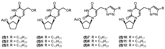
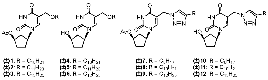
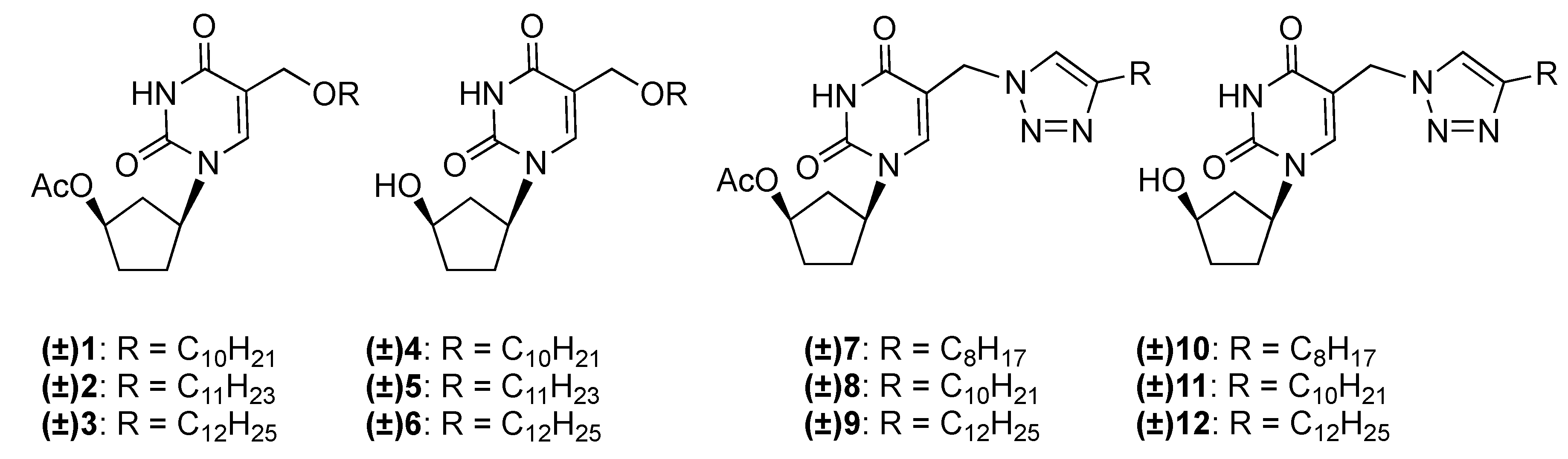
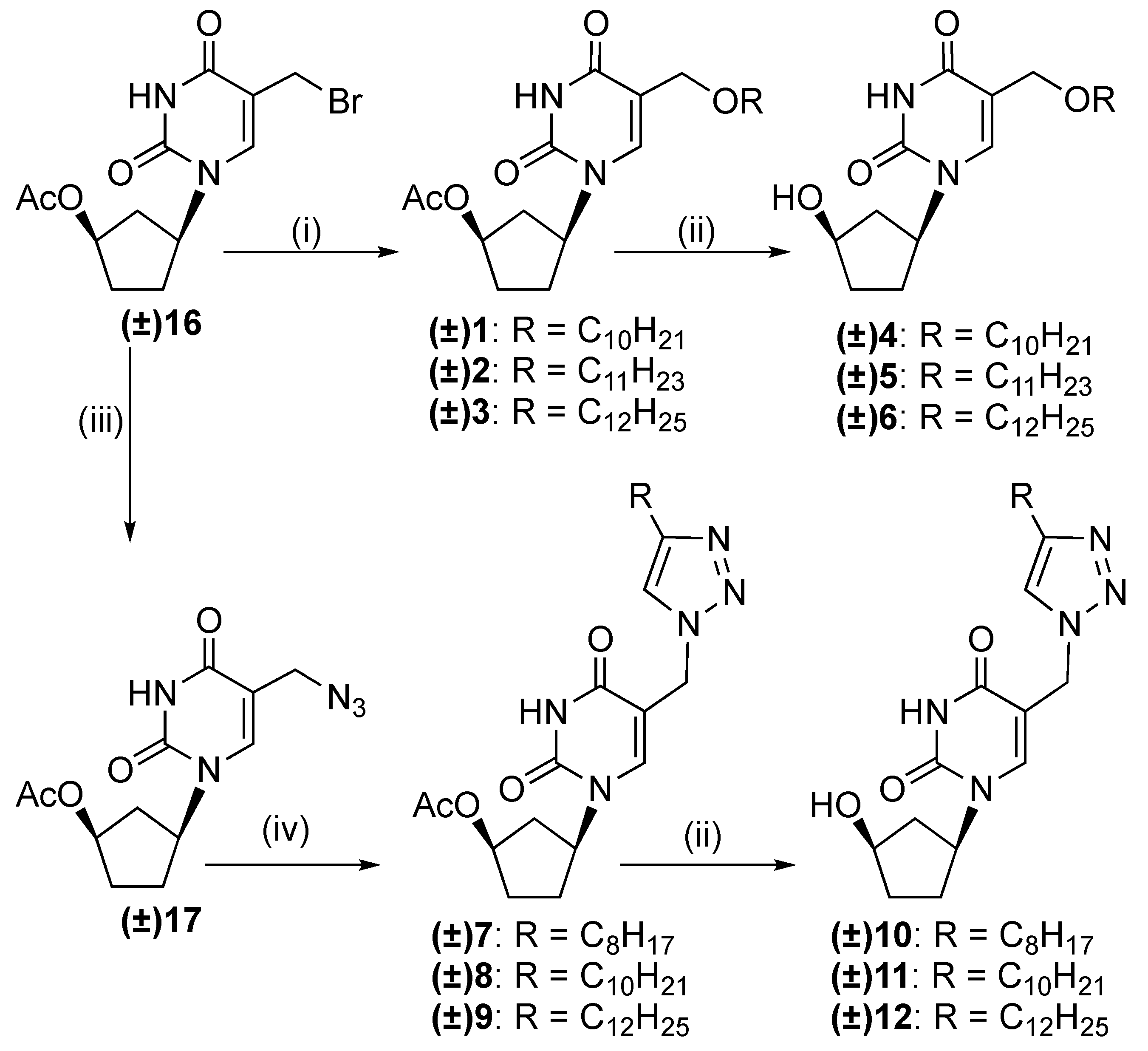
3.1.1. General Procedure for the Synthesis of (±)-1-(4′-Acetoxycyclopent-1′-yl)-5-alkyloxy-methyluracils 13

The corresponding alcohol (1.5 mmol) was added to a solution of 1-(4′-acetoxycyclopent-1′-yl)-5-(bromomethyl)uracil (13, 0.5 mmol) in dry DMF (10 mL). The mixture was stirred for 48 h at 37 °C under argon atmosphere, evaporated under vacuum, and the residue dissolved in CHCl3 (5 mL), loaded onto a silica gel column, and eluted with CHCl3:MeOH (98:2) to provide the crude products which were purified as described below.
(±)-1-(4′-Acetoxycyclopent-1′-yl)-5-decyloxymethyluracil (1). Crude 1 was purified by PLC eluting with EtOAc/hexane (2:1) to give compound (±)-1 as a white powder (92 mg, 45%): Rf 0.40 (98:2 CHCl3:MeOH). 1H-NMR (CDCl3): 8.76 (1H, br. s, NH), 7.49 (1H, s, H-6), 5.23–5.21 (1H, m, H-1′), 5.15–5.11 (1H, m, H-4′), 4.26 (2H, s, 5-CH2O), 3.51 (2H, t, J = 8.0 Hz, CH2CH2(CH2)7CH3), 2.55–2.47 (1H, m, H-а5′), 2.22–2.18, 2.01–1.99 and 1.87–1.83 (4H, 3m, H-2′ and H-3′), 2.08 (3H, s, Ac), 1.78–1.72 (1H, m, H-b5′), 1.61–1.56 (2H, m, CH2CH2(CH2)7CH3), 1.29–1.25 (14H, m, CH2CH2(CH2)7CH3), 0.86 (3H, t, J = 8 Hz, CH2CH2(CH2)7CH3). 13C-NMR (CDCl3): 170.3 (C(O) acetyl), 162.2, 150.9 (C-4, C-2), 138.0 (C-6), 112.9 (C-5), 74.8 (C-4′), 71.6 (5-CH2O), 65.0 (CH2CH2(CH2)7CH3), 54.4 (C-1′), 38.0 (C-5′), 32.0, 31.9, 30.0, 29.8, 29.6 × 2, 29.5, 29.4, 26.2, 22.7, 21.3 (C-2′, C-3′, (CH2)8, CH3C(O)), 14.2 (CH3). HRMS: found m/z 409.2698, calculated C22H36N2O5 [M + H]+ 409.26697.
(±)-1-(4′-Acetoxycyclopent-1′-yl)-5-undecyloxymethyluracil (2). Crude 2 was purified by PLC eluting with EtOAc/hexane (2:1) mixture to give compound (±)-2 as a white powder (99 mg, 47%): Rf 0.41 (98:2 CHCl3:MeOH). 1H-NMR (CDCl3): 8.58 (1H, br. s, NH), 7.49 (1H, s, H-6), 5.25–5.20 (1H, m, H-1′), 5.17–5.11 (1H, m, H-4′), 4.26 (2H, s, 5-CH2O), 3.52 (2H, t, J = 8.0 Hz, CH2CH2(CH2)8CH3), 2.58–2.47 (1H, m, H-а5′), 2.22–2.18, 2.03–1.99 and 1.88–1.83 (4H, 3m, H-2′and H-3′), 2.09 (3H, s, Ac), 1.79–1.72 (1H, m, H-b5′), 1.62–1.58 (2H, m, CH2CH2(CH2)8CH3), 1.30–1.26 (16H, m, CH2CH2(CH2)8CH3), 0.87 (3H, t, J = 8.0 Hz, CH2CH2(CH2)8CH3). 13C-NMR (CDCl3): 170.2 (C(O) acetyl), 162.1, 150.9 (C-4, C-2), 138.0 (C-6), 112.8 (C-5), 74.7 (C-4’), 71.5 (5-CH2O), 64.9 (CH2CH2(CH2)8CH3), 54.3 (C-1′), 37.9 (C-5′), 31.9, 31.8, 29.9, 29.6 × 3, 29.5, 29.3, 29.0, 26.1, 22.7, 21.2 (C-2′, C-3′, (CH2)9, CH3C(O)), 14.1 (CH3). HRMS: found m/z 423.2853, calculated C23H38N2O5 [M + H]+ 423.2853.
(±)-1-(4′-Acetoxycyclopent-1′-yl)-5-dodecyloxymethyluracil (3). Crude 3 was purified by PLC eluting with EtOAc/hexane (2:1) to give compound (±)-3 as a white powder (100 mg, 46%): Rf 0.42 (elution with 98:2 CHCl3:MeOH). 1H-NMR (CDCl3): 8.92 (1H, br. s, NH), 7.43 (1H, s, H-6), 5.18-5.14 (1H, m, H-1′), 5.13-5.06 (1H, m, H-4′), 4.20 (2H, s, 5-CH2O), 3.46 (2H, t, J=8 Hz, CH2CH2(CH2)9CH3), 2.50–2.42 (1H, m, H-а5′), 2.16–2.12, 1.96–1.94 and 1.81–1.77 (4H, 3m, H-2′and H-3′), 2.03 (3H, s, Ac), 1.72–1.67 (1H, m, H-b5′), 1.57–1.50 (2H, m, CH2CH2(CH2)9CH3), 1.25–1.19 (18H, m, CH2CH2(CH2)9CH3), 0.86 (3H, t, J = 8.0 Hz, CH2CH2(CH2)9CH3). 13C-NMR (CDCl3): 170.3 (C(O) acetyl), 162.4, 151.1 (C-4, C-2), 138.1 (C-6), 113.0 (C-5), 74.9 (C-4′), 71.7 (5-CH2O), 65.0 (CH2CH2(CH2)8CH3), 54.5 (C-1′), 38.1 (C-5′), 32.1, 32.0, 30.1, 29.8 × 5, 29.6, 29.5, 26.3, 22.8, 21.4 (C-2′, C-3′, (CH2)10, CH3C(O)), 14.2 (CH3). HRMS: found m/z 437.3004, calculated C24H40N2O5 [M + NH]+ 437.3010.
(±)-1-(4′-Acetoxycyclopent-1′-yl)-5-(azidomethyl)uracil (17). Sodium azide (160 mg, 2.5 mmol) was added to a solution of 1-(4′-acetoxycyclopent-1′-yl)-5-(bromomethyl)uracil (13) (0.5 mmol) in dry DMF (10 mL). The mixture was stirred for 48 h at 37 °C under argon atmosphere, evaporated under vacuum, and the residue dissolved in CHCl3 (5 mL), applied onto a silica gel column, and eluted with CHCl3:MeOH (98:2) to give crude product 17. It was then purified further by PLC eluted with EtOAc/hexane (1:1) to provide the desired compound 17 as off white solid (143 mg, 97.5%). 1H-NMR (CDCl3): 8.43 (1H, br. s, NH), 7.54 (1H, s, H-6), 5.31–5.27 (1H, m, H-1′), 5.22–5.17 (1H, m, H-4′), 4.23 (2H, s, 5-CH2), 2.60–2.53 (1H, m, H-а5′), 2.31–2.27, 2.10–2.05 and 1.93–1.85 (4H, 3m, H-2′and H-3′), 2.07 (3H, s, Ac), 1.81–1.75 (1H, m, H-b5′).

3.1.2. General Procedure for the Synthesis of (±)-1-(4′-Acetoxycyclopent-1′-yl)-5-[(4-alkyl)-1,2,3-triazol-1-yl]methyluracils 79

To a solution of azide 17 (143 mg, 0.49 mmol) and corresponding 1-alkyne (0.75 mmol) in dichloromethane (2 mL), copper sulphate (12.4 mg, 0.05 mmol), sodium ascorbate (30 mg, 0.15 mmol) and H2O (2 mL) were added. The reaction mixture was stirred for 17 h at room temperature. The solvents were evaporated under vacuum and the crude products were purified by PLC eluting with EtOAc/hexane (2:1) to give compounds 79.
(±)-1-(4′-Acetoxycyclopent-1′-yl)-5-[(4-octyl)-1,2,3-triazol-1-yl]methyluracil (7). White powder (100 mg, 47.5%). Rf 0.28 (EtOAc/hexane (2:1)).1H-NMR (CDCl3): 9.53 (1H, s, NH), 7.82 (1H, s, H-6), 7.60 (1H, s, Htriazol), 5.03–5.20 (3H, m, H-1′and 5-CH2N), 5.16–5.08 (1H, m, H-4′), 2.71 (2H, t, J = 8.0 Hz, CH2CH2(CH2)5CH3), 2.59–2.49 (1H, m, H-а5′), 2.20–2.17, 2.03–1.99 and 1.90–1.79 (4H, 3m, H-2′and H-3′), 2.18 (3H, s, Ac), 1.76–1.65 (1H, m, H-b5′), 1.65–1.59 (2H, m, CH2CH2(CH2)5CH3), 1.29–1.24 (10H, m, CH2CH2(CH2)5CH3), 0.88 (3H, t, J=8 Hz, CH2CH2(CH2)5CH3). 13C-NMR (CDCl3): 170.4 (C(O) acetyl), 162.4, 150.5 (C-4, C-2), 148.7 (C-5triazole), 142.4 (C-6), 122.4 (C-4triazole), 109.0 (C-5), 74.4 (C-4′), 55.2 (C-1′), 46.6 (5-CH2N), 38.1 (C-5’), 31.9, 31.8, 30.2, 29.8, 29.3 × 3, 25.4, 22.7, 21.5 (C-2′, C-3′, (CH2)7, CH3C(O)), 14.1 (CH3). HRMS: found m/z 432.2601, calculated C22H33N5O4 [M + H]+ 432.2605.
(±)-1-(4′-Acetoxycyclopent-1′-yl)-5-[(4-decyl)-1,2,3-triazol-1-yl]methyluracil (8), white powder (143 mg, 63.5%). Rf 0.30 (EtOAc/hexane (2:1)).1H-NMR (CDCl3): 9.03 (1H, s, NH), 7.78 (1H, s, H-6), 7.55 (1H, s, Htriazol), 5.26-5.21 (3H, m, 5-CH2N and H-1′), 5.09–5.05 (1H, m, H-4′), 2.68 (2H, t, J = 8.0 Hz, CH2CH2(CH2)7CH3), 2.56–2.48 (1H, m, H-а5′), 2.22–2.17, 2.01–1.99 and 1.89–1.80 (4H, 3m, H-2′and H-3′), 2.15 (3H, s, Ac), 1.76–1.69 (1H, m, H-b5′), 1.66–1.62 (2H, m, CH2CH2(CH2)7CH3), 1.30–1.24 (14H, m, CH2CH2(CH2)7CH3), 0.86 (3H, t, J = 8.0 Hz, CH2CH2(CH2)7CH3). 13C-NMR (CDCl3): 170.4 (C(O) acetyl), 162.5, 150.7 (C-4, C-2), 148.7 (C-5triazole), 142.2 (C-6), 122.1 (C-4triazole), 109.5 (C-5), 74.5 (C-4′), 55.2 (C-1′), 45.2 (5-CH2N), 38.2 (C-5′), 32.0, 31.9, 30.3, 29.7 × 2, 29.4 × 4, 25.7, 22.8, 21.5 (C-2′, C-3′, (CH2)9, CH3C(O)), 14.2 (CH3). HRMS: found m/z 460.2919, calculated C24H37N5O4 [M + H]+ 460.2918.
(±)-1-(4′-Acetoxycyclopent-1′-yl)-5-[(4-dodecyl)-1,2,3-triazol-1-yl]methyluracil (9), white powder (127 mg, 53%). Rf 0.32 (EtOAc/hexane (2:1)).1H-NMR (CDCl3): 9.30 (1H, s, NH), 7.78 (1H, s, H-6), 7.55 (1H, s, Htriazol), 5.21 (3H, m, 5-CH2N and H-1′), 5.09–5.05 (1H, m, H-4′), 2.67 (2H, t, J = 8.0 Hz, CH2CH2(CH2)9CH3), 2.55–2.47 (1H, m, H-а5′), 2.20–2.17, 2.02–1.99 and 1.93–1.83 (4H, 3m, H-2′ and H-3′), 2.15 (3H, s, Ac), 1.76–1.70 (1H, m, H-b5′), 1.65–1.61 (2H, m, CH2CH2(CH2)9CH3), 1.25–1.22 (18H, m, CH2CH2(CH2)9CH3), 0.86 (3H, t, J = 8.0 Hz, CH2CH2(CH2)9CH3). 13C-NMR (CDCl3): 170.4 (C(O) acetyl), 162.5, 150.7 (C-4, C-2), 148.7 (C-5triazole), 142.1 (C-6), 122.0 (C-4triazole), 109.5 (C-5), 74.4 (C-4′), 55.1 (C-1′), 46.1 (5-CH2N), 38.1 (C-5′), 32.0, 31.8, 30.2, 29.7 × 3, 29.6, 29.4 × 4, 25.6, 22.7, 21.4 (C-2′, C-3′, (CH2)11, CH3C(O)), 14.2 (CH3). HRMS: found m/z 488.2232, calculated C26H41N5O4 [M + H]+ 488.2232.

3.1.3. General Procedure for the Synthesis of (±)-1-(4′-Hydroxycyclopent-1′-yl)-5-alkyloxy-methyluracils 46 and (±)-1-(4′-hydroxycyclopent-1′-yl)-5-[(4-alkyl)-1,2,3-triazol-1-yl]methyl-uracils 1012

To a stirred solution of the corresponding acetylated compound 13, 79 (0.15 mmol) in MeOH (5 ml) K2CO3 (52 mg; 0.38 mmol) was added and refluxed during 2–4 h. The progress of the reaction was monitored by TLC in 95:5 CHCl3:MeOH. The reaction mixture was concentrated in vacuum and the residue purified by PLC eluting with 9:1 CHCl3-MeOH to give the desired compounds.
(±)-1-(4′-Hydroxycyclopent-1′-yl)-5-decyloxymethyluracil (4), white solid (47 mg, 85 %): Rf 0.47 (9:1 CHCl3:MeOH). 1H-NMR (CDCl3): 8.72 (1H, br. s, NH), 7.73 (1H, s, H-6), 5.00–4.92 (1H, m, H-1′), 4.39 (1H, m, H-4′), 4.24 (2H, s, 5-CH2O), 3.50 (2H, t, J = 8 Hz, CH2CH2(CH2)7CH3), 2.36–2.34 (1H, m, H-а5′), 2.19–2.16, 1.99–1.89 (4H, 2m, H-2′and H-3′), 1.77–1.72 (1H, m, H-b5′), 1.60–1.57 (2H, m, CH2CH2(CH2)7CH3), 1.28–1.25 (14H, m, CH2CH2(CH2)7CH3), 0.86 (3H, t, J = 8.0 Hz, CH2CH2(CH2)7CH3). 13C-NMR (CDCl3): 162.5, 151.0 (C-4, C-2), 140.8 (C-6), 112.6 (C-5), 72.2 (C-4′), 71.5 (5-CH2O), 65.0 (CH2CH2(CH2)7CH3), 56.4 (C-1′), 40.5 (C-5′), 35.3, 32.0, 29.8, 29.7 × 3, 29.6, 29.4, 26.2, 22.8 (C-2′, C-3′, (CH2)8), 14.2 (CH3). HRMS: found m/z 367.2592, calculated C20H34N2O4 [M + H]+ 367.2591.
(±)-1-(4′-Hydroxycyclopent-1′-yl)-5-undecyloxymethyluracil (5), white solid (51 mg, 88 %): Rf 0.49 (9:1 CHCl3:MeOH). 1H-NMR (CDCl3): 8.56 (1H, br. s, NH), 7.72 (1H, s, H-6), 4.98-4.94 (1H, m, H-1′), 4.40 (1H, m, H-4′), 4.24 (2H, s, 5-CH2O), 3.50 (2H, t, J = 8 Hz, CH2CH2(CH2)8CH3), 2.41–2.33 (2H, m, H-а5′, OH), 2.21–2.14, 2.05–1.96, 1.94–1.87 and 1.78–1.75 (4H, 4m, H-2′and H-3′), 1.74–1.69 (1H, m, H-b5′), 1.62-1.55 (2H, m, CH2CH2(CH2)8CH3), 1.29–1.25 (16H, m, CH2CH2(CH2)8CH3), 0.87 (3H, t, J = 8.0 Hz, CH2CH2(CH2)8CH3). 13C-NMR (CDCl3): 162.4, 151.0 (C-4, C-2), 140.7 (C-6), 112.6 (C-5), 72.2 (C-4′), 71.5 (5-CH2O), 65.0 (CH2CH2(CH2)8CH3), 56.4 (C-1′), 40.5 (C-5′), 35.3, 32.00, 29.8, 29.7 × 4, 29.6, 29.4, 26.2, 22.8 (C-2′, C-3′, (CH2)9), 14.2 (CH3). HRMS: found m/z 381.2749, calculated C21H36N2O4 [M + H]+ 381.2748.
(±)-1-(4′-Hydroxycyclopent-1′-yl)-5-dodecyloxymethyluracil (6), white solid (51.5 mg, 87 %): Rf 0.50 (9:1 CHCl3:MeOH). 1H-NMR (CDCl3): 8.60 (1H, br. s, NH), 7.72 (1H, s, H-6), 4.97–4.94 (1H, m, H-1′), 4.40 (1H, m, H-4′), 4.24 (2H, s, 5-CH2O), 3.50 (2H, t, J = 8 Hz, CH2CH2(CH2)9CH3), 2.41–2.33 (1H, m, H-а5′), 2.21–2.14, 2.05–1.97, 1.94–1.87 and 1.75 (4H, 4m, H-2′and H-3′), 1.75–1.67 (1H, m, H-b5′), 1.62–1.55 (2H, m, CH2CH2(CH2)9CH3), 1.29–1.25 (18H, m, CH2CH2(CH2)9CH3), 0.86 (3H, t, J = 8.0 Hz, CH2CH2(CH2)9CH3). 13C-NMR (CDCl3): 162.4, 151.0 (C-4, C-2), 140.7 (C-6), 112.6 (C-5), 72.2 (C-4′), 71.5 (5-CH2O), 65.0 (CH2CH2(CH2)8CH3), 56.5 (C-1′), 40.4 (C-5′), 35.3, 32.0, 29.7 × 5, 29.6, 29.5, 29.4, 26.2, 22.8 (C-2′, C-3′, (CH2)10), 14.2 (CH3). HRMS: found m/z 395.2904, calculated C22H38N2O4 [M + H]+ 395.2904.
(±)-1-(4′-Hydroxycyclopent-1′-yl)-5-[(4-octyl)-1,2,3-triazol-1-yl]methyluracil (10), white solid (54 mg, 92%): Rf 0.49 (9:1 CHCl3:MeOH). 1H-NMR (CDCl3): 9.41 (1H, s, NH), 8.22 (1H, s, H-6), 7.60 (1H, s, Htriazole), 5.28–5.19 (2H, m, 5-CH2N), 5.13–5.07 (1H, m, H-1′), 4.40 (1H, m, H-4′), 2.67 (2H, t, J = 8.0 Hz, CH2CH2(CH2)5CH3), 2.33–2.25 (1H, m, H-а5′), 2.23–2.19, 1.98–1.84 and 1.67–1.64 (4H, 3m, H-2′and H-3′), 1.74–1.70 (1H, m, H-b5′), 1.62–1.57 (2H, m, CH2CH2(CH2)5CH3), 1.28–1.23 (10H, m, CH2CH2(CH2)5CH3), 0.85 (3H, t, J = 8.0 Hz, CH2CH2(CH2)5CH3). 13C-NMR (CDCl3): 162.8, 151.0 (C-4, C-2), 148.3 (C-5triazole), 144.2 (C-6), 122.5 (C-4triazole), 109.1 (C-5), 72.0 (C-4′), 55.7 (C-1′), 46.5 (5-CH2N), 41.0 (C-5′), 35.2, 31.9, 30.7, 29.8, 29.4 × 2, 29.3, 25.5, 22.7 (C-2′, C-3′, (CH2)7), 14.1 (CH3). HRMS: found m/z 390.2501, calculated C20H31N5O3 [M + H]+ 390.2500.
(±)-1-(4′-Hydroxycyclopent-1′-yl)-5-[(4-decyl)-1,2,3-triazol-1-yl]methyluracil (11), white solid (53 mg, 84%): Rf 0.50 (9:1 CHCl3:MeOH). 1H-NMR (CDCl3): 9.71 (1H, s, NH), 8.17 (1H, s, H-6), 7.54 (1H, s, Htriazole), 5.24-5.19 (2H, m, 5-CH2N), 5.16-5.06 (1H, m, H-1’), 4.41–4.40 (1H, m, H-4′), 2.63 (2H, t, J = 8.0 Hz, CH2CH2(CH2)7CH3), 2.34–2.26 (1H, m, H-а5′), 2.23–2.17, 1.91–1.84 and 1.74–1.65 (4H, 3m, H-2′and H-3′), 1.77–1.73 (1H, m, H-b5′), 1.64–1.57 (2H, m, CH2CH2(CH2)7CH3), 1.28–1.23 (14H, m, CH2CH2(CH2)7CH3), 0.85 (3H, t, J = 8.0 Hz, CH2CH2(CH2)7CH3). 13C-NMR (CDCl3): 162.8, 150.2 (C-4, C-2), 148.2 (C-5triazole), 144.2 (C-6), 122.6 (C-4triazole), 109.0 (C-5), 72.0 (C-4′), 55.8 (C-1′), 46.7 (5-CH2N), 41.0 (C-5′), 35.2, 32.0, 30.7, 29.7, 29.6, 29.4 × 4, 25.5, 22.8 (C-2′, C-3′, (CH2)9), 14.2 (CH3). HRMS: found m/z 418.2078, calculated C22H35N5O3 [M + H]+ 418.2813.
(±)-1-(4′-Hydroxycyclopent-1′-yl)-5-[(4-dodecyl)-1,2,3-triazol-1-yl]methyluracil (12), white solid (519 mg, 88%): Rf 0.52 (9:1 CHCl3:MeOH). 1H-NMR (CDCl3): 9.02 (1H, s, NH), 8.11 (1H, s, H-6), 7.52 (1H, s, Htriazole), 5.24–5.20 (2H, m, 5-CH2N), 5.16–5.04 (1H, m, H-1′), 4.42–4.40 (1H, m, H-4′), 2.65 (2H, t, J = 8.0 Hz, CH2CH2(CH2)9CH3), 2.34–2.28 (1H, m, H-а5′), 2.25–2.18, 1.94-1.83 and 1.69–1.66 (4H, 3m, H-2′and H-3′), 1.73–1.70 (1H, m, H-b5′), 1.64–1.59 (2H, m, CH2CH2(CH2)9CH3), 1.28–1.24 (18H, m, CH2CH2(CH2)9CH3), 0.86 (3H, t, J = 8.0 Hz, CH2CH2(CH2)9CH3). 13C-NMR (CDCl3): 162.6, 150.9 (C-4, C-2), 148.8 (C-5triazole), 143.8 (C-6), 122.0 (C-4triazole), 109.5 (C-5), 72.1 (C-4′), 55.8 (C-1’), 46.1 (5-CH2N), 40.9 (C-5’), 35.3, 32.0, 30.6, 29.7 × 4, 29.5 × 2, 29.4 × 2, 25.8, 22.8 (C-2′, C-3′, (CH2)11), 14.2 (CH3). HRMS: found m/z 446.3127, calculated C24H39N5O3 [M + H]+ 446.3127.

3.2. Antibacterial Experiments

3.2.1. Bacterial Strains

The M. tuberculosis ATCC 25177 and M. bovis ATCC 35737 strains used in these studies were purchased from the American Type Culture Collection (ATCC, Manassas, VA, USA). The laboratory M.tuberculosis H37Rv strain and the MS-115 clinical strain resistant to five first line anti-TB drugs (rifampicin, isoniazid, streptomycin, ethambutol, and pyrazinamide) were acquired from the Russian Central Tuberculosis Research Institute Collection (Moscow, Russia). The bacteria were propagated as recommended by the supplier. B. subtilis ATCC 6633, S. aureus INA 00761 (MRSA), M. smegmatis mc2155, M. smegmatis VKPM Ac 1339, L. mesenteroides VKPM B-4177, P. aeruginosa ATCC 27853, and E. coli ATCC 25922 were purchased from the collection of Gause Institute of New Antibiotics (Moscow, Russia).

3.2.2. U-937 Cells

The U-937 cells used in the cytotoxicity evaluation were obtained from the AIDS Research and Reference Program (Germantown, MD, USA). The cells were cultured as specified by the supplier in RPMI-1640 supplemented with 10% heat inactivated fetal bovine serum, 1% penicillin/streptomycin solution and 1% l-glutamine.

3.2.3. Determination of Minimal Inhibitory Concentration (MIC) against M. tuberculosis ATCC 25177

The susceptibility of the M. tuberculosis ATCC 25177 to the test compounds was evaluated by determining the MIC of each compound using a micro-broth dilution analysis [57,58,59,60]. A standardized inoculum was prepared by direct suspension of bacteria from a Lowenstein-Jensen slant to Middlebrook 7H9 broth with ADC enrichment. The broth culture was incubated for 26 days at 37 °C. Following the incubation, the culture was vortexed with glass beads resulting in an OD595 of 0.18 and then further diluted 1:20 to yield 5 × 10⁶ CFU/mL. One hundred microliters (100 μL) was added in triplicate wells of a 96-well plate containing 100 μL of test compound serially diluted 2-fold in the appropriate broth. This dilution scheme yielded final concentrations of the bacteria estimated to be 2.5 × 10⁶ CFU/mL. The plates were incubated for seven days at 37 °C at which time 30 µL of reazurin solution was added to each well and the cultures were incubated for an additional 24 h at 37 °C. Following the incubation, the plates were visually scored +/- for color change from blue (no growth) to pink (growth). The MIC for each compound was determined as the lowest compound dilution that completely inhibited microbial growth.

3.2.4. Determination of MIC against M. bovis ATCC 35737

The susceptibility of M. bovis ATCC 35737 to the test compounds was evaluated by determining the MIC of each compound using a micro-broth dilution [57]. A standardized inoculum was prepared by direct suspension of bacteria from a Lowenstein-Jensen slant to Middlebrook 7H9 broth with ADC (albumin, dextrose, catalase) enrichment and 40 mM sodium pyruvate. The broth culture was incubated for 23 days at 37 °C. Following the incubation, the culture was vortexed with glass beads resulting in an OD595 = 0.203 which was further diluted 1:10 to yield 3 × 10⁷ CFU/mL. One-hundred microliters (100 μL) was added in triplicate wells of a 96-well plate containing 100 μL of test compound serially diluted 2-fold in the appropriate broth. This dilution scheme yielded final concentrations of the bacteria estimated to be 1.5 × 10⁷ CFU/mL. The plates were incubated for five days at 37 °C at which time 30 µL of reazurin solution was added to each well. The cultures were incubated for an additional 4 days at 37 °C at which time the plates were visually scored +/- for color change from blue (no growth) to pink (growth). The MIC for each compound was determined as the lowest compound dilution that completely inhibited microbial growth.

3.2.5. Antituberculosis Tests with Virulent Strains of M. tuberculosis

The laboratory strain of M. tuberculosis H37Rv susceptible to anti-TB drugs and MDR strain M. tuberculosis MS-115 were used. The MDR strain is resistant toward five first line anti-TB drugs: rifampicin, isoniazid, streptomycin, ethambutol and pyrazinamide. The mycobacteria were transformed into a suspension of single cells at the same growth phase and normalized by CFU [61] and studied using an automated BACTEC MGIT 960 system (BD, Westfield, MA, USA) [62]. Tested compounds were dissolved in DMSO at a concentration of 40 mg/mL (stock solutions). Water and Tween-80 were added to the stock solutions to provide DMSO/Tween-80/H2O (5:0.5:4.5, v/v/v). The concentration of mycobacterial cells was 106 CFU/mL. Each sample, including the control samples lacking the tested compound, were evaluated in triplicate as previously reported [28,29]. The antibacterial activity was analyzed with the TB Exist software [53]. The method is based on comparison of the bacterial growth in the samples, containing the tested compounds, and the control samples diluted 100 times if compared with the initially inoculated culture. The culture is considered susceptible (i.e., the compound is active) if the optical density of the control sample is 400 GU (growth units) and that of the tested sample is less than 100 GU. In addition, using the absolute concentrations method, a delay in the bacterial growth of the tested sample was measured in comparison with the control samples, which reflected a negative effect of the tested compounds at the concentrations lower than MIC on the bacterial viability. The measurements were carried out automatically every hour and registered with the Epicenter software (BD, Westfield, MA, USA).

3.2.6. Antibacterial Effects

The antibacterial effects for each of the compounds as well as rifampicin, isoniazid, ciprofloxacin and amikacin were determined by the micro-broth delution method [63] against B. subtilis ATCC 6633, S. aureus INA 00761 (MRSA), M. smegmatis mc2155, M. smegmatis VKPM Ac 1339, L. mesenteroides VKPM B-4177, P. aeruginosa ATCC 27853, and E. coli ATCC 25922. These strains were purchased from the collection of Gause Institute of New Antibiotics. Two-fold serial dilutions of the compounds in nutrient medium No. 2 Gause (composition (%): glucose-1, peptone-0.5, tryptone-0.3, NaCl-0.5, pH 7.2–7.4) were performed to include concentrations ranging from 300 μg/mL to 1 μg/mL, and the test strains were cultured at 106 cells/mL. All cultures were incubated at 37 °C with the exception of L. mesenteroides VKPM B-4177, which was incubated at 28 °C. The presence of growth and growth intensity were determined after one day in all the strains with the exception of M. smegmatis, which was evaluated after two days of growth.

3.2.7. Evaluation of U-937 Cytotoxicity

U-937 cells plated at 2.5 × 10³ cells/well in a volume of 100 µL were incubated with serially diluted test compounds in a volume of 100 µL for 6 days at 37 °C/5% CO2. Following the incubation, the cells were stained with the tetrazolium dye XTT to determine cellular viability and the plates were read at 460/650 nm on a spectrophotometer. Toxicity values were calculated using linear regression analysis.

3.2.8. Electron Microscopy

For electron microscopy M. tuberculosis H37Rv cells (109 CFU/mL) were grown in enriched Dubois medium with compound 3 dissolved in of DMSO/Twin-80/H2O solution as described above. The final concentration of compound 3 was 60 µg/mL. In control experiments the cells were grown in pure enriched Dubois medium or in the presence of DMSO/Twin-80/H2O solution without compound 3. After 4 days the cells were collected by centrifugation, washed in distilled water and fixed in 2.5% glutaraldehyde. Aliquotes (80 mkL) of cell suspensions were centrifuged in the small centrifuge chambers onto freshly glow-discharged carbon-collodion electron microscopic grids at 2500 g for 20 min [64,65]. Specimens were briefly rinsed in distilled water, stained for 1 min in 1% aqueous uranyl acetate solution [66], air dried and studied on a JEM-100CX electron microscope (JEOL, Peabody, MA, USA) in Core Facility “UNIQEM Collection” of Federal Research Centre “Fundamentals of Biotechnology” of the Russian Academy of Sciences.

4. Conclusions

In summary, twelve new 5′q5 ml-norcarbocyclic derivatives of 5-alkoxymethyl or 5-alkyl-triazolylmethyl uracil were synthesized. The antibacterial activity of the compounds was tested against different classes of bacteria, including both Gram-positive and Gram-negative strains. It was determined that this set of uracil derivatives possess selectivity towards Mycobacteria. Analogues 3, 7 and 8 exhibited activities against two virulent strains of M. tuberculosis: laboratory sensitive strain H37Rv and the MDR clinical strain MS-115, which is known to be clinically resistant to five top antituberculosis drugs. Notably, analogue 7 was 2.5 times more active against the MDR strain. However, all three of the active compounds proved to be quite toxic to the monocytic cell line U937, thus, additional work needs to be undertaken to attenuate this toxicity and improve the activity. TEM studies of M. tuberculosis H37Rv bacterial cells treated with 3 as a representative of this set demonstrated destruction of the bacterial cell wall. The results of these studies thus suggest that the mechanism of action for these compounds is most likely related to interactions with the bacterial cell wall. Ongoing studies currently underway will hopefully provide more insight on this interesting finding and will be reported elsewhere as they become available.

Supplementary Materials

Copies of the NMR spectra are available online.

Author Contributions

A.A.K., E.S.M. and A.L.K. conceived, designed and performed the chemical synthesis; K.W.B., M.W., E.B.L. and M.A.Z. designed and performed cell assays and evaluated biological properties of the compounds; P.N.S. performed HRMS analysis and analyzed the relevant data. A.A.K., E.S.M., E.B.L., M.A.Z., P.N.S., S.N.K., K.L.S-R. and A.L.K. analyzed the data; R.W.B., S.N.K. contributed reagents/materials/analysis tools; K.L.S.-R., A.A.K., E.S.M., E.B.L., P.N.S. and A.L.K. wrote the paper.

Funding

The work was supported by Russian Science Foundation, project No 14-50-00060.

Conflicts of Interest

The authors declare no conflict of interest.

References

  1. Ventola, C.L. The antibiotic resistance crisis: Part 2: Management strategies and new agents. P & T 2015, 40, 344–352. [Google Scholar] [PubMed]
  2. Ventola, C.L. The antibiotic resistance crisis: Part 1: Causes and threats. P & T 2015, 40, 277–283. [Google Scholar] [PubMed]
  3. De Clercq, E.; Li, G. Approved Antiviral Drugs over the Past 50 Years. Clin. Microbiol. Rev. 2016, 29, 695–747. [Google Scholar] [CrossRef] [PubMed] [Green Version]
  4. Rai, D.; Johar, M.; Manning, T.; Agrawal, B.; Kunimoto, D.Y.; Kumar, R. Design and studies of novel 5-substituted alkynylpyrimidine nucleosides as potent inhibitors of mycobacteria. J. Med. Chem. 2005, 48, 7012–7017. [Google Scholar] [CrossRef] [PubMed]
  5. Shmalenyuk, E.R.; Chernousova, L.N.; Karpenko, I.L.; Kochetkov, S.N.; Smirnova, T.G.; Andreevskaya, S.N.; Chizhov, A.O.; Efremenkova, O.V.; Alexandrova, L.A. Inhibition of Mycobacterium tuberculosis strains H37Rv and MDR MS-115 by a new set of C5 modified pyrimidine nucleosides. Bioorg. Med. Chem. 2013, 21, 4874–4884. [Google Scholar] [CrossRef] [PubMed]
  6. Duckworth, B.P.; Wilson, D.J.; Nelson, K.M.; Boshoff, H.I.; Barry, C.E., III; Aldrich, C.C. Development of a selective activity-based probe for adenylating enzymes: Profiling MbtA Involved in siderophore biosynthesis from Mycobacterium tuberculosis. ACS Chem. Biol. 2012, 7, 1653–1658. [Google Scholar] [CrossRef] [PubMed]
  7. Van Calenbergh, S.; Pochet, S.; Munier-Lehmann, H. Drug design and identification of potent leads against mycobacterium tuberculosis thymidine monophosphate kinase. Curr. Top Med. Chem. 2012, 12, 694–705. [Google Scholar] [CrossRef] [PubMed]
  8. Ferrari, V.; Serpi, M. Nucleoside analogs and tuberculosis: New weapons against an old enemy. Future Med. Chem. 2015, 7, 291–314. [Google Scholar] [CrossRef] [PubMed]
  9. Dawadi, S.; Boshoff, H.I.M.; Park, S.W.; Schnappinger, D.; Aldrich, C.C. Conformationally Constrained Cinnolinone Nucleoside Analogues as Siderophore Biosynthesis Inhibitors for Tuberculosis. ACS Med. Chem. Lett. 2018, 9, 386–391. [Google Scholar] [CrossRef] [PubMed]
  10. Bockman, M.R.; Engelhart, C.A.; Dawadi, S.; Larson, P.; Tiwari, D.; Ferguson, D.M.; Schnappinger, D.; Aldrich, C.C. Avoiding Antibiotic Inactivation in Mycobacterium tuberculosis by Rv3406 through Strategic Nucleoside Modification. ACS Infect. Dis. 2018, 4, 1102–1113. [Google Scholar] [CrossRef] [PubMed]
  11. Krajczyk, A.; Zeidler, J.; Januszczyk, P.; Dawadi, S.; Boshoff, H.I.; Barry, C.E., III; Ostrowski, T.; Aldrich, C.C. 2-Aryl-8-aza-3-deazaadenosine analogues of 5′-O-[N-(salicyl)sulfamoyl]adenosine: Nucleoside antibiotics that block siderophore biosynthesis in Mycobacterium tuberculosis. Bioorg. Med. Chem. 2016, 24, 3133–3143. [Google Scholar] [CrossRef] [PubMed]
  12. Dawadi, S.; Kawamura, S.; Rubenstein, A.; Remmel, R.; Aldrich, C.C. Synthesis and pharmacological evaluation of nucleoside prodrugs designed to target siderophore biosynthesis in Mycobacterium tuberculosis. Bioorg. Med. Chem. 2016, 24, 1314–1321. [Google Scholar] [CrossRef] [PubMed] [Green Version]
  13. Bockman, M.R.; Kalinda, A.S.; Petrelli, R.; De la Mora-Rey, T.; Tiwari, D.; Liu, F.; Dawadi, S.; Nandakumar, M.; Rhee, K.Y.; Schnappinger, D.; et al. Targeting Mycobacterium tuberculosis Biotin Protein Ligase (MtBPL) with Nucleoside-Based Bisubstrate Adenylation Inhibitors. J. Med. Chem. 2015, 58, 7349–7369. [Google Scholar] [CrossRef] [PubMed] [Green Version]
  14. Somu, R.V.; Boshoff, H.; Qiao, C.; Bennett, E.M.; Barry, C.E., 3rd; Aldrich, C.C. Rationally designed nucleoside antibiotics that inhibit siderophore biosynthesis of Mycobacterium tuberculosis. J. Med. Chem. 2006, 49, 31–34. [Google Scholar] [CrossRef] [PubMed]
  15. Shakya, N.; Srivastav, N.C.; Bhavanam, S.; Tse, C.; Desroches, N.; Agrawal, B.; Kunimoto, D.Y.; Kumar, R. Discovery of novel 5-(ethyl or hydroxymethyl) analogs of 2′-‘up’ fluoro (or hydroxyl) pyrimidine nucleosides as a new class of Mycobacterium tuberculosis, Mycobacterium bovis and Mycobacterium avium inhibitors. Bioorg. Med. Chem. 2012, 20, 4088–4097. [Google Scholar] [CrossRef] [PubMed]
  16. Shakya, N.; Srivastav, N.C.; Desroches, N.; Agrawal, B.; Kunimoto, D.Y.; Kumar, R. 3′-bromo analogues of pyrimidine nucleosides as a new class of potent inhibitors of Mycobacterium tuberculosis. J. Med. Chem. 2010, 53, 4130–4140. [Google Scholar] [CrossRef] [PubMed]
  17. Johar, M.; Manning, T.; Tse, C.; Desroches, N.; Agrawal, B.; Kunimoto, D.Y.; Kumar, R. Growth inhibition of Mycobacterium bovis, Mycobacterium tuberculosis and Mycobacterium avium in vitro: Effect of 1-beta-D-2′-arabinofuranosyl and 1-(2′-deoxy-2′-fluoro-beta-D-2′-ribofuranosyl) pyrimidine nucleoside analogs. J. Med. Chem. 2007, 50, 3696–3705. [Google Scholar] [CrossRef] [PubMed]
  18. Shmalenyuk, E.R.; Kochetkov, S.N.; Alexandrova, L.A. Novel inhibitors of Mycobacterium tuberculosis growth based on modified pyrimidine nucleosides and their analogues. Russ. Chem. Rev. 2013, 82, 896–915. [Google Scholar] [CrossRef]
  19. Kusaka, T.; Yamamoto, H.; Shibata, M.; Muroi, M.; Kishi, T. Streptomyces citricolor nov. sp. and a new antibiotic, aristeromycin. J. Antibiot. (Tokyo) 1968, 21, 255–263. [Google Scholar] [CrossRef] [PubMed]
  20. Hayashi, M.; Yaginuma, S.; Muto, N.; Tsujino, M. Structures of neplanocins, new antitumor antibiotics. Nucleic Acids Symp. Ser. 1980, 8, s65–s67. [Google Scholar]
  21. Hayashi, M.; Yaginuma, S.; Yoshioka, H.; Nakatsu, K. Studies on neplanocin A, new antitumor antibiotic. II. Structure determination. J. Antibiot. (Tokyo) 1981, 34, 675–680. [Google Scholar] [CrossRef] [PubMed]
  22. Yaginuma, S.; Muto, N.; Tsujino, M.; Sudate, Y.; Hayashi, M.; Otani, M. Studies on neplanocin A, new antitumor antibiotic. I. Producing organism, isolation and characterization. J. Antibiot. (Tokyo) 1981, 34, 359–366. [Google Scholar] [CrossRef] [PubMed]
  23. Hergueta, A.R.; Fernandez, F.; Lopez, C.; Balzarini, J.; De Clercq, E. Novel carbocyclic nucleosides containing a cyclobutyl ring: Adenosine analogues. Chem. Pharm. Bull. (Tokyo) 2001, 49, 1174–1177. [Google Scholar] [CrossRef] [PubMed]
  24. Isabel Nieto, M.; Manuel Blanco, J.; Caamaño, O.; Fernández, F.; Garcia-Mera, X.; López, C.; Balzarini, J.; De Clercq, E. Synthesis and antiviral activity of carbocyclic nucleosides incorporating a modified cyclopentane ring. Part 3: Adenosine and uridine analogues. Nucleosides Nucleotides 1999, 18, 2253–2263. [Google Scholar] [CrossRef] [PubMed]
  25. Nieto, I.; Figueira, M.J.; Blanco, J.M.; Caamano, O.; Fernandez, F.; De Clercq, E.; Balzarini, J. Synthesis of novel carbocyclic nucleosides with a modified cyclopentane ring and evaluation of their antiviral activity. Nucleosides Nucleotides 1999, 18, 641–642. [Google Scholar] [CrossRef] [PubMed]
  26. Nieto, M.I.; Caamano, O.; Fernandez, F.; Gomez, M.; Balzarini, J.; De Clercq, E. Synthesis, antiviral and cytostatic activities, of carbocyclic nucleosides incorporating a modified cyclopentane ring. IV. Adenosine and uridine analogues. Nucleosides Nucleotides Nucleic Acids 2002, 21, 243–255. [Google Scholar] [CrossRef] [PubMed]
  27. Matyugina, E.S.; Khandazhinskaya, A.L.; Kochetkov, S.N. Carbocyclic nucleoside analogues: Classification, target enzymes, mechanisms of action and synthesis. Russ. Chem. Rev. 2012, 81, 729–746. [Google Scholar] [CrossRef]
  28. Matyugina, E.S.; Andreevskaya, S.N.; Smirnova, T.G.; Khandazhinskaya, A.L. Carbocyclic analogues of inosine-5′-monophosphate: Synthesis and biological activity. Acta Naturae 2012, 4, 73–77. [Google Scholar] [PubMed]
  29. Alexandrova, L.; Zicari, S.; Matyugina, E.; Khandazhinskaya, A.; Smirnova, T.; Andreevskaya, S.; Chernousova, L.; Vanpouille, C.; Kochetkov, S.; Margolis, L. Dual-targeted anti-TB/anti-HIV heterodimers. Antiviral Res. 2017, 145, 175–183. [Google Scholar] [CrossRef] [PubMed]
  30. Khandazhinskaya, A.L.; Shirokova, E.A.; Skoblov, Y.S.; Victorova, L.S.; Goryunova, L.Y.; Beabealashvilli, R.S.; Pronyaeva, T.R.; Fedyuk, N.V.; Zolin, V.V.; Pokrovsky, A.G.; et al. Carbocyclic dinucleoside polyphosphonates: Interaction with HIV reverse transcriptase and antiviral activity. J. Med. Chem. 2002, 45, 1284–1291. [Google Scholar] [CrossRef] [PubMed]
  31. Matyugina, E.S.; Valuev-Elliston, V.T.; Babkov, D.A.; Novikov, M.S.; Ivanov, A.V.; Kochetkov, S.N.; Balzarini, J.; Seley-Radtke, K.L.; Khandazhinskaya, A.L. 5′-Nor carbocyclic nucleosides: Surprising nonnucleoside inhibitors of HIV-1 reverse transcriptase. Med. Chem. Comm. 2013, 4, 741–748. [Google Scholar] [CrossRef]
  32. Matyugina, E.S.; Valuev-Elliston, V.T.; Geisman, A.N.; Novikov, M.S.; Chizhov, A.O.; Kochetkov, S.N.; Seley-Radtke, K.L.; Khandazhinskaya, A.L. Structure-activity evaluation of new uracil-based non-nucleoside inhibitors of HIV reverse transcriptase. Med. Chem. Comm. 2013, 4, 1443–1451. [Google Scholar] [CrossRef]
  33. Novikov, M.S.; Valuev-Elliston, V.T.; Babkov, D.A.; Paramonova, M.P.; Ivanov, A.V.; Gavryushov, S.A.; Khandazhinskaya, A.L.; Kochetkov, S.N.; Pannecouque, C.; Andrei, G.; et al. N1,N3-disubstituted uracils as nonnucleoside inhibitors of HIV-1 reverse transcriptase. Bioorg. Med. Chem. 2013, 21, 1150–1158. [Google Scholar] [CrossRef] [PubMed]
  34. Borchardt, R.T. S-Adenosyl-L-Methionine-Dependent Macromolecule Methyltransferases: Potential Targets for the Design of Chemotherapeutic Agents. J. Med. Chem. 1980, 23, 347–357. [Google Scholar] [CrossRef] [PubMed]
  35. Borchardt, R.T.; Creveling, C.R.; Ueland, P.M. Biological Methylation and Drug Design; Humana Press: Clifton, NJ, USA, 1986. [Google Scholar]
  36. Yuan, C.S.; Liu, S.; Wnuk, S.F.; Robins, M.J.; Borchardt, R.T. Design and synthesis of S-adenosylhomocysteine hydrolase inhibitors as broad-spectrum antiviral agents. In Advances in Antiviral Drug Design; De Clercq, E., Ed.; JAI Press Inc.: Greenwich, CT, USA; London, UK, 1996; Volume 2, pp. 41–88. [Google Scholar]
  37. Seley, K.L.; Schneller, S.W.; Rattendi, D.; Bacchi, C.J. (+)-7-Deaza-5′-noraristeromycin as an anti-trypanosomal agent. J. Med. Chem. 1997, 40, 622–624. [Google Scholar] [CrossRef] [PubMed]
  38. Seley, K.L.; Schneller, S.W.; Rattendi, D.; Lane, S.; Bacchi, C.J. Synthesis and antitrypanosomal activities of a series of 7-deaza-5′-noraristeromycin derivatives with variations in the cyclopentyl ring substituents. Antimicrob. Agents Chemother. 1997, 41, 1658–1661. [Google Scholar] [CrossRef] [PubMed]
  39. Schneller, S.W.; Seley, K.L.; Hegde, V.R.; Rajappan, V.P. 5′-Norcarbanucleosides in L-Like Configurations. In Recent Advances in Nucleosides: Chemistry and Chemotherapy; Chu, C.K., Ed.; Elsevier Science: Amsterdam, The Netherlands, 2002; pp. 291–297. [Google Scholar]
  40. Seley, K.L.; Schneller, S.W.; De Clercq, E.; Rattendi, D.; Lane, S.; Bacchi, C.J.; Korba, B. The importance of the 4′-hydroxyl hydrogen for the anti-trypanosomal and antiviral properties of (+)-5′-noraristeromycin and two 7-deaza analogues. Bioorg. Med. Chem. 1998, 6, 797–801. [Google Scholar] [CrossRef]
  41. Seley, K.L.; Schneller, S.W.; Korba, B. Does the anti-hepatitis B virus activity of (+)-5′-noraristeromycin exist in its 4′-epimer and 4′-deoxygenated derivatives? J. Med. Chem. 1998, 41, 2168–2170. [Google Scholar] [CrossRef] [PubMed]
  42. Patil, S.D.; Koga, M.; Schneller, S.W.; Snoeck, R.; De Clercq, E. (+-)-carbocyclic 5′-nor-2′-deoxyguanosine and related purine derivatives: Synthesis and antiviral properties. J. Med. Chem. 1992, 35, 2191–2195. [Google Scholar] [CrossRef] [PubMed]
  43. Patil, S.D.; Schneller, S.W.; Hosoya, M.; Snoeck, R.; Andrei, G.; Balzarini, J.; De Clercq, E. Synthesis and antiviral properties of (+/-)-5′-noraristeromycin and related purine carbocyclic nucleosides. A new lead for anti-human cytomegalovirus agent design. J. Med. Chem. 1992, 35, 3372–3377. [Google Scholar] [CrossRef] [PubMed]
  44. Hegde, V.R.; Seley, K.L.; Schneller, S.W. Carbocyclic 5′-noruridine. Nucleosides Nucleotides Nucleic Acids 2000, 19, 269–273. [Google Scholar]
  45. Hegde, V.R.; Seley, K.L.; Schneller, S.W.; Elder, T.J. 5′-Amino-5′-deoxy-5′-noraristeromycin. J. Org. Chem. 1998, 63, 7092–7094. [Google Scholar] [CrossRef] [PubMed]
  46. Siddiqi, S.M.; Chen, X.; Schneller, S.W.; Ikeda, S.; Snoeck, R.; Andrei, G.; Balzarini, J.; De Clercq, E. Antiviral enantiomeric preference for 5′-noraristeromycin. J. Med. Chem. 1994, 37, 551–554. [Google Scholar] [CrossRef] [PubMed]
  47. Matyugina, E.; Khandazhinskaya, A.; Chernousova, L.; Andreevskaya, S.; Smirnova, T.; Chizhov, A.; Karpenko, I.; Kochetkov, S.; Alexandrova, L. The synthesis and antituberculosis activity of 5′-nor carbocyclic uracil derivatives. Bioorg. Med. Chem. 2012, 20, 6680–6686. [Google Scholar] [CrossRef] [PubMed]
  48. Matyugina, E.; Novikov, M.; Babkov, D.; Ozerov, A.; Chernousova, L.; Andreevskaya, S.; Smirnova, T.; Karpenko, I.; Chizhov, A.; Murthu, P.; et al. 5-Arylaminouracil Derivatives: New Inhibitors of Mycobacterium tuberculosis. Chem. Biol. Drug Des. 2015, 86, 1387–1396. [Google Scholar] [CrossRef] [PubMed]
  49. Trost, B.M.; Kuo, G.-H.; Benneche, T. A Transition-metal Controlled Synthesis of (+/-)-Aristeromycin and (+/-)-2′,3′-diepe-Aristeromycin. An Unusual Directive Effect in Hydroxylations. J. Am. Chem. Soc. 1988, 110, 621–622. [Google Scholar] [CrossRef]
  50. Barwolff, D.; Langer, P. “2′-deoxy-5(hydroxymethy1)uridine and 2′-deoxy-5-formyluridine selective bromination of the 5-methyl group of 5-methylpyrimidine nucleosides”. In Nucleic Acid Chemistry; Townsend, L.B., Tipson, R.S., Eds.; Wiley: New York, NY, USA, 1978; Volume 1, pp. 359–366. [Google Scholar]
  51. Levina, A.S.; Tabatadse, D.R.; Khalimskaya, L.M.; Prichodko, T.A.; Shishkin, G.V.; Alexandrova, L.A.; Zarytova, V.P. Oligonucleotide derivatives bearing reactive and stabilizing groups attached to C5 of deoxyuridine. Bioconjug. Chem. 1993, 4, 319–325. [Google Scholar] [CrossRef] [PubMed]
  52. Lee, B.Y.; Park, S.R.; Jeon, H.B.; Kim, R.S. A new solvent system for efficient synthesis of 1,2,3-triazoles. Tetrahedron Lett. 2006, 47, 5105–5109. [Google Scholar] [CrossRef]
  53. Shakya, N.; Garg, G.; Agrawal, B.; Kumar, R. Chemotherapeutic interventions against tuberculosis. Pharmaceuticals (Basel) 2012, 5, 690–718. [Google Scholar] [CrossRef] [PubMed]
  54. Sadders, B.; Stoker, N.C. (Eds.) Mycobacteria: Biology; John Wiley & Sons: Hoboken, NJ, USA, 2006. [Google Scholar]
  55. Cook, G.M.; Berney, M.; Gebhard, S.; Heinemann, M.; Cox, R.A.; Danilchanka, O.; Niederweis, M. Physiology of mycobacteria. Adv. Microb. Physiol. 2009, 55, 81–182. [Google Scholar] [PubMed]
  56. Zhang, Y.; Yew, W.W. Mechanisms of drug resistance in Mycobacterium tuberculosis. Int. J. Tuberc. Lung Dis. 2009, 13, 1320–1330. [Google Scholar] [PubMed]
  57. Taneja, N.K.; Tyagi, J.S. Resazurin reduction assays for screening of anti-tubercular compounds against dormant and actively growing Mycobacterium tuberculosis, Mycobacterium bovis BCG and Mycobacterium smegmatis. J. Antimicrob. Chemother. 2007, 60, 288–293. [Google Scholar] [CrossRef] [PubMed] [Green Version]
  58. Palomino, J.C.; Martin, A.; Camacho, M.; Guerra, H.; Swings, J.; Portaels, F. Resazurin microtiter assay plate: Simple and inexpensive method for detection of drug resistance in Mycobacterium tuberculosis. Antimicrob. Agents Chemother. 2002, 46, 2720–2722. [Google Scholar] [CrossRef] [PubMed]
  59. Martin, A.; Palomino, J.C. Drug susceptibility testing for Mycobacterium tuberculosis. In Procedure Manual; Available online: http://citeseerx.ist.psu.edu/viewdoc/download;jsessionid=69E596855D1D287F85065BF8EA086DBA?doi=10.1.1.663.5920&rep=rep1&type=pdf (accessed on 30 September 2018).
  60. Systems, T.D. Sensititre. Broth Microdilution (MIC) Method for Rapidly Growing Mycobacteria (RGM), Slowly Growing Nontuberculosis Mycobacteria, Nocardia and other Aerobic Acitomycetes. Available online: http://www.trekds.com/techDocs/techInsert/010-NONTBMYCO_US_GB_V2.8_-_CID8643.pdf (accessed on 30 September 2018).
  61. Andreevskaya, S.N.; Chernousova, L.N.; Smirnova, T.G.; Larinova, E.E.; Kuzmin, A.V. Examining the ex vivo growth in the macrophages of Mycobacterium Tuberculosis of various genotypic clusters. Probl. Tuberk. Bolezn. Legk. 2006, 12, 43–48. [Google Scholar]
  62. Siddiqui, S.H.; Rusch-Gerdes, S. MGIT Procedure Manual; Foundation for Innovative New Diagnostics: Geneva, Switzerland, 2006. [Google Scholar]
  63. Engelkirk, P.; Duben-Engelkirk, J. Laboratory Diagnosis of Infectious Diseases; Lippincott Williams & Wilkins: Philadelphia, PA, USA, 2008; pp. 168–169. [Google Scholar]
  64. Popenko, V.I.; Potekhin, A.A.; Karajan, B.P.; Skarlato, S.O.; Leonova, O.G. The size of DNA molecules and chromatin organization in the macronucleus of the ciliate Didinium nasutum (Ciliophora). J. Eukaryot Microbiol. 2015, 62, 260–264. [Google Scholar] [CrossRef] [PubMed]
  65. Miller, O.L., Jr.; Beatty, B.R. Visualization of nucleolar genes. Science 1969, 164, 955–957. [Google Scholar] [CrossRef] [PubMed]
  66. Harris, R.J. Negative Staining of Thinly Spread Biological Particulates. In Electron Microscopy Methods and Protocols. Methods in Molecular Biology; Kuo, J., Ed.; Springer: Berlin, Germany, 1999. [Google Scholar]
Sample Availability: Samples of the compounds are not available from the authors.
Figure 1. Previously reported anti-M. tuberculosis nucleosides.
Molecules 23 03069 g001
Figure 2. Examples of anti-Mtb 5′-norcarbocyclic nucleosides.
Molecules 23 03069 g002
Figure 3. Target compounds.
Molecules 23 03069 g003
Scheme 1. (i) Pd/C, H2, MeOH; (ii) Ac2O, Py; (iii) Br2, DCE, hv, 83 °C.
Molecules 23 03069 sch001
Scheme 2. (i) ROH; (ii) K2CO3, MeOH; (iii) NaN3, DMF, 40 °C; (iv) RC≡CH, CuSO4·5H2O, sodium ascorbate, CH2Cl2/H2O (1:1).
Molecules 23 03069 sch002
Figure 4. Transmission electron microscopy of M. tuberculosis H37Rv bacterial cells. Control bacterial cells were grown for 4 days in enriched Dubois medium (A) or in enriched Dubois medium containing DMSO/Twin-80/H2O solution without compound 3 (B). Bacterial cells after treatment with compound 3 (50 µg/mL) for 4 days (C and D). Scale bar = 1 µm.
Molecules 23 03069 g004
Table 1. Preliminary evaluation of antimycobacterial activity of compounds 112.
CompoundM. tuberculosis ATCC 25177 MIC99 a (μg/mL)M. bovis ATCC 35737 MIC99 a (μg/mL)M. smegmatis mc2155 MIC99 a (μg/mL)M. smegmatis VKPM Ac 1339 MIC99 a (μg/mL)
(±) 1409409>100>100
(±) 2423423>100>100
(±) 354546767
(±) 4183366>100>100
(±) 5191381>100>100
(±) 6198>3956767
(±) 75454>100>100
(±) 82957676.7
(±) 961488>100>100
(±) 1098195>100>100
(±) 1153105676.7
(±) 1256446>100>100
Rifampicin<0.2<0.2>10010
Streptomycin0.40.4--
Kanamycin1.61.6--
Ciprofloxacin--12
Amikacin--<3.8<3.8
Isoniazid--641
a The concentration of the drug at which the growth of the bacteria is inhibited by 99%.
Table 2. The antimycobacterial activity * of 5′-norcarbocyclic derivatives of 5-substituted uracil and cytotoxicity to U937 cells.
CompoundM. tuberculosis H37Rv MIC99 (μg/mL)M. tuberculosis MS-115 MIC99 (μg/mL)U-937 TC50 (μg/mL)
(±) 3202020.6
(±) 7502023.7
(±) 8505015.1
Rifampicin1>50>500
Isoniazid0.1>100-
Levofloxacin1.51.5-
Nonoxynol-9--5.9
* For more details see the Table in Supplementary Material.

