Correlation of Paraoxonase-1 with the Severity of Crohn’s Disease


Abstract
1. Introduction
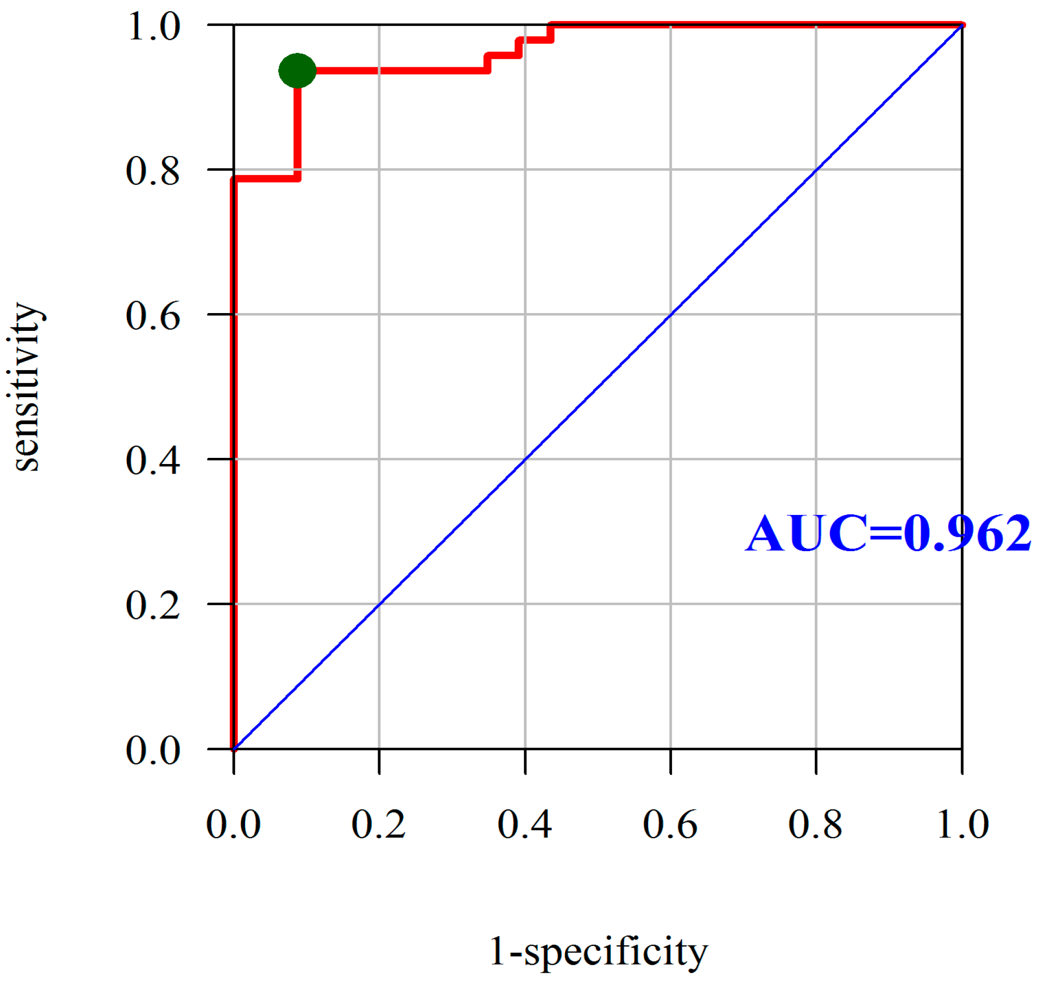
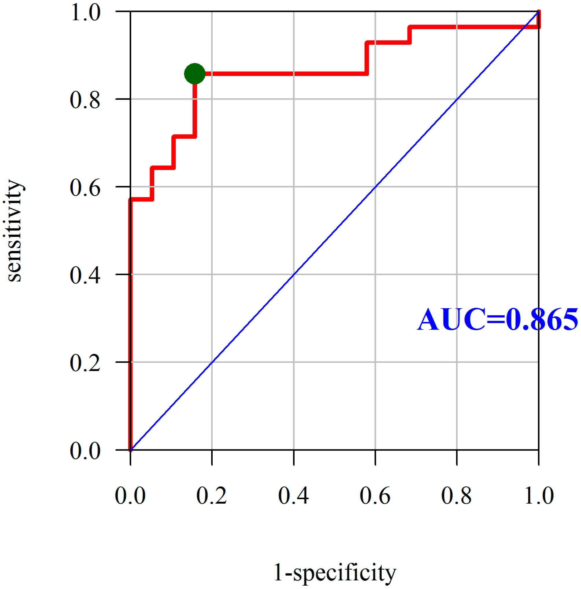
2. Results
3. Discussion
4. Materials and Methods
4.1. Blood Samples
4.2. Theoretical Introduction to the Method
4.3. Determination of MDA
4.4. Details of Determination
4.5. Statistical Analysis
Author Contributions
Funding
Conflicts of Interest
Abbreviations
| AUC | area under the ROC curve |
| BHT | butylated hydroxytoluene |
| CD | Crohn’s disease |
| CDAI | Crohn’s disease activity index |
| CHOD/POD method | liquid cholesterol method |
| CRP | C-reactive protein |
| Hb | hemoglobin |
| HDL | high-density lipoprotein |
| HF | heart failure |
| Ht | hematocrit |
| IBD | inflammatory bowel disease |
| IHD | ischemic heart disease |
| IL | interleukin |
| LDL | low-density lipoprotein |
| LVAD | left ventricular assist device |
| MDA | malondialdehyde |
| PLT | platelet count |
| PON-1 | paraoxonase-1 |
| RA | rheumatoid arthritis |
| RNS | reactive nitrogen species |
| ROS | reactive oxygen species |
| TBA | thiobarbituric acid |
| TBARS | thiobarbituric acid reactive substances |
| TC | total cholesterol |
| TCA | trichloroacetic acid |
| TG | triglyceride |
| TNF-α | tumor necrosis factor |
| VLDL | very low density lipoprotein |
| WBC | white blood cell count |
References
- Tun, G.S.; Cripps, S.; Lobo, A.J. Crohn’s disease: Management in adults, children and young people–concise guidance. Clin. Med. 2018, 18, 231–236. [Google Scholar] [CrossRef] [PubMed]
- You, J.Y. Features and management of very early onset inflammatory bowel disease. Zhongguo Dang Dai Er Ke Za Zhi 2018, 20, 341–345. [Google Scholar] [PubMed]
- Ozhegov, E.V.; Zhivotova, E.Y.; Lebedko, O.A.; Fleishman, M.Y.; Alexeenko, S.A.; Timoshin, S.S. Intensity of proliferative processes and degree of oxidative stress in the mucosa of the ileum in Crohn’s disease. Bull. Exp. Biol. Med. 2012, 152, 420–423. [Google Scholar] [CrossRef] [PubMed]
- Szczeklik, K.; Krzyściak, W.; Cibor, D.; Domagała-Rodacka, R.; Pytko-Polończyk, J.; Mach, T.; Owczarek, D. Markers of lipid peroxidation and antioxidant status in the serum and saliva of patients with active Crohn disease. Pol. Arch. Intern. Med. 2018, 128, 362–370. [Google Scholar] [PubMed]
- Miljkovic, M.; Stefanovic, A.; Simic-Ogrizovic, S.; Vekic, J.; Bogavac-Stanojevic, N.; Cerne, D.; Kocbek, P.; Marc, J.; Jelic-Ivanovic, Z.; Spasojevic-Kalimanovska, V.; et al. Association of Dyslipidemia, Oxidative Stress, and Inflammation with Redox Status in VLDL, LDL, and HDL Lipoproteins in Patients with Renal Disease. Angiology 2018, 3319718780041. [Google Scholar] [CrossRef] [PubMed]
- Santos-Gallego, C.G.; Badimon, J.J.; Rosenson, R.S. Beginning to understand high-density lipoproteins. Endocrinol. Metab. Clin. North. Am. 2014, 43, 913–947. [Google Scholar] [CrossRef] [PubMed]
- Costa, L.G.; Vitalone, A.; Cole, T.B.; Furlong, C.E. Modulation of paraoxonase (PON1) activity. Biochem. Pharmacol. 2005, 69, 541–550. [Google Scholar] [CrossRef] [PubMed]
- Van Lenten, B.J.; Hama, S.Y.; de Beer, F.C.; Stafforini, D.M.; McIntyre, T.M.; Prescott, S.M.; La Du, B.N.; Fogelman, A.M.; Navab, M. Anti-inflammatory HDL becomes pro-inflammatory during the acute phase response. Loss of protective effect of HDL against LDL oxidation in aortic wall cell cocultures. J. Clin. Investig. 1995, 96, 2758–2767. [Google Scholar] [CrossRef] [PubMed]
- Santos-Gallego, C.G.; Giannarelli, C.; Badimón, J.J. Experimental models for the investigation of high-density lipoprotein-mediated cholesterol efflux. Curr. Atheroscler. Rep. 2011, 13, 266–276. [Google Scholar] [CrossRef] [PubMed]
- Borovkova, E.; Antipova, N.V.; Komeenko, T.V.; Shakhparonov, M.I.; Borovkov, I.M. Paraoxonase: The Universal Factor of Antioxidant Defense in Human Body. Vestn. Ross. Akad. Med. Nauk. 2017, 72, 5–10. [Google Scholar] [CrossRef] [PubMed]
- Kirchgesner, J.; Beaugerie, L.; Carrat, F.; Andersen, NN.; Jess, T.; Schwarzinger, M.; BERENICE study group. Increased risk of acute arterial events in young patients and severely active IBD: A nationwide French cohort study. Gut 2018, 67, 1261–1268. [Google Scholar] [CrossRef] [PubMed]
- Voutilainen, M.; Hutri-Kähönen, N.; Tossavainen, P.; Sipponen, T.; Pitkänen, N.; Laitinen, T.; Jokinen, E.; Rönnemaa, T.; Viikari, J.S.A.; Raitakari, O.T.; et al. Low childhood high density lipoprotein cholesterol levels and subsequent risk for chronic inflammatory bowel disease. Dig. Liver Dis. 2018, 50, 348–352. [Google Scholar] [CrossRef] [PubMed]
- Nakhaie, M.R.; Koor, B.E.; Salehi, S.O.; Karimpour, F. Prediction of cardiovascular disease risk using framingham risk score among office workers, Iran, 2017. Saudi J. Kidney Dis. Transplant. 2018, 29, 608–614. [Google Scholar]
- Koutroumpakis, E.; Ramos-Rivers, C.; Regueiro, M.; Hashash, J.G.; Barrie, A.; Swoger, J.; Baidoo, L.; Schwartz, M.; Dunn, M.A.; Koutroubakis, I.E.; et al. Association Between Long-Term Lipid Profiles and Disease Severity in a Large Cohort of Patients with Inflammatory Bowel Disease. Dig. Dis. Sci. 2016, 61, 865–871. [Google Scholar] [CrossRef] [PubMed]
- Vest, A.R.; Kennel, P.J.; Maldonado, D.; Young, J.B.; Mountis, M.M.; Naka, Y.; Colombo, P.C.; Mancini, D.M.; Starling, R.C.; Schulze, P.C. Recovery of Serum Cholesterol Predicts Survival After Left Ventricular Assist Device Implantation. Circ. Heart Fail. 2016, 9, e002881. [Google Scholar] [CrossRef] [PubMed]
- Wu, P.; Jia, F.; Zhang, B.; Zhang, P. Risk of cardiovascular disease in inflammatory bowel disease. Exp. Ther. Med. 2017, 13, 395–400. [Google Scholar] [CrossRef] [PubMed]
- Filimon, A.M.; Negreanu, L.; Doca, M.; Ciobanu, A.; Preda, C.M.; Vinereanu, D. Cardiovascular involvement in inflammatory bowel disease: Dangerous liaisons. World J. Gastroenterol. 2015, 21, 9688–9692. [Google Scholar] [CrossRef] [PubMed]
- Myasoedova, E.; Crowson, C.; Kremers, H.M.; Roger, V.L.; Fitz-Gibbon, P.D.; Therneau, T.M.; Gabriel, S.E. Lipid paradox in rheumatoid arthritis: The impact of serum lipid measures and systemic inflammation on the risk of cardiovascular disease. Ann. Rheum. Dis. 2011, 70, 482–487. [Google Scholar] [CrossRef] [PubMed]
- Rauchhaus, M.; Clark, A.L.; Doehner, W.; Davos, C.; Bolger, A.; Sharma, R.; Coats, A.J.; Anker, S.D. The relationship between cholesterol and survival in patients with chronic heart failure. J. Am. Coll. Cardiol. 2003, 42, 1933–1940. [Google Scholar] [CrossRef] [PubMed]
- Guder, G.; Frantz, S.; Bauersachs, J.; Allolio, B.; Wanner, C.; Koller, M.T.; Ertl, G.; Angermann, C.E.; Störk, S. Reverse epidemiology in systolic and nonsystolic heart failure: Cumulative prognostic benefit of classical cardiovascular risk factors. Circ. Heart Fail. 2009, 2, 563–571. [Google Scholar] [CrossRef] [PubMed]
- Schatz, I.J.; Masaki, K.; Yano, K.; Chen, R.; Rodriguez, B.L.; Curb, J.D. Cholesterol and all-cause mortality in elderly people from the Honolulu Heart Program: A cohort study. Lancet 2001, 358, 351–355. [Google Scholar] [CrossRef]
- Iribarren, C.; Reed, D.M.; Chen, R.; Yano, K.; Dwyer, J.H. Low serum cholesterol and mortality. Which is the cause and which is the effect? Circulation 1995, 92, 2396–2403. [Google Scholar] [CrossRef] [PubMed]
- Cappello, M.; Licata, A.; Calvaruso, V.; Bravatà, I.; Aiello, A.; Torres, D.; Corte, V.D.; Tuttolomondo, A.; Perticone, M.; Licata, G.; et al. Increased expression of markers of early atherosclerosis in patients with inflammatory bowel disease. Eur. J. Intern. Med. 2017, 37, 83–89. [Google Scholar] [CrossRef] [PubMed]
- Bjerrum, J.T.; Steenholdt, C.; Ainsworth, M.; Nielsen, O.H.; Reed, M.A.; Atkins, K.; Günther, U.L.; Hao, F.; Wang, Y. Metabonomics uncovers a reversible proatherogenic lipid profile during infliximab therapy of inflammatory bowel disease. BMC Med. 2017, 15, 184. [Google Scholar] [CrossRef] [PubMed]
- Rodríguez-Carrio, J.; Mozo, L.; López, P.; Nikiphorou, E.; Suárez, A. Anti-High-Density Lipoprotein Antibodies and Antioxidant Dysfunction in Immune-Driven Diseases. Front. Med. 2018, 5, 114. [Google Scholar] [CrossRef] [PubMed]
- Boehm, D.; Krzystek-Korpacka, M.; Neubauer, K.; Matusiewicz, M.; Berdowska, I.; Zielinski, B.; Paradowski, L.; Gamian, A. Paraoxonase-1 status in Crohn’s disease and ulcerative colitis. Inflamm. Bowel. Dis. 2009, 15, 93–99. [Google Scholar] [CrossRef] [PubMed]
- Alves, J.D.; Ames, P.R. Atherosclerosis, oxidative stress and auto-antibodies in systemic lupus erythematosus and primary antiphospholipid syndrome. Immunobiology 2003, 207, 23–28. [Google Scholar] [CrossRef] [PubMed]
- Ibrahim, A.A.; El-Lebedy, D.; Ashmawy, I.; Hady, M.A. Association between paraoxonase-1 gene Q192R and L55M polymorphisms in systemic lupus erythematosus (SLE) and anti-phospholipid syndrome (APS) in a population from Cairo of Egypt. Clin. Rheumatol. 2017, 36, 1305–1310. [Google Scholar] [CrossRef] [PubMed]
- Stanisavljevic, N.; Stojanovich, L.; Marisavljevic, D.; Djokovic, A.; Dopsaj, V.; Kotur-Stevuljevic, J.; Martinovic, J.; Memon, L.; Radovanovic, S.; Todic, B.; et al. Lipid peroxidation as risk factor for endothelial dysfunction in antiphospholipid syndrome patients. Clin. Rheumatol. 2016, 35, 2485–2493. [Google Scholar] [CrossRef] [PubMed]
- Charakida, M.; Besler, C.; Batuca, J.R.; Sangle, S.; Marques, S.; Sousa, M.; Wang, G.; Tousoulis, D.; Alves, J.D.; Loukogeorgakis, S.P.; et al. Vascular abnormalities, paraoxonase activity, and dysfunctional HDL in primary antiphospholipid syndrome. JAMA 2009, 302, 1210–1217. [Google Scholar] [CrossRef] [PubMed]
- Zhu, H.; Zhao, T.; Liu, J. Role of Paraoxonase 1 Activity and Oxidative/Antioxidative Stress Markers in Patients with Acute Cerebral Infarction. Clin. Lab. 2018, 64, 1049–1053. [Google Scholar] [CrossRef] [PubMed]
- Boehm, D.; Krzystek-Korpacka, M.; Neubauer, K.; Matusiewicz, M.; Paradowski, L.; Gamian, A. Lipid peroxidation markers in Crohn’s disease: The associations and diagnostic value. Clin. Chem. Lab. Med. 2012, 50, 1359–1366. [Google Scholar] [CrossRef] [PubMed]
- Maor, I.; Rainis, T.; Lanir, A.; Lavy, A. Oxidative stress, inflammation and neutrophil superoxide release in patients with Crohn’s disease: Distinction between active and non-active disease. Dig. Dis. Sci. 2008, 53, 2208–2214. [Google Scholar] [CrossRef] [PubMed]
- Tanimoto, N.; Kumon, Y.; Suehiro, T.; Ohkubo, S.; Ikeda, Y.; Nishiya, K.; Hashimoto, K. Serum paraoxonase activity decreases in rheumatoid arthritis. Life Sci. 2003, 72, 2877–2885. [Google Scholar] [CrossRef]
- Kumon, Y.; Suehiro, T.; Ikeda, Y.; Hashimoto, K. Human paraoxonase-1 gene expression by HepG2 cells is downregulated by interleukin-1beta and tumor necrosis factor-alpha, but is upregulated by interleukin-6. Life Sci. 2003, 73, 2807–2815. [Google Scholar] [CrossRef]
- Popa, C.; van Tits, L.J.; Barrera, P.; Lemmers, H.L.; van den Hoogen, F.H.; van Riel, P.L.; Radstake, T.R.D.J.; Netea, M.G.; Roest, M.; Stalenhoef, A.F.H. Anti-inflammatory therapy with tumor necrosis factor alpha inhibitors improves high-density lipoprotein cholesterol antioxidative capacity in rheumatoid arthritis patients. Ann. Rheum. Dis. 2009, 68, 868–872. [Google Scholar] [CrossRef] [PubMed]
- Gür, M.; Yıldız, A.; Demirbağ, R.; Yilmaz, R.; Aslan, M.; Özdoğru, I.; Erel, O. Paraoxonase and arylesterase activities in patients with cardiac syndrome X, and their relationship with oxidative stress markers. Coron. Artery Dis. 2007, 18, 89–95. [Google Scholar] [CrossRef] [PubMed]
- Viktorinova, A.; Jurkovicova, I.; Fabryova, L.; Kinova, S.; Koren, M.; Stecova, A.; Svitekova, K. Abnormalities in the relationship of paraoxonase 1 with HDL and apolipoprotein A1 and their possible connection to HDL dysfunctionality in type 2 diabetes. Diabetes Res. Clin. Pract. 2018, 140, 174–182. [Google Scholar] [CrossRef] [PubMed]
- Späh, F. Inflammation in atherosclerosis and psoriasis: Common pathogenic mechanisms and the potential for an integrated treatment approach. Br. J. Dermatol. 2008, 159, 10–17. [Google Scholar] [CrossRef] [PubMed]
- Duan, L.; Rao, X.; Braunstein, Z.; Toomey, A.C.; Zhong, J. Role of Incretin Axis in Inflammatory Bowel Disease. Front. Immunol. 2017, 8, 1734. [Google Scholar] [CrossRef] [PubMed]
- Üstün, Y.; Kilincalp, S.; Çoban, Ş.; Coşkun, Y.; Yüksel, İ.; Ongun, A.; Soykan, İ.; Bektaş, M.; Törüner, M.; Çetinkaya, H.; et al. Evaluation of Early Atherosclerosis Markers in Patients with Inflammatory Bowel Disease. Med. Sci. Monit. 2016, 22, 3943–3950. [Google Scholar] [CrossRef] [PubMed]
- Khan, S.Y.; Awad, E.M.; Oszwald, A.; Mayr, M.; Yin, X.; Waltenberger, B.; Stuppner, H.; Lipovac, M.; Uhrin, P.; Breuss, J. Premature senescence of endothelial cells upon chronic exposure to TNFα can be prevented by N-acetyl cysteine and plumericin. Sci. Rep. 2017, 7, 39501. [Google Scholar] [CrossRef] [PubMed]
- Caliskan, Z.; Gokturk, H.S.; Caliskan, M.; Gullu, H.; Ciftci, O.; Ozgur, G.T.; Guven, A.; Selcuk, H. Impaired coronary microvascular and left ventricular diastolic function in patients with inflammatory bowel disease. Microvasc. Res. 2015, 97, 25–30. [Google Scholar] [CrossRef] [PubMed]
- Rief, P.; Raggam, R.B.; Avian, A.; Hafner, F.; Sareban, M.; Wehrschuetz, M.; Brodmann, M.; Gary, T. Low mean platelet volume is associated with critical limb ischemia in peripheral arterial occlusive disease. Sci. Rep. 2018, 8, 6718. [Google Scholar] [CrossRef] [PubMed]
- Santos-Gallego, C.G.; Vahl, T.P.; Goliasch, G.; Picatoste, B.; Arias, T.; Ishikawa, K.; Njerve, I.U.; Sanz, J.; Narula, J.; Sengupta, P.P.; et al. Sphingosine-1-Phosphate Receptor Agonist Fingolimod Increases Myocardial Salvage and Decreases Adverse Postinfarction Left Ventricular Remodeling in a Porcine Model of Ischemia/Reperfusion. Circulation 2016, 133, 954–966. [Google Scholar] [CrossRef] [PubMed]
- Di Sabatino, A.; Santilli, F.; Guerci, M.; Simeone, P.; Ardizzone, S.; Massari, A.; Giuffrida, P.; Tripaldi, R.; Malara, A.; Liani, R.; Gurini, E.; et al. Oxidative stress and thromboxane-dependent platelet activation in inflammatory bowel disease: Effects of anti-TNF-α treatment. Thromb. Haemost. 2016, 116, 486–495. [Google Scholar] [PubMed]
- Sansanayudh, N.; Numthavaj, P.; Muntham, D.; Yamwong, S.; McEvoy, M.; Attia, J.; Sritara, P.; Thakkinstian, A. Prognostic effect of mean platelet volume in patients with coronary artery disease. A systematic review and meta-analysis. Thromb. Haemost. 2015, 114, 1299–1309. [Google Scholar] [CrossRef] [PubMed]
- Choi, D.H.; Kang, S.H.; Song, H. Mean platelet volume: A potential biomarker of the risk and prognosis of heart disease. Korean J. Intern. Med. 2016, 31, 1009–1017. [Google Scholar] [CrossRef] [PubMed]
- Ayas, Z.Ö.; Can, U. Alteration of mean platelet volume in the pathogenesis of acute ischemic stroke: Cause or consequence? Ideggyogy. Szel. 2018, 71, 49–56. [Google Scholar] [CrossRef] [PubMed]
- Baskol, G.; Baskol, M.; Yurci, A.; Ozbakir, O.; Yucesoy, M. Serum paraoxonase 1 activity and malondialdehyde levels in patients with ulcerative colitis. Cell. Biochem. Funct. 2006, 24, 283–286. [Google Scholar] [CrossRef] [PubMed]
- Tavori, H.; Aviram, M.; Khatib, S.; Musa, R.; Mannheim, D.; Karmeli, R.; Vaya, J. Paraoxonase 1 protects macrophages from atherogenicity of a specific triglyceride isolated from human carotid lesion. Free Radic. Biol. Med. 2011, 51, 234–242. [Google Scholar] [CrossRef] [PubMed]
- Gugliucci, A.; Menini, T. Paraoxonase 1 and HDL maturation. Clin. Chim. Acta 2015, 439, 5–13. [Google Scholar] [CrossRef] [PubMed]
- Karban, A.; Hartman, C.; Eliakim, R.; Waterman, M.; Nesher, S.; Barnett-Griness, O.; Shamir, R. Paraoxonase (PON)1 192R allele carriage is associated with reduced risk of inflammatory bowel disease. Dig. Dis. Sci. 2007, 52, 2707–2715. [Google Scholar] [CrossRef] [PubMed]
- Best, W.R.; Becktel, J.M.; Singleton, J.W.; Kern, F. Development of a Crohn’s disease activity index. National Cooperative Crohn’s Disease Study. Gastroenterology 1976, 70, 439–444. [Google Scholar] [PubMed]
- Walsh, A.; Bryant, R.V.; Travis, S.P. Current best practice for disease activity assessment in IBD. Nat. Rev. Gastroenterol. Hepatol. 2016, 13, 567–579. [Google Scholar] [CrossRef] [PubMed]
- Yoshida, E. The Crohn’s Disease Activity Index, its derivatives and the Inflammatory Bowel Disease Questionnaire: A review of instruments to assess Crohn’s disease. Can. J. Gastroenterol. 1999, 13, 65–73. [Google Scholar] [CrossRef] [PubMed]
- Eckerson, H.W.; Romson, J.; Wyte, C.; La Du, B.N. The human serum paraoxonase polymorphism: Identification of phenotypes by their response to salts. Am. J. Hum. Genet. 1983, 35, 214–227. [Google Scholar] [PubMed]
- Aust, S.D. Thiobarbituric acid assay reactants. In Methods in Toxicology, Vol. 1B: In Vitro Toxicity Indicators; Tyson, C.A., Frazier, J.M., Eds.; Academic Press: San Diego, CA, USA, 1994; pp. 367–376. [Google Scholar]
- Hinkle, D.E.; Wiersma, W.; Jurs, S.G. Applied Statistics for the Behavioral Sciences, 5th ed.; Houghton Mifflin: Boston, MA, USA, 2003. [Google Scholar]
- R Core Team. R: A Language and Environment for Statistical Computing; R Foundation for Statistical Computing: Vienna, Austria, 2017. [Google Scholar]
Sample Availability: Samples of the compounds are not available from the authors. |


| Features | Control Group | CD Group | p * | |||
|---|---|---|---|---|---|---|
| n | % | n | % | |||
| Gender | Women | 13 | 52.00% | 26 | 44.83% | 0.718 χ2 |
| Men | 12 | 48.00% | 32 | 55.17% | ||
| Features | Control Group | CD Group | p ** | |||
| Mean (SD) | Median (quartiles) | Mean (SD) | Median (quartiles) | |||
| Age (years) | 34.16 (9.84) | 30 (26–45) | 35.6 (12.69) | 34 (26.25–40.75) | 0.8 NP | |
| BMI (kg/m2) | 23.49 (3.26) | 22.5 (21.2–25.3) | 22.76 (3.35) | 22.7 (20.33–24.4) | 0.36 P | |
| WBC (G/L) | 5.7 (1.09) | 5.5 (5–6.7) | 7.39 (3.83) | 6.88 (5.11–8.06) | 0.022 NP | |
| Hb (g/dL) | 14.4 (1.37) | 14 (13.5–15.2) | 12.74 (1.8) | 12.95 (11.25–14.2) | <0.001 P | |
| Ht (%) | 42.32 (3.35) | 41.7 (39.7–43.8) | 38.87 (4.39) | 38.95 (35.82–41.9) | 0.001 NP | |
| PLT (G/L) | 258.72 (45.29) | 250 (224–285) | 313.5 (116.54) | 288 (239.25–358.75) | 0.048 NP | |
| CRP (mg/L) | 1.26 (1.21) | 0.76 (0.29–2.41) | 13.51 (15.98) | 6.87 (2.09–17.75) | <0.001 NP | |
| MDA (nmol/g) | 2.88 (0.8) | 2.64 (2.25–3.64) | 6.83 (3.61) | 5.48 (4.17–8.57) | <0.001 NP | |
| TC (mmol/L) | 4.73 (0.39) | 4.88 (4.47–4.98) | 4.35 (0.66) | 4.23 (4.05–4.84) | 0.005 NP | |
| HDL (mmol/L) | 1.14 (0.14) | 1.19 (1.02–1.25) | 0.87 (0.25) | 0.96 (0.62–1.07) | <0.001 NP | |
| LDL (mmol/L) | 2.95 (0.38) | 2.97 (2.79–3.18) | 2.38 (0.49) | 2.25 (1.93–2.81) | <0.001 NP | |
| TG (mmol/L) | 2.76 (0.27) | 2.79 (2.57–2.95) | 1.85 (0.31) | 1.9 (1.64–2.12) | <0.001 NP | |
| Groups | PON-1 (U/L) | p * | |||||||
|---|---|---|---|---|---|---|---|---|---|
| N | Mean | SD | Median | Min | Max | Q1 | Q3 | ||
| A-Control | 23 | 402.56 | 27.73 | 405.31 | 336.57 | 440.42 | 387.59 | 423.36 | <0.001 |
| B-Inactive CD | 19 | 330.47 | 40.03 | 325.24 | 243.13 | 401.36 | 309.54 | 358.16 | A > BCD |
| C-Small active CD | 14 | 262.56 | 63.22 | 281.53 | 156.24 | 352.15 | 209.25 | 301.02 | B > D |
| D-Moderately active CD | 14 | 195.02 | 73.57 | 179.38 | 124.31 | 405.24 | 147.51 | 198.03 | |
| Parameter | Correlation of (PON-1) Activity | |||
|---|---|---|---|---|
| Correlation Coefficient * | p | Dependence | The Power of Dependence | |
| Age (years) | −0.044 | 0.716 | - | - |
| BMI | 0.197 | 0.103 | - | - |
| WBC | −0.262 | 0.029 | negative | very weak |
| Hb | 0.539 | <0.001 | positive | average |
| Ht | 0.48 | <0.001 | positive | weak |
| PLT | −0.326 | 0.006 | negative | weak |
| CRP | −0.61 | <0.001 | negative | average |
| MDA | −0.924 | <0.001 | negative | very strong |
| TC | 0.343 | 0.004 | positive | weak |
| HDL | 0.536 | <0.001 | positive | average |
| LDL | 0.54 | <0.001 | positive | average |
| TG | 0.561 | <0.001 | positive | average |
© 2018 by the authors. Licensee MDPI, Basel, Switzerland. This article is an open access article distributed under the terms and conditions of the Creative Commons Attribution (CC BY) license (http://creativecommons.org/licenses/by/4.0/).
Share and Cite
Szczeklik, K.; Mach, T.; Cibor, D.; Owczarek, D.; Sapa, J.; Papież, M.; Pytko-Polończyk, J.; Krzyściak, W. Correlation of Paraoxonase-1 with the Severity of Crohn’s Disease. Molecules 2018, 23, 2603. https://doi.org/10.3390/molecules23102603
Szczeklik K, Mach T, Cibor D, Owczarek D, Sapa J, Papież M, Pytko-Polończyk J, Krzyściak W. Correlation of Paraoxonase-1 with the Severity of Crohn’s Disease. Molecules. 2018; 23(10):2603. https://doi.org/10.3390/molecules23102603
Chicago/Turabian StyleSzczeklik, Katarzyna, Tomasz Mach, Dorota Cibor, Danuta Owczarek, Jacek Sapa, Monika Papież, Jolanta Pytko-Polończyk, and Wirginia Krzyściak. 2018. "Correlation of Paraoxonase-1 with the Severity of Crohn’s Disease" Molecules 23, no. 10: 2603. https://doi.org/10.3390/molecules23102603
APA StyleSzczeklik, K., Mach, T., Cibor, D., Owczarek, D., Sapa, J., Papież, M., Pytko-Polończyk, J., & Krzyściak, W. (2018). Correlation of Paraoxonase-1 with the Severity of Crohn’s Disease. Molecules, 23(10), 2603. https://doi.org/10.3390/molecules23102603