A New Triterpenoid from Teucrium viscidum


Abstract
:1. Introduction
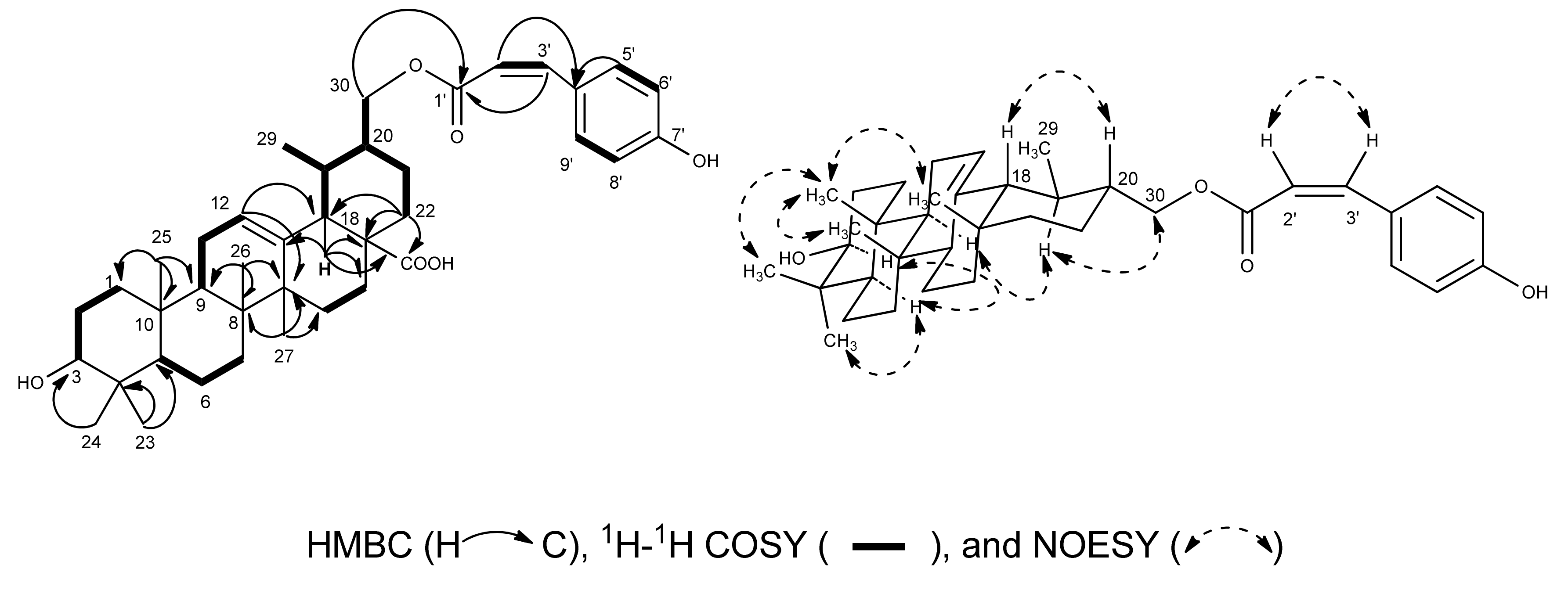
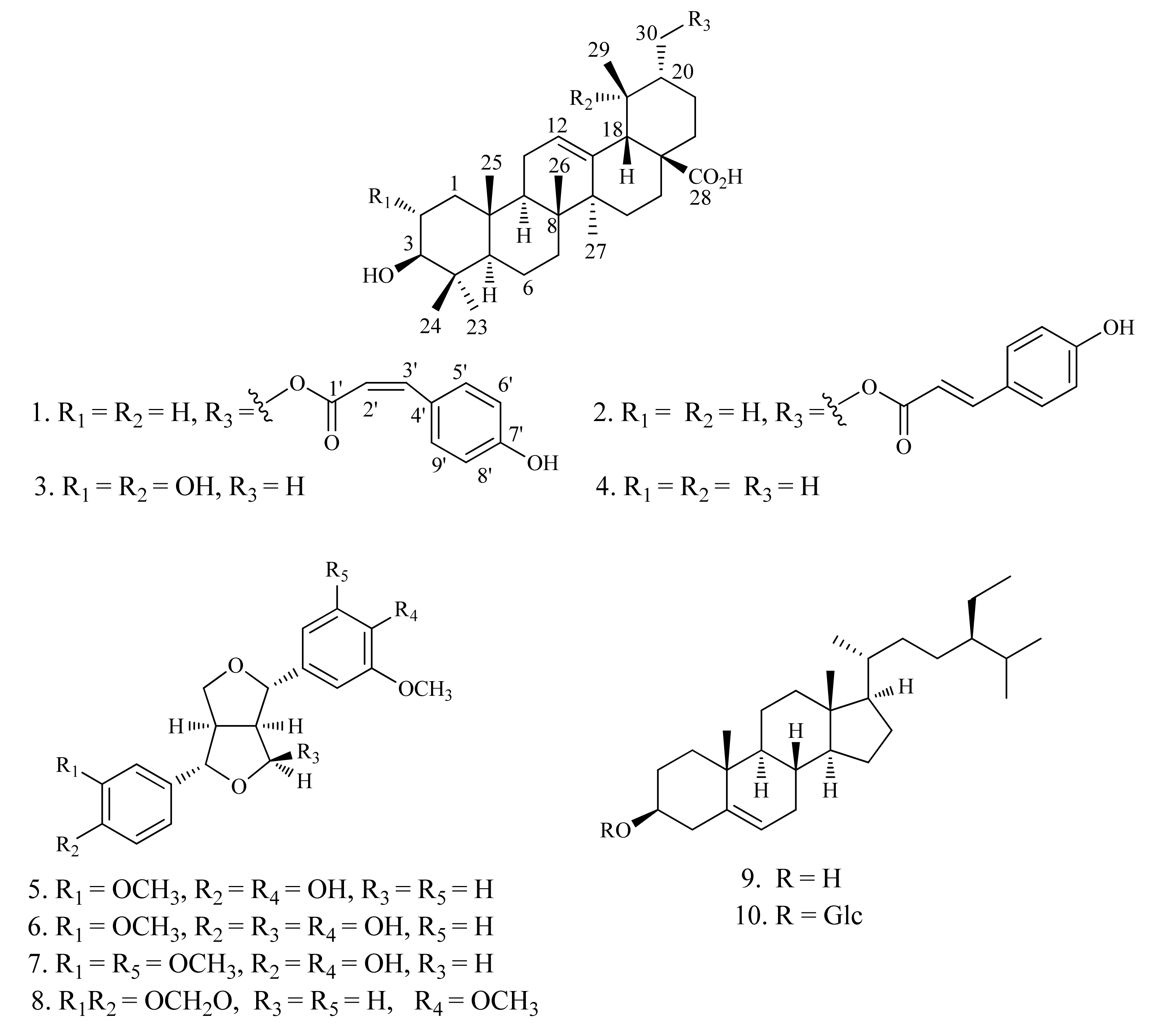
2. Results and Discussion
3. Experimental
3.1. General Procedures
3.2. Plant Material
3.3. Extraction and Isolation
3.4. 11β-HSD1 Inhibitory Assays
4. Conclusions
Supplementary Materials
Acknowledgments
References
- State Administration of Traditional Chinese Medicine. Zhong Hua Ben Cao; Shanghai Science and Technology Publishing Company: Shanghai, China, 1999; pp. 6239–6240. [Google Scholar]
- Chen, Y.P.; Li, C.M.; Sun, H.D. The diterpenoid from Teucrium viscidum. Acta Botanica Yunnanica 1990, 12, 110. [Google Scholar]
- Fujita, E.; Uchida, I.; Fujita, T.; Masaki, N.; Osaki, K. Teucvin, a novel furanoid norditerpene from Teucrium uiscidum var. miquelianum. J. Chem. Soc. Chem. Commun. 1973, 20, 793–794. [Google Scholar] [CrossRef]
- Node, M.; Sai, M.; Fujita, E. Isolation of the diterpenoid teuflin (6-epiteucvin) from Teucrium viscidum var. miquelianum. Phytochemistry 1981, 20, 757–760. [Google Scholar] [CrossRef]
- Uchida, I.; Fujita, T.; Fujita, E. Terpenoids-XXXIV: Teucvidin, a minor norditerpene from Teucrium viscidum var. miquelianum. Tetrahedron 1975, 31, 841–848. [Google Scholar] [CrossRef]
- Siddiqui, B.S.; Begum, S. Two triterpenoids from the leaves of Plumeria obtusa. Phytochemistry 1999, 52, 1111–1115. [Google Scholar] [CrossRef]
- Jang, D.S.; Kim, J.M.; Lee, G.Y.; Kim, J.; Kim, J.S. Ursane-type triterpenoids from the aerial parts of Potentilla discolor. Agric. Chem. Biotechnol. 2006, 49, 48–50. [Google Scholar]
- Cowan, S.; Stewart, M.; Abbiw, D.K.; Latif, Z.; Sarker, S.D.; Nash, R.J. Lignans from Strophanthus gratus. Fitoterapia 2001, 72, 80–82. [Google Scholar] [CrossRef]
- Li, Y.S.; Wang, Z.T.; Zhang, M.; Luo, S.D.; Chen, J.J. A new pinoresinol-type lignan from Ligularia kanaitizensis. Nat. Prod. Res. 2005, 19, 125–129. [Google Scholar] [CrossRef] [PubMed]
- Khan, K.A.; Shoeb, A. A lignan from Lonicera hypoleuca. Phytochemistry 1985, 24, 628–630. [Google Scholar] [CrossRef]
- Latip, J.; Hartley, T.G.; Waterman, P.G. Lignans and coumarins metabolites from Melicopehayesii. Phytochemistry 1999, 51, 107–110. [Google Scholar] [CrossRef]
- Al-Qudah, M.A.; Zarga, M.H.A. Chemical constituents of Sisymbrium irio L. from Jordan. Nat. Prod. Res. 2010, 24, 448–456. [Google Scholar] [CrossRef] [PubMed]
- Thuong, P.T.; Lee, C.H.; Dao, T.T.; Nguyen, P.H.; Kim, W.G.; Lee, S.J.; Oh, W.K. Triterpenoids from the leaves of Diospyros kaki (Persimmon) and their inhibitory effects on protein tyrosine phosphatase 1B. J. Nat. Prod. 2008, 71, 1775–1778. [Google Scholar] [CrossRef] [PubMed]
- Feng, Y.; Huang, S.L.; Dou, W.; Zhang, S.; Chen, J.H.; Shen, Y.; Shen, J.H.; Leng, Y. Emodin, a natural product, selectively inhibits 11β-hydroxysteroid dehydrogenase type 1 and ameliorates metabolic disorder in diet-induced obese mice. Br. J. Pharmacol. 2010, 161, 113–126. [Google Scholar] [CrossRef] [PubMed]
- Rollinger, J.M.; Kratschmar, D.V.; Schuster, D.; Pfisterer, P.H.; Gumy, C.; Aubry, E.M.; Brandstoter, S.; Stuppner, H.; Wolber, G.; Odermatt, A. 11β-Hydroxysteroid dehydrogenase 1 inhibiting constituents from Eriobotrya japonica revealed by bioactivity-guided isolation and computational approaches. Bioorg. Med. Chem. 2010, 18, 1507–1515. [Google Scholar] [CrossRef] [PubMed]
- Yang, H.; Dou, W.; Lou, J.; Leng, Y.; Shen, J. Discovery of novel inhibitors of 11β-hydroxysteroid dehydrogenase type 1 by docking and pharmacophore modeling. Bioorg. Med. Chem. Lett. 2008, 18, 1340–1345. [Google Scholar] [CrossRef] [PubMed]
Sample Availability: Not available. |


| NO. | δH | δC | NO. | δH | δC |
|---|---|---|---|---|---|
| 1α | 1.00 m | 39.6 | 20β | 1.43 m | 44.4 |
| 1β | 1.58 m | ||||
| 2 | 1.86 overlap | 28.6 | 21α | 1.62 m | 26.1 |
| 21β | 1.79 m | ||||
| 3α | 3.48 dd (6.8, 9.2) | 78.6 | 22α | 2.07 overlap | 37.4 |
| 22β | 1.96 overlap | ||||
| 4 | 39.9 | 23 | 1.26 s | 29.3 | |
| 5α | 0.88 m | 56.3 | 24 | 1.05 s | 17.0 |
| 6α | 1.59 m | 19.3 | 25 | 0.92 s | 16.2 |
| 6β | 1.39 m | ||||
| 7α | 1.57 overlap | 34.1 | 26 | 1.08 s | 17.9 |
| 7β | 1.39 overlap | ||||
| 8 | 42.9 | 27 | 1.23 s | 24.4 | |
| 9α | 1.65 m | 48.5 | 28 | 180.2 | |
| 10 | 37.8 | 29 | 1.09 d (6.0) | 17.6 | |
| 11α | 1.64 m | 24.1 | 30a | 4.50 dd (3.2, 11.2) | 68.2 |
| 11β | 1.97 m | 30b | 4.27 dd (7.2, 11.2) | ||
| 12 | 5.52 t (3.2) | 126.7 | 1' | 167.5 | |
| 13 | 139.3 | 2' | 6.09 d (12.4) | 116.9 | |
| 14 | 40.5 | 3' | 7.03 d (12.4) | 144.5 | |
| 15α | 1.25 overlap | 29.1 | 4' | 127.1 | |
| 15β | 2.35 m | ||||
| 16α | 2.14 m | 25.4 | 5', 9' | 8.09 d (8.4) | 134.0 |
| 16β | 2.04 m | ||||
| 17 | 48.3 | 6', 8' | 7.22 d (8.4) | 116.5 | |
| 18β | 2.7 d (11.6) | 53.8 | 7' | 161.1 | |
| 19α | 1.84 overlap | 34.9 |
© 2013 by the authors. This article is an open access article distributed under the terms and conditions of the Creative Commons Attribution license (http://creativecommons.org/licenses/by/3.0/).
Share and Cite
Hao, X.; Zhang, J.; Xia, G.; Xue, Y.; Luo, Z.; Si, Y.; Yao, G.; Zhang, Y. A New Triterpenoid from Teucrium viscidum. Molecules 2013, 18, 1262-1269. https://doi.org/10.3390/molecules18011262
Hao X, Zhang J, Xia G, Xue Y, Luo Z, Si Y, Yao G, Zhang Y. A New Triterpenoid from Teucrium viscidum. Molecules. 2013; 18(1):1262-1269. https://doi.org/10.3390/molecules18011262
Chicago/Turabian StyleHao, Xincai, Jinwen Zhang, Guangxin Xia, Yongbo Xue, Zengwei Luo, Yanyan Si, Guangmin Yao, and Yonghui Zhang. 2013. "A New Triterpenoid from Teucrium viscidum" Molecules 18, no. 1: 1262-1269. https://doi.org/10.3390/molecules18011262
APA StyleHao, X., Zhang, J., Xia, G., Xue, Y., Luo, Z., Si, Y., Yao, G., & Zhang, Y. (2013). A New Triterpenoid from Teucrium viscidum. Molecules, 18(1), 1262-1269. https://doi.org/10.3390/molecules18011262