
Bioactive Glass—An Extensive Study of the Preparation and Coating Methods


 and
and
Abstract
:1. Introduction
2. The Mechanism of Bone Tissue Formation on the Surface of the Bioactive Glass
3. Methods for Obtaining Bioactive Glass
4. Bioactive Glass Deposition Methods
4.1. Enameling
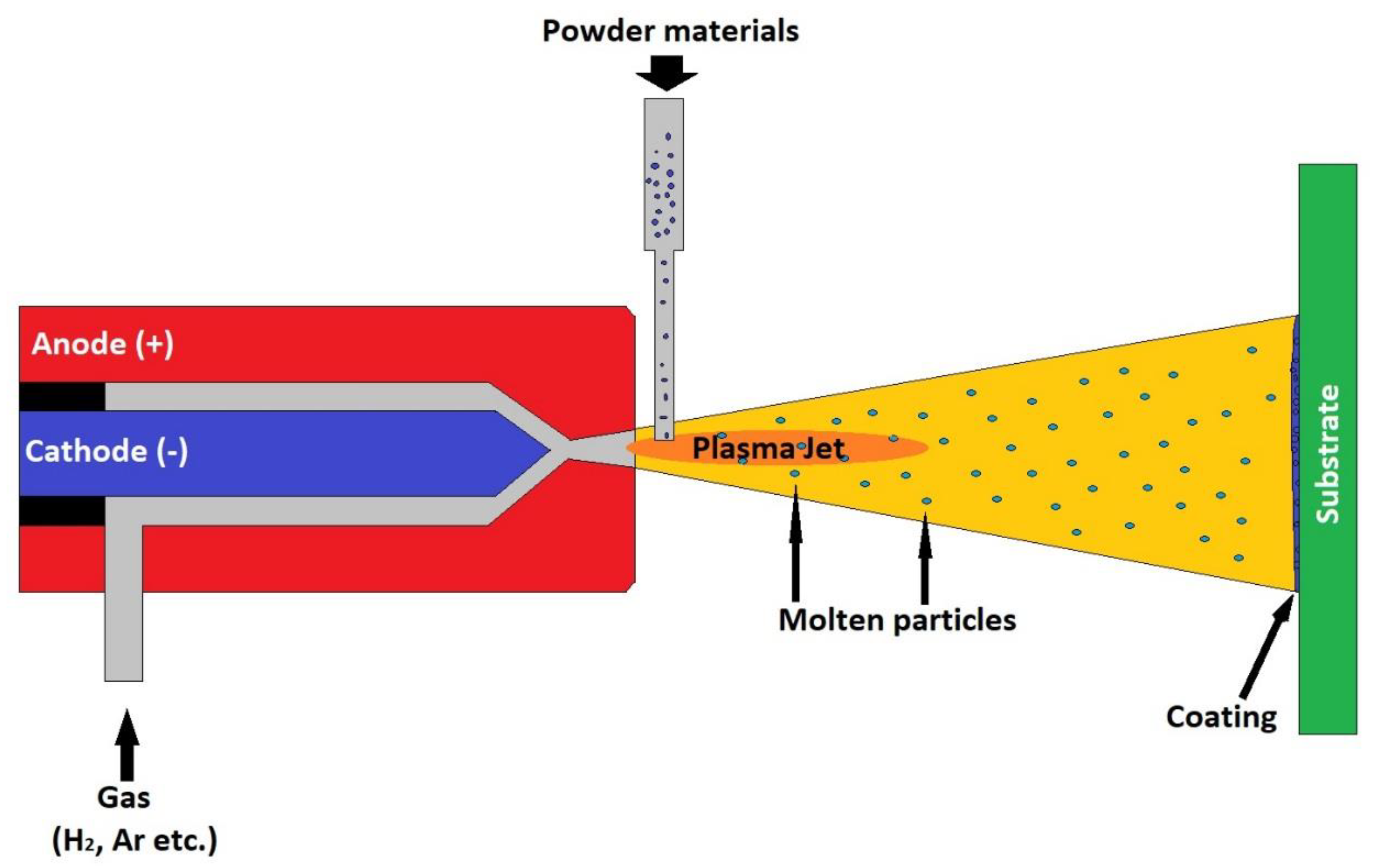
4.2. Thermal Spraying
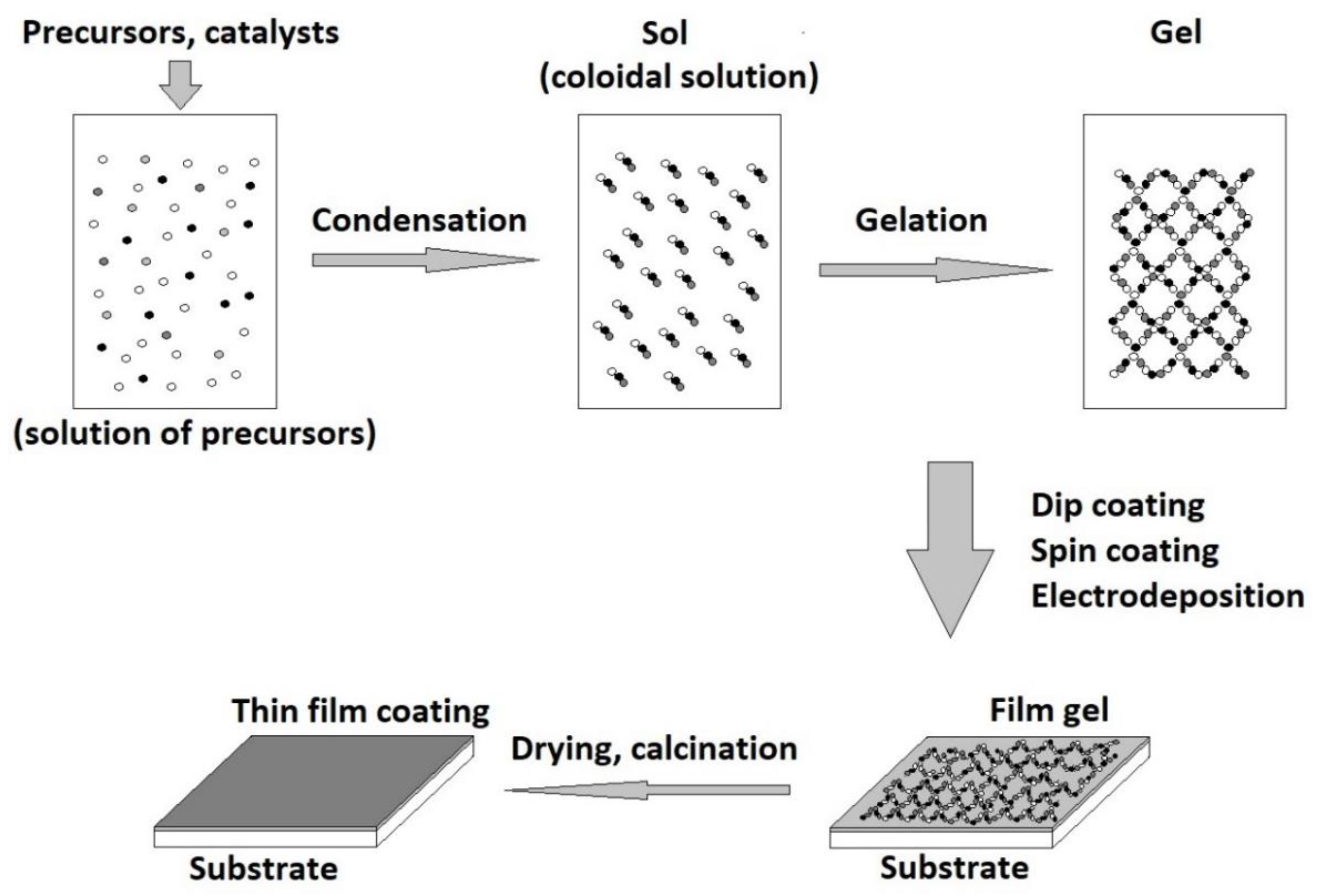
4.3. Sol-Gel Deposition Technique
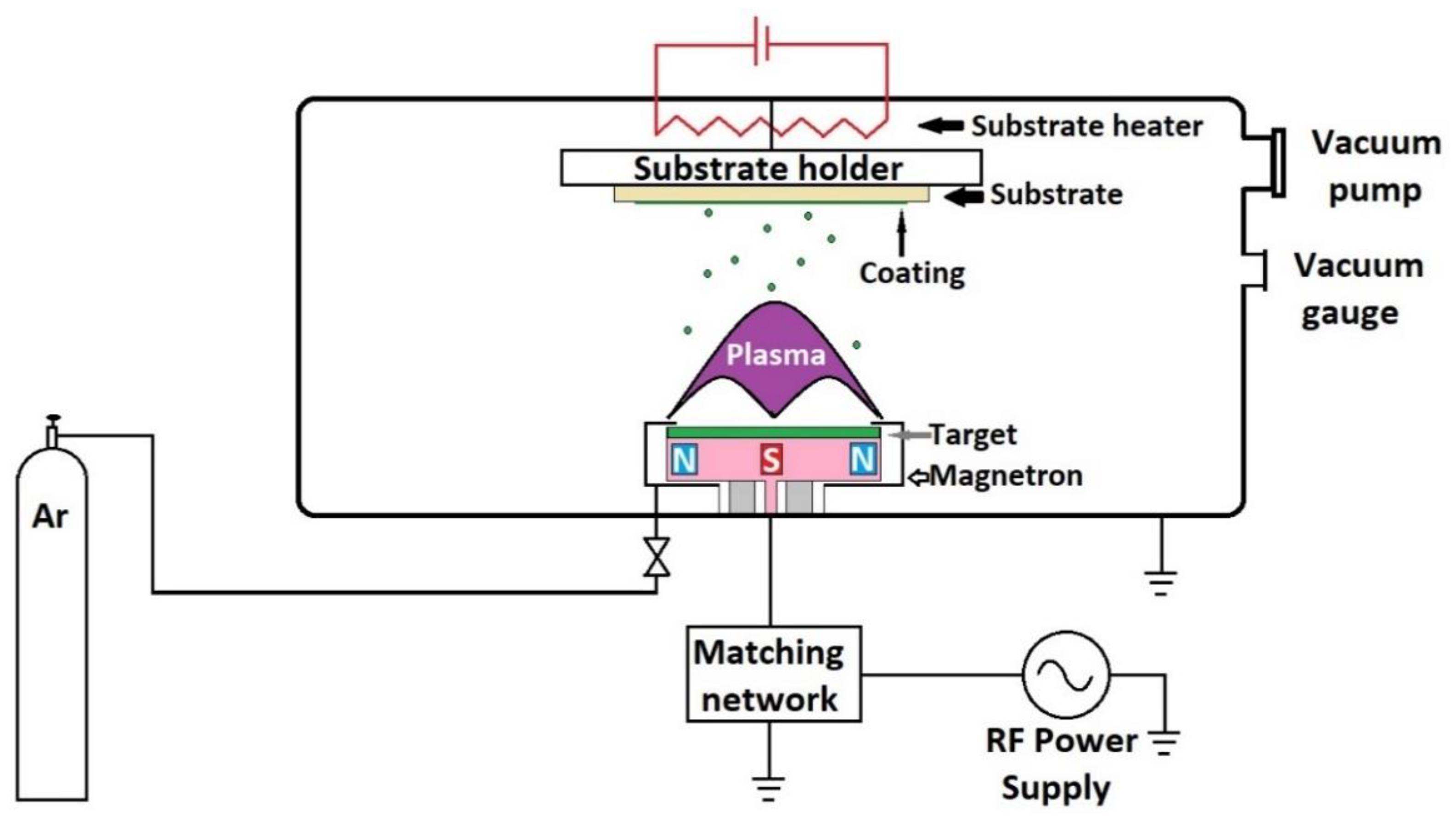
4.4. Radio Frequency Magnetron Sputtering
4.5. Laser Cladding
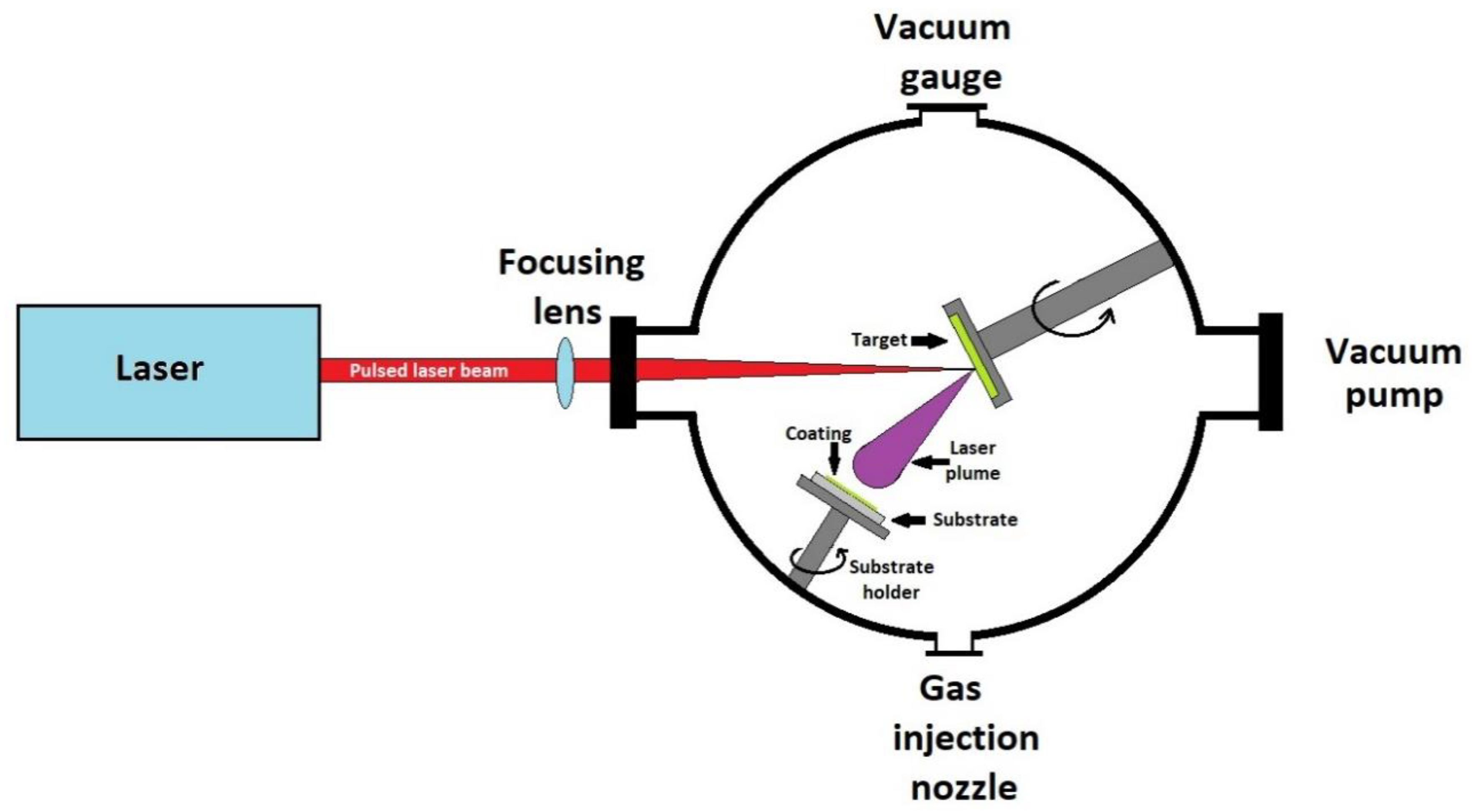
4.6. Pulsed Laser Ablation and Deposition
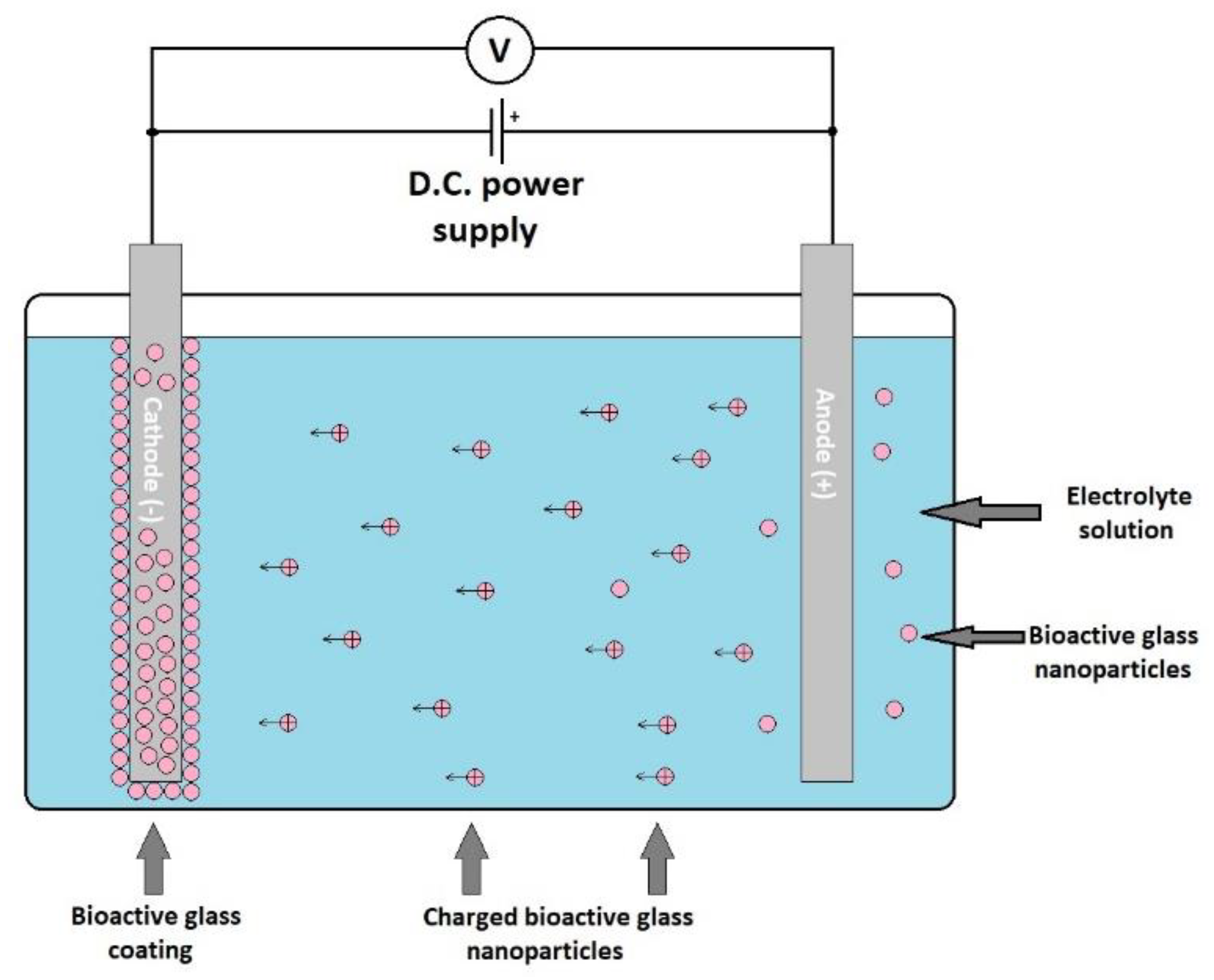
4.7. Electrophoretic Deposition
4.8. Other Bioactive Glass Coating Techniques
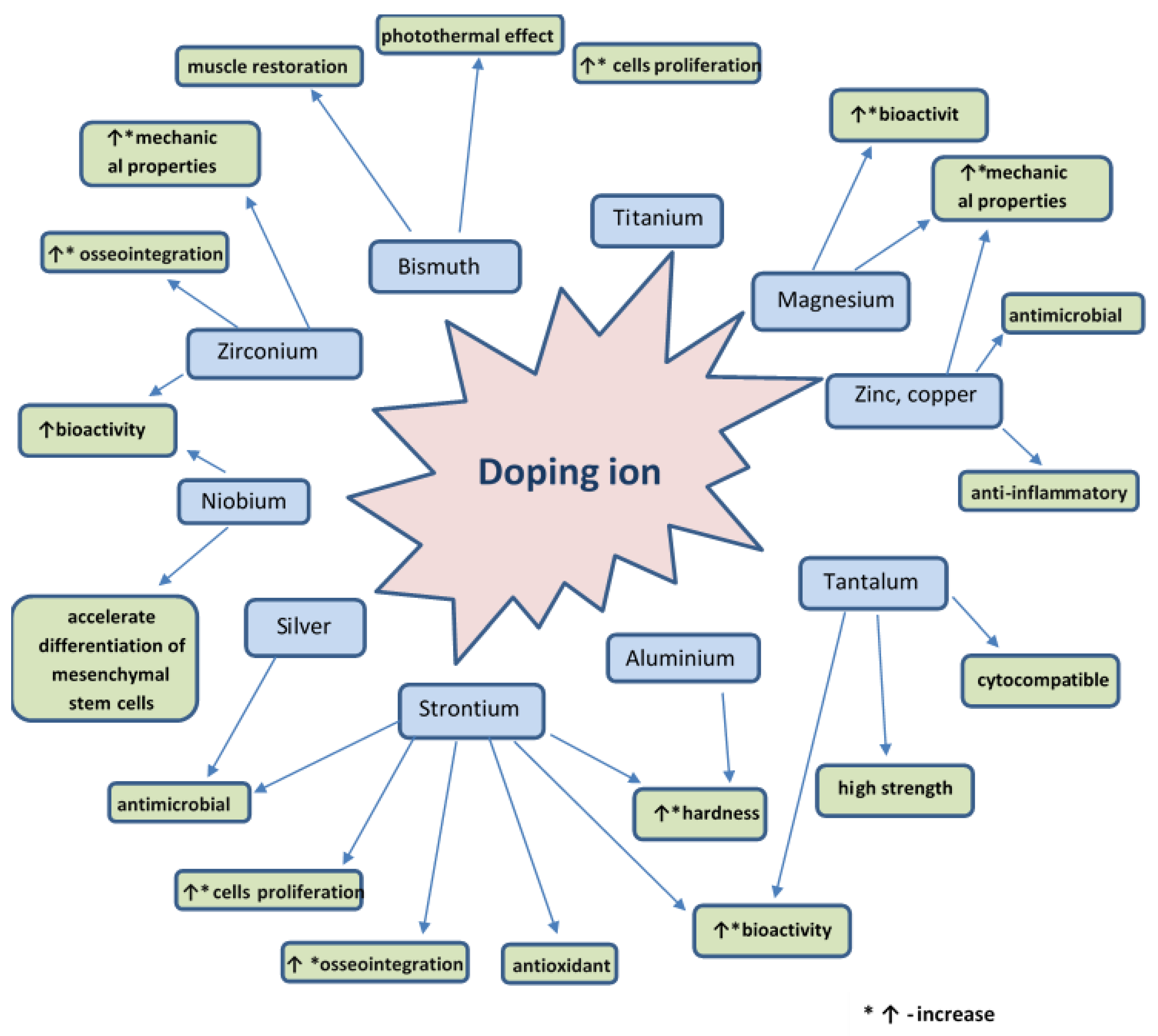
5. Doping Agents
6. Conclusions and Perspectives
Author Contributions
Funding
Institutional Review Board Statement
Informed Consent Statement
Data Availability Statement
Conflicts of Interest
References
- Mesquita-Guimaraes, J.; Henriques, B.; Silva, F.S. Bioactive glass coatings. In Bioactive Glasses. Materials, Properties and Applications; Ylanen, H., Ed.; Woodhead Publishing Series in Biomaterials; Woodhead Publishing: Duxford, UK, 2018; pp. 103–118. [Google Scholar] [CrossRef]
- Sola, A.; Bellucci, D.; Cannillo, V.; Cattini, A. Bioactive glass coatings: A review. Surf. Eng. 2011, 27, 560–572. [Google Scholar] [CrossRef]
- Hench, L.L. The story of Bioglass (R). J. Mater. Sci.-Mater. Med. 2006, 17, 967–978. [Google Scholar] [CrossRef]
- Greenspan, D.C. Glass and medicine: The larry hench story. Int. J. Appl. Glass Sci. 2016, 7, 134–138. [Google Scholar] [CrossRef]
- Moritz, N.; Vallittu, P.K. Bioactive silicate glass in implantable medical devices: From research to clinical applications. In Bioactive Glasses. Fundamentals, Technology and Applications; Aldo, R., Boccaccini, D.S.B., Hupa, L., Eds.; The Royal Society of Chemistry: Lomdon, UK, 2017; pp. 442–470. [Google Scholar]
- Bahrami, M.S.; Eshghinejad, P.; Bakhsheshi-Rad, H.R.; Karamian, E.; Chen, X.B. Electrophoretic deposition of bioglass/graphene oxide composite on Ti-alloy implants for improved antibacterial and cytocompatible properties. Mater. Technol. 2020, 35, 69–74. [Google Scholar] [CrossRef]
- Ranga, N.; Poonia, E.; Jakhar, S.; Sharma, A.K.; Kumar, A.; Devi, S.; Duhan, S. Enhanced antimicrobial properties of bioactive glass using strontium and silver oxide nanocomposites. J. Asian Ceram. Soc. 2019, 7, 75–81. [Google Scholar] [CrossRef] [Green Version]
- Seuss, S.; Heinloth, M.; Boccaccini, A.R. Development of bioactive composite coatings based on combination of PEEK, bioactive glass and Ag nanoparticles with antibacterial properties. Surf. Coat. Technol. 2016, 301, 100–105. [Google Scholar] [CrossRef]
- Echezarreta-Lopez, M.M.; Landin, M. Using machine learning for improving knowledge on antibacterial effect of bioactive glass. Int. J. Pharm. 2013, 453, 641–647. [Google Scholar] [CrossRef]
- Allan, I.; Newman, H.; Wilson, M. Antibacterial activity of particulate Bioglass (R) against supra- and subgingival bacteria. Biomaterials 2001, 22, 1683–1687. [Google Scholar] [CrossRef]
- Krishnan, V. Future Applications of Bioglass. Adv. Struct. Mater. 2016, 53, 317–336. [Google Scholar] [CrossRef]
- Allan, I.; Newman, H.; Wilson, M. Particulate Bioglass (R) reduces the viability of bacterial biofilms formed on its surface in an in vitro model. Clin. Oral Implants Res. 2002, 13, 53–58. [Google Scholar] [CrossRef] [PubMed] [Green Version]
- Wheeler, D.L.; Montfort, M.J.; McLoughlin, S.W. Differential healing response of bone adjacent to porous implants coated with hydroxyapatite and 45S5 bioactive glass. J. Biomed. Mater. Res. 2001, 55, 603–612. [Google Scholar] [CrossRef]
- Migonney, V. History of biomaterials. In Biomaterials; Migonney, V., Ed.; John Wiley & Sons, Inc.: Hoboken, NJ, USA, 2014; pp. 1–10. [Google Scholar] [CrossRef]
- Hench, L.L. Bioactive Glass: Chronology, Characterization, and Genetic Control of Tissue Regeneration. In Advances in Calcium Phosphate Biomaterials; Ben-Nissan, B., Ed.; Springer Series in Biomaterials Science and Engineering; Springer: Berlin/Heidelberg, Germany, 2014; Volume 2, pp. 51–70. [Google Scholar] [CrossRef]
- Amaral, M.; Abreu, C.S.; Oliveira, F.J.; Gomes, J.R.; Silva, R.F. Biotribological performance of NCD coated Si3N4-bioglass composites. Diam. Relat. Mater. 2007, 16, 790–795. [Google Scholar] [CrossRef]
- Dominguez-Trujillo, C.; Ternero, F.; Rodriguez-Ortiz, J.A.; Pavon, J.J.; Montealegre-Melendez, I.; Arevalo, C.; Garcia-Moreno, F.; Torres, Y. Improvement of the balance between a reduced stress shielding and bone rit ingrowth by bioactive coatings onto porous titanium substrates. Surf. Coat. Technol. 2018, 338, 32–37. [Google Scholar] [CrossRef]
- Ananth, K.P.; Suganya, S.; Mangalaraj, D.; Ferreira, J.M.F.; Balamurugan, A. Electrophoretic bilayer deposition of zirconia and reinforced bioglass system on Ti6Al4V for implant applications: An in vitro investigation. Mater. Sci. Eng. C 2013, 33, 4160–4166. [Google Scholar] [CrossRef]
- Li, Z.; Khun, N.W.; Tang, X.Z.; Liu, E.J.; Khor, K.A. Mechanical, tribological and biological properties of novel 45S5 Bioglass (R) composites reinforced with in situ reduced graphene oxide. J. Mech. Behav. Biomed. 2017, 65, 77–89. [Google Scholar] [CrossRef] [PubMed]
- Keranen, P.; Moritz, N.; Alm, J.J.; Ylanen, H.; Kommonen, B.; Aro, H.T. Bioactive glass microspheres as osteopromotive inlays in macrotextured surfaces of Ti and CoCr alloy bone implants: Trapezoidal surface grooves without inlay most efficient in resisting torsional forces. J. Mech. Behav. Biomed. 2011, 4, 1483–1491. [Google Scholar] [CrossRef]
- Drnovsek, N.; Novak, S.; Dragin, U.; Ceh, M.; Gorensek, M.; Gradisar, M. Bioactive glass enhances bone ingrowth into the porous titanium coating on orthopaedic implants. Int. Orthop. 2012, 36, 1739–1745. [Google Scholar] [CrossRef] [Green Version]
- Babu, M.M.; Rao, P.V.; Veeraiah, N.; Prasad, P.S. Effect of Al3+ ions substitution in novel zinc phosphate glasses on formation of HAp layer for bone graft applications. Colloid Surf. B 2020, 185, 110591. [Google Scholar] [CrossRef]
- Popa, A.C.; Stan, G.E.; Husanu, M.A.; Mercioniu, I.; Santos, L.F.; Fernandes, H.R.; Ferreira, J.M.F. Bioglass implant-coating interactions in synthetic physiological fluids with varying degrees of biomimicry. Int. J. Nanomed. 2017, 12, 683–707. [Google Scholar] [CrossRef] [Green Version]
- Profeta, A.C.; Prucher, G.M. Bioactive-glass in periodontal surgery and implant dentistry. Dent. Mater. J. 2015, 34, 559–571. [Google Scholar] [CrossRef] [Green Version]
- Jebahi, S.; Oudadesse, H.; Ben Saleh, G.; Saoudi, M.; Mesadhi, S.; Rebai, T.; Keskes, H.; el Feki, A.; el Feki, H. Chitosan-based bioglass composite for bone tissue healing: Oxidative stress status and antiosteoporotic performance in a ovariectomized rat model. Korean J. Chem. Eng. 2014, 31, 1616–1623. [Google Scholar] [CrossRef]
- Jebahi, S.; Oudadesse, H.; el Feki, H.; Rebai, T.; Keskes, H.; Pellen, P.; el Feki, A. Antioxidative/oxidative effects of strontium-doped bioactive glass as bone graft. In vivo assays in ovariectomised rats. J. Appl. Biomed. 2012, 10, 195–209. [Google Scholar] [CrossRef] [Green Version]
- Price, N.; Bendall, S.P.; Frondoza, C.; Jinnah, R.H.; Hungerford, D.S. Human osteoblast-like cells (MG63) proliferate on a bioactive glass surface. J. Biomed. Mater. Res. 1997, 37, 394–400. [Google Scholar] [CrossRef]
- Choi, A.H.; Ben-Nissan, B.; Matinlinna, J.P.; Conway, R.C. Current Perspectives: Calcium Phosphate Nanocoatings and Nanocomposite Coatings in Dentistry. J. Dent. Res. 2013, 92, 853–859. [Google Scholar] [CrossRef]
- Xuereb, M.; Camilleri, J.; Attard, N.J. Systematic Review of Current Dental Implant Coating Materials and Novel Coating Techniques. Int. J. Prosthodont. 2015, 28, 51–59. [Google Scholar] [CrossRef] [PubMed] [Green Version]
- Baino, F.; Potestio, I. Special Applications of Bioactive Glasses in Otology and Ophthalmology. Adv. Struct. Mater. 2016, 53, 227–248. [Google Scholar] [CrossRef]
- Hench, L.L. Chronology of Bioactive Glass Development and Clinical Applications. New J. Glass Ceram. 2013, 3, 30885. [Google Scholar] [CrossRef] [Green Version]
- Cao, W.P.; Hench, L.L. Bioactive materials. Ceram. Int. 1996, 22, 493–507. [Google Scholar] [CrossRef]
- Oonishi, H.; Hench, L.L.; Wilson, J.; Sugihara, F.; Tsuji, E.; Matsuura, M.; Kin, S.; Yamamoto, T.; Mizokawa, S. Quantitative comparison of bone growth behavior ingranules of Bioglasst, A-W glass-ceramic, and hydroxyapatite. J. Biomed. Mater. Res. 2000, 51, 37–46. [Google Scholar] [CrossRef]
- Xynos, I.D.; Edgar, A.J.; Buttery, L.D.K.; Hench, L.L.; Polak, J.M. Gene-expression profiling of human osteoblasts following treatment with the ionic products of Bioglass 45S5 dissolution. J. Biomed. Mater. Res. 2001, 55, 151–157. [Google Scholar] [CrossRef]
- Baino, F.; Yamaguchi, S. The Use of Simulated Body Fluid (SBF) for Assessing Materials Bioactivity in the Context of Tissue Engineering: Review and Challenges. Biomimetics 2020, 5, 57. [Google Scholar] [CrossRef]
- Catauro, M.; Papale, F.; Bollino, F. Coatings of titanium substrates with xCaO center dot (1−x)SiO2 sol-gel materials: Characterization, bioactivity and biocompatibility evaluation. Mater. Sci. Eng. C 2016, 58, 846–851. [Google Scholar] [CrossRef]
- Manavitehrani, I.; Fathi, A.; Wang, Y.W.; Maitz, P.K.; Mirmohseni, F.; Cheng, T.L.; Peacock, L.; Little, D.G.; Schindeler, A.; Dehghani, F. Fabrication of a Biodegradable Implant with Tunable Characteristics for Bone Implant Applications. Biomacromolecules 2017, 18, 1736–1746. [Google Scholar] [CrossRef]
- Wang, L.P.; Long, N.J.; Li, L.H.; Lu, Y.; Li, M.; Cao, J.K.; Zhang, Y.; Zhang, Q.Y.; Xu, S.H.; Yang, Z.M.; et al. Multi-functional bismuth-doped bioglasses: Combining bioactivity and photothermal response for bone tumor treatment and tissue repair. Light Sci. Appl. 2018, 7, 1. [Google Scholar] [CrossRef] [PubMed] [Green Version]
- Granito, R.N.; Renno, A.C.; Ravagnani, C.; Bossini, P.S.; Mochiuti, D.; Jorgetti, V.; Driusso, P.; Peitl, O.; Zanotto, E.D.; Parizotto, N.A.; et al. In vivo biological performance of a novel highly bioactive glass-ceramic (Biosilicate (R)): A biomechanical and histomorphometric study in rat tibial defects. J. Biomed. Mater. Res. B 2011, 97b, 139–147. [Google Scholar] [CrossRef] [PubMed]
- El-Meliegy, E.; Hamzawy, E.M.A.; El-Kady, A.M.; Salama, A.; El-Rashedi, A. Development and bioactivity evaluation of bioglasses with low Na2O content based on the system Na2O-CaO-MgO-P2O5-SiO2. J. Mater. Sci. Mater. Med. 2012, 23, 2069–2080. [Google Scholar] [CrossRef]
- Rivadeneira, J.; Gorustovich, A. Bioactive glasses as delivery systems for antimicrobial agents. J. Appl. Microbiol. 2017, 122, 1424–1437. [Google Scholar] [CrossRef] [Green Version]
- Kundu, B.; Nandi, S.K.; Dasgupta, S.; Datta, S.; Mukherjee, P.; Roy, S.; Singh, A.K.; Mandal, T.K.; Das, P.; Bhattacharya, R.; et al. Macro-to-micro porous special bioactive glass and ceftriaxone-sulbactam composite drug delivery system for treatment of chronic osteomyelitis: An investigation through in vitro and in vivo animal trial. J. Mater. Sci. Mater. Med. 2011, 22, 705–720. [Google Scholar] [CrossRef]
- Tan, Y.B.; Wang, X.H.; Wu, Q.H.; Yan, W.Q. Early peri-implant osteogenesis with functionally graded nanophase hydroxyapatite/bioglass coating on Ti alloys. Key Eng. Mater. 2007, 330, 553–556. [Google Scholar] [CrossRef]
- Tite, T.; Popa, A.C.; Chirica, I.M.; Stuart, B.W.; Galca, A.C.; Balescu, L.M.; Popescu-Pelin, G.; Grant, D.M.; Ferreira, J.M.F.; Stan, G.E. Phosphate bioglass thin-films: Cross-area uniformity, structure and biological performance tailored by the simple modification of magnetron sputtering gas pressure. Appl. Surf. Sci. 2021, 541, 148640. [Google Scholar] [CrossRef]
- Chakraborty, J.; Sengupta, S.; Ray, S.; Ghosh, S.; Kapoor, R.; Gouri, S.P.; Pande, G.; Datta, S. Multifunctional gradient coatings of phosphate-free bioactive glass on SS316L biomedical implant materials for improved fixation. Surf. Coat. Technol. 2014, 240, 437–443. [Google Scholar] [CrossRef]
- Lin, F.H.; Huang, Y.Y.; Hon, M.H.; Wu, S.C. Fabrication and Biocompatibility of a Porous Bioglass Ceramic in a Na2o-Cao-Sio2-P2o5 System. J. Biomed. Eng. 1991, 13, 328–334. [Google Scholar] [CrossRef]
- Koller, G.; Cook, R.J.; Thompson, I.D.; Watson, T.F.; Di Silvio, L. Surface modification of titanium implants using bioactive glasses with air abrasion technologies. J. Mater. Sci. Mater. Med. 2007, 18, 2291–2296. [Google Scholar] [CrossRef]
- Skallevold, H.E.; Rokaya, D.; Khurshid, Z.; Zafar, M.S. Bioactive Glass Applications in Dentistry. Int. J. Mol. Sci. 2019, 20, 5960. [Google Scholar] [CrossRef] [Green Version]
- De Aza, P.N.; De Aza, A.H.; Pena, P.; De Aza, S. Bioactive glasses and glass-ceramics. Bol.-Soc. Esp. Ceram. Vidr. 2007, 46, 45–55. [Google Scholar] [CrossRef]
- Chen, Q.Z.; Blaker, J.J.; Boccaccini, A.R. Bioactive and mechanically strong Bioglass (R)-poly(D,L-lactic acid) composite coatings on surgical sutures. J. Biomed. Mater. Res. B 2006, 76b, 354–363. [Google Scholar] [CrossRef]
- Han, I.; Lee, I.S.; Choi, J.H.; Baik, H.K. Thinfilm deposition and characteristics of calcium-silicates bioglass. Key Eng. Mater. 2007, 343, 649–652. [Google Scholar] [CrossRef]
- Singh, R.K.; Srinivasan, A.; Kothiyal, G.P. Evaluation of CaO-SiO2-P2O5-Na2O-Fe2O3 bioglass-ceramics for hyperthermia application. J. Mater. Sci. Mater. Med. 2009, 20, 147–151. [Google Scholar] [CrossRef]
- Bhakta, S.; Faira, P.E.; Salata, L.A.; Neto, P.J.D.; Miller, C.A.; van Noort, R.; Reaney, I.M.; Brook, I.M.; Hatton, P.V. Determination of relative in vivo osteoconductivity of modified potassium fluorrichterite glass-ceramics compared with 45S5 bioglass. J. Mater. Sci. Mater. Med. 2012, 23, 2521–2529. [Google Scholar] [CrossRef] [PubMed]
- Haftbaradaran-Esfahani, M.; Ahmadian, M.; Nassajpour-Esfahani, A.H. Fabrication and characterization of porous biomedical Vitallium alloy with 58S bioglass coating prepared by sol-gel method. Appl. Surf. Sci. 2020, 506, 144959. [Google Scholar] [CrossRef]
- Anand, A.; Lalzawmliana, V.; Kumar, V.; Das, P.; Devi, K.B.; Maji, A.K.; Kundu, B.; Roy, M.; Nandi, S.K. Preparation and in vivo biocompatibility studies of different mesoporous bioactive glasses. J. Mech. Behav. Biomed. 2019, 89, 89–98. [Google Scholar] [CrossRef] [PubMed]
- Zhang, P.; Yang, K.; Zhou, Z.Y.; Zhu, X.R.; Li, W.C.; Cao, C.L.; Zhou, K.; Liao, L.; Ai, F.R. Customized Borosilicate Bioglass Scaffolds With Excellent Biodegradation and Osteogenesis for Mandible Reconstruction. Front. Bioeng. Biotech. 2020, 8, 610284. [Google Scholar] [CrossRef] [PubMed]
- Bagherpour, I.; Naghib, S.M.; Yaghtin, A.H. Synthesis and characterisation of nanostructured hardystonite coating on stainless steel for biomedical application. IET Nanobiotechnol. 2018, 12, 895–902. [Google Scholar] [CrossRef]
- Wajda, A.; Sitarz, M. Structural and microstructural studies of zinc-doped glasses from NaCaPO4-SiO2 system. J. Non-Cryst. Solids 2016, 441, 66–73. [Google Scholar] [CrossRef]
- Mozafari, M.; Rabiee, M.; Azami, M.; Maleknia, S. Biomimetic formation of apatite on the surface of porous gelatin/bioactive glass nanocomposite scaffolds. Appl. Surf. Sci. 2010, 257, 1740–1749. [Google Scholar] [CrossRef]
- Dinaryand, P.; Seyedjafari, E.; Shafiee, A.; Jandaghi, A.B.; Doostmohammadi, A.; Fathi, M.H.; Farhadian, S.; Soleimani, M. New Approach to Bone Tissue Engineering: Simultaneous Application of Hydroxyapatite and Bioactive Glass Coated on a Poly(L-lactic acid) Scaffold. ACS Appl. Mater. Interfaces 2011, 3, 4518–4524. [Google Scholar] [CrossRef]
- Sepulveda, P.; Jones, J.R.; Hench, L.L. In vitro dissolution of melt-derived 45S5 and sol-gelderived 58S bioactive glasses. J. Biomed. Mater. Res. 2002, 61, 301–311. [Google Scholar] [CrossRef]
- Biernat, M.; Ciolek, L.; Dzierzynska, M.; Ozieblo, A.; Sawicka, J.; Deptula, M.; Bauer, M.; Kamysz, W.; Pikula, M.; Jaegermann, Z.; et al. Porous chitosan/ZnO-doped bioglass composites as carriers of bioactive peptides. Int. J. Appl. Ceram. Technol. 2020, 17, 2807–2816. [Google Scholar] [CrossRef]
- Theodorou, G.; Goudouri, O.M.; Kontonasaki, E.; Chatzistavrou, X.; Papadopoulou, L.; Kantiranis, N.; Paraskevopoulos, K.M. Comparative bioactivity study of 45S5 and 58S bioglasses in organic and inorganic environment. Bioceram. Dev. Appl. 2011, 1, 1–4. [Google Scholar] [CrossRef]
- Durgalakshmi, D.; Ajay Rakkesh, R.; Aruna, P.; Ganesana, S.; Balakumar, S. Bioactivity and hemocompatibility of sol-gel bioactive glass synthesized under different catalytic conditions. New J. Chem. 2020, 44, 21026–21037. [Google Scholar] [CrossRef]
- Lombardi, M.; Gremillard, L.; Chevalier, J.; Lefebvre, L.; Cacciotti, I.; Bianco, A.; Montanaro, L. A comparative study between melt-derived and sol-gel synthesized 45S5 bioactive glasses. Key Eng. Mater. 2013, 541, 15–30. [Google Scholar] [CrossRef]
- Gouveia, P.F.; Mesquita-Guimaraes, J.; Galarraga-Vinueza, M.E.; Souza, J.C.M.; Silva, F.S.; Fredel, M.C.; Boccaccini, A.R.; Detsch, R.; Henriques, B. In-vitro mechanical and biological evaluation of novel zirconia reinforced bioglass scaffolds for bone repair. J. Mech. Behav. Biomed. 2021, 114, 104164. [Google Scholar] [CrossRef]
- Teghil, R.; Curcio, M.; De Bonis, A. Substituted Hydroxyapatite, Glass, and Glass-Ceramic Thin Films Deposited by Nanosecond Pulsed Laser Deposition (PLD) for Biomedical Applications: A Systematic Review. Coatings 2021, 11, 811. [Google Scholar] [CrossRef]
- Ducheyne, P. Bioglass Coatings and Bioglass Composites as Implant Materials. J. Biomed. Mater. Res. 1985, 19, 273–291. [Google Scholar] [CrossRef] [PubMed]
- Kim, C.Y.; Lee, J.W. Surface bio-modification of titanium implants by an enamel process. J. Ceram. Process. Res. 2005, 6, 338–344. [Google Scholar]
- Bharati, S.; Soundrapandian, C.; Basu, D.; Datta, S. Studies on a novel bioactive glass and composite coating with hydroxyapatite on titanium based alloys: Effect of gamma-sterilization on coating. J. Eur. Ceram. Soc. 2009, 29, 2527–2535. [Google Scholar] [CrossRef]
- Pazo, A.; Saiza, E.; Tomsiaa, P. Silicate glass coatings on Ti-based implants. Acta Mater. 1998, 46, 2551–2558. [Google Scholar] [CrossRef]
- Fuchs, G.A. The Biological and Biomechanical Properties of Metal Implants Coated with Bioglass Ceramic, as Exemplified in the Simple-Model of a Loaded, Cement-Free Total Hip-Prosthesis. Biomed. Tech. 1982, 27, 24–29. [Google Scholar] [CrossRef]
- Lacefield, W.R.; Hench, L.L. The Bonding of Bioglass to a Cobalt-Chromium Surgical Implant Alloy. Biomaterials 1986, 7, 104–108. [Google Scholar] [CrossRef]
- Kudo, K.; Miyasawa, M.; Fujioka, Y.; Kamegai, T.; Nakano, H.; Seino, Y.; Ishikawa, F.; Shioyama, T.; Ishibashi, K. Clinical-Application of Dental Implant with Root of Coated Bioglass-Short-Term Results. Oral Surg. Oral Med. Oral Pathol. 1990, 70, 18–23. [Google Scholar] [CrossRef]
- Andersson, O.H.; Karlsson, K.H.; Hero, H.; Vedel, E.; Yliurpo, A.; Pajamaki, K.J.J.; Lindholm, T.S. Bioactive Double Glass Coatings for Co-Cr-Mo Alloy. J. Mater. Sci. Mater. Med. 1995, 6, 242–247. [Google Scholar] [CrossRef]
- Hench, L.L.; Greenspan, D.C. Bioglass Coated Al2O3 Ceramics. U.S. Patent 4103002, 25 July 1978. [Google Scholar]
- Greenspan, D.C.; Hench, L.L. Chemical and mechanical behavior of Bioglass coated alumina. Biomed. Mater. Res. 1976, 10, 503. [Google Scholar] [CrossRef]
- Griss, P.; Werner, E.; Heimke, G.; Rautekreinsen, U. Comparative Experimental Investigations with Bioglass LL Hench and Al2O3-Ceramic Coated with Mod Bioglass. II. Results of Experiments with Loaded Implants. Arch. Orthop. Traum. Surg. 1978, 92, 199–210. [Google Scholar] [CrossRef]
- Turley, P.K.; Shapiro, P.A.; Moffett, B.C. The Loading of Bioglass-Coated Aluminum-Oxide Implants to Produce Sutural Expansion of the Maxillary Complex in the Pigtail Monkey (Macaca-Nemestrina). Arch. Oral Biol. 1980, 25, 459–469. [Google Scholar] [CrossRef]
- Smith, J.R. Bone Dynamics Associated with the Controlled Loading of Bioglass-Coated Aluminum-Oxide Endosteal Implants. Am. J. Orthod. Dentofac. 1979, 76, 618–636. [Google Scholar] [CrossRef]
- Ignatius, A.; Peraus, M.; Schorlemmer, S.; Augat, P.; Burger, W.; Leyen, S.; Claes, L. Osseointegration of alumina with a bioactive coating under load-bearing and unloaded conditions. Biomaterials 2005, 26, 2325–2332. [Google Scholar] [CrossRef] [PubMed]
- Kirsten, A.; Hausmann, A.; Weber, M.; Fischer, J.; Fischer, H. Bioactive and Thermally Compatible Glass Coating on Zirconia Dental Implants. J. Dent. Res. 2015, 94, 297–303. [Google Scholar] [CrossRef] [Green Version]
- Rohr, N.; Nebe, J.B.; Schmidli, F.; Muller, P.; Weber, M.; Fischer, H.; Fischer, J. Influence of bioactive glass-coating of zirconia implant surfaces on human osteoblast behavior in vitro. Dent. Mater. 2019, 35, 862–870. [Google Scholar] [CrossRef]
- Gomez-Vega, J.M.; Saiz, E.; Tomsia, A.P.; Marshall, G.W.; Marshall, S.J. Bioactive glass coatings with hydroxyapatite and Bioglass (R) particles on Ti-based implants. 1. Processing. Biomaterials 2000, 21, 105–111. [Google Scholar] [CrossRef]
- Peddi, L.; Brow, R.K.; Brown, R.F. Bioactive borate glass coatings for titanium alloys. J. Mater. Sci. Mater. Med. 2008, 19, 3145–3152. [Google Scholar] [CrossRef]
- Mistry, S.; Kundu, D.; Datta, S.; Basu, D. Comparison of bioactive glass coated and hydroxyapatite coated titanium dental implants in the human jaw bone. Aust. Dent. J. 2011, 56, 68–75. [Google Scholar] [CrossRef]
- Bobzin, K.; Kopp, N.; Wiesner, S.; Puidokas, S.; Anavar, S.S.; Fischer, H.; Korsten, A.; Schickle, K. Investigation of a bioactive Ti-Co-based brazing coating on oxide high-performance ceramics in medical technology. Mater. Werkst 2014, 45, 504–511. [Google Scholar] [CrossRef]
- Mistry, S.; Roy, R.; Kundu, B.; Datta, S.; Kumar, M.; Chanda, A.; Kundu, D. Clinical Outcome of Hydroxyapatite Coated, Bioactive Glass Coated, and Machined Ti6Al4V Threaded Dental Implant in Human Jaws: A Short-Term Comparative Study. Implant Dent. 2016, 25, 252–260. [Google Scholar] [CrossRef]
- Rodriguez, O.; Matinmanesh, A.; Phull, S.; Schemitsch, E.H.; Zalzal, P.; Clarkin, O.M.; Papini, M.; Towler, M.R. Silica-Based and Borate-Based, Titania-Containing Bioactive Coatings Characterization: Critical Strain Energy Release Rate, Residual Stresses, Hardness, and Thermal Expansion. J. Funct. Biomater. 2016, 7, 32. [Google Scholar] [CrossRef]
- Fujino, S.; Tokunaga, H.; Hata, S.; Esaiz, A.P.T. Graded glass coatings for Co-Cr implant alloys. J. Mater. Sci. 2005, 40, 2499–2503. [Google Scholar] [CrossRef]
- Henao, J.; Poblano-Salas, C.; Monsalve, M.; Corona-Castuera, J.; Barceinas-Sanchez, O. Bio-active glass coatings manufactured by thermal spray: A status report. J. Mater. Res. Technol. 2019, 8, 4965–4984. [Google Scholar] [CrossRef]
- Fauchais, P.; Vardelle, M.; Vardelle, A.; Goutier, S. What Do We Know, What are the Current Limitations of Suspension Plasma Spraying? J. Therm. Spray Technol. 2015, 24, 1120–1129. [Google Scholar] [CrossRef] [Green Version]
- Jordan Eric, H.; Chen, J.; Maurice, G. The Solution Precursor Plasma Spray (SPPS) Process: A Review with Energy Considerations. J. Therm. Spray Technol. 2015, 24, 1153–1165. [Google Scholar] [CrossRef]
- Karthikeyan, J. The advantages and disadvantages of the cold spray coating process. In The Cold Spray Materials Deposition Process; Woodhead Publishing Series; Woodhead Publishing: Southston, UK, 2007; pp. 62–71. [Google Scholar]
- Fan, W.; Bai, Y. Review of Suspension and Solution Precursor Plasma Sprayed Thermal Barrier Coatings. Ceram. Int. 2016, 42, 14299–14312. [Google Scholar] [CrossRef]
- Killinger, A.; Müller, P.; Gadow, R. What Do We Know, What are the Current Limitations of Suspension HVOF Spraying? J. Therm. Spray Technol. 2015, 24, 1130–1142. [Google Scholar] [CrossRef]
- Monsalvea, M.; Ageorgesa, H.; Lopezb, E.; Vargasb, F.; Bolivar, F. Bioactivity and mechanical properties of plasma-sprayed coatings ofbioglass powder. Surf. Coat. Technol. 2013, 220, 60–66. [Google Scholar] [CrossRef]
- Lindgren, V.; Galea, V.P.; Nebergall, A.; Greene, M.E.; Rolfson, O.; Malchau, H. Radiographic and Clinical Outcomes of Porous Titanium-Coated and Plasma-Sprayed Acetabular Shells. J. Bone Jt. Surg. Am. 2018, 100, 1673–1681. [Google Scholar] [CrossRef] [PubMed]
- Marghussian, V.K.; Mesgar, A.S.-M. Effects of composition on crystallization behaviour and mechanical properties of bioactive glass-ceramics in the MgO-CaO-SiO2-P2O5 system. Ceram. Int. 2000, 26, 415–420. [Google Scholar] [CrossRef]
- Goller, G. The effect of bond coat on mechanical properties ofplasma sprayed bioglass-titanium coatings. Ceram. Int. 2004, 30, 351–355. [Google Scholar] [CrossRef]
- Cannillo, V.; Sola, A. Different approaches to produce coatings with bioactive glasses:Enamelling vs. plasma spraying. J. Eur. Ceram. Soc. 2010, 30, 2031–2039. [Google Scholar] [CrossRef]
- Lopez-Sastre, A.; Gonzalo-Orden, J.M.; Altonaga, J.A.R.; Altonaga, J.R.; Orden, M.A. Coating titanium implants with bioglass and with hydroxyapatite—A comparative study in sheep. Int. Orthop. 1998, 22, 380–383. [Google Scholar] [CrossRef] [Green Version]
- Verne, E.; Ferraris, M.; Ventrella, A.; Paracchini, L.; Krajewskic, A.; Ravaglioli, A. Sintering and Plasma Spray Deposition of Bioactive Glass-Matrix Composites for Medical Applications. J. Eur. Ceram. Soc. 1998, 18, 363–372. [Google Scholar] [CrossRef]
- Jallot, E.; Benhayoune, H.; Kilian, L.; Irigaray, J.L.; Barbotteau, Y.; Balossier, G.; Bonhomme, P. Dissolution Kinetics, Selective Leaching, and Interfacial Reactions of a Bioglass Coating Enriched in Alumina. J. Colloid Interface Sci. 2001, 233, 83–90. [Google Scholar] [CrossRef]
- Li, M.Q.; Zhang, R.; Wang, J.P.; Yang, S.Q. Study of different biocomposite coatings on Ti alloy by a subsonic thermal spraying technique. Biomed. Mater. 2007, 2, 1–5. [Google Scholar] [CrossRef]
- Newman, S.D.; Lotfibakhshaiesh, N.; O’Donnell, M.; Walboomers, F.; Horwood, N.; Jansen, J.A.; Amis, A.A.; Cobb, J.P.; Stevens, M.M. Enhanced Osseous Implant Fixation with Strontium-Substituted Bioactive Glass Coating. Tissue Eng. Part A 2014, 20, 1–8. [Google Scholar] [CrossRef] [PubMed]
- Kitsugi, T.; Nakamura, T.; Oka, M.; Senaha, Y.; Goto, T.; Shibuya, T. Bone-bonding behavior of plasma-sprayed coatings of Bioglass(R), AW-glass ceramic, and tricalcium phosphate on titanium alloy. J. Biomed. Mater. Res. 1996, 30, 261–269. [Google Scholar] [CrossRef]
- Carvalho, F.L.S.; Borges, C.S.; Branco, J.R.T.; Pereira, M.M. Structural analysis of hydroxyapatite bioactive glass composite coatings obtained by plasma spray processing. J. Non-Cryst. Solids 1999, 247, 64–68. [Google Scholar] [CrossRef]
- Canas, E.; Vicent, M.; Bannier, E.; Carpio, P.; Orts, M.J.; Sánchez, E. Effect of particle size on processing of bioactive glass powder for atmospheric plasma spraying. J. Eur. Ceram. Soc. 2016, 36, 837–845. [Google Scholar] [CrossRef]
- Calvo, V.L.; Cabedo, M.V.; Bannier, E.; Recacha, E.C.; Boccaccini, A.R.; Arias, L.C.; Vilches, E.S. 45S5 bioactive glass coatings by atmospheric plasma spraying obtained from feedstocks prepared by different routes. J. Mater. Sci. 2014, 49, 7933–7942. [Google Scholar] [CrossRef] [Green Version]
- Poirier, T.; Planche, M.P.; Landemarre, O.; Coddet, C. Particles Spreading Phenomena in the Caseof Glass Thermal Spraying. J. Therm. Spray Technol. 2008, 17, 564–573. [Google Scholar] [CrossRef]
- Cai, F.; Miyata, C.; Huang, X.; Yang, Q. Microstructure, bioactivity and wear resistance of sintered composite Co-Cr-Mo/Bioglass((R)) for medical implant applications. Int. J. Surf. Sci. Eng. 2014, 8, 264–281. [Google Scholar] [CrossRef]
- Sergi, R.; Bellucci, D.; Cannillo, V. A Comprehensive Review of Bioactive Glass Coatings: State of the Art, Challenges and Future Perspectives. Coatings 2020, 10, 757. [Google Scholar] [CrossRef]
- Xiao, Y.F.; Song, L.; Liu, X.G.; Huang, Y.; Huang, T.; Wu, Y.; Chen, J.Y.; Wu, F. Nanostructured bioactive glass-ceramic coatings deposited by the liquid precursor plasma spraying process. Appl. Surf. Sci. 2011, 257, 1898–1905. [Google Scholar] [CrossRef]
- Altomare, L.; Bellucci, D.; Bolelli, G.; Bonferroni, B.; Cannillo, V.; De Nardo, L.; Gadow, R.; Killinger, A.; Lusvarghi, L.; Sola, A.; et al. Microstructure and in vitro behaviour of 45S5 bioglass coatings deposited by high velocity suspension flame spraying (HVSFS). J. Mater. Sci. Mater. Med. 2011, 22, 1303–1319. [Google Scholar] [CrossRef] [PubMed]
- Bano, S.; Ahmed, I.; Grant, D.M.; Nommeots-Nomm, A.; Hussain, T. Effect of processing on microstructure, mechanical properties and dissolution behaviour in SBF of Bioglass (45S5) coatings deposited by Suspension High Velocity Oxy Fuel (SHVOF) thermal spray. Surf. Coat. Technol. 2019, 372, 229–238. [Google Scholar] [CrossRef]
- Liu, X.R.; Li, Y.Q.; Li, S.J.; Lin, Y.C.; Li, V.L.; Chen, Y.H.; Lin, C.P.; Keerthi, M.; Shih, S.J.; Chung, R.J. Polyelectrolyte multilayer coatings for short/long-term release of antibacterial agents. Surf. Coat. Technol. 2020, 393, 125696. [Google Scholar] [CrossRef]
- Mesquita-Guimaraes, J.; Leite, M.A.; Souza, J.C.M.; Henriques, B.; Silva, F.S.; Hotza, D.; Boccaccini, A.R.; Fredel, M.C. Processing and strengthening of 58S bioactive glass-infiltrated titaniascaffolds. J. Biomed. Mater. Res. Part A 2016, 105, 590–600. [Google Scholar] [CrossRef]
- Li, R.; Clark, A.E.; Hench, L.L. An Investigation of Bioactive Glass Powders by Sol-Gel Processing. J. Appl. Biomater. 1991, 2, 231–239. [Google Scholar] [CrossRef]
- Chen, W.; Long, T.; Guo, Y.J.; Zhu, Z.A.; Guo, Y.P. Hydrothermal synthesis of hydroxyapatite coatings with oriented nanorod arrays. RSC Adv. 2014, 4, 185–191. [Google Scholar] [CrossRef]
- Shankhwar, N.; Kothiyal, G.P.; Srinivasan, A. Influence of phosphate precursors on the structure, crystallization behaviour and bioactivity of sol-gel derived 45S5 bioglass. RSC Adv. 2015, 5, 100762–100768. [Google Scholar] [CrossRef]
- Vichery, C.; Nedelec, J.-M. Bioactive Glass Nanoparticles: From Synthesis to Materials Design for Biomedical Applications. Materials 2016, 9, 288. [Google Scholar] [CrossRef] [PubMed] [Green Version]
- Chen, C.Z.; Meng, X.G.; Yu, H.J.; Yang, H.; He, T.; Wang, D.G.; Zhao, S.G. Preparation and Development of Bioglass by Sol-Gel Method. Key Eng. Mater. 2014, 591, 34–39. [Google Scholar] [CrossRef]
- Tian, B.; Chen, W.; Dong, Y.F.; Marymont, J.V.; Lei, Y.; Ke, Q.F.; Guo, Y.P.; Zhu, Z.N. Silver nanoparticle-loaded hydroxyapatite coating: Structure, antibacterial properties, and capacity for osteogenic induction in vitro. RSC Adv. 2016, 6, 8549–8562. [Google Scholar] [CrossRef]
- Snyder, K.L.; Holmes, H.R.; VanWagner, M.J.; Hartman, N.J.; Rajachar, R.M. Development of vapor deposited silica sol-gel particles for use as a bioactive materials system. J. Biomed. Mater. Res. A 2013, 101, 1682–1693. [Google Scholar] [CrossRef] [PubMed]
- Gomez-Vega, J.M.; Hozumi, A.; Sugimura, H.; Takai, O. Ordered mesoporous silica coatings that induce apatite formation in vitro. Adv. Mater. 2001, 13, 822–825. [Google Scholar] [CrossRef]
- Singh, B.N.; Pramanik, K. Development of novel silk fibroin/polyvinyl alcohol/sol-gel bioactive glass composite matrix by modified layer by layer electrospinning method for bone tissue construct generation. Biofabrication 2017, 9, 015028. [Google Scholar] [CrossRef] [PubMed]
- Seitz, J.M.; Collier, K.; Wulf, E.; Bormann, D.; Bach, F.W. Comparison of the Corrosion Behavior of Coated and Uncoated Magnesium Alloys in an In Vitro Corrosion Environment. Adv. Eng. Mater. 2011, 13, B313–B323. [Google Scholar] [CrossRef]
- Boccaccini, A.R.; Stamboulis, A.G.; Rashid, A.; Roether, J.A. Composite surgical sutures with bioactive glass coating. J. Biomed. Mater. Res. B 2003, 67b, 618–626. [Google Scholar] [CrossRef]
- Fathi, M.H.; Mohammadi, A.D. Preparation and characterization of sol-gel bioactive glass coating for improvement of biocompatibility of human body implant. Mater. Sci. Eng. A 2008, 474, 128–133. [Google Scholar] [CrossRef]
- Fathi, M.H.; Doostmohammadi, A. Bioactive glass nanopowder and bioglass coating for biocompatibility improvement of metallic implant. J. Mater. Process. Technol. 2009, 209, 1385–1391. [Google Scholar] [CrossRef]
- Sebdani, M.M.; Fathi, M.H. Novel hydroxyapatite-forsterite-bioglass nanocomposite coatings with improved mechanical properties. J. Alloy Compd. 2011, 509, 2273–2276. [Google Scholar] [CrossRef]
- Chen, Q.; Cordero-Arias, L.; Roether, J.A.; Cabanas-Polo, S.; Virtanen, S.; Boccaccini, A.R. Alginate/Bioglass (R) composite coatings on stainless steel deposited by direct current and alternating current electrophoretic deposition. Surf. Coat. Technol. 2013, 233, 49–56. [Google Scholar] [CrossRef]
- Pourhashem, S.; Afshar, A. Double layer bioglass-silica coatings on 316L stainless steel by sol-gel method. Ceram. Int. 2014, 40, 993–1000. [Google Scholar] [CrossRef]
- Omar, S.; Repp, F.; Desimone, P.M.; Weinkamer, R.; Wagermaier, W.; Cere, S.; Ballarre, J. Sol-gel hybrid coatings with strontium-doped 45S5 glass particles for enhancing the performance of stainless steel implants: Electrochemical, bioactive and in vivo response. J. Non-Cryst. Solids 2015, 425, 1–10. [Google Scholar] [CrossRef]
- Tabia, Z.; Bricha, M.; El Mabrouk, K.; Vaudreuil, S. Manufacturing of a metallic 3D framework coated with a bioglass matrix for implant applications. J. Mater. Sci. 2021, 56, 1658–1672. [Google Scholar] [CrossRef]
- Ye, X.Y.; Cai, S.; Dou, Y.; Xu, G.H.; Huang, K.; Ren, M.G.; Wang, X.X. Bioactive glass-ceramic coating for enhancing the in vitro corrosion resistance of biodegradable Mg alloy. Appl. Surf. Sci. 2012, 259, 799–805. [Google Scholar] [CrossRef]
- Shen, S.B.; Cai, S.; Xu, G.H.; Li, Y.; Zhang, T.; Zhang, M. Bond strength and corrosion resistance of bioglass coated magnesium alloy fabricated by uniaxial pressing and microwave hybrid heating. Mater. Des. 2015, 86, 610–615. [Google Scholar] [CrossRef]
- Lalk, M.; Reifenrath, J.; Rittershaus, D.; Bormann, D.; Meyer-Lindenberg, A. Biocompatibility and degradation behaviour of degradable magnesium sponges coated with bioglass-method establishment within the framework of a pilot study. Mater. Werkst 2010, 41, 1025–1034. [Google Scholar] [CrossRef]
- Yang, Y.Y.; Michalczyk, C.; Singer, F.; Virtanen, S.; Boccaccini, A.R. In vitro study of polycaprolactone/bioactive glass composite coatings on corrosion and bioactivity of pure Mg. Appl. Surf. Sci. 2015, 355, 832–841. [Google Scholar] [CrossRef]
- Li, H.; Chen, S.Y.; Wu, Y.; Jiang, J.; Ge, Y.S.; Gao, K.; Zhang, P.Y.; Wu, L.X. Enhancement of the osseointegration of a polyethylene terephthalate artificial ligament graft in a bone tunnel using 58S bioglass. Int. Orthop. 2012, 36, 191–197. [Google Scholar] [CrossRef] [Green Version]
- Jugowiec, D.; Lukaszczyk, A.; Cieniek, L.; Kot, M.; Reczynska, K.; Cholewa-Kowalska, K.; Pamula, E.; Moskalewicz, T. Electrophoretic, deposition and characterization of composite chitosan-based coatings incorporating bioglass and sol-gel glass particles on the Ti-13Nb-13Zr alloy. Surf. Coat. Technol. 2017, 319, 33–46. [Google Scholar] [CrossRef]
- Scislowska-Czarnecka, A.; Menaszek, E.; Szaraniec, B.; Kolaczkowska, E. Ceramic modifications of porous titanium: Effects on macrophage activation. Tissue Cell 2012, 44, 391–400. [Google Scholar] [CrossRef] [PubMed]
- Prefac, G.A.; Milea, M.L.; Vadureanu, A.M.; Muraru, S.; Dobrin, D.I.; Isopencu, G.O.; Jinga, S.I.; Raileanu, M.; Bacalum, M.; Busuioc, C. CeO2 Containing Thin Films as Bioactive Coatings for Orthopaedic Implants. Coatings 2020, 10, 642. [Google Scholar] [CrossRef]
- Gomez-Vega, J.M.; Hozumi, A.; Saiz, E.; Tomsia, A.P.; Sugimura, H.; Takai, O. Bioactive glass-mesoporous silica coatings on Ti6Al4V through enameling and triblock-copolymer-templated sol-gel processing. J. Biomed. Mater. Res. 2001, 56, 382–389. [Google Scholar] [CrossRef]
- Mirhosseini, N.; Crouse, P.L.; Li, L.; Garrod, D. Combined laser/sol-gel synthesis of calcium silicate coating on Ti-6Al-4V substrates for improved cell integration. Appl. Surf. Sci. 2007, 253, 7998–8002. [Google Scholar] [CrossRef]
- Say, Y.; Aksakal, B. Enhanced corrosion properties of biological NiTi alloy by hydroxyapatite and bioglass based biocomposite coatings. J. Mater. Res. Technol. 2020, 9, 1742–1749. [Google Scholar] [CrossRef]
- Das, I.; Medda, S.K.; De, G.; Fagerlund, S.; Hupa, L.; Puska, M.A.; Vallittu, P.K. Hierarchically Designed Bioactive Glassy Nanocoatings for the Growth of Faster and Uniformly Dense Apatite. J. Am. Ceram. Soc. 2015, 98, 2428–2437. [Google Scholar] [CrossRef]
- Ibrahim, M.Z.; Sarhan, A.A.D.; Yusuf, F.; Hamdi, M. Biomedical materials and techniques to improve the tribological, mechanical and biomedical properties of orthopedic implants—A review article. J. Alloy Compd. 2017, 714, 636–667. [Google Scholar] [CrossRef]
- Saino, E.; Maliardi, V.; Quartarone, E.; Fassina, L.; Benedetti, L.; De Angelis, M.G.C.; Mustarelli, P.; Facchini, A.; Visai, L. In Vitro Enhancement of SAOS-2 Cell Calcified Matrix Deposition onto Radio Frequency Magnetron Sputtered Bioglass-Coated Titanium Scaffolds. Tissue Eng. Part A 2010, 16, 995–1008. [Google Scholar] [CrossRef]
- Berbecaru, C.; Alexandru, H.V.; Stan, G.E.; Marcov, D.A.; Pasuk, I.; Ianculescu, A. First stages of bioactivity of glass-ceramics thin films prepared by magnetron sputtering technique. Mater. Sci. Eng. B 2010, 169, 101–105. [Google Scholar] [CrossRef]
- Stan, G.E.; Pasuk, I.; Husanu, M.A.; Enculescu, I.; Pina, S.; Lemos, A.F.; Tulyaganov, D.U.; El Mabrouk, K.; Ferreira, J.M.F. Highly adherent bioactive glass thin films synthetized by magnetron sputtering at low temperature. J. Mater. Sci. Mater. Med. 2011, 22, 2693–2710. [Google Scholar] [CrossRef]
- Stan, G.E.; Popa, A.C.; Galca, A.C.; Aldica, G.; Ferreira, J.M.F. Strong bonding between sputtered bioglass-ceramic films and Ti-substrate implants induced by atomic inter-diffusion post-deposition heat-treatments. Appl. Surf. Sci. 2013, 280, 530–538. [Google Scholar] [CrossRef]
- Popa, A.C.; Stan, G.E.; Enculescu, M.; Tanase, C.; Tulyaganov, D.U.; Ferreira, J.M.F. Superior biofunctionality of dental implant fixtures uniformly coated with durable bioglass films by magnetron sputtering. J. Mech. Behav. Biomed. 2015, 51, 313–327. [Google Scholar] [CrossRef]
- Popa, A.C.; Marques, V.M.F.; Stan, G.E.; Husanu, M.A.; Galca, A.C.; Ghica, C.; Tulyaganov, D.U.; Lemos, A.F.; Ferreira, J.M.F. Nanomechanical characterization of bioglass films synthesized by magnetron sputtering. Thin Solid Films 2014, 553, 166–172. [Google Scholar] [CrossRef]
- Stuart, B.; Gimeno-Fabra, M.; Joel Segal, I.A.; Grant, D.M. Preferential sputtering in phosphate glass systems for the processing of bioactive coatings. Thin Solid Films 2015, 589, 534–542. [Google Scholar] [CrossRef]
- van Oirschot, B.A.J.A.; Alghamdi, H.S.; Narhi, T.O.; Anil, S.; Aldosari, A.A.; van den Beucken, J.J.J.P.; Jansen, J.A. In vivo evaluation of bioactive glass-based coatings on dental implants in a dog implantation model. Clin. Oral Implants Res. 2014, 25, 21–28. [Google Scholar] [CrossRef] [PubMed]
- Perez-Tanoira, R.; Garcia-Pedrazuela, M.; Hyyrynen, T.; Soininen, A.; Aarnisalo, A.; Nieminen, M.T.; Tiainen, V.M.; Konttinen, Y.T.; Kinnari, T.J. Effect of S53P4 bone substitute on staphylococcal adhesion and biofilm formation on other implant materials in normal and hypoxic conditions. J. Mater. Sci. Mater. Med. 2015, 26. [Google Scholar] [CrossRef]
- Bibby, J.K.; Bubb, N.L.; Wood, D.J.; Mummery, P.M. Fluorapatite-mullite glass sputter coated Ti6Al4V for biomedical applications. J. Mater. Sci. Mater. Med. 2005, 16, 379–385. [Google Scholar] [CrossRef] [PubMed]
- Lee, S.; Goh, B.T.; Wolke, J.; Tideman, H.; Stoelinga, P.; Jansen, J. Soft tissue adaptation to modified titanium surfaces. J. Biomed. Mater. Res. A 2010, 95a, 543–549. [Google Scholar] [CrossRef]
- Chanchareonsook, N.; Tideman, H.; Lee, S.; Hollister, S.J.; Flanagan, C.; Jansen, J.A. Mandibular reconstruction with a bioactive-coated cementless Ti6Al4V modular endoprosthesis in Macaca fascicularis. Int. J. Oral Max. Surg. 2014, 43, 758–768. [Google Scholar] [CrossRef]
- Pou, J.; Lusquinos, F.; Comesana, R.; Boutinguiza, M. Production of biomaterial coatings by laser-assisted processes. In Advances in Laser Materials Processing; Woodhead Publishing: Sawston, UK, 2010. [Google Scholar] [CrossRef]
- Kuo, P.H.; Joshi, S.S.; Lu, X.N.; Ho, Y.H.; Xiang, Y.; Dahotre, N.B.; Du, J.C. Laser coating of bioactive glasses on bioimplant titanium alloys. Int. J. Appl. Glass Sci. 2019, 10, 307–320. [Google Scholar] [CrossRef]
- Lee, S.H.; Oh, J.Y.; Mukaeyama, S.; Sun, S.H.; Nakano, T. Preparation of Titanium Alloy/Bioactive Glass Composite for Biomedical Applications via Selective Laser Melting. Mater. Trans. 2019, 60, 1779–1784. [Google Scholar] [CrossRef]
- Comesaña, R.; Quintero, F.; Lusquiños, F.; Pascual, M.J.; Boutinguiza, M.; Durán, A.; Pou, J. Laser cladding of bioactive glass coatings. Acta Biomater. 2010, 9, 953–961. [Google Scholar] [CrossRef]
- Baino, F.; Montealegre, M.A.; Orlygsson, G.; Novajra, G.; Vitale-Brovarone, C. Bioactive glass coatings fabricated by laser cladding on ceramic acetabular cups: A proof-of-concept study. J. Mater. Sci. 2017, 52, 9115–9128. [Google Scholar] [CrossRef]
- Ma, J.; Chen, C.Z.; Yao, L.; Bao, Q.H. Characterization of some methods of preparation for bioactive glass coating on implants. Surf. Rev. Lett. 2006, 13, 93–102. [Google Scholar] [CrossRef]
- Zhao, Y.F.; Chen, C.Z.; Wang, D.G. The current techniques for preparing bioglass coatings. Surf. Rev. Lett. 2005, 12, 505–513. [Google Scholar] [CrossRef]
- Liste, S.; Serra, J.; González, P.; Borrajo, J.P.; Chiussi, S.; León, B.; Pérez-Amor, M. The role of the reactive atmosphere in pulsed laser deposition of bioactive glass films. Thin Solid Films 2004, 453, 224–228. [Google Scholar] [CrossRef]
- Sanz, C.K.; dos Santos, A.R.; da Silva, M.H.P.; Marcal, R.; Tute, E.M.; Meza, E.L.; Mello, A.; Borghi, F.F.; Camargo, S.A.D. Niobo-phosphate bioactive glass films produced by pulsed laser deposition on titanium surfaces for improved cell adhesion. Ceram. Int. 2019, 45, 18052–18058. [Google Scholar] [CrossRef]
- Berbecaru, C.; Alexandru, H.V.; Ianculescu, A.; Popescu, A.; Socol, G.; Sima, F.; Mihailescu, I. Bioglass thin films for biomimetic implants. Appl. Surf. Sci. 2009, 255, 5476–5479. [Google Scholar] [CrossRef]
- Floroian, L.; Sima, F.; Florescu, M.; Badea, M.; Popescu, A.C.; Serban, N.; Mihailescu, I.N. Double layered nanostructured composite coatings with bioactive silicate glass and polymethylmetacrylate for biomimetic implant applications. J. Electroanal. Chem. 2010, 648, 111–118. [Google Scholar] [CrossRef]
- D’Alessio, L.; Teghil, R.; Zaccagnino, M.; Zaccardo, I.; Ferro, D.; Marotta, V. Pulsed laser ablation and deposition of bioactive glass as coating material for biomedical applications. Appl. Surf. Sci. 1999, 138, 527–532. [Google Scholar] [CrossRef]
- Czel, G.; Teghil, R.; Janovszky, D. Pulsed laser deposition of bioglass coatings on dental implants. Mater. Sci. Forum 2003, 414–415, 9–14. [Google Scholar] [CrossRef]
- Tanaskovic, D.; Jokic, B.; Socol, G.; Popescu, A.; Mihailescu, I.N.; Petrovic, R.; Janackovic, D. Synthesis of functionally graded bioactive glass-apatite multistructures on Ti substrates by pulsed laser deposition. Appl. Surf. Sci. 2007, 254, 1279–1282. [Google Scholar] [CrossRef]
- Wang, D.G.; Chen, C.Z.; Yang, X.X.; Ming, X.C.; Zhang, W.L. Effect of bioglass addition on the properties of HA/BG composite films fabricated by pulsed laser deposition. Ceram. Int. 2018, 44, 14528–14533. [Google Scholar] [CrossRef]
- Floroian, L.; Ristoscu, C.; Mihailescu, N.; Negut, I.; Badea, M.; Ursutiu, D.; Chifiriuc, M.C.; Urzica, I.; Dyia, H.M.; Bleotu, C.; et al. Functionalized Antimicrobial Composite Thin Films Printing for Stainless Steel Implant Coatings. Molecules 2016, 21, 740. [Google Scholar] [CrossRef] [Green Version]
- Mihailescu, N.; Stan, G.E.; Ristoscu, C.; Sopronyi, M.; Mihailescu, I.N. Bioactive glass thin films synthesized by advanced pulsed laser techniques. J. Phys. Conf. Ser. 2016, 764, 012020. [Google Scholar] [CrossRef]
- Wang, D.G.; Xiao, F.H.; Li, Y.; Ming, X.C.; Zhai, J.Q.; Chen, C.Z. Properties of HA-based composite films fabricated by pulsed laser deposition with an in-situ heat treatment. Surf. Coat. Technol. 2020, 394, 125863. [Google Scholar] [CrossRef]
- Pishbin, F.; Simchi, A.; Ryan, M.P.; Boccaccini, A.R. A study of the electrophoretic deposition of Bioglass (R) suspensions using the Taguchi experimental design approach. J. Eur. Ceram. Soc. 2010, 30, 2963–2970. [Google Scholar] [CrossRef]
- Khanmohammadi, S.; Ojaghi-Ilkhchi, M.; Farrokhi-Rad, M. Development of bioglass coating reinforced with hydroxyapatite whiskers on TiO2 nanotubes via electrophoretic deposition. Ceram. Int. 2021, 47, 1333–1343. [Google Scholar] [CrossRef]
- You, C.K.; Meng, X.W.; Kwon, T.Y.; Yang, Y.Z.; Ong, J.L.; Kim, S.; Kim, K.H. Development of hydroxyapatite thin film on titanium substrate by electrophoretic deposition. Key Eng. Mater. 2005, 284–286, 901–904. [Google Scholar] [CrossRef]
- Radice, S.; Kern, P.; Burki, G.; Michler, J.; Textor, M. Electrophoretic deposition of zirconia-Bioglass (R) composite coatings for biomedical implants. J. Biomed. Mater. Res. A 2007, 82a, 436–444. [Google Scholar] [CrossRef]
- Cho, J.; Cannio, M.; Boccaccini, A.R. The Electrophoretic Deposition of Bioglasso (R)/Carbon Nanotube composite layers for bioactive coatings. Int. J. Mater. Prod. Technol. 2009, 35, 260–270. [Google Scholar] [CrossRef]
- Schausten, M.C.; Meng, D.C.; Telle, R.; Boccaccini, A.R. Electrophoretic deposition of carbon nanotubes and bioactive glass particles for bioactive composite coatings. Ceram. Int. 2010, 36, 307–312. [Google Scholar] [CrossRef]
- Ma, R.; Li, Y.; Zhitomirsky, I. Electrophoretic Deposition of Hyaluronic Acid and Composite Films for Biomedical Applications. JOM 2010, 62, 72–75. [Google Scholar] [CrossRef]
- Pishbin, F.; Simchi, A.; Ryan, M.P.; Boccaccini, A.R. Electrophoretic deposition of chitosan/45S5 Bioglass (R) composite coatings for orthopaedic applications. Surf. Coat. Technol. 2011, 205, 5260–5268. [Google Scholar] [CrossRef]
- Pishbin, F.; Mourino, V.; Flor, S.; Kreppel, S.; Salih, V.; Ryan, M.P.; Boccaccini, A.R. Electrophoretic Deposition of Gentamicin-Loaded Bioactive Glass/Chitosan Composite Coatings for Orthopaedic Implants. ACS Appl. Mater. Interfaces 2014, 6, 8796–8806. [Google Scholar] [CrossRef]
- Estrada-Cabrera, E.; Torres-Ferrer, L.R.; Aztatzi-Aguilar, O.G.; De Vizcaya-Ruiz, A.; Meraz-Rios, M.A.; Zarate-Trivino, D.G.; Arizmendi-Morquecho, A.; Bugallo, A.D.; Prokhorov, E.; Luna-Barcenas, G. Chitosan-bioglass coatings on partially nanostructured anodized Ti-6Al-4V alloy for biomedical applications. Surf. Coat. Technol. 2019, 375, 468–476. [Google Scholar] [CrossRef]
- Molaei, A.; Yousefpour, M. Preparation of Chitosan-based nanocomposites and biomedical investigations in bone tissue engineering. Int. J. Polym. Mater. Polym. Biomater. 2019, 68, 701–713. [Google Scholar] [CrossRef]
- Singh, S.; Singh, G.; Bala, N. Electrophoretic deposition of bioactive glass composite coating on biomaterials and electrochemical behavior study: A review. Mater. Today Proc. 2018, 5, 20160–20169. [Google Scholar] [CrossRef]
- Meyer, N.; Rivera, L.R.; Ellis, T.; Qi, J.H.; Ryan, M.P.; Boccaccini, A.R. Bioactive and Antibacterial Coatings Based on Zein/Bioactive Glass Composites by Electrophoretic Deposition. Coatings 2018, 8, 27. [Google Scholar] [CrossRef] [Green Version]
- Rehman, M.A.U.; Bastan, F.E.; Nawaz, A.; Nawaz, Q.; Wadood, A. Electrophoretic deposition of PEEK/bioactive glass composite coatings on stainless steel for orthopedic applications: An optimization for in vitro bioactivity and adhesion strength. Int. J. Adv. Manuf. Technol. 2020, 108, 1849–1862. [Google Scholar] [CrossRef]
- Yadav, V.S.; Sankar, M.R.; Pandey, L.M. Coating of bioactive glass on magnesium alloys to improve its degradation behavior: Interfacial aspects. J. Magnes. Alloys 2020, 8, 999–1015. [Google Scholar] [CrossRef]
- Tan, F.; Naciri, M.; Al-Rubeai, M. Osteoconductivity and Growth Factor Production by MG63 Osteoblastic Cells on Bioglass-Coated Orthopedic Implants. Biotechnol. Bioeng. 2011, 108, 454–464. [Google Scholar] [CrossRef]
- Tan, F.; Nasiri, M.; Al-Rubeai, M. In vitro osteoblastic cell activity on Bioglass-derived bioceramic surfaces deposited by a novel coblast technique. J. Biotechnol. 2010, 150, S105. [Google Scholar] [CrossRef]
- Bellucci, D.; Bianchi, M.; Graziani, G.; Gambardella, A.; Berni, M.; Russo, A.; Cannillo, V. Pulsed Electron Deposition of nanostructured bioactive glass coatings for biomedical applications. Ceram. Int. 2017, 43, 15862–15867. [Google Scholar] [CrossRef]
- Beltran, A.M.; Alcudia, A.; Begines, B.; Rodriguez-Ortiz, J.A.; Torres, Y. Porous titanium substrates coated with a bilayer of bioactive glasses. J. Non-Cryst. Solids 2020, 544, 120206. [Google Scholar] [CrossRef]
- Beltran, A.M.; Begines, B.; Alcudia, A.; Rodriguez-Ortiz, J.A.; Torres, Y. Biofunctional and Tribomechanical Behavior of Porous Titanium Substrates Coated with a Bioactive Glass Bilayer (4555-1393). ACS Appl. Mater. Interfaces 2020, 12, 30170–30180. [Google Scholar] [CrossRef] [PubMed]
- Costa, R.C.; Souza, J.G.S.; Cordeiro, J.M.; Bertolini, M.; de Avila, E.D.; Landers, R.; Rangel, E.C.; Fortulan, C.A.; Retamal-Valdes, B.; da Cruz, N.C.; et al. Synthesis of bioactive glass-based coating by plasma electrolytic oxidation: Untangling a new deposition pathway toward titanium implant surfaces. J. Colloid Interface Sci. 2020, 579, 680–698. [Google Scholar] [CrossRef]
- Panahi, Z.; Tamjid, E.; Rezaei, M. Surface modification of biodegradable AZ91 magnesium alloy by electrospun polymer nanocomposite: Evaluation of in vitro degradation and cytocompatibility. Surf. Coat. Technol. 2020, 386, 125461. [Google Scholar] [CrossRef]
- Shafiee, B.M.; Torkaman, R.; Mahmoudi, M.; Emadi, R.; Derakhshan, M.; Karamian, E.; Tavangarian, F. Surface Modification of 316L SS Implants by Applying Bioglass/Gelatin/Polycaprolactone Composite Coatings for Biomedical Applications. Coatings 2020, 10, 1220. [Google Scholar] [CrossRef]
- Cacciotti, I. Bivalent cationic ions doped bioactive glasses: The influence of magnesium, zinc, strontium and copper on the physical and biological properties. J. Mater. Sci. 2017, 52, 8812–8831. [Google Scholar] [CrossRef]
- Vrouwenvelder, W.C.A.; Groot, C.G.; Degroot, K. Better Histology and Biochemistry for Osteoblasts Cultured on Titaniumdoped Bioactive Glass—Bioglass 45s5 Compared with Iron-Containing, Titanium-Containing, Fluorine-Containing and Boron-Containing Bioactive Glasses. Biomaterials 1994, 15, 97–106. [Google Scholar] [CrossRef]
- Jurczyk, K.; Kubicka, M.M.; Ratajczak, M.; Jurczyk, M.U.; Niespodziana, K.; Nowak, D.M.; Gajecka, M.; Jurczyk, M. Antibacterial activity of nanostructured Ti-45S5 bioglass-Ag composite against Streptococcus mutans and Staphylococcus aureus. Trans. Nonferrous Met. Soc. China 2016, 26, 118–125. [Google Scholar] [CrossRef]
- Jurczyk, K.; Jurczyk, M.U. Nanostructured surfaces in biomaterials. In Nanobiomaterials; Woodhead Publishing: Southston, UK, 2018; pp. 179–195. [Google Scholar] [CrossRef]
- Gavinho, S.R.; Prezas, P.R.; Ramos, D.J.; Sa-Nogueira, I.; Borges, J.P.; Lanca, M.C.; Silva, J.C.; Henriques, C.M.R.; Pires, E.; Kumar, J.S.; et al. Nontoxic glasses: Preparation, structural, electrical and biological properties. Int. J. Appl. Ceram. Technol. 2019, 16, 1885–1894. [Google Scholar] [CrossRef]
- Galdos, M.V.G.; Pastore, J.I.; Ballarre, J.; Cere, S.M. Dual-surface modification of titanium alloy with anodizing treatment and bioceramic particles for enhancing prosthetic devices. J. Mater. Sci. 2017, 52, 9151–9165. [Google Scholar] [CrossRef]
- Fu, T.; Sun, J.M.; Zhao, Y.T.; Wang, L.J.; Zhou, Y.C.; Ma, X. Hydrothermally crystallized Sr-containing bioactive glass film and its cytocompatibility. Ceram. Int. 2017, 43, 13689–13695. [Google Scholar] [CrossRef]
- Bargavi, P.; Chitra, S.; Durgalakshmi, D.; Radha, G.; Balakumar, S. Zirconia reinforced bio-active glass coating by spray pyrolysis: Structure, surface topography, in-vitro biological evaluation and antibacterial activities. Mater. Today Commun. 2020, 25, 101253. [Google Scholar] [CrossRef]
- Say, Y.; Aksakal, B. Effects of hydroxyapatite/Zr and bioglass/Zr coatings on morphology and corrosion behaviour of Rex-734 alloy. J. Mater. Sci. Mater. Med. 2016, 27, 105. [Google Scholar] [CrossRef]
- Araujo, M.S.; Bartolome, J.F.; Mello-Castanho, S. Tribological and mechanical behaviour of 45S5 Bioglass (R)-based compositions containing alumina and strontium. Ceram. Int. 2020, 46, 24347–24354. [Google Scholar] [CrossRef]
- Abushahba, F.; Soderling, E.; Aalto-Setala, L.; Sangder, J.; Hupa, L.; Narhi, T.O. Antibacterial properties of bioactive glass particle abraded titanium against Streptococcus mutans. Biomed. Phys. Eng. Express 2018, 4, 045002. [Google Scholar] [CrossRef]
- Burtscher, S.; Krieg, P.; Killinger, A.; Al-Ahmad, A.; Seidenstucker, M.; Latorre, S.H.; Bernstein, A. Thin Degradable Coatings for Optimization of Osteointegration Associated with Simultaneous Infection Prophylaxis. Materials 2019, 12, 3495. [Google Scholar] [CrossRef] [PubMed] [Green Version]
- Baino, F.; Hamzehlou, S.; Kargozar, S. Bioactive Glasses: Where Are We and Where Are We Going? J. Funct. Biomater. 2018, 9, 25. [Google Scholar] [CrossRef] [PubMed] [Green Version]
- Babu, M.M.; Prasad, P.S.; Bindu, S.H.; Prasad, A.; Rao, P.V.; Govindan, N.P.; Veeraiah, N.; Ozcan, M. Investigations on Physico-Mechanical and Spectral Studies of Zn2+ Doped P2O5-Based Bioglass System. J. Compos. Sci. 2020, 4, 129. [Google Scholar] [CrossRef]
- Soundrapandian, C.; Bharati, S.; Basu, D.; Datta, S. Studies on novel bioactive glasses and bioactive glass-nano-HAp composites suitable for coating on metallic implants. Ceram. Int. 2011, 37, 759–769. [Google Scholar] [CrossRef]
- Khodaei, M.; Nejatidanesh, F.; Shirani, M.J.; Valanezhad, A.; Watanabe, I.; Savabi, O. The effect of the nano-bioglass reinforcement on magnesium based composite. J. Mech. Behav. Biomed. 2019, 100, 103396. [Google Scholar] [CrossRef]
- Yang, Y.W.; Lu, C.F.; Peng, S.P.; Shen, L.D.; Wang, D.; Qi, F.W.; Shuai, C.J. Laser additive manufacturing of Mg-based composite with improved degradation behaviour. Virtual Phys. Prototyp. 2020, 15, 278–293. [Google Scholar] [CrossRef]
- Daguano, J.K.M.E.; Strecker, K.; Ziemath, E.C.; Rogero, S.O.; Fernandes, M.H.V.; Santos, C. Effect of partial crystallization on the mechanical properties and cytotoxicity of bioactive glass from the 3CaO center dot P2O5-SiO2-MgO system. J. Mech. Behav. Biomed. 2012, 14, 78–88. [Google Scholar] [CrossRef] [PubMed]
- Samudrala, R.K.; Azeem, P.A. Preliminary biological evaluation of tantalum containing soda lime borosilicate bioactive glasses. J. Alloys Compd. 2019, 810, 151853. [Google Scholar] [CrossRef]
- Yu, C.X.; Zhuang, J.J.; Dong, L.Q.; Cheng, K.; Weng, W.J. Effect of hierarchical pore structure on ALP expression of MC3T3-E1 cells on bioglass films. Colloid Surf. B 2017, 156, 213–220. [Google Scholar] [CrossRef]
- Adamek, G. Tantalum-45S5Bioglass composite foams prepared in thermal dealloying process. Int. J. Refract. Met. Hard Mater. 2019, 81, 58–62. [Google Scholar] [CrossRef]
- Alhalawani, A.M.F.; Mehrvar, C.; Stone, W.; Waldman, S.D.; Towler, M.R. A novel tantalum-containing bioglass. Part II. Development of a bioadhesive for sternal fixation and repair. Mater. Sci. Eng. C 2017, 71, 401–411. [Google Scholar] [CrossRef]
- Ning, C.Y.; Wang, Y.J.; Lu, W.W.; Chen, X.F.; Wu, G.; Zhao, N.R. Microstructure and mechanical performances of plasma-sprayed functionally gradient HA-ZrO2-Bioglass coatings. Key Eng. Mater. 2005, 280–283, 1893–1898. [Google Scholar]
- Ning, C.Y.; Wang, Y.J.; Lu, W.W.; Qiu, Q.X.; Lam, R.W.M.; Chen, X.F.; Chiu, K.Y.; Ye, J.D.; Wu, G.; Wu, Z.H.; et al. Nano-structural bioactive gradient coating fabricated by computer controlled plasma-spraying technology. J. Mater. Sci. Mater. Med. 2006, 17, 875–884. [Google Scholar] [CrossRef]
- Babu, M.M.; Prasad, P.S.; Rao, P.V.; Bindu, S.H.; Prasad, A.; Veeraiah, N.; Ozcan, M. Influence of ZrO2 Addition on Structural and Biological Activity of Phosphate Glasses for Bone Regeneration. Materials 2020, 13, 4058. [Google Scholar] [CrossRef]
- Aldini, N.N.; Fini, M.; Giavaresi, G.; Torricelli, P.; Martin, L.; Giardino, R.; Ravaglioli, A.; Krajewski, A.; Mazzocchi, M.; Dubini, B.; et al. Improvement in zirconia osseointegration by means of a biological glass coating: An in vitro and in vivo investigation. J. Biomed. Mater. Res. 2002, 61, 282–289. [Google Scholar] [CrossRef]
- Yang, D.W.; Zhu, J.W.; Wu, W.G. Improvement of biocompatibility and osteointegration of zirconia implants by means of bioactive glass coating: An experimental study in animal. Key Eng. Mater. 2007, 336–338, 1679–1682. [Google Scholar] [CrossRef]
- Nogueira, L.B.; Campos, T.P.R. Synthesis, chemical characterization and radiological response of Ho and HoZr bioglass seeds. J. Sol.-Gel. Sci. Technol. 2016, 77, 688–698. [Google Scholar] [CrossRef]
- Souza, L.; Lopes, J.H.; Encarnacao, D.; Mazali, I.O.; Martin, R.A.; Camilli, J.A.; Bertran, C.A. Comprehensive in vitro and in vivo studies of novel melt-derived Nb-substituted 45S5 bioglass reveal its enhanced bioactive properties for bone healing. Sci. Rep. 2018, 8, 12808. [Google Scholar] [CrossRef] [PubMed]
- Lopes, J.H.; Souza, L.P.; Domingues, J.A.; Ferreira, F.V.; Hausen, M.D.; Camilli, J.A.; Martin, R.A.; Duek, E.A.D.; Mazali, I.O.; Bertran, C.A. In vitro and in vivo osteogenic potential of niobium-doped 45S5 bioactive glass: A comparative study. J. Biomed. Mater. Res. B 2020, 108, 1372–1387. [Google Scholar] [CrossRef] [PubMed]
- Prasad, S.; Vyas, V.K.; Harijan, P.K.; Ershad, M.; Pyare, R. Investigating in vitro bioactivity, magnetic and mechanical properties of iron and cobalt oxide reinforced (45S5-HA) biocomposite. J. Aust. Ceram. Soc. 2018, 54, 411–421. [Google Scholar] [CrossRef]
- Li, X.L.; Zhitomirsky, I. Deposition of poly(methyl methacrylate) and composites containing bioceramics and bioglass by dip coating using isopropanol-water co-solvent. Prog. Org. Coat. 2020, 148, 105883. [Google Scholar] [CrossRef]
- Floroian, L.; Samoila, C.; Badea, M.; Munteanu, D.; Ristoscu, C.; Sima, F.; Negut, I.; Chifiriuc, M.C.; Mihailescu, I.N. Stainless steel surface biofunctionalization with PMMA-bioglass coatings: Compositional, electrochemical corrosion studies and microbiological assay. J. Mater. Sci. Mater. Med. 2015, 26, 195. [Google Scholar] [CrossRef] [PubMed]
- Floroian, L.; Florescu, M.; Sima, F.; Popescu-Pelin, G.; Ristoscu, C.; Mihailescu, I.N. Synthesis of biomaterial thin films by pulsed laser technologies: Electrochemical evaluation of bioactive glass-based nanocomposite coatings for biomedical applications. Mater. Sci. Eng. C 2012, 32, 1152–1157. [Google Scholar] [CrossRef]
- Negut, I.; Floroian, L.; Ristoscu, C.; Mihailescu, C.N.; Rosca, J.C.M.; Tozar, T.; Badea, M.; Grumezescu, V.; Hapenciuc, C.; Mihailescu, I.N. Functional Bioglass-Biopolymer Double Nanostructure for Natural Antimicrobial Drug Extracts Delivery. Nanomaterials 2020, 10, 385. [Google Scholar] [CrossRef] [Green Version]
- Boulila, S.; Oudadesse, H.; Badraoui, R.; Lefeuvre, B.; Mabrouk, M.; Chaabouni, K.; Mostafa, A.; Makni-Ayedi, F.; Barroug, A.; Rebai, T.; et al. Antioxidative/oxidative effects and retarding osteoconductivity of ciprofloxacin-loaded porous polyvinyl alcohol/bioactive glass hybrid. Med. Biol. Eng. Comput. 2017, 55, 17–32. [Google Scholar] [CrossRef]


Publisher’s Note: MDPI stays neutral with regard to jurisdictional claims in published maps and institutional affiliations. |
© 2021 by the authors. Licensee MDPI, Basel, Switzerland. This article is an open access article distributed under the terms and conditions of the Creative Commons Attribution (CC BY) license (https://creativecommons.org/licenses/by/4.0/).
Share and Cite
Maximov, M.; Maximov, O.-C.; Craciun, L.; Ficai, D.; Ficai, A.; Andronescu, E. Bioactive Glass—An Extensive Study of the Preparation and Coating Methods. Coatings 2021, 11, 1386. https://doi.org/10.3390/coatings11111386
Maximov M, Maximov O-C, Craciun L, Ficai D, Ficai A, Andronescu E. Bioactive Glass—An Extensive Study of the Preparation and Coating Methods. Coatings. 2021; 11(11):1386. https://doi.org/10.3390/coatings11111386
Chicago/Turabian StyleMaximov, Maxim, Oana-Cristina Maximov, Luminita Craciun, Denisa Ficai, Anton Ficai, and Ecaterina Andronescu. 2021. "Bioactive Glass—An Extensive Study of the Preparation and Coating Methods" Coatings 11, no. 11: 1386. https://doi.org/10.3390/coatings11111386
APA StyleMaximov, M., Maximov, O.-C., Craciun, L., Ficai, D., Ficai, A., & Andronescu, E. (2021). Bioactive Glass—An Extensive Study of the Preparation and Coating Methods. Coatings, 11(11), 1386. https://doi.org/10.3390/coatings11111386