Review

Coating Technologies for Copper Based Antimicrobial Active Surfaces: A Perspective Review

by
Naveen Bharadishettar
,
Udaya Bhat K
* and
Devadas Bhat Panemangalore
Department of Metallurgical and Materials Engineering, National Institute of Technology Karnataka, Surathkal, Mangalore 575025, India
*
Author to whom correspondence should be addressed.
Metals 2021, 11(5), 711; https://doi.org/10.3390/met11050711
Submission received: 18 March 2021 / Revised: 12 April 2021 / Accepted: 13 April 2021 / Published: 26 April 2021

Abstract

:
Microbial contamination of medical devices and treatment rooms leads to several detrimental hospital and device-associated infections. Antimicrobial copper coatings are a new approach to control healthcare-associated infections (HAI’s). This review paper focuses on the efficient methods for depositing highly adherent copper-based antimicrobial coatings onto a variety of metal surfaces. Antimicrobial properties of the copper coatings produced by various deposition methods including thermal spray technique, electrodeposition, electroless plating, chemical vapor deposition (CVD), physical vapor deposition (PVD), and sputtering techniques are compared. The coating produced using different processes did not produce similar properties. Also, process parameters often could be varied for any given coating process to impart a change in structure, topography, wettability, hardness, surface roughness, and adhesion strength. In turn, all of them affect antimicrobial activity. Fundamental concepts of the coating process are described in detail by highlighting the influence of process parameters to increase antimicrobial activity. The strategies for developing antimicrobial surfaces could help in understanding the mechanism of killing the microbes.

1. Introduction

Today, in the 21st century, despite being in the era of advanced healthcare technology, hospital-acquired infections (HAIs) are becoming a severe problem in the healthcare field. Hospital-associated infections are causing severe health issues in patients, caretakers, and hospital staff. This has become a major concern of global public health. Over 7.6% of the mixed patient population in developed countries are affected by HAIs . The European Centre for Disease Prevention and Control estimated that in Europe, every year about 4.1 million patients are affected by HAIs. As a result, an estimate of 16 million days of stay at the hospital, 37 thousand deaths, and an annual financial loss of up to 7 billion Euros occur every year. Whereas in the USA, the estimated HAIs incidence rate is 4.5% in 2002 (1.7 million patients). This results in approximately 99,000 deaths as well as an economic impact of 35 to 45 billion dollars per year. The data collected from 173 intensive care units (ICUs) from Africa, Asia, Europe, and Latin America shows that the crude mortality rate is as high as 18.5%, 23.6%, and 29.3% for healthcare-associated urinary tract infections, healthcare-associated bloodstream infections, and ventilator-associated infections, respectively. Studies showed that an additional 5 to 29.5 days of hospital stay for the patients affected with HAIs [1,2]. These details show that HAIs are a serious matter requiring serious attention. About 20–40% of HAIs are due to direct contact with the contaminated surfaces or the affected patients via health workers in the hospital [3].
A recent survey depicts that the patients admitted to hospital wards earlier occupied by carriers of vancomycin-resistant Enterococcus or methicillin-resistant Staphylococcus aureus (MRSA) have increased risk of acquiring the identical pathogen by environmental contamination [4]. Because of these complications, as far as controlling the infection is focused, incorporation of features to existing surfaces with current design practices offers a new ray of hope that will lead to economically better outcomes [5]. Copper-based antimicrobial surfaces are facilitating a renaissance in combating the epidemiology of HAIs and it is now of great research interest. Due to recent advances in biotechnology and material science methodologies, several mechanisms are present to design and modify surfaces with better antimicrobial properties.
The first recorded use of copper in the medical field as an antimicrobial material is mentioned in the Smith papyrus, one of the oldest books known [6]. From the days of human civilization, earlier to 5th millennia B.C, copper and copper compounds have been treated for burns, headaches, ear infections, intestinal worms, sterilize chest wounds and drinking water, and for hygiene management in general. Today, the spread of infection through microbial activity is ever-present in healthcare, nursing homes, animal breeding facilities, and food processing plants. This has motivated the necessity for different approaches for making active pathogenic microorganisms inactive. One such alternative is the use of copper surfaces in hygiene-sensitive zones [6,7]. Soft metals, like copper, are functionally defined by their polarizability, which allows them to associate closely with sulfhydryl groups. Copper has a significant attraction for protein thiols, and they are toxic to bacteria. Toxicity is coarsely in proportion to their affinities to sulfur.
Pure copper, copper oxides, and cuprous compounds like Cu2S, CuI, and CuCl have high levels of biocidal efficiency. But, compared to cupric compounds, cuprous compounds are showing higher antimicrobial activity [8]. Copper gained more significance due to its environmental friendliness and the continuous killing power for pathogens [9]. In addition to that, copper is the only solid metal touch surface that can be used in hospitals approved by the Environmental Protection Agency (EPA). Compared to other antimicrobial materials, copper can offer an antimicrobial activity 24/7 [8]. The antimicrobial efficacy of copper is scientifically far more effective than silver-containing coatings. Moreover, copper has advantages like safe to use and not wearing out [10]. Copper remains effective even after repeated wet and dry abrasion and recontamination conditions. Natural oxidation does not impair the efficacy of copper. Copper has excellent mechanical properties as well as complete recyclability [11]. Copper and silver coatings have been applied too frequently to touch surfaces in the hospital to destroy nosocomial disease [12]. Copper-coated stainless steel can offer reduced grain size and better surface area [13], and improved mechanical properties [14] compared to bulk copper.
Touch surfaces in hospitals made of copper are effective in preventing the growth and survival of microorganisms [15,16,17]. Copper is oxidized when exposed to dry air; however, this does not affect its antimicrobial properties, which make it suitable for prolonged exposures under those conditions [18]. Copper in its pure form can kill 99.9% of bacteria after an hour, but 60% pure Cu takes two hours for similar antimicrobial efficacy [19]. The purity of the copper and composition of the copper alloy play a key role in reducing bacterial contamination [20]. Despite, the intense interest in copper-based antimicrobial coatings, there is still study needed to be focused on factors affecting antimicrobial efficacy. The copper coating is a well-established technology to reduce antimicrobial activity and may be an appropriate technique to substitute or modify the current stainless steel and aluminum surfaces in healthcare and hospital environments. Very few coating technologies are focused on the development of copper-coated antimicrobial active surfaces [21].
Copper’s antimicrobial properties have been recognized for thousands of years and coated copper (nanoparticles) surfaces offer excellent antibacterial activity. Copper coatings failed commercially in the past due to their complex multi-step preparations [22]. However, developments in the instrumentation and understanding of the process have made the coating process simple. The ease of the process and economy would allow us to commercialize these coatings as an adaptable coating technique for clinics, the food and pharmaceutical industry, tourism (airports, rail, and bus terminals), and even in domestic applications. This article focuses on copper-based antimicrobial surfaces developed by thermal spray technique, electrodeposition, electroless plating, chemical vapor deposition (CVD), physical vapor deposition (PVD), and sputtering techniques covering property modifications to reduce microbial activity on the surfaces, particularly healthcare settings to reduce hospital-acquired infections (HAI’s).
Most of the attention was focused on the copper coatings deposited on a metal substrate like stainless steel and aluminum, which are commonly used in hospital elements like doors, knobs, handles, rails, hospital furniture’s, medical equipments like scissors, knife, sterilizers, small autoclaves, medical refrigerators, and freezers. Considerable commercial investment and academic research groups do largely believe that surface modification in clinical settings can reduce microbial species numbers [23]. This review firstly concentrates on major strategies for designing antibacterial coatings—which are divided into three categories: (a) Antibacterial agent release (such as silver and copper ion release), (b) contact killing (adhered bacteria are killed by disruption of their cell membrane), and (c) anti-adhesion/bacteria-repelling (bacteria repel due to the superhydrophobic nature of surface) [24].
Microbes are the main source of infections. Microbes causing infections are generally referred to as pathogens. Hence, common pathogens used to test the antimicrobial surfaces are briefed. The development of a variety of coating technology has been central to the evaluation of antimicrobial coatings. It allows optimized development of new methods to deposit the copper on different substrates. This review presents an overview of common coating technologies used in the fabrication of copper coatings with a brief description of their working principles and their functional properties of the coatings.

1.1. Methods to Reduce the Growth of the Microbes

In practice, the growth of the microbes can be reduced in two ways: (a) Conventional (intervention), this involves a regular cleaning by using disinfectants. The problem with the intervention approach is the use of disinfectants has a deficiency in residual effect. Disinfectants like detergents, organosilanes, and light-activated photosynthesizer like TiO2 belongs to this group. This is economic and convenient. But many questions decide its efficiency in real life. The second type is (b) novel one, modification of surfaces to prevent contamination and microbial growth [25]. Another approach is the use of air ionizers to control the spread of infections in hospital environments. The physical effect of ions in the air is that they charge medical equipment negatively and then they repel airborne microbes [26]. Nanocomposite thin film formulations are loaded with the cell wall damaging enzymes called antimicrobial enzymes. A particular study explored that reusable antimicrobial film containing carbon nanotube-lysostaphin combination could kill 99% of MRSA in 2 h. These enzymes are released slowly and they minimize the growth of microbes [27]. The harmful extremophile bacteria are recently tested on copper to remove contaminated surfaces using biological machining with a significant removal rate [28]. E. Diaz-Tena et al. have studied simultaneous culture and biomachining of copper using Acid thiobacillus ferrooxidans and Sulfobacillus thermosulfidooxidans culture medium [29]. Copper surfaces are cleaned by metal etching via microorganisms [30].

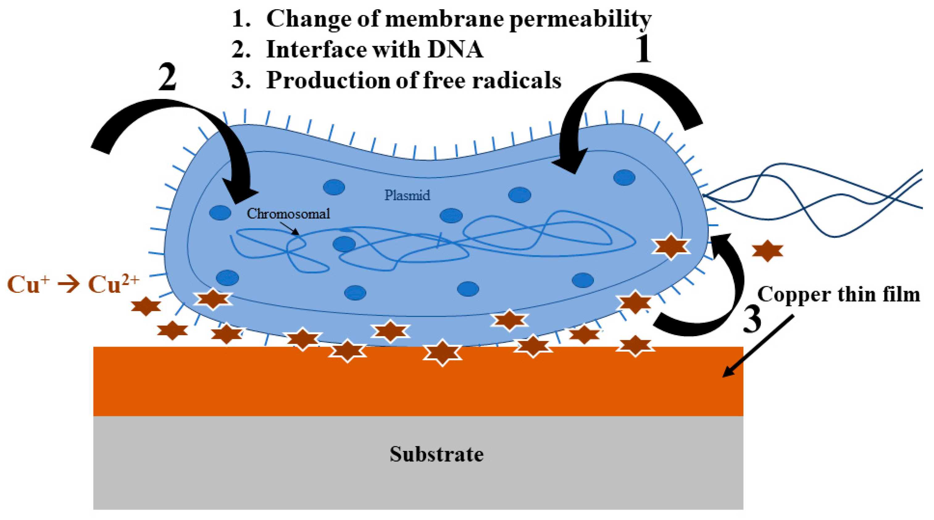
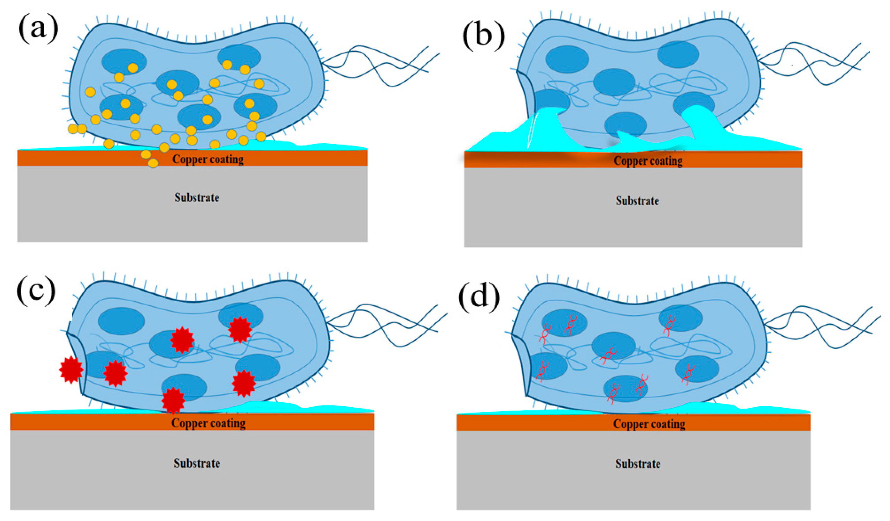
1.2. Mechanism of Microbe Killing

Since from olden times, metals like copper, silver, mercury, arsenic, and antimony are effective materials to inhibit surface infection due to contact killing. Hence, these metals are referred to as antimicrobial metals [31]. Copper is one among other antimicrobial metals widely used due to its extremely high toxicity and biocidal nature (biocidal means biochemical that can kill life by poisoning) which kills the microbes at a faster rate. Unlike other antimicrobial metals, use of the copper as an antimicrobial metal has the following advantages [32].
  • Copper is a stable metal and it can be used in different forms, such as particles, ions absorbed or exchanged, in carrier salts, hybrid structures, composites, and alloys.
  • Copper can encapsulate nanoparticles as a thin sheath on a variety of metals as a coating. These copper nanostructures or alloys can be easily prepared and handled.
Copper has a long history as antimicrobial metal. Today, more than 30 types of copper-based proteins are existing in living organisms [33]. Though the use of copper and copper-based alloys as an antimicrobial agent is for long period, the mechanism of the effect of copper or copper ion to kill the bacteria is not fully understood to date. But it is well agreed that the copper atoms or copper ions (exist in Cu+ or Cu2+) are the cause for injury of a microbial membrane (stage I in contact killing) [31]. Copper can employ toxic effects towards bacteria by different means: (a) By binding or blocking functional groups in microbes. (b) by introducing essential copper ions or atoms in enzymes (c) by involving chemical reactions that are toxic and harmful. The effect of these action/s leads to damage of biological membranes and protein DNA, complications in the enzyme functions, oxidative stresses, and cellular processes. One property of Cu is that it has a maximum affinity for biological molecules, and hence it can dislocate other ions (atoms) from the biological molecules. Cu occurs in either oxidized Cu2+ or reduced Cu+ state. Cu+ has an attraction for thiols and thioether groups and Cu2+ attributes to preferred coordination to imidazole or oxygen groups. These Cu2+ ions can contribute to a wide range of interactions with proteins causing various biochemical reactions [34]. By considering the strength of the copper ions as Lewis acids, one can easily understand the metal toxicity towards the microbes [31].
Another approach for metal toxicity is oxidative stress. Here, metal damages the cell by cellular oxidative stress damage. This means damage of microbes caused by plenty of oxidants like oxygen ions, reactive oxygen species (ROS), and free radicals. Peroxide groups are examples of ROS species. A certain amount of ROS is necessary for living activity [35]. ROS are highly active and they involve in oxidation reactions with organic molecules. Metals like copper, chromium, and iron are called redox-active metals. From Fenton-like reactions kinetics [36], redox-active metals produce hydroxyl groups (OH*), hydrogen peroxide (H2O2), and superoxide (O2) [33].
Fenton-like reactions are as below:
Cu2+ + H2O2 ↔ Cu⋯OOH+ + H+
Cu⋯OOH+ → HO2. + Cu+
Cu+ + H2O2 → OH. + OH + Cu2+
OH. + RH → R. + H2O
Many works of literature state that contact killing is initiated by a very high local concentration of Cu ions from the copper surface, causing high toxicity towards microbes which leads to rupture of the cell membrane [33]. At the interface, copper-mediated ROS generation occurs largely in the periplasm of Enterococcus Coli, subsequently, more ROS generation and then cell destruction and degradation of plasmid and DNA chromosomes occurs [37]. Copper ions can easily accept or donate electrons that lead to the chemical reactions; hence, microbes are killed due to oxidation damage [38]. Copper ions cause damage to the outer or/and an inner membrane that can lead to the outflow of intracellular components like glutamate which affects cell apoptosis and potassium content [39]. Most of the studies agree that the primary attack of copper is the cell membrane in bacteria [16,40]. The microbial membranes contain polymers with electronegative chemical groups that serve as spots for the absorption of metal cations [41].

2. Strategies for Designing Antimicrobial Coatings

2.1. Antimicrobial Agent Release Coatings

Copper ions that are released from the copper coatings are treated as antimicrobial agents. Using an energy-dependent mechanism, Cu compounds or copper ions released from the surface are transported through the cell that kills both adhered and adjacent bacteria. The cell membrane becomes progressively porous due to the outflow of essential cell content such as potassium, amino acids, nucleotides [41,42]. The antimicrobial activity has been mostly recognized as the liberation of Cu ions from its surface. The release rate of ions depends on the chemistry of Cu surfaces [43]. Literature reports that the ion release is the driving force for the antimicrobial properties of the antimicrobial nanoparticles. Copper ions released from the surface induce the production of the ROS that causes further cell damage (see Figure 1) [33]. The release of ions affects the integrity of the bacterial membrane, develops intracellular oxidative stress, which is nontoxic, resulting in the death of the microorganisms [44]. There is an improvement in the antimicrobial activity by the Cu oxide surface due to the significant release of Cu ions from the Cu oxide surface. It’s reported that copper ions released by copper nanocoatings may attack the negatively charged bacteria cell wall, causing rupture. Consequently, protein denaturation and cell death follow [45,46,47].

2.2. Contact Killing

Copper can kill undesirable viruses and bacteria by doing physical contact with them (see Figure 2a). Most of the studies agree that the primary target of attack of copper is the cell wall in the bacteria [16,40]. This is attributed to the presence of polymers with highly electronegative chemical groups (e.g., peptidoglycan, phospholipid, teichoic or teichuronic acids, and lipoteichoic acid groups) on the bacterial membrane that serves as sites for absorption of metal cations. The antimicrobial activity of the Cu ions is due to a positive charge developing electrostatic attraction between the negatively charged cell membrane of microbes with surfaces [41]. Microbes are killed on the copper within hours. Higher copper concentration [48], higher room temperature [49], and higher relative humidity [50] increase the efficiency of contact killing. Bacteria are sensitive to contact killing and various mechanisms for microbe killing by copper and silver have been reported [33,51] Oxidation Behavior of the copper makes more toxicity to bacteria in the contact killing mechanism. The electrochemical phenomenon is well-studied for the copper surfaces [52]. The cell membrane becomes progressively porous due to the outflow of essential cell content such as potassium, amino acids, nucleotides as shown in Figure 2b. ROS are catalyzed by Cu (II) complexes, which cause permanent cell damages by a variety of mechanisms such as oxidative damage, lipid peroxidation, and inhibition of respiration (see Figure 2c) [53]. Cu species leads to rapid DNA destruction and cell death (see Figure 2d) [54]. It appears that there is a variation with respect to the key toxic principle that depends upon the system, experimental conditions, and the bacteria under consideration [52]. A key activity in the contact killing process seems to be the Cu ion release from the copper surfaces.

2.3. Anti-Adhesion/Bacterial Repelling

The adhesion and spread of bacteria on the surfaces of the materials and succeeding biofilm growth creates challenges in healthcare settings (see Figure 3). Anti-adhesion coatings benefit to prevent biofilm development using non-cytotoxic mechanisms. Mechanism of microbial adhesion model can be described in two stages:
Stage I: In this initial stage, the reactions are rapid and reversible that is governed by non-specific physical and chemical interactions.
Stage II: Secondary “looking” stage involves species-specific physicochemical interactions [55].
The design of antimicrobial coatings has been a long-lasting effort and several approaches have been undertaken to limit colonization of bacteria on the coated surfaces. The development of superhydrophobicity surfaces can decrease the adhesion force between microbe and coated surfaces. Due to this, the bacteria will be easily removed before the surface develops a thick biofilm [56]. Surface patterns observed in nature have been the motivation source to generate artificial superhydrophobic (Repelling action) surfaces. Superhydrophobic surfaces inspired by the lotus leaf are attributed to the hierarchical micro/nanostructures leading to an anti-adhesive microbial surface [57]. Nie et al. proposed that superhydrophobic surfaces increase the biocidal activity of copper coatings [58]. Zohra et al. proposed a possible scheme for the bacterial biofilm development on the surfaces of dental and bone repair devices and its treatment and prevention [59]. Important strategies proposed for the development of antimicrobial surfaces presented in Table 1.

3. Copper Coatings as Antimicrobial Surfaces

A surface that makes contact in real-time applications should be composed of Cu or its alloys. The use of pure solid copper is expensive. Hence, Cu coatings are preferred over bulk because of cost and the ability to manipulate the properties of the surface. The properties of thin-film copper coatings mainly depend on four facets: (1) Use of correct copper source, (2) transport of copper species, (3) condensation of the copper species on the substrate, and (4) correct type of substrate. The nature of the coating is strongly affected by the copper source (purity, shapes, etc.) and the substrate used (development of residual stress, texture, etc.) [80]. There are multiple techniques available for the coating of copper. Amongst them, Chemical vapor deposition (copper source is copper salt), Physical vapor deposition (evaporation and condensation of the copper species), Electrodeposition, Electroless (copper salt in aqueous medium) deposition, Sputtering, and Thermal spray techniques (pure copper as copper source) are widely used methods for depositing the copper. Not only an optimum deposition method is identified, but also, it is important to develop a Cu coating with increased physicochemical and high antimicrobial properties. Ideally, the copper-coated surfaces should be permanent (i.e., not peeling), not wearing out, and work under hospital conditions. Functional properties of the coating depend on parameters, like deposition temperature, rate of deposition, deposition environment, the distance between the source and the substrate. In this review, a study on the influence of process parameters affecting antimicrobial performance is also presented. The advantages and disadvantages of different coating routes for copper deposition which could affect the antimicrobial activity are represented in Table 2.

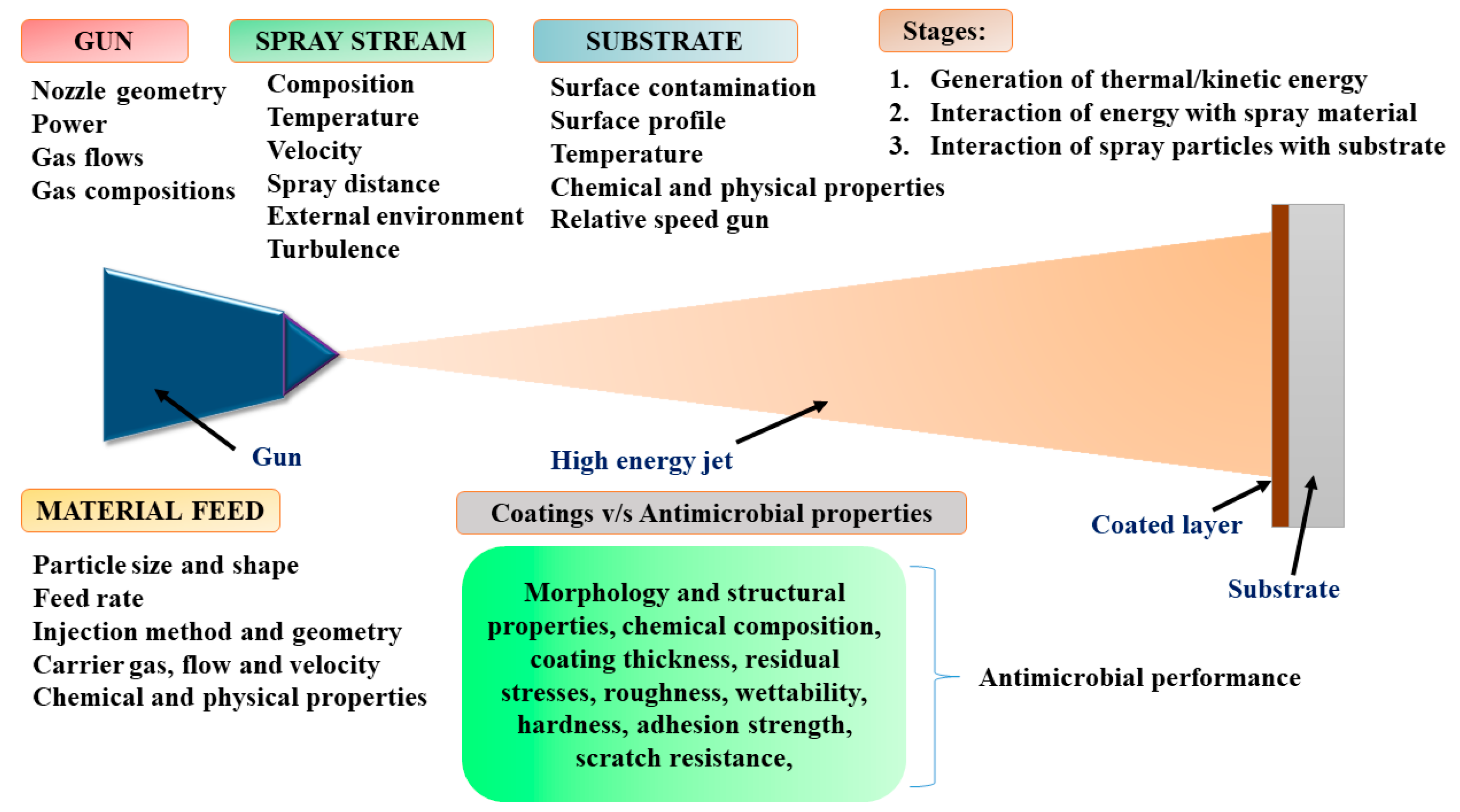
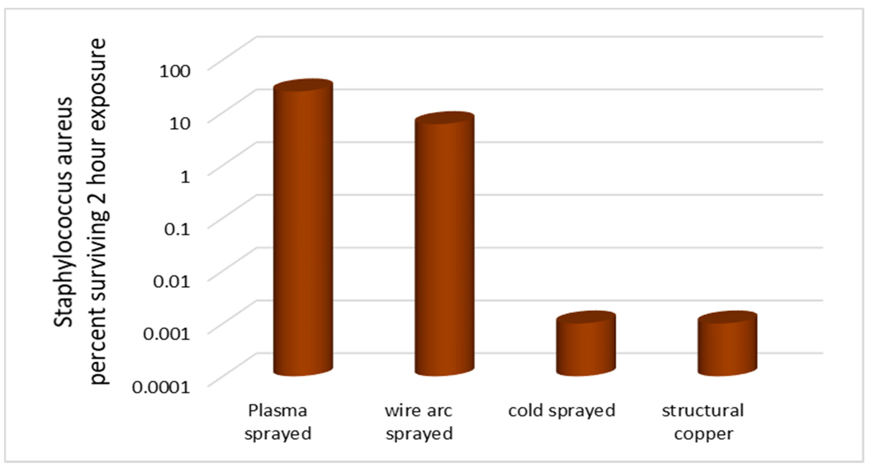
3.1. Thermal Spray Techniques

Traditionally, thermal spray coatings are widely used as protective coatings. However, antimicrobial coatings include deposited coatings which have an active, integrated, and new functionality beyond the traditional coatings. Antimicrobial coatings are developed to attend to its functionality, for instance, its capability to kill bacteria or prevent infections on the touch surface applications. Tremendous efforts are carried out to control the infections from hospital touch surfaces, both towards the elimination of bacterial activity as well as preventing their colonization. Thermal spray techniques are a convenient practice for obtaining copper coatings, which involves the development of the thick copper coatings (approximate thickness range 20 μm to several mm) with good corrosion and high adhesion and minimum wear with low cost [81]. The metallic and non-metallic materials in the form of wire, powder, and rod are heated up to their molten or semi-molten state and then a high jet is used to impart kinetic energy to the molten particles. The resulting molten droplets or particles are accelerated towards the prepared substrate. Those molten particles flow into thin lamellar particles adhering to the substrate surface and rapidly cool down to form a solid splat. They form adhesive bonds with the substrate and cohesive with each other. Many process parameters need to be optimized to produce proper coatings for antimicrobial applications. They are shown in Figure 4. [82]. The most commonly used thermal spray techniques are: (a) Plasma spraying, (b) Wire arc spraying, (c) Flame spraying, and (d) Kinetic or cold spraying. The microstructure of thermally sprayed copper coatings depends on several factors such as the initial particle size, the individual impact velocity, impingement angle, and the Cu powder microstructure [83]. A typical microstructure of the thermal sprayed coating is as shown in Figure 5.
The plasma spray technique uses direct current arc or radio frequency discharge as a thermal source required for the deposition process. The elevated temperature at the jet core (~14,000 K) produces high proportions of particles melted and it gives rise to excellent deposition characteristics like coating density, bond strength, and less porosity compared to other thermal spraying techniques [84]. Vacuum plasma spray (VPS) coatings (<100 Pa) are developed to reduce the detrimental effects like oxidation and undesired contamination in the coatings [85]. Vacuum spray coatings offer better columnar structure and increased deposition rate compared to the conventional PVD.
In wire arc spraying, two consumable conductive wires are fed automatically between which a direct electric current arc is established at the wire tip in an atomizing gas stream. Immediately, the material gets molten and the molten metal is accelerated towards the substrate surface by the stream of atomizing gas. The type of spray gas (such as compressed air or nitrogen) used as atomizing gas describes the oxide content and also influences the hardness of the coatings [86]. The adhesion strength to the substrate is the essential criterion deciding the quality of thermally sprayed coatings. Two criteria required to achieve good bond strength between the coatings and the substrate are:
(a)
The particles must be in a fully molten state.
(b)
They should possess sufficient velocity [87,88].
Potential benefits of the twin wire arc method are (i) less expensive thermal spraying process and (ii) capability to produce dense coatings with excellent deposition rate. The cold spray technique involves the transmission of high kinetic energy into the feedstock particles, which results in strong bonding upon impingement at the target surface. This technique utilizes pressurized gases with helium and nitrogen [89]. Cold sprayed coatings exhibit better metallographic features, show dense coating with the absence of pores and oxides. The cold sprayed technique has low thermal influence and this method can use feed materials, like nanocrystalline materials, oxidation sensitive, and metastable materials [83]. A comparative study has been carried out for the copper coatings developed by wire arc spray (moderate speed, molten/heat-softened), cold spray (high speed, kinetically deposited solid-state powder particles), and plasma spray for antimicrobial applications [90]. Powder spray techniques exhibited the significance of the deposited structure. The cold spray technique demonstrated greater antimicrobial potential as compared to wire arc spray and plasma spray due to high dislocation density and enhanced ionic copper diffusivity offered by the higher impact velocity of the sprayed particles (see Figure 6) [90]. The dense microstructure, greater thickness, and low porosity coatings developed by using the cold gas spray technique presented the improved antimicrobial efficacy against Staphylococcus aureus with complete mortality of the bacteria after a time interval of 10 min [91]. Copper coatings on carbon steels showed excellent corrosion resistance when immersed in chloride solution for 1100 h [92]. The characteristics of the twin wire arc sprayed Cu coatings (such as increased surface roughness, enhanced surface free energy, minimal contact angle, and materials characteristics such as chemical composition and ultrafine grain microstructure, lattice defects, and specific oxygen species) determine the efficiency of antimicrobial activity of the surfaces against ATCC 25922 Escherichia coli and ATCC 292133 Staphylococcus aureus pathogens. In spray techniques, the microstructure growth of the deposit will be influenced by the solidification behavior [93]. A study of antimicrobial activity of copper, copper alloy (CuNi35, CuSn10) and copper composite (CuTiO210) coatings on the stainless steel formed by plasma spray technique confirm that the satin finishing of the coated surfaces developed for protective applications has a beneficial effect of antibacterial activity [94]. A comparative study on microstructure, adhesion strength, microhardness, and elastic and plastic response of the copper and the copper alloy (Cu4%Sn, Cu17%Al 1%Fe1 and Cu17%Ni 9.6Zn) coatings on the 316 stainless steel substrates developed by wire arc spray technique has been carried out [95]. Rough and cold sprayed surfaces are having more noticeable antimicrobial activity than that of the plasma/wire-arc sprayed Cu surfaces [96]. Copper coatings deposited on the stainless steel (Type 316) by wire arc spray method revealed excellent antimicrobial behavior (97% after 6 h) which is attributed to the presence of ultrafine grains, micropores, and crystallographic defects [97]. Nanosized particles exhibit better biological and chemical activity, hardness, electrical conductivity and possess enhanced active surface area in comparison with micrometer-sized particles. The microbial effectiveness of the Cu nanoparticles is related to their size and high surface-to-volume ratio. CuO nanoparticles produced by using thermal plasma technology were efficacious in enhancing antimicrobial activity [45].
In the case of the flame spray technique, combustion of fuel gases or liquid fuel in the presence of oxygen is used to melt the spray coating material. The combustion produces expanding gas flow at a pressure close to 1 MPa and passes through a converging and diverging nozzle. It creates the required jet to accelerate the material towards the targeted surface. The typical operating temperature for the flame spray technique is around 3000 K and the jet velocity at the entrance of the barrel is up to 100 m/s. The feedstock material in the form of powder is injected into the gas stream. However, based on the variations in the requirement of the coatings, several variations are developed. Different techniques are detonation Gun (D-Gun) spraying, high-velocity oxy-fuel spraying (HVOF), high-velocity air fuel spraying (HVAF), suspension/solution high-velocity oxy-fuel spraying (SHVOF). Wire high-velocity oxy-fuel (W-HVOF) technique is employed to produce copper coatings on low carbon steels with thickness below 40 microns through wire feedstock rather than using an expensive powder. A systematic structural and microstructural study was carried out using SEM and XRD to understand the surface and cross-section of the coatings. Optimization of process parameters concerning mechanical properties of the copper coatings is well briefed [98]. However, in context to antimicrobial applications, the copper deposits produced from thermal spray techniques are not explored much.

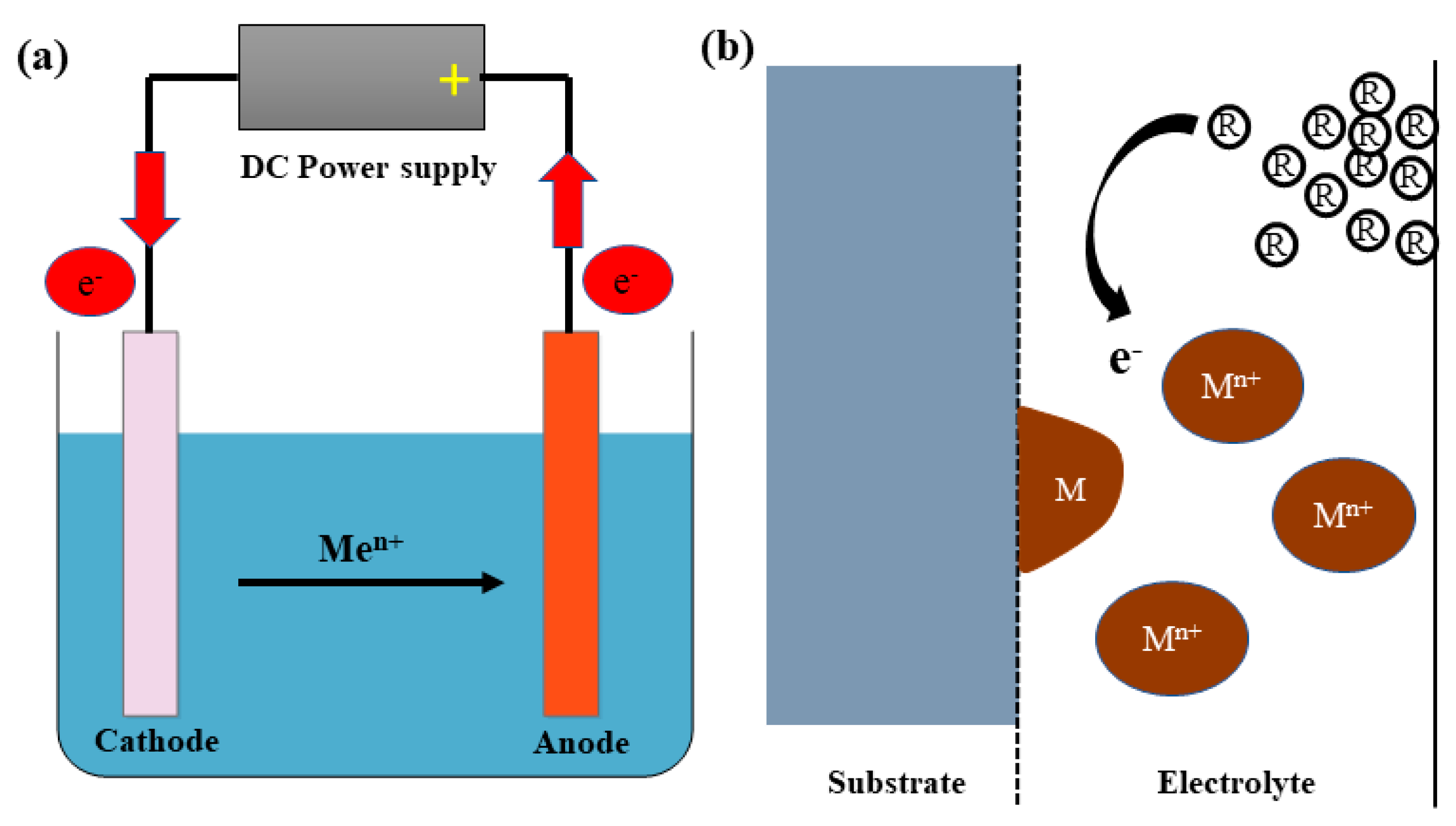
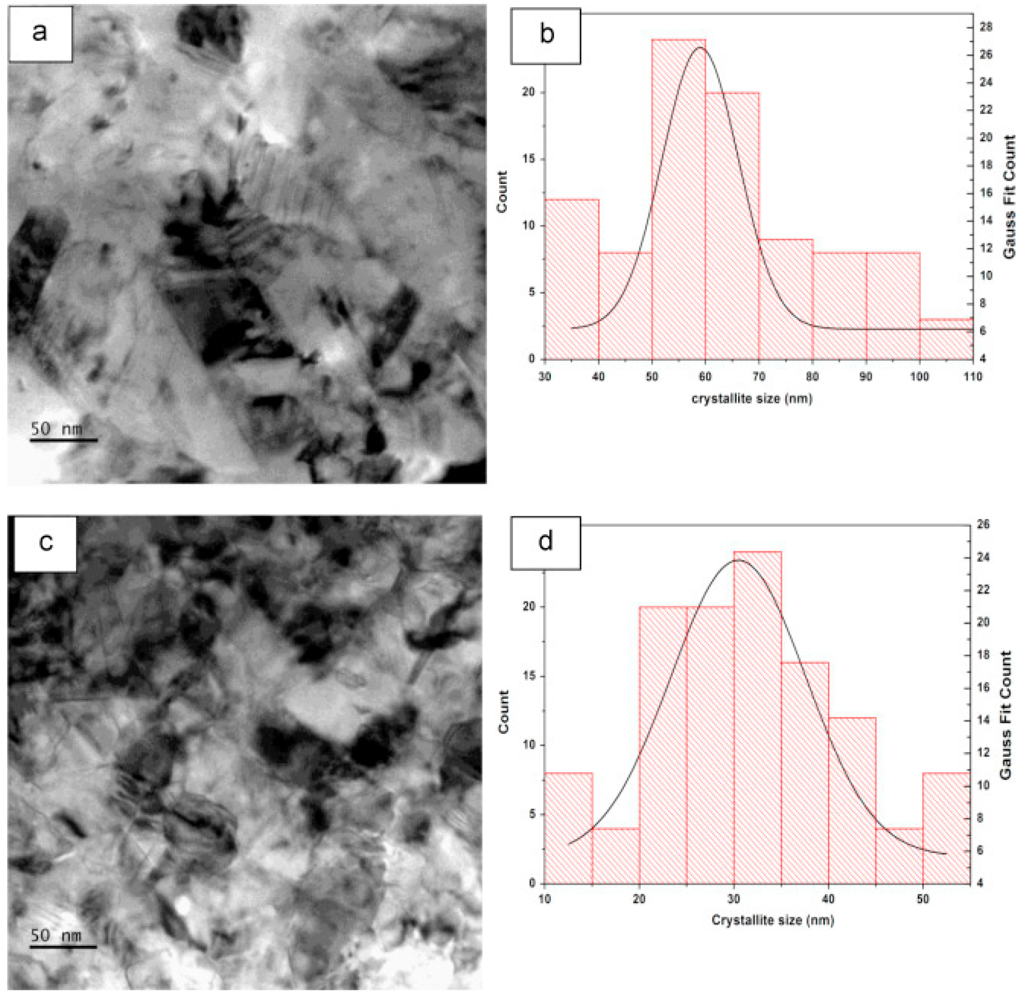
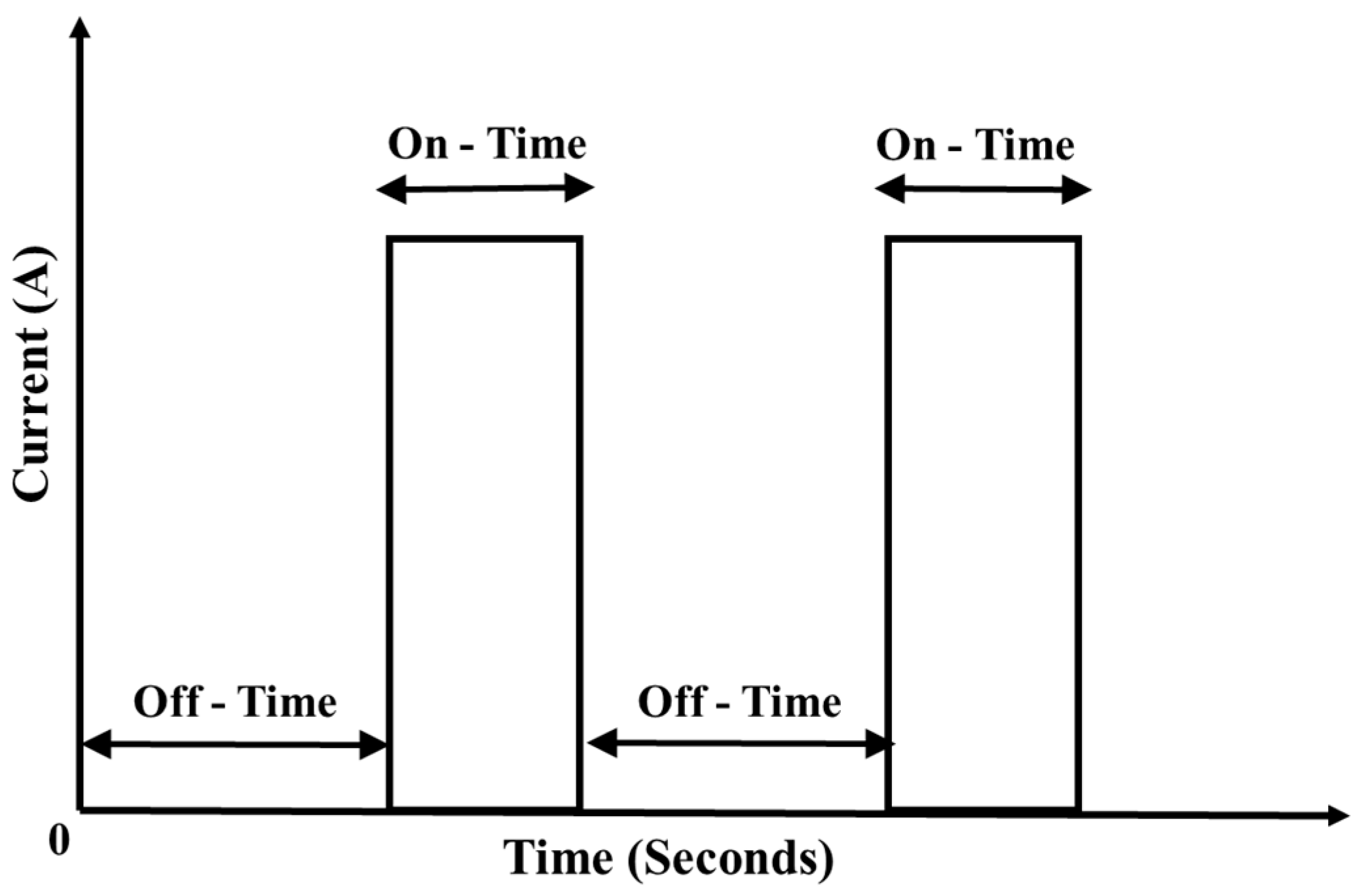
3.2. Electrodeposition Techniques

The electrodeposition of copper and copper-based alloys is over 176 years old; the first activities being stated in 1842 (Faraday’s Laws of Electrolysis in 1834). Copper is the commonly electrodeposited metal for use in different applications. As a process, electrodeposition uses electric current as the source of energy and the current passes through a closed electrochemical circuit. As a result, deposition of the anodic species (metal ions to be deposited) occurs on to the cathodic metal substrate. The copper plate used as sacrificial anode helps in supplying constant metallic ion density in the electrolytic solution and the component to be coated is connected as the cathode. Electro dissolution takes place at the anode, whereas electrodeposition takes place at the cathode (see Figure 7a).
The electrolytic bath contains the metal ion source as the copper salt in the electrolyte. Grain refiners, hardeners, brighteners, buffering agents, and leveling agents are other additives. By varying the process parameters such as pH, current density, bath composition, bath temperature, and deposition time, properties of the coating namely, morphology, composition, crystal size, crystal orientation, residual stress, etc. may be controlled. Most of the literature reported that the copper coatings could be electroplated from different standard baths. These baths could be cyanide or non-cyanide in nature. Non-cyanide baths include sulfate bath, phosphate bath, and chloride bath. Recently, electrodeposited copper coatings have proved their antimicrobial functionality [99].
Advantages of the electrodeposition technique are easy control of the coating thickness, ability to deposit on the complexes shapes, economics, and feasibility of alloy deposition, low operating temperature, and deposition of the non-equilibrium materials. Recent investigation has highlighted electroplating as an attractive approach for the preparation of nanostructured coatings due to its cost-effective and less equipment-intensive method. The copper nanoparticles were successfully electroplated on the stainless steel substrate using pure acidic copper sulfate solution. Cyclic voltammetry and chronoamperometry methods revealed information on the nucleation and growth kinetics of the copper coatings on the stainless steel. The density of copper nuclei, particle size, and surface area is highly influenced by the deposition time [18]. Augustin et al. investigated the effect of current density during copper electrodeposition on the aluminum metal substrate on microstructure, hardness, and wettability of the textured copper coatings for antimicrobial application.The formation of nanocrystals in copper coatings causes improvement in the scratch resistance and the surface microhardness (see Figure 8a–d) [100].
Extensive research work has been carried out on the effect of current density, the role of the complexing agents, and the bath chemistry to understand morphology change and grain refinement of the electrodeposited copper [101,102,103,104,105,106,107]. EDTA solution consisting of 0.01 M Cu2+ ions with a pH = 8 was used to deposit Cu on the stainless steel substrate at −1.1 V vs. Ag/AgCl for 15 min. A high percentage of Cu, uniform coating, high surface roughness, and nanosized structures in the coatings play a major role in the contact killing of bacteria [108]. The reason for the poor adhesion of the copper coatings on the mild steel substrate from acid and non-cyanide electrolytes is due to the contact exchange of the copper ions-iron pair. In an acid electrolytic medium, the large value of current exchange causes poor adhesion. In the case of non-cyanide electrolytes, iron is passivated. Hence, adhesion between the substrate and the coating is insufficient because of the presence of a separate passive film at the substrate [109]. Response surface methodology (RMS) is utilized to predict and tune the process parameters to improve the adhesion strength of the substrate made of austenitic stainless steel. The modeling approach is used to tune the process parameters to obtain maximum adhesion strength of 10 N [110].
Marco Zeiger et al. investigated the influence of electrodeposited Cu surface on the performance of contact killing of E. coli. It was proved that the copper coatings prepared by electrodeposition killed bacteria rapidly than the copper surfaces prepared by other coating techniques. Electroplated copper surfaces enhanced the release of ionic copper compared to the coatings prepared by using other methods. Copper ions released by the coupons into the aqueous phase could be measured by using atomic absorption spectroscopy (AAS) [111]. Higher current density attributes multiple layers of copper nodules having enhanced amounts of defects, like dislocations, twins, and stacking faults. The structural details of the electrodeposited copper coatings were studied by using TEM and nodules have fine crystallites in the range of 60 nm [112]. If the substrate is changed, surface properties such as resistivity, surface energy, corrosion-resistant behavior, and residual stress of the electroplated Cu will also be modified.
Crystal shapes, structure, the energy of the electrodeposited copper coatings primarily control the electrochemical, mechanical, and tribological characteristics. Nucleation and growth mechanism of the electrodeposited pure Cu from the acidic CuSO4 bath is reported [113] and to alter the topography, the microstructure and grain refinement, the role of surfactants and ligands in acidic and non-cyanide baths have been studied [114]. The surface morphology of the electroplated copper varies with overpotential value irrespective of the chosen substrate. Lower overpotential favors growth in a layer-by-layer fashion, whereas higher overpotential promotes potential dendritic growth followed by multi-directional growth, similar to that of a cauliflower. Type of the substrate played a key role in the deposited crystal shape [115].
Constant research being focused on the improvement of property of the electrodeposited coatings for any given application. However, pulsed electrodeposition, periodic reverse, and asymmetric alternating current plating techniques have promised their potential characteristics for functional coatings. In pulsed electrodeposition, a series of pulses of either potential or current is alternated between different values. Pulses are equal in magnitude, duration, and polarity separated by zero current (see Figure 9) This favors nucleation of fresh grains and also attributes an increase in the number of nuclei per unit surface area. They give better properties of coatings compared to conventional electrodeposited coatings. It is possible to control the thickness of the film in the atomic scale and composition of the coating by controlling the pulse parameters [116].

3.3. Electroless Plating

Electroless deposition is also known as autocatalytic or chemical plating. It is a non-electrolytic method involving several parallel reactions in an aqueous plating solution without using an electric potential. A simultaneous reducing and oxidizing reaction in solution causes spontaneous deposition of films on conductive and/or non-conductive substrates. Typical copper plating solution consists of metal ions (Cupric salts such as copper chloride, copper sulfate, and copper nitrate), a stabilizer, or additives like complexing agents and reducing agents. Minimum necessary components of electroless deposition bath are the source of metal ions and reducing agents. In general, electroless deposition of copper is characterized by the reduction of Cu metal ions on the substrate immersed in a bath of an aqueous solution of the copper metal ions. Commonly used reducing agents in electroless copper deposition baths are formaldehyde, dimethylamine borane, borohydride, hypophosphite, hydrazine, sugars (sucrose, glucose, etc.), and dithionite [77]. Formaldehyde is used in traditional electroless copper plating as the reducing agent due to its ability to facilitate good quality of deposits. Though, formaldehyde results in a combination of cost and effectively coated surfaces, it suffers from limitations like environmental and health concerns. Electroless plating is slower and expensive process compared to electrolytic deposition [117]. Alkaline amines (encouraging faster plating rate, ≥2 μm/20 min), tartrate salts (encouraging lower plating rate, ≥0.5 μm/20 min), ethylenediaminetetraacetic acid (EDTA) (for controlled plating rate and grain structure) are commonly used complexing or chelating agents for the copper electroless plating [117].
Half-cell reaction for electroless plating of copper (II):
Cu2+ + 2e → Cu
HCOO + 2H2 + 2e → HCHO + 3OH
The net reaction could be written as,
Cu2+ + 2HCHO + 4OH → Cu + 2HCOO + 2H2O +H2
The stability of the process fully depends on the substrate material. The actual reduction reaction of the metal takes place when an appropriate pre-treated surface is made available. Surface properties of the electroless copper plated materials depend on the plating bath composition/constituents, pH of the bath, deposition time, and deposition temperature. Extensive research work has been completed on the role of additives in the electroless deposition of copper [118]. Additives are used to improve the film morphology by controlling the plating rate. Additives can enhance or diminish the deposition rate resulting in simultaneous modification of physical properties [119,120]. Enhanced mechanical bonding increases adhesion between the EL coating and the substrate. Microstructural growth and bonding mechanism starts with the formation of globules in nano-size range on certain preferred location (nucleation stage) and then develop laterally to cover the whole surface along thickness direction with high uniformity (growth stage) [121]. The microstructure developed by electroless deposition is columnar as shown in Figure 10 [112]. Also, multiple crystallites are existing within columnar grains. Deposition temperature and pH of the plating solution affect the color of the coating and deposition rate. Increasing deposition temperature results in an increase in deposition rate. Thus, improved morphology is possible and crystal growth becomes stable with a decrease in internal stress. An increase in plating bath temperature attributes better adhesion strength [122]. S. aureus exhibited lower resistance compared to E. coli against copper nanoparticles prepared by electroless deposition [123]. Compared to electrodeposition, the morphology of the electroless deposited copper surfaces have a homogenous, dense, and bright appearance irrespective of the bath parameters and the substrate [118]. Yet, the contact killing mechanism and microbial activity phenomenon on the electroless Cu deposits are still not understood. The major advantage of electroless and electroplating with the coating techniques for antimicrobial touch surface applications is the existence of large-scale commercial plating outfits.

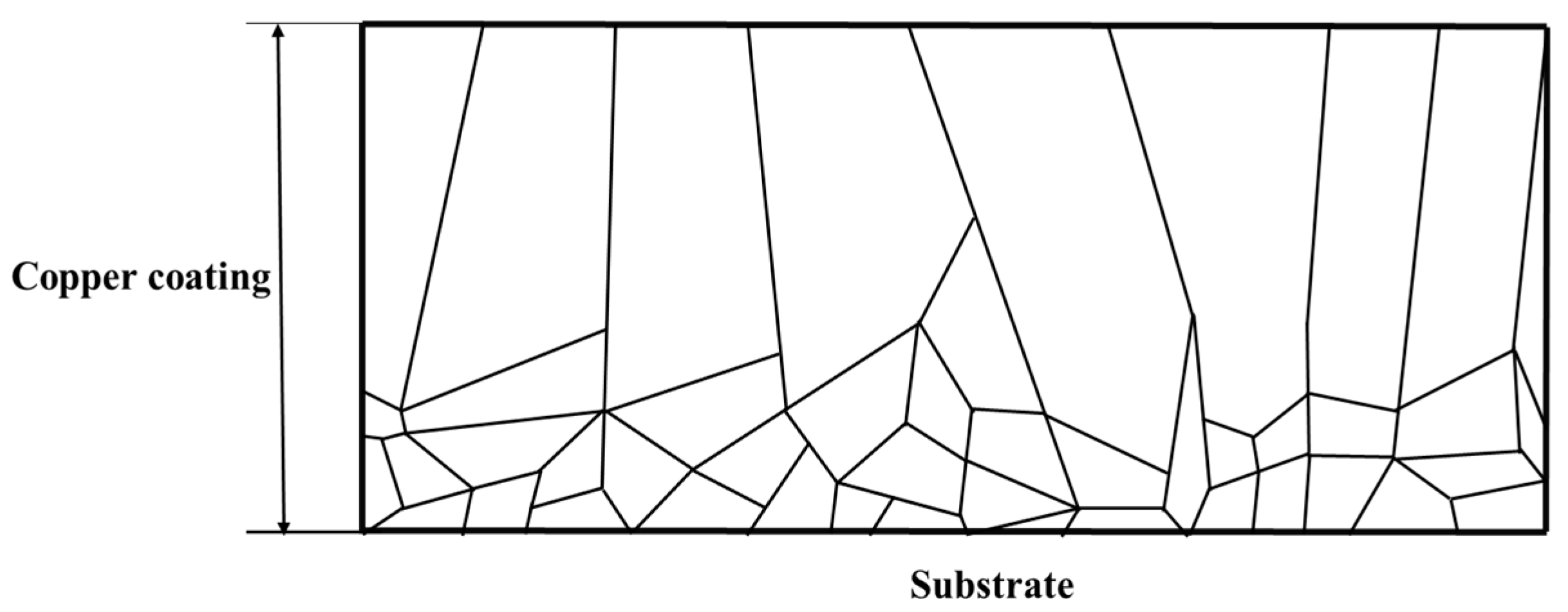
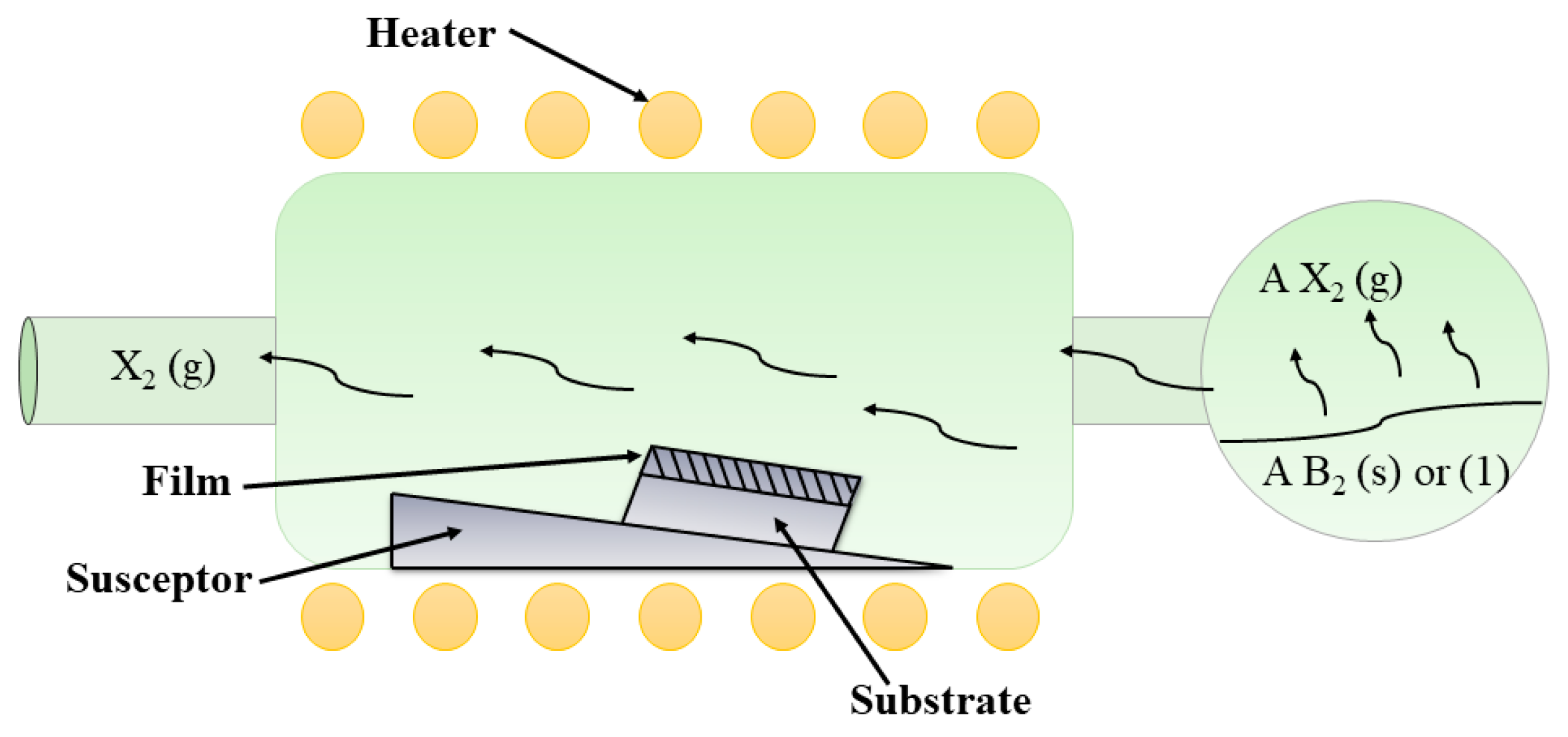
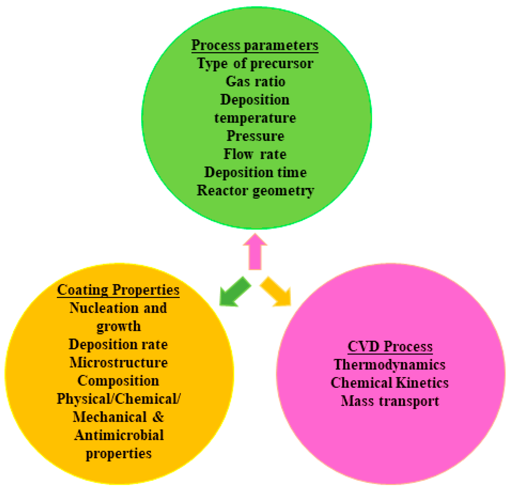
3.4. Chemical Vapor Deposition

Chemical vapor deposition (CVD) coatings have successively acquired broader scope with importance on the deposition aspects for fabricating functional semiconducting and antimicrobial coatings with enhanced surface properties. In chemical vapor deposition (CVD), the heated substrate surface is exposed to gaseous molecules (used as volatile precursors). It reacts chemically and decomposes on the substrate (wafer) surface to produce a thin coating or stable solid layer in an activated environment (heat, light, plasma). At the same time, volatile by-products are formed. They are removed by the flow of gas through the reaction chamber (see Figure 11). There are two distinct varieties in CVD: (a) Heterogeneous CVD, where, the reaction takes place very near to the surface of the substrate and reacting species is directly deposited on the substrate surface. (b) Homogeneous CVD, involves a homogenous gas-phase reaction, where gaseous molecules will get transported to the substrate surface and get deposited. The chemical reaction of the gas molecules takes place in the presence of inert carrier gas and it helps in controlling the reaction rate.
The major factors that affect the morphology and surface properties of the coatings are substrate temperature, presence of the carrier gas, the velocity of the gas flow, and type of precursor used (see Figure 12). In CVD, coatings can be prepared in hot wall reactors or cold wall reactors operating above atmospheric pressure, with or without carrier gasses and temperature ranging from 200 °C to 1600 °C. Several reactions such as oxidation, reduction, hydrolysis, pyrolysis and a combination of these can occur in a CVD process. Reasonably, CVD allows uniform coating on any substrate surface at once, on both sides of the substrate surface or substrate of large size and/or complex shape. In contrast, sputtering and PVD can deposit the substrate surface which is directly placed in front of the PVD source due to the nature of the line-of-sight deposition process [124].
Copper acetylacetonate and copper hexaflouroacetyleacctonate (Cu(hfac)) are commonly used precursors for copper deposition [125,126]. Accurate control and monitoring are required on deposition process parameters to produce quality coatings. Coating-substrate adhesion strength issues could be addressed by avoiding surface contamination, the attack of corrosive unreacted precursors, and/or formation of by-products [127]. Microstructural growth of the CVD coatings depends on two mechanisms: (a) Surface kinetics (b) mass transport. Usually, the CVD technique employing higher deposition temperatures and lower deposition rates imposes limitations on the kinetics of grain growth, consequently yield coarser grain size with lattice distortion. The columanr-equiaxed, nodular, smooth and uniform morphology was obaserved in Cu coatings deposited by CVD using Cu amidinate [128] and hexafluoroacetylacetonate (Cu(HFAc)2) precursors. However, deposition temperature and concentration of reactive species (supersaturation) affect the nucleation and growth kinetics. The concentration of reactive species is affected by the partial pressure of the active gaseous species and total pressure in the reactor. Larger grain sizes in CVD (compared to PVD) lead to coarser morphology as a function of thickness [129]. Thin Cu-SiO2 films developed on the glass substrate by flame-assisted chemical vapor deposition (FACVD) were evaluated against a variety of highly resistant strains including Escherichia coli, Pseudomonas aeruginosa (d). Staphylococcus aureus and Vancomycin-resistant Enterococcus fascism. SEM results exhibited nanostructured coatings within the silica matrix, which enhances the antimicrobial activity. Cu-SiO2 coatings can also be deposited on the substrates like metals and ceramics and demonstrate potential applications in antibacterial environments [130]. The high antimicrobial effectiveness of the Cu2O films than that of the pure Cu films was proved against Staphylococcus aureus (S. aureus) and E. coli. The Cu2O and pure copper films were deposited via aerosol-assisted CVD using Cu(NO3)2 as a precursor [131]. The development of superhydrophobic antibacterial Cu-coated polymer films via aerosol-assisted CVD showed a tremendous reduction in bacterial cell adhesion as compared to the control [132].
A commercial limitation of the CVD coatings for antimicrobial application is that the complex and large-size components cannot be coated easily. Nevertheless, the high purity, the robust films imparted by CVD have found applications in certain components such as sanitary valves and food processing products which are non-regular in shape, are small, and exhibit enhanced wear.

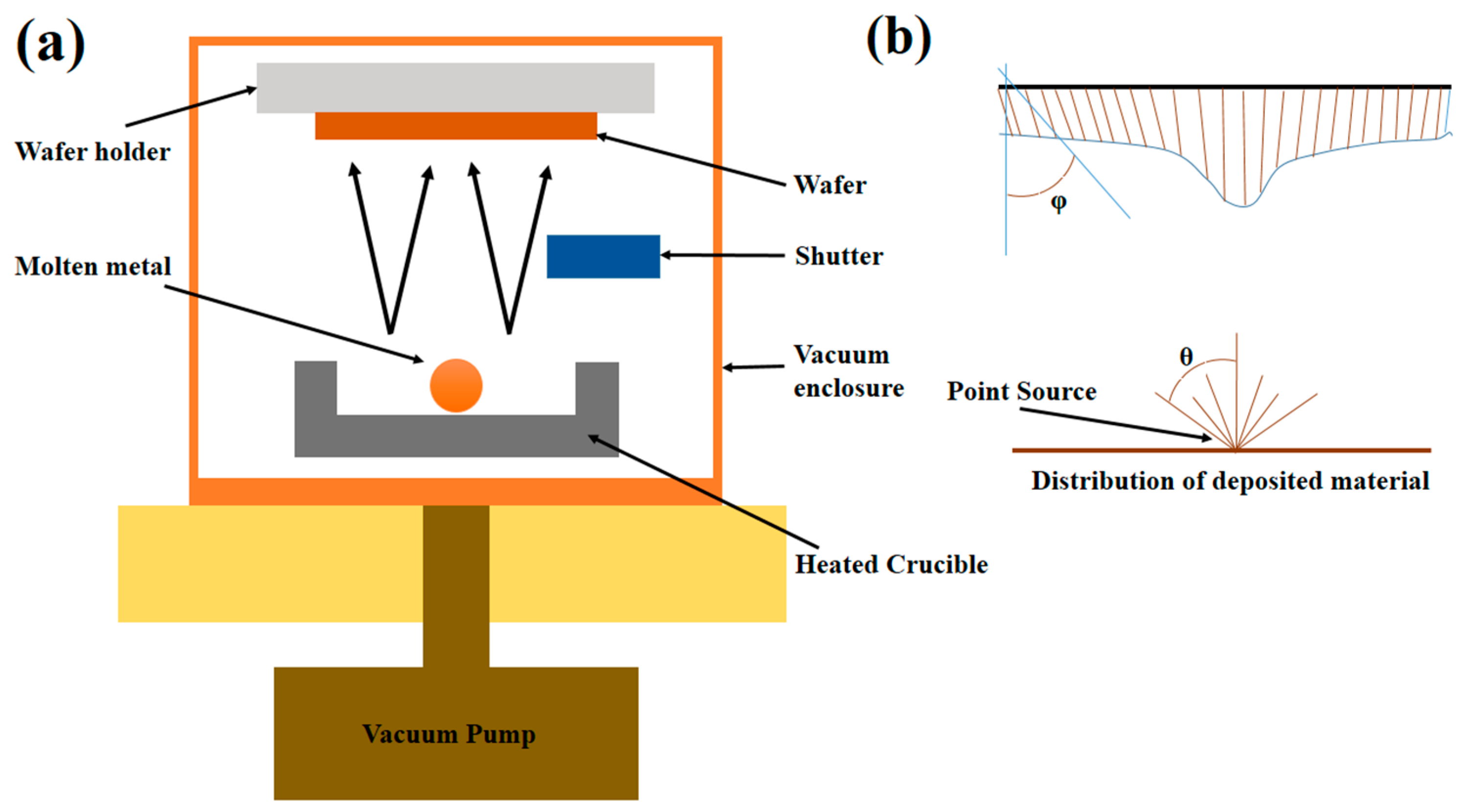
3.5. Physical Vapor Deposition

This involves thermal evaporation and condensation. Vaporized copper atoms (from solid copper) are allowed to deposit over the substrate without colliding with the residual gas molecules. Electric current as a source of energy is utilized (through filament) to vaporize solid copper kept in a crucible made of refractory material. The copper evaporates and vapor flux is directed outward and condenses on the sample kept in the line of sight. Components are to be deposited are enclosed in the vacuum chamber. The pressure level inside the vacuum chamber decides the vaporizing temperature.
Simple and inexpensive PVD has a couple of serious limitations: First is physical vapor deposition is a line of sight of the process and hence maximum coating thickness may be reached at the line of symmetry and thickness is reduced rapidly as one would go away. By modifying the geometry of the copper source, introducing a high vaporization rate by promoting collision between vaporized copper atoms, and the use of proper shutter plates, some of these limitations could be partially overcome. The second is due to differences in the values of coefficient of thermal expansion, difficult to handle the evaporating Cu sample in the crucible.
Physical vapor deposition is a line of process, if the substrate has a complex geometry, there will be geometrical shadowing and some portion of the component would be improperly covered (see Figure 13a,b) [133]. Deposition of the copper via thermal evaporation is a cost-effective and fast method. In contrast, reported literature on PVD (thermal evaporation and condensation) for copper deposition is relatively less due to the process difficulties involved in melting and evaporating high conducting metal like Cu. Arresting the dislocation motion is the key source of strengthening Cu thin films. To enhance the mechanical strength to a larger extent, introducing defects in the form of precipitates and grain boundaries has proven to be effective. Also, twin boundaries act as barriers to the motion of dislocations. High-strength Cu films could be deposited by tailoring twin boundaries [112,134]. The ratio of the substrate temperature to the melting temperature of deposited metal significantly affects the microstructure and properties of the coating. Nanocrystalline films could be produced from low substrate temperature using vapor deposition [135]. The physical and chemical condition during the PVD reaction can strongly affect the morphology, composition, residual stresses of the coatings [136]. During E-beam PVD, the substrate temperature and morphology of the film are two critical problems impacting the behavior of the film [137].

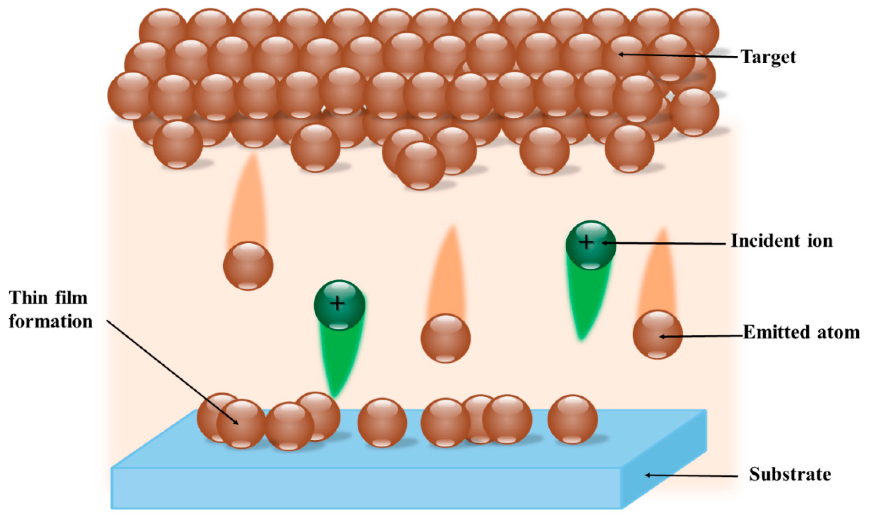
3.6. Sputtering Technique

Coated copper touch surfaces are getting a significant role in health care settings due to their proven antimicrobial activity acquired during hospital-acquired infection. From an economic point of view, the copper-coated element must be cheaper compared to the identical element made using bulk copper or any other antimicrobial material. The sputtering technique is one important route for copper coating, offering considerably improved functional properties than the thicker coatings fabricated using other techniques [138]. Sputter deposition is a type of physical deposition process wherein, the copper (pure copper or copper alloy) target (cathode) is bombarding with energetic Ar+ particles. Subsequently, the neutral copper atoms, copper ions, cluster of atoms, free electrons are knocked out from the target material. This process is called sputtering. A widely used bombarding species is argon ions. Those sputtered atoms are in turn deposited on the substrate (anode surface) due to momentum. The coating material is allowed to reach the vapor phase, without altering its chemical compositions (see Figure 14) [139].
Sputtering uses high energy sources, like DC voltage, magnetic field (magnetron), radio frequency (RF) to convert gas into plasma. The positive gas ions in the plasma get accelerated towards the target and erupt the copper particles eventually on the substrate. The most promising properties of the sputtered copper coatings like thickness, structure and morphology, hardness, and texture could be controlled by varying important process parameters like the pressure inside the chamber, voltage, distance between the substrate and the target, deposition time, and target current. Different types of sputtering are (a) magnetron sputtering, (b) RF sputtering, (c) ion-assisted deposition, and (d) pulsed laser deposition. In real-time use, DC magnetron and/or RF magnetron combinations give good deposition rates and high-quality deposits [140]. The durability of coatings is affected by the lower value of the adhesion bond between the copper film and the substrate. This is due to rich oxide layer formation on the substrate material when exposed to the environment before coating. Efforts are made on improving the adhesion strength of DC magnetron sputtered coatings. There is an improvement in the adhesion properties and structure of the sputtered copper thin films deposited on the pre zinc layer [140]. The antibacterial efficacy of copper-based TiN films deposited on stainless steel substrate processed using dual magnetron sputtering was studied [141].
Nanostructured coatings exhibited enhanced hardness and thermal stability at the nanolayers [142]. The improved bacterial killing rate has been reported for nanostructured coatings. The sputtered copper layer thickness range of 35 nm to 150 nm was confirmed from 4.6 min to 16.3 min of sputtering of copper on glass substrate below the 5 × 10⁻⁵ Torr of partial pressure of the sputtering chamber [143]. The mechanical stability of alloy films deposited from the sputtering technique needed to be addressed against Gram-positive and Gram-negative strains [144]. Nanostructured copper coatings successfully deposited on tantalum substrate for effective killing of Staphylococcus aureus and E. coli [145]. Magnetron sputtering is becoming a rather established technique and is now applied at the industrial level to produce hard coatings on the tools, functional coatings, and decorative treatment of the surfaces. Comparision between thermal spray, electrodeposition, electroless plating, CVD, PVD-sputtering and PVD- EB depostion is given in Table 3.

4. Concluding Remarks and Future Perspectives

Recently, the antimicrobial coatings market has got increased demand in health care applications. Copper coatings are being considered as materials with the highest likelihood of success for antimicrobial activity. Many researchers have investigated the effectiveness of copper and copper alloys as antimicrobial materials. Terms like deposition technique and structural design are involved in the development of the coating process to obtain cost-efficient functional products making them commercially available and attainable to all types of market. The development of coatings and surfaces that can actively kill the microbes is an important component of maintaining hygiene in the hospital environment and various coating routes have proved the antimicrobial activity of copper surfaces. In view of the excellent antimicrobial property of copper coatings, purity of copper, high surface roughness, defects like nanotwins, stacking fault regions have increased the copper ion release, beneficial for killing the microbes and improved hardness. There is strong evidence that nanostructured copper coatings exhibited the highest toxicity to the bacterial membrane. Copper coatings developed by thermal spray, electroless deposition, electrodeposition, PVD, CVD, and sputtering were effective in killing the range of microbes involved in HAIs. Furthermore, the surface texture, nanostructure, and wettability nature of coatings helped to understand the mechanism of antimicrobial activity. Typically, columnar growth is found in copper coatings that could increase the rate of killing the microbes. The homogenous, dense coatings showed improved antimicrobial activity than the coatings with high porosity and non-uniformity. The limitation with copper deposition by various coating methods is excessive copper is highly dangerous, the adhesion strength of the coating to the substrate and in case electrodeposition and electroless deposition, use of cyanide bath have waste management and water pollution issues. The influence of metallurgical properties of the copper coatings on antimicrobial activity has not been investigated. Industries and the research community can bring more research into retaining the aesthetic nature of copper coatings without compromising the antimicrobial functionality. The life of the coating is most important. Long term performance of antimicrobial coatings is not addressed in the laboratory studies. However, a big step has to be taken to transform the experimental studies into the real market. More studies to be carried out on the influence of process parameters increasing the efficacy of antimicrobial activity from various deposition techniques and its commercial models. Also, antibacterial products must be performed as a necessary upturn in human health. Designers should always think about prospects to upgrade the surfaces to antimicrobial surfaces. These surfaces must not be waxed, painted, lacquered, or varnished.

Author Contributions

Writing original draft and editing, N.B.; conceptualization, finalization of the manuscript, editing and reviewing, U.B.K.; editing and reviewing, D.B.P. All authors have read and agreed to the published version of the manuscript.

Funding

This research received no external funding.

Institutional Review Board Statement

Not applicable.

Data Availability Statement

Not applicable.

Conflicts of Interest

The authors declare no conflict of interest.

References

  1. World Health Organization. Report on the Burden of Endemic Health Care-Associated Infection Worldwide; World Health Organization: Geneva, Switzerland, 2011; pp. 1–40. [Google Scholar]
  2. Klevens, R.M.; Edwards, J.R.; Richards, C.L.; Horan, T.C.; Gaynes, R.P.; Pollock, D.A.; Cardo, D.M. Estimating health care-associated infections and deaths in U.S hospitals, 2002. Public Health Rep. 2007, 122, 160–166. [Google Scholar] [CrossRef] [PubMed]
  3. Weber, D.J.; Rutala, W.A.; Miller, M.B.; Huslage, K.; Sickbert-Bennett, E. Role of hospital surfaces in the transmission of emerging health care-associated pathogens: Norovirus, Clostridium difficile, and Acinetobacter species. Am. J. Infect. Control 2010, 38, S25–S33. [Google Scholar] [CrossRef] [PubMed]
  4. Datta, R.; Platt, R.; Yokoe, D.S.; Huang, S.S. Environmental cleaning intervention and risk of acquiring multidrug-resistant organisms from prior room occupants. Arch. Intern. Med. 2011, 171, 491–494. [Google Scholar] [CrossRef] [PubMed] [Green Version]
  5. EPA. U.S. Environmental Protection Agency. Available online: https://www3.epa.gov (accessed on 28 August 2020).
  6. Dollwet, H.H.A.; Sorenson, J.R.J. Historic uses of copper compounds in medicine. Trace Elem. Med. 1985, 2, 80–87. [Google Scholar]
  7. Dick, R.J.; Wray, J.A.; Johnston, H.N. A Literature and Technology Search on the Bacteriostatic and Sanitizing Properties of Copper and Copper Alloy Surfaces; International Copper Research Association: New York, NY, USA, 1973; Volume 212. [Google Scholar]
  8. Sunada, K.; Minoshima, M.; Hashimoto, K. Highly efficient antiviral and antibacterial activities of solid-state cuprous compounds. J. Hazard. Mater. 2012, 235, 265–270. [Google Scholar] [CrossRef] [PubMed]
  9. Yasuyuki, M.; Kunihiro, K.; Kurissery, S.; Kanavillil, N. Antibacterial properties of nine pure metals: A laboratory study using Staphylococcus aureus and Escherichia coli. J. Bio Adhes. Biofilm Res. 2010, 26, 851–858. [Google Scholar] [CrossRef] [PubMed]
  10. Introduction to Antimicrobial Copper. Cu+, Antimicrobial Copper. 2014. Available online: http://www.antimicrobialcopper.org/uk/introduction-to-antimicrobial-copper (accessed on 5 October 2016).
  11. European Copper Institute. Mechanical Properties of Copper. 2016. Available online: http://www.copperalliance.eu/about-copper/properties/mechanical-properties (accessed on 2 April 2020).
  12. Xu, F.F.; Imlay, J.A. Silver (I), mercury (II), cadmium (II), and zinc (II) target exposed enzymic iron-sulfur clusters when they toxify Escherichia coli. Appl. Environ. Microbiol. 2012, 78, 3614–3621. [Google Scholar] [CrossRef] [PubMed] [Green Version]
  13. Lu, L.; Chen, X.; Huang, X.; Lu, K. Revealing the Maximum Strength in Nanotwinned Copper. Science 2009, 323, 607–610. [Google Scholar] [CrossRef] [PubMed]
  14. Lu, L.; Shen, Y.; Chen, X.; Qian, L.; Lu, K. Ultrahigh Strength and High Electrical Conductivity in Copper. Science 2004, 304, 422–427. [Google Scholar] [CrossRef] [PubMed] [Green Version]
  15. Eastman, J.A.; Weertman, J.R. Elastic and tensile behavior of nano- crystalline copper and palladium. Acta Mater. 1997, 45, 4019–4025. [Google Scholar]
  16. Warnes, S.L.; Caves, V.; Keevil, C.W. Mechanism of copper surface toxicity in Escherichia coli O157: H7 and Salmonella involves immediate membrane depolarization followed by slower rate of DNA destruction which differs from that observe. Environ. Microbiol. 2012, 14, 1730–1743. [Google Scholar] [CrossRef] [PubMed]
  17. Marais, F.; Mehtar, S.; Chalkley, L. Antimicrobial efficacy of copper touch surfaces in reducing environmental bioburden in a South African community healthcare facility. J. Hosp. Infect. 2010, 74, 80–82. [Google Scholar] [CrossRef]
  18. Norziehana, N.; Isa, C.; Mohd, Y.; Hafizudden, M.; Zaki, M. Characterization of copper coating electrodeposited on stainless steel substrate. Int. J. Electrochem. Sci. 2017, 12, 6010–6021. [Google Scholar] [CrossRef]
  19. Stranak, V.; Wulff, H.; Ksirova, P.; Zietz, C.; Drache, S.; Cada, M.; Hubicka, Z.; Bader, R.; Tichy, M.; Helm, C.A.; et al. Ionized vapor deposition of antimicrobial Ti-Cu films with controlled copper release. Thin Solid Films 2014, 550, 389–394. [Google Scholar] [CrossRef]
  20. Michels, H.; Moran, W.; Michel, J. Antimicrobial Properties of Copper Alloy Surfaces, With a Focus on Hospital-Acquired Infections. Int. J. Metalcast. 2008, 2, 47–56. [Google Scholar] [CrossRef]
  21. Von Recum, H.A.; Cyphert, E.L. Emerging technologies for long-term antimicrobial device coatings: Advantages and limitations. Exp. Biol. Med. 2017, 242, 788–798. [Google Scholar]
  22. Longano, D.; Ditaranto, N.; Sabbatini, L.; Torsi, L.; Cioff, N. Synthesis and Antimicrobial Activity of Copper Nanomaterials. In Nano-Antimicrobials; Springer: Berlin/Heidelberg, Germany, 2012; pp. 85–117. [Google Scholar]
  23. Salgado, C.D.; Sepkowitz, K.A.; John, J.F.; Cantey, J.R.; Attaway, H.H.; Freeman, K.D.; Sharpe, P.A.; Michels, H.T.; Schmidt, M.G. Copper surfaces reduce the rate of healthcare-acquired infections in the intensive care unit. Infect. Control Hosp. Epidemiol. 2013, 34, 479–486. [Google Scholar] [CrossRef] [PubMed] [Green Version]
  24. Cloutier, M.; Montavani, D.; Rosei, F. Antimicrobial Coatings: Challenges, Perspectives and Opportunities. Trends Biotechnol. 2015, 33, 637–652. [Google Scholar] [CrossRef] [PubMed]
  25. Curtis, M.D.; Donskey, J. Does improving surface cleaning and disinfection reduce health care-associated infections? Am. J. Infect. Control 2013, 41, S12–S19. [Google Scholar]
  26. Shepherd, S.J.; Beggs, C.B.; Smith, C.F.; Kerr, K.G.; Noakes, C.J.; Sleigh, P.A. Effect of negative air ions on the potential for bacterial contamination of plastic medical equipment. BMC Infect. Dis. 2010, 10, 92. [Google Scholar] [CrossRef] [PubMed] [Green Version]
  27. Pangule, R.C.; Brooks, S.J.; Dinu, C.Z.; Bale, S.S.; Salmon, S.L.; Zhu, G.; Metzger, D.W.; Kane, R.S.; Dordick, J.S. Antistaphylococcal nanocomposite films based on enzyme-nanotube conjugates. ACS Nano 2010, 4, 3993–4000. [Google Scholar] [CrossRef] [Green Version]
  28. Diaz-Tena, E.; Rodriguez-Ezquerro, A.; Lopez de Lacalle Marcaide, L.N.; Gurtubay Bustinduy, L.; Elias Saenz, A. A sustainable process for material removal on pure copper by use of extremophile bacteria. J. Clean. Prod. 2014, 84, 752–760. [Google Scholar] [CrossRef]
  29. Diaz-Tena, E.; Gallastegui, G.; Hipperdinger, M.; Donati, E.R.; Rojo, N.; Santaolalla, A.; Ramierz, M.; Barona, A.; Elias, A. Simultaneous Culture and Biomachining of Copper in MAC Medium: A comparison between Acidithiobacillus ferrooxidans and Sulfobacillus thermosulfidooxidans. ACS Sustain. Chem. Eng. 2018, 6, 17026–17034. [Google Scholar] [CrossRef] [Green Version]
  30. Diaz-Tena, E.; Barona, A.; Gallastegui, G.; Rodriguez, A.; Norberto Lopez de Lacalle, L.; Elias, A. Biomachining: Metal etching via microorgaisms. Crit. Rev. Biotechnol. 2015, 37, 323–332. [Google Scholar] [CrossRef] [PubMed]
  31. Hobman, J.L.; Crossman, L.C. Bacterial antimicrobial metal ion resistance. J. Med. Microbiol. 2015, 64, 471–491. [Google Scholar] [CrossRef]
  32. Palza, H. Antimicrobial Polymers with Metal Nanoparticles. Int. J. Mol. Sci. 2015, 16, 2099–2116. [Google Scholar] [CrossRef] [Green Version]
  33. Gregor, G.; Rensing, C.; Solioz, M. Metallic Copper as Antimicrobial Surfaces. Am. Soc. Microbiol. 2011, 77, 1541–1547. [Google Scholar]
  34. Thiele, J.; Richard, A.; Dennis, F. Copper: An essential metal in biology. Curr. Biol. 2011, 21, 877–883. [Google Scholar]
  35. Schieber, M.; Chandel, N.S. ROS Function in Redox Signaling and Oxidative Stress. Curr. Biol. 2014, 24, R453–R462. [Google Scholar] [CrossRef] [PubMed] [Green Version]
  36. Shaobin, W. A Comparative study of Fenton and Fenton-like reaction kinetics in decolourisation of wastewater. Dyes Pigment. 2008, 76, 714–720. [Google Scholar]
  37. Macomber, L.; Rensing, C.; Imlay, J.A. Intracellular copper does not catalyze the formation of oxidative DNA damage in Escherichia coli. J. Bacteriol. 2007, 189, 1616–1626. [Google Scholar] [CrossRef] [PubMed] [Green Version]
  38. Massa, M. Membrane-associated redox cycling of copper mediates hydroperoxide toxicity in Escherichia coli. Biochim. Biophys. Acta Bioenerg. 1993, 1144, 77–84. [Google Scholar]
  39. Borkow, G.; Okon-Levy, N.; Gabbay, J. Copper oxide impregnated wound dressing: Biocidal and safety studies. Wounds 2010, 22, 301–310. [Google Scholar] [PubMed]
  40. Santo, C.E.; Lam, E.W.; Elowsky, C.G.; Quaranta, D.; Domaille, D.W.; Chang, C.J.; Grass, G. Bacterial killing by dry metallic copper surfaces. Appl. Environ. Microbiol. 2011, 77, 794–802. [Google Scholar] [CrossRef] [PubMed] [Green Version]
  41. Lemire, J.A.; Harrison, J.J.; Turner, R.J. Antimicrobial activity of metals: Mechanisms, molecular targets and applications. Nat. Rev. Microbiol. 2013, 11, 371–384. [Google Scholar] [CrossRef] [PubMed]
  42. Ohsumi, Y.; Kitamoto, K.; Anraku, Y. Changes induced in the permeability barrier of the yeast plasma membrane by cupric ion. J. Bacteriol. 1988, 170, 2676–2682. [Google Scholar] [CrossRef] [Green Version]
  43. Warnes, S.L. Laboratory Studies to Investigate the Efficacy and Mechanism of Action of Copper Alloys to Kill a Range of Bacterial Pathogens. Ph.D. Thesis, University of Southampton, Southampton, UK, 2014. [Google Scholar]
  44. Sotiriou, G.A.; Pratsinis, S.E. Antibacterial activity of nanosilver ions and particles. Environ. Sci. Technol. 2010, 44, 5649–5654. [Google Scholar] [CrossRef]
  45. Ren, G.; Hu, D.; Cheng, E.W.C.; Vargas-Reus, M.A.; Reip, P.; Allaker, R.P. Characterization of copper oxide nanoparticles for antimicrobial applications. Int. J. Antimicrob. Agents 2009, 33, 587–590. [Google Scholar] [CrossRef]
  46. Ruparelia, J.P.; Chatterjee, A.K.; Duttagupta, S.P.; Mukherji, S. Strain specificity in antimicrobial activity of silver and copper nanoparticles. Acta Biomater. 2008, 4, 707–716. [Google Scholar] [CrossRef] [PubMed]
  47. Rhim, J.W.; Wang, L.F.; Hong, S.I. Preparation and characterization of agar/silver nanoparticles composite films with antimicrobial activity. Food Hydrocoll. 2003, 33, 327–335. [Google Scholar] [CrossRef]
  48. Wilks, S.A.; Michels, H.; Keevil, C.W. The survival of Escherichia coli O157 on a range of metal surfaces. Int. J. Food Microbiol. 2005, 105, 445–454. [Google Scholar] [CrossRef] [PubMed]
  49. Elguindi, J.; Wagner, J.; Rensing, C. Genes involved in copper resistance influence survival of Pseudomonas aeruginosa on copper surfaces. J. Appl. Microbiol. 2009, 106, 1448–1455. [Google Scholar] [CrossRef] [Green Version]
  50. Michels, H.T.; Noyce, J.O.; Keevil, C.W. Effects of temperature and humidity on the efficacy of methicillin-resistant Staphylococcus aureus challenged antimicrobial materials containing silver and copper. Lett. Appl. Microbiol. 2009, 49, 191–195. [Google Scholar] [CrossRef] [Green Version]
  51. Silvestry-Rodriguez, N.; Sicairos-Ruelas, E.E.; Gerba, C.P.; Bright, K.R. Silver as a Disinfectant. In Reviews of Environmental Contamination and Toxicology; Ware, G.W., Ed.; Springer: Berlin/Heidelberg, Germany, 2007; pp. 23–45. [Google Scholar]
  52. Hans, M.; Mathews, S.; Mucklich, F.; Solioz, M. Physicochemical properties of copper important for its antibacterial activity and development of unified model. Biointerphases 2016, 11, 018902. [Google Scholar] [CrossRef] [PubMed]
  53. Nandakumar, R.; Espirito Santo, C.; Madayiputhiya, N.; Grass, G. Quantitative proteomic profiling of the Escherichia coli response to metallic copper surfaces. Biometals 2011, 24, 429–444. [Google Scholar] [CrossRef] [PubMed]
  54. Warnes, S.L.; Green, S.M.; Michels, H.T.; Keevil, C.W. Biocidal Efficacy of Copper Alloys against Pathogenic Enterococci Involves Degradation of Genomic and Plasmid DNAs. Appl. Environ. Microbiol. 2010, 76, 5390–5401. [Google Scholar] [CrossRef] [Green Version]
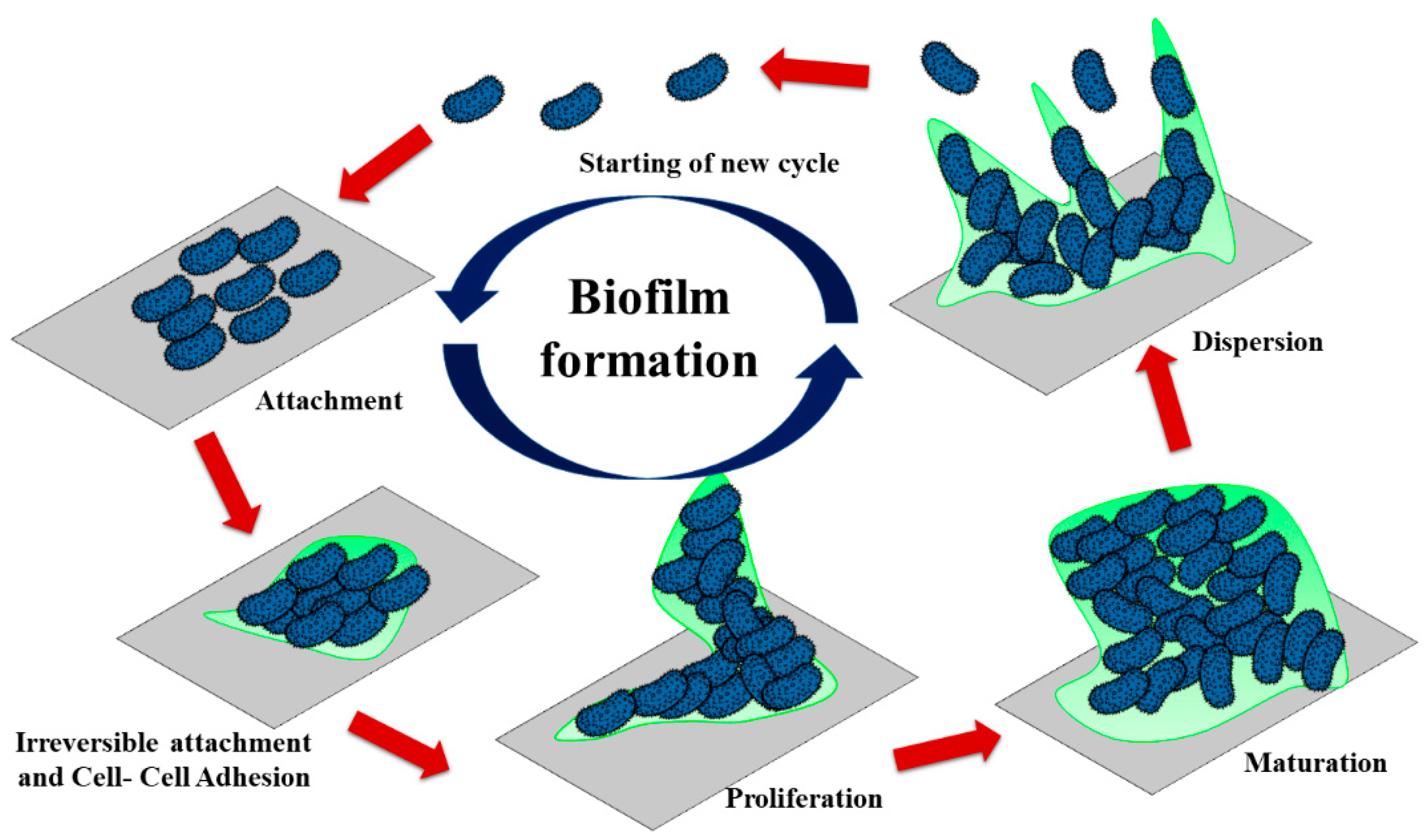
  55. Dunne, W.M. Bacterial Adhesion: Seen any good biofilms lately? Clin. Microbiol. Rev. 2002, 15, 155–166. [Google Scholar] [CrossRef] [PubMed] [Green Version]
  56. Crick, C.R.; Ismail, S.; Pratten, J.; Parkin, I.P. An investigation into bacterial attachment to an elastomeric super hydrophobic surface prepared via aerosol assisted deposition. Thin Solid Films 2011, 519, 3722–3727. [Google Scholar] [CrossRef]
  57. Zhang, X.; Wang, L.; Levanen, E. Superhydrophobic surfaces for the reduction of bacterial adhesion. RSC Adv. 2013, 3, 12003–12020. [Google Scholar] [CrossRef]
  58. Nie, Y.; Kalapos, C.; Nie, X.; Murphy, M.; Hussein, R.; Zhang, L. Superhydrophilicity and Antibacterial property of a Cu dotted oxide coating surface. Ann. Clin. Microbial. 2010, 9, 1–10. [Google Scholar]
  59. Khatoon, Z.; McTiernan, C.D.; Suuronen, E.J.; Mah, T.-F.; Alarcona, E.I. Bacterial biofilm formation on implantable devices and approaches to its treatment and prevention. Heliyon 2018, 4, e01067. [Google Scholar] [CrossRef] [Green Version]
  60. Elizabeth, E.; Baranwal, G.; Krishnan, A.G.; Menon, D.; Nair, M. ZnO Nanoparticle incorporated nanostructured metallic titanium for increased mesenchymal stem cell response and antibacterial activity. Nanotechnology 2014, 25, 115101. [Google Scholar] [CrossRef] [PubMed]
  61. Hans, M.; Erbe, A.; Mathewas, S.; Chen, Y.; Solioz, M.; Mucklich, F. Role of copper oxides in contact killing of bacteria. Langmuir 2013, 29, 16160–16166. [Google Scholar] [CrossRef] [PubMed]
  62. Holinka, J.; Pilz, M.; Kubista, B.; Presterl, E.; Windhager, R. Effects of selenium coating of orthopedic implant surfaces on bacterial adherence and osteoblastic cell growth. Bone Jt. J. 2013, 95, 678–682. [Google Scholar] [CrossRef] [PubMed]
  63. Koseki, H.; Asahara, T.; Shida, T.; Yoda, I.; Horiuchi, H.; Baba, K.; Osaki, M. Clinical and histomorphometrical study on titanium dioxide-coated external fixation pins. Int. J. Nanomed. 2013, 8, 593–599. [Google Scholar] [CrossRef] [PubMed] [Green Version]
  64. Cheng, L.; Li, Y.; Huo, K.; Gao, B.; Xiong, W. Long lasting in vivo and in vitro antibacterial ability of nanostructured titania coating incorporated with silver nanoparticles. J. Biomed. Mater. Res. 2014, 102, 3488–3499. [Google Scholar] [CrossRef] [PubMed]
  65. Thallinger, B.; Prasetyo, E.N.; Nyalnhongo, G.S.; Guebitz, G.M. Antimicrobial enzymes: An emerging strategy to fight microbes and microbial biofilms. Biotechnol. J. 2013, 8, 97–109. [Google Scholar] [CrossRef]
  66. Li, B.; McKeague, A.L. Emerging ideas: Interleukin-12 nanocoating’s prevent open fracture-associated infections. Clin. Orthop. Relat. Res. 2011, 469, 3262–3265. [Google Scholar] [CrossRef] [PubMed] [Green Version]
  67. Rapsch, K.; Bier, F.F.; Tadros, M.; Nickisch-Rosenegk, M.V. Identification of antimicrobial peptides and immobilization strategy suitable for a covalent surface coating with biocompatible properties. Bioconjug. Chem. 2014, 25, 308–319. [Google Scholar] [CrossRef] [PubMed]
  68. Norowski, P.A.; Courtney, H.S.; Babu, J.; Haggard, W.O.; Bumgardner, J.D. Chitosan coatings deliver antimicrobials from titanium implants: A preliminary study. Implant. Dent. 2011, 20, 56–67. [Google Scholar] [CrossRef] [PubMed]
  69. Antoci, V.; Adams, C.S.; Parvizi, J.; Ducheyne, P.; Shapiro, I.M.; Hickok, N.J. Covalently attached vancomycin provides a nanoscale antibacterial surface. Clin. Orthop. Relat. Res. 2007, 461, 81–87. [Google Scholar] [CrossRef]
  70. Glinel, K.; Thebault, P.; Humblot, V.; Pradier, C.M.; Jouenne, T. Antibacterial surfaces developed from bio-inspired approaches. Acta Biomater. 2012, 8, 1670–1684. [Google Scholar] [CrossRef]
  71. He, T.; Chan, V. Covalent layer-by-layer assembly of polyethyleneimine multilayer for antibacterial applications. J. Biomed. Mater. Res. Part A 2010, 95, 454–464. [Google Scholar] [CrossRef]
  72. Rizzello, L.; Pompa, P.P. Nanosilver-based antibacterial drugs and devices: Mechanisms, methodological drawbacks, and guidelines. Chem. Soc. Rev. 2013, 7, 1501–1518. [Google Scholar] [CrossRef] [PubMed]
  73. Gottenbos, B.; van der Mei, H.C.; Klatter, F.; Grijpma, D.W.; Feihen, J.; Nieuwenhuis, P.; Busscher, H.J. Positively charged biomaterials exert antimicrobial effects on gram-negative bacilli in rats. Biomaterials 2003, 24, 2707–2710. [Google Scholar] [CrossRef]
  74. Bernthal, N.M.; Stavrakis, A.I.; Billi, F.; Cho, J.S.; Kremen, T.J.; Simon, S.I.; Cheung, A.L.; Finerman, G.A.; Lieberman, J.R.; Adams, J.S.; et al. A mouse model of post-arthroplasty Staphylococcus aureus joint infection to evaluate in vivo the efficacy of antimicrobial implant coatings. PLoS ONE 2010, 5, e12580. [Google Scholar] [CrossRef] [Green Version]
  75. Zhu, H.; Guo, Z.; Liu, W. Adhesion behaviors on super hydrophobic surfaces. Chem. Commun. 2014, 18, 3900–3913. [Google Scholar] [CrossRef]
  76. Shida, T.; Koeski, H.; Yoda, I.; Horiuchi, H.; Sakoda, H.; Osaki, M. Adherence ability of Staphylococcus epidermidis on prosthetic biomaterials: An in vitro study. Int. J. Nanomed. 2013, 8, 3955–3961. [Google Scholar]
  77. Pandit, V.; Zuidema, J.M.; Venuto, K.N.; Macione, J.; Dai, G.; Gilbert, R.J.; Kotha, S.P. Evaluation of multifunctional polysaccharide hydrogels with varying stiffness for bone tissue engineering. Tissue Eng. A 2013, 19, 2452–2463. [Google Scholar] [CrossRef] [PubMed] [Green Version]
  78. Yu, Q.; Cho, J.; Shivapooja, P.; Ista, L.K.; Lopez, G.P. Nanopatterned smart polymer surfaces for controlled attachment, killing, and release of bacteria. ACS Appl. Master Interfaces 2013, 5, 9295–9304. [Google Scholar] [CrossRef] [PubMed]
  79. Parvizi, J.; Antoci, V., Jr.; Hickok, N.J.; Shapiro, I.M. Self-protective smart orthopedic implants. Expert Rev. Med. Devices 2007, 4, 55–64. [Google Scholar] [CrossRef]
  80. Bhat, U.; Augustin, A.; Bhat, S.; Udupa, K.R. Preparation and characterization of copper thin films for Antimicrobial applications. In Microscopy Applied to Materials Sciences and Life Sciences; Apple Academic Press Inc.: Waretown, NJ, USA, 2018; pp. 91–110. [Google Scholar]
  81. Singh, H.; Sidhu, T.S.; Kalsi, S.B.S.; Karthikeyan, J. Development of cold spray from innovation to emerging future coating technology. J. Braz. Soc. Mech. Sci. Eng. 2013, 35, 231–245. [Google Scholar] [CrossRef]
  82. Tejero-Martin, D.; Rezvani Rad, M.; McDonald, A.; Hussain, T. Beyond Traditional Coatings: A Review on Thermal-Sprayed Functional and Smart coatings. J. Therm. Spray Technol. 2019, 28, 598–644. [Google Scholar] [CrossRef] [Green Version]
  83. Borchers, C.; Gartner, F.; Stoltenhof, T.; Assadi, H.; Kreye, H. Microstructural and macroscopic properties of cold sprayed copper coatings. J. Appl. Phys. 2003, 93, 10064. [Google Scholar] [CrossRef]
  84. Crawmer, D.E. Thermal Spray Processes. In Handbook of Thermal Spray Technology; ASM International: Russell Township, OH, USA, 2013; pp. 54–76. [Google Scholar]
  85. Smith, M.F.; Hall, A.C.; Fleetwood, J.D.; Meyer, P. Very Low-Pressure Plasma Spray—A Review of an Emerging Technology in the Thermal Spray Community. Coatings 2011, 1, 117–132. [Google Scholar] [CrossRef] [Green Version]
  86. Jandin, G.; Liao, H.; Feng, Z.Q.; Coddet, C. Correlations between operating conditions, microstructure and mechanical properties of twin wire arc sprayed steel coatings. Mater. Sci. Eng. 2003, 349, 298–305. [Google Scholar] [CrossRef] [Green Version]
  87. Wang, X.; Heberlein, J.; Pfender, E.; Gerberich, W. Effect of Nozzle Configuration, Gas Pressure, and Gas Type on Coating Properties in Wire Arc Spray. J. Therm. Spray Technol. 1999, 8, 565–575. [Google Scholar] [CrossRef]
  88. Pourmousa, A.; Mostaghimi, J.; Abedini, A.; Chandra, S. Particle size distribution in a wire-arc spraying system. J. Therm. Spray Technol. 2005, 14, 502–510. [Google Scholar] [CrossRef]
  89. Davis, J.R. Cold Spray Process. In Thermal Spray Technology; ASM International: Russell Township, OH, USA, 2004; pp. 77–84. [Google Scholar]
  90. Champagne, V.; Sundberg, K.; Helfritch, D. Kinetically deposited copper antimicrobial surfaces. Coatings 2019, 9, 257. [Google Scholar] [CrossRef] [Green Version]
  91. Sundberg, K.L. Application of Materials Characterization, Efficacy Testing, and Modelling Methods on Copper Cold Spray Coatings for Optimized Antimicrobial Properties. Ph.D. Thesis, Worcester Polytechnic Institute, Worcester, MA, USA, 2019. [Google Scholar]
  92. Da Silva, F.S.; Cinca, N.; Dosta, S.; Cano, I.; Guilemany, J.; Caires, C.; Lima, A.; Silva, C.; Oliveira, S.; Caires, A.; et al. Corrosion resistance and antibacterial properties of copper coatings deposited by cold gas spray. Surf. Coat. Technol. 2019, 361, 292–301. [Google Scholar] [CrossRef]
  93. Sharifahmadian, O.; Salimijazi, H.R.; Fathi, M.H.; Mostaghimi, J.; Pershin, L. Relationship between surface properties and antibacterial behavior of wire arc spray copper coatings. Surf. Coat. Technol. 2013, 233, 74–79. [Google Scholar] [CrossRef]
  94. Worna, A.; Bilewska, K.; Lis, M.; Kaminska, M.; Olszewski, T.; Pajzderski, P.; Więcław, G.; Jaśkiewicz, M.; Kamysz, W. Antimicrobial properties of protective coatings produced by plasm spraying technique. Surf. Coat. Technol. 2017, 318, 332–340. [Google Scholar]
  95. Ibrahim, M.; Al-Athel, K.; Fazal, A.; Arif, M. Strength and Hardness Assessment of Copper and Copper Alloy Coatings on Stainless Steel Substrates. In Proceedings of the ASME 2016 International Mechanical Engineering Congress and Exposition, Phoenix, AZ, USA, 11–17 November 2016. [Google Scholar]
  96. Champagne, V.K.; Helfritch, D.J. A Demonstration of the antimicrobial effectiveness of various copper surfaces. J. Biol. Eng. 2013, 7, 8. [Google Scholar] [CrossRef] [PubMed] [Green Version]
  97. Sharifahmadian, O.; Salimijazi, H.R.; Fathi, M.H.; Mostaghimi, J.; Pershin, L. Study of the Antibacterial Behavior of Wire Arc Sprayed Copper Coatings. J. Therm. Spray Technol. 2013, 22, 371–379. [Google Scholar] [CrossRef]
  98. Tailor, S.; Modi, A.; Modi, S.C. Thermally Sprayed Thin Copper Coatings by W-HVOF. J. Therm. Spray Technol. 2019, 28, 273–282. [Google Scholar] [CrossRef]
  99. Augustin, A.; Thaira, H.; Bhat, K.U.; Udupa, K.R. Effect of Electrodeposited Copper Thin Film on the Morphology and Cell Death of E. coli; Electron Microscopic Study. In Biotechnology and Biochemical Engineering; Springer: Singapore, 2016; pp. 227–232. [Google Scholar]
  100. Augustin, A.; Huilgol, P.; Udupa, K.R.; Bhat, K.U. Effect of current density during electrodeposition on microstructure and hardness of textured Cu coating in the application Al touch surface. J. Mech. Behav. Biomed. Mater. 2016, 63, 352. [Google Scholar] [CrossRef]
  101. Schwarzacher, W. Electrodeposition: A technology for the future. Electrochem. Soc. Interface 2016, 15, 32–35. [Google Scholar] [CrossRef]
  102. Rasmussen, A.A.; Jensen, J.A.D.; Horsewell, A.; Somers, M.A. Microstructure in electrodeposited copper layers; the role of the substrate. Electrochim. Acta. 2001, 47, 67–74. [Google Scholar] [CrossRef]
  103. Nikolic, N.D.; Popov, K.I.; Pavlovic, L.J.; Pavlovic, M.G. Morphologies of copper deposits obtained by the electrodeposition at high overpotentials. Surf. Coat. Technol. 2006, 201, 560–566. [Google Scholar] [CrossRef]
  104. Milchev, A.; Zapryanova, T. Nucleation and growth of copper under combined charge transfer and diffusion limitations. Electrochim. Acta Part I 2006, 51, 433–438. [Google Scholar] [CrossRef]
  105. Ibañez, A.; Fatás, E. Mechanical and structural properties of electrodeposited copper and their relation with the electrodeposition parameters. Surf. Coat. Technol. 2005, 191, 7–16. [Google Scholar] [CrossRef]
  106. Shekar, R. Synergistic effect of additives on electrodeposition of copper from cyanide free electrolytes and its structural and morphological characteristics. Trans. Nonferrous Met. Soc. China 2017, 27, 1665–1676. [Google Scholar] [CrossRef]
  107. Shao, W.; Pattanaik, G.; Zangari, G. Influence of chloride anions on the mechanism of copper electrodeposition from acidic sulfate electrolytes. J. Electrochem. Soc. 2007, 154, D201–D207. [Google Scholar] [CrossRef]
  108. Norziehana, N.; Isa, C.; Mohd, Y.; Mohamad, S.A.S.; Zaki, M.H.M. Antibacterial Activity of Copper Coating Electrodeposited on 304 stainless steel. AIP Conf. Proc. 2017, 1901, 020009. [Google Scholar]
  109. Ivshin, Y.V.; Shaikhutdinova, F.N.; Sysoev, V.A. Electrodeposition of Copper on Mild Steel: Peculiarities of the Process. Surf. Eng. Appl. Electrochem. 2017, 54, 452–458. [Google Scholar]
  110. Haider, F.I.; Ani, M.H.; Mahmood, M.H. Modelling and Optimization of Copper Electroplating Adhesion Strength. In IOP Conference Series: Materials Science and Engineering, Proceedings of the 5th International Conference on Nanomaterials and Materials Engineering, Bali, Indonesia, 1–3 April 2017; IOP Publishing Ltd.: Bristol, UK, 2017; Volume 204. [Google Scholar]
  111. Zeiger, M.; Solioz, M.; Edongue, H.; Arzt, E.; Schneider, A.S. Surface structure influences contact killing of bacteria by copper. Microbiol. Open 2014, 3, 327–332. [Google Scholar] [CrossRef]
  112. Augustin, A.; Bhat, U.K.; Rajendra Udupa, K.K.; Hegade, C.A. Electron microscopic study of nodules formed during electrodeposition of copper on aluminium. In Materials Science Forum; Trans Tech Publications Ltd.: Freienbach, Switzerland, 2015; pp. 371–374. [Google Scholar]
  113. Scholz, F. Active sites of heterogeneous nucleation understood as chemical reaction sites. Electrochem. Commun. 2011, 13, 932–933. [Google Scholar] [CrossRef]
  114. Kumar, S.K.; Biswas, K. Effect of thiourea on grain refinement and defect structure of the pulsed electrodeposited nanocrystalline copper. Surf. Coat. Technol. 2013, 214, 8–18. [Google Scholar] [CrossRef]
  115. Banthiaa, S.; Sengupta, S.; Mallik, M.; Das, S.; Das, K. Substrate effect on electrodeposited copper morphology and crystal shapes. Surf. Eng. 2018, 34, 485–492. [Google Scholar] [CrossRef]
  116. Chandrasekar, M.S.; Pushpavanam, M. Pulse and pulse reverse plating—Conceptual, advantages and applications. Electrochim. Acta 2008, 53, 3313–3322. [Google Scholar] [CrossRef]
  117. Deckert, C.A. Electroless Copper Plating A Review: Part, I. Plat. Surf. Eng. 1995, 82, 48–55. [Google Scholar]
  118. Ghosh, S. Electroless copper deposition: A critical review. Thin Solid Films 2019, 669, 641–658. [Google Scholar] [CrossRef]
  119. Li, J.; Hayden, H.; Kohl, P.A. The influence of 2,2′-dipyridyl on non-formaldehyde electroless copper plating. Electrochem. Acta 2004, 49, 1789–1795. [Google Scholar] [CrossRef]
  120. Gan, X.; Wu, Y.; Liu, L.; Hu, W. Effects of K4Fe(CN)6 on electroless copper plating using hypophosphite as reducing agent. J. Appl. Electrochem. 2007, 37, 899–904. [Google Scholar] [CrossRef]
  121. Luo, L.M.; Lu, Z.L.; Huang, X.M.; Tan, X.Y.; Ding, X.Y.; Cheng, J.G.; Zhu, L.; Wu, Y.-C. Electroless copper plating on PC engineering plastic with a novel palladium-free surface activation process. Surf. Coat. Technol. 2014, 251, 69–73. [Google Scholar] [CrossRef]
  122. Oita, M.; Matsuoka, M.; Iwakura, C. The deposition rate and morphology of electroless copper film from solutions containing 2,2′-dipyridyl. Electrochim. Acta 1997, 42, 1435–1440. [Google Scholar] [CrossRef]
  123. Argueta–Figueroa, L.; Morales-Luckie, R.A.; Scougall-Vilchis, R.J.; Olea-Mejia, O.F. Synthesis, characterization and antibacterial activity of copper, nickel and bimetallic Cu–Ni nanoparticles for potential use in dental materials. Prog. Nat. Sci. Mater. Int. 2014, 24, 321–328. [Google Scholar] [CrossRef] [Green Version]
  124. BuFaroosha, M. Precursors for Chemical Vapor Deposition of Copper. Doctoral Dissertations, Louisiana State University, Baton Rouge, LA, USA, 2002. No. 1437. [Google Scholar]
  125. Shima, K.; Shimizu, H.; Momose, T.; Shimogaki, Y. Comparative Study on Cu-CVD Nucleation Using -diketonato and Amidinato Precursors for Sub-10-nm-Thick Continuous Film Growth. ECS J. Solid-State Sci. Technol. 2015, 4, P305–P313. [Google Scholar] [CrossRef]
  126. Chiou, J.-C.; Chen, Y.-J.; Chen, M.-C. Copper chemical vapor deposition from Cu(hexafluoroacetylacetonate)trimethylvinylsilane. J. Electron. Mater. 1994, 23, 383–390. [Google Scholar] [CrossRef]
  127. Choy, K.L. Chemical vapor deposition of coatings. Prog. Mater. Sci. 2003, 48, 57–170. [Google Scholar] [CrossRef]
  128. Aviziotis, I.G.; Cheimarios, N.; Vahlas, C.; Boudouvis, A.G. Experimental and computational investigation of chemical vapor deposition of Cu from Cu amidinate. Surf. Coat. Technol. 2013, 230, 273–278. [Google Scholar] [CrossRef] [Green Version]
  129. Quinto, D.T. Mechanical property and structure relationships in hard coatings for cutting tools. J. Vac. Sci. Technol. 1988, 6, 2149–2157. [Google Scholar] [CrossRef]
  130. Varghese, S.; El Fakhri, S.O.; Sheel, D.W.; Sheel, P.; Frederick, J.; Bolton, E.; Foster, H.A. Antimicrobial activity of novel nanostructured Cu-SiO2 coatings prepared by chemical vapor deposition against hospital related pathogens. AMB Express 2013, 3, 53. [Google Scholar] [CrossRef] [Green Version]
  131. Hassan, I.A.; Parkin, I.P.; Nairb, S.P.; Carmalt, C.J. Antimicrobial activity of copper and copper(i) oxide thin films deposited via aerosol-assisted CVD. J. Mater. Chem. B 2014, 2, 2855–2860. [Google Scholar] [CrossRef]
  132. Ozkan, E.; Crick, C.C.; Taylor, A.; Allan, E.; Parkin, I.P. Copper-based water repellent and antibacterial coatings by aerosol assisted chemical vapor deposition. R. Soc. Chem. 2016, 7, 5126–5131. [Google Scholar]
  133. Romig, A., Jr. A time dependent regular solution model for the thermal evaporation of an Al-Mg alloy. 2. J. Appl. Phys. 1987, 62, 503–508. [Google Scholar] [CrossRef]
  134. Alam, M.M.; Mia, N.H.; Hasan, R.; Shahinuzzaman, M.; Islam, M.K.; Khan, M. Nasir Uddin. Study of Structural and Morphological Properties of Vacuum coated copper (Cu) metal thin film. Mater. Sci. Appl. 2015, 6, 753–759. [Google Scholar]
  135. Ustinov, A.I.; Movachan, B.A.; Polishchuk. Formation of Nano quasi crystalline Al-Cu-Fe Coatings at Electron Beam Physical Vapor Deposition. Scr. Mater. 2004, 50, 533–537. [Google Scholar] [CrossRef]
  136. Singh, J.; Wolfe, D.E. Nano and macro-structured component fabrication by electron beam—Physical vapor deposition (EB-PVD). J. Mater. Sci. 2005, 40, 1–26. [Google Scholar] [CrossRef]
  137. Komalakrishna, H.; Augustin, A.; Bhat, U.K. Electron Beam Deposition of Copper Thin Film on Aluminium Substrate and Its Characterization. Am. J. Mater. Sci. 2015, 5, 9–24. [Google Scholar]
  138. Kelly, P.; Arnell, R. Magnetron sputtering: A review of recent developments and applications. Vacuum 2000, 56, 159–172. [Google Scholar] [CrossRef]
  139. Thornton, J.A. High-rate thick film growth. Annu. Rev. Mater. Sci. 1977, 7, 239–260. [Google Scholar] [CrossRef]
  140. Augustin, A.; Rajendra Udupa, K.; Bhat, U.K. Effect of Pre-Zinc Coating on the Properties and Structure of DC magnetron sputtered copper thin film on aluminium. Am. J. Mater. Sci. 2015, 5, 58–61. [Google Scholar]
  141. Tian, X.B.; Wang, Z.M.; Yang, S.Q.; Luo, Z.J.; Ricky, K.Y.; Fu, P.; Chu, K. Antibacterial copper containing titanium nitride films produced by dual magnetron sputtering. Surf. Coat. Technol. 2007, 201, 8606–8609. [Google Scholar] [CrossRef]
  142. Rodriguez-Barrero, S.; Fernandez–Larrinoa, J.; Azkona, I.; Lopez de Lacalle, L.N.; Polvorosa, R. Enhanced Performance of Nanostructred Coatings for Drilling by Droplet Ellimination. Mater. Manuf. Process 2016, 31, 593–602. [Google Scholar] [CrossRef]
  143. Abdullah, M.R.; Harun, N.H.; Ibrahim, S.N.; Abdul, A. Thin Film Coating of Copper Nanoparticles with DC Magnetron Sputtering via Physical Vapor Deposition. AIP Conf. Proc. 2019, 2129, 020120. [Google Scholar]
  144. Musil, J.; Blazek, J.; Fajfrlik, K.; Cerstvy, R.; Proksova, S. Antibacterial Cr-Cu-O films prepared by reactive magnetron sputtering. Appl. Surf. Sci. 2013, 276, 660–666. [Google Scholar] [CrossRef]
  145. Wang, S.; Zhu, W.; Yu, P.; Wang, X.; He, T.; Tan, G.; Ning, C. Antibacterial nanostructured copper coatings deposited on tantalum by magnetron sputtering. Mater. Technol. 2015, 30, B120–B125. [Google Scholar] [CrossRef]
  146. Benetti, G.; Cavaliere, E.; Banfi, F.; Gavioli, L. Antimicrobial Nanostructured Coatings: A Gas Phase Deposition and Magnetron Sputtering Perspective. Materials 2020, 13, 784. [Google Scholar] [CrossRef] [Green Version]
Figure 1. Event of the release of copper ion for coated copper surface during bacteria interaction.
Metals 11 00711 g001
Figure 2. Illustration on the events in contact killing (a) Breaking of the inner and outer membrane (b) Damage by oxidation caused by the ROS (c) Deactivation of the essential enzymes (d) Destruction of the deoxyribonucleic acid (DNA).
Metals 11 00711 g002
Figure 3. Schematic view of biofilm formation on the surface.
Metals 11 00711 g003
Figure 4. Process parameters tuning for better antimicrobial properties in the thermal spray process.
Metals 11 00711 g004
Figure 5. Typical microstructures of a thermally sprayed coating.
Metals 11 00711 g005
Figure 6. Percent Staphylococcus aureus surviving after two-hour exposure to the copper surface [90].
Metals 11 00711 g006
Figure 7. A schematic diagram for (a) the Electrolytic deposition process (b) the Electroless deposition process. Simplicity, economic and easy adaptability of both processes can attract many industries.
Metals 11 00711 g007
Figure 8. (a) Bright-field TEM micrographs of copper nanocoatings deposited at j = 2 Adm−2 (b) crystallite distribution of features presented in Figure 8a, (c) Bright-field TEM micrographs of copper nanocoating are deposited at j = 10 Adm−2 (d) Crystallite distribution to Figure 8c. (Reproduced with permission from ref. [100] copyright 2016, Journal of the Mechanical Behaviour of Biomedical Materials).
Metals 11 00711 g008
Figure 9. Current (A) versus time (Seconds) diagram in the case of pulse electrodeposition.
Metals 11 00711 g009
Figure 10. Sketch showing microstructure formation during atomistic deposition on a metal substrate using an electroless deposition process.
Metals 11 00711 g010
Figure 11. Schematic diagram of the chemical vapor deposition process.
Metals 11 00711 g011
Figure 12. Sketch showing coating property correlation with chemical vapor deposition (CVD) process parameters.
Metals 11 00711 g012
Figure 13. (a) Instrumentation and configuration of the PVD setup. (b) Sketch showing the distribution of coating concerning vapor flux which is due to line-of-sight limitation.
Metals 11 00711 g013
Figure 14. Sputtering technique on an atomic scale.
Metals 11 00711 g014
Table 1. Important strategies proposed for the development of antimicrobial surfaces.
Antibacterial FunctionalityTested ExamplesReference
Antibacterial agent release Nanoparticles[33]
Copper oxides[45]
Contact killingInorganicZinc ion[60]
Copper ion[61]
Selenium ion[62]
Titanium dioxide[63]
Silver nanoparticles[64]
OrganicEnzymes[65]
Cytokine[66]
Signaling, inhibiting, and antimicrobial peptides[67]
Chitosan derivatives[68]
Coated or covalently linked antibiotics[69]
Other Non-antibiotic bactericidal substances[70]
CombinedMultilayer coatings [71]
Synergy material intensification[72]
Positively charged polymers[73]
Anti-adhesion/Bacterial Repelling Anti-adhesive polymers [74]
Superhydrophobic surfaces[75]
Nanopatterned surfaces[76]
Hydrogels[77]
Smart CoatingsPassiveNanostructured “smart” material[78]
ActiveConcept: Sensors conjoined to Nano containers [79]
Table 2. Advantages and disadvantages of different coating routes for copper deposition which could affect the antimicrobial activity.
TechniqueAdvantagesInconveniences
Thermal Spraying Low cost
High deposition rates
Excellent adhesion strength
High temperature induces decomposition, oxidation of Cu could affect the antimicrobial activity.
The line of sight technique leads to nonuniformity
Amorphous coatings due to rapid cooling
Interface separation and spalling of coating between the coating and substrate
Electrodeposition Uniform distribution of coating
Simple technique
Can easily tune the coating morphologies
Can operate at low temperature
The coating is possible for highly complex objects
Controlled nanoporous coatings
Low tear strength
Poor adhesion
Surface pretreatment required to deposit Cu on metal substrates
Alkaline and acidic baths suffer environmental disposal issues
Electroless depositionSimple process
Low temperature
Coats highly irregular objects
Coating pure Cu is difficult
Limited salts for Cu plating
Physical vapor deposition Less prone to crack formation
Thin layers
More adherent than thermal spray
Expensive method
Difficulty producing nanoporous coatings
Chemical vapor deposition Can easily control precursor concentrations to fabricate functionally graded coatingsExpensive method
The use of volatile gases cause the formation of contamination on Cu surfaces that could reduce the rate of bacteria-killing
Limited coating composition
Sputtering Homogenous coating
Uniform coating on flat substrates
High adhesion
Dense deposits
Expensive technique and time-consuming
Line of sight issues
Amorphous coatings
Table 3. Comparison between thermal spray, electrodeposition, electroless plating, CVD, physical vapor deposition (PVD)—sputtering, EB-PVD deposition routes.
Deposition
Method
Ideal Process Parameters for Cu DepositionSubstrate Heating (°C) and Substrate PreparationCoating Rate and Coating ThicknessType of Adhesion Bonding and Surface RoughnessTypical Microstructure for Antimicrobial Application
Thermal SprayTechnique
[90,91,92,93,94,95,96,97,98]
Plasma Arc: Temperature: 1500–2500 °C
Velocity: 100-400 m/s
Porosity: ~5%
Oxides: ~2%
Wire Arc: Temperature: 1500–2500 °C
Velocity: 50–100 m/s
Porosity: ~10%
Oxides: ~15%
Cold Spray: Temperature: 150–400 °C
Velocity: 500–100 m/s
Porosity: ~10%
Oxides: ~15%
Flame spray: Temperature: 1010–1175 °C
Porosity: ~1%
Oxides: ~15%
Copper particles size is less than 20 μm with 99% purity for thermally sprayed coatings.
Usually ~10% of Copper melting temperature
Carbon steel, Stainless steel widely used substrates for antimicrobial applications.
>100 μm/min Approximately
Coating thickness: 30 μm–300 μm
Mechanical
Interlocking and
Very rough coatings,
higher bacterial killing rate.
Good adhesion strength
between substrate and
coating due to high surface roughness of the substrate.
Deformed lamella
Electro
deposition
[99,100,102,103,104,105,106,108,109,112,115]
Current density range: 1 Adm−2 to 10 Adm−2
High current density will lead to formation
of defects like nanotwins and staking faults regions which could improve the hardness
of the coatings.
pH range: For acidic bath: 1.5 to 4
for alkaline bath 12 to 14
Cannot heat substrate while deposition.
Stainless steel, Aluminium, mild steel are the good
substrates for Cu electrodeposition.
<0.05 μm/min
(<3 μm/h)
Thickness in the range of 1 μm to
few microns.
Mechanical interlocking.
Surface contamination, oxide layer on the substrate decreases the adhesion Strength.
Surface roughness depends
on substrate pre-treatment
Cu coatings shows Columnar structures and Nanograined structures good for antimicrobial activity
Electroless Plating
[118,122]
Coating time: Few minutes to hours.
Additives are commonly used in the bath to refine the film morphology and deposition rate.
Optimum bath temperature: 50 °C–80 °C
Higher the pH coating quality degrades.
Cannot heat
substrate while deposition.
Suitable substrates: Aluminium, glass, polymers, mild steel, and stainless steel.
<0.03 μm/min
(<2 μm/h)
30 min to one hour plating time required to achieve >1 μm to few microns thickness of Cu coating
Mechanical interlocking
Smooth, dense, bright Cu coatings irrespective of substrate type.
Columnar-equiaxed, Nano grained, uniform thickness
Chemical Vapor Deposition (CVD)
[124,125,128,130,131,132]
Type of precursor used for Cu deposition:
Copper (I) amidinate [Cu(i-Pr-Me-AMD)]2, hexafluoroacetylacetonate (Cu (HFAc)2)
Carrier gas: N2 gas.
Deposition time: 1 h
200 °C–500 °C
Suitable substrates: Stainless steel, silicon, SiO2, borosilicate glass
<0.08 μm/min
(<5 μm/h)
Coating thickness: 80 nm to 270 nm
Coating thickness: 40 nm
(100% antimicrobial activity under 2 h)
Diffusion bondingColumnar-equiaxed
nodular and smooth, uniform morphology.
Morphology can be tuned by substrate roughness
and substrate heating
Columnar structure is
good for antimicrobial activity
Electron Beam Deposition-PVD
[134,135,136,137]
Vacuum level: 1.5 × 10⁵ mbar
Source and substrate distance: 10 cm
Coating time: 1 h
RT to 1200 °C
(flexible)
Suitable Substrates: Titanium, Aluminium, Stainless steel, low carbon steel
Deposition rate: 1 Å/sDiffusion bondingColumnar-equiaxed,
Nano sized grains
Increases the surface area—more beneficial for antimicrobial activity.
Sputtering Technique
[141,142,143,144,145,146]
Base pressure:
5 × 10⁻⁵ Pa to 1.3 × 10⁻⁴ Pa
Working pressure range:
0.012 mbar to 0.04 mbar
Mass flow rate of Ar gas:
Substrate rotation speed: Low speed
(100 rpm)
<600 °C
(flexible)
Suitable Substrates: Titanium, Aluminium, Stainless steel, low carbon steel.
<0.08 μm/min
(<5 μm/h)
Diffusion bondingColumnar, uniform, adherent coating observed in the range of 50 W to 150 W
Publisher’s Note: MDPI stays neutral with regard to jurisdictional claims in published maps and institutional affiliations.

