Recent Advancements in Nanoparticle Based Imaging and Therapy

A special issue of Journal of Nanotheranostics (ISSN 2624-845X).

Deadline for manuscript submissions: closed (30 September 2021) | Viewed by 22056

Special Issue Editors


E-Mail Website
Guest Editor
Department of Health Technology, Technical University of Denmark, Roskilde, Denmark
Interests: application of radiotheranostics to oncology and neurology; molecular imaging; continuous production of PET radionuclides using flow technologies; nanotechnology; underdeveloped radionuclides for radiotheranostics

E-Mail Website
Guest Editor
Department of Radiology, Mayo Clinic, Rochester, MN, USA
Interests: molecular imaging; radiochemistry; isotope production; cGMP; radiopharmaceuticals; nanotechnology; medicinal and synthetic organic chemistry

Special Issue Information

Dear Colleagues,

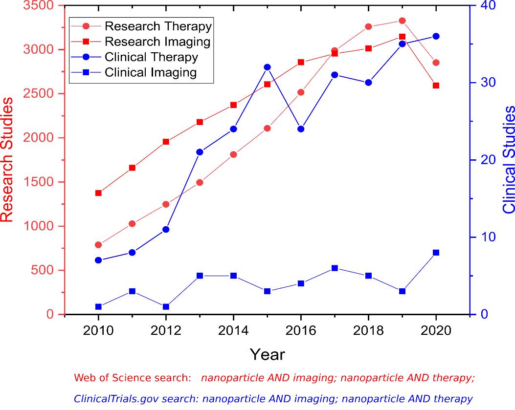
Nanoparticles are defined as a nanosized material, typically under 100 nm. Over the last two decades, there has been an unabated interest in expanding the role of nanotechnology into medicine. The nanosized formulations are designed to improve the target site accumulation, thus maximizing the drug’s efficacy and minimizing toxicity. For therapy, nanoparticles can act as delivery vehicles carrying drugs to their biological targets. When loaded with the appropriate contrast agent or radionuclides, nanoparticles can serve as optical, magnetic resonance imaging (MRI), single-photon emission computed tomography (SPECT), and positron emission tomography (PET) imaging probes. The continuous research and development effort led to remarkable scientific progress but also highlighted considerable clinical challenges. The cytotoxicity, limited in-vivo efficacy, and immune response to nanoparticles remain major hurdles for clinical translation. The other challenge is the need for appropriate characterization of nanoparticles for clinical use and the issue of scale-up and batch-to-batch variations. The dichotomy between academic research and the clinical translation is further reflected in the number of corresponding publications over the years as shown in Figure 1. Based on this analysis, only ~1% of nanoparticle-based research is translated into clinical therapy studies, and ~0.4% of imaging research is translated into clinical imaging studies. Therefore, we propose this thematic issue to inform and guide the scientific community by inviting experts in the field to provide their critical findings, systematic, perspective, and education reviews to reduce this gap and overcome major challenges faced in the clinical translation of nanoparticle-based therapy and imaging.
text

Research and clinical publications in the field of nanotheranostics.

This thematic issue will cover the following:

  • Targeted nanotheranostics for imaging and drug delivery applications
  • Novel nanoplatforms for imaging and or radiotheranostics applications
  • Nanosized multimodal imaging probes and superparamagnetic iron oxide (SPIO) nanoparticles
  • Novel approaches to loading, controlled release, chelation, and conjugation strategy of theranostic radionuclides and conjugates
  • Preclinical evaluation of novel nanoparticles
  • Novel biochemical mechanism for nanoparticle internalization (disease-specific or case study)
  • Radiomics-based patient stratification strategies
  • Translating preclinical efficacy and toxicology to clinical outcome
  • Regulatory challenges and approach towards clinical translation

Dr. Fedor Zhuravlev
Dr. Mukesh K. Pandey
Guest Editors

Manuscript Submission Information

Manuscripts should be submitted online at www.mdpi.com by registering and logging in to this website. Once you are registered, click here to go to the submission form. Manuscripts can be submitted until the deadline. All submissions that pass pre-check are peer-reviewed. Accepted papers will be published continuously in the journal (as soon as accepted) and will be listed together on the special issue website. Research articles, review articles as well as short communications are invited. For planned papers, a title and short abstract (about 100 words) can be sent to the Editorial Office for announcement on this website.

Submitted manuscripts should not have been published previously, nor be under consideration for publication elsewhere (except conference proceedings papers). All manuscripts are thoroughly refereed through a single-blind peer-review process. A guide for authors and other relevant information for submission of manuscripts is available on the Instructions for Authors page. Journal of Nanotheranostics is an international peer-reviewed open access quarterly journal published by MDPI.

Please visit the Instructions for Authors page before submitting a manuscript. The Article Processing Charge (APC) for publication in this open access journal is 1000 CHF (Swiss Francs). Submitted papers should be well formatted and use good English. Authors may use MDPI's English editing service prior to publication or during author revisions.

Keywords

  • nanoparticles
  • imaging
  • radiotherapy
  • nanotheranostics

Benefits of Publishing in a Special Issue

  • Ease of navigation: Grouping papers by topic helps scholars navigate broad scope journals more efficiently.
  • Greater discoverability: Special Issues support the reach and impact of scientific research. Articles in Special Issues are more discoverable and cited more frequently.
  • Expansion of research network: Special Issues facilitate connections among authors, fostering scientific collaborations.
  • External promotion: Articles in Special Issues are often promoted through the journal's social media, increasing their visibility.
  • e-Book format: Special Issues with more than 10 articles can be published as dedicated e-books, ensuring wide and rapid dissemination.

Further information on MDPI's Special Issue polices can be found here.

Published Papers (3 papers)

Order results
Result details
Select all
Export citation of selected articles as:

Research

Jump to: Review

12 pages, 1594 KiB  
Communication
Surface Adsorption of the Alpha-Emitter Astatine-211 to Gold Nanoparticles Is Stable In Vivo and Potentially Useful in Radionuclide Therapy
by Emanuel Sporer, Christian B. M. Poulie, Sture Lindegren, Emma Aneheim, Holger Jensen, Tom Bäck, Paul J. Kempen, Andreas Kjaer, Matthias M. Herth and Andreas I. Jensen
J. Nanotheranostics 2021, 2(4), 196-207; https://doi.org/10.3390/jnt2040012 - 1 Oct 2021
Cited by 6 | Viewed by 3769
Abstract
Targeted α-therapy (TAT) can eradicate tumor metastases while limiting overall toxicity. One of the most promising α-particle emitters is astatine-211 (211At). However, 211At-carbon bonds are notoriously unstable in vivo and no chelators are available. This hampers its adoption in TAT. [...] Read more.
Targeted α-therapy (TAT) can eradicate tumor metastases while limiting overall toxicity. One of the most promising α-particle emitters is astatine-211 (211At). However, 211At-carbon bonds are notoriously unstable in vivo and no chelators are available. This hampers its adoption in TAT. In this study, the stability of 211At on the surface of gold nanoparticles (AuNPs) was investigated. The employed AuNPs had sizes in the 25–50 nm range. Radiolabeling by non-specific surface-adsorption in >99% radiochemical yield was achieved by mixing 211At and AuNPs both before and after polyethylene glycol (PEG) coating. The resulting 211At-AuNPs were first challenged by harsh oxidation with sodium hypochlorite, removing roughly 50% of the attached 211At. Second, incubation in mouse serum followed by a customized stability test, showed a stability of >95% after 4 h in serum. This high stability was further confirmed in an in vivo study, with comparison to a control group of free 211At. The AuNP-associated 211At showed low uptake in stomach and thyroid, which are hallmark organs of uptake of free 211At, combined with long circulation and high liver and spleen uptake, consistent with nanoparticle biodistribution. These results support that gold surface-adsorbed 211At has high biological stability and is a potentially useful delivery system in TAT. Full article
(This article belongs to the Special Issue Recent Advancements in Nanoparticle Based Imaging and Therapy)
Show Figures

Graphical abstract

Review

Jump to: Research

26 pages, 3377 KiB  
Review
Theranostic Applications of Nanoparticle-Mediated Photoactivated Therapies
by Shalini Sharma, Andrei V. Zvyagin and Indrajit Roy
J. Nanotheranostics 2021, 2(3), 131-156; https://doi.org/10.3390/jnt2030009 - 3 Aug 2021
Cited by 10 | Viewed by 4173
Abstract
Nanoparticle-mediated light-activated therapies, such as photodynamic therapy and photothermal therapy, are earnestly being viewed as efficient interventional strategies against several cancer types. Theranostics is a key hallmark of cancer nanomedicine since it allows diagnosis and therapy of both primary and metastatic cancer using [...] Read more.
Nanoparticle-mediated light-activated therapies, such as photodynamic therapy and photothermal therapy, are earnestly being viewed as efficient interventional strategies against several cancer types. Theranostics is a key hallmark of cancer nanomedicine since it allows diagnosis and therapy of both primary and metastatic cancer using a single nanoprobe. Advanced in vivo diagnostic imaging using theranostic nanoparticles not only provides precise information about the location of tumor/s but also outlines the narrow time window corresponding to the maximum tumor-specific drug accumulation. Such information plays a critical role in guiding light-activated therapies with high spatio-temporal accuracy. Furthermore, theranostics facilitates monitoring the progression of therapy in real time. Herein, we provide a general review of the application of theranostic nanoparticles for in vivo image-guided light-activated therapy in cancer. The imaging modalities considered here include fluorescence imaging, photoacoustic imaging, thermal imaging, magnetic resonance imaging, X-ray computed tomography, positron emission tomography, and single-photon emission computed tomography. The review concludes with a brief discussion about the broad scope of theranostic light-activated nanomedicine. Full article
(This article belongs to the Special Issue Recent Advancements in Nanoparticle Based Imaging and Therapy)
Show Figures

Figure 1

31 pages, 4216 KiB  
Review
Use of Superparamagnetic Iron Oxide Nanoparticles (SPIONs) via Multiple Imaging Modalities and Modifications to Reduce Cytotoxicity: An Educational Review
by Nicholas R. Nelson, John D. Port and Mukesh K. Pandey
J. Nanotheranostics 2020, 1(1), 105-135; https://doi.org/10.3390/jnt1010008 - 9 Dec 2020
Cited by 50 | Viewed by 12716
Abstract
The aim of the present educational review on superparamagnetic iron oxide nanoparticles (SPIONs) is to inform and guide young scientists and students about the potential use and challenges associated with SPIONs. The present review discusses the basic concepts of magnetic resonance imaging (MRI), [...] Read more.
The aim of the present educational review on superparamagnetic iron oxide nanoparticles (SPIONs) is to inform and guide young scientists and students about the potential use and challenges associated with SPIONs. The present review discusses the basic concepts of magnetic resonance imaging (MRI), basic construct of SPIONs, cytotoxic challenges associated with SPIONs, shape and sizes of SPIONs, site-specific accumulation of SPIONs, various methodologies applied to reduce cytotoxicity including coatings with various materials, and application of SPIONs in targeted delivery of chemotherapeutics (Doxorubicin), biotherapeutics (DNA, siRNA), and positron emission tomography (PET) imaging applications. Full article
(This article belongs to the Special Issue Recent Advancements in Nanoparticle Based Imaging and Therapy)
Show Figures

Figure 1

Back to TopTop