Article

Metagenomic Changes of Gut Microbiota following Treatment of Helicobacter pylori Infection with a Simplified Low-Dose Quadruple Therapy with Bismuth or Lactobacillus reuteri

1
Dipartimento di Medicina, Chirurgia e Farmacia, University of Sassari, Viale San Pietro 8, 07100 Sassari, Italy
2
Dipartimento di Scienze Biomediche, University of Sassari, Viale San Pietro 43B, 07100 Sassari, Italy
3
CEMAD Digestive Disease Center—Fondazione Policlinico Universitario Agostino Gemelli IRCCS, 00168 Rome, Italy
*
Author to whom correspondence should be addressed.
Nutrients 2022, 14(14), 2789; https://doi.org/10.3390/nu14142789
Submission received: 6 June 2022 / Revised: 29 June 2022 / Accepted: 4 July 2022 / Published: 6 July 2022
(This article belongs to the Special Issue Role of Lactobacillus and Probiotics in Human Health and Diseases)

Abstract

:
Background: Probiotic supplementation to antibiotic regimens against Helicobacter pylori infection has been proposed to improve eradication rate and to decrease detrimental effects on gut microbiota. Aims: To evaluate microbiota modifications due to a low-dose quadruple therapy with bismuth or Lactobacillus reuteri. Methods: Forty-six patients infected with H. pylori were prospectively enrolled in a single-centre, randomized controlled trial to receive b.i.d. with meals for 10 days low-dose quadruple therapy consisting of rabeprazole 20 mg and bismuth (two capsules of Pylera® plus 250 mg each of tetracycline and metronidazole), or the same dose of rabeprazole and antibiotics plus Gastrus® (L. reuteri), one tablet twice-a-day for 27 days. Stool samples were collected at the enrolment, at the end and 30–40 days after the treatment. Gut microbiota composition was investigated with 16S rRNA gene sequencing. Results: Eradication rate was by ITT 78% in both groups, and by PP analysis 85.7% and 95.5% for Gastrus® and bismuth group, respectively. Alpha and beta diversity decreased at the end of treatment and was associated with a reduction of bacterial genera beneficial for gut homeostasis, which was rescued 30–40 days later in both groups, suggesting a similar impact of the two regimens in challenging bacterial community complexity. Conclusions: Low-dose bismuth quadruple therapy proved to be effective with lower costs and amount of antibiotics and bismuth. Gastrus® might be an option for patients with contraindications to bismuth. L. reuteri was unable to significantly counteract dysbiosis induced by antibiotics. How to administer probiotics to prevent gut microbiota alterations remains an open question.

1. Introduction

Helicobacter pylori infection is common among populations and can cause devastating injury to human beings [1,2]. H. pylori infection is etiologically associated with gastritis, peptic disease, gastric cancer and mucosa-associated lymphoid tissue lymphoma [3]. For this reason, its diagnosis and treatment are strongly recommended [3]. However, only a small number of regimens reliably yield cure rates higher than 95%, due to H. pylori resistant strains, patients’ non-compliance and adverse events [4,5]. Side effects related to treatment may be the result of detrimental perturbations of the gut microbiota induced by antibiotics. The use of proton pump inhibitors (PPIs), an important component of anti-H. pylori regimens, also demonstrated to significantly change the gastric and intestinal microbial milieu [6]. To enhance efficacy of antimicrobial therapy against the bacterium, researchers moved considerable interest to using probiotics in addition to antibiotic regimens. For example, probiotics can reliably increase the cure rate ≥ 90% in regimens usually achieving cure rates of about 80% [7,8,9,10,11,12,13]. A meta-analysis including 5792 patients reported that probiotic supplementation improved the eradication rate by 10% (p < 0.00001) and significantly reduced side effects and gastrointestinal symptoms compared with controls; however, a significant between-study heterogeneity (I2 = 75%) was detected [13]. Similar results were reported in the meta-analysis performed by Shi et al. [10]. Probiotics given before, throughout and within the two weeks following the eradication treatment enhanced eradication rate, especially when combined with the bismuth quadruple regimen [10]. These results were further confirmed in 16 additional randomized controlled trials (RCTs) recruiting a total of 2466 patients [11]. The bismuth quadruple therapy supplemented with probiotic showed a statistically significant higher eradication rate compared to patients treated without probiotic supplementation (p = 0.0001) and Lactobacillus showed the highest eradication rate in comparison to other genera [11]. Also in children, Lactobacillus adjunct to triple therapy was able to increase H. pylori eradication rates and to significantly reduce the incidence of diarrhea related to therapy [9]. The adjunct of bismuth to amoxicillin, clarithromycin and PPIs determined a statistically significant reduction of alpha diversity and an increase in relative abundance of Bifidobacterium adolescentis and Enterococcus faecium in the gut microbiota [14]. In a prospective, randomized, double-blind, placebo controlled trial, supplementation of triple therapy with non-viable L. reuteri DSM17648 exerted a beneficial effect on the gut microbiota and reduced gastrointestinal symptoms, although not improving the eradication rate of H. pylori [15]. The esomeprazole, amoxicillin, furazolidone and bismuth b.i.d. regimen supplemented with E. faecium and Bacillus subtilis three times a day for four weeks, in a multicenter RCT, was associated with a faster taxa level restoration and increased Bacillus and Lactobacillales taxa [16]. Interestingly, an increase of Lactobacillus and Bifidobacterium has been reported to be also associated with H. pylori eradication after quadruple therapy containing bismuth [17].
In an open-label pilot study conducted in Sardinia, supplementation of Gastrus® (L. reuteri DSM 17938 and L. reuteri ATCC PTA 6475) (BioGaia, Stockholm, Sweden), to the traditional quadruple therapy, was able to achieve an eradication of 84.8% and 79.6% according to per protocol (PP) and intention-to-treat (ITT) analysis, respectively [18].
In this study, we aimed (i) to confirm the efficacy of a low dose quadruple therapy containing bismuth versus L. reuteri on H. pylori eradication; (ii) to assess the compliance and the occurrence of adverse effects in patients treated with the two regimes; and (iii) to compare the impact of the two therapy protocols on the gut microbiota.

2. Materials and Methods
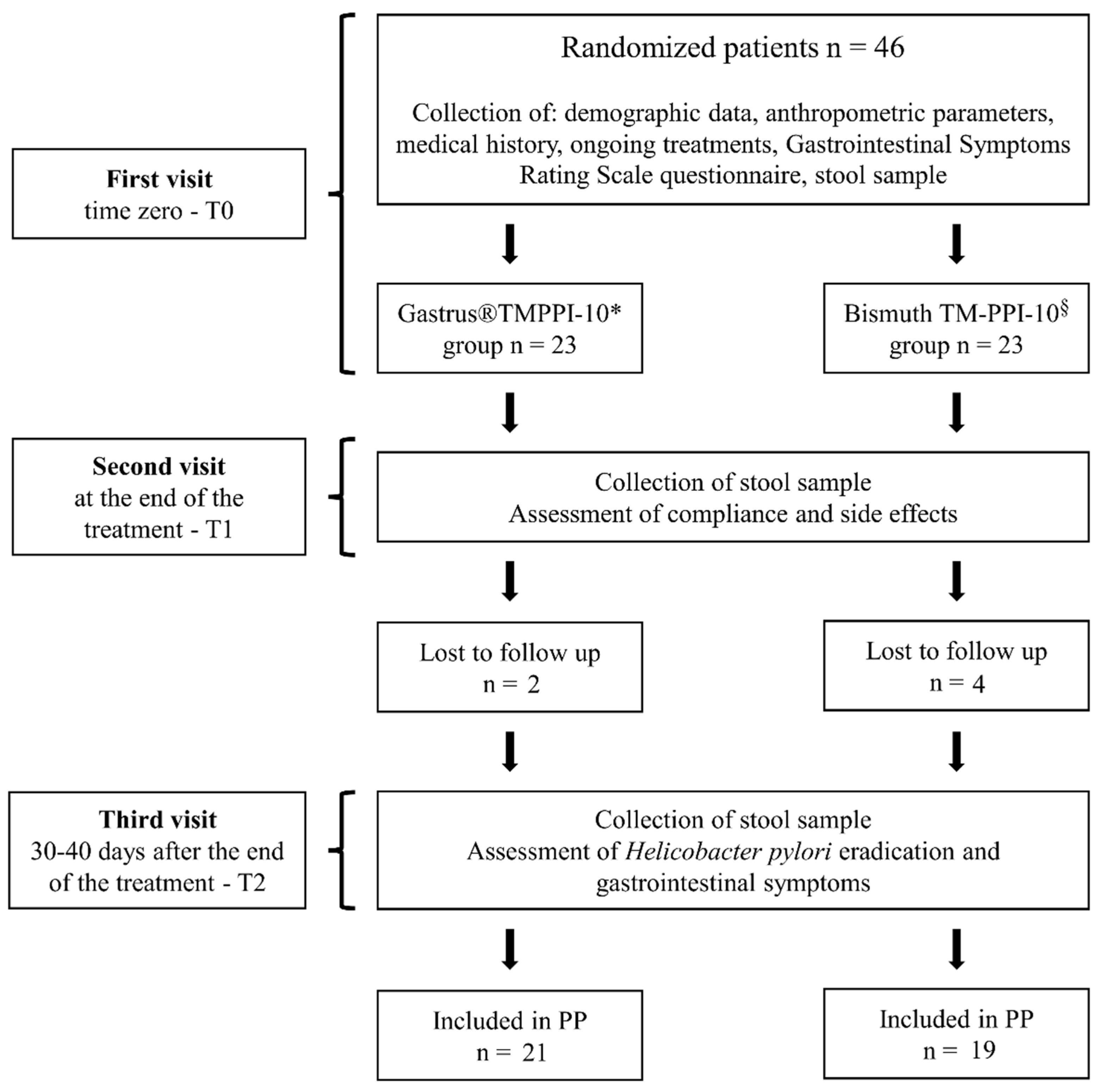
2.1. Study Design

Adult patients with H. pylori infection were randomized in a prospective open-label controlled-trial to receive a bismuth-based treatment or a probiotic-based treatment. Randomization was generated using the GraphPad software [19]. The treatment regimen was kept in a sealed envelope and opened before allocation of the patient. The study was conducted at the Department of Internal Medicine, Gastroenterology section, University of Sassari, Italy.
For each patient, three visits were scheduled. In the first visit (T0), the following information/samples were collected: (i) demographic data and anthropometric parameters, including body height, weight, as well as a detailed medical history and medications taken; (ii) a fecal sample; (iii) gastrointestinal symptoms, evaluated by using the Gastrointestinal Symptoms Rating Scale (GSRS) questionnaire, and treatment allocation (Figure 1).
In the second visit, after the end of the treatment (T1), the compliance and side effects were assessed and a second stool sample collected. In the final visit, 30–40 days after the end of treatment (T2), the last stool sample was collected, the efficacy of treatment checked and gastrointestinal symptoms re-evaluated (Figure 1).

2.2. Patient Eligibility

Patients older than 18 years referred to upper endoscopy or to a gastroenterology visit for dyspeptic symptoms, found positive for H. pylori infection and available to give stool samples, were invited to participate in the trial and to sign the informed consent.

2.3. Ethical Issues

The study was conducted in accordance with the Declaration of Helsinki, and approved by the Institutional Review Board of the local “Comitato Etico ASL No. 1 di Sassari” (Prot. No. 2358/CE). This study was registered on Clinical-Trials.gov, number NCT00669955. There was no pharmaceutical company influence in any phase of the study.

2.4. Exclusion Criteria

Presence of malignancy, pregnancy or lactation, clinically significant diseases, a recent gastroenteritis or bowel prep for colonoscopy were considered exclusion criteria. Patients who had a history of drug or alcohol abuse were also excluded. Allergy to any component of treatment regimens used in the trial or PPIs, anti-secretory drugs, antibiotics or probiotics taken one month preceding the enrollment were additional exclusion criteria.

2.5. H. pylori Status

Patients were defined as positive for H. pylori infection based on the detection of bacteria on gastric specimens and/or a positive 13C-Urea Breath Test (13C-UBT) (delta value greater than 5‰) and/or a positive stool antigen test. Eradication was confirmed by a negative 13C-UBT or a negative stool antigen test, both performed 30–40 days after treatment completion.

2.6. Medication

Both regimens included: rabeprazole 20 mg, tetracycline 500 mg and metronidazole 500 mg twice a day. One regimen was supplemented with bismuth subcitrate potassium 140 mg (Pylera®), 2 capsules twice a day, and the other one with Gastrus® (BioGaia), 1 chewable tablet in the morning and in the afternoon. Both regimens were given with the midday and evening meals for 10 days with the exception of Gastrus® tablets that were given for 27 days (Table 1).

2.7. Patients’ Compliance and Side Effects

At the time of enrollment, patients were instructed to return all drug containers after completion of therapy. More than 90% of the total recommended dose taken was considered excellent. Complained side effects were recorded after the end of treatment.

2.8. Characterization of the Gut Microbiota

Fecal samples were kept at 4 °C for a maximum of four hours after collection and stored at −80 °C until processed.
DNA was extracted from fecal samples with the QIAamp Fast DNA Stool Mini Kit (Qiagen, Hilden, Germany). DNA quantification was performed using a Qubit™ Fluorometer with the dsDNA High Sensitivity assay kit (Thermo Fisher Scientific, Waltham, MA, USA). Amplicon libraries were constructed using Illumina’s recommendations as previously described, amplifying the variable region 4 (V4) of the 16S rRNA gene [20]. Library quality control validation was performed through the TapeStation 4150 using the D1000 ScreenTape System (Agilent Technologies, Santa Clara, CA, USA). Subsequently, libraries (average size 425 bps) were quantified with a Qubit kit, normalized and pooled. DNA sequencing was performed in service using a MiSeq sequencer with the MiSeq Reagent Kit v2 (Illumina, San Diego, CA, USA), in paired-end modality and with 251 bases in each direction. The bioinformatic analysis of 16S rRNA gene sequencing data was performed as detailed elsewhere [20]. The Amplicon Sequence Variant (ASV) table was mapped against pre-formatted SILVA 138 SSURef NR99 database, using a 99% identity criterion.
In order to assess community richness and diversity, the ASV table was multiply rarefied to 90% of the minimum sample reads, and alpha and beta diversity were computed using phyloseq and visualized with ggplot2 (v.3.3.3) in R [21]. Specifically, alpha diversity and richness were calculated through Shannon’s index and number of observed ASVs, respectively. Beta diversity was calculated using weighted and unweighted UniFrac distances [22] and visualized by principal coordinates’ analysis (PCoA) plots.

2.9. Statistical Analysis

The eradication rates of H. pylori infection in the two groups were assessed by ITT, including all eligible patients enrolled in the study, and PP analysis, which excluded patients lost to follow-up, by using the Wald estimator. The significance threshold was set at p < 0.05. Ninety-five percent confidence intervals (95% CIs) were also calculated. Modification of symptoms after treatment in the two groups was categorized as: amelioration, disappearance, no modification and worsening, and compared.
The ASV data were aggregated based on the taxonomic assignment and subjected to differential statistical analysis between groups using the edgeR algorithm available on the Galaxy-P platform (v3.34.0 + galaxy1) [23,24,25], setting default parameters, except for trimmed mean of M-values as normalization [26]. In case of samples from the same patient analyzed at different times, the variable “patient” was added as additional factor and an additive linear model was used. Only patients with a documented H. pylori eradication were considered for the differential analysis. A correction for multiple tests was applied by calculating the False Discovery Rate (FDR) [27], setting FDR < 0.05 as a significant threshold. Moreover, only taxa present in at least 50% of the samples in a minimum of one group were considered as statistically significant. Differences in alpha diversity between groups were assessed by using the Wilcoxon rank sum test, setting a paired test in case of comparison between two different time points for the same patient. Significance testing between groups for beta diversity was assessed via permutational multivariate analysis of variance (PERMANOVA) using the vegan package (v.2.5-7) in R [28]. The heatmap illustrating the distribution of the ASVs belonging to Lactobacillus was created with the web application Morpheus (https://software.broadinstitute.org/morpheus/, accessed on 15 November 2021).

3. Results

3.1. Eradication Rate

Forty-six patients were randomized: 23 patients in the Gastrus® group (mean age 57.2 ± 11.5 years; 14 women), and 23 in the bismuth group (mean age 51.2 ± 13.1 years; 16 women). The proportion of current and former smokers was similar in both groups (8.7% current smokers vs. 8.7%, and 28.6% former smokers vs. 30.4%, respectively); according to ITT analysis, the eradication rate was identical in both groups (78% vs. 78%), while, for PP, was 85.7% in the Gastrus® group and 95.5% in the bismuth group, respectively (Table 2). Failure patients were treated with Pylera® at the full dosage as indicated by the Pharma.

3.2. Compliance and Side Effects

Among the 40 patients who completed the trial, side effects including nausea, diarrhea, bloating, fatigue, bitter taste and dark stool, albeit not severe, were complained more frequently in the bismuth group compared with Gastrus® group (26.3% vs. 9.5%; however, this difference was not statistically significant.
The compliance was kept higher than 90% in 93% and 91% of patients assigned to Gastrus® and bismuth groups, respectively, within the whole treatment period.
The improvement and/or disappearance of gastrointestinal symptoms, recorded according to the GSRS questionnaire, was superior in the Gastrus® group compared to bismuth group (68% vs. 53%), albeit not significantly.

3.3. Gut Microbiota Changes

Fifty-eight fecal samples were collected from 22 patients (9 from Gastrus® group and 13 from bismuth group). Stool samples from patients lost to the follow up, or for whom the intervention was ineffective, as well as samples not passing the quality control, were excluded from the differential analysis. A total of 3,566,070 reads were obtained (61,484 on average per sample), corresponding to 2732 ASVs. These, in turn, were taxonomically assigned to 51 phyla, 93 families, and 212 genera, as described in Supplementary Dataset S1.
Alpha diversity and richness of the bacterial community, evaluated by the Shannon’s index and number of observed ASVs, respectively, did not differ between the two study groups before (T0), at the end of treatment (T1), and 30–40 days after the end of treatment (T2) (Supplementary Figure S1).
In addition, differences among the three time points within the same group were investigated. In both Gastrus® and bismuth groups, alpha diversity (Shannon’s index), and richness (number of observed ASVs) dropped at time T1 compared to baseline (Gastrus®, p = 0.002; bismuth, p = 0.0078) and partially recovered at T2 (Gastrus®, p = 0.002; bismuth, p = 0.0313) although to a lesser extent in the bismuth group.
Beta diversity between and within groups was also investigated through PCoA performed with ASV data. PCoA plots did not show significant difference between groups, according to both weighted and unweighted UniFrac (Supplementary Figures S2 and S3). Differences were more pronounced when comparing the three time points within the same group, either with weighted or with unweighted UniFrac within Gastrus® and bismuth groups (p = 0.001) (Supplementary Figures S4 and S5).
A dramatic impact of the treatments on the gut microbiota composition was detected in both groups. Consistently with alpha-diversity dynamics, 30–40 days after treatment (T2), all gut bacterial members had recovered abundance levels similar to those measured at T0, with the sole exception of Barnesiella (in Gastrus® group only). T1 was the only time point at which a few bacterial taxa showed a significantly different abundance between Gastrus® group and bismuth group; specifically, Akkermansiaceae and the corresponding phylum Verrucomicrobiota, as well as Acidaminococcaceae, were more abundant in the bismuth group (Table 3).
Detailed lists of differential taxa are provided in Table 4 (at the genus level) and in Supplementary Tables S1 and S2 (at the phylum and family levels, respectively).
Several microbial taxa significantly changed in abundance following drug treatment (T1 vs. T0), e.g., Gram-negative Proteobacteria and Desulfobacterota, varied similarly in both treatment groups. Proteobacteria were detected in all samples at T0 and T1 and their relative abundance increased, as expected, after antibiotic treatment. Desulfobacterota, a former proteobacterial class, recently recognized as a novel phylum, were detected as well with a reduced relative abundance and in a lower number of samples (Supplementary Dataset S1). The decrease of Verrucomicrobiota in T1 was more evident in the Gastrus® group (FDR < 0.000001) compared with bismuth group (FDR < 0.02); consistently, Akkermansiaceae and Akkermansia significantly changed only in the Gastrus® group. No variation of Firmicutes was observed in any group, given the opposite behavior of families belonging to this phylum after antibiotic treatment (see below). Finally, a decrease of the Bacteroidota phylum was observed in the bismuth group at T1. Looking at genera, Enterococcus was found as more abundant at T1 in both groups. Furthermore, a decreased abundance of Akkermansia, Alistipes, Subdoligranulum, Coprococcus and Parabacteroides was detected in the Gastrus® group, as well as an increase of Lactobacillus and Granulicatella. A decrease of taxa generally considered beneficial, such as Roseburia and Fusicatenibacter, was observed in the bismuth group.
Despite Gastrus® supplementation given for 27 days (i.e., up to 17 days after antibiotic withdrawal), no significant differences were observed at T2 between the two groups.
Lactobacillus colonization from T0 to T2 was further explored by comparing the ASVs assortments and their relative abundances in both study groups (Supplementary Figure S5). Samples from Gastrus® group did not show colonization by a predominant Lactobacillus ASV, neither at T1 nor at T2. Clearly, different ASVs of Lactobacillus contributed to the taxon increase at T1 in both sample groups, suggesting the involvement of different Lactobacillus strains.

4. Discussion

In this RCT, the low-dose bismuth quadruple therapy given b.i.d. for 10 days achieved an eradication rate of 94.7% by PP analysis according to previous studies conducted in the same geographical area [18]. The cure rate decreased to 78% by ITT analysis. Replacement of bismuth with Gastrus® proved to be less effective failing to cure nearly 10% of patients. As previously observed, these data highlight that L. reuteri DSM 17938 and L. reuteri ATCC PTA 6475 were not able to completely replace the bismuth action against H. pylori [18]. The choice of L. reuteri in substitution of bismuth was based on the evidence that this bacterium produces reuterin (3-hydroxypropionaldehyde), an antimicrobial compound against H. pylori [29]. Moreover, L. reuteri hampers H. pylori colonization of human gastric mucosa through the inhibition of H. pylori binding to glycolipid receptors [30]. Yao et al., in an elegant study published last year, elucidated the underlying mechanism of bismuth action against H. pylori [31]. Functional analysis showed that bismuth downregulates CagA and VacA virulence proteins, impairs antioxidant response and compromises purine, pyrimidine, amino acid and carbon metabolic pathways involved in bacterial growth. Moreover, bismuth inhibits H. pylori colonization breaking off the assembly of the bacterial flagellum [31]. These findings may explain the superiority of bismuth with respect to L. reuteri in terms of efficacy against H. pylori.
In addition, low-dose bismuth resulted in being highly effective, as an empirical regimen, similarly to high-dose bismuth quadruple therapy. For example, in 2100 European patients treated with Pylera® (single-capsule bismuth quadruple therapy), according to the Pharma instructions (i.e., three capsules every six h for 10 days), eradication rate was greater than 90% in clinical practice. However, 60 patients complained of severe adverse events, with 1.7% of them requiring to stop the treatment [32]. In our study, patients were exposed to a regimen containing a low dose quadruple therapy and avoided almost 10 gr of bismuth, 5 gr of metronidazole and 5 gr of tetracycline and severe side effects were not experienced. However, the Gastrus® group complained fewer side effects compared to the bismuth group, as previously reported [18]. Moreover, a greater percentage of patients claimed gastrointestinal symptoms improvement and/or disappearance in the Gastrus® group compared to the bismuth group. Interestingly, in both study groups, the eradication rate by ITT analysis was identical.
The 16S metagenomic sequencing revealed that alpha diversity significantly changed soon after eradication compared with baseline. Alpha diversity is an ecological, cumulative measure of the complexity of the microbial community, used as a hallmark of homeostasis and resilience of the gut microbiota, being inversely correlated with mucosal inflammation and permeability of the intestinal barrier [33]. Treatment-dependent disruption of the gut microbiota, highlighted by the drop of alpha diversity at T1, showed a recovery 30–40 days after treatment, albeit not complete. The beta diversity followed a similar pattern, confirming a possible restoration at the end of intervention in each study group.
Given that weighted UniFrac accounts for the abundance of the observed microorganisms (number of reads assigned to an ASV), this result may suggest that the two regimens, either supplemented with L. reuteri strains or with bismuth, are both somehow able to shape biodiversity.
While the dynamics of alpha diversity were similar, some key members of the gut microbiota showed Gastrus®- or bismuth-specific changes of their relative abundance. Both treatments were associated with an increase of Proteobacteria, a large phylum of Gram-negative bacteria, mostly facultative aerobes/anaerobes, including several pathobionts. Members of this phylum were not detected as differentially abundant at family or genus levels, suggesting that most Proteobacteria members were evenly modified by treatment, with the exception of Morganellaceae that showed a significant increase of its relative abundance only in the Gastrus® group. It is worth noting that opportunistic genera belong to this family and their increase in antibiotic-dysrupted microbiota has already been reported. Similarly, Bandy and Zhang reported an increased opportunistic genus in (Morganella, Proteus, and Providencia) dysbiosis due to antibiotics, indicating that bismuth may be more effective in controlling the opportunistic blooming of Morganellaceae during antibiotic treatment [34,35]. Recently, a large group of Proteobacteria members were reclassified in the novel phylum Desulfobacteroidota, based on phylogeny and metabolic functions [36]. In the present study, we observed for the first time an opposite behavior of Proteobacteria and Desulfobacteroidota under antibiotic treatment, highlighting a different response of the two phyla to gut environmental changes.
According to a previous study aimed at evaluating the short- and long-term effect of bismuth quadruple therapy on the gut microbiota in a pediatric population allergic to penicillin [37], we observed a significant decrease of the Bacteroidota phylum, including the butyrate-producer Butyricimonas, after bismuth treatment. Instead, Alistipes and Parabacteroides were significantly reduced in the Gastrus® group. In addition, a decrease of other specific Bacteroidota members, including Bacteroides, Barnesiella and Odoribacter, was observed in both treatment groups. Although Bacteroidota members showed different susceptibility to bismuth, given the cumulative effect at phylum level, the overall response to bismuth treatment was higher.
Opposite trends were observed for different genera belonging to Firmicutes, determining, in turn, a lack of the overall phylum variation. In the bismuth group, there was a significant increase of Enterococcus, Granulicatella and Lactobacillus. The aerobic pathobiont Enterococcus, together with Proteobacteria members, represent a hallmark of gut microbiota dysbiosis. Granulicatella, a nutritional variant Streptococcus, has been reported to increase after H. pylori eradication and to contribute to the progression of precancerous gastric lesions to cancer [38]; additionally, it might potentially cause endocarditis [39]. Studies on Lactobacillus modification in the gut microbiota determined by bismuth quadruple therapy with or without probiotics reported contrasting results [17,40,41]. In our study, bismuth caused a significantly increased Lactobacillus at the end of treatment, while Gastrus® supplementation did not.
In a recent study conducted in Taiwan, fecal samples obtained at the end of therapy from patients treated with bismuth quadruple therapy (q.i.d. for 14 days) displayed a decreased abundance of Verrucomicrobia and Bacteroidota, similarly to our findings [42]. The same regimen given for 10 days, six weeks after H. pylori eradication, caused a statistically significant decrease of Akkermansia, Bifidobacterium, B. fragilis and Faecalibacterium prausnitzii [43]. Akkermansia spp. are recognized as beneficial members of the gut microbiota, capable of growing on mucin as a sole source of nutrients and are generally associated with a healthy gut mucosa [44]. It is worth noting that we observed a less pronounced reduction of Verrucomicrobia and the correlated family Akkermansiaceae in the bismuth group.
Interestingly, Barnesiella showed a dramatic reduction of its relative abundance even 30–40 days after treatment with the Gastrus® regimen, with a fold change almost identical to that observed immediately after treatment. Barnesiella is an important genus for the gut mucosal health, especially when the microbiota is challenged by antibiotics, as it is reported to be associated with pathobionts’ eradication [45] and to reduce susceptibility to colitis induction in mice [46].
Although in the literature there are studies demonstrating that probiotic supplementation may reduce the detrimental effects of antibiotics in the gut microbiota [47,48], in our study, abundance increment or reduction did not dramatically differ in the probiotic group compared to the bismuth group.
A number of limitations in our study need to be mentioned. First, the study design was open-labelled, which may have hampered minimizing the researcher’s bias as well as the placebo effect. However, although this may have impacted eradication rate, it may have hardly influenced the gut microbiota changes. The relatively small size of treatment groups may have theoretically reduced the statistical power, although in a previous study [18], enrolling a total of 99 patients, similar eradication rates were obtained by using the same two treatment regimens. Moreover, stool samples from non-responders were not sequenced and the number of stool samples failing quality control was not negligible (14 out of 36 samples). Although detailed instructions on stool collection were given to the patients, after an accurate control, we were forced to exclude several samples because they were stored at room temperature, or for more than 2 h, and this may have affected results.

5. Conclusions

Overall, our findings prove that twice-a-day low-dose bismuth quadruple therapy given for 10 days is able to achieve high eradication rates with lower costs, amount of antibiotics and bismuth. However, a proper non-inferiority study against high doses would be required to confirm that both are equally effective.
Replacement of bismuth with Gastrus® might be an option for patients with contraindications to bismuth. However, L. reuteri in the method of administration used in this study was not able to significantly counteract alterations induced in the gut microbiota by antibiotics, still leaving open the question on how to administer probiotics (dosage, timing, for how long, which strain, or a combination of strains) to prevent dysbiosis.

Supplementary Materials

The following supporting information can be downloaded at: https://www.mdpi.com/article/10.3390/nu14142789/s1, Dataset S1: List of identified ASVs, phyla, families, genera; Figure S1: Alpha diversity and richness between group A and group B at the three different time points; Figure S2: Beta diversity between group A and group B based at the three different time points; Figure S3: Beta diversity between group A and group B based at the three different time points; Figure S4: Beta diversity within group A and group B based on the three time points; Figure S5: Lactobacillus ASVs distribution among patients; Table S1: List of differential abundant phyla when comparing T0 vs. T1 and T2; Table S2: List of differential abundant families when comparing T0 vs. T1 and T2.

Author Contributions

Conceptualization, M.P.D. and S.U.; methodology, S.B., M.A. and A.T.; software, M.A.; validation, M.P.D., S.U. and G.M.P.; formal analysis, S.B., G.M.P. and S.U.; investigation, M.P.D., R.S., C.N., M.L., M.A. and A.T.; resources, M.P.D.; data curation, M.P.D.; writing—original draft preparation, M.P.D., G.M.P. and S.U.; writing—review and editing, M.P.D., G.M.P., M.A., A.T. and S.U.; visualization, G.M.P.; supervision, M.P.D.; project administration, M.P.D.; funding acquisition, M.P.D. All authors have read and agreed to the published version of the manuscript.

Funding

This research received no external funding.

Institutional Review Board Statement

The study was conducted in accordance with the Declaration of Helsinki, and approved by the Institutional Review Board of the local “Comitato Etico ASL No. 1 di Sassari” (Prot. No. 2358/CE). This study was registered on Clinical-Trials.gov, number NCT00669955. There was no pharmaceutical company influence in any phase of the study.

Informed Consent Statement

Informed consent was obtained from all subjects involved in the study.

Data Availability Statement

The datasets presented in this study can be found in online repositories. The names of the repository/repositories and accession number can be found at: https://www.ebi.ac.uk/ena, PRJEB35028.

Acknowledgments

We would like to thank BioGaia (Stockholm, Sweden) for supplying Gastrus® for this study.

Conflicts of Interest

The authors declare no conflict of interest.

References

  1. Kikuchi, S.; Dore, M.P. Epidemiology of Helicobacter pylori Infection. Helicobacter 2005, 10 (Suppl. S1), 1–4. [Google Scholar] [CrossRef]
  2. Wu, J.Y.; Lee, Y.C.; Graham, D.Y. The eradication of Helicobacter pylori to prevent gastric cancer: A critical appraisal. Expert Rev. Gastroenterol. Hepatol. 2019, 13, 17–24. [Google Scholar] [CrossRef]
  3. Lee, Y.C.; Dore, M.P.; Graham, D.Y. Diagnosis and Treatment of Helicobacter pylori Infection. Annu. Rev. Med. 2022, 73, 183–195. [Google Scholar] [CrossRef]
  4. Dore, M.P.; Marras, L.; Maragkoudakis, E.; Nieddu, S.; Manca, A.; Graham, D.Y.; Realdi, G. Salvage therapy after two or more prior Helicobacter pylori treatment failures: The super salvage regimen. Helicobacter 2003, 8, 307–309. [Google Scholar] [CrossRef]
  5. Shiotani, A.; Lu, H.; Dore, M.P.; Graham, D.Y. Treating Helicobacter pylori effectively while minimizing misuse of antibiotics. Cleve Clin. J. Med. 2017, 84, 310–318. [Google Scholar] [CrossRef] [Green Version]
  6. Imhann, F.; Bonder, M.J.; Vich Vila, A.; Fu, J.; Mujagic, Z.; Vork, L.; Tigchelaar, E.F.; Jankipersadsing, S.A.; Cenit, M.C.; Harmsen, H.J.; et al. Proton pump inhibitors affect the gut microbiome. Gut 2016, 65, 740–748. [Google Scholar] [CrossRef] [Green Version]
  7. Dore, M.P.; Bibbo, S.; Pes, G.M.; Francavilla, R.; Graham, D.Y. Role of Probiotics in Helicobacter pylori Eradication: Lessons from a Study of Lactobacillus reuteri Strains DSM 17938 and ATCC PTA 6475 (Gastrus(R)) and a Proton-Pump Inhibitor. Can. J. Infect. Dis. Med. Microbiol. 2019, 2019, 3409820. [Google Scholar] [CrossRef] [Green Version]
  8. Dore, M.P.; Goni, E.; Di Mario, F. Is There a Role for Probiotics in Helicobacter pylori Therapy? Gastroenterol. Clin. North Am. 2015, 44, 565–575. [Google Scholar] [CrossRef]
  9. Fang, H.R.; Zhang, G.Q.; Cheng, J.Y.; Li, Z.Y. Efficacy of Lactobacillus-supplemented triple therapy for Helicobacter pylori infection in children: A meta-analysis of randomized controlled trials. Eur. J. Pediatr. 2019, 178, 7–16. [Google Scholar] [CrossRef]
  10. Shi, X.; Zhang, J.; Mo, L.; Shi, J.; Qin, M.; Huang, X. Efficacy and safety of probiotics in eradicating Helicobacter pylori: A network meta-analysis. Medicine 2019, 98, e15180. [Google Scholar] [CrossRef]
  11. Si, X.B.; Lan, Y.; Qiao, L. A meta-analysis of randomized controlled trials of bismuth-containing quadruple therapy combined with probiotic supplement for eradication of Helicobacter pylori. Zhonghua Nei Ke Za Zhi 2017, 56, 752–759. [Google Scholar] [CrossRef] [PubMed]
  12. Yu, M.; Zhang, R.; Ni, P.; Chen, S.; Duan, G. Efficacy of Lactobacillus-supplemented triple therapy for H. pylori eradication: A meta-analysis of randomized controlled trials. PLoS ONE 2019, 14, e0223309. [Google Scholar] [CrossRef] [PubMed] [Green Version]
  13. Zhang, M.; Zhang, C.; Zhao, J.; Zhang, H.; Zhai, Q.; Chen, W. Meta-analysis of the efficacy of probiotic-supplemented therapy on the eradication of H. pylori and incidence of therapy-associated side effects. Microb. Pathog. 2020, 147, 104403. [Google Scholar] [CrossRef]
  14. Olekhnovich, E.I.; Manolov, A.I.; Samoilov, A.E.; Prianichnikov, N.A.; Malakhova, M.V.; Tyakht, A.V.; Pavlenko, A.V.; Babenko, V.V.; Larin, A.K.; Kovarsky, B.A.; et al. Shifts in the Human Gut Microbiota Structure Caused by Quadruple Helicobacter pylori Eradication Therapy. Front. Microbiol. 2019, 10, 1902. [Google Scholar] [CrossRef] [PubMed] [Green Version]
  15. Yang, C.; Liang, L.; Lv, P.; Liu, L.; Wang, S.; Wang, Z.; Chen, Y. Effects of non-viable Lactobacillus reuteri combining with 14-day standard triple therapy on Helicobacter pylori eradication: A randomized double-blind placebo-controlled trial. Helicobacter 2021, 26, e12856. [Google Scholar] [CrossRef]
  16. Tang, B.; Tang, L.; Huang, C.; Tian, C.; Chen, L.; He, Z.; Yang, G.; Zuo, L.; Zhao, G.; Liu, E.; et al. The Effect of Probiotics Supplementation on Gut Microbiota after Helicobacter pylori Eradication: A Multicenter Randomized Controlled Trial. Infect. Dis. Ther. 2021, 10, 317–333. [Google Scholar] [CrossRef]
  17. He, C.; Peng, C.; Wang, H.; Ouyang, Y.; Zhu, Z.; Shu, X.; Zhu, Y.; Lu, N. The eradication of Helicobacter pylori restores rather than disturbs the gastrointestinal microbiota in asymptomatic young adults. Helicobacter 2019, 24, e12590. [Google Scholar] [CrossRef]
  18. Dore, M.P.; Bibbo, S.; Loria, M.; Salis, R.; Manca, A.; Pes, G.M.; Graham, D.Y. Twice-a-day PPI, tetracycline, metronidazole quadruple therapy with Pylera(R) or Lactobacillus reuteri for treatment naive or for retreatment of Helicobacter pylori. Two randomized pilot studies. Helicobacter 2019, 24, e12659. [Google Scholar] [CrossRef]
  19. GraphPad Software, S.D., California USA. Available online: http://www.graphpad.com/quickcalcs/randomize2/ (accessed on 2 November 2021).
  20. Bibbo, S.; Abbondio, M.; Sau, R.; Tanca, A.; Pira, G.; Errigo, A.; Manetti, R.; Pes, G.M.; Dore, M.P.; Uzzau, S. Fecal Microbiota Signatures in Celiac Disease Patients With Poly-Autoimmunity. Front. Cell Infect. Microbiol. 2020, 10, 349. [Google Scholar] [CrossRef]
  21. Wickham, H. ggplot2: Elegant Graphics for Data Analysis; Springer: New York, NY, USA, 2016. [Google Scholar]
  22. Lozupone, C.; Lladser, M.E.; Knights, D.; Stombaugh, J.; Knight, R. UniFrac: An effective distance metric for microbial community comparison. ISME J. 2011, 5, 169–172. [Google Scholar] [CrossRef] [Green Version]
  23. Boekel, J.; Chilton, J.M.; Cooke, I.R.; Horvatovich, P.L.; Jagtap, P.D.; Kall, L.; Lehtio, J.; Lukasse, P.; Moerland, P.D.; Griffin, T.J. Multi-omic data analysis using Galaxy. Nat. Biotechnol. 2015, 33, 137–139. [Google Scholar] [CrossRef]
  24. Liu, R.; Holik, A.Z.; Su, S.; Jansz, N.; Chen, K.; Leong, H.S.; Blewitt, M.E.; Asselin-Labat, M.L.; Smyth, G.K.; Ritchie, M.E. Why weight? Modelling sample and observational level variability improves power in RNA-seq analyses. Nucleic Acids Res. 2015, 43, e97. [Google Scholar] [CrossRef] [PubMed]
  25. Galaxy/Proteomics. Available online: https://proteomics.usegalaxy.eu (accessed on 2 December 2021).
  26. Robinson, M.D.; Oshlack, A. A scaling normalization method for differential expression analysis of RNA-seq data. Genome Biol. 2010, 11, R25. [Google Scholar] [CrossRef] [PubMed] [Green Version]
  27. Benjamini, Y.; Drai, D.; Elmer, G.; Kafkafi, N.; Golani, I. Controlling the false discovery rate in behavior genetics research. Behav. Brain Res. 2001, 125, 279–284. [Google Scholar] [CrossRef] [Green Version]
  28. Oksanen, J.; Simpson, G.L.; Blanchet, F.G.; Kindt, R.; Legendre, P.; Minchin, P.R.; O’Hara, R.B.; Solymos, P.; Stevens, H.H.; Szoecs, E.; et al. vegan: Community Ecology Package. Available online: https://CRAN.R-project.org/package=vegan (accessed on 2 November 2021).
  29. Mukai, T.; Asasaka, T.; Sato, E.; Mori, K.; Matsumoto, M.; Ohori, H. Inhibition of binding of Helicobacter pylori to the glycolipid receptors by probiotic Lactobacillus reuteri. FEMS Immunol. Med. Microbiol. 2002, 32, 105–110. [Google Scholar] [CrossRef] [PubMed] [Green Version]
  30. Chung, T.C.; Axelsson, L.; Lindgren, S.E.; Dobrogosz, W.J. In Vitro Studies on Reuterin Synthesis by Lactobacillus reuteri. Microb. Ecol. Health Dis. 1989, 2, 137–144. [Google Scholar] [CrossRef] [Green Version]
  31. Yao, X.; Xiao, S.; Zhou, L. Integrative proteomic and metabolomic analyses reveal the mechanism by which bismuth enables Helicobacter pylori eradication. Helicobacter 2021, 26, e12846. [Google Scholar] [CrossRef]
  32. Nyssen, O.P.; Perez-Aisa, A.; Castro-Fernandez, M.; Pellicano, R.; Huguet, J.M.; Rodrigo, L.; Ortun, J.; Gomez-Rodriguez, B.J.; Pinto, R.M.; Areia, M.; et al. European Registry on Helicobacter pylori management: Single-capsule bismuth quadruple therapy is effective in real-world clinical practice. United Eur. Gastroenterol. J. 2021, 9, 38–46. [Google Scholar] [CrossRef]
  33. Ley, D.; Desseyn, J.L.; Gouyer, V.; Plet, S.; Tims, S.; Renes, I.; Mischke, M.; Gottrand, F. Early life nutrition influences susceptibility to chronic inflammatory colitis in later life. Sci. Rep. 2019, 9, 18111. [Google Scholar] [CrossRef] [Green Version]
  34. Bandy, A. Ringing bells: Morganella morganii fights for recognition. Public Health 2020, 182, 45–50. [Google Scholar] [CrossRef]
  35. Zhang, Z.; Li, M.; Cui, B.; Chen, X. Antibiotic Disruption of the Gut Microbiota Enhances the Murine Hepatic Dysfunction Associated With a High-Salt Diet. Front. Pharmacol. 2022, 13, 829686. [Google Scholar] [CrossRef] [PubMed]
  36. Waite, D.W.; Chuvochina, M.; Pelikan, C.; Parks, D.H.; Yilmaz, P.; Wagner, M.; Loy, A.; Naganuma, T.; Nakai, R.; Whitman, W.B.; et al. Proposal to reclassify the proteobacterial classes Deltaproteobacteria and Oligoflexia, and the phylum Thermodesulfobacteria into four phyla reflecting major functional capabilities. Int. J. Syst. Evol. Microbiol. 2020, 70, 5972–6016. [Google Scholar] [CrossRef] [PubMed]
  37. Zhou, Y.; Ye, Z.; Lu, J.; Miao, S.; Lu, X.; Sun, H.; Wu, J.; Wang, Y.; Huang, Y. Long-term changes in the gut microbiota after 14-day bismuth quadruple therapy in penicillin-allergic children. Helicobacter 2020, 25, e12721. [Google Scholar] [CrossRef] [PubMed]
  38. Sung, J.J.Y.; Coker, O.O.; Chu, E.; Szeto, C.H.; Luk, S.T.Y.; Lau, H.C.H.; Yu, J. Gastric microbes associated with gastric inflammation, atrophy and intestinal metaplasia 1 year after Helicobacter pylori eradication. Gut 2020, 69, 1572–1580. [Google Scholar] [CrossRef] [Green Version]
  39. Holloway, V.; Jacob, G.; Hayes, N. Challenges in the diagnosis and management of Granulicatella elegans endocarditis in a 9-year-old child. BMJ Case Rep. 2021, 14, e240079. [Google Scholar] [CrossRef]
  40. Niu, Z.Y.; Li, S.Z.; Shi, Y.Y.; Xue, Y. Effect of gastric microbiota on quadruple Helicobacter pylori eradication therapy containing bismuth. World J. Gastroenterol. 2021, 27, 3913–3924. [Google Scholar] [CrossRef]
  41. Yuan, Z.; Xiao, S.; Li, S.; Suo, B.; Wang, Y.; Meng, L.; Liu, Z.; Yin, Z.; Xue, Y.; Zhou, L. The impact of Helicobacter pylori infection, eradication therapy, and probiotics intervention on gastric microbiota in young adults. Helicobacter 2021, 26, e12848. [Google Scholar] [CrossRef]
  42. Hsu, P.I.; Pan, C.Y.; Kao, J.Y.; Tsay, F.W.; Peng, N.J.; Kao, S.S.; Wang, H.M.; Tsai, T.J.; Wu, D.C.; Chen, C.L.; et al. Helicobacter pylori eradication with bismuth quadruple therapy leads to dysbiosis of gut microbiota with an increased relative abundance of Proteobacteria and decreased relative abundances of Bacteroidetes and Actinobacteria. Helicobacter 2018, 23, e12498. [Google Scholar] [CrossRef]
  43. Yildiz, S.S.; Yalinay, M.; Karakan, T. Bismuth-based quadruple Helicobacter pylori eradication regimen alters the composition of gut microbiota. Infez Med. 2018, 26, 115–121. [Google Scholar]
  44. Macchione, I.G.; Lopetuso, L.R.; Ianiro, G.; Napoli, M.; Gibiino, G.; Rizzatti, G.; Petito, V.; Gasbarrini, A.; Scaldaferri, F. Akkermansia muciniphila: Key player in metabolic and gastrointestinal disorders. Eur. Rev. Med. Pharmacol. Sci. 2019, 23, 8075–8083. [Google Scholar] [CrossRef]
  45. Bilinski, J.; Grzesiowski, P.; Sorensen, N.; Madry, K.; Muszynski, J.; Robak, K.; Wroblewska, M.; Dzieciatkowski, T.; Dulny, G.; Dwilewicz-Trojaczek, J.; et al. Fecal Microbiota Transplantation in Patients With Blood Disorders Inhibits Gut Colonization With Antibiotic-Resistant Bacteria: Results of a Prospective, Single-Center Study. Clin. Infect. Dis. 2017, 65, 364–370. [Google Scholar] [CrossRef] [PubMed]
  46. Butera, A.; Di Paola, M.; Pavarini, L.; Strati, F.; Pindo, M.; Sanchez, M.; Cavalieri, D.; Boirivant, M.; De Filippo, C. Nod2 Deficiency in mice is Associated with Microbiota Variation Favouring the Expansion of mucosal CD4+ LAP+ Regulatory Cells. Sci. Rep. 2018, 8, 14241. [Google Scholar] [CrossRef] [PubMed] [Green Version]
  47. Oh, B.; Kim, B.S.; Kim, J.W.; Kim, J.S.; Koh, S.J.; Kim, B.G.; Lee, K.L.; Chun, J. The Effect of Probiotics on Gut Microbiota during the Helicobacter pylori Eradication: Randomized Controlled Trial. Helicobacter 2016, 21, 165–174. [Google Scholar] [CrossRef] [PubMed]
  48. Wang, F.; Feng, J.; Chen, P.; Liu, X.; Ma, M.; Zhou, R.; Chang, Y.; Liu, J.; Li, J.; Zhao, Q. Probiotics in Helicobacter pylori eradication therapy: Systematic review and network meta-analysis. Clin. Res. Hepatol. Gastroenterol. 2017, 41, 466–475. [Google Scholar] [CrossRef] [PubMed]
Figure 1. Study design of the controlled trial. The symbols * and § represent the treatment according to the Table 1.
Nutrients 14 02789 g001
Table 1. Regimens used in the trial.
Gastrus® GroupBismuth Group
Rabeprazole 20 mg 1 cp Rabeprazole 20 mg 1 cp
Metronidazole 250 mg 2 cp Metronidazole 250 mg 1 cp
Tetracycline 250 mg 2 cp Tetracycline 250 mg 1 cp
(b.i.d.: midday and evening meals)Pylera® 140 mg/125 mg/125 mg 2 cp
for 10 days(b.i.d.: midday and evening meals)
Gastrus® 1 tablet in the morning and 1 tablet in the afternoon for 27 daysfor 10 days
Table 2. Intervention status of 46 patients enrolled in the trial.
StatusGastrus® Group 1Bismuth Group 1
Received intervention2323
Lost to follow-up24
Completed trial2119
Intervention ineffective31
Cure Rate ITT 278.3% (18/23)78.3% (18/23)
95% CI 4-ITT61.4–95.161.4–95.1
Cure Rate PP 385.7% (18/21)94.7% (18/19)
95% CI 4-PP71.4–10085.6–100
1 Treatment regimens are specified in Table 1. 2 ITT: intention-to-treat analysis; 3 PP: per protocol analysis; 4 CI: Confidence Interval.
Table 3. List of differential abundant taxa between Gastrus® group and Bismuth group. Taxa with statistically significant difference (FDR < 0.05) identified in at least 50% of the samples in at least one group.
Taxonomic LevelTaxaTime T1
logFC (Gastrus®/Bismuth) 1FDR 1
PhylumVerrucomicrobiota−7.240.02329
FamilyAkkermansiaceae−9.300.02715
Acidaminococcaceae−11.140.02182
1 FC: Fold Changes; FDR: False Discovery Rate.
Table 4. List of differential abundant genera when comparing T0 vs. T1 and T2. Genera with statistically significance (FDR < 0.05), identified in at least 50% of the samples in at least one group.
GenusGastrus® GroupBismuth Group
T1 < T0T2 < T0T1 < T0T2 < T0
logFC (T1/T0)FDRlogFC (T2/T0)FDRlogFC (T1/T0)FDRlogFC (T2/T0)FDR
Adlercreutzia −6.600.03803
Akkermansia−11.370.00730
Alistipes−12.180.00548
Anaerostipes −5.740.02256
Barnesiella−13.470.00280−12.180.01277−6.940.00996
Bilophila −8.200.00962
Butyricimonas −8.330.00962
Collinsella −8.050.00960
Coprococcus−9.700.04882
Flavonifractor −7.230.04680
Fusicatenibacter −8.800.00102
Intestinimonas −7.420.00962
Lachnoclostridium −7.060.00962
Megasphaera −10.410.00962
Monoglobus−11.230.00548 −8.520.00167
Odoribacter−10.650.03474 −9.560.00088
Oscillibacter −7.380.03803
Parabacteroides−9.440.04882
Parasutterella −6.630.00996
Romboutsia −7.800.00962
Roseburia −9.080.00167
Ruminococcus−8.070.04882 −9.230.00167
Subdoligranulum−11.220.03586
GenusT1 > T0T2 > T0T1 > T0T2 > T0
logFC (T1/T0)FDRlogFC (T2/T0)FDRlogFC (T1/T0)FDRlogFC (T2/T0)FDR
Enterococcus10.980.00548 12.810.00276
Granulicatella 5.560.04680
Lactobacillus 7.320.04909
Publisher’s Note: MDPI stays neutral with regard to jurisdictional claims in published maps and institutional affiliations.

