
Synthesis of Metal–Organic Frameworks Quantum Dots Composites as Sensors for Endocrine-Disrupting Chemicals


Abstract
1. Introduction
1.1. Metal–Organic Frameworks
1.2. Quantum Dots
1.3. Endocrine-Disrupting Chemicals
Bisphenol A (BPA)
1.4. Heavy Metals
2. Metal–Organic Frameworks
2.1. Metal–Organic Frameworks as Potential Sensors for Pollutants in the Environment
2.2. Quantum Dots as Sensors for Environmental Pollutants and Other Toxic Substances
2.3. Compounds Used as Sensors in the Detection and Removal of Bisphenol A (BPA) from the Environment
2.4. Metal–Organic Framework Quantum Dots Composites as Sensors for Environmental Pollutants and Other Compounds
3. Conclusions
Author Contributions
Funding
Conflicts of Interest
References
- Ryo, O.; Susumu, K. Porous Coordination Polymers/Metal-Organic Frameworks. In Handbook of Solid-State Chemistry; Dronskowski, R., Kikkawa, S., Stein, A., Eds.; Wiley-VCH: Weinheim, Germany, 2017; pp. 141–164. [Google Scholar] [CrossRef]
- Batten, S.R.; Champness, N.R.; Chen, X.M.; Garcia-Martinez, J.; Kitagawa, S.; Öhrström, L.; O’Keeffe, M.; Suh, M.P.; Reedijk, J. Terminology of Metal–Organic Frameworks and coordination polymers. Pure Appl. Chem. 2013, 85, 1715–1724. [Google Scholar] [CrossRef]
- Tranchemontagne, D.J.; Mendoza-Cortes, J.L.; O’Keeffeand, M.; Yaghi, O.M. Secondary building units, nets and bonding in the chemistry of metal–organic frameworks. Chem. Soc. Rev. 2009, 38, 1257–1283. [Google Scholar] [CrossRef]
- Chaikittisilp, W.; Ariga, K.; Yamauchi, Y. A new family of carbon materials: Synthesis of MOF-derived nonporous carbons and their promising applications. J. Mater. Chem. A 2013, 1, 14–19. [Google Scholar] [CrossRef]
- Jones, N.B.; Gibbons, B.; Morris, A.J.; Morris, J.R.; Troya, D. Reversible Dissociation for Effective Storage of Diborane Gas within the UiO-66-NH2 Metal–Organic Framework. ACS Appl. Mater. Interfaces 2022, 14, 8322–8332. [Google Scholar] [CrossRef]
- Xia, Y.-P.; Wang, C.-X.; Yu, M.-H.; Bu, X.-H. A unique 3D microporous MOF constructed by cross-linking 1D coordination polymer chains for effectively selective separation of CO2/CH4 and C2H2/CH4. Chin. Chem. Lett. 2021, 32, 1153–1156. [Google Scholar] [CrossRef]
- Han, M.; Tang, X.; Wang, P.; Zhao, Z.; Ba, X.; Jiang, Y.; Zhang, X. Metal-Organic Frameworks Decorated Cu2O Heterogeneous Catalysts for Selective Oxidation of Styrene. Catalysts 2022, 12, 487. [Google Scholar] [CrossRef]
- Zhang, J.; Wang, Y.; Wang, H.; Zhong, D.; Lu, T. Enhancing photocatalytic performance of metal-organic frameworks for CO2 reduction by a bimetallic strategy. Chin. Chem. Lett. 2022, 33, 2065–2068. [Google Scholar] [CrossRef]
- Zhang, X.; Yang, Y.; Qin, P.; Han, L.; Zhu, W.; Duan, S.; Lu, M.; Cai, Z. Facile preparation of nano-g-C3N4/UiO-66-NH2 composite as sorbent for high-efficient extraction and preconcentration of food colorants prior to HPLC analysis. Chin. Chem. Lett. 2022, 33, 903–906. [Google Scholar] [CrossRef]
- Qin, P.; Zhu, W.; Han, L.; Zhang, X.; Zhao, B.; Zhang, X.; Lu, M. Monodispersed mesoporous SiO2@metal-organic framework (MSN@MIL-101(Fe)) composites as sorbent for extraction and preconcentration of phytohormones prior to HPLC-DAD analysis. Microchim. Acta 2020, 187, 367. [Google Scholar] [CrossRef]
- Qin, P.; Han, L.; Zhang, X.; Li, M.; Li, D.; Lu, M.; Cai, Z. MIL-101(Fe)-derived magnetic porous carbon as sorbent for stir bar sorptive-dispersive microextraction of sulfonamides. Microchim. Acta 2021, 188, 340. [Google Scholar] [CrossRef]
- Wang, Y.; Li, Q.; Deng, M.; Chen, K.; Wang, J. Self-assembled metal-organic frameworks nanocrystals synthesis and application for plumbagin drug delivery in acute lung injury therapy. Chin. Chem. Lett. 2022, 33, 324–327. [Google Scholar] [CrossRef]
- Jaheon, K.; Banglin, C.; Reineke, T.M.; Hailian, L.; Mohamed, E.; David, B.M.; Michael, O.; Yaghi, O.M. Assembly of Metal-Organic Frameworks from large organic and inorganic secondary building units: New examples and simplifying principles for complex structures. J. Am. Chem. Soc. 2001, 123, 8239–8247. [Google Scholar] [CrossRef]
- Tella, A.C.; Aaron, I. Syntheses and applications of Metal-Organic Frameworks materials: A review. Acta Chim. Pharm. Indica 2012, 2, 74–81, ISSN 2277-288X. [Google Scholar]
- Lustig, W.P.; Mukherjee, S.; Rudd, N.D.; Desai, A.V.; Li, J.; Ghosh, S.K. Metal–Organic Frameworks: Functional luminescent and photonic materials for sensing applications. Chem. Soc. Rev. 2017, 46, 3242–3285. [Google Scholar] [CrossRef] [PubMed]
- Han, L.; Liu, X.; Zhang, X.; Li, M.; Li, D.; Qin, P.; Tian, S.; Lu, M.; Cai, Z. Preparation of multivariate zirconia metal-organic frameworks for highly efficient adsorption of endocrine disrupting compounds. J. Hazard. Mater. 2022, 424, 127559. [Google Scholar] [CrossRef]
- Qin, P.; Chen, D.; Li, M.; Li, D.; Gao, Y.; Zhu, S.; Mu, M.; Lu, M. Melamine/MIL-101(Fe)-derived magnetic carbon nanotube-decorated nitrogen-doped carbon materials as sorbent for rapid removal of organic dyes from environmental water sample. J. Mol. Liq. 2022, 359, 119231. [Google Scholar] [CrossRef]
- Han, L.; Zhang, X.; Li, D.; Li, M.; Qin, P.; Tian, S.; Wang, Y.; Lu, M.; Ca, Z. Fabrication of stable multivariate metal-organic frameworks with excellent adsorption performance toward bisphenols from environmental samples. Talanta 2021, 235, 122818. [Google Scholar] [CrossRef]
- Falcaro, P.; Ricco, R.; Yazdi, A.; Imaz, I.; Furukawa, S.; Maspoch, D.; Ameloot, R.; Evans, J.D.; Doonan, C.J. Application of metal and metal oxide nanoparticles@MOFs. Coord. Chem. Rev. 2016, 307, 237–254. [Google Scholar] [CrossRef]
- Wang, J.; Jiu, J.T.; Araki, T.; Nogi, M.; Sugahara, T.; Nagao, S.; Koga, H.; He, P.; Suganuma, K. Silver nanowire electrodes: Conductivity improvement without post-treatment and application in capacitive pressure sensors. Nano-Micro Lett. 2015, 7, 51–58. [Google Scholar] [CrossRef]
- Yang, Z.; Li, Z.H.; Xu, M.H.; Ma, Y.J.; Zhang, J.; Su, Y.J.; Gao, F.; Wei, H.; Zhang, L.Y. Controllable synthesis of fluorescent carbon dots and their detection application as nanoprobes. Nano-Micro Lett. 2013, 5, 247–259. [Google Scholar] [CrossRef]
- Moldovan, O.; Iniguez, B.; Deen, M.J.; Marsal, L.F. Graphene electronic sensors: Review of recent developments and future challenges. IET Circuits Devices Syst. 2015, 9, 446–453. [Google Scholar] [CrossRef]
- Chen, X.; Zhou, G.; Mao, S.; Chen, J. Rapid detection of nutrients with electronic sensors: A review. Environ. Sci. Nano 2018, 5, 837–862. [Google Scholar] [CrossRef]
- Fang, Y.; Wang, E. Electrochemical biosensors on platforms of graphene. Chem. Commun. 2013, 49, 9526–9539. [Google Scholar] [CrossRef] [PubMed]
- Mao, S.; Wen, Z.H.; Ci, S.Q.; Guo, X.R.; Ostrikov, K.; Chen, J.H. Perpendicularly oriented MoSe2/graphene nanosheets as advanced electrocatalysts for hydrogen evolution. Small 2015, 11, 414–419. [Google Scholar] [CrossRef] [PubMed]
- Malhotra, R.; Patel, V.; Vaque, J.P.; Gutkind, J.S.; Rusling, J.F. Ultrasensitive electrochemical immunosensor for oral cancer biomarker IL-6 using carbon nanotube forest electrodes and multilabel amplification. Anal. Chem. 2010, 82, 3118–3123. [Google Scholar] [CrossRef] [PubMed]
- Bo, Z.; Yuan, M.; Mao, S.; Chen, X.; Yan, J.H.; Cen, K.F. Decoration of vertical graphene with tin dioxide nanoparticles for highly sensitive room temperature formaldehyde sensing. Sens. Actuators B Chem. 2018, 256, 1011–1020. [Google Scholar] [CrossRef]
- Mao, S.; Pu, H.H.; Chang, J.B.; Sui, X.Y.; Zhou, G.H.; Ren, R.; Chen, Y.T.; Chen, J.H. Ultrasensitive detection of orthophosphate ions with reduced graphene oxide/ferritin field-effect transistor sensors. Environ. Sci. Nano 2017, 4, 856–863. [Google Scholar] [CrossRef]
- Fang, X.; Ren, H.X.; Zhao, H.; Li, Z.X. Ultrasensitive visual and colorimetric determination of dopamine based on the prevention of etching of silver nanoprisms by chloride. Microchim. Acta 2017, 184, 415–421. [Google Scholar] [CrossRef]
- Zhou, Y.; Wang, S.X.; Zhang, K.; Jiang, X.Y. Visual detection of copper (II) by azide- and alkyne-functionalized gold nanoparticles using click chemistry. Angew. Chem. 2008, 47, 7454–7456. [Google Scholar] [CrossRef]
- Arya, S.K.; Saha, S.; Ramirez-Vick, J.E.; Gupta, V.; Bhansali, S.; Singh, S.P. Recent advances in ZnO nanostructures and thin films for biosensor applications: Review. Anal. Chim. Acta 2012, 737, 1–21. [Google Scholar] [CrossRef]
- Huo, X.; Liu, P.; Zhu, J.; Liu, X.; Ju, H. Electrochemical immunosensor constructed using TiO2 nanotubes as immobilization scaffold and tracing tag. Biosens. Bioelectron. 2016, 85, 698–706. [Google Scholar] [CrossRef] [PubMed]
- Liu, S.; Zhang, J.; Tu, W.; Bao, J.; Dai, Z. Using ruthenium polypyridyl functionalized ZnO mesocrystals and gold nanoparticle dotted graphene composite for biological recognition and electrochemiluminescence biosensing. Nanoscale 2013, 6, 2419–2425. [Google Scholar] [CrossRef] [PubMed]
- Zhang, W.-H.; Ma, W.; Long, Y.-T. Redox-Mediated Indirect Fluorescence Immunoassay for the Detection of Disease Biomarkers Using Dopamine-Functionalized Quantum Dots. Anal. Chem. 2016, 88, 5131–5136. [Google Scholar] [CrossRef] [PubMed]
- Wang, G.Q.; Chen, Z.P.; Chen, L.X. Mesoporous silica-coated gold nanorods: Towards sensitive colorimetric sensing of ascorbic acid via target-induced silver overcoating. Nanoscale 2011, 3, 1756–1759. [Google Scholar] [CrossRef]
- Cha, J.H.; Han, J.I.; Choi, Y.; Yoon, D.S.; Oh, K.W.; Lim, G. DNA hybridization electrochemical sensor using conducting polymer. Biosens. Bioelectron. 2003, 18, 1241–1247. [Google Scholar] [CrossRef]
- Xu, Y.; Meng, J.; Meng, L.X.; Dong, Y.; Cheng, Y.X.; Zhu, C.J. A highly selective fluorescence-based polymer sensor incorporating an (r, r)-salen moiety for Zn2+ detection. Chem. Eur. J. 2010, 16, 12898–12903. [Google Scholar] [CrossRef]
- Chidambaram, A.; Stylianou, K.C. Electronic metal–organic framework sensors. Inorg. Chem. Front. 2018, 5, 979–998. [Google Scholar] [CrossRef]
- Stavila, V.; Talin, A.A.; Allendorf, M.D. MOF-based electronic and opto-electronic devices. Chem. Soc. Rev. 2014, 43, 5994–6010. [Google Scholar] [CrossRef]
- Yong, K.-T. Quantum dots for biophotonics. Theranostics 2012, 2, 629–630. [Google Scholar] [CrossRef]
- Yong, K.T.; Wang, Y.; Roy, I.; Rui, H.; Swihart, M.T.; Law, W.; Kwak, S.K.; Ye, L.; Liu, J.; Mahajan, S.D.; et al. Preparation of quantum dot/drug nanoparticles formulations for traceable targeted delivery and therapy. Theranostics 2012, 2, 681–694. [Google Scholar] [CrossRef]
- Guo, W.; Chen, N.; Tu, Y.; Dong, C.; Zhang, B.; Hu, C.; Chang, J. Synthesis of ZnCu-In-S/ZnS core/shell quantum dots with inhibited blue-shift photoluminescence and applications for tumour targeted bioimaging. Theranostics 2013, 3, 99–108. [Google Scholar] [CrossRef] [PubMed]
- Jara, D.H.; Yoon, S.J.; Stamplecoskie, K.G.; Kamat, P.V. Size-Dependent Photovoltaic Performance of CuInS2 Quantum Dot-Sensitized Solar Cells. Chem. Mater. 2014, 26, 7221–7228. [Google Scholar] [CrossRef]
- Shen, S.; Wang, Q. Rational Tuning the Optical Properties of Metal Sulphide Nanocrystals and Their Applications. Chem. Mater. 2013, 25, 166–1178. [Google Scholar] [CrossRef]
- Stolle, C.J.; Harvey, T.B.; Pernik, D.R.; Hibbert, J.I.; Du, J.; Rhee, D.J.; Akhavan, V.A.; Schaller, R.D.; Korgel, B.A. Multiexciton Solar Cells of CuInSe2 Nanocrystals. J. Phys. Chem. Lett. 2014, 5, 304–309. [Google Scholar] [CrossRef] [PubMed]
- Stolle, C.J.; Schaller, R.D.; Korgel, B.A. Efficient Carrier Multiexciton in Colloidal CuInSe2 Nanocrystals. J. Phys. Chem. Lett. 2014, 5, 3169–3174. [Google Scholar] [CrossRef]
- Bruchez, M., Jr.; Moronne, M.; Gin, P.; Weiss, S.; Alivisatos, A.P. Semiconductor nanocrystals as fluorescent biological labels. Science 1998, 281, 2013–2016. [Google Scholar] [CrossRef]
- GhoshMitra, S.; Diercks, D.R.; Mills, N.C.; Hynds, D.L.; Ghosh, S. Excellent biocompatibility of semiconductor quantum dots encased in multifunctional poly (N-isopropylacrylamide) nanoreservoirs and nuclear specific labelling of growing neurons. Appl. Phys. Lett. 2011, 98, 103702–103703. [Google Scholar] [CrossRef]
- Dabbousi, B.O.; Rodriguez-Viejo, J.; Mikulec, F.V.; Heine, J.R.; Mattoussi, H.; Ober, R. (CdSe) ZnS Core − shell quantum dots: Synthesis and characterization of a size series of highly luminescent nanocrystalline. J. Phys. Chem. B 1997, 101, 9463–9475. [Google Scholar] [CrossRef]
- Bakalova, R.; Ohba, H.; Zhelev, Z. Quantum dots as photosensitizers. Nat. Biotechnol. 2004, 22, 1360–1369. [Google Scholar] [CrossRef]
- Chan, W.C.; Nie, S. Quantum dot bioconjugates for ultrasensitive monoisotopic detection. Science 1998, 281, 2016–2018. [Google Scholar] [CrossRef]
- Azzazy, H.M.; Mansour, M.M.; Kazmierczak, S.C. From diagnostics to therapy: Prospects of quantum dots. Clin. Biochem. 2007, 40, 917–927. [Google Scholar] [CrossRef]
- Deerinck, T.J. The application of fluorescent quantum dots to confocal, multiphoton, and electron microscopic imaging. Toxicol. Pathol. 2008, 36, 112–116. [Google Scholar] [CrossRef]
- McCarthy, M.M. Estradiol and the developing brain. Physiol. Rev. 2008, 88, 91–124. [Google Scholar] [CrossRef]
- Barrett, E.S.; Partisaul, H.B. Endocrine Disrupting Chemicals and Behaviour: Re-evaluating science at a critical turning point. Horm. Behav. 2017, 96, A1–A6. [Google Scholar] [CrossRef] [PubMed]
- International Food Safety Authorities Network (Infosan). Bisphenol A (BPA)—Current State of Knowledge and Future Actions by WHO and FAO; World Health Organization: Geneva, Switzerland, 2009; pp. 1–6. Available online: https://cansa.org.za/files/2009/10/WHO-Summary-Infosan-Bisphenol-A-Nov2009.pdf (accessed on 12 July 2022).
- Vandenberg, L.N.; Chahoud, I.; Heindel, J. Urinary, circulating, and tissue biomonitoring studies indicate widespread exposure to bisphenol A. Environ. Health Perspect. 2010, 118, 1055–1070. [Google Scholar] [CrossRef]
- Mendiola, J.; Jorgensen, N.; Andersson, A.M. Are environmental levels of bisphenol A associated with reproductive function in fertile men? Environ. Health Perspect. 2010, 118, 1286–1291. [Google Scholar] [CrossRef]
- Schug, T.T.; Vogel, S.A.; Vandenberg, L.N.; Bisphenol, A. Dioxins and Health: Including Other Persistent Organic Pollutants and Endocrine Disruptors; Wiley: Hoboken, NJ, USA, 2012; Volume 3, pp. 381–414. ISBN 978-0-470-60529-5. [Google Scholar]
- Mok-Lin, E.; Ehrlich, S.; Williams, P.L. Urinary bisphenol A concentrations and ovarian response among women undergoing IVF. Int. J. Androl. 2010, 33, 385–393. [Google Scholar] [CrossRef]
- Ehrlich, S.; Williams, P.L.; Missmer, S.A. Urinary bisphenol A concentrations and early reproductive health outcomes among women undergoing IVF. Hum. Reprod. 2012, 27, 3583–3592. [Google Scholar] [CrossRef]
- Boyle, C.A.; Boulet, S.; Schieve, L.A. Trends in the prevalence of developmental disabilities in US children, 1997–2008. Pediatr. Res. 2011, 127, 1034–1042. [Google Scholar] [CrossRef] [PubMed]
- Tchounwou, P.B.; Yedjou, C.G.; Patlolla, A.K.; Sutton, D.J. Heavy Metals Toxicity and the Environment. Pub. Med. Cent. 2012, 101, 133–164. [Google Scholar] [CrossRef]
- Arruti, A.; Fernández-Olmo, I.; Irabien, A. Evaluation of the contribution of local sources to trace metals levels in urban PM2.5 and PM10 in the Cantabria region (Northern Spain). J. Environ. Monit. 2010, 12, 1451–1458. [Google Scholar] [CrossRef] [PubMed]
- Kreno, L.E.; Leong, K.; Farha, O.K.; Allendorf, M.; Van Duyne, R.P.; Hupp, J.T. Metal–Organic Framework Materials as Chemical Sensors. Chem. Rev. 2012, 112, 1105–1125. [Google Scholar] [CrossRef] [PubMed]
- Zhang, Q.X.; Wang, F.; Zhang, H.X.; Zhang, Y.Y.; Liu, M.L.; Liu, Y. Universal Ti3C2 MXenes based self-standard ratiometric fluorescence resonance energy transfer platform for highly sensitive detection of exosomes. Anal. Chem. 2018, 90, 12737–12744. [Google Scholar] [CrossRef]
- Jin, M.; Mou, Z.L.; Zhang, R.L.; Liang, S.S.; Zhang, Z.Q. An efficient ratiometric fluorescence sensor based on metal-organicframeworks and quantum dots for highly selective detection of 6-mercaptopurine. Biosens. Bioelectron. 2017, 91, 162–168. [Google Scholar] [CrossRef] [PubMed]
- Ning, Z.; Zheng, Y.; Pan, D.; Zhang, Y.; Shen, Y. Coupling aptazyme and catalytic hairpin assembly for cascaded dual signal amplified electrochemiluminescence biosensing. Biosens. Bioelectron. 2020, 150, 111945–111951. [Google Scholar] [CrossRef]
- Sha, H.; Yan, B. Design of a ratiometric fluorescence sensor based on metal organic frameworks and Ru (bpy)32+ doped silica composites for 17β-Estradiol detection. J. Colloid Interface Sc. 2021, 583, 50–57. [Google Scholar] [CrossRef]
- Wen, J.; Jiang, D.; Shan, X.; Wang, W.; Xu, F.; Shiigi, H.; Chen, Z. Ternary electrochemiluminescence biosensor based on black phosphorus quantum dots doped perylene derivative and metal organic frameworks as a coreaction accelerator for the detection of chloramphenicol. Microchem. J. 2022, 172, 106927. [Google Scholar] [CrossRef]
- Zhang, C.; Zhang, S.; Yang, Y.; Yu, H.; Dong, X. Highly sensitive H2S sensors based on Metal-Organic Framework driven γ-Fe2O3 on reduced graphene oxide composites at room temperature. Sens. Actuators B 2020, 325, 128804. [Google Scholar] [CrossRef]
- He, Y.; Wang, Y.; Yang, X.; Xie, S.; Yuan, R.; Chai, Y. Metal-Organic Frameworks combining CoFe2O4 magnetic nanoparticles as highly efficient sers sensing platform for ultrasensitive detection of n-terminal pro-brain natriuretic peptide. Appl. Mater. Inter. 2016, 8, 7683–7690. [Google Scholar] [CrossRef]
- Lou, X.; Hong, Y.; Chen, S.; Leung, C.W.T.; Zhao, N.; Bo, S.; Lam, J.W.Y.; Tong, B. A selective glutathione probe based on AIE fluorogen and its application in enzymatic activity assay. Sci. Rep. 2014, 4, 4272. [Google Scholar] [CrossRef]
- Das, K.; Sarkar, S.; Das, P.K. Fluorescent indicator displacement assay: Ultrasensitive detection of glutathione and selective cancer cell imaging. ACS Appl. Mater. Inter. 2016, 8, 25691–25701. [Google Scholar] [CrossRef] [PubMed]
- Wang, R.; Chen, L.; Liu, P.; Zhang, Q.; Wang, Y. Sensitive near-infrared fluorescent probes for thiols based on Se-N bond cleavage: Imaging in living cells and tissues. Chem. Eur. J. 2012, 18, 11343–11349. [Google Scholar] [CrossRef] [PubMed]
- Niu, L.; Guan, Y.; Chen, Y.; Wu, L.; Tung, C.; Yang, Q. BODIPY-based ratiometric fluorescent sensor for highly selective detection of glutathione over cysteine and homocysteine. J. Am. Chem. Soc. 2012, 134, 18928–18931. [Google Scholar] [CrossRef] [PubMed]
- Wei, M.; Yin, P.; Shen, Y.; Zhang, L.; Deng, J.; Xue, S.; Li, H.; Guo, B.; Zhang, Y.; Yao, S. A new turn-on fluorescent probe for selective detection of glutathione and cysteine in living cells. Chem. Commun. 2013, 49, 4640–4642. [Google Scholar] [CrossRef]
- Wang, F.; Guo, Z.; Li, X.; Li, X.; Zhao, C. Development of a small molecule probe capable of discriminating cysteine, homocysteine, and glutathione with three distinct turn-on fluorescent outputs. Chem. Eur. J. 2015, 20, 11471–11478. [Google Scholar] [CrossRef]
- Marshall, R.J.; Hobday, C.L.; Murphie, C.F.; Griffin, S.L.; Morrison, C.A.; Moggach, S.A.; Forgan, R.S. Amino acids as highly efficient modulators for single crystals of zirconium and hafnium metal-organic frameworks. J. Mater. Chem. A 2016, 4, 6955–6962. [Google Scholar] [CrossRef]
- Zhu, J.; Xia, T.; Cui, Y.; Yang, Y.; Qian, G. A turn-on MOF-based luminescent sensor for highly selective detection of glutathione. J. Solid State Chem. 2019, 270, 317–323. [Google Scholar] [CrossRef]
- Pestka, J.J.; Smolinski, A.T. Deoxynivalenol: Toxicology and potential effects on humans. J. Toxicol. Environ. Health B 2005, 8, 39–69. [Google Scholar] [CrossRef]
- Song, Y.; Xu, M.; Lina, Z.L.; HuLinghao, H.; Zhang, H.; Du, M. A bimetallic CoNi-based metal-organic framework as efficient platform for label-free impedimetric sensing toward hazardous substances. Sensor Actuat. B Chem. 2020, 311, 127927. [Google Scholar] [CrossRef]
- Rajendran, S.; Manoj, D.; Raju, K.; Dionysiou, D.D.; Naushad, M.; Gracia, F.; Cornejo, L.; Gracia-Pinilla, M.A.; Ahamad, T. Influence of mesoporous defect induced mixedvalent NiO (Ni2+/Ni3+)-TiO2 nanocomposite for non-enzymatic glucose biosensors. Sensor Actuat. B Chem. 2018, 264, 27–37. [Google Scholar] [CrossRef]
- Tang, J.; Zhong, X.; Li, H.; Li, Y.; Pan, F.; Xu, B. In-situ and selectively laser reduced graphene oxide sheets as excellent conductive additive for high-rate capability LiFePO4 lithium ion batteries. J. Power Sources 2019, 412, 677–682. [Google Scholar] [CrossRef]
- Greathouse, J.A.; Ockwig, N.W.; Criscenti, L.J.; Guilinger, T.R.; Pohl, P.; Allendorf, M.D. Computational screening of metal–organic frameworks for large-molecule chemical sensing. Phys. Chem. Chem. Phys. 2010, 12, 12621–12629. [Google Scholar] [CrossRef] [PubMed]
- Nagarkar, S.S.; Joarder, B.; Chaudhari, A.K.; Mukherjee, S.; Ghosh, S.K. Highly selective detection of nitro explosives by a luminescent metal-organic framework. Angew. Chem. Int. Ed. 2013, 52, 2881–2885. [Google Scholar] [CrossRef] [PubMed]
- Eddaoudi, M.; Kim, J.; Rosi, N.; Vodak, D.; Wachter, J.; O’Keeffe, M.; Yaghi, O.M. Systematic design of pore size and functionality in isoreticular MOFs and their application in methane storage. Science 2002, 295, 469–474. [Google Scholar] [CrossRef] [PubMed]
- Abedi, S.; Tehrani, A.A.; Morsali, A. Mechanochemical synthesis of isorecticular metal–organic frameworks and comparative study of their potential for nitrobenzene sensing. New J. Chem. 2015, 39, 5108–5111. [Google Scholar] [CrossRef]
- Rowsell, J.L.C.; Yaghi, O.M. Effects of functionalization, catenation, and variation of the metal oxide and organic linking units on the low-pressure hydrogen adsorption properties of Metal-Organic Frameworks. J. Am. Chem. Soc. 2006, 128, 1304–1316. [Google Scholar] [CrossRef]
- Deng, H.; Doonan, C.J.; Furukawa, H.; Ferreira, R.B.; Towne, J.; Knobler, C.B.; Wang, B.; Yaghi, O.M. Multiple Functional Groups of Varying Ratios in Metal-Organic Frameworks. Science 2010, 327, 846–850. [Google Scholar] [CrossRef]
- Boyd, S.A.; Sheng, G.; Teppen, B.J.; Johnston, C.T. Mechanisms for the adsorption of substituted nitrobenzenes by smectite clays. Environ. Sci. Technol. 2001, 35, 4227–4235. [Google Scholar] [CrossRef]
- Zhou, X.; Li, H.; Xiao, H.; Li, L.; Zhao, Q.; Yang, T.; Zuo, J.; Huang, W. A microporous luminescent europium Metal–Organic Framework for nitroexplosive sensing. Dalton Trans. 2013, 42, 5718–5723. [Google Scholar] [CrossRef]
- Sailor, M.J.; Trogler, W.C.; Sohn, H.; Calhoun, R.M. Photoluminescent Polymetalloles as Chemical Sensors. U.S. Patent No. 7482,168 B2, 27 January 2009. [Google Scholar]
- Toal, S.J.; Trogler, W.C. Polymer sensors for nitroaromatic explosives detection. J. Mater. Chem. 2006, 16, 2871–2883. [Google Scholar] [CrossRef]
- Rose, A.; Zhu, Z.; Madigan, C.F.; Swager, T.M.; Bulovic, V. Sensitivity gains in chemosensing by lasing action in organic polymers. Nature 2005, 434, 876–879. [Google Scholar] [CrossRef] [PubMed]
- Thomas, S.W.; Joly, G.D.; Swager, T.M. Chemical Sensors Based on Amplifying Fluorescent Conjugated Polymers. Chem. Rev. 2007, 107, 1339–1386. [Google Scholar] [CrossRef] [PubMed]
- Yang, J.S.; Swager, T.M. Fluorescent Porous Polymer Films as TNT Chemosensors: Electronic and Structural Effects. J. Am. Chem. Soc. 1998, 120, 11864–11873. [Google Scholar] [CrossRef]
- Swager, T.M. The Molecular Wire Approach to Sensory Signal Amplification. Acc. Chem. Res. 1998, 31, 201–207. [Google Scholar] [CrossRef]
- Ferey, G. Hybrid porous solids: Past, present, future. Chem. Soc. Rev. 2008, 37, 191–214. [Google Scholar] [CrossRef]
- Pramanik, S.; Zheng, C.; Zhang, X.; Emge, T.J.; LI, J. New microporous metal-organic framework demonstrating unique selectivity for detection of high explosives and aromatic compounds. J. Am. Chem. Soc. 2011, 133, 4153–4155. [Google Scholar] [CrossRef]
- Rao, C.N.R.; Sood, A.K.; Subrahmanyam, K.S.; Govindaraj, A. Graphene: The new two-dimensional nanomaterial. Angew. Chem. Int. Ed. 2009, 48, 7752–7777. [Google Scholar] [CrossRef]
- Ou, C. The Effect of Graphene/Ag Nanoparticles Addition on the Performances of Organic Solar Cells. J. Mater. Sci. Chem. Eng. 2015, 3, 30–35. [Google Scholar] [CrossRef][Green Version]
- Ponomarenko, L.A.; Schedin, F.; Katsnelson, M.I.; Yang, R.; Hill, E.W.; Novoselov, K.S.; Geim, A.K. Chaotic Dirac billiard in graphene quantum dots. Science 2008, 320, 356–358. [Google Scholar] [CrossRef]
- Li, X.; Wang, X.; Zhang, L.; Lee, S.; Dai, H. Chemically derived, ultrasmooth graphene nanoribbon semiconductors. Science 2008, 319, 1229–1232. [Google Scholar] [CrossRef]
- Pan, D.; Zhang, J.; Li, Z.; Wu, M. Hydrothermal Route for Cutting Graphene Sheets into Blue-Luminescent Graphene Quantum Dots. Adv. Mater. 2010, 22, 734–738. [Google Scholar] [CrossRef]
- Qin, J.; Li, D.; Miao, Y.; Yan, G. Detection of phosphate based on phosphorescence of Mn doped ZnS quantum dots combined with cerium (III). J. Pharm. Biomed. Anal. 2010, 51, 1108–1112. [Google Scholar] [CrossRef]
- Zhao, H.X.; Liu, L.Q.; Liu, Z.D.; Wang, Y.; Zhao, X.J.; Huang, C.Z. Highly selective detection of phosphate in very complicated matrixes with an off–on fluorescent probe of europium-adjusted carbon dots. Chem. Commun. 2011, 47, 2604–2606. [Google Scholar] [CrossRef] [PubMed]
- Bai, J.-M.; Zhang, L.; Liang, R.-P.; Qiu, J.-D. Graphene Quantum Dots Combined with Europium Ions as Photoluminescent Probes for Phosphate Sensing. Chem. Eur. J. 2013, 19, 3822–3826. [Google Scholar] [CrossRef]
- Jose, V.R.; Sundararajan, P.; Vasudevan, P.R.R.; Thabang, C.L.; Rodney, M.; Sabu, T.; Oluwatobi, S.O. Selective and sensitive detection of Cu2+ ions in the midst of other metal ions using glutathione capped CuInS2/ZnS quantum dots. Phys. E Low-Dimens. Syst. Nanostruct. 2022, 136, 182–188. [Google Scholar] [CrossRef]
- Ma, J.; He, W.; Han, X.; Hua, D. Amidoximated fluorescent polymer-based sensor for detection of trace uranyl ion in aqueous solution. Talanta 2017, 168, 10–15. [Google Scholar] [CrossRef] [PubMed]
- Luo, Y.; Liu, X.J.; Li, J.K. Updating techniques on controlling mycotoxins—A review. Food Contr. 2018, 89, 123–132. [Google Scholar] [CrossRef]
- Wang, J.; Li, D.; Qiu, Y.; Liu, X.; Zhang, X.; Huang, L.; Wen, H.; Hu, J. Synergistic effect of fluorescence recovery and enhancement on ultrasensitive visual assay of cyanide anions based on N-Acetyl-L-Cysteine-capped CdTe quantum dots and carbon dots. Sens. Actuators B Chem. 2019, 301, 126984. [Google Scholar] [CrossRef]
- Zhou, X.; Ma, P.; Wang, A.; Yu, C.; Qian, T.; Wu, S.; Shen, J. Dopamine fluorescent sensors based on polypyrole/graphene quantum dots core/shell hybrids. Biosens. Bioelectron. 2014, 64, 404–410. [Google Scholar] [CrossRef]
- Taylor, A.A.; Tsuji, J.S.; Garry, M.R.; McArdle, M.E.; Goodfellow, W.L.; Adams, W.J.; Menzie, C.A. Critical Review of Exposure and Effects: Implications for Setting Regulatory Health Criteria for Ingested Copper. Environ. Manag. 2020, 65, 131–159. [Google Scholar] [CrossRef]
- Varghese, R.J.; Parani, S.; Remya, V.R.; Maluleke, R.; Thomas, S.; Oluwafemi, O.S. Sodium alginate passivated CuInS2/ZnS QDs encapsulated in the mesoporous channels of amine modified SBA 15 with excellent photostability and biocompatibility. Int. J. Biol. Macromol. 2020, 161, 94–97. [Google Scholar] [CrossRef]
- Lushchak, V.I. Glutathione homeostasis and functions: Potential targets for medical interventions. J. Amino Acids 2012, 2012, 736837. [Google Scholar] [CrossRef] [PubMed]
- Rasoulzadeh, F.; Amjadi, A. The chemoluminescence of AgInS2 quantum dots and its application as a sensing platform for glutathione assay. J. Photochem. Photobiol. A 2021, 420, 113493–113498. [Google Scholar] [CrossRef]
- Bokare, A.D.; Choi, W. Singlet-oxygen generation in alkaline periodate solution. Environ. Sci. Technol. 2015, 49, 14392–14400. [Google Scholar] [CrossRef]
- Chen, X.; He, L.; Wang, Y.; Liu, B.; Tang, Y. Trace analysis of uranyl ion (UO22+) in aqueous solution by fluorescence turn-on detection via aggregation induced emission enhancement effect. Anal. Chim. Acta 2014, 847, 55–60. [Google Scholar] [CrossRef] [PubMed]
- He, W.; Hua, D. Spectrographic sensors for uranyl detection in the environment. Talanta 2019, 201, 317–329. [Google Scholar] [CrossRef] [PubMed]
- Zhang, H.; Cheng, X.; Chen, L.; Mo, F.; Xu, L.; Fu, F. Magnetic beads-based DNA hybridization chain reaction amplification and DNAzyme recognition for colorimetric detection of uranyl ion in seafood. Anal. Chim. Acta 2017, 956, 63–69. [Google Scholar] [CrossRef]
- Brina, R.; Miller, A.G. Direct detection of trace levels of uranium by laser-induced kinetic phosphorimetry. Anal. Chem. 1992, 64, 1413–1418. [Google Scholar] [CrossRef]
- Richard, A.; Cauzid, J.; Cathelineau, M.; Boiron, M.-C.; Mercadier, J.; Cuney, M. Synchrotron XRF and XANES investigation of uranium speciation and element distribution in fluid inclusions from unconformity-related uranium deposits. Geofluids 2013, 13, 101–111. [Google Scholar] [CrossRef]
- Ruan, C.; Luo, W.; Wang, W.; Gu, B. Surface-enhanced Raman spectroscopy for uranium detection and analysis in environmental samples. Anal. Chim. Acta 2007, 605, 80–86. [Google Scholar] [CrossRef]
- Sadeghi, S.; Davami, A. Ternary deep eutectic solvent modified cadmium selenide quantum dots as a selective fluorescent probe for sensing of uranyl ions in water samples. J. Mol. Liq. 2020, 316, 113753. [Google Scholar] [CrossRef]
- Zhang, Q.; De Oliveira Vigier, K.; Royer, S.; Jérôme, F. Deep eutectic solvents: Syntheses, properties and applications. Chem. Soc. Rev. 2012, 41, 7108–7118. [Google Scholar] [CrossRef] [PubMed]
- Li, X.; Row, K.H. Development of deep eutectic solvents applied in extraction and separation. J. Sep. Sci. 2016, 39, 3505–3520. [Google Scholar] [CrossRef]
- Wang, Y.; Yu, M.; Yang, K.; Lu, J.; Chen, L. Simple synthesis of luminescent CdSe quantum dots from ascorbic acid and selenium dioxide. Luminescence 2015, 30, 1375–1379. [Google Scholar] [CrossRef]
- Wang, X.; Niessner, R.; Tang, D.P.; Knopp, D. Nanoparticle-based immunosensors and immunoassays for aflatoxins. Anal. Chim. Acta 2016, 912, 10–23. [Google Scholar] [CrossRef]
- Guo, X.D.; Wen, F.; Zheng, N.; Luo, Q.J.; Wang, H.W.; Wang, H.; Li, S.L.; Wang, J.Q. Development of an ultrasensitive aptasensor for the detection of aflatoxin B. Biosens. Bioelectron. 2014, 56, 340–344. [Google Scholar] [CrossRef]
- Lu, X.; Wang, C.; Qian, J.; Ren, C.; An, K.; Wang, K. Target-driven switch-on fluorescence aptasensor for trace aflatoxin B1 determination based on highly fluorescent ternary CdZnTe quantum dots. Anal. Chim. Acta 2019, 1047, 163–171. [Google Scholar] [CrossRef] [PubMed]
- Taya, P.; Maiti, B.; Kumar, V.; De, P.; Satapathi, S. Design of a novel FRET based fluorescent chemosensor and their application for highly sensitive detection of nitroaromatics. Sen. Actuator. B Chem. 2018, 255, 2628–2634. [Google Scholar] [CrossRef]
- Zhao, D.; Fang, Y.; Wang, H.Y.; He, Z.K. Synthesis and characterization of high quality water-soluble CdTe: Zn2+ quantum dots capped by N-acetyl-l-cysteine via hydrothermal method. J. Mater. Chem. 2011, 21, 13365–13370. [Google Scholar] [CrossRef]
- Meng, C.; Lan, Z.; Liu, N.A.; Xu, Y.; Wu, A. Novel ratiometric fluorescence probe for highly sensitive and specific detection of Chlorotetracycline among tetracycline antibiotics. Anal. Chim. Acta 2019, 1089, 144–151. [Google Scholar] [CrossRef]
- Xu, S.; Ding, J.; Chen, L. A fluorescent material for the detection of chlortetracycline based on molecularly imprinted silica-graphitic carbon nitride composite. Anal. Bioanal. Chem. 2018, 410, 7103–7112. [Google Scholar] [CrossRef] [PubMed]
- Miao, H.; Wang, Y.; Yang, X. Carbon dots derived from tobacco for visually distinguishing and detecting three kinds of tetracyclines. Nanoscale 2018, 10, 8139–8145. [Google Scholar] [CrossRef]
- Bu, X.; Fu, Y.; Jiang, X.; Jin, H.; Gui, R. Self-assembly of DNA-templated copper nanoclusters and carbon dots for ratiometric fluorometric and visual determination of arginine and acetaminophen with a logic-gate operation. Microchim. Acta 2020, 187, 154. [Google Scholar] [CrossRef] [PubMed]
- Chen, X.; Lin, J.; Zhuang, Y.; Huang, S.; Chen, J.; Han, Z. Dual-mode turn-on ratiometric fluorescence sensor based on carbon dots and CuInS2/ZnS quantum dots for detection of Chlorotetracycline. Spectrochim. Acta Part A Mol. Biol. Spectrosc. 2022, 270, 120851. [Google Scholar] [CrossRef]
- Yusoff, N.; Pandikumar, A.; Ramaraj, R.; Lim, H.N.; Huang, N.M. Gold nanoparticle based optical and electrochemical sensing of dopamine. Microchim. Acta 2015, 182, 2091–2114. [Google Scholar] [CrossRef]
- Amiri, M.; Dadfarnia, S.; Haji Shabani, A.M.; Sadjadi, S. Non-enzymatic sensing of dopamine by localized surface plasmon resonance using carbon dots functionalized gold nanoparticles. J. Pharm. Biomed. Anal. 2019, 172, 223–229. [Google Scholar] [CrossRef]
- Zhu, Q.; Chen, Y.; Wang, W.; Zhang, H.; Ren, C.; Chen, H.; Chen, X. A sensitive biosensor for dopamine determination based on the unique catalytic chemiluminescence of metal-organic framework HKUST-1. Sens. Actuators B. Chem. 2015, 210, 500–507. [Google Scholar] [CrossRef]
- Jagadeesh, J.S.; Natarajan, S. Schizophrenia Interaction between Dopamine, Serotonin, Glutamate, GABA and Norepinephrine. Res. J. Pharm. Biol. Chem. Sci. 2013, 4, 1267–1271, ISSN 0975-8585. [Google Scholar]
- Álvarez-Martos, E.E.; Ferapontova, A. DNA sequence obtained by replacement of the dopamine RNA aptamer bases is not an aptamer. Biochem. Biophys. Res. Commun. 2017, 489, 381–385. [Google Scholar] [CrossRef]
- Omar, N.A.S.; Fen, Y.W. Recent development of SPR spectroscopy as potential method for diagnosis of dengue virus E-protein. Sens. Rev. 2018, 38, 106–116. [Google Scholar] [CrossRef]
- Lin, J.; Cai, X.; Liu, Z.; Liu, N.; Xie, M.; Zhou, B.; Wang, H.; Guo, Z. Anti-liquid interfering and bacterially antiadhesive strategy for highly stretchable and ultrasensitive strain sensors based on Cassie-Baxter wetting state. Adv. Funct. Mater. 2020, 30, 1–10. [Google Scholar] [CrossRef]
- Chen, Z.; Zhao, D.; Ma, R.; Zhang, X.; Rao, J.; Yin, Y.; Wang, X.; Yi, F. Flexible temperature sensors based on carbon nanomaterials. J. Mater. Chem. B 2021, 9, 1941–1964. [Google Scholar] [CrossRef] [PubMed]
- Eddin, F.B.K.; Fen, Y.W.; Omar, N.A.S.; Liew, J.Y.C.; Daniyal, W.M.E.M.M. Femtomolar detection of dopamine using surface plasmon resonance sensor based on chitosan/graphene quantum dots thin film. Spectrochim. Acta Part A Mol. Biomol. Spec. 2021, 263, 120202. [Google Scholar] [CrossRef]
- Arumugasamy, S.K.; Govindaraju, S.; Yun, K. Electrochemical sensor for detecting dopamine using graphene quantum dots incorporated with multiwall carbon nanotubes. Appl. Surf. Sci. 2020, 508, 145294. [Google Scholar] [CrossRef]
- Aoun, B.S. Nanostructured carbon electrode modified with N-doped graphene quantum dots-chitosan nanocomposite: A sensitive electrochemical dopamine sensor. R. Soc. Open Sci. 2017, 4, 1–12. [Google Scholar] [CrossRef]
- Weng, S.; Liang, D.; Qiu, H.; Liu, Z.; Lin, Z.; Zheng, Z.; Liu, A.; Chen, W.; Lin, X. Sensors and Actuators B: Chemical A unique turn-off fluorescent strategy for sensing dopamine based on formed polydopamine (pDA) using graphene quantum dots (GQDs) as fluorescent probe. Sens. Actuators B. Chem. 2015, 221, 7–14. [Google Scholar] [CrossRef]
- Jha, R.; Sharma, A.K. High-performance sensor based on surface plasmon resonance with chalcogenide prism and aluminium for detection in infrared. Opt. Lett. 2009, 34, 749–755. [Google Scholar] [CrossRef]
- Cennamo, N.; Massarotti, D.; Galatus, R.; Conte, L.; Zeni, L. Performance Comparison of Two Sensors Based on Surface Plasmon Resonance in a Plastic Optical Fiber. Sensors 2013, 13, 721–735. [Google Scholar] [CrossRef]
- Mazmanian, K.; Sargsyan, K.; Grauffel, C.d.; Dudev, T.; Lim, C. Preferred hydrogen bonding partners of cysteine: Implications for regulating cys functions. J. Phys. Chem. B 2016, 120, 10288–10296. [Google Scholar] [CrossRef]
- Refsum, H.; Ueland, P.; Nygård, O.; Vollset, S. Homocysteine and cardiovascular disease. Annu. Rev. Med. 1998, 49, 31–62. [Google Scholar] [CrossRef]
- Van Meurs, J.B.; Dhonukshe-Rutten, R.A.; Pluijm, S.M.; van der Klift, M.; de Jonge, R.; Lindemans, J.; de Groot, L.C.; Hofman, A.; Witteman, J.C.; van Leeuwen, J.P. Homocysteine levels and the risk of osteoporotic fracture. N. Engl. J. Med. 2004, 350, 2033–2041. [Google Scholar] [CrossRef]
- Seshadri, S.; Beiser, A.; Selhub, J.; Jacques, P.F.; Rosenberg, I.H.; D’Agostino, R.B.; Wilson, P.W.; Wolf, P.A. Plasma homocysteine as a risk factor for dementia and Alzheimer’s disease. N. Engl. J. Med. 2002, 346, 476–483. [Google Scholar] [CrossRef] [PubMed]
- Jacobsen, D.W. Homocysteine, and vitamins in cardiovascular disease. Clin. Chem. 1998, 44, 1833–1843. [Google Scholar] [CrossRef] [PubMed]
- Wang, J.; Zhang, F.; Yang, L.; Wang, B.; Song, X. A red-emitting fluorescent probe for sensing and imaging biothiols in living cells. J. Lumin. 2021, 234, 117994. [Google Scholar] [CrossRef]
- Reja, S.I.; Minoshima, M.; Hori, Y.; Kikuchi, K. Near-infrared fluorescent probes: A next-generation tool for protein-labelling applications. Chem. Sci. 2021, 12, 3437–34478. [Google Scholar] [CrossRef]
- Zhou, S.; Xu, H.; Gan, W.; Yuan, Q. Graphene quantum dots: Recent progress in preparation and fluorescence sensing applications. RSC Adv. 2016, 6, 110775–110788. [Google Scholar] [CrossRef]
- Khan, Z.G.; Patil, P.O. Design, and synthesis of poly-L-lysine-functionalized graphene quantum dots sensor for specific detection of cysteine and homocysteine Materials. Chem. Phy. 2022, 276, 125383. [Google Scholar] [CrossRef]
- Zhan, X.; Hu, S.; Wang, J.; Chen, H.; Chen, X.; Yang, J.; Yang, H.; Su, Z. One-pot electrodeposition of Metal-Organic Frameworks composite accelerated by gold nanoparticles and electroreduced carbon dots for electroanalysis of bisphenol A in real plastic samples. Sens. Actuators B 2021, 346, 130499. [Google Scholar] [CrossRef]
- Xu, Z.; Zhang, L.; Long, L.; Zhu, S.; Chen, M.; Ding, L.; Cheng, Y. Metal Organic Frame-Upconverting Nanoparticle Assemblies for the FRET Based Sensor Detection of Bisphenol A in High-Salt Foods. Front. Bioeng. Biotechnol. 2020, 8, 626269. [Google Scholar] [CrossRef]
- Yang, H.; Wang, B.; Liu, J.; Cheng, J.; Yu, L.; Yu, J.; Wang, P.; Li, J.; Su, X. Sensitive and Selective Detection of Bisphenol Compounds in a Fluorescent Metal-Organic Framework”. Sens. Actuators B 2020, 314, 128048. [Google Scholar] [CrossRef]
- Zeng, Y.H.; Yang, J.Q.; Wu, K.B. Electrochemistry and determination of epinephrine using a mesoporous Al-incorporated SiO2 modified electrode. Electrochim. Acta 2008, 53, 4615–4620. [Google Scholar] [CrossRef]
- Yin, H.S.; Zhou, Y.L.; Ai, S.Y. Preparation, and characteristic of cobalt phthalocyanine modified carbon paste electrode for bisphenol A detection. J. Electroanal. Chem. 2009, 626, 80–87. [Google Scholar] [CrossRef]
- Chen, Y.; Li, W.; Jianlin, L.; Zhuo, S.; Jiao, S.; Wang, S.; Jialong, S.; Qianjin, L.; Zheng, T. Stable three-dimensional porous silicon-carbon-gold composite film for enrichment and directly electrochemical detection of bisphenol A. Microchem. J. 2021, 171, 106881. [Google Scholar] [CrossRef]
- Wang, F.; Yang, J.; Wu, K. Mesoporous silica-based electrochemical sensor for sensitive determination of environmental hormone bisphenol A. Anal. Chim Acta 2009, 638, 23–28. [Google Scholar] [CrossRef]
- Galarneau, A.; Cangiotti, M.; Renzo, F.; Fajula, F.; Ottaviani, M.F. Synthesis of Micelle Templated Silico−Aluminas with Different Alumina Contents. J. Phys. Chem. B 2006, 110, 4058–4065. [Google Scholar] [CrossRef]
- Yang, L.; Zhao, H.; Fan, S.; Bingchan, L.; Li, C.P. A highly sensitive electrochemical sensor for simultaneous determination of hydroquinone and bisphenol A based on the ultrafine Pd nanoparticle@TiO2 functionalized SiC. Anal. Chim. Acta 2014, 852, 28–36. [Google Scholar] [CrossRef]
- Yah, W.O.; Takahara, A.; Lvov, Y.M. Selective modification of halloysite lumen with octadecylphosphonic acid: New inorganic tubular micelle. J. Am. Chem. Soc. 2012, 134, 1853–1859. [Google Scholar] [CrossRef]
- Cavallaro, G.; Lazzara, G.; Milioto, S. Dispersions of nanoclays of different shapes into aqueous and solid biopolymeric matrices. Extended physicochemical study. Langmuir 2011, 27, 1158–1167. [Google Scholar] [CrossRef]
- Niu, X.; Yang, W.; Wang, G.; Rena, J.; Guo, H.; Gao, J. A novel electrochemical sensor of bisphenol A based on stacked graphene nanofibers/gold nanoparticles composite modified glassy carbon electrode. Electrochim. Acta 2013, 98, 167–175. [Google Scholar] [CrossRef]
- Huang, C.X.; Wu, Y.T.; Chen, J.S.; Han, Z.Z.; Wang, J.; Pan, H.B.; Du, M. Synthesis and electrocatalytic activity of 3Au-1Pd alloy nanoparticles/graphene composite for bisphenol A detection. Electroanalysis 2012, 24, 1416–1425. [Google Scholar] [CrossRef]
- Liu, F.; Dai, Y.; Zhang, S.; Li, J.; Zhao, C.; Wang, Y.; Liu, C.; Sun, J. Modification and application of mesoporous carbon adsorbent for removal of endocrine disruptor bisphenol A in aqueous solutions. J. Mater. Sci. 2018, 53, 2337–2350. [Google Scholar] [CrossRef]
- Sui, Q.; Huang, J.; Liu, Y.S.; Chang, X.; Ji, G.; Deng, S.; Xie, T.; Yu, G. Rapid removal of bisphenol A on highly ordered mesoporous carbon. J. Environ. Sci. China 2011, 23, 177–182. [Google Scholar] [CrossRef]
- Liu, F.; Gao, Y.; Zhang, S.; Yan, X.; Fan, F.T.; Zhao, C.C.; Sun, J. Application of mesoporous carbon and modified mesoporous carbon for treatment of DMF sewage. J. Nanopart. Res. 2016, 18, 38. [Google Scholar] [CrossRef]
- Zhang, L.; Wen, Y.P.; Yao, Y.Y.; Wang, Z.F.; Duan, X.M.; Xu, J.K. Electrochemical sensor based on f-SWCNT and carboxylic group functionalized PEDOT for the sensitive determination of bisphenol A. Chin. Chem. Lett. 2014, 25, 517–522. [Google Scholar] [CrossRef]
- Wang, J.; Musameh, M.; Lin, Y. Solubilization of carbon nanotubes by nafion toward the preparation of amperometric biosensors. J. Am. Chem. Soc. 2003, 125, 2408–2409. [Google Scholar] [CrossRef] [PubMed]
- Wu, W.; Zhu, H.R.; Fan, L.Z. Sensitive dopamine recognition by boronic acid functionalized multi-walled carbon nanotubes. Chem. Commun. 2007, 2345–2347. [Google Scholar] [CrossRef] [PubMed]
- Gooding, J.J.; Wibowo, R.; Liu, J.Q. Protein electrochemistry using aligned carbon nanotube arrays. J. Am. Chem. Soc. 2003, 125, 9006–9007. [Google Scholar] [CrossRef]
- Trojanowicz, M. Analytical applications of carbon nanotubes: A review. TrAC Trends Anal. Chem. 2006, 25, 480–489. [Google Scholar] [CrossRef]
- Goldoni, A.; Larciprete, R.; Petaccia, L.; Lizzit, S. Single-wall carbon nanotube interaction with gases: Sample contaminants and environmental monitoring. J. Am. Chem. Soc. 2003, 125, 11329–11333. [Google Scholar] [CrossRef]
- Wang, J. Carbon-nanotube based electrochemical biosensors: A review. Electroanalysis 2005, 17, 7–14. [Google Scholar] [CrossRef]
- Allam, E.A.; Ali, A.S.M.; Elsharkawy, R.M.; Mahmoud, M.E. Framework of nano-metal oxides N-NiO@N-Fe3O4@N-ZnO for adsorptive removal of atrazine and bisphenol-A from wastewater: Kinetic and adsorption studies. Environ. Nanotech. Monitor. Manag. 2021, 16, 100481. [Google Scholar] [CrossRef]
- Zhao, Z.; Wu, Q.; Nie, T.; Zhou, W. Quantitative evaluation of relationships between adsorption and partition of atrazine in biochar-amended soils with biochar characteristics. RSC. Adv. 2019, 9, 4162–4171. [Google Scholar] [CrossRef] [PubMed]
- Agdi, K.; Bouaid, A.; Esteban, A.M.; Hernando, P.F.; Azmani, A.; Camara, C. Removal of atrazine and four organophosphorus pesticides from environmental waters by diatomaceous earth–remediation method. J. Environ. Monit. 2000, 2, 420–423. [Google Scholar] [CrossRef] [PubMed]
- Bhatnagar, A.; Ioannis, A. Adsorptive removal of bisphenol A (BPA) from aqueous solution: A review. Chemosphere 2017, 168, 885–902. [Google Scholar] [CrossRef]
- Hacıosmanoglu, G.G.; Dogruel, T.; Genç, S.; Oner, E.T.; Oner, E.T.; Can, Z.S. Adsorptive removal of bisphenol A from aqueous solutions using phosphonated levan. J. Hazard. Mater. 2019, 374, 43–49. [Google Scholar] [CrossRef] [PubMed]
- Olivera, S.; Hu, C.; Nagananda, G.S.; Reddy, N.; Venkatesh, K.; Muralidhara, H.B.; Asiri, A.M. The adsorptive removal of Cr (VI) ions and antibacterial activity studies on hydrothermally synthesized iron oxide and zinc oxide nanocomposite. J. Taiwan Inst. Chem. Eng. 2018, 93, 342–349. [Google Scholar] [CrossRef]
- Lee, B.; Koo, S. Preparation of silver nanoparticles on the surface of fine magnetite particles by a chemical reduction. J. Ind. Eng. Chem. 2011, 17, 762–766. [Google Scholar] [CrossRef]
- Lima, M.S.; Cruz-Filho, J.F.; Noleto, L.F.G.; Silva, L.J.; Costa, T.M.S.; Luz, G.E., Jr. Synthesis, characterization, and catalytic activity of Fe3O4@ WO3/SBA-15 on photodegradation of the acid dichlorophenoxyacetic (2,4-D) under UV irradiation. J. Environ. Chem. Eng. 2020, 8, 104145. [Google Scholar] [CrossRef]
- Xie, S.; Li, X.; Wang, L.; Zhu, F.; Zhao, X.; Yuan, T.; Liu, Q.; Chen, X. High quantum-yield carbon dots embedded metal-organic frameworks for selective and sensitive detection of dopamine. Microchem. J. 2021, 160, 105718. [Google Scholar] [CrossRef]
- Zhang, Y.; Liu, J.; Wu, X.; Tao, W.; Li, Z. Ultrasensitive detection of Cr (VI) (Cr2O7−2/CrO4−2) ions in water environment with a fluorescent sensor based on metal-organic frameworks combined with sulphur quantum dots. Anal. Chim. Acta 2020, 1131, 68–79. [Google Scholar] [CrossRef]
- Fu, X.; Li, H.; Lv, R.; Hong, D.; Yang, B.; Gu, W.; Liu, X. Synthesis of Mn2+ doped ZnS quantum dots/ZIF-8 composite and its applications as a fluorescent probe for sensing Co2+ and dichromate. J. Solid State Chem. 2018, 264, 35–41. [Google Scholar] [CrossRef]
- Yang, J.; Hu, X.; Liu, Y.; Zhang, W. Fabrication of a carbon quantum dots-immobilized zirconium-based metalorganic framework composite fluorescence sensor for highly sensitive detection of 4-nitrophenol. Microporous Mesoporous Mater. 2019, 274, 149–154. [Google Scholar] [CrossRef]
- Long, Z.; Jia, J.; Wang, S.; Kou, L.; Hou, X.; Sepaniak, M.J. Visual enantioselective probe based on metal organic framework incorporating quantum dots. Microchem. J. 2013, 110, 764–769. [Google Scholar] [CrossRef]
- Zhang, M.; Pu, Z.-J.; Chen, X.-L.; Gong, X.-L.; Zhu, A.-X.; Yuan, L.-M. Chiral recognition of a 3D chiral nanoporous Metal–Organic Framework. Chem. Commun. 2013, 49, 5201–5203. [Google Scholar] [CrossRef]
- Lin, X.; Gao, G.; Zheng, L.; Chi, Y.; Chen, G. Encapsulation of Strongly Fluorescent Carbon Quantum Dots in Metal-Organic Frameworks for Enhancing Chemical Sensing. Anal. Chem. 2014, 86, 1223–1228. [Google Scholar] [CrossRef] [PubMed]
- Nouar, F.; Eckert, J.; Eubank, J.F.; Forster, P.; Eddaoudi, M.J. Zeolite-like Metal−Organic Frameworks (ZMOFs) as Hydrogen Storage Platform: Lithium and Magnesium Ion-Exchange and H2-(rho-ZMOF) Interaction Studies. Am. Chem. Soc. 2009, 131, 2864–2870. [Google Scholar] [CrossRef] [PubMed]
- Rani, S.; Sharma, B.; Kapoor, S.; Malhotra, R.; Varma, R.S.; Dilbaghi, N. Construction of silver quantum dot immobilized Zn-MOF-8 composite for electrochemical sensing of 2,4-dinitrotoluene. Appl. Sci. 2019, 9, 4952. [Google Scholar] [CrossRef]
- Espey, M.G.; Sun, A.Y.; Pooput, C.; Kirk, K.L.; Krishna, M.C.; Khosh, D.S.; Drisko, J.; Levine, P.M. Pharmacologic ascorbate synergizes with gemcitabine in preclinical models of pancreatic cancer. Free Radical Biol. Med. 2011, 50, 1610–1619. [Google Scholar] [CrossRef]
- Huang, Z.N.; Jiao, Z.; Teng, J.; Liu, Q.; Yuan, M.M.; Jiao, F.P.; Jiang, X.Y.; Yu, J.G. A novel electrochemical sensor based on self-assembled platinum nanochains-Multi-walled carbon nanotubes-graphene nanoparticles composite for simultaneous determination of dopamine and ascorbic acid. Ecotoxicol. Environ. Saf. 2019, 172, 167–175. [Google Scholar] [CrossRef]
- Shenoy, N.; Creagan, E.; Witzig, T.; Levine, M. Ascorbic Acid in Cancer Treatment: Let the Phoenix Fly. Canc. Cell 2018, 34, 700–706. [Google Scholar] [CrossRef]
- Kouakanou, L.; Xu, Y.; Peters, C.; He, J.; Wu, Y.; Yin, Z.; Kabelitz, D. Vitamin C promotes the proliferation and effector functions of human γδ T cells. Cell. Mol. Immunol. 2020, 17, 462–473. [Google Scholar] [CrossRef] [PubMed]
- Adewale, A.T.; Falk Libby, E.; Fu, L.; Lenzie, A.; Boitet, E.R.; Birket, S.E.; Petty, C.F.; Johns, J.D.; Mazur, M.; Tearney, G.J.; et al. Novel Therapy of Bicarbonate, Glutathione, and Ascorbic Acid Improves Cystic Fibrosis Mucus Transport. Am. J. Respir. Cell Mol. Biol. 2020, 63, 1–10. [Google Scholar] [CrossRef] [PubMed]
- Meng, H.M.; Zhang, X.B.; Yang, C.; Kuai, H.; Mao, G.J.; Gong, L.; Zhang, W.; Feng, S.; Chang, J. Efficient Two-Photon Fluorescence Nanoprobe for Turn-On Detection and Imaging of Ascorbic Acid in Living Cells and Tissues. Anal. Chem. 2016, 88, 6057–6063. [Google Scholar] [CrossRef] [PubMed]
- Kong, X.; Gong, Y.; Fan, Z. The Sensitive Turn-On Fluorescence Detection of Ascorbic Acid Based on Iron (III)-Modulated Nitrogen-Doped Graphene Quantum Dots. J. Fluoresc. 2016, 26, 1755–1762. [Google Scholar] [CrossRef]
- Lee, M.H.; Kim, J.S.; Sessler, J.L. Small molecule-based ratiometric fluorescence probes for cations, anions, and biomolecules. Chem. Soc. Rev. 2015, 44, 4185–4191. [Google Scholar] [CrossRef] [PubMed]
- Chen, L.; Liu, D.; Peng, J.; Du, Q.; He, H. Ratiometric fluorescence sensing of metal-organic frameworks: Tactics and perspectives. Coord. Chem. Rev. 2020, 404, 213113. [Google Scholar] [CrossRef]
- Chen, L.; Zheng, L.; Wang, F.; Yi, S.; Liu, D.; Huang, X.; Chen, R.; He, H. A ratiometric fluorescence sensor based on metal-organic frameworks and quantum dots for detection of ascorbic acid. Opt. Mat. 2021, 121, 111622. [Google Scholar] [CrossRef]


| No. | MOFs Sensors | Analytes | Linear Range | Limit of Detection | Effect on Luminescence | Ref. |
|---|---|---|---|---|---|---|
| 1. | MIL-53-NH2 | 17β-Estradiol (E2) | 0.5–1000 nM | 0.2–0.3 μM | Quenching | [58] |
| 2. | Co-Ni/MOF | Choramphenicol | 1.0 × 10⁻¹³ M to 1.0 × 10⁻⁶ M | 2.9 × 10⁻¹⁴ M | Enhancing | [59] |
| 3. | Ƴ-Fe2O3/rGO | H2S | - | - | Enhancing | [60] |
| 4. | IRMOF@Au-tetrapods | NT-proBNPS | 1 fg·mL−1 to 1 ηg·mL−1 | 0.75 fg·mL−1 | Enhancing | [61] |
| 5. | UiO-67-sbdc | GSH | - | 107.2 µM | Enhancing | [69] |
| 6. | Co-Ni-MOF | Deoxynivalenol (DON), Salbutamol (SAL) | 0.001 to 0.5 ng·mL−1 | 0.05–0.30 pg mL−1 | Enhancing | [73] |
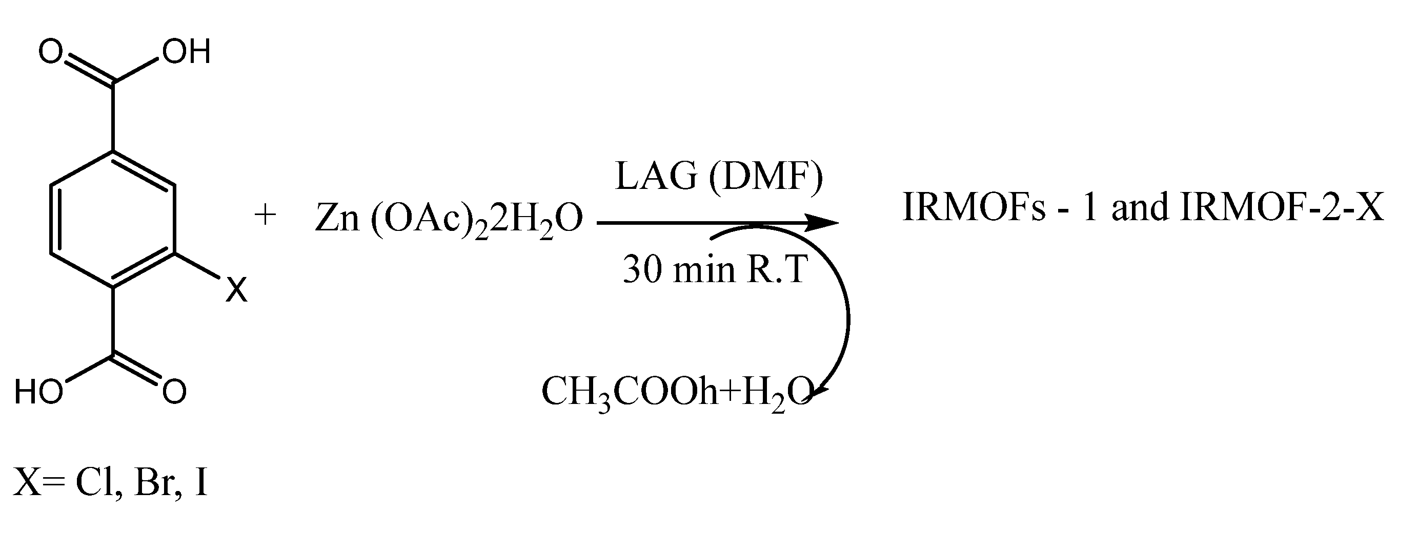
| 7. | IRMOF-2-X | PhNO2 | - | - | Quenching | [79,80,81] |
| 8. | Zn2(oba)2(bpy). | Nitroaromatics | - | - | Quenching | [89] |
| No. | Quantum Dots Sensors | Analytes | Linear Range | Limit of Detection | Effect on Luminescence | Ref. |
|---|---|---|---|---|---|---|
| 1. | Graphene oxide (GO) | Phosphate (Pi) | 15–37 µM | 0.1 µM | Quenching | [93] |
| 2. | CuInS2/ZnS | Copper (Cu2+) | 0–70 nM | 63 nM | Quenching | [99] |
| 3. | AgInS2 | Glutathione (GSH) | 3 × 10⁻⁴ to 2.5 × 10⁻³ mol·L−1 | 2.8 × 10⁻¹⁰ mol·L−1 | Enhancing | [102] |
| 4. | TDES-CdSe | Uranyl ion (UO22+) | 10–50 nM | 5.7 nM | Quenching | [109] |
| 5. | CdZnTe | Aflatoxin 1 (AFB1) | 50–100 ng·mL−1 | 20 ng·mL−1 | Enhancing | [110] |
| 6. | CDs/ | Chlorotetracycline (CTC) | 1–70 µM | 0.46 µM | Enhancing | [111] |
| CuInS2/ZnS | 1–50 µM | 0.36 µM | Ignorable | |||
| 7. | CS-GQDs | Dopamine (DA) | - | - | Enhancing | [112] |
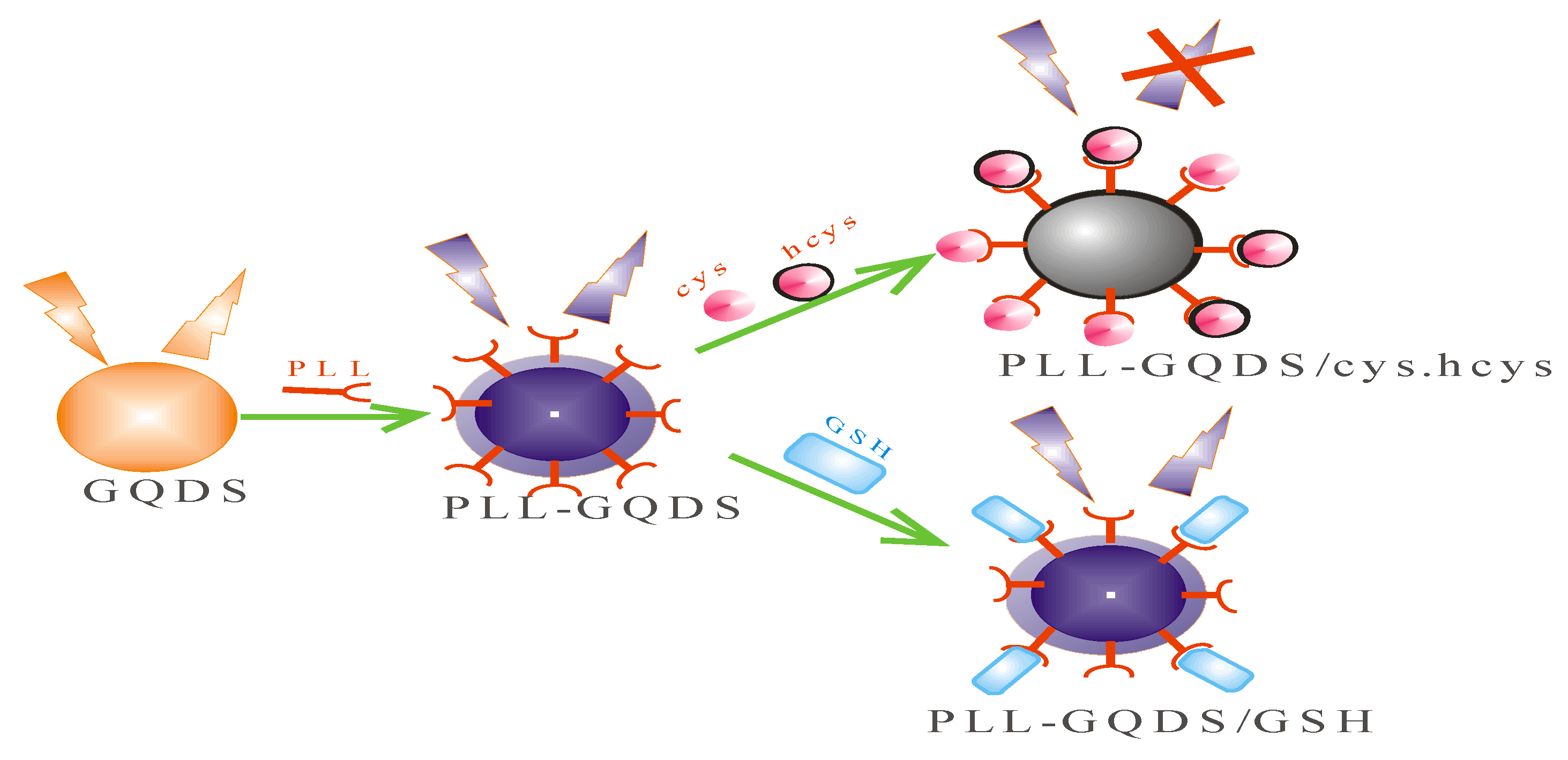
| 8. | PLL-GQDs | Cysteine (cys) | 0–150 nM | 2.38 nM. | Quenching | [113] |
| Homocysteine (hcys) | 0–100 nM | 1.94 nM | Quenching |
| No. | Sensors/Probes | Analytes | Linear Range | Limit of Detection | Effect on Luminescence | Ref. |
|---|---|---|---|---|---|---|
| 1. | AuNPs- ErCDs-MOFs | Bisphenol A (BPA) | 7.0 × 10⁻⁸ to 5 × 10⁻⁷ mol/L | 32 nmol/L | Enhancing | [151] |
| 2. | MOF-UCNPs | Bisphenol A (BPA) | 0.1–100 nM | 0.02 nM | Quenching | [152] |
| 3. | Ga-MOF | Bisphenol A (BPA) | 320–382 nm | 26.36 nM | Enhancing | [153] |
| 4. | (3D PS-C-Au) electrode | Bisphenol A (BPA) | 5.0 × 10⁻⁹ mol/L to 1.0 × 10⁻⁵ mol/L | 3.5 × 10⁻⁹ mol/L | Enhancing | [154] |
| 5. | MCM-41 | Bisphenol A (BPA) | 2.2 × 10⁻⁷ mol/L to 8.8 × 10⁻⁵ mol/L | 3.8 × 10⁻⁸ mol/L | Enhancing | [155] |
| 6. | Pd@TiO2-SiC-GCE | Bisphenol A (BPA) | 0.01–200 µM | 4.3 nM | Enhancing | [158] |
| 7. | N-modified mesoporous carbon (NMC), | Bisphenol A (BPA) | - | - | Enhancing | [164] |
| mesoporous carbon (MC) | Enhancing | |||||
| 8. | f-SWCNT/PC4/GCE | Bisphenol A (BPA) | 0.099–5.794 µmol/L | 0.032 µmol/L | Enhancing | [165] |
| 9. | N-NiO@NFe3O4@N-ZnO | Bisphenol A (BPA) | - | - | Quenching | [166] |
| No. | Sensors/Probes | Analytes | Linear Range | Limit of Detection | Effect on Luminescence | Refs. |
|---|---|---|---|---|---|---|
| 1. | CDs@ZIF-8 | dopamine (DA) | 0.1–200 nM | 16.64 nM | Enhancing | [182] |
| 2. | SQDs@MOFs | Cr (VI), | below the threshold | 0.16 μM | Enhancing | [183] |
| (Cr2O72−/Cr2O42−) | 0.17 μM | |||||
| 3. | Mn2+ ZnS@ ZIF-8 | Co2+ | 0.27 μM | Quenching | [184] | |
| human albumin (HAS) | 0.22 μM | |||||
| 4. | amine-CQDs@MOFs | 4-nitrophenol (4-NP) | 0.01–2.0 µM | 3.5 nM | Enhancing | [185] |
| 5. | CdTe@Zn2camph2bipy | L-tartaric | - | - | Quenching | [186] |
| D- and L-dimethyl tartrates | ||||||
| 6. | BPEI-CQDs/ZIF-8 | Cu2+ | 2 nM to 1000 nM | 80 pM | Quenching | [188] |
| 7. | Zn-MOF-8@AgQDs | 2,4-dinitritoluene | 0.0002 µM to 0.9 µM | 0.041 µM | Enhancing | [190] |
| 8. | UiO-66-NH2@CdTe | ascorbic acid (AA) | 200–1200 µM | 39.5 µM | Enhancing/quenching | [201] |
Publisher’s Note: MDPI stays neutral with regard to jurisdictional claims in published maps and institutional affiliations. |
© 2022 by the authors. Licensee MDPI, Basel, Switzerland. This article is an open access article distributed under the terms and conditions of the Creative Commons Attribution (CC BY) license (https://creativecommons.org/licenses/by/4.0/).
Share and Cite
Ajibade, P.A.; Oloyede, S.O. Synthesis of Metal–Organic Frameworks Quantum Dots Composites as Sensors for Endocrine-Disrupting Chemicals. Int. J. Mol. Sci. 2022, 23, 7980. https://doi.org/10.3390/ijms23147980
Ajibade PA, Oloyede SO. Synthesis of Metal–Organic Frameworks Quantum Dots Composites as Sensors for Endocrine-Disrupting Chemicals. International Journal of Molecular Sciences. 2022; 23(14):7980. https://doi.org/10.3390/ijms23147980
Chicago/Turabian StyleAjibade, Peter A., and Solomon O. Oloyede. 2022. "Synthesis of Metal–Organic Frameworks Quantum Dots Composites as Sensors for Endocrine-Disrupting Chemicals" International Journal of Molecular Sciences 23, no. 14: 7980. https://doi.org/10.3390/ijms23147980
APA StyleAjibade, P. A., & Oloyede, S. O. (2022). Synthesis of Metal–Organic Frameworks Quantum Dots Composites as Sensors for Endocrine-Disrupting Chemicals. International Journal of Molecular Sciences, 23(14), 7980. https://doi.org/10.3390/ijms23147980