Article

Copper (II) Heterocyclic Thiosemicarbazone Complexes as Single-Source Precursors for the Preparation of Cu9S5 Nanoparticles: Application in Photocatalytic Degradation of Methylene Blue

1
Department of Inorganic Chemistry, Faculty of Science, University of Yaounde I, Yaounde P.O. Box 812, Cameroon
2
Institut Jean Lamour UMR 7198 CNRS, Université de Lorraine Campus Artem, 2 Allée André Guinier, 54000 Nancy, France
*
Authors to whom correspondence should be addressed.
Catalysts 2022, 12(1), 61; https://doi.org/10.3390/catal12010061
Submission received: 26 November 2021 / Revised: 15 December 2021 / Accepted: 27 December 2021 / Published: 6 January 2022
(This article belongs to the Special Issue Advances in Supported Catalytic Nanomaterials)

Abstract

:
In this study, two copper(II) complexes, [Cu(C6H8N3S2)2]Cl2 (1) and [Cu(C7H10N3S2)2]Cl2·H2O (2), were synthesized from 2-(thiophen-2-ylmethylene)hydrazine-1-carbothioamide (L1H) and 2-(1-(thiophen-2-yl)ethylidene)hydrazine-1-carbothioamide (L2H) respectively and characterized using various spectroscopic techniques and elemental analyses. The as-prepared complexes were used as single-source precursors for the synthesis of oleylamine-capped (OLA@CuxSy), hexadecylamine-capped (HDA@CuxSy), and dodecylamine-capped (DDA@CuxSy) copper sulphide nanoparticles (NPs) via the thermolysis method at 190 °C and 230 °C and then characterized using powder X-ray diffraction (p-XRD), UV-visible spectroscopy, transmission electron microscopy (TEM), and scanning electron microscopy (SEM). The p-XRD diffraction patterns confirmed the formation of crystalline rhombohedral digenite Cu9S5 with the space group R-3m. The TEM images showed the formation of nanoparticles of various shapes including hexagonal, rectangular, cubic, truncated-triangular, and irregularly shaped Cu9S5 nanomaterials. The SEM results showed aggregates and clusters as well as the presence of pores on the surfaces of nanoparticles synthesized at 190 °C. The UV-visible spectroscopy revealed a general blue shift observed in the absorption band edge of the copper sulphide NPs, as compared to bulk CuxSy, with energy band gaps ranging from 2.52 to 3.00 eV. Energy-dispersive X-ray spectroscopy (EDX) confirmed the elemental composition of the Cu9S5 nanoparticles. The nanoparticles obtained at 190 °C and 230 °C were used as catalysts for the photocatalytic degradation of methylene blue (MB) under UV irradiation. Degradation rates varying from 47.1% to 80.0% were obtained after 90 min of exposure time using only 10 mg of the catalyst, indicating that Cu9S5 nanoparticles have potential in the degradation of organic pollutants (dyes).

1. Introduction

In recent decades, there has been an increasing interest in semiconductor metal chalcogenide nanocrystals (NCs) due to their various applications in nanoscience and nanotechnology [1,2]. Their unique properties make them a subject of intense research and are strongly influenced by morphology, phase, and surface properties [1,2,3,4]. Amongst metal chalcogenides, copper sulphide, which is a p-type semiconductor with a tuneable band-gap ranging from 1.2 eV to 2.0 eV, has become highly sought after due to its wide range of stoichiometric compositions and phases. These phases vary from the copper-rich chalcocite phase (Cu2S) to the copper-deficient phases (covellite (CuS), anilite (Cu1.75S), digenite (Cu1.8S), and djurleite (Cu1.96S)) [5,6,7,8,9]. Copper-containing nanomaterials have been shown to possess potential applications in solar cells [5], photocatalysis [6], supercapacitors [10], nanometre-scale switches, and high-capacity cathode materials in lithium secondary batteries, superconductor, thermoelectric cooling material, and solar-energy absorption [11,12]. These varied applications of copper sulphide semiconductor materials stem from their unique optical, electronic, and photocatalytic properties that are influenced by their morphology, their stoichiometric composition, and their crystalline phases, which are tuneable by various reaction parameters such as their capping agent, the nature of their precursor, and their reaction temperature [7,8,9,13]. Capping agents stabilize the surface of copper sulphide to prevent agglomeration and to obtain monodisperse nanoparticles (NPs) [14,15]. However, controlling the phase and the morphology of copper sulphide NPs has been difficult to achieve.
Significant efforts have been devoted to phase-controlled synthesis of copper sulphide NPs [13,14,15]. Several methods have been developed for structure-controlled synthesis of copper sulphide nanoparticles, including microwave irradiation [16], solvothermal [17], and thermolysis of a single-source precursor [18,19]. Among these synthetic routes, the thermolysis of single-source precursors has been shown to yield high-quality nanoparticles due to the presence of the preformed metal–sulphur bond in the molecule, which can control the chemical composition and physical properties of the as-prepared metal chalcogenide NPs [15,20,21,22,23]. This method regulates the particle size and shape by varying reaction conditions such as temperature, precursor concentration, and capping group [7,8,24]. The choice of precursors plays an important role on the morphology and phase of copper sulphide NPs [16]. Thermolysis of dithiocarbamates [6], xanthates [25], dithiolates [26], and thiobiuret [27] compounds have been explored as single-source precursors for the preparation of copper sulphide nanoparticle. Despite the various reports on the synthesis of copper sulphide NPs, the effects of capping agents on crystallinity, phase, size, shape, optical, and photocatalytic properties have not been fully explored. Recently, our group reported the use of thiourea Cu(II) complexes, N-morpholine-N-benzoylthioureatocopper(II), and N-pyrrolidine-N-benzoylthioureatocopper(II) complexes as single-source precursors for the preparation of copper sulphide NPs with various morphologies [8,9], and our results indicate that the formation of the roxbyite phase (Cu1.75S) was solvent and temperature dependent [9]. Studies have also reported the use of Cu(II) complexes for the photodegradation of organic dyes in aqueous solution [28,29].
Copper(II) heterocyclic thiosemicarbazone complexes have been extensively studied for their antimicrobial, anticancer, and antiviral properties [30,31,32,33,34,35]. However, little attention has been paid to their use as a single source precursor for the preparation of CuxSy nanoparticles. Heterocyclic thiosemicarbazones are chelating ligands with the ability to form air-stable and less toxic complexes possessing the pre-formed metal-sulfur (M-S) bonds. We hereby report on the synthesis of two heterocyclic thiosemicarbazone copper (II) complexes and their use as single-source precursors to prepare CuxSy NPs at 190 °C and 230 °C, using oleylamine (OLA), hexadecylamine (HDA), and dodecylamine (DDA) as capping agents. We also report on the use of OLA-capped CuxSy, HDA-capped CuxSy, and DDA-capped CuxSy NPs obtained as photocatalysts for the degradation of methylene-blue (MB) dye under UV irradiation.

2. Results and Discussion

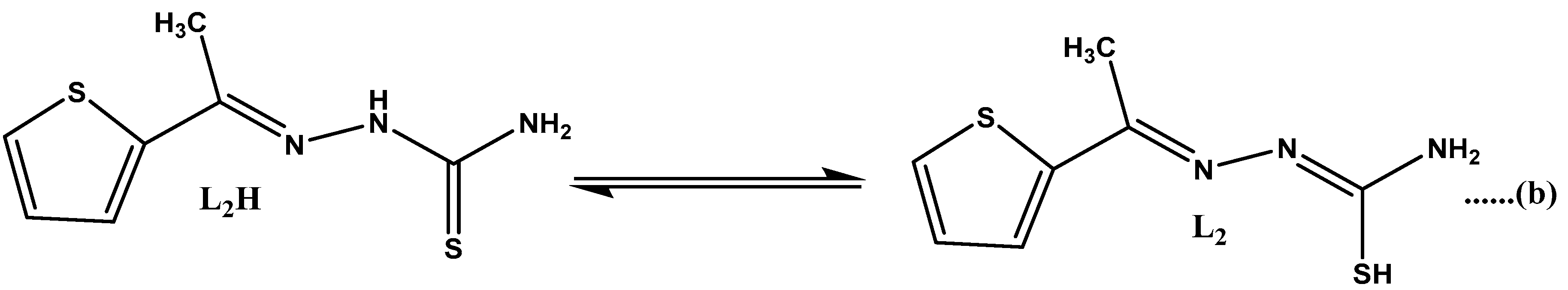
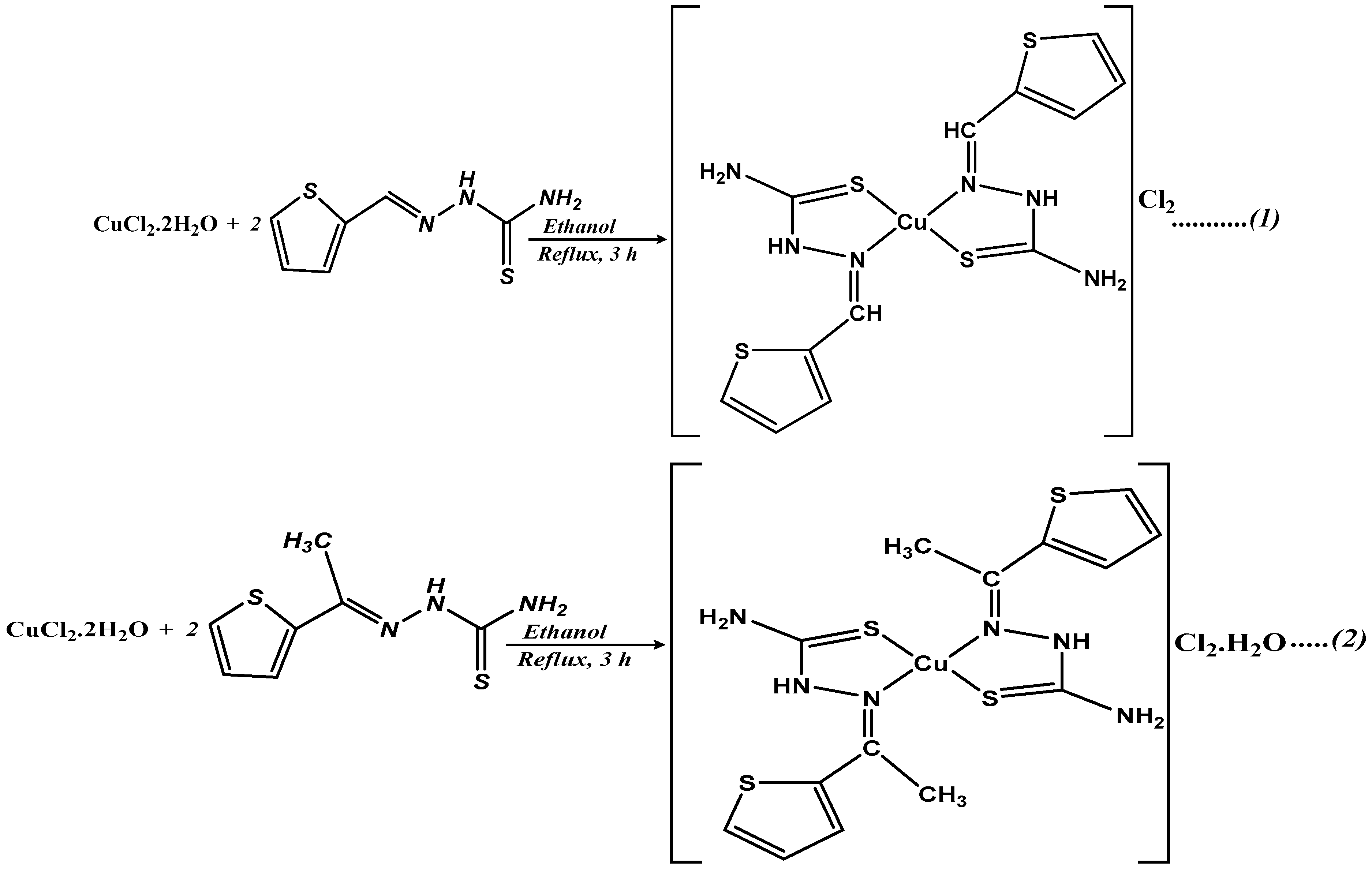
2.1. Characterisation of the Ligands and Complexes

The ligands and their corresponding Cu(II) complexes were obtained in appropriate yields, and microanalysis confirmed their purity. All the compounds were coloured, air-stable, and soluble in dimethyl sulphoxide (DMSO). The molar conductance values of the complexes recorded in the DMSO as a solvent for complex 1 ([Cu(C6H8N3S2)2]Cl2) and complex 2 ([Cu(C7H10N3S2)2]Cl2·H2O), indicating the electrolytic nature of both complexes [36,37].

2.1.1. Infrared Study

The bonding mode of the ligands in each Cu (II) complex was determined by comparing the Fourier-transform infrared (FTIR) spectrum of the ligand with that of the corresponding metal complex. The FTIR spectra of the ligands and their corresponding Cu(II) complexes are presented in Figure S1. The FTIR spectra of the free ligands exhibited a band within the range 1582–1595 cm−1 assigned to ν(C=N). In the spectra of complexes 1 and 2, this band shifted to higher frequencies by ca. 6–16 cm−1 (1602–1608 cm−1), suggesting the coordination of the azomethine nitrogen to the central metal atom. In addition, this coordination was supported by the presence of ν(M-N) vibrations at approximately 515–540 cm−1. In the IR spectra of the ligands, the ν(S-H) band that usually appears at approximately 2570 cm−1 [38,39] was absent while the ν(C=S) bands at 830 and 835 cm−1 were present. These ν(C=S) bands had shifted to lower frequencies by ca. 4–10 cm−1 (820–831 cm−1) in the spectra of complexes 1 and 2. This shift was attributed to the thiocarbonyl ν(C=S) stretching and bending modes of vibrations and confirmed the coordination of the sulphur atom to the metal ion (M-S) [38,39]. The band that appeared around 3248 cm−1 in the spectrum of the complex with the ligand (L2) was attributed to water of crystallisation [38].

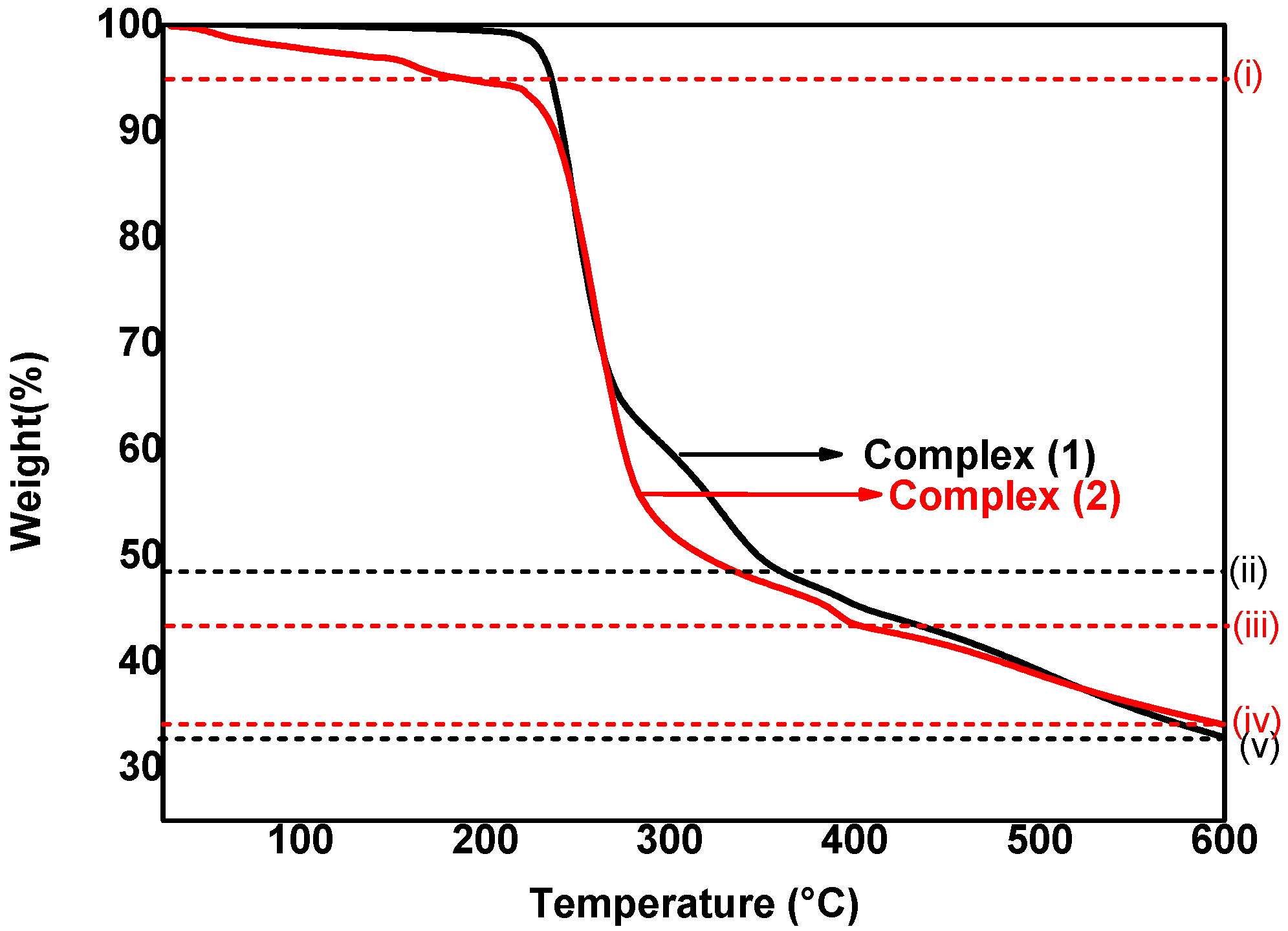
2.1.2. Thermal Decomposition Studies of Precursors

The TGA curves of complexes (1) and (2) are shown in Figure 1. The thermal decomposition curve of complex 1 showed two decomposition steps. The first decomposition occurred within a temperature range of 211–276 °C with a mass loss of 35.9% (calculated (calc): 36.8%), which corresponded to the degradation of the ligand molecule (L1) (curve ii in Figure 1). The second decomposition step involved a weight loss of 31.3% (calc: 30.8%) within a temperature range of 276–525 °C, which was attributed to the loss of the organic moiety and chloride (curve v in Figure 1). The final residues comprised copper sulphide and carbon (found: 31.5%; calc: 32.4%) [30,38]. The thermal decomposition curve of complex 2 showed three decomposition steps. The first step between 50 °C and 144 °C corresponds to the loss of a water molecule (found: 3.3%; calc: 3.2%) (curve i in Figure 1). The second decomposition step between 178 °C and 284 °C corresponds to the loss of a ligand (L2) molecule and a chloride ion (found: 41.6%; calc: 42.5%) (curve iii in Figure 1), while the third decomposition step, which occurred between 305 °C and 530 °C (Found: 21.03%; calc: 20.2%), corresponds to the decomposition of an organic moiety (curve iv in Figure 1). The final residues of 34.07% (calc: 34.1%) were attributed to the mixture of copper sulphide and carbon residue [30,38].

2.2. Characterisation of Copper Sulphide Nanoparticles

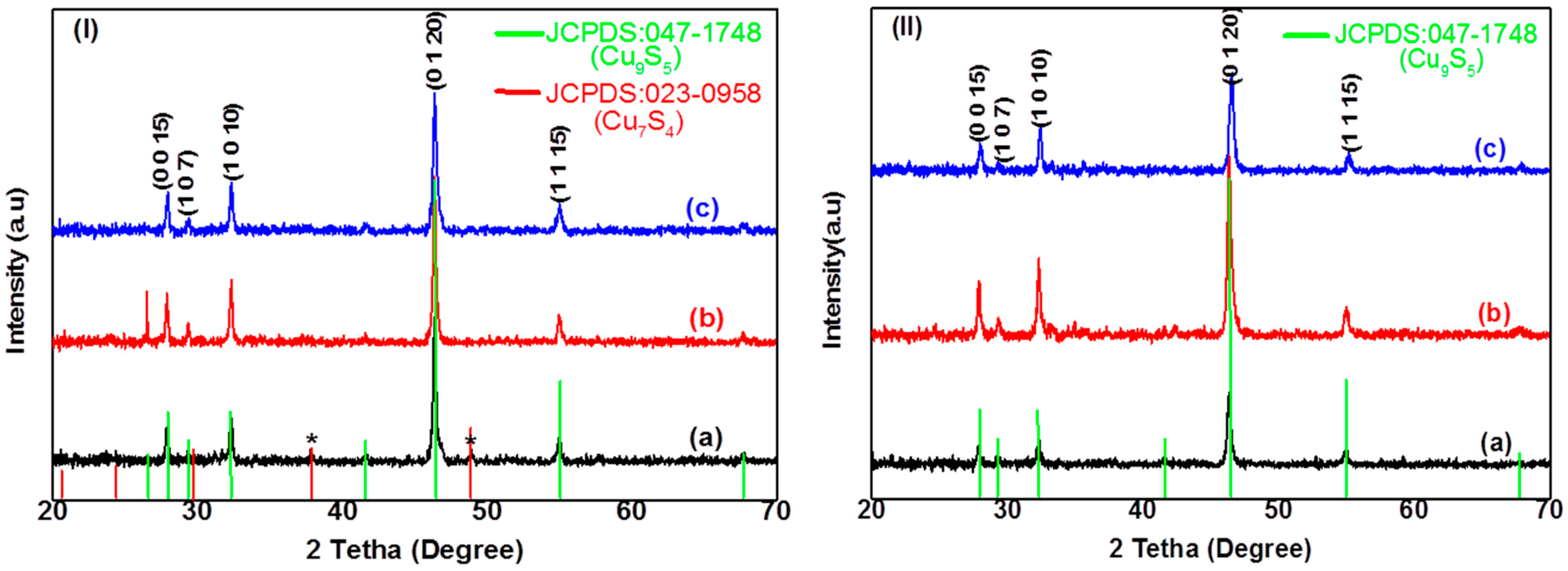
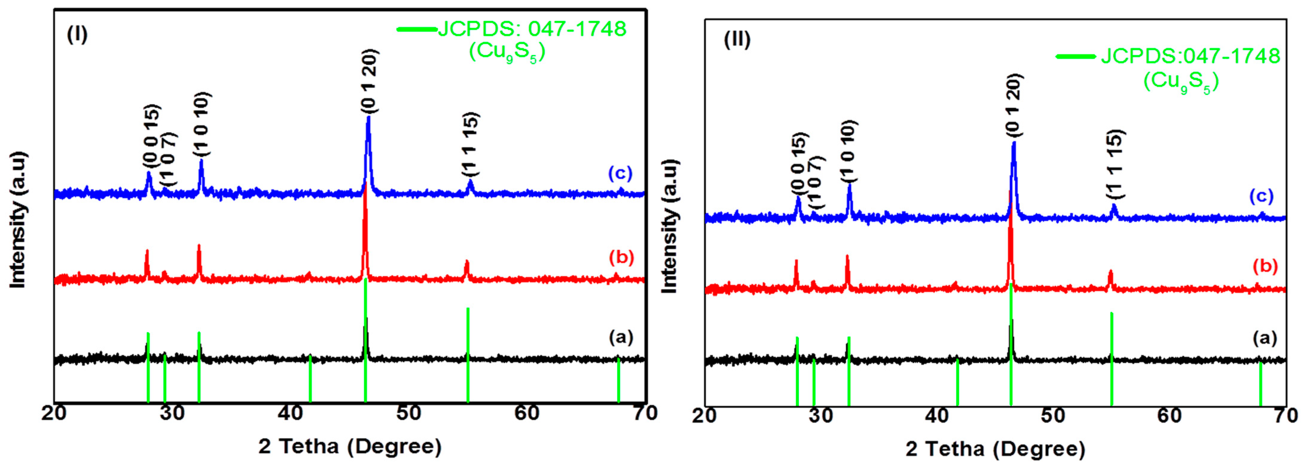
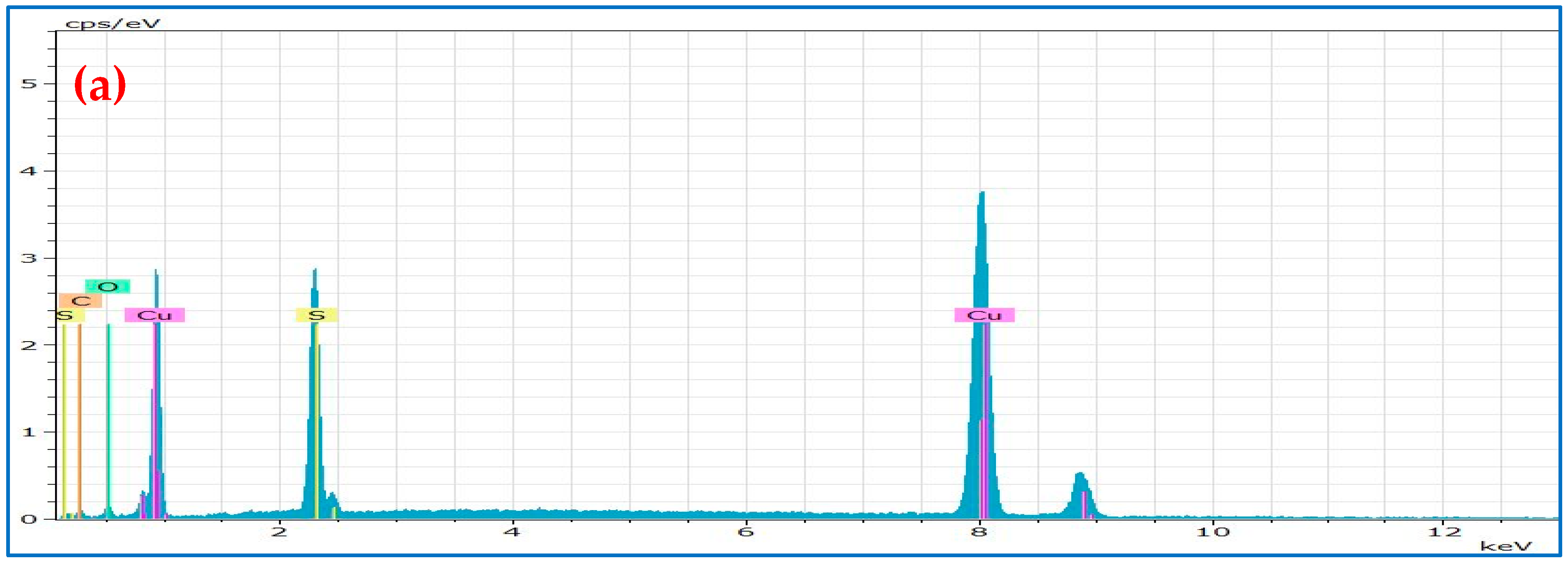
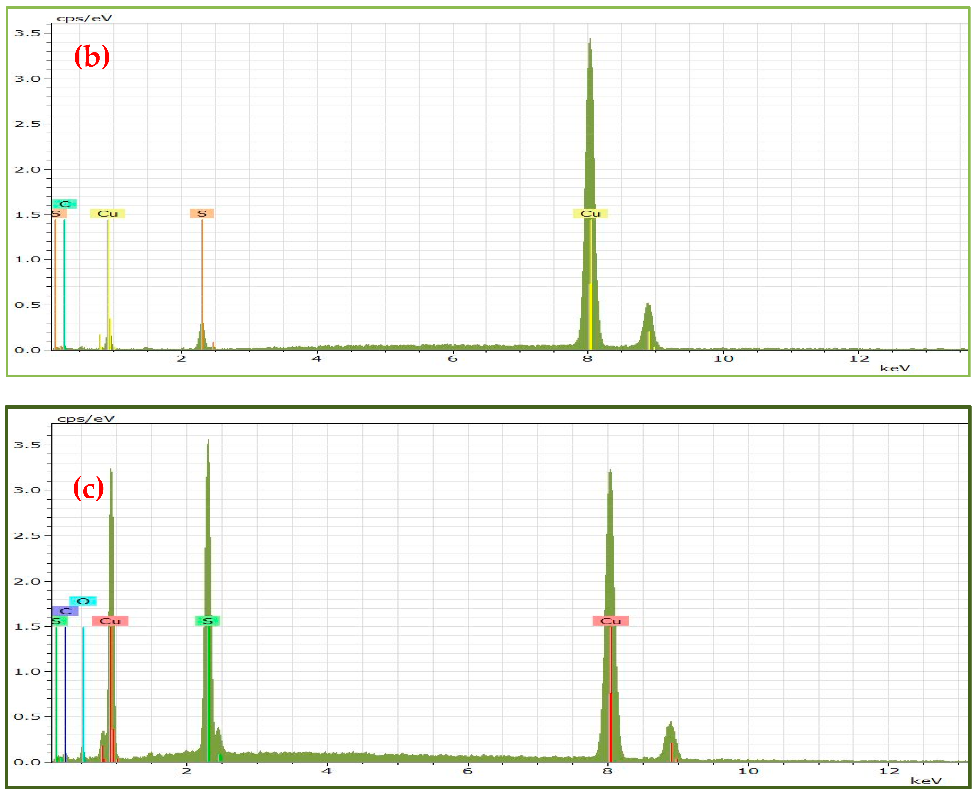
2.2.1. Structural Characterization of CuxSy Nanoparticles

The powder X-ray diffraction (p-XRD) patterns of copper sulphide NPs synthesized using complexes 1 and 2 in OLA (C18), HDA (C16), and DDA (C10) at 190 °C are presented in Figure 2. When complex 1 was used as a precursor with OLA, HDA, and DDA as capping agents, the p-XRD patterns (Figure 2I(a–c)) with the lattice planes (0 0 15), (1 0 7), (1 0 10), (0 1 20), and (1 1 15) were indexed to pure rhombohedral digenite Cu9S5 with space group R-3m. In addition, the diffractogram of copper sulphide NPs synthesized with OLA as capping agents showed extra peaks (denoted *, Figure 2I(a)) that were attributed to the roxbyite Cu7S4 phase, (JCPDS: 023-0958). When complex 2 was used, the rhombohedral digenite Cu9S5 with space group R-3m was also observed with all three capping agents, with no other phases present, which confirmed the phase purity of the Cu9S5 NPs (Figure 2II(a–c)). The p-XRD results agreed with the reported data for copper sulphide nanostructures synthesized from copper(II) piperidine dithiocarbamate precursors using a single-source precursor route [15,40].
Crystalline nanoparticles were also obtained when the reaction temperature was increased to 230 °C using complexes 1 and 2 as well as all the capping agents (Figure 3). The rhombohedral phase (Cu9S5, digenite) was also obtained with the lattice planes (0 0 15), (1 0 7), (1 0 10), (0 1 20), and (1 1 15) (JCPDS: 047-1748). The rhombohedral phase (Cu9S5, digenite) has been commonly reported for copper chalcogenide NPs [41].
EDX analysis were performed to confirm the elemental composition of the synthesized CuxSy NPs and results are presented in Figure 4, Table 1 and Table 2. The EDX spectra showed the presence of Cu and S, as well as other constituent elements such as C and O, when complexes 1 and 2 were employed as precursors. The presence of the carbon peak was attributed to the capping agents OLA, HDA, and DDA as well as the double-sided carbon tape that had been used for mounting the nanoparticles. The presence of Cu and S indicated the formation of copper sulphide NPs. The Cu/S ratios approximately corresponded to the expected compositions, with a slightly copper-rich stoichiometry in the OLA@Cu9S5 and the HDA@Cu9S5 NPs using complex 1. The high percentage of Cu could be attributed to the excessive absorption of copper ions onto the surface of NPs. Similar results were recently obtained by Murendeni et al. [15]. The EDX spectra of the DDA@Cu9S5 NPs confirmed that the primary elemental constituents of the nanoparticles were copper and sulphur in a molar ratio of 1.83:1 (Cu/S), which agreed with the stoichiometric ratio in the digenite phase. A similar trend was observed for the OLA@Cu9S5 NPs when complex 2 was used as precursor (Figure S2). The results showed Cu/S ratios that represented sulphur-rich and copper-deficient stoichiometries and thus indicated the nonstoichiometric compositions in the structures of the samples (Figure S2) of the HDA@Cu9S5 and DDA@Cu9S5 NPs. The presence of oxygen on the EDX spectrum of Cu9S5 NPs prepared in OLA and DDA could be explained by the absorption of oxygen from the air during sample preparation for analysis. The results of the composition of CuxSy nanoparticles when OLA, HDA, and DDA were used as capping agents at 230 °C (Figures S3a–c and S4a–c) were comparable to those obtained at 190 °C. A general trend was observed where a decrease in the carbon chain of the primary amine-capping agent (OLA–DDA), C18–C12, had resulted in alteration of the composition (Cu/S ratio) in the copper sulphide NPs.

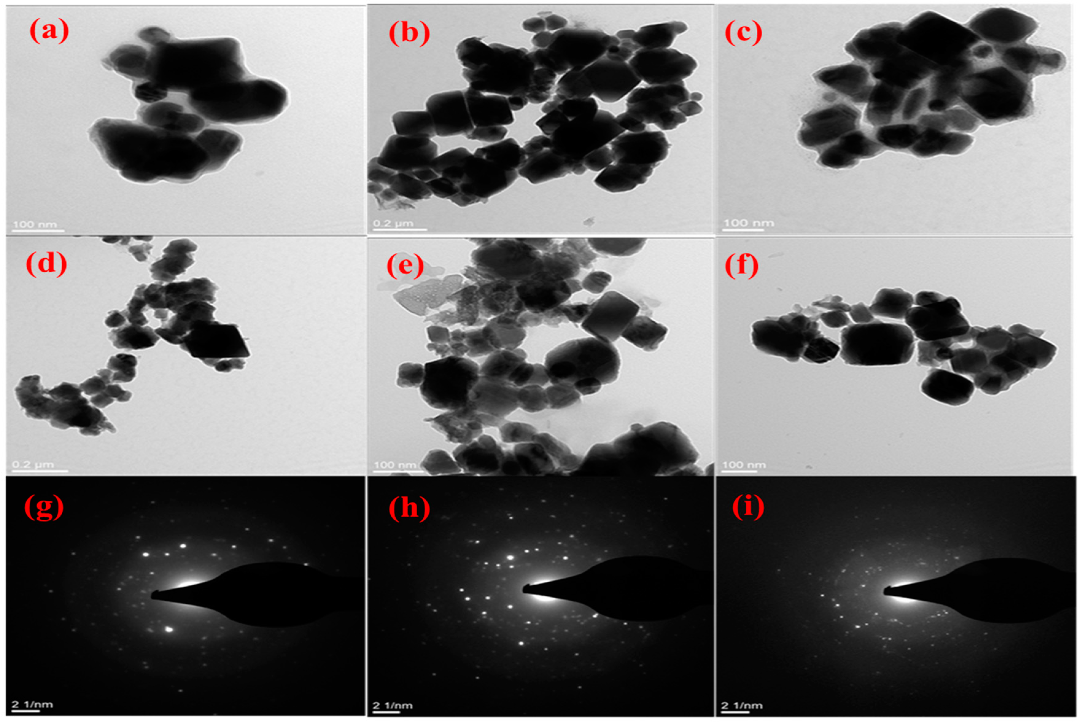
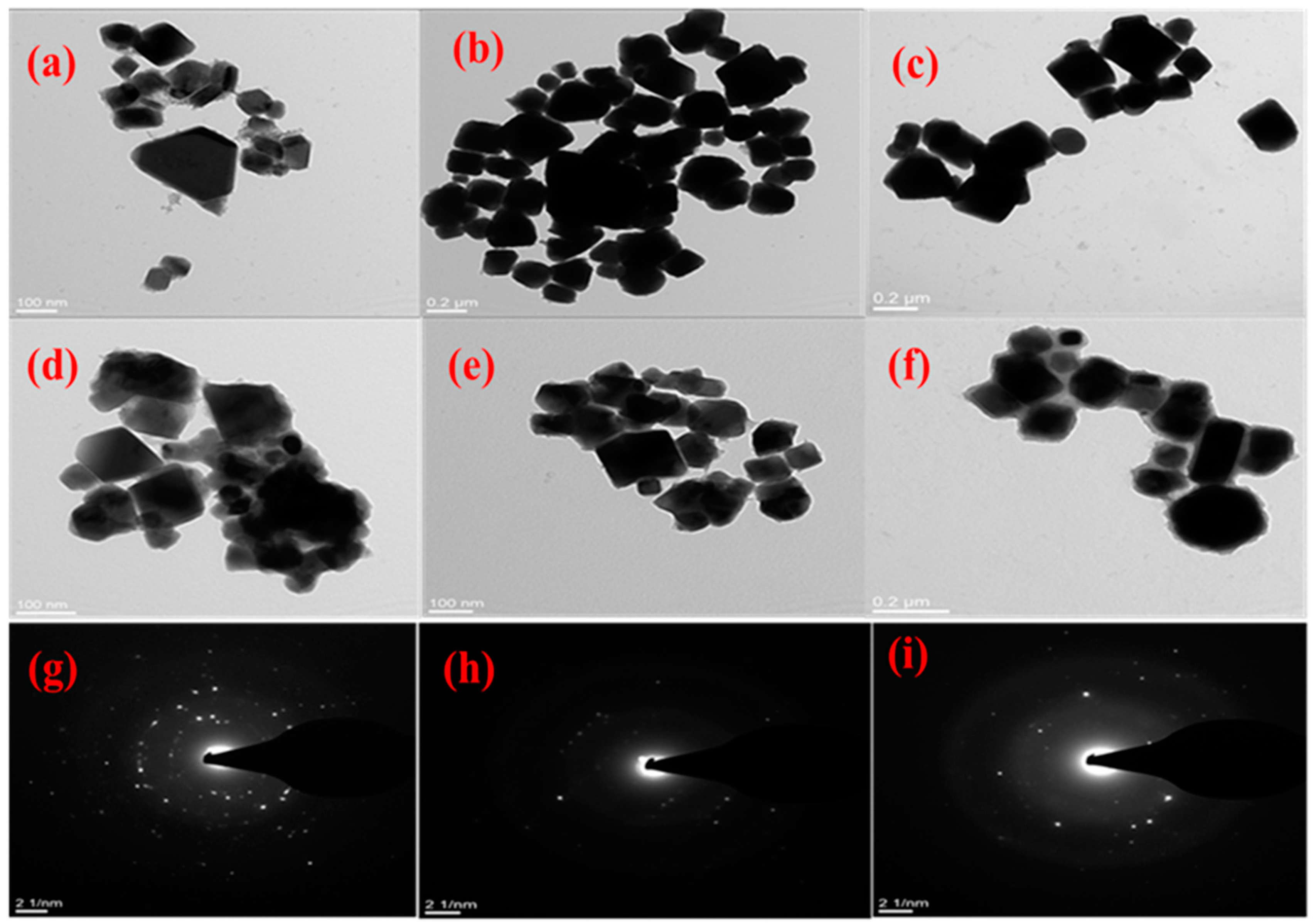
2.2.2. Morphological Characterisation of CuxSy Nanoparticles

The morphology of the synthesized copper sulphide NPs was investigated by TEM analyses, and the results are given in Figure 5 and Figure 6. When OLA (C18) was used as a stabilising agent, agglomerated irregularly shaped particles were formed with sizes ranging from 49 to 80 nm using complex 1 as a precursor (Figure 5a). The large particles resulting from the interparticle aggregation were also formed. When the stabilizing agent was changed to HDA (C16), a mixture of rectangular and cubic-shaped copper sulphide NPs with sizes ranging from 46 to 134 nm was formed (Figure 5b). When DDA (C12) was used, rectangular and irregularly shaped particles with sizes ranging from 53 to 154 nm were produced (Figure 5c). The properties of the capping group had some influence on the size of the nanoparticles through the dynamics of attaching and detaching [42].
The influence of the precursor was studied by replacing complex 1 with complex 2 under similar reaction conditions. A slight difference was observed in the morphologies of the copper sulphide NPs. When OLA was used, agglomerated, shapeless particles were formed with estimated sizes ranging from 32 to 75 nm (Figure 5d). When HDA was used, a mixture of semi-spherical and rectangular particles with estimated sizes of 43–125 nm (Figure 5e) was produced. DDA as a capping agent produced irregular cubic–shaped particles with sizes ranging 23–125 nm (Figure 5f). The morphologies of copper sulphide reported in this work were comparable to those previously reported [4,43].
An increase in the reaction temperature resulted in an improvement in the quality and crystallinity of the copper sulphide NPs, and various shapes were formed (Figure 6). When OLA was used as a capping agent, a mixture of rectangular and truncated triangle-shaped copper sulphide NPs were observed with sizes of approximately 40–120 nm (Figure 6a) using complex 1. The use of HDA resulted in the formation of irregular cubic- and rectangular-shaped particles with estimated sizes of 101–200 nm, and DDA produced irregular cubic NPs with sizes of 61–119 nm (Figure 6b,c). The variations in the morphologies of the copper sulphide NPs were attributed to the influence of the length of the alkyl chains of the capping agents, which appeared to control the resultant sizes and shapes [44]. When complex 2 was used as a precursor under similar synthetic conditions, agglomerated NPs were formed in OLA while HAD resulted in irregular cubic NPs with sizes of 45–115 nm (Figure 6e). Under the same conditions with DDA, rectangular and cubic NPs with an estimated size of 66–225 nm were formed (Figure 6f). The selected area electron diffraction (SAED) of the nanoparticles from complexes 1 and 2, prepared at 190 °C and 230 °C, confirmed the crystalline nature of the nanoparticles Figure 5g–i, Figure 6g–i, Figures S5a–c and S6a–c). The particle size distributions of Cu9S5 nanoparticles are presented in Figure 7 and Figure S7c–f.
The SEM analyses were conducted to investigate the surface morphology of Cu9S5 nanoparticles using complexes 1 and 2 as precursors, and the results are presented in Figure 8. When complex 1 was thermolysed at 190 °C using OLA, HDA, and DDA as capping agents, agglomerated, granular Cu9S5 particles were formed (Figure 8a–c). In HDA, a less porous surface was observed (Figure 8b) while a porous surface was observed in DDA (Figure 8c). When complex 2 was used as a precursor, clusters were formed in OLA (Figure 8d), agglomerated seeds in HDA (Figure 8e), and agglomerated, porous, irregularly spherical NPs in DDA (Figure 8f).
The SEM images of the Cu9S5 nanoparticles obtained at 230 °C are shown in Figure S8. Agglomerated, irregularly spherical particles were formed in OLA (Figure S8a) while rock-shaped particles were formed in both HDA and DDA (Figure S8b,c). A similar trend (i.e., rock-shaped particles) was observed when complex 2 was thermolysed in OLA, HDA, and DDA (Figure S8e,f).

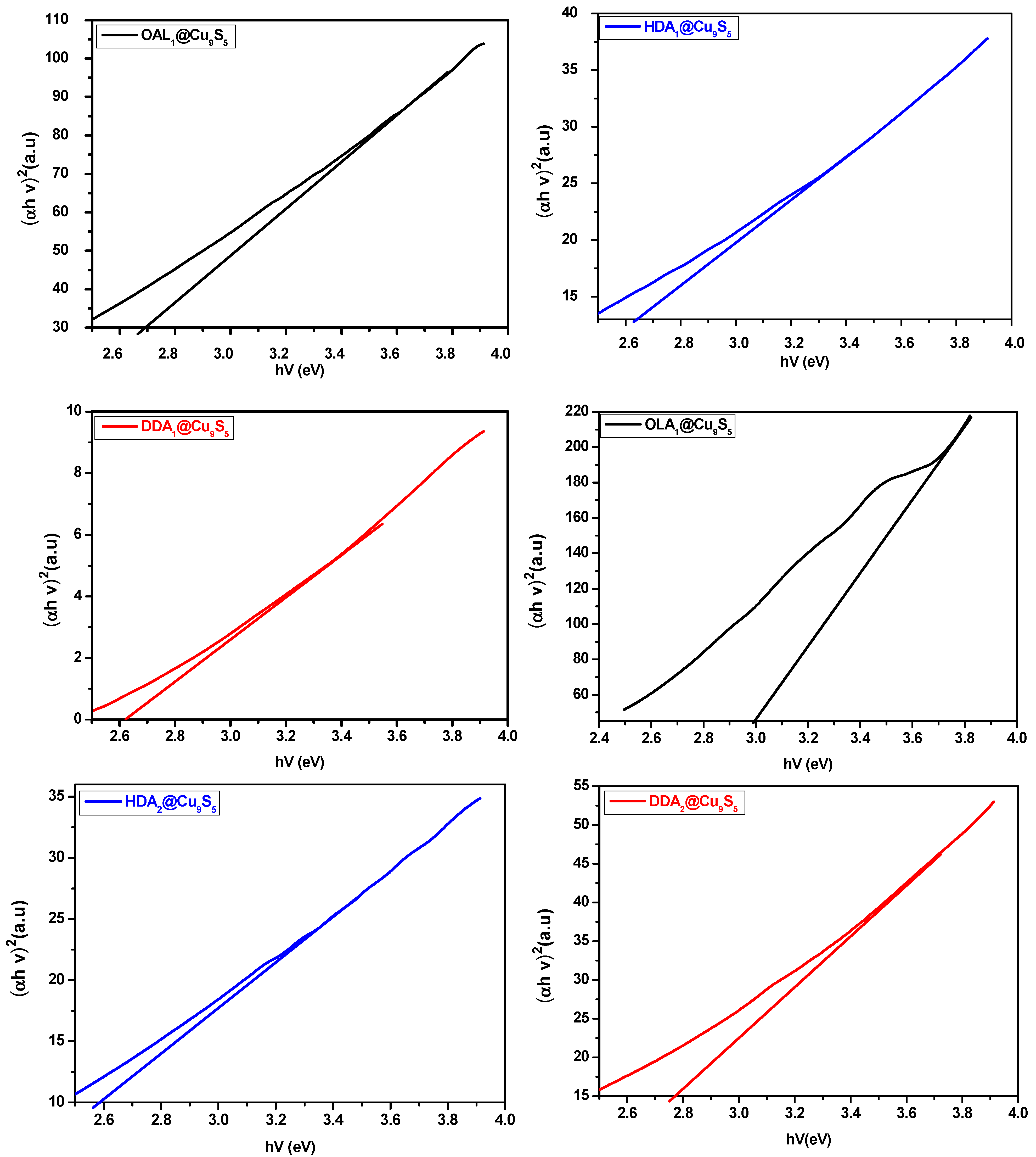
2.2.3. Optical Properties of CuxSy Nanoparticles

The ultraviolet-visible (UV–Vis) absorption spectra and their corresponding Tauc plots for copper sulphide NPs synthesized in OLA, HDA, and DDA and using complexes 1 and 2 at 190 °C are presented in Figure S9a,b and Figure 9 respectively. All the samples exhibited broad absorption in the visible region (400–650 nm) and extended into the near-infrared region at approximately 800 nm. The energies obtained were within 2.57–2.70 eV range for complex 1 and 2, 2.55–3.00 eV for complex 2. These values correspond to a blue shift as compared CuxSy bulk values (1.2–2.5 eV) due to quantum confinement effect of the CuxSy NPs [45]. The obtained materials showed variations in their optical properties according to the capping agent and precursor used. This difference in energy bandgap was attributed to the stoichiometric variations and the arrangement of the cations and anions in the atomic structures of the compounds [4]. This blue shift was more pronounced in OLA@CuxSy (2.70 eV and 3.00 eV) than in HDA@CuxSy (2.64 eV and 2.58) and DDA@CuxSy (2.57 eV and 2.55) NPs when complexes 1 and 2 were used, respectively, as precursors. This could be due to the differences in the carbon chains of the capping molecules, as OLA is a C18 branched amine while HDA and DDA are, respectively, C16 and C12 linear amines.
The same trend in the optical properties was observed when the reaction temperature was raised to 230 °C with a slight decrease in the absorption-band edges, as compared to 190 °C; the bandgap energies varied within the range of 2.52–2.76 eV for complex 1 and 2.56–2.83 eV for complex 2. This result was consistent with previous research [4]. The absorption spectra with their corresponding Tauc plots are presented in Figures S10a,b and S11.

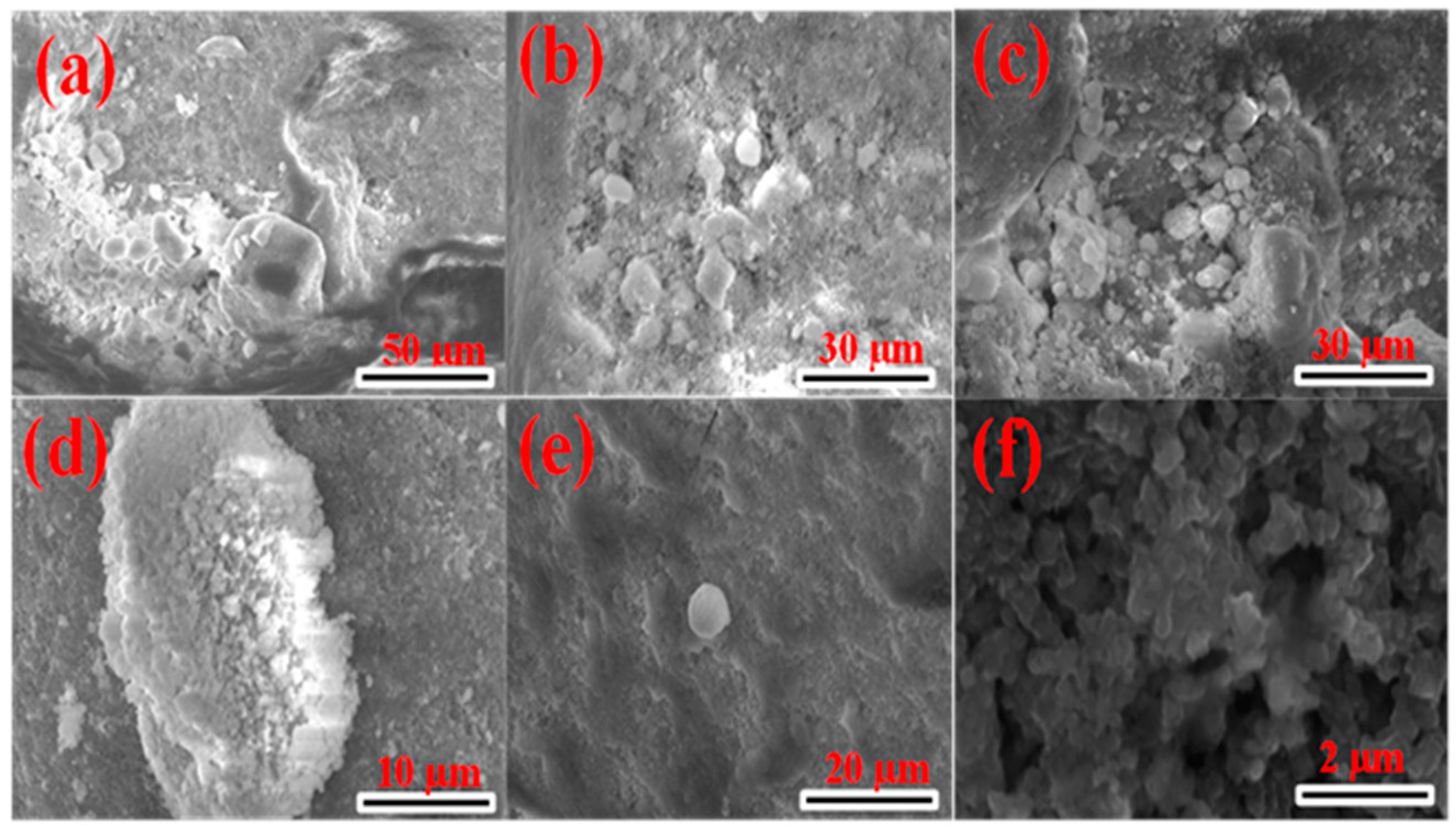
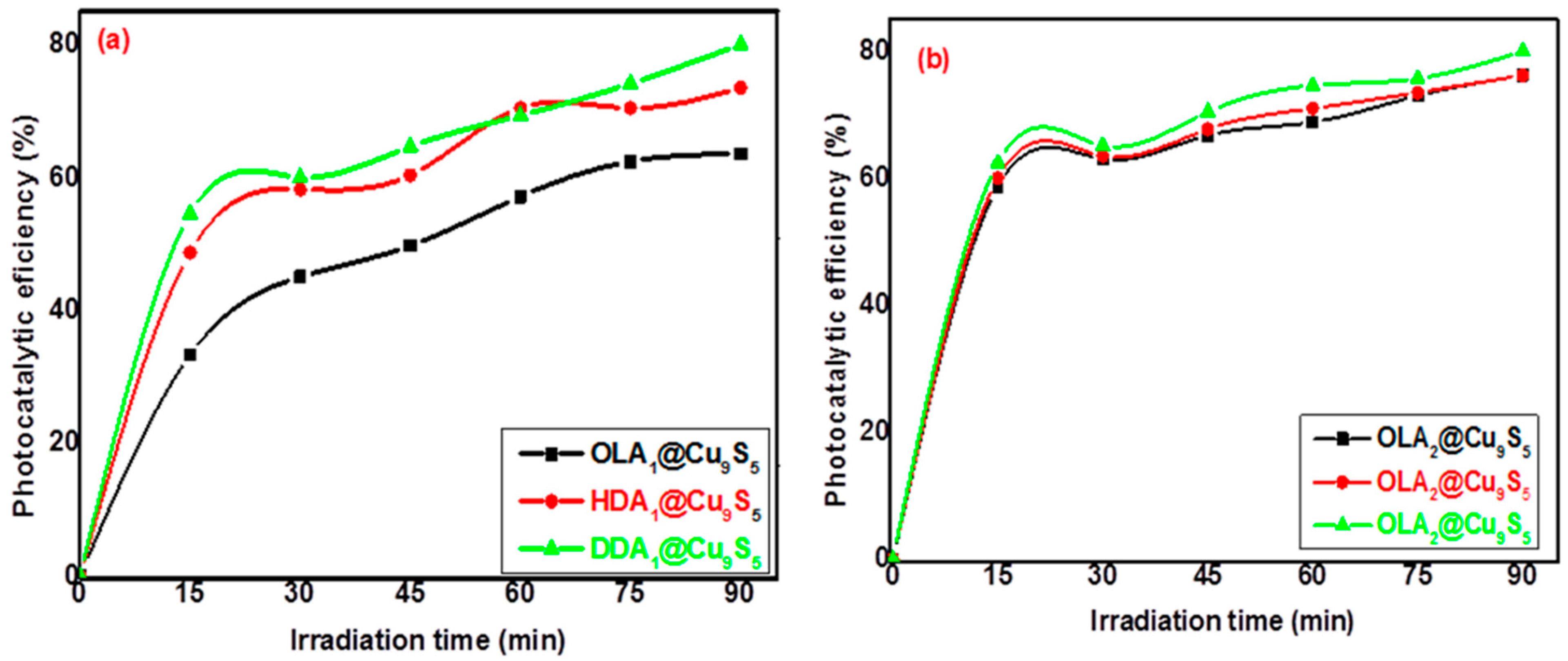
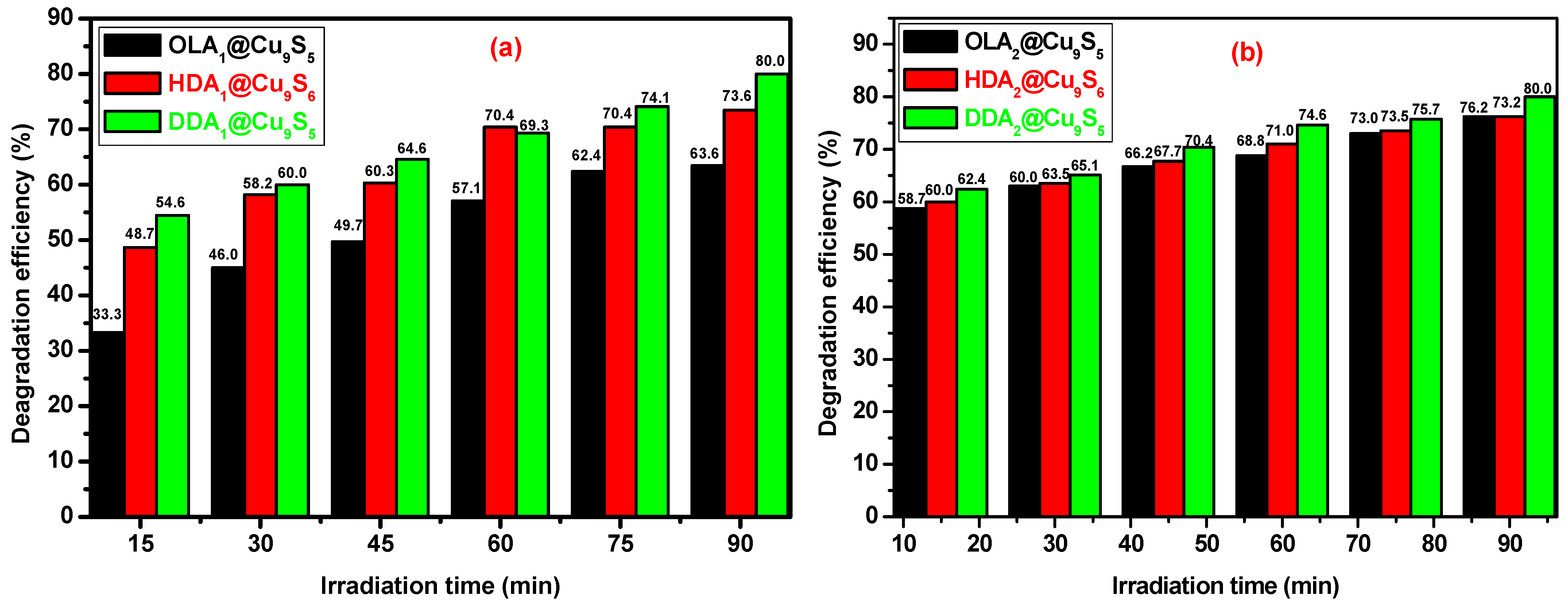
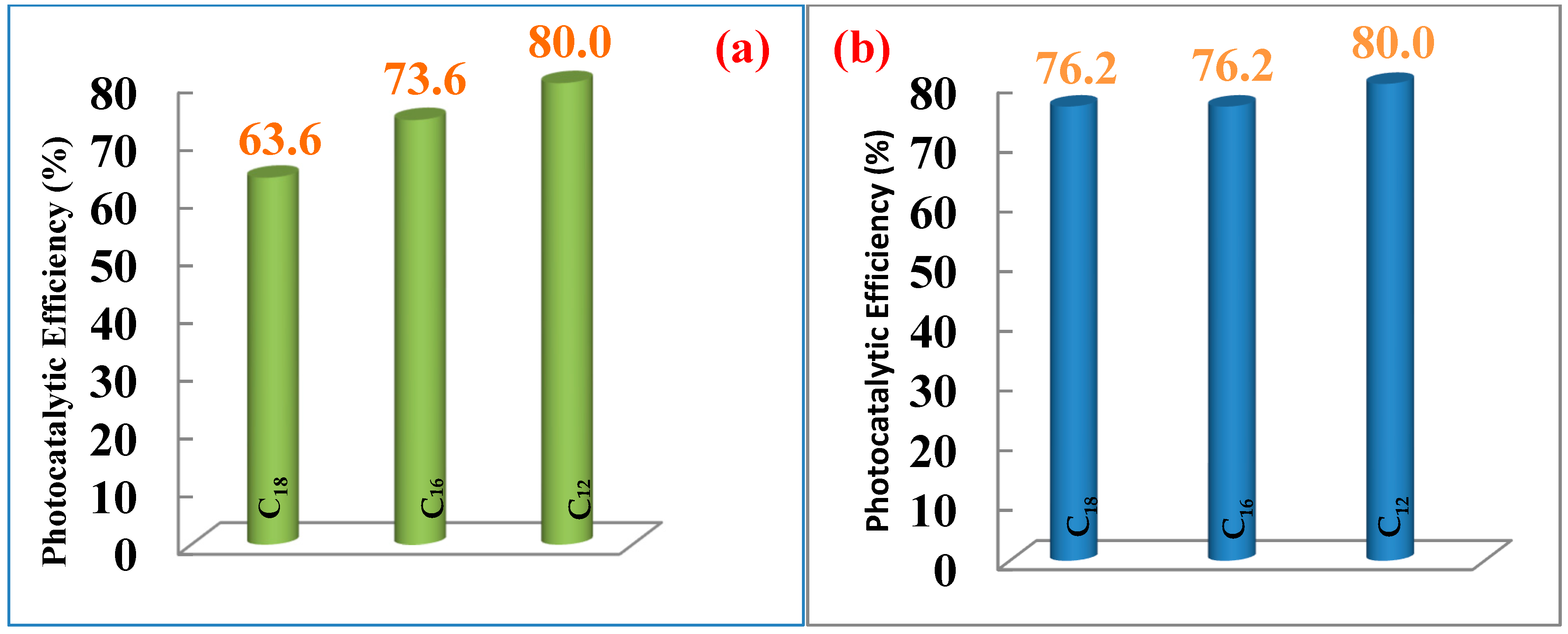
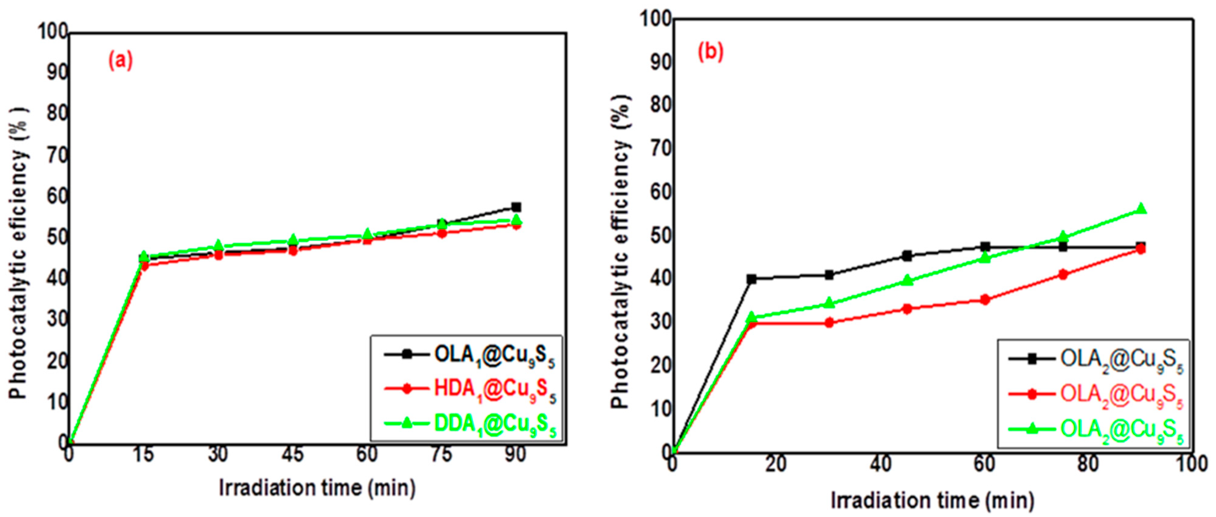
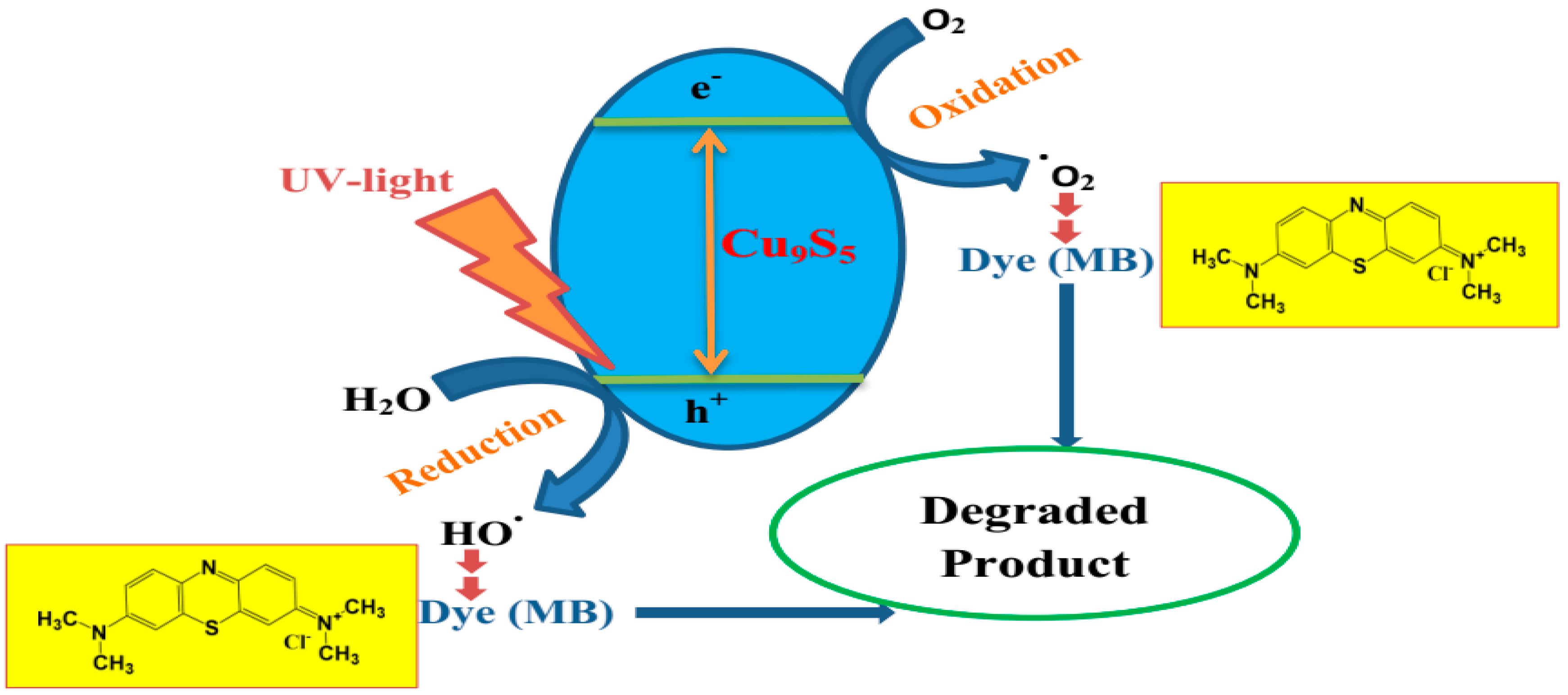
2.3. Photocatalytic Activity of CuxSy Nanoparticles on Methylene-Blue Dye

The photocatalytic activities of Cu9S5 NPs synthesized at 190 °C and 230 °C were examined using a synthetic effluent containing methylene blue (MB). Figure 10, Figures S12 and S13 display the absorption spectra of the aliquots collected at different time intervals (i.e., at 15, 30, 45, 60, 75, and 90 min) after UV irradiation using a mixed solution of Cu9S5 NPs and MB. It was observed that as the irradiation time increased, there was a decrease in the absorption peak at 663 nm, which indicated MB degradation. The OLA1@Cu9S5, HDA1@Cu9S5, and DDA1@Cu9S5 NPs obtained at 190 °C using complex 1 showed degradation efficiencies of 63.6%, 73.6%, and 80.0%, respectively, after 90 min of UV irradiation (Table 3, Figure 10a and Figure 11a). A slight increase was observed in degradation rates when complex 2 was used as a precursor under the same reaction conditions, which indicated the effect of the precursor type on the NPs’ photocatalytic properties. Degradation rates of 76.2%, 76.2%, and 80.0% were obtained for OLA2@Cu9S5, HDA2@Cu9S5, and DDA2@Cu9S5 NPs, respectively (Table 3, Figure 10b and Figure 11b).
The results also showed an increase in the degradation rates for MB with a decrease in the length of the carbon chain of the capping agent used (Figure 12). The Cu9S5 NPs produced from DDA (C12) exhibited higher photocatalytic activity, as compared to copper sulphide NPs obtained with HDA (C16) and OLA (C18). This may have been related to the steric properties of the ligand, which may have affected its surface coverage of the Cu9S5 NPs. The enhancement of photocatalytic activities may have been due to the narrowing of the bandgap as well as the reduction in the electron–hole pair recombination and the porous surface [46]. The results obtained showed improved photocatalytic activities in copper sulphide NPs with higher degradation rates, compared to those reported by Ajibade et al. [47].
An increase in the reaction temperature from 190 °C to 230 °C resulted in a decrease in the photocatalytic properties of the copper sulphide NPs (Table 4 and Figure 13). When complex 1 was used as a precursor, degradation rates of 57.7%, 53.4%, and 54.5% were obtained for the OLA1@Cu9S5, HDA1@Cu9S5, and DDA1@Cu9S5 NPs, respectively. A similar trend was observed for complex 2 when the reaction temperature was increased from 190 °C to 230 °C, yielding degradation rates of 47.6%, 47.1%, and 56.1% for the OLA2@Cu9S5, HDA2@Cu9S5, and DDA2@Cu9S5 NPs, respectively, which were slightly lower than values obtained for Complex 1 at same temperature.
The photocatalytic efficiency of the Cu9S5 NPs on the photodegradation of MB was associated with their sufficient bandgap energy and their efficient production of electron–hole pairs under UV irradiation. It has been reported that the oxidative potential of a material may be reduced with a decrease in the bandgap energy, and this may impact the photocatalytic efficiency of the material [48]. Figure 14 attempts to describe the mechanism of the photocatalytic process, starting from the creation of electron-hole pairs as Cu9S5 nanoparticles receive UV irradiation. The excited electrons create O2- radicals from dissolved oxygen while electron-hole participate in the oxidation of surface-adsorbed water to form hydroxyl radicals (HO.) which react with the dye molecule [48]. The degradation efficiencies of OLA1@Cu9S5, HDA1@Cu9S5, and DDA1@Cu9S5 NPs obtained at 190 °C suggested that copper sulphide nanoparticles could be exploited as potential and effective photocatalysts for the removal of organic pollutants from water effluents. A summary of the results obtained is presented in Table 5.

3. Materials and Methods Experimental

3.1. Chemicals

Hexadecylamine (HDA, 90%), dodecylamine (DDA, 90%), oleylamine (OLA, 70%), 2-thiophenecarboxaldehyde (99%), 2-acethylthiophene (99%), thiosemicarbazide (99%), copper chloride dihydrate (98%), ethanol (99.5%), methanol (99.5%), toluene (99.5%), and acetone (99.5%) were obtained from a commercial source and used without any further purification.

3.2. Instrumentation

Melting points were determined using an SMP11 melting-point-measurement instrument. Microanalysis (C, H, and N) data were obtained from a Perkin Elmer automated model 2400 series II CHNS/0 analyser. Fourier-transform infrared (FTIR) spectra were recorded on an FTIR Perkin Elmer 400 spectrometer from 4000 cm−1 to 450 cm−1. Thermal analyses were conducted using a Perkin Elmer Pyris 6 TGA device up to 600 °C. X-ray diffraction (XRD) measurements were performed using X’Pert MPD diffractometer with Cu Kα radiation (λ = 1.5406 Å). TEM analyses were performed using a CM200-FEI at 200 KV. Scanning electron microscopy (SEM) analyses were performed using a Quanta 650 FEG-FEI. Elemental compositions of CuxSy were obtained using an energy-dispersive X-ray (EDX). Optical absorption measurements were conducted using an Ocean Insight FX-VIS-IRS-ES spectrophotometer at room temperature.

3.3. Preparation of Thiosemicarbazone Ligands

Thiosemicarbazone ligands were prepared using a method described in the literature, with slight modification [32,33].

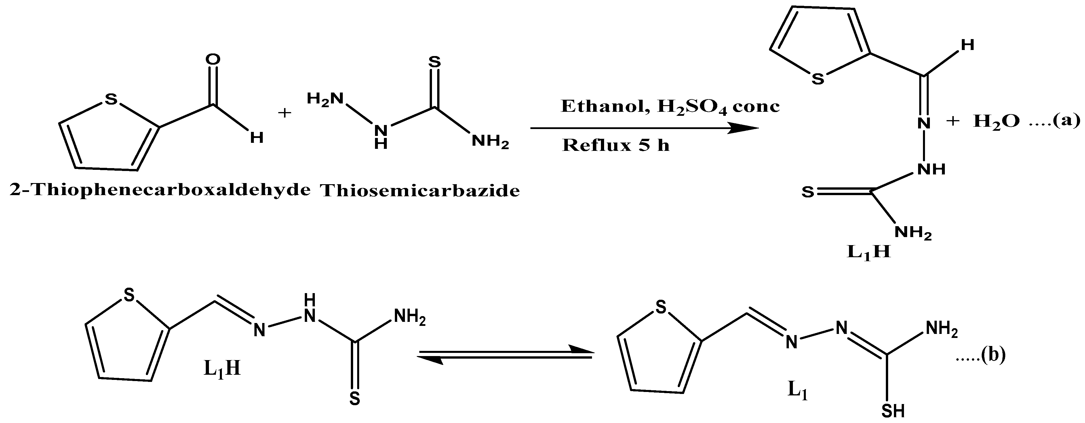
3.3.1. Synthesis of 2-Thiophenecarboxaldehyde Thiosemicarbazone (L1H) Ligand

A mass of 0.38 g (3 mmol) of 2-thiophenecarboxaldehyde was dissolved in 15 mL of hot ethanol and then added dropwise into a solution containing 0.33 g (3 mmol) of thiosemicarbazide dissolved in 25 mL hot ethanol in a round-bottom flask. The reaction mixture was then stirred continuously, and 2–3 drops of concentrated H2SO4 were added. The resulting mixture was heated under reflux at 80 °C for 5 h. The yellow precipitate obtained was filtered, washed with ethanol, and then dried in air. The reaction equation is represented in Scheme 1. Yield: 69%. Melting point: 182 ± 2 °C. Anal. Calc. for (C6H7N3S2): C, 38.69; H, 4.33; N, 22.56. Found: C, 38.90; H, 3.81; N, 22.68. Significant Infrared (IR) bands: (cm−1): ν(NH2): 3361, ν(N-H): 3125, ν(C=N): 1582, ν(N-N): 1044, ν(C=S): 835.

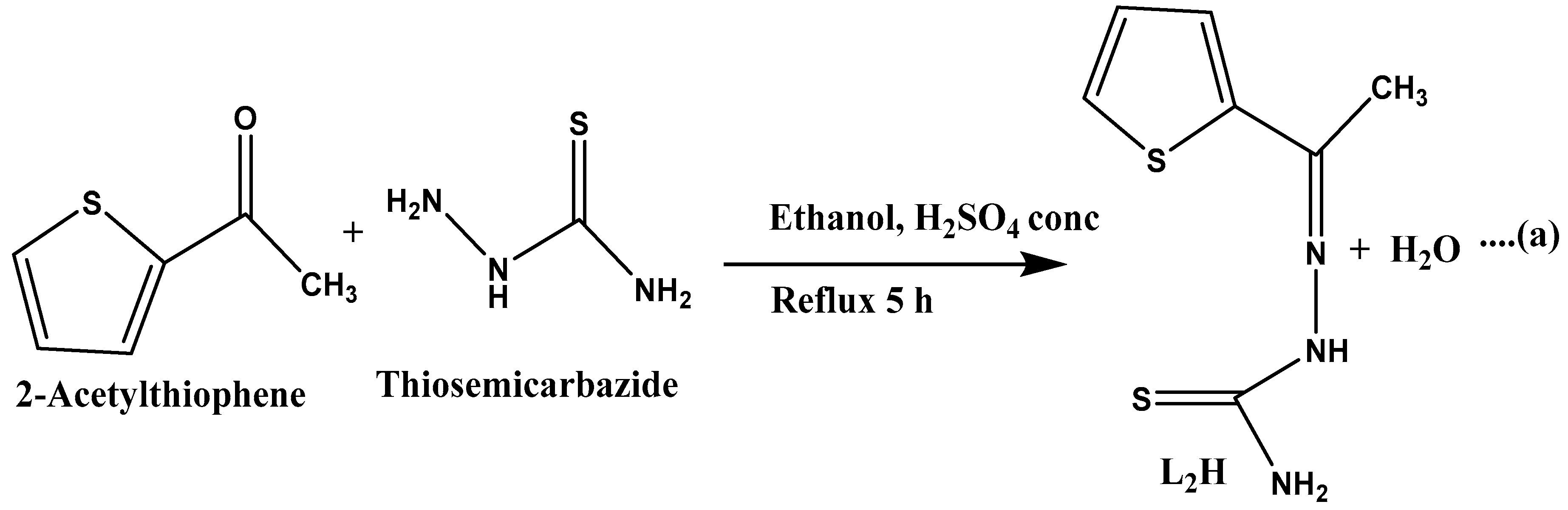
3.3.2. Synthesis of 2-Acetylthiophene Thiosemicarbazone (L2H) Ligand

The same reaction procedure as described above was used for the synthesis of L2H using a mass of 0.34 g (3 mmol) of 2-acetylthiophene. The light-yellow precipitate obtained was filtered, washed with ethanol, and then dried in air. The reaction equation is represented in Scheme 2. Yield: 95.4%. Melting point: 146 ± 2 °C. Anal. Calc. for (C7H9N3S2): C, 41, 97; H, 5.03; N, 20.58. Found: C, 40.82; H, 4.79; N, 20.17. Significant IR bands: (cm−1): ν(NH2): 3409, ν(N-H): 3157, ν(C=N): 1594, ν(N-N): 1040, ν(C=S): 830.

3.4. Preparation of Cu(II) Complexes (Complex 1 and 2)

A hot ethanolic solution (20 mL) of 2-(thiophen-2-ylmethylene)hydrazine-1-carbothioamide ligand (L1H) (1.10 g, 6 mmol) was added dropwise to a hot ethanolic solution (20 mL) of CuCl2·2H2O (0.52 g, 3 mmol). The mixture was stirred and heated under reflux for 3 h at 80 °C (Scheme 3). The precipitate obtained was allowed to cool at room temperature and then separated by filtration, washed with ethanol, and then dried at room temperature to obtain complex 1 as a pale-yellow powder. Yield: 71.3%. Melting point > 300 °C. Anal. Calc. for [Cu(C6H8N3S2)2]Cl2: C, 28.54; H, 2.79; N, 16.64. Found: C, 28.77; H, 2.86; N, 16.84. Significant IR bands (cm−1): ν(NH2): 3422-3235, ν(C=N): 1608, ν(N-N): 1034, ν(C=S): 831, ν(M-N):555.
The same method was used for the synthesis of complex 2 using the ligand 2-(1-(thiophen-2-yl) ethylidene)hydrazine-1-carbothioamide (L2H) (1,20 g, 6 mmol) (Scheme 3). Complex 2 was obtained as a pale-yellow powder. Yield: 59%. Melting point > 300 °C. Anal. Calc. for [Cu(C7H10N3S2)2]Cl2·H2O: C, 30.51; H, 3.66; N 15.25. Found: C, 29.87; H, 3.29; N, 15.25. Significant IR bands(cm−1): ν (NH2): 3415-3254, ν (C=N): 1602, ν(N-N): 1109, ν(C=S): 820, (M-N): 513.

3.5. Preparation of Copper Sulphide Nanoparticles

In a typical synthetic method employing hot-injection thermolysis, 3.0 mL of oleylamine (OLA) was placed in a three-necked flask and purged with N2. The capping agent was slowly heated to the desired temperature (i.e., 190 °C or 230 °C), and a mass 200 mg of complex 1 dissolved in 3.0 mL of OLA was injected into the heated OLA in a three-necked flask at 190 °C while stirring. A decrease in temperature of approximately 20 °C was observed, and the solution turned brown in colour. The reaction temperature was maintained at 190 °C, and after 30 min of stirring, heating was stopped, and methanol was added to flocculate the copper sulphide NPs [8,9]. The precipitate was separated by centrifugation and then dispersed in toluene to obtain dark-brownish OLA-capped CuxSy nanoparticles (OLA@CuxSy).
The above reaction procedure was repeated at a reaction temperature of 230 °C, varying the capping groups from hexadecylamine (HDA) to dodecylamine (DDA) and replacing the precursor with complex 2 in Scheme 3.

3.6. Photocatalytic Studies

The photocatalytic activities of OLA-, HDA-, and DDA-capped Cu9S5 NPs were evaluated by degrading methylene blue (MB) as a test contaminant in an aqueous solution under ultraviolet (UV)-light irradiation value of 60 W/m2. A 5 mg of MB solution was prepared in a 500 mL volumetric flask using distilled water (10 ppm). A total of 10 mg of the catalyst was dispersed in 50 mL of aqueous MB solution in a Pyrex beaker. Prior to irradiation, the solution was stirred magnetically in the dark for 15 min to attain adsorption–desorption equilibrium. The resulting solution was then exposed to ultraviolet (UV)-light irradiation (λ = 368 nm) for 90 min. Aliquots were collected at 15 min interval and centrifuged to obtain the resulting solution for absorption analysis. The photodegradation efficiency (η) was calculated using Equation (1) [49].
η = Ao At Ao × 100  
where η is the photodegradation efficiency, Ao and At are the absorbance values, respectively, before and after 15, 30, 45, 60, 75, and 90 min of illumination and recorded at the maximum absorbance wavelength for MB (λ = 663 nm) using a UV-Vis-NIR spectrophotometer.

4. Conclusions

Heterocyclic copper(II) thiosemicarbazone complexes were successfully used as single-source precursors for the synthesis of copper sulphide nanoparticles via hot-injection method at 190 °C and 230 °C while using OLA, HDA, and DDA as capping agents. The effects of quantum confinement observed in the energy bandgap of the synthesized copper sulphide NPs were solvent, temperature, and precursor-type dependant. The OLA-capped copper sulphide NPs had a higher energy bandgap of 3.06 eV at 190 °C. The same trend was observed in the formation of various morphologies of copper sulphide nanoparticles, which were influenced by the reaction temperature. Crystalline copper sulphide NPs reported here demonstrates t better photocatalytic properties of these system compared to those reported in recent studies. The degradation rates of MB under UV irradiation were affected by the length of the carbon chain of the primary amine-capping molecule and the type of precursor, and a maximum of 80% was achieved. This study confirmed the potential of synthesized copper sulphide nanophotocatalysts in the treatment of water that has been contaminated with organic pollutants.

Supplementary Materials

The following are available online at https://www.mdpi.com/article/10.3390/catal12010061/s1, Figure S1: Infrared spectra of ligands and their corresponding complexes. Figure S2: EDX spectra of CuxSy nanoparticles prepared in (a) OLA, (b) HDA and (c) DDA at 190 °C using complex 2. Figure S3: EDX spectra of CuxSy nanoparticles prepared in (a) OLA, (b) HDA, and (c) DDA at 230 °C using complex 1. Figure S4: EDX spectra of CuxSy nanoparticles prepared in (a) OLA, (b) HDA, and (c) DDA at 230 °C using complex 2. Figure S5: SAED patterns of CuxSy nanoparticles prepared at 190 °C in (a,c) HDA and (b,d) DDA using complex 1 and complex 2. Figure S6: SAED patterns of CuxSy nanoparticles prepared at 230 °C in (a,c) HDA and (b,d) DDA using complex 1 and complex 2. Figure S7: Distribution size of CuxSy nanoparticles prepared in (a) OLA, (b) HDA and (c) DDA using complexes 1 at 190 °C. Figure S8: SEM images of CuxSy nanoparticles prepared in (a, d) OLA, (b,e) HDA, and (c,f) DDA at 230 °C using the complexes 1 and 2 as precursors. Figure S9: UV-visible-NIR of CuxSy nanoparticles prepared in OLA, HDA, and DDA at 190 °C using (a) complex 1 and (b) complex 2 as single-source precursors. Figure S10: UV-visible NIR of CuxSy nanoparticles prepared in OLA, HDA, and DDA at 230 °C using (a) complex 1 and (b) complex 2 as single source precursors. Figure S11: Tauc plots of CuxSy nanoparticles prepared in OLA, HDA, and DDA at 230 °C using complexes 1 and 2. Figure S12: UV-Vis absorption spectra for methylene blue photodegradation using CuxSy nanoparticles synthesized at 190 °C. Figure S13: UV-Vis absorption spectra for methylene blue photodegradation using CuxSy nanoparticles synthesized at 230 °C.

Author Contributions

Conceptualization, L.D.N., F.C. and P.T.N.; methodology, A.P.Y.; formal analysis, T.G., S.F. and K.I.Y.K.; data curation, A.P.Y.; writing—original draft preparation, A.P.Y., T.G. and K.I.Y.K.; writing—review and editing, L.D.N., S.F., F.C. and P.T.N.; funding acquisition, L.D.N., F.C. and P.T.N.; project management, P.T.N. All authors have read and agreed to the published version of the manuscript.

Funding

This research received financial assistance from the Royal Society–DFID capacity building initiative (Grant N°AQ140001) (PTN) and the OWSD for the OWSD Early Career fellowship (UNESCO and the International Development Research Centre, Ottawa, Canada) (LDN). The views expressed herein do not necessarily represent those of UNESCO, IDRC, or the Board of Governors.

Data Availability Statement

Data are contained within the article and the Supplementary Materials section.

Acknowledgments

The authors are grateful for the mobility bursary (A.P.Y.), which was provided by the University of Lorraine through the Institute Jean Lamour (IJL). The authors would also like to thank the Department of Chemistry of the University of Zululand for TGA and microanalyses.

Conflicts of Interest

The authors declare no conflict of interest.

References

  1. Onwudiwe, D.C. Microwave-assisted synthesis of PbS nanostructures. Heliyon 2019, 5, e01413. [Google Scholar] [CrossRef] [PubMed] [Green Version]
  2. Jiade, P.A.A.; Nqombolo, A. Synthesis and structural studies of nickel sulphide and palladium sulphide nanocrystals. Chalcogenide Lett. 2016, 13, 427–434. [Google Scholar]
  3. Chakraborty, P.; Adhikary, J.; Chatterjee, S.; Biswas, B.; Chattopadhyay, T. Facile syntheses of copper sulfide nanoparticles: Antibacterial and antifungal activity study. Rasayan J. Chem. 2016, 9, 77–83. [Google Scholar]
  4. Olatunde, O.C.; Onwudiwe, D.C. Temperature Controlled Evolution of Pure Phase Cu9S5 Nanoparticles by Solvothermal Process. Front. Mater. 2021, 8, 211. [Google Scholar] [CrossRef]
  5. Lei, H.; Qin, P.; Ke, W.; Guo, Y.; Dai, X.; Chen, Z. Performance enhancement of polymer solar cells with high work function CuS modifed ITO as anodes. Org. Electron. 2015, 22, 173–179. [Google Scholar] [CrossRef]
  6. Srinivas, B.; Kumar, B.G.; Muralidharan, K. Stabilizer free copper sulphide nanostructures for rapid photocatalytic decomposition of rhodamine B. J. Mol. Catal. A Chem. 2015, 410, 8–18. [Google Scholar] [CrossRef]
  7. Zhang, K.; Khan, M.W.; Zuo, X.; Yang, Q.; Tang, H.; Wu, M. Controllable Synthesis and Photoelectric Properties of Interconnected and Self-Assembled Nanocomposite of Porous Hollow Cu7S4/CuS and Nitrogen-Doped Graphene Oxide. Electrochim. Acta 2019, 307, 64–75. [Google Scholar] [CrossRef]
  8. Khan, M.D.; Akhtar, M.; Malik, M.A.; Revaprasadu, N.; O’Brien, P. New examples of phase control in the preparation of copper sulfde nanoparticles and deposition of thin flms by AACVD from bis (piperidinedithiocarbamato) copper(II) complex. ChemistrySelect 2018, 3, 2943–2950. [Google Scholar] [CrossRef]
  9. Ketchemen, K.I.Y.; Khan, M.D.; Mlowe, S.; Nyamen, L.D.; Ndifon, P.T.; O’Brien, P.; Revaprasadu, N. Tailoring Shape and Crystallographic Phase of Copper Sulfide Nanostructures Using Novel Thiourea Complexes as Single Source Precursors. J. Inorg. Organomet. Polym. Mater. 2019, 29, 917–927. [Google Scholar] [CrossRef]
  10. Hsu, Y.-K.; Chen, Y.-C.; Lin, Y.-G. Synthesis of copper sulfde nanowire arrays for high-performance supercapacitors. Electrochim. Acta 2014, 139, 401–407. [Google Scholar] [CrossRef]
  11. Pei, L.Z.; Wang, J.F.; Tao, X.X. Synthesis of CuS and Cu1.1Fe1.1S2 crystals and their electrochemical properties. Mater. Charact. 2011, 62, 354–359. [Google Scholar] [CrossRef]
  12. Ayodhya, D.; Venkatesham, M.; Kumari, A.S.; Reddy, G.B.; Ramakrishna, D.; Veerabhadram, G. Photocatalytic degradation of dye pollutants under solar, visible and UV lights using green synthesised CuS nanoparticles. J. Exp. Nanosci. 2016, 11, 418–432. [Google Scholar] [CrossRef]
  13. Senthilkumar, M.; Babu, S.M. Synthesis and characterization of hexagonal faceted copper sulfide (Cu1.8S) nanodisks. Mater. Sci. Semicond. Process. 2015, 40, 203–208. [Google Scholar] [CrossRef]
  14. Ajibade, P.A.; Botha, N.L. Synthesis and structural studies of copper sulfide nanocrystals. Results Phys. 2016, 6, 581–589. [Google Scholar] [CrossRef] [Green Version]
  15. Ravele, M.P.; Oyewo, O.A.; Onwudiwe, D.C. Controlled Synthesis of CuS and Cu9S5 and Their Application in the Photocatalytic Mineralization of Tetracycline. Catalysts 2021, 11, 899. [Google Scholar] [CrossRef]
  16. Liao, X.-H.; Chen, N.-Y.; Xu, S.; Yang, S.-B.; Zhu, J.-J. A microwave assisted heating method for the preparation of copper sulfide nanorods. J. Cryst. Growth 2003, 252, 593–598. [Google Scholar] [CrossRef]
  17. Li, J.; Zhao, H.; Chen, X.; Jia, H.; Zheng, Z. In situ fabricate Cu2S thin film with hierarchical petal-like nanostructures. Mater. Res. Bull 2013, 48, 2940–2943. [Google Scholar] [CrossRef]
  18. Cheng, Z.; Wang, S.; Si, D.; Geng, B. Controlled synthesis of copper sulfide 3D nanoarchitectures through a facile hydrothermal route. J. Alloys Compd. 2010, 492, 44–49. [Google Scholar] [CrossRef]
  19. Botha, N.L.; Ajibade, P.A. Effect of temperature on crystallite sizes of copper sulfide nanocrystals prepared from copper (II) dithiocarbamate single source precursor. Mater. Sci. Semicond. Processing 2016, 43, 149–154. [Google Scholar] [CrossRef]
  20. Shen, S.; Zhang, Y.; Peng, L.; Xu, B.; Du, Y.; Deng, M.; Xu, H.; Wang, Q. Generalized synthesis of metal sulfide nanocrystals from single-source precursors: Size, shape and chemical composition control and their properties. CrystEngComm 2011, 13, 4572–4579. [Google Scholar] [CrossRef]
  21. Jen-La Plante, I.; Zeid, T.W.; Yang, P.; Mokari, T. Synthesis of metal sulfide nanomaterials via thermal decomposition of single-source precursors. J. Mater. Chem. 2010, 20, 6612–6617. [Google Scholar] [CrossRef] [Green Version]
  22. Paca, A.M.; Ajibade, P.A. Synthesis and structural studies of iron sulphide nanocomposites from iron(III) dithiocarbamate single source precursors. Mater. Chem. Phys. 2017, 202, 143–150. [Google Scholar] [CrossRef]
  23. Ma, Y.; Wan, H.; Ye, Y.; Chen, L.; Li, H.; Zhou, H.; Chen, J. In-situ synthesis of size tunable silver sulfide nanoparticles to improve tribological properties of the polytetrafluoroethylene-based nanocomposite lubricating coatings. Tribol. Int. 2020, 148, 106324. [Google Scholar] [CrossRef]
  24. Pawar, A.S.; Masikane, S.C.; Mlowe, S.; Garje, S.S.; Akerman, M.P.; Revaprasadu, N. Zinc thiosemicarbazone complexes: Single source precursors for alkylamine capped ZnS nanoparticles. Inorg. Chim. Acta 2017, 463, 7–13. [Google Scholar] [CrossRef]
  25. Khan, M.D.; Malik, M.A.; Akhtar, J.; Mlowe, S.; Revaprasadu, N. Phase pure deposition of flower-like thin flms by aerosol assisted chemical vapor deposition and solvent mediated structural transformation in copper sulfde nanostructures. Thin Solid Films 2017, 638, 338–344. [Google Scholar] [CrossRef]
  26. Du, X.-S.; Yu, Z.-Z.; Dasari, A.; Ma, J.; Meng, Y.-Z.; Mai, Y.-W. Facile synthesis and assembly of Cu2S nanodisks to corncoblike nanostructures. Chem. Mater. 2006, 18, 5156–5158. [Google Scholar] [CrossRef]
  27. Abdelhady, A.L.; Ramasamy, K.; Malik, M.A.; O’Brien, P.; Haighb, S.J.; Raftery, J. New routes to copper sulfide nanostructures and thin films. J. Mater. Chem. 2011, 21, 17888–17895. [Google Scholar] [CrossRef]
  28. Krishna, P.M.; Reddy, N.B.G.; Kottam, N.; Yallur, B.C.; Katreddi, H.R. Design and Synthesis of Metal Complexes of (2E)-2-[(2E)-3-Phenylprop-2-en-1-ylidene]hydrazine carbothioamide and Their Photocatalytic Degradation of Methylene Blue. Sci. World J. 2013, 2013, 828313. [Google Scholar] [CrossRef] [Green Version]
  29. Jain, M.; Babar, D.G.; Garje, S.S. Ligand-based stoichiometric tuning in copper sulfde nanostructures and their catalytic ability. Appl. Nanosci. 2019, 9, 353–367. [Google Scholar] [CrossRef]
  30. Gaber, A.; Refat, M.S.; Belal, A.A.M.; El-Deen, I.M.; Hassa, N.; Zakaria, R.; Alhomrani, M.; Alamri, A.S.; Alsanie, W.F.; Saied, E.M. New Mononuclear and Binuclear Cu(II), Co(II), Ni(II), and Zn(II) Thiosemicarbazone Complexes with Potential Biological Activity: Antimicrobial and Molecular Docking Study. Molecules 2021, 26, 2288. [Google Scholar] [CrossRef]
  31. Wilson, J.T.; Jiang, X.; McGill, B.C.; Lisic, E.C.; Deweese, J.E. Examination of the Impact of Copper(II) α-(N)-Heterocyclic Thiosemicarbazone Complexes on DNA Topoisomerase Iiα. Chem. Res. Toxicol. 2016, 29, 649–658. [Google Scholar] [CrossRef]
  32. Conner, D.; Medawala, W.; Stephens, M.T.; Morris, W.H.; Deweese, J.E.; Kent, P.L.; Rice, J.J.; Jiang, X.; Lisic, E.C. Cu(II) Benzoylpyridine Thiosemicarbazone Complexes: Inhibition of Human Topoisomerase IIα and Activity against Breast Cancer Cells. Open J. Inorg. Chem. 2016, 6, 146. [Google Scholar] [CrossRef] [Green Version]
  33. Lisic, E.C.; Rand, V.G.; Ngo, L.; Kent, P.; Rice, J.; Gerlach, D.; Papish, E.T.; Jiang, X. Cu(II) Propionyl-Thiazole Thiosemicarbazone Complexes: Crystal Structure, Inhibition of Human Topoisomerase IIα, and Activity against Breast Cancer Cells. Open J. Med. Chem. 2018, 8, 30–46. [Google Scholar] [CrossRef] [Green Version]
  34. Sarker, D.; Hossen, M.F.; Zahan1, M.K.; Haque, M.M.; Zamir, R.; Asraf, M.A. Synthesis, Characterization, Thermal Analysis and Antibacterial Activity of Cu (II) and Ni(II) Complexes with Thiosemicarbazone Derived from Thiophene-2-aldehyde. J. Mater. Sci. Res. Rev. 2020, 5, 15–25. [Google Scholar]
  35. Mohan, C.; Kumar, V.; Kumari, S. synthesis, characterization, and antibacterial activity of schiff bases derived from thiosemicarbazide, 2-acetylthiophene and thiophene-2-aldehyde. Int. Res. J. Pharm. 2018, 9, 153–158. [Google Scholar] [CrossRef]
  36. Tyagi, M.; Chandra, S.; Tyagi, P. Mn(II) and Cu(II) complexes of a bidentate Schiff’s base ligand: Spectral, thermal, molecular modelling and mycological studies. Spectrochim. Acta A Mol. Biomol. Spectrosc. 2014, 117, 1–8. [Google Scholar] [CrossRef] [PubMed]
  37. Chandra, S.; Goel, S.; Dwivedi, S.D. spectroscopic and biological studies on newly synthesized copper(II) and nickel(II) complexes with p-dimethylaminobanzaldehyde semicarbazone and p-dimethylaminobanzaldehyde thiosemicarbazone. Int. J. Appl. biol. Pharm. 2012, 3, 149–156. [Google Scholar]
  38. El-Saied, F.; El-Aarag, B.; Salem, T.; Said, G.; Khalifa, S.A.M.; El-Seedi, H.R. Synthesis, Characterization, and In Vivo Anti-Cancer Activity of New Metal Complexes Derived from Isatin-N(4) antipyrinethiosemicarbazone Ligand Against Ehrlich Ascites Carcinoma Cells. Molecules 2019, 24, 3313. [Google Scholar] [CrossRef] [Green Version]
  39. Pahontu, E.; Fala, V.; Gulea, A.; Poirier, D.; Tapcov, V.; Rosu, T. Synthesis and Characterization of Some New Cu(II), Ni(II) and Zn(II) Complexes with Salicylidene Thiosemicarbazones: Antibacterial, Antifungal and in Vitro Antileukemia Activity. Molecules 2013, 18, 8812–8836. [Google Scholar] [CrossRef] [Green Version]
  40. Jing, M.; Li, F.; Chen, M.; Zhang, J.; Long, F.; Jing, L.; Lv, X.; Ji, X.; Wu, T. Facile synthetic strategy to uniform Cu9S5 embedded into carbon: A novel anode for sodium-ion batteries. J. Alloys Compd. 2018, 762, 473–479. [Google Scholar] [CrossRef]
  41. Mbewana-Ntshanka, N.G.; Moloto, M.J.; Mubiayi, P.K. Antimicrobial Activity of the Synthesized of Copper Chalcogenide Nanoparticles. J. Nanotechnol. 2021, 2021, 6675145. [Google Scholar] [CrossRef]
  42. Onwudiwe, D.C.; Hrubaru, M.; Ebenso, E.E. Synthesis, Structural and Optical Properties of TOPO and HDA Capped Cadmium Sulphide Nanocrystals, and the Effect of Capping Ligand Concentration. J. Nanomater. 2015, 9, 143632. [Google Scholar] [CrossRef] [Green Version]
  43. Akhtar, M.; Alghamdi, Y.; Akhtar, J.; Aslam, Z.; Revaprasadu, N.; Malik, M.A. Phase controlled synthesis of copper sulfide nanoparticles by colloidal and non-colloidal methods. Mater. Chem. Phys. 2016, 180, 404–412. [Google Scholar] [CrossRef]
  44. Ketchemen, K.I.Y.; Mlowe, S.; Nyamen, L.D.; Ndifon, P.T.; Revaprasadu, N. Comparative study on the effect of precursors on the morphology and electronic properties of CdS nanoparticles. Turk. J. Chem. 2021, 45, 400–409. [Google Scholar] [CrossRef]
  45. Botha, N.L.; Ajibade, P.A. Optical and structural characterization of copper sulphide nanoparticles from copper(II) piperidine dithiocarbamate. Opt. Quantum Electron. 2020, 52, 337. [Google Scholar] [CrossRef]
  46. Mintcheva, N.; Gicheva, G.; Panayotova, M.; Kulinich, S.A. Room-Temperature Synthesis of ZnS Nanoparticles Using Zinc Xanthates as Molecular Precursors. Materials 2020, 13, 171. [Google Scholar] [CrossRef] [Green Version]
  47. Ajibade, P.A.; Sikakane, B.M.; Botha, N.L.; Oluwalana, A.E.; Omondi, B. Synthesis and crystal structures of bis(dibenzyl dithiocarbamato)Cu(II) and Ag(I) complexes: Precursors for Cu1.8S and Ag2S nano-photocatalysts. J. Mol. Struct. 2020, 1221, 128791. [Google Scholar] [CrossRef]
  48. Pal, M.; Mathews, N.R.; Sanchez-Mora, E.; Pal, U.; Paraguay-Delgado, F.; Mathew, X. Synthesis of CuS nanoparticles by a wet chemical route and their photocatalytic activity. J. Nanoparticle Res. 2015, 17, 301. [Google Scholar] [CrossRef]
  49. Yepseu, A.P.; Isac, L.; Nyamen, L.D.; Cleymand, F.; Duta, A.; Ndifon, P.T. Optical and Photocatalytic Properties of CuxS/ZnO Composite Thin Films Deposited by Robotic Spray Pyrolysis Deposition. J. Nanomater 2021, 2021, 9975600. [Google Scholar] [CrossRef]
Figure 1. Thermogravimetric analysis (TGA) graph of complexes 1 and 2.
Catalysts 12 00061 g001
Figure 2. p-XRD patterns of CuxSy nanoparticles prepared in (a) OLA, (b) HDA, and (c) DDA at 190 °C using (I) complex 1 and (II) complex 2.
Catalysts 12 00061 g002
Figure 3. p-XRD patterns of CuxSy nanoparticles prepared in (a) OLA, (b) HDA, and (c) DDA at 230 °C using (I) complex 1 and (II) complex 2.
Catalysts 12 00061 g003
Figure 4. EDX spectra of CuxSy nanoparticles prepared in (a) OLA, (b) HDA, and (c) DDA at 190 °C using complex 1.
Catalysts 12 00061 g004aCatalysts 12 00061 g004b
Figure 5. TEM images of CuxSy nanoparticles prepared in (a,d) OLA, (b,e) HDA, and (c,f) DDA at 190 °C using complexes 1 and 2; SAED patterns of nanoparticles prepared in (g) OLA, (h) HDA and (i) DDA at 190 °C from complex 1.
Catalysts 12 00061 g005
Figure 6. TEM images of CuxSy nanoparticles prepared in (a,d) OLA, (b,e) HDA, and (c,f) DDA at 190 °C using complexes 1 and 2; SAED patterns of nanoparticles prepared in (g) OLA, (h) HDA, and (i) DDA at 230 °C from complex 1.
Catalysts 12 00061 g006
Figure 7. Particle size distribution for CuxSy nanoparticles prepared in (a) OLA, (b) HDA, and (c) DDA at 230 °C using complex 2.
Catalysts 12 00061 g007
Figure 8. SEM images of CuxSy nanoparticles prepared in (a,d) OLA, (b,e) HDA, and (c,f) DDA at 190 °C using the complexes 1 and 2.
Catalysts 12 00061 g008
Figure 9. Tauc bandgap graphs of CuxSy nanoparticles prepared in OLA, HDA, and DDA at 190 °C while using either complex 1 or 2.
Catalysts 12 00061 g009
Figure 10. Degradation-efficiency curves of MB at different irradiation times using (a) complex 1 and (b) complex 2 at 190 °C.
Catalysts 12 00061 g010
Figure 11. Histograms of degradation efficiencies of MB at different irradiation times using (a) complex 1 and (b) complex 2 at 190 °C.
Catalysts 12 00061 g011
Figure 12. Histograms of degradation efficiencies of MB using copper sulphide nanoparticles at 190 °C versus carbon chain of capping agents using (a) complex 1 and (b) complex 2.
Catalysts 12 00061 g012
Figure 13. Degradation efficiency curves of MB at different irradiation times using (a) complex 1 and (b) complex 2 as precursors at 230 °C.
Catalysts 12 00061 g013
Figure 14. Mechanism of MB degradation by CuxSy NPs under UV irradiation.
Catalysts 12 00061 g014
Scheme 1. Equation of synthesis of the ligand, 2-thiophenecarboxaldehyde thiosemicarbazone (L1H) (a) and resonance forms also shown in (b).
Catalysts 12 00061 sch001
Scheme 2. Equation of synthesis of the ligand, 2-acetylthiophene thiosemicarbazone (L2H) (a) and resonance forms also shown in (b).
Catalysts 12 00061 sch002aCatalysts 12 00061 sch002b
Scheme 3. Equation for the synthesis of (1) complex 1 and (2) complex 2.
Catalysts 12 00061 sch003
Table 1. EDX analysis of OLA@Cu9S5, HDA@Cu9S5, and DDA@Cu9S5 NPs obtained at 190 °C using complex 1 and complex 2.
OLA1@Cu9S5HDA1@Cu9S5DDA1@Cu9S5OLA2@Cu9S5HDA2@Cu9S5DDA2@Cu9S5
ElementWeight %ElementWeight %ElementWeight %ElementWeight %ElementWeight %ElementWeight %
Cu49.50Cu61.06Cu46.47Cu27.77Cu31.67Cu31.07
S25.44S27S25.33S13.90S18.50S17.66
O4.92O/O6.61O7.90O19.52O19.22
C20.14C11.94C21.5C50.43C30.31C32.05
Cu/S1.94:1Cu/S2.26:1Cu/S1.83:1Cu/S1.99:1Cu/S1.71:1Cu/S1.76:1
Table 2. EDX analysis of OLA@Cu9S5, HDA@Cu9S5, and DDA@Cu9S5 NPs obtained at 230 °C using complex 1 and complex 2.
OLA1@Cu9S5HDA1@Cu9S5DDA1@Cu9S5OLA2@Cu9S5HDA2@Cu9S5DDA2@Cu9S5
ElementWeight %ElementWeight %ElementWeight %ElementWeight %ElementWeight %ElementWeight %
Cu59.86Cu60.53Cu61.56Cu58.81Cu59.57Cu56.54
S31.94S33.27S32.78S3197S33.32S29.56
O8.20O6.20O5.66O9.22O7.11O13.90
C59.86C/C/C/C/C/
Cu/S1.87:1Cu/S1.81:1Cu/S1.87:1Cu/S1.84:1Cu/S1.80:1Cu/S1.91:1
Table 3. Degradation efficiencies of OLA@Cu9S5, HDA@Cu9S5, and DDA@Cu9S5 NPs obtained at 190 °C using complexes 1 and 2.
t (min) 153045607590
OLA1@Cu9S5η (%)33.346.049.757.162.463.6
HDA1@Cu9S5η (%)48.758.260.370.470.473.6
DDA1@Cu9S5η (%)54.660.064.669.374.180.0
OLA2@Cu9S5η (%)58.760.066.768.873.076.2
HDA2@Cu9S5η (%)60.063.567.771.073.576.2
DDA2@Cu9S5η (%)62.465.170.474.675.780.0
Table 4. Degradation efficiencies of OLA@Cu9S5, HDA@Cu9S5, and DDA@Cu9S5 NPs obtained at 230 °C using complexes 1 and 2.
t (min) 153045607590
OLA1@Cu9S5η (%)45.046.647.649.753.457.7
HDA1@Cu9S5η (%)43.446.047.149.751.353.4
DDA1@Cu9S5η (%)45.548.149.550.853.454.5
OLA2@Cu9S5η (%)40.241.245.547.647.647.6
HDA2@Cu9S5η (%)30.030.133.335.441.247.1
DDA2@Cu9S5η (%)31.234.439.745.049.756.1
Table 5. Summary of the reaction conditions, sizes, shapes optical, and photocatalytic properties of copper sulphide NPs.
ComplexesCapping AgentReaction
Temp
(°C)
Energy Bandgap
(eV)
Phase
(Formula)
MorphologyAverage Size (nm)Degradation Rates (%) after 90 min
1OLA1902.70Mixture of Cu9S5 & Cu7S5Agglomerated Irregular49–8063.6
HDA1902.64Cu9S5Rectangular and cubic46–13473.6
DDA1902.57Cu9S5Rectangular and irregular53–15480.0
OLA2302.60 Cu9S5Rectangular and truncated40–12057.7
HDA2302.76Cu9S5Irregular cubic and rectangular101–20053.4
DDA2302.64Cu9S5Irregular cubic61–11954.5
2OLA1903.00 Cu9S5Agglomerated32–7576.2
HDA1902.58Cu9S5Semi-spherical and rectangular43–12576.2
DDA1902.55Cu9S5Irregular cubic49–19680.0
OLA2302.83 Cu9S5Agglomerated34–12847.6
HDA2302.76Cu9S5Irregular cubic45–11547.1
DDA2302.73Cu9S5Rectangular and cubic66–22556.1
Publisher’s Note: MDPI stays neutral with regard to jurisdictional claims in published maps and institutional affiliations.

