Co-Microencapsulation of Flavonoids from Yellow Onion Skins and Lactic Acid Bacteria Lead to Multifunctional Ingredient for Nutraceutical and Pharmaceutics Applications

 ,
,  ,
, 
Abstract
1. Introduction
2. Materials and Methods
2.1. Materials
2.2. Methods
2.2.1. Flavonoid Extraction from Yellow Onion Skins
2.2.2. Microencapsulation of Flavonoids and LAB
2.2.3. Determination of Selected Phytochemical Profile of the Extract and Co-Microencapsulated Powder
2.2.4. Encapsulation Efficiency and Viability of LAB
2.2.5. Determination of Physical Properties of the Microencapsulated Powder
Powder Solubility
Powder Hygroscopicity
2.2.6. In Vitro Assays
α-Amylase Inhibition
Lipase Inhibition
Lipoxygenase Inhibition
Determination of Bioavailability of Bioactives
2.2.7. In Vitro Cytotoxicity of the Co-Microencapsulated Powder
Cell Culture and Treatment
Cell Viability
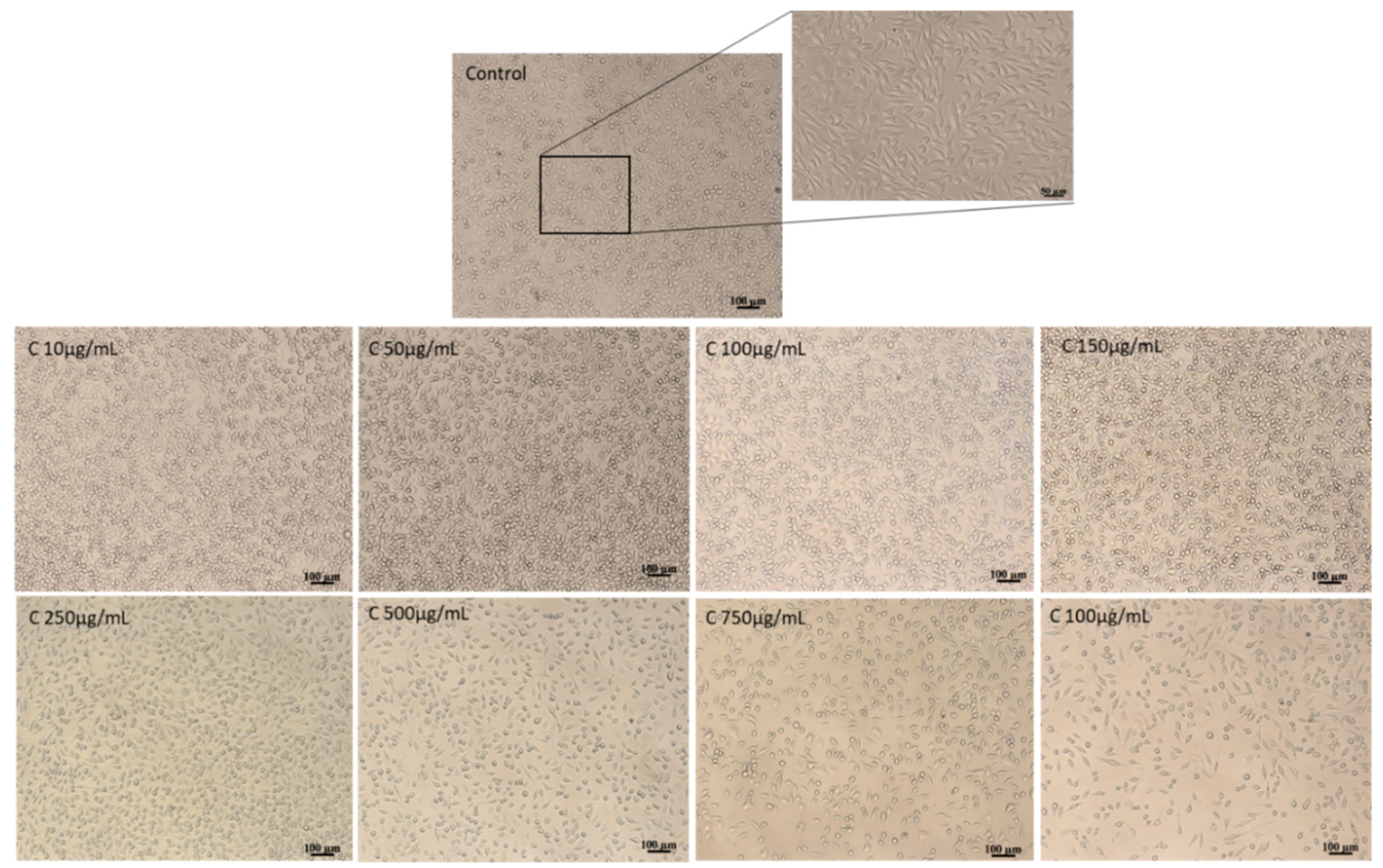
Cell Morphology
2.2.8. Formulation of a Soft Cheese with Added Value
2.2.9. Statistical Analyses
3. Results and Discussions
3.1. Extract Characterization, Co-Microencapsulation and Global Phytochemical Characterization of the Powder
3.2. Solubility and Hygroscopicity of the Co-Microencapsulated Powder
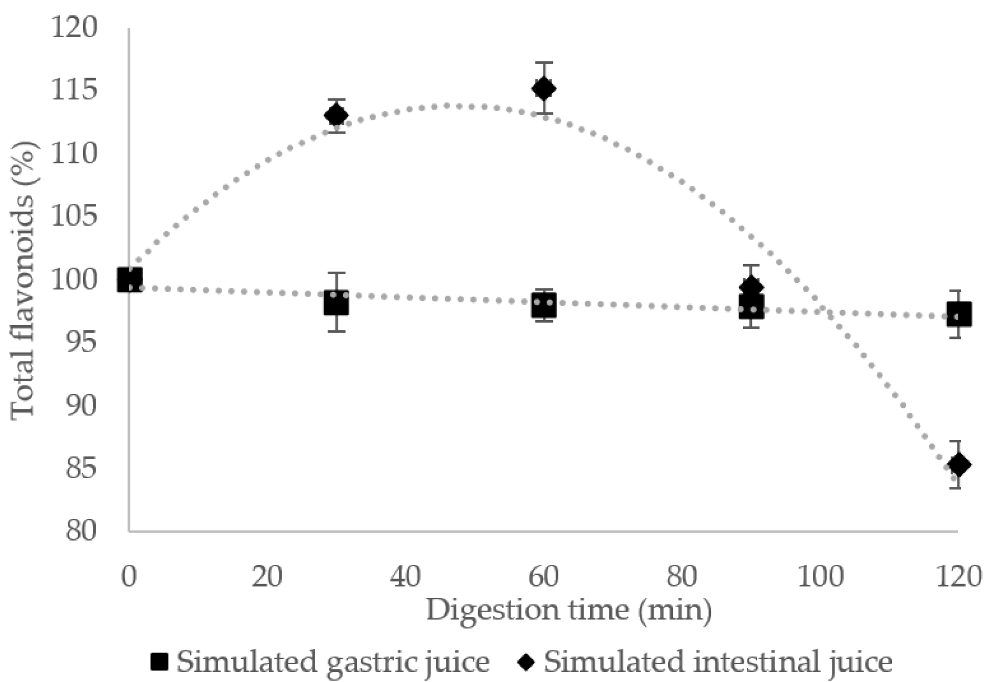
3.3. Flavonoids Stability in Gastrointestinal Simulated Conditions
3.4. Anti-Diabetic and Anti-Inflammatory Potential
3.4.1. α-Amylase Inhibitory Activity
3.4.2. Lipase Inhibitory Activity
3.4.3. Lipoxygenase Inhibitory Activity
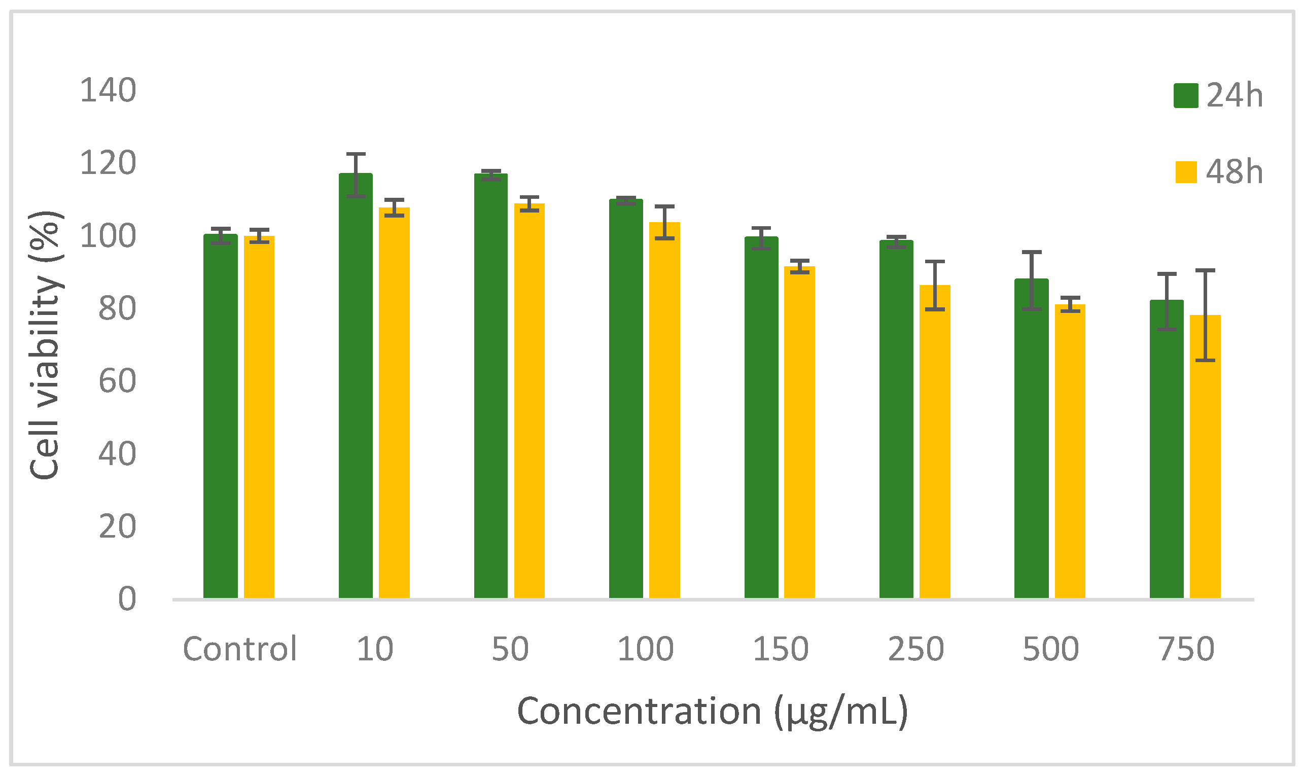
3.5. In Vitro Cytotoxicity of the Microencapsulated Powders
3.6. Characterization of New Formulated Food Product
4. Conclusions
Author Contributions
Funding
Acknowledgments
Conflicts of Interest
References
- Sak, K. Cytotoxicity of dietary flavonoids on different human cancer types. Pharmacogn. Rev. 2014, 8, 122. [Google Scholar] [CrossRef] [PubMed]
- Costamagna, M.S.; Zampini, I.C.; Alberto, M.R.; Cuello, S.; Torres, S.; Pérez, J.; Quispe, C.; Schmeda-Hirschmann, G.; Isla, M.I. Polyphenols rich fraction from Geoffroea decorticans fruits flour affects key enzymes involved in metabolic syndrome, oxidative stress and inflammatory process. Food Chem. 2016, 190, 392–402. [Google Scholar] [CrossRef]
- Milea, A.S.; Aprodu, I.; Vasile, A.M.; Barbu, V.; Râpeanu, G.; Bahrim, G.E.; Stănciuc, N. Widen the functionality of flavonoids from yellow onion skins through extraction and microencapsulation in whey proteins hydrolysates and different polymers. J. Food Eng. 2019, 251, 29–35. [Google Scholar] [CrossRef]
- Tsali, A.; Goula, A.M. Valorization of grape pomace: Encapsulation and storage stability of its phenolic extract. Powder Technol. 2018, 340, 197–207. [Google Scholar] [CrossRef]
- Kerry, G.R.; Patra, J.K.; Gouda, S.; Park, Y.; Shin, H.-S.; Das, G. Benefaction of probiotics for human health: A review. J. Food Drug Anal. 2018, 26, 927–939. [Google Scholar] [CrossRef] [PubMed]
- Vasile, A.M.; Milea, S.A.; Enachi, E.; Barbu, V.; Cîrciumaru, A.; Bahrim, G.E.; Râpeanu, G.; Stănciuc, N. Functional enhancement of bioactives from black beans and lactic acid bacteria into an innovative food ingredient by co-microencapsulation. Food Bioprocess Technol. 2020, 13, 978–987. [Google Scholar] [CrossRef]
- Gonzales, G.B.; Smagghe, G.; Grootaert, C.; Zotti, M.; Raes, K.; Camp, J.V. Flavonoid interactions during digestion, absorption, distribution and metabolism: A sequential structure–activity/property relationship-based approach in the study of bioavailability and bioactivity. Drug Metab. Rev. 2015, 47, 175–190. [Google Scholar] [CrossRef]
- Oancea, A.M.; Hasan, M.; Vasile, A.M.; Barbu, V.; Enachi, E.; Bahrim, G.; Râpeanu, G.; Silvi, S.; Stănciuc, N. Functional evaluation of microencapsulated anthocyanins from sour cherries skins extract in whey proteins isolate. LWT Food Sci. Technol. 2018, 95, 129–134. [Google Scholar] [CrossRef]
- Saénz, C.; Tapia, S.; Chávez, J.; Robert, P. Microencapsulation by spray drying of bioactive compounds from cactus pear (Opuntia ficus-indica). Food Chem. 2009, 114, 616–622. [Google Scholar] [CrossRef]
- Colín-Cruz, M.A.; Pimentel-González, D.J.; Carrillo-Navas, H.; Alvarez-Ramírez, J.; Guadarrama-Lezama, A.Y. Co-encapsulation of bioactive compounds from blackberry juice and probiotic bacteria in biopolymeric matrices. LWT Food Sci. Technol. 2019, 110, 94–101. [Google Scholar] [CrossRef]
- ISO 8261 IDF122:2001. Milk and Milk Products–General Guidance for the Preparation of Test Samples, Initial Suspensions and Decimal Dilutions for Microbiological Examination, 2nd ed.; International Standard Association: Geneva, Switzerland, 2001. [Google Scholar]
- Hussain, S.A.; Hameed, A.; Nazir, Y.; Naz, T.; Wu, Y.; Suleria, H.A.R.; Song, Y. Microencapsulation and the Characterization of Polyherbal Formulation (PHF) Rich in Natural Polyphenolic Compounds. Nutrients 2018, 10, 843. [Google Scholar] [CrossRef] [PubMed]
- Gaspar, A.; Craciunescu, O.; Trif, M.; Moisei, M.; Moldovan, L. Antioxidant and anti-inflammatory properties of active compounds from Arnica montana L. Rom. Biotechnol. Lett. 2014, 19, 9353–9365. [Google Scholar]
- Singh, V.; Krishan, P.; Shri, R. Extraction of antioxidant phytoconstituents from onion waste. J. Pharmacogn. Phytochem. 2016, 6, 502–505. [Google Scholar]
- Akdeniz, B.; Sumnu, G.; Sahin, S. Microencapsulation of phenolic compounds extracted from onion (Allium cepa) skin. J. Food Process. Preserv. 2018, 42, e13648. [Google Scholar] [CrossRef]
- Gebara, C.; Chaves, K.S.; Ribeiro, M.C.E.; Souza, F.N.; Grosso, C.R.F.; Gigante, M.L. Viability of Lactobacillus acidophilus La5 in pectin-whey protein microparticles during exposure to simulated gastrointestinal conditions. Food Res. Int. 2013, 51, 872–878. [Google Scholar] [CrossRef]
- Cai, S.; Zhao, M.; Fang, Y.; Nishinari, K.; Phillips, G.O.; Jiang, F. Microencapsulation of Lactobacillus acidophilus CGMCC1.2686 via emulsification/internal gelation of alginate using Ca-EDTA and CaCO3 as calcium sources. Food Hydrocoll. 2014, 39, 295–300. [Google Scholar] [CrossRef]
- Vodnar, D.C.; Socaciu, C. Selenium enriched green tea increase stability of Lactobacillus casei and Lactobacillus plantarum in chitosan coated alginate microcapsules during exposure to simulated gastrointestinal and refrigerated conditions. LWT Food Sci. Technol. 2014, 57, 406–411. [Google Scholar] [CrossRef]
- Horincar, G.; Aprodu, I.; Barbu, V.; Râpeanu, G.; Bahrim, G.E.; Stănciuc, N. Interactions of flavonoids from yellow onion skins with whey proteins: Mechanisms of binding and microencapsulation with different combinations of polymers. Spectrochim. Acta A 2019, 215, 158–167. [Google Scholar] [CrossRef]
- Tonon, R.V.; Brabet, C.; Hubinger, M.D. Influence of process conditions on the physicochemical properties of acai (Euterpe oleraceae Mart.) powder produced by spray drying. J. Food Eng. 2008, 88, 411–418. [Google Scholar] [CrossRef]
- Syamaladevi, R.M.; Insan, S.K.; Dhawan, S.; Andrews, P.; Sablani, S.S. Physicochemical Properties of Encapsulated Red Raspberry (Rubus idaeus) Powder: Influence of High-Pressure Homogenization. Dry. Technol. 2012, 30, 484–493. [Google Scholar] [CrossRef]
- Khazaei, K.M.; Jafari, S.M.; Ghorbani, M.; Kakhki, A.H. Application of maltodextrin and gum Arabic in microencapsulation of saffron petal’s anthocyanins and evaluating their storage stability and color. Carbohydr. Polym. 2014, 105, 57–62. [Google Scholar] [CrossRef]
- Milea, Ș.A.; Dima, C.V.; Enachi, E.; Dumitrașcu, L.; Barbu, V.; Bahrim, G.E.; Barbu, V.V.; Alexe, P.; Stănciuc, N. Combination of freeze drying and molecular inclusion techniques improves the bioaccessibility of microencapsulated anthocyanins from black rice (Oryza sativa L.) and lavender (Lavandula angustifolia L.) essential oils in a model food system. Int. J. Food Sci. Technol. 2020. [Google Scholar] [CrossRef]
- Sun, D.; Huang, S.; Cai, S.; Cao, J.; Han, P. Digestion property and synergistic effect on biological activity of purple rice (Oryza sativa L.) anthocyanins subjected to a simulated gastrointestinal digestion in vitro. Food Res. Int. 2015, 78, 114–123. [Google Scholar] [CrossRef]
- Kim, I.; Moon, J.K.; Hur, S.J.; Lee, J. Structural changes in mulberry (Morus Microphylla. Buckl) and chokeberry (Aronia melanocarpa) anthocyanins during simulated in vitro human digestion. Food Chem. 2020, 318, 126449. [Google Scholar] [CrossRef]
- Xiao, J.; Chen, T.; Cao, H. Flavonoid glycosylation and biological benefits. Biotechnol. Adv. 2014, 32, 1145–1156. [Google Scholar] [CrossRef]
- Global Health Estimates 2016: Disease Burden by Cause, Age, Sex, by Country and by Region, 2000–2016; World Health Organization: Geneva, Switzerland, 2018.
- Abete, I.; Goyenechea, E.; Zulet, M.A.; Martinez, J.A. Obesity and metabolic syndrome: Potential benefit from specific nutritional components. Nutr. Metab. Cardiovasc. Dis. 2011, 21, B1–B15. [Google Scholar] [CrossRef]
- de la Garza, A.L.; Milagro, F.I.; Boque, N.; Campión, J.; Martínez, J.A. Natural inhibitors of pancreatic lipase as new players in obesity treatment. Planta Med. 2011, 77, 773–785. [Google Scholar] [CrossRef]
- Kim, J.; Lee, K.; Lee, H. Polyphenols in Human Health and Disease: Polyphenols Suppress and Modulate Inflammation: Possible Roles in Health and Disease; Elsevier: Amsterdam, The Netherlands, 2014. [Google Scholar]
- Shi, G.Q.; Yang, J.; Liu, J.; Liu, S.N.; Song, H.X.; Zhao, W.E.; Liu, Y.Q. Isolation of flavonoids from onion skin and their effects on K562 cell viability. Bangladesh J. Pharmacol. 2016, 11, S18–S25. [Google Scholar] [CrossRef]
- Ma, C.; Gong, G.; Liu, Z.; Ma, A.; Chen, Z. Stimulatory effects of tea supplements on the propagation of Lactobacillus casei in milk. Int. Dairy J. 2015, 43, 1–6. [Google Scholar] [CrossRef]
- Yee, W.L.; Yee, C.L.; Lin, N.K.; Phing, P.L. Microencapsulation of Lactobacillus acidophilus NCFM incorporated with mannitol and its storage stability in mulberry tea. Ciência E Agrotecnologia 2019, 43, e005819. [Google Scholar] [CrossRef]


| Selected Phytochemical | Extract | Powder |
|---|---|---|
| Total flavonoid content (mg quercetin equivalents (QE)/g dry weight (DW)) | 229.14 ± 3.05 a | 89.49 ± 4.12 b |
| Total polyphenol content (mg gallic acid equivalents (GAE)/g DW) | 96.06 ± 2.70 a | 34.17 ± 1.79 b |
| Antioxidant activity (mM Trolox/g DW) | 101.19 ± 0.53 a | 39.27 ± 0.45 b |
| Sample | Flavonoids, mg QE/g DW | Polyphenols, mg GAE/g DW | Antioxidant Activity, mM Trolox/g DW | |||
|---|---|---|---|---|---|---|
| 0 | 21 Days | 0 | 21 Days | 0 | 21 Days | |
| V1 | 4.81 ± 0.32 * | 2.75 ± 0.23 | 4.68 ± 0.24 | 3.06 ± 0.24 | 1.95 ± 0.01 | 1.81 ± 0.01 |
| V2 | 5.87 ± 0.22 | 3.16 ± 0.11 | 5.15 ± 0.29 | 3.60 ± 0.14 | 2.01 ± 0.01 | 1.83 ± 0.01 |
Publisher’s Note: MDPI stays neutral with regard to jurisdictional claims in published maps and institutional affiliations. |
© 2020 by the authors. Licensee MDPI, Basel, Switzerland. This article is an open access article distributed under the terms and conditions of the Creative Commons Attribution (CC BY) license (http://creativecommons.org/licenses/by/4.0/).
Share and Cite
Milea, Ș.A.; Vasile, M.A.; Crăciunescu, O.; Prelipcean, A.-M.; Bahrim, G.E.; Râpeanu, G.; Oancea, A.; Stănciuc, N. Co-Microencapsulation of Flavonoids from Yellow Onion Skins and Lactic Acid Bacteria Lead to Multifunctional Ingredient for Nutraceutical and Pharmaceutics Applications. Pharmaceutics 2020, 12, 1053. https://doi.org/10.3390/pharmaceutics12111053
Milea ȘA, Vasile MA, Crăciunescu O, Prelipcean A-M, Bahrim GE, Râpeanu G, Oancea A, Stănciuc N. Co-Microencapsulation of Flavonoids from Yellow Onion Skins and Lactic Acid Bacteria Lead to Multifunctional Ingredient for Nutraceutical and Pharmaceutics Applications. Pharmaceutics. 2020; 12(11):1053. https://doi.org/10.3390/pharmaceutics12111053
Chicago/Turabian StyleMilea, Ștefania Adelina, Mihaela Aida Vasile, Oana Crăciunescu, Ana-Maria Prelipcean, Gabriela Elena Bahrim, Gabriela Râpeanu, Anca Oancea, and Nicoleta Stănciuc. 2020. "Co-Microencapsulation of Flavonoids from Yellow Onion Skins and Lactic Acid Bacteria Lead to Multifunctional Ingredient for Nutraceutical and Pharmaceutics Applications" Pharmaceutics 12, no. 11: 1053. https://doi.org/10.3390/pharmaceutics12111053
APA StyleMilea, Ș. A., Vasile, M. A., Crăciunescu, O., Prelipcean, A.-M., Bahrim, G. E., Râpeanu, G., Oancea, A., & Stănciuc, N. (2020). Co-Microencapsulation of Flavonoids from Yellow Onion Skins and Lactic Acid Bacteria Lead to Multifunctional Ingredient for Nutraceutical and Pharmaceutics Applications. Pharmaceutics, 12(11), 1053. https://doi.org/10.3390/pharmaceutics12111053