Therapeutic Potential of Volatile Terpenes and Terpenoids from Forests for Inflammatory Diseases
Abstract
1. Introduction
1.1. Terpenes and Terpenoids
1.2. Inflammation
2. Anti-Inflammatory Effects of Volatile Terpene and Terpenoids in Forests
2.1. Regulation of Pro-Inflammatory Mediators
2.2. Regulation of Transcription Factors Involved in Inflammatory Responses
2.3. Signal Transduction and Direct Targets of Terpene Compounds
2.4. Function of Terpene Compounds against Oxidative Stress
2.5. Autophagy
2.6. Functions of Terpenes on Other Pathways
3. Therapeutic Potentials of Volatile Forest Terpene and Terpenoids on Inflammatory Diseases: Respiratory Inflammation, Atopic Dermatitis, Arthritis, and Neuroinflammation
3.1. Respiratory Inflammation
3.2. Atopic Dermatitis
3.3. Arthritis
3.4. Neuroinflammation
4. Conclusions
Author Contributions
Funding
Conflicts of Interest
Abbreviations
| AD | atopic dermatitis |
| Aif1 | allograft inflammatory factor 1 |
| Aβ | amyloid β |
| BVOCs | biogenic volatile organic compounds |
| BZD | benzodiazepine |
| CB(2)R | cannabinoid type 2 receptor |
| COX-2 | cyclooxygenase-2 |
| DSS | dextran sulfate sodium |
| ER | endoplasmic reticulum |
| ERK | extracellular signal-regulated kinase |
| GSH | glutathione |
| hTRPA1 | human transient receptor potential cation channel, member A1 |
| I/R | ischemia/reperfusion |
| IL-1β | interleukin-1β |
| IL-6 | interleukin-6 |
| iNOS | inducible NO synthase |
| JNK | c-jun N-terminal kinase |
| LPS | lipopolysaccharide |
| MAPK | mitogen-activated protein kinase |
| MMP-1 | matrix metallopeptidase-1 |
| NF-κB | nuclear factor-κB |
| NLRP3 | NACHT, LRR and PYD domains-containing protein 3 |
| NMDA | N-methyl-D-aspartic acid |
| NO | nitric oxide |
| Nrf2 | nuclear factor erythroid 2-related factor 2 |
| OA | osteoarthritis |
| OVA | ovalbumin |
| PGE-2 | prostaglandin E2 |
| PPARγ | peroxisome proliferator activated receptor gamma |
| RA | rheumatoid arthritis |
| ROS | reactive oxygen species |
| t-BHP | tert-butyl hydroperoxide |
| TGF 1 | transforming growth factor type 1 |
| TLRs | toll-like receptors |
| TMPP | tetramethylpyrazine phosphate |
| TNBS | 2,4,6-trinitrobenzene sulfonic acid |
| TNF-α | tumor necrosis factor-alpha |
| TRPVs | transient receptor potential vanilloids |
References
- Gershenzon, J.; Dudareva, N. The function of terpene natural products in the natural world. Nat. Chem. Biol. 2007, 3, 408–414. [Google Scholar] [CrossRef] [PubMed]
- Zulak, K.G.; Bohlmann, J. Terpenoid biosynthesis and specialized vascular cells of conifer defense. J. Integr. Plant Biol. 2010, 52, 86–97. [Google Scholar] [CrossRef]
- Unsicker, S.B.; Kunert, G.; Gershenzon, J. Protective perfumes: The role of vegetative volatiles in plant defense against herbivores. Curr. Opin. Plant Biol. 2009, 12, 479–485. [Google Scholar] [CrossRef]
- Heil, M. Indirect defence via tritrophic interactions. N. Phytol. 2008, 178, 41–61. [Google Scholar] [CrossRef]
- Martin, D.; Rojo, A.I.; Salinas, M.; Diaz, R.; Gallardo, G.; Alam, J.; de Galarreta, C.M.R.; Cuadrado, A. Regulation of heme oxygenase-1 expression through the phosphatidylinositol 3-kinase/Akt pathway and the Nrf2 transcription factor in response to the antioxidant phytochemical carnosol. J. Biol. Chem. 2004, 279, 8919–8929. [Google Scholar] [CrossRef]
- Pichersky, E.; Sharkey, T.D.; Gershenzon, J. Plant volatiles: A lack of function or a lack of knowledge? Trends Plant Sci. 2006, 11, 421. [Google Scholar] [CrossRef]
- Dicke, M.; van Loon, J. Multitrophic effects of herbivore-induced plant volatiles in an evolutionary context. Entomol. Exp. Appl. 2000, 97, 237–249. [Google Scholar] [CrossRef]
- Guimarães, A.G.; Serafini, M.R.; Quintans-Júnior, L.J. Terpenes and derivatives as a new perspective for pain treatment: A patent review. Expert. Opin. Ther. Pat. 2014, 24, 243–265. [Google Scholar] [CrossRef]
- Lange, B.M.; Ahkami, A. Metabolic engineering of plant monoterpenes, sesquiterpenes and diterpenes—Current status and future opportunities. Plant Biotechnol. J. 2013, 11, 169–196. [Google Scholar] [CrossRef]
- Mewalal, R.; Rai, D.K.; Kainer, D.; Chen, F.; Külheim, C.; Peter, G.F.; Tuskan, G.A. Plant-derived terpenes: A feedstock for specialty biofuels. Trends Biotechnol. 2017, 35, 227–240. [Google Scholar] [CrossRef]
- Dubey, V.S.; Bhalla, R.; Luthra, R. An overview of the non-mevalonate pathway for terpenoid biosynthesis in plants. J. Biosci. 2003, 28, 637–646. [Google Scholar] [CrossRef]
- Kirby, J.; Keasling, J.D. Biosynthesis of plant isoprenoids: Perspectives for microbial engineering. Annu. Rev. Plant Biol. 2009, 60, 335–355. [Google Scholar] [CrossRef]
- International Union of Pure and Applied Chemistry. Compendium of Chemical Terminology, 2nd ed.; The “Gold Book”; McNaught, A.D., Wilkinson, A., Eds.; Blackwell Scientific Publications: Oxford, UK, 1997. [Google Scholar]
- Cho, K.S.; Lim, Y.-R.; Lee, K.; Lee, J.; Lee, J.H.; Lee, I.-S. Terpenes from forests and human health. Toxicol. Res. 2017, 33, 97–106. [Google Scholar] [CrossRef]
- Thoppil, R.J.; Bishayee, A. Terpenoids as potential chemopreventive and therapeutic agents in liver cancer. World J. Hepatol. 2011, 3, 228–249. [Google Scholar] [CrossRef]
- Huang, M.; Lu, J.-J.; Huang, M.-Q.; Bao, J.-L.; Chen, X.-P.; Wang, Y.-T. Terpenoids: Natural products for cancer therapy. Expert. Opin. Investig. Drugs 2012, 21, 1801–1818. [Google Scholar] [CrossRef]
- Zhu, W.; Liu, X.; Wang, Y.; Tong, Y.; Hu, Y. Discovery of a novel series of α-terpineol derivatives as promising anti-asthmatic agents: Their design, synthesis, and biological evaluation. Eur. J. Med. Chem. 2018, 143, 419–425. [Google Scholar] [CrossRef]
- Guenther, A.; Hewitt, C.N.; Erickson, D.; Fall, R.; Geron, C.; Graedel, T.; Harley, P.; Klinger, L.; Lerdau, M.; Mckay, W.A.; et al. A global model of natural volatile organic compound emissions. J. Geophys. Res. Atmos. 1995, 100, 8873–8892. [Google Scholar] [CrossRef]
- Helmig, D.; Klinger, L.F.; Guenther, A.; Vierling, L.; Geron, C.; Zimmerman, P. Biogenic volatile organic compound emissions (BVOCs). Chemosphere 1999, 38, 2163–2187. [Google Scholar] [CrossRef]
- Fall, R. Abundant oxygenates in the atmosphere: A biochemical perspective. Chem. Rev. 2003, 103, 4941–4952. [Google Scholar] [CrossRef]
- Bai, J.; Baker, B.; Liang, B.; Greenberg, J.; Guenther, A. Isoprene and monoterpene emissions from an Inner Mongolia grassland. Atmos. Environ. 2006, 40, 5753–5758. [Google Scholar] [CrossRef]
- Peñuelas, J.; Staudt, M. BVOCs and global change. Trends Plant Sci. 2010, 15, 133–144. [Google Scholar] [CrossRef] [PubMed]
- Pokorska, O.; Dewulf, J.; Amelynck, C.; Schoon, N.; Šimpraga, M.; Steppe, K.; Van Langenhove, H. Isoprene and terpenoid emissions from Abies alba: Identification and emission rates under ambient conditions. Atmos. Environ. 2012, 59, 501–508. [Google Scholar] [CrossRef]
- Aydin, Y.M.; Yaman, B.; Koca, H.; Dasdemir, O.; Kara, M.; Altiok, H.; Dumanoglu, Y.; Bayram, A.; Tolunay, D.; Odabasi, M.; et al. Biogenic volatile organic compound (BVOC) emissions from forested areas in Turkey: Determination of specific emission rates for thirty-one tree species. Sci. Total Environ. 2014, 490, 239–253. [Google Scholar] [CrossRef] [PubMed]
- Holdern, M.W.; Westberg, H.H.; Zimmerman, P.R. Analysis of monoterpene hydrocarbons in rural atmospheres. J. Geophys. Res. Ocean. 1979, 84, 5083–5088. [Google Scholar] [CrossRef]
- Warneck, P. Chemistry of the Natural Atmosphere; Academic Press: Cambridge, MA, USA, 1988; p. 242. [Google Scholar]
- Bao, H.; Kondo, A.; Kaga, A.; Tada, M.; Sakaguti, K.; Inoue, Y.; Shimoda, Y.; Narumi, D.; Machimura, T. Biogenic volatile organic compound emission potential of forests and paddy fields in the Kinki region of Japan. Environ. Res. 2008, 106, 156–169. [Google Scholar] [CrossRef]
- Zuo, Z.; Weraduwage, S.M.; Lantz, A.T.; Sanchez, L.M.; Weise, S.E.; Wang, J.; Childs, K.L.; Sharkey, T.D. Isoprene acts as a signaling molecule in gene networks important for stress responses and plant growth. Plant Physiol. 2019, 180, 124–152. [Google Scholar] [CrossRef]
- Zuo, Z.; Weraduwage, S.M.; Lantz, A.T.; Sanchez, L.M.; Weise, S.E.; Wang, J.; Childs, K.L.; Sharkey, T.D.; Fuchs, H.; Hofzumahaus, A.; et al. Experimental evidence for efficient hydroxyl radical regeneration in isoprene oxidation. Plant Physiol. 2013, 6, 1023–1026. [Google Scholar]
- Fineschi, S.; Loreto, F.; Staudt, M.; Peñuelas, J. Diversification of volatile isoprenoid emissions from trees: Evolutionary and ecological perspectives. In Biology, Controls and Models of Tree Volatile Organic Compound Emissions; Niinemets, Ü, Monson, R.K., Eds.; Springer: Dordrecht, The Netherlands, 2013; pp. 1–20. [Google Scholar]
- Lange, B.M. The Evolution of plant secretory structures and emergence of terpenoid chemical diversity. Annu. Rev. Plant Biol. 2015, 66, 139–159. [Google Scholar] [CrossRef]
- Staudt, M.; Lhoutellier, L. Volatile organic compound emission from holm oak infested by gypsy moth larvae: Evidence for distinct responses in damaged and undamaged leaves. Tree Physiol. 2007, 27, 1433–1440. [Google Scholar] [CrossRef]
- Geron, C.; Rasmussen, R.; Arnts, R.R.; Guenther, A. A review and synthesis of monoterpene speciation from forests in the United States. Atmos. Environ. 2000, 34, 1761–1781. [Google Scholar] [CrossRef]
- Lee, J.; Cho, K.S.; Jeon, Y.; Kim, J.B.; Lim, Y.; Lee, K.; Lee, I.-S. Characteristics and distribution of terpenes in South Korean forests. J. Ecol. Environ. 2017, 41, 19. [Google Scholar] [CrossRef]
- Noe, S.M.; Hüve, K.; Niinemets, Ü.; Copolovici, L. Seasonal variation in vertical volatile compounds air concentrations within a remote hemiboreal mixed forest. Atmos. Chem. Phys. 2012, 12, 3909–3926. [Google Scholar] [CrossRef]
- Helmig, D.; Daly, R.W.; Milford, J.; Guenther, A. Seasonal trends of biogenic terpene emissions. Chemosphere 2013, 93, 35–46. [Google Scholar] [CrossRef] [PubMed]
- Staudt, M.; Lhoutellier, L. Monoterpene and sesquiterpene emissions from Quercus coccifera exhibit interacting responses to light and temperature. Biogeosciences 2011, 8, 2757–2771. [Google Scholar] [CrossRef]
- Helmig, D.; Ortega, J.; Duhl, T.; Tanner, D.; Guenther, A.; Harley, P.; Wiedinmyer, C.; Milford, J.; Sakulyanontvittaya, T. Sesquiterpene emissions from pine trees--identifications, emission rates and flux estimates for the contiguous United States. Environ. Sci. Technol. 2007, 41, 1545–1553. [Google Scholar] [CrossRef] [PubMed]
- Liu, T.; Zhang, L.; Joo, D.; Sun, S.-C. NF-κB signaling in inflammation. Signal Transduct. Target. Ther. 2017, 2, 17023. [Google Scholar] [CrossRef]
- Lawrence, T. The nuclear factor NF-kappaB pathway in inflammation. Cold Spring Harb. Perspect. Biol. 2009, 1, a001651. [Google Scholar] [CrossRef]
- Kyriakis, J.M.; Avruch, J. Mammalian MAPK signal transduction pathways activated by stress and inflammation: A 10-year update. Physiol. Rev. 2012, 92, 689–737. [Google Scholar] [CrossRef]
- Mittal, M.; Siddiqui, M.R.; Tran, K.; Reddy, S.P.; Malik, A.B. Reactive oxygen species in inflammation and tissue injury. Antioxid. Redox Signal. 2014, 20, 1126–1167. [Google Scholar] [CrossRef]
- Warnatsch, A.; Tsourouktsoglou, T.-D.; Branzk, N.; Wang, Q.; Reincke, S.; Herbst, S.; Gutierrez, M.; Papayannopoulos, V. Reactive oxygen species localization programs inflammation to clear microbes of different size. Immunity 2017, 46, 421–432. [Google Scholar] [CrossRef]
- Qian, M.; Fang, X.; Wang, X. Autophagy and inflammation. Clin. Transl. Med. 2017, 6, 24. [Google Scholar] [CrossRef] [PubMed]
- Hasnain, S.Z.; Lourie, R.; Das, I.; Chen, A.C.-H.; McGuckin, M.A. The interplay between endoplasmic reticulum stress and inflammation. Immunol. Cell Biol. 2012, 90, 260–270. [Google Scholar] [CrossRef] [PubMed]
- Yoon, W.-J.; Lee, N.H.; Hyun, C.-G. Limonene suppresses lipopolysaccharide-induced production of nitric oxide, prostaglandin E2, and pro-inflammatory cytokines in RAW 264.7 macrophages. J. Oleo Sci. 2010, 59, 415–421. [Google Scholar] [CrossRef]
- Rehman, M.U.; Tahir, M.; Khan, A.Q.; Khan, R.; Oday-O-Hamiza; Lateef, A.; Hassan, S.K.; Rashid, S.; Ali, N.; Zeeshan, M.; et al. d-limonene suppresses doxorubicin-induced oxidative stress and inflammation via repression of COX-2, iNOS, and NFκB in kidneys of Wistar rats. Exp. Biol. Med. 2014, 239, 465–476. [Google Scholar] [CrossRef]
- Shin, M.; Liu, Q.F.; Choi, B.; Shin, C.; Lee, B.; Yuan, C.; Song, Y.J.; Yun, H.S.; Lee, I.-S.; Koo, B.-S.; et al. Neuroprotective effects of limonene (+) against Aβ42-induced neurotoxicity in a Drosophila model of Alzheimer’s disease. Biol. Pharm. Bull. 2019, 43, 409–417. [Google Scholar] [CrossRef]
- Rufino, A.T.; Ribeiro, M.; Sousa, C.; Judas, F.; Salgueiro, L.; Cavaleiro, C.; Mendes, A.F. Evaluation of the anti-inflammatory, anti-catabolic and pro-anabolic effects of E-caryophyllene, myrcene and limonene in a cell model of osteoarthritis. Eur. J. Pharmacol. 2015, 750, 141–150. [Google Scholar] [CrossRef]
- Ramalho, T.R.; de Oliveira, T.R.; de Oliveira, M.T.P.; de Araujo Lima, A.L.; Bezerra-Santos, C.R.; Piuvezam, M.R. Gamma-terpinene modulates acute inflammatory response in mice. Planta Med. 2015, 81, 1248–1254. [Google Scholar]
- Ramalho, T.R.; Filgueiras, L.R.; de Oliveira, M.T.P.; de Araujo Lima, A.L.; Bezerra-Santos, C.R.; Jancar, S.; Piuvezam, M.R. Gamma-terpinene modulation of LPS-stimulated macrophages is dependent on the PGE2/IL-10 axis. Planta Med. 2016, 82, 1341–1345. [Google Scholar]
- Siqueira, H.D.S.; Neto, B.S.; Sousa, D.P.; Gomes, B.S.; da Silva, F.V.; Cunha, F.V.M.; Wanderley, C.W.S.; Pinheiro, G.; Cândido, A.G.F.; Wong, D.V.T.; et al. αphellandrene, a cyclic monoterpene, attenuates inflammatory response through neutrophil migration inhibition and mast cell degranulation. Life Sci. 2016, 160, 27–33. [Google Scholar] [CrossRef]
- De Christo Scherer, M.M.; Marques, F.M.; Figueira, M.M.; Peisino, M.C.O.; Schmitt, E.F.P.; Kondratyuk, T.P.; Endringer, D.C.; Scherer, R.; Fronza, M. Wound healing activity of terpinolene and α-phellandrene by attenuating inflammation and oxidative stress in vitro. J. Tissue Viability 2019, 28, 94–99. [Google Scholar] [CrossRef]
- Juergens, U.R.; Stober, M.; Schmidt-Schilling, L.; Kleuver, T.; Vetter, H. Antiinflammatory effects of euclyptol (1.8-cineole) in bronchial asthma: Inhibition of arachidonic acid metabolism in human blood monocytes ex vivo. Eur. J. Med. Res. 1998, 3, 407–412. [Google Scholar] [PubMed]
- Juergens, U.R.; Stober, M.; Vetter, H. Inhibition of cytokine production and arachidonic acid metabolism by eucalyptol (1.8-cineole) in human blood monocytes in vitro. Eur. J. Med. Res. 1998, 3, 508–510. [Google Scholar] [PubMed]
- Bastos, V.P.D.; Gomes, A.S.; Lima, F.J.B.; Brito, T.S.; Soares, P.M.G.; Pinho, J.P.M.; Silva, C.S.; Santos, A.A.; Souza, M.H.L.P.; Magalhães, P.J.C. Inhaled 1,8-cineole reduces inflammatory parameters in airways of ovalbumin-challenged guinea pigs. Basic Clin. Pharmacol. Toxicol. 2011, 108, 34–39. [Google Scholar] [CrossRef] [PubMed]
- Zhao, C.; Sun, J.; Fang, C.; Tang, F. 1,8-cineol attenuates LPS-induced acute pulmonary inflammation in ice. Inflammation 2014, 37, 566–572. [Google Scholar] [CrossRef] [PubMed]
- Khan, A.; Vaibhav, K.; Javed, H.; Tabassum, R.; Ahmed, M.E.; Khan, M.M.; Khan, M.B.; Shrivastava, P.; Islam, F.; Siddiqui, M.S.; et al. 1,8-cineole (eucalyptol) mitigates inflammation in amyloid Beta toxicated PC12 cells: Relevance to Alzheimer’s disease. Neurochem. Res. 2014, 39, 344–352. [Google Scholar] [CrossRef] [PubMed]
- Kim, K.Y.; Lee, H.S.; Seol, G.H. Eucalyptol suppresses matrix metalloproteinase-9 expression through an extracellular signal-regulated kinase-dependent nuclear factor-kappa B pathway to exert anti-inflammatory effects in an acute lung inflammation model. J. Pharm. Pharmacol. 2015, 67, 1066–1074. [Google Scholar] [CrossRef] [PubMed]
- Lee, H.-S.; Park, D.-E.; Song, W.-J.; Park, H.-W.; Kang, H.-R.; Cho, S.-H.; Sohn, S.-W. Effect of 1.8-cineole in Dermatophagoides pteronyssinus-Stimulated bronchial epithelial cells and mouse model of asthma. Biol. Pharm. Bull. 2016, 39, 946–952. [Google Scholar] [CrossRef]
- Kennedy-Feitosa, E.; Okuro, R.T.; Pinho Ribeiro, V.; Lanzetti, M.; Barroso, M.V.; Zin, W.A.; Porto, L.C.; Brito-Gitirana, L.; Valenca, S.S. Eucalyptol attenuates cigarette smoke-induced acute lung inflammation and oxidative stress in the mouse. Pulm. Pharmacol. Ther. 2016, 41, 11–18. [Google Scholar] [CrossRef]
- Li, Y.; Lai, Y.; Wang, Y.; Liu, N.; Zhang, F.; Xu, P. 1, 8-cineol protect against influenza-virus-induced pneumonia in mice. Inflammation 2016, 39, 1582–1593. [Google Scholar] [CrossRef]
- Somade, O.T.; Ajayi, B.O.; Safiriyu, O.A.; Oyabunmi, O.S.; Akamo, A.J. Renal and testicular up-regulation of pro-inflammatory chemokines (RANTES and CCL2) and cytokines (TNF-α, IL-1β, IL-6) following acute edible camphor administration is through activation of NF-kB in rats. Toxicol. Rep. 2019, 6, 759–767. [Google Scholar] [CrossRef]
- Juhas, S.; Cikos, S.; Czikkova, S.; Vesela, J.; Il’kova, G.; Hajek, T.; Domaracka, K.; Domaracky, M.; Bujnakova, D.; Rehak, P.; et al. Effects of borneol and thymoquinone on TNBS-induced colitis in mice. Folia Biol. (Praha). 2008, 54, 1–7. [Google Scholar] [PubMed]
- Liu, R.; Zhang, L.; Lan, X.; Li, L.; Zhang, T.-T.; Sun, J.-H.; Du, G.-H. Protection by borneol on cortical neurons against oxygen-glucose deprivation/reperfusion: Involvement of anti-oxidation and anti-inflammation through nuclear transcription factor κappaB signaling pathway. Neuroscience 2011, 176, 408–419. [Google Scholar] [CrossRef] [PubMed]
- Zhong, W.; Cui, Y.; Yu, Q.; Xie, X.; Liu, Y.; Wei, M.; Ci, X.; Peng, L. Modulation of LPS-stimulated pulmonary inflammation by borneol in murine acute lung injury model. Inflammation 2014, 37, 1148–1157. [Google Scholar] [CrossRef] [PubMed]
- Wang, B.; Chen, T.; Xue, L.; Wang, J.; Jia, Y.; Li, G.; Ren, H.; Wu, F.; Wu, M.; Chen, Y. Methamphetamine exacerbates neuroinflammatory response to lipopolysaccharide by activating dopamine D1-like receptors. Int. Immunopharmacol. 2019, 73, 1–9. [Google Scholar] [CrossRef]
- De Oliveira, M.G.B.; Marques, R.B.; de Santana, M.F.; Santos, A.B.D.; Brito, F.A.; Barreto, E.O.; De Sousa, D.P.; Almeida, F.R.C.; Badauê-Passos, D., Jr.; Antoniolli, Â.R.; et al. α-terpineol reduces mechanical hypernociception and inflammatory response. Basic Clin. Pharmacol. Toxicol. 2012, 111, 120–125. [Google Scholar] [CrossRef]
- Zhang, Z.; Shen, P.; Lu, X.; Li, Y.; Liu, J.; Liu, B.; Fu, Y.; Cao, Y.; Zhang, N. In Vivo and In Vitro Study on the efficacy of terpinen-4-ol in dextran sulfate sodium-induced mice experimental colitis. Front. Immunol. 2017, 8, 558. [Google Scholar] [CrossRef]
- Ning, J.; Xu, L.; Zhao, Q.; Zhang, Y.; Shen, C. The protective effects of terpinen-4-ol on LPS-induced acute lung injury via activating PPAR-γ. Inflammation 2018, 41, 2012–2017. [Google Scholar] [CrossRef]
- Huo, M.; Cui, X.; Xue, J.; Chi, G.; Gao, R.; Deng, X.; Guan, S.; Wei, J.; Soromou, L.W.; Feng, H.; et al. Anti-inflammatory effects of linalool in RAW 264.7 macrophages and lipopolysaccharide-induced lung injury model. J. Surg. Res. 2013, 180, e47–e54. [Google Scholar] [CrossRef]
- Li, Y.; Lv, O.; Zhou, F.; Li, Q.; Wu, Z.; Zheng, Y. Linalool inhibits LPS-induced inflammation in BV2 microglia cells by activating Nrf2. Neurochem. Res. 2015, 40, 1520–1525. [Google Scholar] [CrossRef]
- Sabogal-Guáqueta, A.M.; Osorio, E.; Cardona-Gómez, G.P. Linalool reverses neuropathological and behavioral impairments in old triple transgenic Alzheimer’s mice. Neuropharmacol. 2016, 102, 111–120. [Google Scholar] [CrossRef]
- Lee, S.-C.; Wang, S.-Y.; Li, C.-C.; Liu, C.-T. Anti-inflammatory effect of cinnamaldehyde and linalool from the leaf essential oil of Cinnamomum osmophloeum Kanehira in endotoxin-induced mice. J. Food Drug Anal. 2018, 26, 211–220. [Google Scholar] [CrossRef] [PubMed]
- Barrera-Sandoval, A.M.; Osorio, E.; Cardona-Gómez, G.P. Microglial-targeting induced by intranasal linalool during neurological protection postischemia. Eur. J. Pharmacol. 2019, 857, 172420. [Google Scholar] [CrossRef] [PubMed]
- Kim, M.-G.; Kim, S.-M.; Min, J.-H.; Kwon, O.-K.; Park, M.-H.; Park, J.-W.; Ahn, H.I.; Hwang, J.-Y.; Oh, S.-R.; Lee, J.-W.; et al. Anti-inflammatory effects of linalool on ovalbumin-induced pulmonary inflammation. Int. Immunopharmacol. 2019, 74, 105706. [Google Scholar] [CrossRef] [PubMed]
- Yang, H.; Zhao, R.; Chen, H.; Jia, P.; Bao, L.; Tang, H. Bornyl acetate has an anti-inflammatory effect in human chondrocytes via induction of IL-11. IUBMB Life 2014, 66, 854–859. [Google Scholar] [CrossRef]
- Rogerio, A.P.; Andrade, E.L.; Leite, D.F.P.; Figueiredo, C.P.; Calixto, J.B. Preventive and therapeutic anti-inflammatory properties of the sesquiterpene alpha-humulene in experimental airways allergic inflammation. Br. J. Pharmacol. 2009, 158, 1074–1087. [Google Scholar] [CrossRef]
- Cho, J.Y.; Chang, H.-J.; Lee, S.-K.; Kim, H.-J.; Hwang, J.-K.; Chun, H.S. Amelioration of dextran sulfate sodium-induced colitis in mice by oral administration of β-caryophyllene, a sesquiterpene. Life Sci. 2007, 80, 932–939. [Google Scholar] [CrossRef]
- Askari, V.R.; Shafiee-Nick, R. The protective effects of β-caryophyllene on LPS-induced primary microglia M1/M2 imbalance: A mechanistic evaluation. Life Sci. 2019, 219, 40–73. [Google Scholar] [CrossRef]
- Youssef, D.A.; El-Fayoumi, H.M.; Mahmoud, M.F. Beta-caryophyllene protects against diet-induced dyslipidemia and vascular inflammation in rats: Involvement of CB2 and PPAR-γ receptors. Chem. Biol. Interact. 2019, 297, 16–24. [Google Scholar] [CrossRef]
- Alberti, T.B.; Barbosa, W.L.R.; Vieira, J.L.F.; Raposo, N.R.B.; Dutra, R.C. (-)-β-caryophyllene, a CB2 receptor-selective phytocannabinoid, suppresses motor paralysis and neuroinflammation in a murine model of multiple sclerosis. Int. J. Mol. Sci. 2017, 18, 691. [Google Scholar] [CrossRef]
- Ojha, S.; Javed, H.; Azimullah, S.; Haque, M.E. β-Caryophyllene, a phytocannabinoid attenuates oxidative stress, neuroinflammation, glial activation, and salvages dopaminergic neurons in a rat model of Parkinson disease. Mol. Cell. Biochem. 2016, 418, 59–70. [Google Scholar] [CrossRef]
- Guo, K.; Mou, X.; Huang, J.; Xiong, N.; Li, H. Trans-caryophyllene suppresses hypoxia-induced neuroinflammatory responses by inhibiting NF-κB activation in microglia. J. Mol. Neurosci. 2014, 54, 41–48. [Google Scholar] [CrossRef]
- Liu, H.; Song, Z.; Liao, D.; Zhang, T.; Liu, F.; Zhuang, K.; Luo, K.; Yang, L. Neuroprotective effects of trans-caryophyllene against kainic acid induced seizure activity and oxidative stress in mice. Neurochem. Res. 2015, 40, 118–123. [Google Scholar] [CrossRef] [PubMed]
- Hu, Y.; Zeng, Z.; Wang, B.; Guo, S. Trans-caryophyllene inhibits amyloid β (Aβ) oligomer-induced neuroinflammation in BV-2 microglial cells. Int. Immunopharmacol. 2017, 51, 91–98. [Google Scholar] [CrossRef] [PubMed]
- Kim, D.-S.; Lee, H.-J.; Jeon, Y.-D.; Han, Y.-H.; Kee, J.-Y.; Kim, H.-J.; Shin, H.-J.; Kang, J.; Lee, B.S.; Kim, S.-H.; et al. Alpha-pinene exhibits anti-inflammatory activity through the suppression of MAPKs and the NF-κB pathway in mouse peritoneal macrophages. Am. J. Chin. Med. 2015, 43, 731–742. [Google Scholar] [CrossRef] [PubMed]
- Chi, G.; Wei, M.; Xie, X.; Soromou, L.W.; Liu, F.; Zhao, S. Suppression of MAPK and NF-κB pathways by Limonene contributes to attenuation of lipopolysaccharide-induced inflammatory responses in acute lung injury. Inflammation 2013, 36, 501–511. [Google Scholar] [CrossRef] [PubMed]
- Greiner, J.F.-W.; Müller, J.; Zeuner, M.-T.; Hauser, S.; Seidel, T.; Klenke, C.; Grunwald, L.-M.; Schomann, T.; Widera, D.; Sudhoff, H.; et al. 1,8-cineol inhibits nuclear translocation of NF-κB p65 and NF-κB-dependent transcriptional activity. Biochim. Biophys. Acta Mol. Cell Res. 2013, 1833, 2866–2878. [Google Scholar] [CrossRef]
- Somade, O.T.; Ajayi, B.O.; Tajudeen, N.O.; Atunlute, E.M.; James, A.S.; Kehinde, S.A. Camphor elicits up-regulation of hepatic and pulmonary pro-inflammatory cytokines and chemokines via activation of NF-kB in rats. Pathophysiology 2019, 26, 305–313. [Google Scholar] [CrossRef]
- Wu, Q.; Yu, L.; Qiu, J.; Shen, B.; Wang, D.; Soromou, L.W.; Feng, H. Linalool attenuates lung inflammation induced by Pasteurella multocida via activating Nrf-2 signaling pathway. Int. Immunopharmacol. 2014, 21, 456–463. [Google Scholar] [CrossRef]
- Ma, J.; Xu, H.; Wu, J.; Qu, C.; Sun, F.; Xu, S. Linalool inhibits cigarette smoke-induced lung inflammation by inhibiting NF-κB activation. Int. Immunopharmacol. 2015, 29, 708–713. [Google Scholar] [CrossRef]
- Yadav, N.; Chandra, H. Suppression of inflammatory and infection responses in lung macrophages by eucalyptus oil and its constituent 1,8-cineole: Role of pattern recognition receptors TREM-1 and NLRP3, the MAP kinase regulator MKP-1, and NFκB. PLoS ONE 2017, 12, e0188232. [Google Scholar] [CrossRef]
- Yin, C.; Liu, B.; Wang, P.; Li, X.; Li, Y.; Zheng, X.; Tai, Y.; Wang, C.; Liu, B. Eucalyptol alleviates inflammation and pain responses in a mouse model of gout arthritis. Br. J. Pharmacol. 2019. [Google Scholar] [CrossRef]
- Caceres, A.I.; Liu, B.; Jabba, S.V.; Achanta, S.; Morris, J.B.; Jordt, S.-E. Transient receptor potential cation channel subfamily M member 8 channels mediate the anti-inflammatory effects of eucalyptol. Br. J. Pharmacol. 2017, 174, 867–879. [Google Scholar] [CrossRef]
- Zhang, X.; Xu, F.; Liu, L.; Feng, L.; Wu, X.; Shen, Y.; Sun, Y.; Wu, X.; Xu, Q. (+)-borneol improves the efficacy of edaravone against DSS-induced colitis by promoting M2 macrophages polarization via JAK2-STAT3 signaling pathway. Int. Immunopharmacol. 2017, 53, 1–10. [Google Scholar] [CrossRef] [PubMed]
- Wang, L.; Liang, Q.; Lin, A.; Wu, Y.; Min, H.; Song, S.; Wang, Y.; Wang, H.; Yi, L.; Gao, Q. Borneol alleviates brain injury in sepsis mice by blocking neuronal effect of endotoxin. Life Sci. 2019, 232, 116647. [Google Scholar] [CrossRef] [PubMed]
- Sherkheli, M.A.; Schreiner, B.; Haq, R.; Werner, M.; Hatt, H. Borneol inhibits TRPA1, a proinflammatory and noxious pain-sensing cation channel. Pak. J. Pharm. Sci. 2015, 28, 1357–1363. [Google Scholar] [PubMed]
- Gertsch, J.; Leonti, M.; Raduner, S.; Racz, I.; Chen, J.-Z.; Xie, X.-Q.; Altmann, K.-H.; Karsak, M.; Zimmer, A. Beta-caryophyllene is a dietary cannabinoid. Proc. Natl. Acad. Sci. USA 2008, 105, 9099–9104. [Google Scholar] [CrossRef] [PubMed]
- Bento, A.F.; Marcon, R.; Dutra, R.C.; Claudino, R.F.; Cola, M.; Leite, D.F.P.; Calixto, J.B. β-caryophyllene inhibits dextran sulfate sodium-induced colitis in mice through CB2 receptor activation and PPARγ pathway. Am. J. Pathol. 2011, 178, 1153–1166. [Google Scholar] [CrossRef] [PubMed]
- Horváth, B.; Mukhopadhyay, P.; Kechrid, M.; Patel, V.; Tanchian, G.; Wink, D.A.; Gertsch, J.; Pacher, P. β-Caryophyllene ameliorates cisplatin-induced nephrotoxicity in a cannabinoid 2 receptor-dependent manner. Free Radic. Biol. Med. 2012, 52, 1325–1333. [Google Scholar] [CrossRef]
- Porres-Martínez, M.; González-Burgos, E.; Carretero, M.E.; Gómez-Serranillos, M.P. Major selected monoterpenes α-pinene and 1,8-cineole found in Salvia lavandulifolia (Spanish sage) essential oil as regulators of cellular redox balance. Pharm. Biol. 2015, 53, 921–929. [Google Scholar] [CrossRef]
- Bai, J.; Zheng, Y.; Wang, G.; Liu, P. Protective effect of D-limonene against oxidative stress-induced cell damage in human lens epithelial cells via the p38 pathway. Oxid. Med. Cell. Longev. 2016, 2016, 5962832. [Google Scholar] [CrossRef]
- Roberto, D.; Micucci, P.; Sebastian, T.; Graciela, F.; Anesini, C. Antioxidant activity of Limonene on normal murine lymphocytes: Relation to H2O2 modulation and cell proliferation. Basic Clin. Pharmacol. Toxicol. 2010, 106, 38–44. [Google Scholar] [CrossRef]
- Quintans-Júnior, L.; Moreira, J.C.F.; Pasquali, M.A.B.; Rabie, S.M.S.; Pires, A.S.; Schröder, R.; Rabelo, T.K.; Santos, J.P.A.; Lima, P.S.S.; Cavalcanti, S.C.H.; et al. Antinociceptive activity and redox profile of the monoterpenes (+)-camphene, p-cymene, and geranyl acetate in experimental models. ISRN Toxicol. 2013, 2013, 459530. [Google Scholar] [CrossRef] [PubMed]
- Tiwari, M.; Kakkar, P. Plant derived antioxidants-geraniol and camphene protect rat alveolar macrophages against t-BHP induced oxidative stress. Toxicol. Vitr. 2009, 23, 295–301. [Google Scholar] [CrossRef] [PubMed]
- Hwang, E.; Ngo, H.T.T.; Park, B.; Seo, S.-A.; Yang, J.-E.; Yi, T.-H. Myrcene, an aromatic volatile compound, ameliorates human skin extrinsic aging via regulation of MMPs production. Am. J. Chin. Med. 2017, 45, 1113–1124. [Google Scholar] [CrossRef] [PubMed]
- Cutillas, A.-B.; Carrasco, A.; Martinez-Gutierrez, R.; Tomas, V.; Tudela, J. Thymus mastichina L. essential oils from Murcia (Spain): Composition and antioxidant, antienzymatic and antimicrobial bioactivities. PLoS ONE 2018, 13, e0190790. [Google Scholar] [CrossRef] [PubMed]
- De Oliveira, T.M.; de Carvalho, R.B.F.; da Costa, I.H.F.; de Oliveira, G.A.L.; de Souza, A.A.; de Lima, S.G.; de Freitas, R.M. Evaluation of p-cymene, a natural antioxidant. Pharm. Biol. 2015, 53, 423–428. [Google Scholar] [CrossRef] [PubMed]
- Agus, H.H.; Sengoz, C.O.; Yilmaz, S. Oxidative stress-mediated apoptotic cell death induced by camphor in sod1-deficient Schizosaccharomyces pombe. Toxicol. Res. (Camb). 2018, 8, 216–226. [Google Scholar] [CrossRef] [PubMed]
- Sabogal-Guáqueta, A.M.; Hobbie, F.; Keerthi, A.; Oun, A.; Kortholt, A.; Boddeke, E.; Dolga, A. Linalool attenuates oxidative stress and mitochondrial dysfunction mediated by glutamate and NMDA toxicity. Biomed. Pharmacother. 2019, 118, 109295. [Google Scholar] [CrossRef]
- Elmann, A.; Mordechay, S.; Rindner, M.; Larkov, O.; Elkabetz, M.; Ravid, U. Protective effects of the essential oil of Salvia fruticosa and its constituents on astrocytic susceptibility to hydrogen peroxide-induced cell death. J. Agric. Food Chem. 2009, 57, 6636–6641. [Google Scholar] [CrossRef] [PubMed]
- Ames-Sibin, A.P.; Barizão, C.L.; Castro-Ghizoni, C.V.; Silva, F.M.S.; Sá-Nakanishi, A.B.; Bracht, L.; Bersani-Amado, C.A.; Marçal-Natali, M.R.; Bracht, A.; Comar, J.F. β-caryophyllene, the major constituent of copaiba oil, reduces systemic inflammation and oxidative stress in arthritic rats. J. Cell. Biochem. 2018, 119, 10262–10277. [Google Scholar] [CrossRef] [PubMed]
- Yu, X.; Lin, H.; Wang, Y.; Lv, W.; Zhang, S.; Qian, Y.; Deng, X.; Feng, N.; Yu, H.; Qian, B. d-limonene exhibits antitumor activity by inducing autophagy and apoptosis in lung cancer. Onco. Targets. Ther. 2018, 11, 1833–1847. [Google Scholar] [CrossRef]
- Berliocchi, L.; Chiappini, C.; Adornetto, A.; Gentile, D.; Cerri, S.; Russo, R.; Bagetta, G.; Corasaniti, M.T. Early LC3 lipidation induced by d-limonene does not rely on mTOR inhibition, ERK activation and ROS production and it is associated with reduced clonogenic capacity of SH-SY5Y neuroblastoma cells. Phytomedicine 2018, 40, 98–105. [Google Scholar] [CrossRef]
- Russo, R.; Cassiano, M.G.V.; Ciociaro, A.; Adornetto, A.; Varano, G.P.; Chiappini, C.; Berliocchi, L.; Tassorelli, C.; Bagetta, G.; Corasaniti, M.T. Role of D-Limonene in autophagy induced by bergamot essential oil in SH-SY5Y neuroblastoma cells. PLoS ONE 2014, 9, e113682. [Google Scholar] [CrossRef] [PubMed]
- Lenis-Rojas, O.A.; Robalo, M.P.; Tomaz, A.I.; Carvalho, A.; Fernandes, A.R.; Marques, F.; Folgueira, M.; Yáñez, J.; Vázquez-García, D.; López Torres, M.; et al. RuII(p-cymene) compounds as effective and selective anticancer candidates with no toxicity in Vivo. Inorg. Chem. 2018, 57, 13150–13166. [Google Scholar] [CrossRef] [PubMed]
- Edeler, D.; Arlt, S.; Petković, V.; Ludwig, G.; Drača, D.; Maksimović-Ivanić, D.; Mijatović, S.; Kaluđerović, G.N. Delivery of [Ru(η6-p-cymene)Cl2{Ph2P(CH2)3SPh-κP}] using unfunctionalized and mercapto functionalized SBA-15 mesoporous silica: Preparation, characterization and in vitro study. J. Inorg. Biochem. 2018, 180, 155–162. [Google Scholar] [CrossRef]
- Ağuş, H.H.; Yilmaz, S.; Şengöz, C.O. Crosstalk between autophagy and apoptosis induced by camphor in Schizosaccharomyces pombe. Turkish J. Biol. Turk Biyol. Derg. 2019, 43, 382–390. [Google Scholar] [CrossRef] [PubMed]
- Yu, B.; Ruan, M.; Liang, T.; Huang, S.-W.; Yu, Y.; Cheng, H.-B.; Shen, X.-C. The synergic effect of tetramethylpyrazine phosphate and borneol for protecting against ischemia injury in cortex and hippocampus regions by modulating apoptosis and autophagy. J. Mol. Neurosci. 2017, 63, 70–83. [Google Scholar] [CrossRef] [PubMed]
- Chang, T.-L.; Liou, P.-S.; Cheng, P.-Y.; Chang, H.-N.; Tsai, P.-J. Borneol andluteolin from Chrysanthemum morifolium regulate ubiquitin signal degradation. J. Agric. Food Chem. 2018, 66, 8280–8290. [Google Scholar] [CrossRef] [PubMed]
- Yang, H.; Woo, J.; Pae, A.N.; Um, M.Y.; Cho, N.-C.; Park, K.D.; Yoon, M.; Kim, J.; Lee, C.J.; Cho, S. α-pinene, a major constituent of pine tree oils, enhances non-rapid eye movement sleep in mice through GABA-benzodiazepine receptors. Mol. Pharmacol. 2016, 90, 530–539. [Google Scholar] [CrossRef] [PubMed]
- Xu, Q.; Li, M.; Yang, M.; Yang, J.; Xie, J.; Lu, X.; Wang, F.; Chen, W. α-pinene regulates miR-221 and induces G(2)/M phase cell cycle arrest in human hepatocellular carcinoma cells. Biosci. Rep. 2018, 38, BSR20180980. [Google Scholar] [CrossRef]
- Woo, J.; Yang, H.; Yoon, M.; Gadhe, C.G.; Pae, A.N.; Cho, S.; Lee, C.J. 3-Carene, a phytoncide from pine tree has a sleep-enhancing effect by targeting the GABA(A)-benzodiazepine receptors. Exp. Neurobiol. 2019, 28, 593–601. [Google Scholar] [CrossRef] [PubMed]
- Kobayashi, N.F.K. Macrophages in inflammation. Curr. Drug Targets Inflamm. Allergy 2005, 4, 281–286. [Google Scholar]
- Archer, C.W.; Francis-West, P. The chondrocyte. Int. J. Biochem. Cell Biol. 2003, 35, 401–404. [Google Scholar] [CrossRef]
- Baeuerle, P.A.; Baichwal, V.R. NF-kappa B as a frequent target for immunosuppressive and anti-inflammatory molecules. Adv. Immunol. 1997, 65, 111–137. [Google Scholar] [PubMed]
- Hur, J.; Pak, S.C.; Koo, B.-S.; Jeon, S. Borneol alleviates oxidative stress via upregulation of Nrf2 and Bcl-2 in SH-SY5Y cells. Pharm. Biol. 2013, 51, 30–35. [Google Scholar] [CrossRef] [PubMed]
- Lou, J.; Cao, G.; Li, R.; Liu, J.; Dong, Z.; Xu, L. β-caryophyllene attenuates focal cerebral ischemia-reperfusion injury by Nrf2/HO-1 pathway in rats. Neurochem. Res. 2016, 41, 1291–1304. [Google Scholar] [CrossRef]
- Porres-Martinez, M.; Gonzalez-Burgos, E.; Carretero, M.E.; Gomez-Serranillos, M.P. In vitro neuroprotective potential of the monoterpenes alpha-pinene and 1,8-cineole against H2O2-induced oxidative stress in PC12 cells. Z. Naturforsch. C. 2016, 71, 191–199. [Google Scholar] [CrossRef]
- Jiang, Z.; Guo, X.; Zhang, K.; Sekaran, G.; Cao, B.; Zhao, Q.; Zhang, S.; Kirby, G.M.; Zhang, X. The essential oils and eucalyptol from Artemisia vulgaris L. prevent acetaminophen-induced liver injury by activating Nrf2-Keap1 and enhancing APAP clearance through non-toxic metabolic pathway. Front. Pharmacol. 2019, 10, 782. [Google Scholar] [CrossRef]
- Xu, H.; Blair, N.T.; Clapham, D.E. Camphor activates and strongly desensitizes the transient receptor potential vanilloid subtype 1 channel in a vanilloid-independent mechanism. J. Neurosci. 2005, 25, 8924–8937. [Google Scholar] [CrossRef]
- Sherkheli, M.A.; Benecke, H.; Doerner, J.F.; Kletke, O.; Vogt-Eisele, A.K.; Gisselmann, G.; Hatt, H. Monoterpenoids induce agonist-specific desensitization of transient receptor potential vanilloid-3 (TRPV3) ion channels. J. Pharm. Pharm. Sci. 2009, 12, 116–128. [Google Scholar] [CrossRef]
- Riera, C.E.; Menozzi-Smarrito, C.; Affolter, M.; Michlig, S.; Munari, C.; Robert, F.; Vogel, H.; Simon, S.A.; le Coutre, J. Compounds from Sichuan and Melegueta peppers activate, covalently and non-covalently, TRPA1 and TRPV1 channels. Br. J. Pharmacol. 2009, 157, 1398–1409. [Google Scholar] [CrossRef] [PubMed]
- Kaimoto, T.; Hatakeyama, Y.; Takahashi, K.; Imagawa, T.; Tominaga, M.; Ohta, T. Involvement of transient receptor potential A1 channel in algesic and analgesic actions of the organic compound limonene. Eur. J. Pain 2016, 20, 1155–1165. [Google Scholar] [CrossRef] [PubMed]
- Bujak, J.K.; Kosmala, D.; Szopa, I.M.; Majchrzak, K.; Bednarczyk, P. Inflammation, cancer and immunity-implication of TRPV1 channel. Front. Oncol. 2019, 9, 1087. [Google Scholar] [CrossRef] [PubMed]
- Turcotte, C.; Blanchet, M.-R.; Laviolette, M.; Flamand, N. The CB(2) receptor and its role as a regulator of inflammation. Cell. Mol. Life Sci. 2016, 73, 4449–4470. [Google Scholar] [CrossRef]
- Trachootham, D.; Lu, W.; Ogasawara, M.A.; Nilsa, R.-D.V.; Huang, P. Redox regulation of cell survival. Antioxid. Redox Signal. 2008, 10, 1343–1374. [Google Scholar] [CrossRef]
- Balaban, R.S.; Nemoto, S.; Finkel, T. Mitochondria, oxidants, and aging. Cell 2005, 120, 483–495. [Google Scholar] [CrossRef]
- Schieber, M.; Chandel, N.S. ROS function in redox signaling and oxidative stress. Curr. Biol. 2014, 24, R453–R462. [Google Scholar] [CrossRef]
- Miguel, M.G. Antioxidant and anti-inflammatory activities of essential oils: A short review. Molecules 2010, 15, 9252–9287. [Google Scholar] [CrossRef]
- Pisoschi, A.M.; Pop, A.; Cimpeanu, C.; Predoi, G. Antioxidant capacity determination in plants and plant-derived products: A review. Oxid. Med. Cell. Longev. 2016, 2016, 9130976. [Google Scholar] [CrossRef]
- Lin, J.-J.; Hsu, S.-C.; Lu, K.-W.; Ma, Y.-S.; Wu, C.-C.; Lu, H.-F.; Chen, J.-C.; Lin, J.-G.; Wu, P.-P.; Chung, J.-G. Alpha-phellandrene-induced apoptosis in mice leukemia WEHI-3 cells in vitro. Environ. Toxicol. 2016, 31, 1640–1651. [Google Scholar] [CrossRef]
- Ravanan, P.; Srikumar, I.F.; Talwar, P. Autophagy: The spotlight for cellular stress responses. Life Sci. 2017, 188, 53–67. [Google Scholar] [CrossRef]
- Allen, E.A.; Baehrecke, E.H. Autophagy in animal development. Cell Death Differ. 2020, 27, 1–16. [Google Scholar] [CrossRef]
- Deretic, V.; Levine, B. Autophagy balances inflammation in innate immunity. Autophagy 2018, 14, 243–251. [Google Scholar] [CrossRef] [PubMed]
- Zhang, Q.; Wu, D.; Wu, J.; Ou, Y.; Mu, C.; Han, B.; Zhang, Q. Improved blood–brain barrier distribution: Effect of borneol on the brain pharmacokinetics of kaempferol in rats by in vivo microdialysis sampling. J. Ethnopharmacol. 2015, 162, 270–277. [Google Scholar] [CrossRef] [PubMed]
- Yu, B.; Ruan, M.; Liang, T.; Huang, S.-W.; Liu, S.-J.; Cheng, H.-B.; Shen, X.-C. Tetramethylpyrazine phosphate and borneol combination therapy synergistically attenuated ischemia-reperfusion injury of the hypothalamus and striatum via regulation of apoptosis and autophagy in a rat model. Am. J. Transl. Res. 2017, 9, 4807–4820. [Google Scholar] [PubMed]
- Suh, K.S.; Chon, S.; Choi, E.M. Limonene attenuates methylglyoxal-induced dysfunction in MC3T3-E1 osteoblastic cells. Food Agric. Immunol. 2017, 28, 1256–1268. [Google Scholar] [CrossRef]
- Martin, H. Role of PPAR-gamma in inflammation. Prospects for therapeutic intervention by food components. Mutat. Res. Mol. Mech. Mutagen. 2010, 690, 57–63. [Google Scholar] [CrossRef]
- Tabet, N. Acetylcholinesterase inhibitors for Alzheimer’s disease: Anti-inflammatories in acetylcholine clothing! Age Ageing 2006, 35, 336–338. [Google Scholar] [CrossRef]
- Kalb, A.; von Haefen, C.; Sifringer, M.; Tegethoff, A.; Paeschke, N.; Kostova, M.; Feldheiser, A.; Spies, C.D. Acetylcholinesterase inhibitors reduce neuroinflammation and -degeneration in the cortex and hippocampus of a surgery stress rat model. PLoS ONE 2013, 8, e62679. [Google Scholar] [CrossRef]
- Martinez, F.D. Genes, environments, development and asthma: A reappraisal. Eur. Respir. J. 2007, 29, 179–184. [Google Scholar] [CrossRef]
- World Health Organization. Asthma. Fact sheet No. 307; Geneva, Swizerland. 2011. Available online: https://www.who.int/en/news-room/fact-sheets/detail/asthma. (accessed on 10 January 2020).
- Douglas, J.G. Asthma; Manson Pub: London, UK, 2010. [Google Scholar]
- Ober, C.; Hoffjan, S. Asthma genetics 2006: The long and winding road to gene discovery. Genes Immun. 2006, 7, 95–100. [Google Scholar] [CrossRef] [PubMed]
- Quirt, J.; Hildebrand, K.J.; Mazza, J.; Noya, F.; Kim, H. Asthma. Allergy Asthma. Clin. Immunol. 2018, 14, 50. [Google Scholar] [CrossRef] [PubMed]
- Hirota, R.; Nakamura, H.; Bhatti, S.A.; Ngatu, N.R.; Muzembo, B.A.; Dumavibhat, N.; Eitoku, M.; Sawamura, M.; Suganuma, N. Limonene inhalation reduces allergic airway inflammation in Dermatophagoides farinae-treated mice. Inhal. Toxicol. 2012, 24, 373–381. [Google Scholar] [CrossRef]
- Chi, T.; Ji, X.; Xia, M.; Rong, Y.; Qiu, F.Z.L. Effect of six extractions from Wuhu decoction on isolated tracheal smooth muscle in guinea pig. Zhong Guo Shi Yan Fang Ji Xue Za Zhi 2009, 15, 52–55. [Google Scholar]
- Falk, A.A.; Hagberg, M.T.; Lof, A.E.; Wigaeus-Hjelm, E.M.; Wang, Z.P. Uptake, distribution and elimination of alpha-pinene in man after exposure by inhalation. Scand. J. Work. Environ. Health 1990, 16, 372–378. [Google Scholar] [CrossRef]
- Worth, H.; Dethlefsen, U. Patients with asthma benefit from concomitant therapy with cineole: A placebo-controlled, double-blind trial. J. Asthma 2012, 49, 849–853. [Google Scholar] [CrossRef] [PubMed]
- Juergens, U.R. Anti-inflammatory properties of the monoterpene 1.8-cineole: Current evidence for co-medication in inflammatory airway diseases. Drug Res. (Stuttg). 2014, 64, 638–646. [Google Scholar] [CrossRef]
- Chen, N.; Sun, G.; Yuan, X.; Hou, J.; Wu, Q.; Soromou, L.W.; Feng, H. Inhibition of lung inflammatory responses by bornyl acetate is correlated with regulation of myeloperoxidase activity. J. Surg. Res. 2014, 186, 436–445. [Google Scholar] [CrossRef]
- Thomas, M. Allergic rhinitis: Evidence for impact on asthma. BMC Pulm. Med. 2006, 6 (Suppl. 1), S4. [Google Scholar] [CrossRef]
- Nam, S.-Y.; Chung, C.; Seo, J.-H.; Rah, S.-Y.; Kim, H.-M.; Jeong, H.-J. The therapeutic efficacy of α-pinene in an experimental mouse model of allergic rhinitis. Int. Immunopharmacol. 2014, 23, 273–282. [Google Scholar] [CrossRef]
- Rohr, A.C.; Wilkins, C.K.; Clausen, P.A.; Hammer, M.; Nielsen, G.D.; Wolkoff, P.; Spengler, J.D. Upper airway and pulmonary effects of oxidation products of (+)- α –pinene, d –limonene, and isoprene in BALB/c mice. Inhal. Toxicol. 2002, 14, 663–684. [Google Scholar] [CrossRef] [PubMed]
- Cakmak, S.; Dales, R.E.; Liu, L.; Kauri, L.M.; Lemieux, C.L.; Hebbern, C.; Zhu, J. Residential exposure to volatile organic compounds and lung function: Results from a population-based cross-sectional survey. Environ. Pollut. 2014, 194, 145–151. [Google Scholar] [CrossRef] [PubMed]
- National Institute of Arthritis and Musculoskeletal and Skin Diseases. Handout on Health: Atopic Dermatitis (A Type of Eczema). 2013. Available online: https://www.niams.nih.gov/health-topics/atopic-dermatitis/advanced. (accessed on 5 January 2020).
- Lee, G.N. and J. Atopic Dermatitis and Cytokines: The immunoregulatory and therapeutic implications of cytokines in atopic dermatitis-part II: Negative regulation and cytokine therapy in atopic dermatitis. Recent Pat. Inflamm. Allergy Drug Discov. 2012, 6, 248–261. [Google Scholar] [CrossRef] [PubMed]
- Jin, H.; He, R.; Oyoshi, M.; Geha, R.S. Animal models of atopic dermatitis. J. Invest. Dermatol. 2009, 129, 31–40. [Google Scholar] [CrossRef]
- Amagai, Y.; Katsuta, C.; Nomura, Y.; Oida, K.; Matsuda, K.; Jang, H.; Ahn, G.; Hamasaki, T.; Matsuda, H.; Tanaka, A. Amelioration of atopic-like skin conditions in NC/Tnd mice by topical application with distilled Alpinia intermedia Gagnep extracts. J. Dermatol. 2017, 44, 1238–1247. [Google Scholar] [CrossRef]
- Kang, N.-J.; Han, S.-C.; Yoon, S.-H.; Sim, J.-Y.; Maeng, Y.H.; Kang, H.-K.; Yoo, E.-S. Cinnamomum camphora leaves alleviate allergic skin inflammatory responses In Vitro and In Vivo. Toxicol. Res. 2019, 35, 279–285. [Google Scholar] [CrossRef]
- Mihara, S.; Yamamoto, T.N.O. Inhibitory effects of the volatile compounds on allergic rhinitis—a consideration of the role of TRPA1 expressed in sensory nerve cell. Fragr J. 2013, 41, 59–65. [Google Scholar]
- Takaishi, M.; Uchida, K.; Fujita, F.; Tominaga, M. Inhibitory effects of monoterpenes on human TRPA1 and the structural basis of their activity. J. Physiol. Sci. 2014, 64, 47–57. [Google Scholar] [CrossRef]
- Wei, Q.J.; Wei, C.N.; Harada, K.; Minamoto, K.; Okamoto, Y.; Otsuka, M.; Ueda, A. Evaluation of allergenicity of constituents of myoga using the murine local lymph node assay. Int. J. Immunopathol. Pharmacol. 2010, 23, 463–470. [Google Scholar] [CrossRef]
- Pesonen, M.; Suomela, S.; Kuuliala, O.; Henriks-Eckerman, M.-L.; Aalto-Korte, K. Occupational contact dermatitis caused by D-limonene. Contact Dermatitis 2014, 71, 273–279. [Google Scholar] [CrossRef]
- Dharmagunawardena, B.; Takwale, A.; Sanders, K.J.; Cannan, S.; Rodger, A.; Ilchyshyn, A. Gas chromatography: An investigative tool in multiple allergies to essential oils. Contact Dermatitis 2002, 47, 288–292. [Google Scholar] [CrossRef] [PubMed]
- Matura, M.; Sköld, M.; Börje, A.; Andersen, K.E.; Bruze, M.; Frosch, P.; Goossens, A.; Johansen, J.D.; Svedman, C.; White, I.R.; et al. Selected oxidized fragrance terpenes are common contact allergens. Contact Dermatitis 2005, 52, 320–328. [Google Scholar] [CrossRef] [PubMed]
- Rudbäck, J.; Bergström, M.A.; Börje, A.; Nilsson, U.; Karlberg, A.-T. α-terpinene, an antioxidant in tea tree oil, autoxidizes rapidly to skin allergens on air exposure. Chem. Res. Toxicol. 2012, 25, 713–721. [Google Scholar] [CrossRef] [PubMed]
- Guo, Q.; Wang, Y.; Xu, D.; Nossent, J.; Pavlos, N.J.; Xu, J. Rheumatoid arthritis: Pathological mechanisms and modern pharmacologic therapies. Bone Res. 2018, 6, 15. [Google Scholar] [CrossRef]
- Walia, M.; Mann, T.S.; Kumar, D.; Agnihotri, V.K.; Singh, B. Chemical composition and in vitro cytotoxic activity of essential oil of leaves of Malus domestica growing in western Himalaya (India). Evidence Based Complement. Altern. Med. 2012, 2012, 649727. [Google Scholar] [CrossRef]
- Rufino, A.T.; Ribeiro, M.; Judas, F.; Salgueiro, L.; Lopes, M.C.; Cavaleiro, C.; Mendes, A.F. Anti-inflammatory and chondroprotective activity of (+)-α-pinene: Structural and enantiomeric selectivity. J. Nat. Prod. 2014, 77, 264–269. [Google Scholar] [CrossRef]
- Cho, K.S.; Lee, J.H.; Cho, J.; Song*, G.-H.C. and G.J. Autophagy modulators and neuroinflammation. Curr. Med. Chem. 2018, 25, 1–25. [Google Scholar]
- Tansey, M.G.; McCoy, M.K.; Frank-Cannon, T.C. Neuroinflammatory mechanisms in Parkinson’s disease: Potential environmental triggers, pathways, and targets for early therapeutic intervention. Exp. Neurol. 2007, 208, 1–25. [Google Scholar] [CrossRef]
- Kraft, A.D.; Harry, G.J. Features of microglia and neuroinflammation relevant to environmental exposure and neurotoxicity. Int. J. Environ. Res. Public Health 2011, 8, 2980–3018. [Google Scholar] [CrossRef]
- Cabral, G.A.; Griffin-Thomas, L. Emerging role of the cannabinoid receptor CB2 in immune regulation: Therapeutic prospects for neuroinflammation. Expert Rev. Mol. Med. 2009, 11, e3. [Google Scholar] [CrossRef]
- Youssef, D.A.; El-Fayoumi, H.M.; Mahmoud, M.F. Beta-caryophyllene alleviates diet-induced neurobehavioral changes in rats: The role of CB2 and PPAR-γ receptors. Biomed. Pharmacother. 2019, 110, 145–154. [Google Scholar] [CrossRef] [PubMed]
- Singh, H.P.; Batish, D.R.; Kaur, S.; Arora, K.; Kohli, R.K. Alpha-pinene inhibits growth and induces oxidative stress in roots. Ann. Bot. 2006, 98, 1261–1269. [Google Scholar] [CrossRef] [PubMed]
- Somade, O.; Ogunberu, D.; Fakayode, T.; Animashaun, A. Edible camphor-induced histopathological changes in hippocampus and cerebral cortex following oral administration into rats. J. Interdiscip. Histopathol. 2017, 5, 7–11. [Google Scholar] [CrossRef]
- Fritz, T.M.; Burg, G.; Krasovec, M. Allergic contact dermatitis to cosmetics containing Melaleuca alternifolia (tea tree oil). Ann. Dermatol. Venereol. 2001, 128, 123–126. [Google Scholar]
- Valente, J.; Zuzarte, M.; Gonçalves, M.J.; Lopes, M.C.; Cavaleiro, C.; Salgueiro, L.; Cruz, M.T. Antifungal, antioxidant and anti-inflammatory activities of Oenanthe crocata L. essential oil. Food Chem. Toxicol. 2013, 62, 349–354. [Google Scholar] [CrossRef]
| Type | Name (Synonym, Molecular Formula) | Ref | |||
|---|---|---|---|---|---|
| Monoterpene | α-Pinene (C10H16) | β-Pinene (C10H16) | 3-Carene (C10H16) | d-Limonene (C10H16) | [24,33,34,35,36,37] |
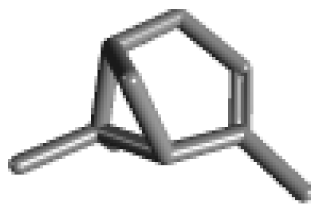 | 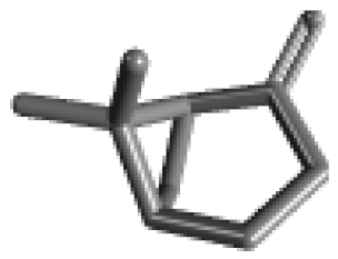 |  |  | ||
| Camphene (C10H16) | Myrcene (C10H16) | α-Terpinene (C10H16) | γ-Terpinene (C10H16) | ||
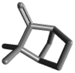 | 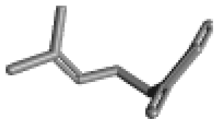 |  |  | ||
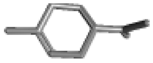
| α-Phellandrene (C10H16) | β-Phellandrene (C10H16) | p-Cymene (C10H14) | Sabinene (C10H16) | ||
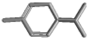 |  |  |  | ||
| Terpinolene (C10H16)  | |||||
| Oxygenated monoterpene | 1,8-Cineole (Eucalyptol, C10H18O) | Camphor (C10H16O) | Borneol (C10H18O) | α-Terpineol (C10H18O) | [34,37] |
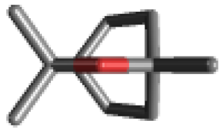 | 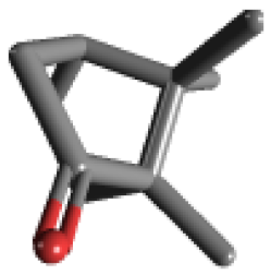 | 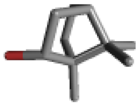 |  | ||
| Terpinen-4-ol (C10H18O) | Linalool (C10H18O) | Linalool-oxide (C10H18O2) | |||
 |  | 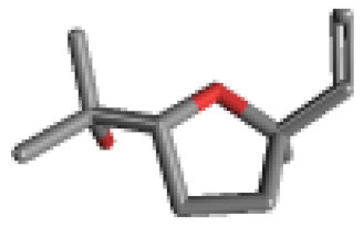 | |||
| Monoterpene derivatives | Bornyl acetate (C12H20O2) | [34] | |||
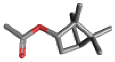 | |||||
| Sesquiterpene | Humulene (α-Caryophyllene, C15H24) | β-Caryophyllene (trans-Caryophyllene, C15H24) | [34,37,38] | ||
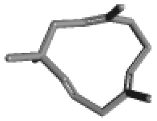 |  | ||||
| Related Inflammatory Activities | Name | Mechanism of Action | Experimental Protocol | Animal Tested | Ref. |
|---|---|---|---|---|---|
| Pro-inflammatory mediator | d-Limonene | TNF-α, IL-1β and IL-6 ↓ | LPS-stimulation | Raw 264.7 cell line | [46] |
| NF-kB, COX-2, iNOS and Nitrite levels ↓ | Doxorubicin-induced inflammation | Wistar rats | [47] | ||
| NO levels ↓ | Aβ42 expressed heads | Fruit fly | [48] | ||
| NO and iNOS levels ↓ | In vitro treatment | Human chondrocytes | [49] | ||
| Myrcene | NO and iNOS levels ↓ | In vitro treatment | Human chondrocytes | [49] | |
| γ-Terpinene | TNF-α and IL-1β ↓ | Carrageenan-induced peritonitis model | Swiss mice | [50] | |
| IL-1β, IL-6↓ and IL-10, COX-2, PGE2 ↑ | LPS-Stimulation | Macrophages from mice | [51] | ||
| α-Phellandrene | TNF-α and IL-6 ↓ | Carrageenan injection in air pouch cavities | Wistar rats or swiss mice | [52] | |
| IL-6 and TNF-α ↓ NO production ↓ | LPS-stimulation | Raw 264.7 cell line | [53] | ||
| Terpinolene | Pro-inflammatory cytokines IL-6 and TNF-α ↓ NO production ↓ | LPS-stimulation | Raw 264.7 cell line | [53] | |
| 1,8-Cineole | Production of LTB4 and PGE2 from monocytes ex vivo | Stimulated with the calcium ionophore A23187 measured ex vivo | Blood monocytes of patients with bronchial asthma | [54] | |
| TNF-α and IL-1β, leukotriene B4 and thromboxane B2 ↓ | LPS-and IL1β-stimulation in vitro | Human monocytes | [55] | ||
| Levels of TNFα and IL-1β in BALF ↓ | Experimental model of airways allergic inflammation | Ovalbumin (OVA)-challenged Guinea pigs | [56] | ||
| TNF-α and IL-1β ↓ and IL-10 ↑ | Mouse LPS-induced acute lung injury model | ICR mice | [57] | ||
| NO ↓ TNF-α, IL-1β and IL-6 ↓ | Aβ (25-35) treatment | PC 12 cell line | [58] | ||
| MMP-9 ↓ TNF-α, IL-6 and NO ↓ | LPS-induced acute lung injury mouse model | BALB/C mice | [59] | ||
| Production of interleukin IL-4, IL-13 and IL-17A in BALF after Derp challenge ↓ | House dust mite (HDM)- induced murine asthma model | BEAS-2B cell line | [60] | ||
| IL-1β, IL-6 and TNF-α in BALF ↓ | Short-term cigarette smoke (CS) exposure | C57BL/6 mice | [61] | ||
| IL-4, IL-5, IL-10, and MCP-1 in nasal lavage fluids ↓ IL-1β, IL-6, TNF-α, and IFN-γ in lung tissues ↓ | Mice infected with influenza A virus | BALB/C mice | [62] | ||
| Camphor | TNF-α, IL-1β and IL-6 in Kidney, testes, liver and lung ↑ | An acute administration | Wistar rats | [63] | |
| Borneol | IL-1β and IL-6 mRNA expression in colon tissue ↓ | TNBS-induced colitis | ICR mouse | [64] | |
| The elevation of NO, the increase of inducible iNOS enzymatic activity and the upregulation of iNOS expression ↓ | In vitro ischemic model of oxygen-glucose deprivation followed by reperfusion | Wistar rats | [65] | ||
| TNF-α, IL-1β, and IL-6 ↓ | Mouse LPS-induced acute lung injury model | Raw 264.7 cell line BALB/c mice | [66] | ||
| CD16 and CD206 expressions and levels of IL-1β, IL-6, TNF-a, and IL-10 proteins ↓ | LPS-stimulated mouse microglia and septic mice | C57BL/6 mice | [67] | ||
| α-Terpineol | Nitrite production ↓ | LPS-stimulation | Peritoneal macrophage | [68] | |
| Terpinen-4-ol | NF-κB and NLRP3 inflammasome ↓ | Dextran sulfate sodium-induced colitis | C57BL/6 mice | [69] | |
| LPS-induced phosphorylation of IκBα and NF-κB p65 ↓ The expression of PPAR-γ ↑ | Mouse LPS-induced acute lung injury model | BALB/c mice | [70] | ||
| Linalool | The production of LPS-induced TNF-α and IL-6 ↓ | LPS-stimulation | Raw 264.7 cell line | [71] | |
| LPS-induced TNF-α, IL-1β, NO, and PGE2 ↓ | LPS-stimulated microglia cells. | Murine BV2 cell line | [72] | ||
| The levels of the pro-inflammatory markers p38 MAPK, NOS2, COX2 and IL-1β ↓ | Triple transgenic model of Alzheimer’s disease mice | 3xTg-AD mice | [73] | ||
| Endotoxin-induced levels of peripheral nitrate/nitrite, IL-1β, IL-18, TNF-α, IFN-γ, and HMGB-1 ↓ Nitrate/nitrite, IL-1β, TNF-α, and IFN-γ in spleen and MLNs ↓ | Endotoxin-injection | C57BL/6J mice | [74] | ||
| Microgliosis and decreased COX2, IL-1β and Nrf2 markers in the cerebral cortex and hippocampus ↓ | Focal ischemia | Wistar rats | [75] | ||
| Levels of iNOS expression in the lung tissues caused by OVA exposure ↓ | Experimental model of airways allergic inflammation | OVA-challenged mice | [76] | ||
| Bornyl acetate | IL-1β-mediated up-regulation of IL-6, IL-8, MMP-1, and MMP-13 ↓ | In vitro treatment | Human chondrocytes | [77] | |
| Humulene | IL-5, CCL11 and leukotriene B4 levels in bronchoalveolar lavage fluid ↓ IL-5 production in mediastinal lymph nodes (In vitro assay) ↓ | Experimental model of airways allergic inflammation | OVA-challenged mice | [78] | |
| β-Caryophyllene | The serum level of IL-6 protein as well as the level of IL-6 mRNA in the tissue ↓ | Dextran sulfate sodium-induced colitis | BALB/c mice | [79] | |
| Anti-inflammatory (IL-10, Arg-1, and urea) and anti-oxidant GSH parameters ↑and the inflammatory (IL-1β, TNF-α, PGE2, iNOS and NO) and ROS biomarkers ↓ | LPS-stimulation | Primary microglia cell lines (C57BL/6) | [80] | ||
| The elevated TNF-α, NF-κB, and iNOS ↓ | Rats fed a high fat/fructose diet to induce insulin resistance and obesity | Wistar rats | [81] | ||
| The iNOS in the lumbar spinal cord ↓ | Experimental autoimmune encephalomyelitis, a murine model of multiple sclerosis | C57BL/6 mice | [82] | ||
| Pro-inflammatory cytokines and inflammatory mediators such as COX-2 and iNOS ↓ | Rotenone-treated rat model of Parkinson disease | Wistar rats | [83] | ||
| Hypoxia-induced cytotoxicity as well as IL-1β, TNF-α and IL-6 ↓ | Hypoxia exposure | Murine BV2 cell line | [84] | ||
| TNF-α and IL-1β ↓ | Kainic acid-induced seizure activity and oxidative stress | Mouse model | [85] | ||
| NO and PGE2 production ↓ iNOS and COX-2 ↓ Secretion of pro-inflammatory cytokines ↓ | Aβ-treated microglia | Murine BV2 cell line | [86] | ||
| Transcription factors | α-Pinene | NF-κB ↓ | LPS-stimulation | Mouse peritoneal macrophages | [87] |
| d-Limonene | NF-κB ↓ | LPS-induced acute lung injury | BALB/c mice | [88] | |
| Doxorubicin-induced inflammation in kidneys | Wistar rats | [47] | |||
| In vitro treatment | Human chondrocytes | [49] | |||
| Myrcene | NF-κB ↓ | In vitro treatment | Human chondrocytes | [49] | |
| 1,8-Cineole | Nuclear translocation of NF-κB p65 ↓ Expression of NF-κB target genes ↓ Protein levels of IκBα in an IKK-independent matter ↑ LPS-associated loss of interaction between NF-κB p65 and IκBα ↑ | LPS-stimulation | U373 and HeLa cell lines | [89] | |
| The expression of NF-κB p65 ↓ | Mouse LPS-induced acute lung injury model | ICR mice | [57] | ||
| LPS-induced acute lung injury mouse model | BALB/C mice | [59] | |||
| Short-term cigarette smoke (CS) exposure | C57BL/6 mice | [61] | |||
| Mice infected with influenza A virus | BALB/C mice | [62] | |||
| Camphor | The expressions of renal, testicular, hepatic and pulmonary NF-kB ↑ | An acute administration in male | Wistar rats | [90] | |
| Borneol | Phosphorylation of NF-κB and IκBa ↓ | Mouse LPS-induced acute lung injury model | Raw 264.7 cell line BALB/c mice | [66] | |
| Linalool | Nuclear Nrf-2 protein translocation ↑ | Pneumonia model infected by Pasteurella multocida | A549 cell line C57BL/6J mice | [91] | |
| LPS-induced NF-κB activation ↓ Nuclear translocation of Nrf2 ↑ | LPS-stimulated microglia cells. | Murine BV2 cell line | [72] | ||
| CS-induced NF-κB activation ↓ | Cigarette smoke -induced acute lung inflammation | C57BL/6 mice | [92] | ||
| The activation of NF-κB ↓ | Endotoxin-injection | C57BL/6J mice | [74] | ||
| The activation of NF-κB in the lung tissues caused by OVA exposure ↓ | Experimental model of airways allergic inflammation | OVA-challenged mice | [76] | ||
| Humulene | The NF-kB and the AP-1 activation ↓ | Experimental model of airways allergic inflammation | OVA-challenged mice | [78] | |
| β-Caryophyllene | Hypoxia-induced the activation of NF-κB ↓ | Cultured microglia under hypoxia | Murine BV2 cell line | [84] | |
| Aβ1-42-induced phosphorylation and degradation of IκBα, nuclear translocation of p65, and NF-κB transcriptional activity ↓ | Aβ-treated microglia | Murine BV2 cell line | [86] | ||
| Signal transduction | α-Pinene | ERK and JNK ↓ | LPS-stimulation | Mouse peritoneal macrophages | [87] |
| d-Limonene | p38, JNK, ERK ↓ | LPS-induced acute lung injury | BALB/c mice | [88] | |
| p38 and JNK activation ↓ | In vitro treatment | Human chondrocytes | [49] | ||
| Myrcene | p38 and JNK activation ↓ | In vitro treatment | Human chondrocytes | [49] | |
| 1,8-Cineole | Phosphorylated JNK in U373 cells ↓ | LPS-stimulation | U373 and HeLa cell lines | [89] | |
| TREM-1, NLRP3 of the inflammasome ↓ Phosphorylation of the transcription factor NF-κB and p38↓MKP-1 phosphatase, a negative regulator of MAPKs ↓ | LPS-induced the murine lung alveolar macrophage inflammation model | MH-S cell line | [93] | ||
| NLRP3 inflammasome activation and pro-inflammatory cytokine productions induced by MSU in ankle tissues in vivo ↓ MSU-induced upregulation of TRPV1 expression in ankle tissues and dorsal root ganglion neurons innervating the ankle ↓ | A mouse model of gout arthritis was established via MSU injection into the ankle joint | BALB/c mice | [94] | ||
| Inflammatory cytokines (IL-1β, TNF-α and IL-6) ↓ | LPS-induced pulmonary inflammation | C57BL/6 | [95] | ||
| Borneol | Phosphorylation of p38 and JNK ↓ | Mouse LPS-induced acute lung injury model | Raw 264.7 cell line BALB/c mice | [66] | |
| The activation of M2 macrophages in a STAT3-dependent manner ↑ | DSS-induced colitis | Raw 264.7 cell line | [96] | ||
| NF-κB and p38 signaling ↓ | LPS-stimulated microglia | C57BL/6 mice | [97] | ||
| TRPA1 mediated cationic currents ↓ | In vitro treatment | In heterologous expression systems like Xenopus oocytes and in neurons cultured from trigeminal ganglia | [98] | ||
| Linalool | Phosphorylation of IκBα protein, p38, c-JNK, and ERK ↓ | LPS-stimulation | Raw 264.7 cell line | [71] | |
| β-Caryophyllene | Functional agonist of CB(2)R | LPS-stimulation | CB2-expressiong HL60 cell line | [99] | |
| Activation of ERK 1/2, NF-κB, IκB-kinase α/β ↓ Involvement of CB2 and the PPARγ pathway | DSS-induced colitis | CD1 mice | [100] | ||
| Cisplatin-induced renal inflammatory response (chemokines MCP-1 and MIP-2, cytokines TNF-α and IL-1β, adhesion molecule ICAM-1, and neutrophil and macrophage infiltration) through a CB(2)R-dependent pathway ↓ | Cisplatin-induced nephropathy model | C57BL/6J | [101] | ||
| Activation of NF-κB and the secretion of inflammatory cytokines ↓ | Hypoxia exposure | Murine BV2 cell line | [84] | ||
| Oxidative stress | α-Pinene | ROS formation and lipid peroxidation induced by H2O2-stimulated oxidative damage ↓ | H2O2-stimulated oxidative stress | U373-MG cells | [102] |
| d-Limonene | ROS formation/ caspase-3/caspase-9 activation/p38 MAPK phosphorylation ↓ The Bcl-2/Bax ratio induced by H2O2-stimulated oxidative damage ↑ | H2O2-stimulated oxidative stress | Human lens epithelial cells | [103] | |
| Catalase and peroxidase activities of cell antioxidant enzymes ↑ | Lymphoid cell suspensions from lymph nodes | BALB/c mice | [104] | ||
| Camphene | Strong antioxidant effect and high scavenging activities against different free radicals | The nonenzymatic antioxidant capacity | Swiss mice | [105] | |
| The cell viability and GSH content and restored the mitochondrial membrane potential ↑ NO release and ROS generation ↓ | t-BHP stressed alveolar macrophages | Wistar rats | [106] | ||
| Myrcene | ROS, MMP-1, MMP-3, and IL-6, and increased TGF 1 and type I procollagen secretions ↓ The phosphorylation of various MAPK-related signaling molecules ↓ | In vitro treatment | UVB-irradiated human dermal fibroblasts | [107] | |
| α-Terpinene | The best antioxidant compounds in ABTS, chelating power and DPPH assays | In vitro antioxidation assay | [108] | ||
| γ-Terpinene | The best antioxidant compounds in ABTS and DPPH assays | In vitro antioxidation assay | [108] | ||
| α-Phellandrene | The intracellular oxidative stress environment ↓ | Mice leukemia | WEHI-3 cell line | [108] | |
| O2-production ↓ | LPS-stimulation | Raw 264.7 cell line | [53] | ||
| p-Cymene | SOD and catalase activity significantly ↑ | Intraperitoneal treatment with 0.05% Tween 80 | Swiss mice | [109] | |
| Terpinolene | O2-production ↓ | LPS-stimulation | Raw 264.7 cell line | [53] | |
| 1,8-Cineole | ROS formation and lipid peroxidation induced by H2O2-stimulated oxidative damage ↓ | H2O2-stimulated oxidative stress | U373-MG cells | [102] | |
| Camphor | Excessive ROS production and mitochondrial impairment ↑ | Oxidative stress-mediated apoptotic cell death | Schizosaccharomyces pombe | [110] | |
| Linalool | The best antioxidant compounds in ORAC and Chelating power assay | In vitro antioxidation assay | [108] | ||
| Oxidative stress and mitochondrial dysfunction mediated by glutamate and NMDA toxicity ↓ | Oxidative stress and mitochondrial dysfunction | HT-22 cells | [111] | ||
| Humulene | H2O2-induced astrocytic cell death ↓ | Primary astrocytes from cerebral cortices | Neonatal wistar rats | [112] | |
| β-Caryophyllene | H2O2-induced astrocytic cell death ↓ | Primary astrocytes from cerebral cortices | Neonatal wistar rats | [112] | |
| Rates of ROS production and the associated respiratory activity in freshly isolated hepatic mitochondria ↓ | Development of adjuvant arthritis | Holtzman rats | [113] | ||
| Autophagy | d-Limonene | Expression of apoptosis and autophagy-related genes ↑ | In vitro and vivo treatment | BALB/c mice A549 and H1299 cell lines | [114] |
| LC3 lipidation ↑ and clonogenic capacity ↓ | In vitro treatment | SH-SY5Y, HepG2 and MCF7 cell lines | [115] | ||
| LC3 II↑ and p62 levels ↓ | In vitro treatment | SH-SY5Y and MCF7 cell lines | [116] | ||
| p-Cymene | Autophagolysosomes ↑ and proliferation ↓ Anti-tumor metallodrug candidates | In vitro treatment | A2780 ovarian and MCF7 and MDAMB231 breast | [117] | |
| Autophagy with materials containing Ru complex ↑ | In vitro treatment | B16 and B16-F10 cell lines | [118] | ||
| Camphor | Autophagy and apoptotic cell death ↑ | In vitro treatment | Schizosaccharomyces pombe | [119] | |
| Borneol and TMPP | LC3 II/I, pAMPK, mTOR, and ULK1 in hypothalamus, and pAMPK, mTOR, ULK1, Beclin1, and Bax in striatum ↑ | Surgical induction of GCIR | Sprague-Dawley rats | [118] | |
| Cortex autophagy by modulating pAMPK in the pAMPK-mammalian target of mTOR-ULK1 signaling pathway ↑ | [120] | ||||
| Borneol and Luteolin | E1, p62, and ubiquitin levels ↓ Accumulation of toxic aggregates, cell death ↑ | In vitro treatment | HepG2 cell line | [121] | |
| Other activities | α-Pinene | Sleep enhancing property through a direct binding to GABAA BZD receptors | Pentobarbital-induced sleep | ICR and C57BL/6N mice | [122] |
| G2/M-phase cell cycle arrest miR-221 expression level ↓ CDKN1B/p27-CDK1 and ATM-p53-Chk2 pathways ↑ | In vitro treatment | HepG2 cell line | [123] | ||
| 3-Carene | sleep duration ↑ and sleep latency ↓ GABAA receptor-mediated synaptic responses ↑ | Pentobarbital-induced sleep | ICR and C57BL/6N mice | [124] | |
| 1,8-Cineole | Acetylcholinesterase activities ↓ | In vitro antioxidation assay | [108] | ||
| Bornyl acetate | Lipoxygenase ↓ | In vitro antioxidation assay | [108] | ||
| Limonene | Lipoxygenase ↓ | In vitro antioxidation assay | [108] | ||
| β-Caryophyllene | VCAM-1 ↓, and restored vascular eNOS/iNOS expression balance PPAR-γ agonist | high fat/fructose diet-induced dyslipidemia and vascular inflammation | Wistar rats | [81] | |
© 2020 by the authors. Licensee MDPI, Basel, Switzerland. This article is an open access article distributed under the terms and conditions of the Creative Commons Attribution (CC BY) license (http://creativecommons.org/licenses/by/4.0/).
Share and Cite
Kim, T.; Song, B.; Cho, K.S.; Lee, I.-S. Therapeutic Potential of Volatile Terpenes and Terpenoids from Forests for Inflammatory Diseases. Int. J. Mol. Sci. 2020, 21, 2187. https://doi.org/10.3390/ijms21062187
Kim T, Song B, Cho KS, Lee I-S. Therapeutic Potential of Volatile Terpenes and Terpenoids from Forests for Inflammatory Diseases. International Journal of Molecular Sciences. 2020; 21(6):2187. https://doi.org/10.3390/ijms21062187
Chicago/Turabian StyleKim, Taejoon, Bokyeong Song, Kyoung Sang Cho, and Im-Soon Lee. 2020. "Therapeutic Potential of Volatile Terpenes and Terpenoids from Forests for Inflammatory Diseases" International Journal of Molecular Sciences 21, no. 6: 2187. https://doi.org/10.3390/ijms21062187
APA StyleKim, T., Song, B., Cho, K. S., & Lee, I.-S. (2020). Therapeutic Potential of Volatile Terpenes and Terpenoids from Forests for Inflammatory Diseases. International Journal of Molecular Sciences, 21(6), 2187. https://doi.org/10.3390/ijms21062187


