Article

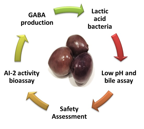
Ιn Vitro Screening of γ-Aminobutyric Acid and Autoinducer-2 Signalling in Lactic Acid Bacteria Exhibiting Probiotic Potential Isolated from Natural Black Conservolea Olives

by
Foteini Pavli
1,
Eleni Gkana
1,
Oluwabunmi Adebambo
2,
Kimon-Andreas Karatzas
2,
Efstathios Panagou
1 and
George-John E. Nychas
1,*
1
Laboratory of Microbiology Biotechnology of Foods, Department of Food Science and Human Nutrition, School of Food, Biotechnology and Development, Agricultural University of Athens, 11855 Athens, Greece
2
Department of Food and Nutritional Sciences, University of Reading, Reading RG6 6AP, UK
*
Author to whom correspondence should be addressed.
Foods 2019, 8(12), 640; https://doi.org/10.3390/foods8120640
Submission received: 3 November 2019 / Revised: 30 November 2019 / Accepted: 1 December 2019 / Published: 4 December 2019
(This article belongs to the Section Food Microbiology)

Abstract

:
In the present study, 33 strains of lactic acid bacteria (LAB) previously isolated from natural black Conservolea olives were assessed for their probiotic potential in vitro, as well as for their autoinducer-2 (AI-2) activity under standard growth conditions and the production of γ-aminobutyric acid (GABA). The probiotic tests included the in vitro resistance to low pH and resistance to bile salts, the evaluation of bile salt hydrolase activity, as well as safety tests regarding their possible haemolytic activity and their antimicrobial activity against pathogens. The results indicated that 17 strains were able to survive in low pH and in the presence of bile, with 15 of them also exhibiting partial bile salt hydrolase activity. None of the strains exhibited haemolytic activity or inhibited the growth of any of the examined pathogens. Moreover, the strains displayed generally low AI-2 activity under the growth conditions tested, regardless of the species. Interestingly, in contrast to what has been found in most foods, none of the isolates were found to produce GABA after 48 h of growth. The results from the AI-2 activity and extracellular GABA detection were considered as unexpected for LAB with probiotic attributes.

Graphical Abstract

1. Introduction

Lactic acid bacteria (LAB) are the most important group of probiotic bacteria, followed by Bifidobacteria, and some species of Enterococci and Bacillus, although the safety of the latter remains a controversy. Many beneficial effects of probiotics have been associated with lowering of cholesterol, controlling symptoms of lactose intolerance, stimulating the immune system, relieving constipation, preventing antibiotic-associated and traveller’s diarrhoea, and prevention of Clostridium difficile infections [1,2,3,4,5]. Such bacteria have been isolated from various sources, including fermented dairy products [6,7], non-dairy products [8,9,10,11,12], and from the intestinal tract of healthy individuals or infants [13,14,15]. Fermented products of plant origin, such as table olives, are considered an excellent source of probiotic bacteria and also great vehicles for probiotic delivery, with many studies confirming their potential [16,17,18,19]. A series of in vitro tests could be performed as a first step in order to assess the probiotic potential of these strains. Such tests could include acid and bile resistance, the production of antimicrobial compounds, and bile salt hydrolase activity, although further tests and clinical studies are also required at a later stage.
Cell-to-cell communication, also called quorum sensing, is the process of signalling that enables bacteria to control their gene expression and regulate their activities. A “universal” signal-molecule called autoinducer-2 (AI-2) could be present in gram-positive and gram-negative bacteria, supporting inter-species communication. The expression of various phenotypes including virulence factors, biofilm formation, light production, and stress resistance are only some of the functions that are linked to AI-2 in many bacterial species [20,21]. This is of a particular interest in the case of probiotic bacteria, due to their exposure to a variety of environmental challenges, including low pH, the presence of bile and enzymes. Lebeer [22] reported that AI-2 production had an impact on several physiological functions of the probiotic Lactobacillus rhamnosus GG. In another study, Lactobacillus paraplantarum L-ZS9 was found to overexpress luxS gene and produce AI-2 that enhances heat, bile, and salt resistance, while also having an effect on its biofilm formation [23]. Furthermore, other studies have related AI-2 activity with acid tolerance in probiotic bacteria [24].
γ-Aminobutyric acid (GABA) is an important metabolite produced by the glutamate decarboxylase system (GAD) in various bacteria through the decarboxylation of glutamate under acidic conditions. Thus, quantification of GABA could be an important tool to assess acid resistance of some probiotic bacteria, which is important because survival in the acidic pH of the stomach is required for gut colonization in the host [25]. Antianxiety, antidepressant, antihypertensive, and tranquilizing roles have been attributed to GABA, thus far [26,27]. Due to the aforementioned beneficial effects, much attention has been paid to the development of foods enriched with GABA. Several microorganisms have been examined for their potential to produce GABA through the GAD system, such as Lactobacillus brevis and Lactococcus lactis. Furthermore, it has been reported that strains of Lactobacillus buchneri, Lactobacillus paracasei, and Lactobacillus plantarum isolated from traditional cheeses have the ability to produce GABA [28,29]. It has to be noted that the ability of LAB to produce GABA varies among species and strains [30]. Therefore, screening of LAB for their ability to synthesize GABA is of special interest for the development of novel GABA-enriched functional foods, and this ability of LAB might be considered as an important probiotic trait in the near future [31].
In the present study, 33 strains of LAB were examined for their probiotic potential using a series of in vitro tests. The strains that exhibited good performance in the probiotic tests were then screened for AI-2 activity under standard growth conditions, followed by another screening with regard to their ability to produce GABA.

2. Materials and Methods

A total of 33 strains of LAB, previously isolated from natural black Conservolea olives during storage in different packaging conditions, as well as two reference strains, namely Lactobacillus casei Shirota (ACA-DC 6002) and Lactobacillus rhamnosus GG (ATCC 53103), kindly provided by Prof. E. Tsakalidou (Laboratory of Dairy Science, Agricultural University of Athens) were screened for their probiotic potential following a series of in vitro tests. The studied strains were 8 Lactobacillus plantarum, 20 Lactobacillus pentosus, 1 Lactobacillus paraplantarum, 2 Lactobacillus corinyformis, and 2 Pediococcus ethanolidurans (Table 1), that were previously isolated, identified, and characterized [32]. Strains were stored in 20% glycerol at −80 °C for long-term storage, and were routinely cultured at 30 °C in de Man, Rogosa, and Sharpe (MRS) broth (Biolife, Milan, Italy) or agar (Biolife, Italy) for 24 or 18 h and 72 h, respectively.

2.1. Low pH Assay, Bile Salts Assay, and Bile Salt Hydrolase (BSH) Activity

The tests to assess the resistance of the strains to low pH and bile salts, as well as the bile salt hydrolase (BSH) activity, were performed according to Argyri et al. [10]. Briefly, for the resistance to low pH, bacterial cells from overnight cultures (18 h) were harvested by centrifugation (5000× g, 15 min, 4 °C), washed twice with phosphate-buffered saline (PBS) (pH 7.2), and finally re-suspended in PBS solution with a pH of 2.5. After incubation for 0, 1, 2, and 3 h at 37 °C under stirring conditions, resistance to low pH was assessed in terms of viable colony counts enumerated on MRS agar. The strains that exhibited final counts ≥6 log CFU/mL at a pH of 2.5 for 3 h were selected to be screened for bile salt resistance. For the bile salt resistance test, the same procedure was applied, with the final resuspension being in PBS solution with a pH 8, containing 0.5% bile salts (Oxoid, Hampshire, UK). After incubation for 0, 1, 2, 3, and 4 h at 37 °C under stirring conditions, resistance was assessed in terms of viable colony counts enumerated on MRS agar. The strains that exhibited final counts ≥6 log CFU/mL under these conditions were selected to be screened for BSH activity. For the BSH activity, the bacterial cultures were streaked on MRS agar containing 0.5% taurodeoxycholic acid (TDCA; Sigma, St. Louis, MO, USA). The hydrolysis effect was assessed by different colony morphology in comparison to the control MRS plates (without TDCA) after anaerobic incubation at 37 °C for 48 h. The results were expressed as no hydrolysis or partial hydrolysis. The assays of low pH and bile were performed in triplicate, whereas the test for BSH activity was performed in duplicate.

2.2. Safety Assessment of the Selected Strains

The selected strains from the previous tests were further evaluated regarding their potential haemolytic activity and antimicrobial activity according to Argyri et al. [10]. Fresh bacterial cultures were streaked on Columbia agar plates (Lab M Limited, Lancashire, United Kingdom) containing 5% w/v horse blood and incubated for 48 h at 30 °C. After incubation, the plates were examined for signs of α-haemolysis (green-hued zones around colonies), β-haemolysis (clear zones around colonies), or γ-haemolysis (no zones around colonies). With regard to the antimicrobial activity, all strains were tested against the following pathogens: Listeria monocytogenes ATCC 13932; Listeria monocytogenes FMCC B-129; Listeria monocytogenes 23UD, kindly provided by Prof L. Cocolin [33]; Listeria monocytogenes H7550, kindly provided by Prof. S. Kathariou; Salmonella enterica subsp. enterica serovar Enteritidis P167807; Salmonella enterica subsp. enterica serovar Typhimurium 4/74 [34]; Salmonella enterica FMCC B-64; Salmonella enteritidis ATCC 13076; Escherichia coli FMCC B-13; Escherichia coli NCTC 13127; Escherichia coli ATCC 35150; and Escherichia coli ATCC 25922. The tests for haemolytic and antimicrobial activity were performed in duplicate.

2.3. Screening for AI-2 Activity

For this assay, two strains of Vibrio harveyi were used: V. harveyi BAA-1117 (ATCC BB-170) as biosensor and V. harveyi BAA-1119 (ATCC BB152) as positive control. Vibrio strains were cultured in Autoinducer Bioassay (AB) broth [35], and incubated with agitation (160 rpm) at 30 °C for 24 h. The cultures used directly in the bioassay were prepared by transferring a single colony from AB agar in 10 mL of AB broth, and incubating with agitation (160 rpm) at 30 °C for 16 h. For the growth of the LAB strains, quarter-strength brain heart infusion (BHI) broth (Lab M Limited, Lancashire, United Kingdom) was used as previously reported [36]. The AI-2 activity bioassay was performed as described previously [37]. In this study, 10 μL of sterile growth medium was used as a negative control, whereas 10 μL of the cell-free supernatant (CFS) from V. harveyi BA-1119 strain was used as a positive control to verify the bioassay. The microplates were incubated at 30 °C and luminescence was measured every 15 min using a Synergy HT multi-mode microplate reader (Biotek, Winooski, VT, USA). Measurements were collected until the negative control exhibited an increase in luminescence. AI-2 activity was expressed as relative AI-2 activity, which was calculated as the ratio of luminescence of the test sample (CFSLAB) to that of the control (negative) sample. The bioassay was performed in triplicate with four technical replicates each.

2.4. Detection of Extracellular GABA

Single colonies from each strain, previously grown on MRS agar, were obtained and inoculated in MRS broth, followed by incubation anaerobically at 37 °C overnight. These cultures were used as the inoculum 1% (v/v) to prepare the cultures used for the GABA determination in MRS broth supplemented with 10 mM L-glutamic acid (Sigma-Aldrich, Poole, United Kingdom) and were incubated under the same conditions. The population of each strain together with the pH values with the presence of L-glutamic acid after 48 h incubation were recorded, whereas the supernatant was collected from each culture with centrifugation (13,000 rpm for 10 min). Then, 10 μL of the supernatant was incubated with 90 μL of the assay mixture in each well of a 96-well microtiter plate, as described previously [38,39]. Additionally, standard solutions containing 0, 1, 2, 3, 4, 5, 6, 7, 8, 9, and 10 mM GABA were added to 90 μL of the assay mixture on the same microtiter plate, in order to obtain a standard curve for GABA determination. The microtiter plate was incubated at 37 °C for 180 min using a Sunrise Spectrophotometer (Tecan, Männedorf, Switzerland), and the optical density (OD) at 340 nm was measured every 120 s. The concentration of GABA in the supernatant was calculated using the calibration curves generated by the standard solutions. The test for GABA detection was performed in triplicate.

2.5. Statistical Analysis

The statistical analysis was performed using SPSS for Windows, Version 16.0 (SPSS Inc., Chicago, IL, USA). Regarding the resistance to low pH and resistance to bile salts, analysis of variance (ANOVA) for final bacterial counts of each strain (per hour and in total) was performed and means were separated with Tukey’s HSD test. The Tukey post hoc test was also used to compare the means of GABA concentration and AI-2 activity, expressed as the ratio of luminescence of the test sample to the control sample. All differences were reported at a significance level of 0.05.

3. Results and Discussion

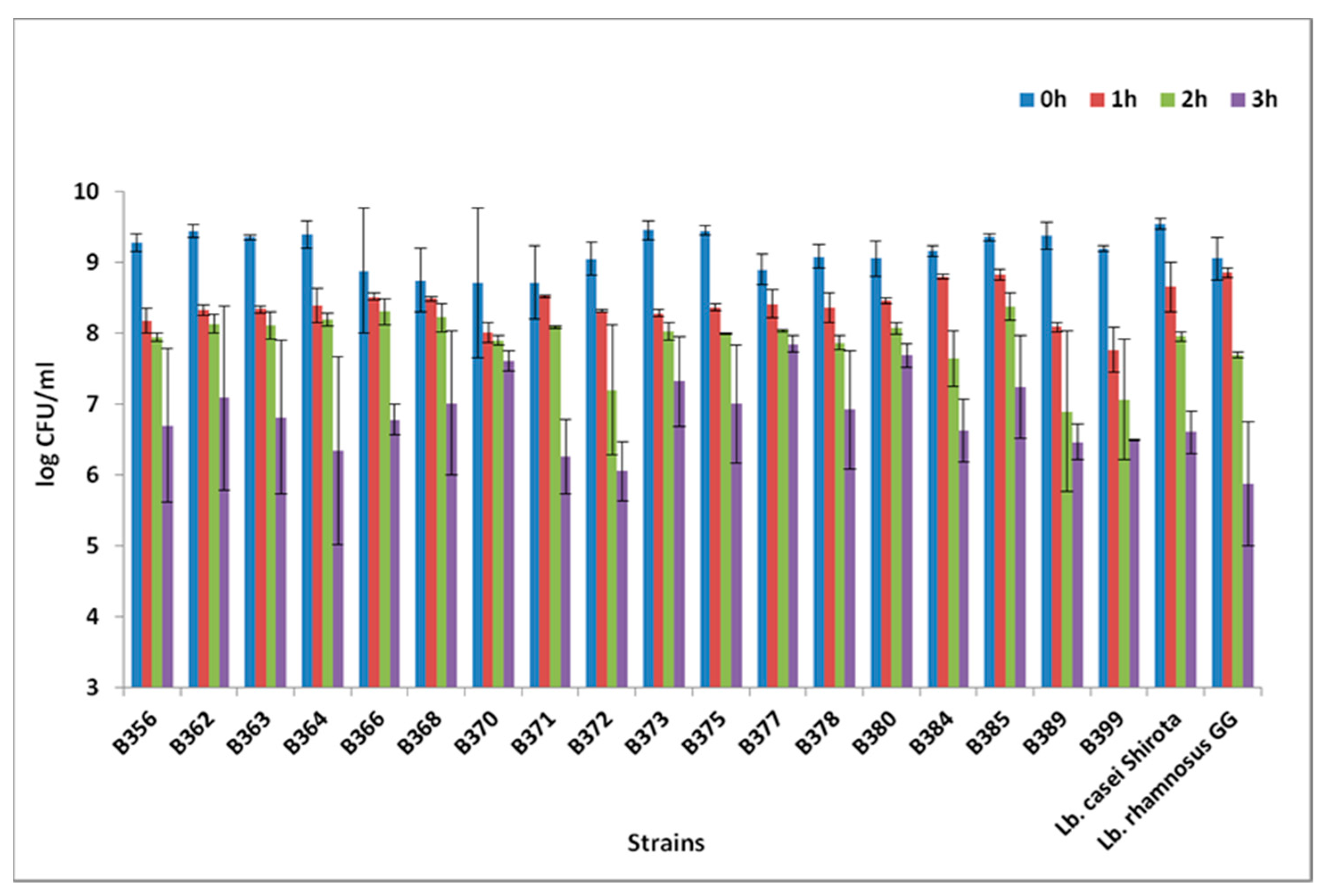
3.1. Low pH Assay, Bile Salts Assay, and BSH Activity

Out of the 33 LAB strains, 18 exhibited high population counts after exposure to pH of 2.5 for a total of 3 h. Amongst these, eight strains (five Lactobacillus pentosus and three Lactobacillus plantarum) showed the highest population ≥7 log CFU/mL under the acidic conditions tested (Figure 1). Statistically significant differences in the bacterial counts were detected after 1 h in low pH (p < 0.05). Results from low pH resistance were in agreement with other studies, where Lactobacillus pentosus and Lactobacillus plantarum strains were able to maintain their viability after exposure to low pH; however, a variation in the acid resistance among different strains was also observed [6,8,10]. Regarding the resistance to bile salts, out of the 18 strains, only 1 (Pediococcus ethanolidurans B389) showed final population <6 log CFU/mL after exposure to bile salts for 4 h. A total of 16 out of 17 strains exhibited a very low log reduction <1, whereas one strain, Lactobacillus pentosus B362, showed a slightly higher log reduction (1.14 log CFU/mL; Figure 2). The resistance to bile salts is a prerequisite for probiotic characterization, and guarantees that the cells could reach the intestinal tract alive [40]. Although generally lactobacillus species are able to tolerate bile concentrations normally encountered in the host (0.1%–0.5%), great variability in bile resistance has been reported at genus and species level [41,42]. These observations confirm the hypothesis that bile resistance is a strain-dependent characteristic [43]. It has to be noted that bacterial resistance to low pH and bile salts in vitro is not necessarily similar to the in vivo behaviour [44].
BSH was examined on those strains that exhibited satisfactory performance in the low pH and bile resistance tests. A total of 15 out of 17 strains exhibited partial BSH activity, expressed as differentiated colony morphology recorded on TDCA-MRS agar plates compared to the control MRS agar plates. BSH activity might be a beneficial attribute for a potentially probiotic strain and is often associated with the resistance to bile salts due to the reduction in serum cholesterol or cholesterol solubility and absorption [45]. However, further studies are needed to confirm that other risks for the host are kept to a minimum when excessive amount of probiotic bacteria is consumed. Furthermore, apart from live cells, dead or non-growing lactobacilli cells have been reported to have the ability to reduce cholesterol from media [46].

3.2. Safety Assessment of the Selected Strains

Haemolytic activity is one of the screening tests performed for probiotic characterization. Absence of haemolytic activity is considered as a safety requirement for the selection of probiotic strains. In the present study, none of the 17 strains, previously selected for their good performance in low pH and bile assays, exhibited α- or β-haemolysis, whereas all of them exhibited γ-haemolysis (no haemolysis) (Table 2). The findings are similar to those of previous studies regarding the safety of the LAB [6,10,47,48,49], although some exceptions do exist.
The antimicrobial activity against pathogens is deemed as a desirable trait, although not obligatory for probiotic characterization. From the 17 selected LAB strains, none of them were found to inhibit the growth of the 12 pathogens tested, according to the well-diffusion method. These results are in agreement with previous studies [6,10,50], where no antimicrobial activity was reported. On the contrary, significant antimicrobial activity of the LAB against two strains of Listeria monocytogenes and low or moderate antimicrobial activity against Bacillus cereus was reported in another study [48].

3.3. AI-2 Activity

The AI-2 activity for the tested strains exhibited values ranging from 0.32- to 1.57-fold compared to the negative control, whereas the bacterial counts were estimated from 6.42 to 8.30 log CFU/mL after 20 h of incubation time in ¼ strength BHI (Table 3). According to the results obtained, the selected strains exhibiting probiotic potential did not show detectable AI-2 activity under these growth conditions. Generally, diverse results have been reported with regard to the ability of LAB to produce AI-2 molecules [24,36,51]. The detection of AI-2 molecules has been proven to be growth-medium dependent [52,53]. The luxS gene, responsible for AI-2 production, is subject to catabolic repression by glucose, thus AI-2 molecules are difficult to be detected in a growth medium containing glucose [54]. De Keersmaecher Vanderleyden [53] suggested that a final concentration of 2 mM of glucose present in the CFS could cause an inhibition to the light production in the bioassay, whereas other sugars such as galactose did not. Furthermore, the acidic conditions present in CFS could also have an impact on the bioassay, as reported in a previous study [53]. Another critical factor in the detection of AI-2 molecules is the growth stage. The best point in growth to detect AI-2 molecules in LAB is during the late exponential phase and/or the stationary phase, as the molecules are still considered to be present. This information was taken into consideration in the current study, and the supernatant was collected after 20 h of incubation at 30 °C, whereas the bacterial population was confirmed with plate counting.
Many potentially probiotic bacteria such as Bifidobacterium and Lactobacillus having a luxS homologue can produce AI-2 molecules. Lactobacillus rhamnosus GG has been thoroughly studied regarding its ability to produce AI-2 and regulate its physiology [22], suggesting that the luxS gene has a central metabolic role in this strain. In a previous study [24], Lactobacillus rhamnosus GG and Lactobacillus salivarius UCC118 were found to produce AI-2 signal molecules under standard growth conditions, reaching the maximum concentration at the late exponential and stationary phase, respectively. In the same study, the AI-2 activity after an acidic shock with pH of 3 and 4 showed an increase in Lactobacillus acidophilus NCFM and Lactobacillus rhamnosus GG. Such an observation supported the hypothesis that the LuxS-mediated quorum sensing via AI-2 activity possibly plays an important role in the stress tolerance response of Lactobacillus species. Park et al. [51] reported various intensities of AI-2 activity in fermented kimchi products and also from the LAB obtained from these products. Strains of Lactobacillus plantarum, Lactobacillus brevis, Lactobacillus fermentum and Lactobacillus garlicum exhibited significant AI-2 activity, which was considered by the authors as an interesting characteristic for the future of fermented foods [51]. Although QS is generally reported in LAB participating in food fermentations [55], it is possible that the most dominant QS system is that of autoinducing peptides (AIP), especially for the case of fermented vegetables such as table olives [56,57,58,59].

3.4. Detection of Extracellular GABA

Screening of LAB for their ability to produce GABA is important for the food industry, as GABA-producing strains could be utilised as starters or adjunct cultures in fermented foods, developing GABA-enriched functional products. GABA is synthesized and exported by the glutamic acid decarboxylase (GAD) system, which is a very potent acid resistance mechanism. The GAD enzyme catalyses the proton-consuming decarboxylation of L-glutamate to GABA, which subsequently is exported by a glutamate/GABA antiporter that also imports another glutamate molecule to initiate another cycle of glutamate decarboxylation [39]. Furthermore, the GAD system can decarboxylate intracellular L-glutamate pools to produce intracellular GABA, which can be metabolised to succinate through the GABA shunt [60]. Several studies have indicated the presence of GAD system in lactic acid bacteria [29]. Generally, GABA synthesis in bacteria is related to enhanced resistance under acidic conditions. In the present study, 17 strains of LAB, with good probiotic attributes, were screened for their ability to produce GABA in vitro. Extracellular GABA was not detected in any of the strains tested (concentrations between 0.16–0.66 mM/mL) under the growth conditions tested, as presented in Table 3.
Measurements of extracellular GABA (GABAe) as means of quantification of the GAD system activity could potentially indicate the acid resistance of a specific microorganism. However, the quantification of the intracellular GABA (GABAi) is also important for the investigation of the GAD system [25]. It has to be noted that although the GAD system is widely distributed in LAB, the ability of LAB to produce GABA varies significantly [61]. Similar conclusions were made previously, for GABA production in Listeria monocytogenes, where different strains export GABA in different media and environmental conditions, suggesting that diverse activation signals present in different niches might activate the GAD system in different strains [62].
Several factors are considered to have a detrimental effect on GABA synthesis in vitro, such as the incubation temperature, the incubation time, and the glutamate concentrations [61,63]. The optimum temperature range for GABA synthesis is 30–37 °C, whereas at 45 °C or more, GABA is not detected, possibly due to difficulties in bacterial growth. For the detection of the highest GABA concentrations, the optimum incubation time is 48 h, whereas extra incubation time does not result in an increase in GABA values [63]. In our study, the bacterial counts of the examined strains clearly showed that the presence of L-glutamic acid did not affect their growth (9.22–9.66 log CFU/mL) (Table 3).
In a study of Yunes et al. [64], 135 human-derived strains of lactobacilli and bifidobacteria were isolated and screened for their potential to synthesize GABA. A total of 43% of the isolates were determined as GABA-producers, with the strains assigned to Lactobacillus plantarum, Lactobacillus brevis, Bifidobacterium adolescentis, Bifidobacterium angulatum, and Bifidobacterium dentium. On the contrary, Barrett et al. [65] reported that from the 91 human-derived lactobacilli and bifidobacteria, only 4% were GABA producers, including Lactobacillus brevis, B. dentium, Bifidobacterium infantis, and B. adolescentis, with Lactobacillus brevis being the one with the highest conversion ability of monosodium glutamate to GABA. Furthermore, isolates from various foods such as Italian cheeses [29], selected dairy products [66], artisanal Zlatar Cheese [67], or Nostrano cheeses made from raw alpine milk [28] were positive in GABA production by 13.86%, 50%, 28%, or 70%, respectively. The work on Italian cheeses has shown that gorgonzola and pecorino harboured a high number of GABA-producing LAB. In addition, the type of milk used for cheese manufacturing, together with the ripening period, had an impact on the GABA concentrations.
Interestingly, in our work we found no isolate producing any GABA. This is in contrast with the situation in isolates from human colon and dairy products. This might be related to the fact that LAB strains isolated from dairy products might have a higher potential in GABA production compared to strains isolated from non-dairy products [68]. Cheese possesses specific characteristics that favour the natural presence of GABA-producing LAB. The high content of L-glutamate (17.5% of the total amino acid content) in milk caseins is metabolized from LAB during the ripening process [69]. In addition to strain variability, GABA production is affected by many other factors such as the temperature, the pH, the medium composition, and other environmental factors [29,70,71]. Work by Villegas et al. [63] has shown that 3 out of 19 strains (15%) originating from amaranth and quinoa showed GABA producing ability that was low but still significantly higher than the absence of GABA producers found in this study. It is well known that the amount of GABA available in fruits and vegetables is relatively low compared to other sources such as dairy and meat products [63,72]. In a study of Karatzas et al. [39], it was reported that L. monocytogenes, although producing GABA in rich media, is unable to export GABA in a defined medium supplemented with L-glutamate. This suggests that additional factors present in a nutrient-rich environment play an important role in the function of the GAD system.

4. Conclusions

A total of 17 strains were chosen as good candidates for potentially probiotic applications, as adjunct or co-starter cultures, although more tests are required to further assess their potential as probiotics. AI-2 signal molecules and extracellular GABA were not detected under the in vitro growth conditions tested; however, further research is needed to better understand the systems involved and the mechanisms triggering their production in LAB. The lack of GABA-producing strains isolated from natural black olives seems unique among various fermented foods, although the functionality of the GAD system or the genes involved have not yet been studied.

Author Contributions

Funding acquisition, G.-J.E.N.; Methodology, F.P., E.G. and O.A.; Writing—review & editing, F.P., K.-A.K., E.P. and G.-J.E.N.

Conflicts of Interest

The authors declare no conflict of interest.

References

  1. Choi, E.A.; Chang, H.C. Cholesterol-lowering effects of a putative probiotic strain Lactobacillus plantarum EM isolated from kimchi. LWT Food Sci. Technol. 2015, 62, 210–217. [Google Scholar] [CrossRef]
  2. He, T.; Priebe, M.G.; Zhong, Y.; Huang, C.; Harmsen, H.J.M. Effects of yogurt and bifidobacteria supplementation on the colonic microbiota in lactose intolerant subjects. J. Appl. Microbiol. 2007, 104, 595–604. [Google Scholar] [CrossRef] [PubMed]
  3. Kim, S.E.; Choi, S.C.; Park, K.S.; Park, M.I.; Shin, J.E. Change of Fecal Flora and Effectiveness of the Short-term VSL#3 Probiotic Treatment in Patients with Functional Constipation. J. Neurogastroenterol. Motil. 2015, 21, 111–120. [Google Scholar] [PubMed] [Green Version]
  4. McFarland, L.V. Probiotics for the primary and secondary prevention of C. difficile Infections: A meta-analysis and systematic review. Antibiotics 2015, 4, 160–178. [Google Scholar] [CrossRef] [PubMed] [Green Version]
  5. McFarland, L.V. Meta-analysis of probiotics for the prevention of traveler’s diarrhea. Travel Med. Infect. Dis. 2007, 5, 97–105. [Google Scholar] [CrossRef] [PubMed]
  6. Maragkoudakis, P.A.; Zoumpopoulou, G.; Miaris, C.; Kalantzopoulos, G.; Pot, B.; Tsakalidou, E. Probiotic potential of Lactobacillus strains isolated from dairy products. Int. Dairy J. 2006, 16, 189–199. [Google Scholar] [CrossRef]
  7. Zago, M.; Fornasari, M.E.; Carminati, D.; Burns, P.; Suàrez, V.; Vinerola, G.; Reinheimer, G.; Giraffa, G. Characterisation and probiotic potential of Lactobacillus plantarum strains isolated from cheeses. Food Microbiol. 2011, 28, 1033–1040. [Google Scholar] [CrossRef]
  8. Pavli, F.G.; Argyri, A.A.; Papadopoulou, O.S.; Nychas, G.-J.E.; Chorianopoulos, N.G.; Tassou, C.C. Probiotic potential of lactic acid bacteria from traditional fermented dairy and meat products: Assessment by in vitro tests and molecular characterization. J. Probiotics Health 2016, 4, 157. [Google Scholar] [CrossRef]
  9. Botta, C.; Langerholc, T.; Cencič, A.; Cocolin, L. In vitro selection and characterization of new probiotic candidates from table olive microbiota. PLoS ONE 2014, 9, e94457. [Google Scholar] [CrossRef]
  10. Argyri, A.A.; Zoumpopoulou, G.; Karatzas, K.-A.G.; Tsakalidou, E.; Nychas, G.-J.E.; Panagou, E.Z.; Tassou, C.C. Selection of potential probiotic lactic acid bacteria from fermented olives by in vitro tests. Food Microbiol. 2013, 33, 282–291. [Google Scholar] [CrossRef]
  11. Vitali, B.; Minervini, G.; Rizzello, C.G.; Spisni, E.; Maccaferri, S.; Brigidi, P.; Gobbetti, M.; Di Cagno, R. Novel probiotic candidates for humans isolated from raw fruits and vegetables. Food Microbiol. 2012, 31, 116–125. [Google Scholar] [CrossRef] [PubMed]
  12. Pennacchia, C.; Ercolini, D.; Blaiotta, G.; Pepe, O.; Mauriello, G.; Villani, F. Selection of Lactobacillus strains from fermented sausages for their potential use as probiotics. Meat Sci. 2004, 67, 309–317. [Google Scholar] [CrossRef] [PubMed]
  13. Kirtzalidou, E.; Pramateftaki, P.; Kotsou, M.; Kyriacou, A. Screening for lactobacilli with probiotic properties in the infant gut microbiota. Anaerobe 2011, 17, 440–443. [Google Scholar] [CrossRef] [PubMed]
  14. Vizoso-Pinto, M.G.; Franz, C.M.; Schillinger, U.; Holzapfel, W.H. Lactobacillus spp. with in vitro probiotic properties from human faeces and traditional fermented products. Int. J. Food Microbiol. 2006, 109, 205–214. [Google Scholar] [CrossRef] [PubMed]
  15. Martin, R.; Jiménez, E.; Olivares, M.; Marín, M.L.; Fernández, L.; Xaus, J.; Rodríguez, J.M. Lactobacillus salivarius CECT 5713, a potential probiotic strain isolated from infant feces and breast milk of a mother-child pair. Int. J. Food Microbiol. 2006, 112, 35–43. [Google Scholar] [CrossRef] [PubMed]
  16. Peres, C.M.; Peres, C.; Hernández-Mendoza, A.; Malcata, F.X. Review on fermented plant materials as carriers and sources of potentially probiotic lactic acid bacteria-With an emphasis on table olives. Trends Food Sci. Technol. 2012, 26, 31–42. [Google Scholar] [CrossRef]
  17. Hurtado, A.; Reguant, C.; Bordons, A.; Rozès, N. Lactic acid bacteria from fermented table olives. Food Microbiol. 2012, 31, 1–8. [Google Scholar] [CrossRef]
  18. Argyri, A.A.; Nisiotou, A.A.; Mallouchos, A.; Panagou, E.Z.; Tassou, C.C. Performance of two potential probiotic Lactobacillus strains from the olive microbiota as starters in the fermentation of heat shocked green olives. Int. J. Food Microbiol. 2014, 171, 68–76. [Google Scholar] [CrossRef]
  19. Blana, V.A.; Grounta, A.; Tassou, C.C.; Nychas, G.-J.E.; Panagou, E.Z. Inoculated fermentation of green olives with potential probiotic Lactobacillus pentosus and Lactobacillus plantarum starter cultures isolated from industrially fermented olives. Food Microbiol. 2014, 38, 208–218. [Google Scholar] [CrossRef]
  20. Xavier, K.B.; Bassler, B.L. LuxS quorum sensing: More than just a numbers game. Curr. Opin. Microbiol. 2003, 6, 191–197. [Google Scholar] [CrossRef]
  21. Lebeer, S.; Verhoeven, T.L.A.; Vélez, M.P.; Vanderleyden, J.; De Keersmaecker, S.C.J. Impact of environmental and genetic factors on biofilm formation by the probiotic strain Lactobacillus rhamnosus GG. Appl. Environ. Microbiol. 2007, 73, 6768–6775. [Google Scholar] [CrossRef] [PubMed] [Green Version]
  22. Lebeer, S.; De Keersmaecker, S.C.J.; Verhokomeven, T.L.A.; Fadda, A.A.; Marchal, K.; Vanderleyden, J. Functional Analysis of luxS in the Probiotic Strain Lactobacillus rhamnosus GG Reveals a Central Metabolic Role Important for Growth and Biofilm Formation. J. Bacteriol. 2007, 189, 860–871. [Google Scholar] [CrossRef] [PubMed] [Green Version]
  23. Liu, L.; Wu, R.; Zhang, J.; Li, P. Overexpression of luxS Promotes Stress Resistance and Biofilm Formation of Lactobacillus paraplantarum L-ZS9 by Regulating the Expression of Multiple Genes. Front. Microbiol. 2018, 9, 2628. [Google Scholar] [CrossRef] [PubMed]
  24. Moslehi-Jenabian, S.; Gori, K.; Jespersen, L. AI-2 signalling is induced by acidic shock in probiotic strains of Lactobacillus spp. Int. J. Food Microbiol. 2009, 135, 295–302. [Google Scholar] [CrossRef] [PubMed]
  25. O’Byrne, C.P.; Feehily, C.; Ham, R.; Karatzas, K.A.G. A modified rapid enzymatic microtiter plate assay, for the quantification of intracellular γ-aminobutyric acid and succinate semialdehyde in bacterial cells. J. Microbiol. Methods 2011, 84, 137–139. [Google Scholar] [CrossRef]
  26. Foster, A.C.; Kemp, J.A. Glutamate- and GABA-based CNS therapeutics. Curr. Opin. Pharmacol. 2006, 6, 7–17. [Google Scholar] [CrossRef]
  27. Möhler, H. The GABA system in anxiety and depression and its therapeutic potential. Neuropharmacology 2012, 62, 42–53. [Google Scholar] [CrossRef]
  28. Franciosi, E.; Carafa, I.; Nardin, T.; Schiavon, S.; Poznanski, E.; Cavazza, A.; Larcher, A.; Tuohy, K.M. Biodiversity and γ-Aminobutyric Acid Production by Lactic Acid Bacteria Isolated from Traditional Alpine Raw Cow’s Milk Cheeses. BioMed Res. Int. 2015, 2015, 625740. [Google Scholar] [CrossRef] [Green Version]
  29. Siragusa, S.; De Angelis, M.; Di Cagno, R.; Rizzello, C.G.; Coda, R.; Gobbetti, M. Synthesis of γ-Aminobutyric Acid by Lactic Acid Bacteria Isolated from a Variety of Italian Cheeses. Appl. Environ. Microbiol. 2007, 73, 7283–7290. [Google Scholar] [CrossRef] [Green Version]
  30. Park, K.-B.; Oh, S.-H. Production of yogurt with enhanced levels of gamma-aminobutyric acid and valuable nutrients using lactic acid bacteria and germinated soybean extract. Bioresour. Technol. 2006, 98, 1675–1679. [Google Scholar] [CrossRef]
  31. Ohmori, T.; Tahara, M.; Ohshima, T. Mechanism of gamma-aminobutyric acid (GABA) production by a lactic acid bacterium in yogurt-sake. Process Biochem. 2018, 74, 21–27. [Google Scholar] [CrossRef]
  32. Doulgeraki, A.I.; Hondrodimou, O.; Iliopoulos, V.; Panagou, E.Z. Lactic acid bacteria and yeast heterogeneity during aerobic and modified atmosphere packaging storage of natural black Conservolea olives in polyethylene pouches. Food Control 2012, 26, 49–57. [Google Scholar] [CrossRef]
  33. Cocolin, L.; Stella, S.; Nappi, R.; Bozzetta, E.; Cantoni, C.; Comi, G. Analysis of PCR-based methods for characterization of Listeria monocytogenes strains isolated from different sources. Int. J. Food Microbiol. 2005, 103, 167–178. [Google Scholar] [CrossRef] [PubMed]
  34. Hoiseth, S.K.; Stocker, B.A. Aromatic-dependent Salmonella typhimurium are non-virulent and effective as live vaccines. Nature 1981, 291, 238–239. [Google Scholar] [CrossRef] [PubMed]
  35. Lu, L.; Hume, M.E.; Pillai, S.D. Autoinducer-2-like activity associated with foods and its interaction with food additives. J. Food Prot. 2004, 67, 1457–1462. [Google Scholar] [CrossRef] [PubMed]
  36. Blana, V.A.; Doulgeraki, A.I.; Nychas, G.-J.E. Autoinducer-2-like Activity in Lactic Acid Bacteria Isolated from Minced Beef Packaged under Modified Atmospheres. J. Food Prot. 2011, 74, 631–635. [Google Scholar] [CrossRef] [PubMed]
  37. Surette, M.G.; Bassler, B.L. Quorum sensing in Escherichia coli and Salmonella typhimurium. Proc. Natl. Acad. Sci. USA 1998, 95, 7046–7050. [Google Scholar] [CrossRef] [Green Version]
  38. Tsukatani, T.; Higuchi, T.; Matsumoto, K. Enzyme-based microtiter plate assay for γ-aminobutyric acid: Application to the screening of γ-aminobutyric acid-producing lactic acid bacteria. Anal. Chim. Acta 2005, 540, 293–297. [Google Scholar] [CrossRef]
  39. Karatzas, K.-A.G.; Brennan, O.; Heavin, S.; Morrissey, J.; O’Byrne, C.P. Intracellular Accumulation of High Levels of γ-Aminobutyrate by Listeria monocytogenes 10403S in Response to Low pH: Uncoupling of γ-Aminobutyrate Synthesis from Efflux in a Chemically Defined Medium. Appl. Environ. Microbiol. 2010, 76, 3529–3537. [Google Scholar] [CrossRef] [Green Version]
  40. Taranto, M.P.; Perez-Martinez, G.; Font de Valdez, G. Effect of bile acid on the cell membrane functionality of lactic acid bacteria for oral administration. Res. Microbiol. 2006, 157, 720–725. [Google Scholar] [CrossRef]
  41. Jacobsen, C.N.; Rosenfeldt Nielsen, V.; Hayford, A.E.; Moller, P.L.; Michaelsen, K.F.; Pærregaard, A.; Sandström, B.; Tvede, M.; Jakobsen, M. Screening of probiotic activities of forty-seven strains of Lactobacillus spp. by in vitro techniques and evaluation of the colonization ability of five selected strains in humans. Appl. Environ. Microbiol. 1999, 65, 4949–4956. [Google Scholar] [PubMed]
  42. Papadimitriou, K.; Zoumpopoulou, G.; Foligné, B.; Alexandraki, V.; Kazou, M.; Pot, B.; Tsakalidou, E. Discovering probiotic microorganisms: In vitro, in vivo, genetic and omics approaches. Front. Microbiol. 2015, 6, 58. [Google Scholar] [CrossRef] [PubMed] [Green Version]
  43. Begley, M.; Gahan, C.G.M.; Hill, C. The interaction between bacteria and bile. FEMS Microbiol. 2005, 29, 625–651. [Google Scholar] [CrossRef] [PubMed] [Green Version]
  44. Morelli, L. In vitro assessment of probiotic bacteria: From survival to functionality. Int. Dairy J. 2007, 17, 1278–1283. [Google Scholar] [CrossRef]
  45. Begley, M.; Hill, C.; Gahan, C.G.M. Bile Salt Hydrolase Activity in Probiotics. Appl. Environ. Microbiol. 2006, 72, 1729–1738. [Google Scholar] [CrossRef] [Green Version]
  46. Liong, M.T.; Shah, N.P. Acid and Bile tolerance and the cholesterol removal ability of lactobacilli strains. J. Dairy Sci. 2005, 88, 55–66. [Google Scholar] [CrossRef] [Green Version]
  47. Zoumpopoulou, G.; Foligne, B.; Christodoulou, K.; Grangette, C.; Pot, B.; Tsakalidou, E. Lactobacillus fermentum ACA-DC 179 displays probiotic potential in vitro and protects against trinitrobenzene sulfonic acid (TNBS)-induced colitis and Salmonella infection in murine models. Int. J. Food Microbiol. 2008, 121, 18–26. [Google Scholar] [CrossRef]
  48. Ruiz-Moyano, S.; Martín, A.; Benito, M.J.; Casquete, R.; Serradilla, M.J.; Córdoba, M.D.G. Safety and functional aspects of pre-selected lactobacilli for probiotic use in Iberian dry-fermented sausages. Meat Sci. 2009, 83, 460–467. [Google Scholar] [CrossRef]
  49. Tejero-Sariňena, S.; Barlow, J.; Costabile, A.; Gibson, G.R.; Rowland, I. In vitro evaluation of the antimicrobial activity of a range of probiotics against pathogens: Evidence for the effects of organic acids. Anaerobe 2012, 18, 530–538. [Google Scholar] [CrossRef]
  50. Rubio, R.; Jofré, A.; Martín, B.; Aymerich, T.; Garriga, M. Characterization of lactic acid bacteria isolated from infant faeces as potential probiotic starter cultures for fermented sausages. Food Microbiol. 2014, 38, 303–311. [Google Scholar] [CrossRef]
  51. Park, H.; Shin, H.; Lee, K.; Holzapfel, W. Autoinducer-2 properties of kimchi are associated with lactic acid bacteria involved in its fermentation. Int. J. Food Microbiol. 2016, 225, 38–42. [Google Scholar] [CrossRef] [PubMed]
  52. Surette, M.G.; Bassler, B.L. Quorum sensing in Escherichia coli, Salmonella typhimurium and Vibrio harveyi: A new family of genes responsible for autoinducer production. Proc. Natl. Acad. Sci. USA 1999, 96, 1639–1644. [Google Scholar] [CrossRef] [PubMed] [Green Version]
  53. De Keersmaecker, S.C.J.; Vanderleyden, J. Constraints on detection of autoinducer-2 (AI-2) signalling molecules using Vibrio harveyi as a reporter. Microbiology 2003, 149, 1953–1956. [Google Scholar] [CrossRef] [PubMed] [Green Version]
  54. Ammor, M.S.; Michaelidis, C.; Nychas, G.-J.E. Insights into the role of quorum sensing in food spoilage. J. Food Prot. 2008, 71, 1510–1525. [Google Scholar] [CrossRef]
  55. Johansen, P.; Jespersen, L. Impact of quorum sensing on the quality of fermented foods. Curr. Opin. Food Sci. 2017, 13, 16–25. [Google Scholar] [CrossRef]
  56. Ruiz-Barba, J.L.; Caballero-Guerrero, B.; Maldonado-Barragán, A.; Jiménez-Díaz, R. Coculture with specific bacteria enhances survival of Lactobacillus plantarum NC8, an autoinducer-regulated bacteriocin producer, in olive fermentations. Food Microbiol. 2010, 27, 413–417. [Google Scholar] [CrossRef] [Green Version]
  57. Caballero-Guerrero, B.; Lucena-Padrós, H.; Maldonado-Barragán, A.; Ruiz-Barba, J.L. High-salt brines compromise autoinducer-mediated bacteriocinogenic Lactobacillus plantarum survival in Spanish-style green olive fermentations. Food Microbiol. 2013, 33, 90–96. [Google Scholar] [CrossRef]
  58. Rizzello, C.G.; Filannino, P.; Di Cagno, R.; Calasso, M.; Gobbeti, M. Quorum-sensing regulation of constitutive plantaricin by Lactobacillus plantarum strains under a model system for vegetables and fruits. Appl. Environ. Microbiol. 2014, 80, 777–787. [Google Scholar] [CrossRef] [Green Version]
  59. Doulgeraki, A.I.; Paraskevopoulos, N.; Nychas, G.J.E.; Panagou, E.Z. An in vitro study of Lactobacillus plantarum strains for the presence of plantaricin genes and their potential control of the table olive microbiota. Antonie van Leeuwenhoek 2013, 103, 821–832. [Google Scholar] [CrossRef]
  60. Feehily, C.; O’Byrne, C.P.; Karatzas, K.-A.G. Functional γ-Aminobutyrate Shunt in Listeria monocytogenes: Role in Acid Tolerance and Succinate Biosynthesis. Appl. Environ. Microbiol. 2013, 79, 74–80. [Google Scholar] [CrossRef] [Green Version]
  61. Komatsuzaki, N.; Shima, J.; Kawamoto, S.; Momose, H.; Kimura, T. Production of γ-aminobutyric acid (GABA) by Lactobacillus paracasei isolated from traditional fermented foods. Food Microbiol. 2005, 22, 497–504. [Google Scholar] [CrossRef]
  62. Karatzas, K.-A.G.; Suur, L.; O’Byrne, C.P. Characterisation of the intracellular glutamate decarboxylase system: Analysis of its function, transcription, and role in the acid resistance of various strains of Listeria monocytogenes. Appl. Environ. Microbiol. 2012, 78, 3571–3579. [Google Scholar] [CrossRef] [PubMed] [Green Version]
  63. Villegas, J.M.; Brown, L.; de Giori, G.S.; Hebert, E.M. Optimization of batch culture conditions for GABA production by Lactobacillus brevis CRL 1942, isolated from quinoa sourdough. LWT Food Sci. Technol. 2016, 67, 22–26. [Google Scholar] [CrossRef]
  64. Yunes, R.A.; Poluektova, E.U.; Dyachkova, M.S.; Klimina, K.M.; Kovtun, A.S.; Averina, O.V.; Orlova, V.S.; Danilenko, V.N. GABA production and structure of gadB/gadC genes in Lactobacillus and Bifidobacterium strains from human microbiota. Anaerobe 2016, 42, 197–204. [Google Scholar] [CrossRef]
  65. Barrett, E.; Ross, R.P.; O’Toole, P.W.; Fitzgerald, G.F.; Stanton, C. γ-Aminobutyric acid production by culturable bacteria from the human intestine. J. Appl. Microbiol. 2012, 113, 411–417. [Google Scholar] [CrossRef]
  66. Valenzuela, J.A.; Flórez, A.B.; Vázquez, L.; Vasek, O.M.; Mayo, B. Production of γ-aminobutyric acid (GABA) by lactic acid bacteria strains isolated from traditional, starter-free dairy products made of raw milk. Benef. Microbes 2019, 10, 579–589. [Google Scholar] [CrossRef]
  67. Sokovic Bajic, S.; Djokic, J.; Dinic, M.; Veljovic, K.; Golic, N.; Mihajlovic, S.; Tolinacki, M. GABA-Producing Natural Dairy Isolate from Artisanal Zlatar Cheese Attenuates Gut Inflammation and Strengthens Gut Epithelial Barrier in vitro. Front. Microbiol. 2019, 10, 527. [Google Scholar] [CrossRef]
  68. Shan, Y.; Man, C.X.; Han, X.; Li, L.; Guo, Y.; Deng, Y.; Li, T.; Zhang, L.W.; Jiang, Y.J. Evaluation of improved γ-aminobutyric acid production in yogurt using Lactobacillus plantarum NDC75017. J. Dairy Sci. 2015, 98, 2138–2149. [Google Scholar] [CrossRef] [Green Version]
  69. Renes, E.; Linares, D.M.; González, L.; Fresno, J.M.; Tornadijo, M.E.; Stanton, C. Production of conjugated linoleic acid and gamma-aminobutyric acid by autochthonous lactic acid bacteria and detection of the genes involved. J. Funct. Foods 2017, 34, 340–346. [Google Scholar] [CrossRef]
  70. Zhuang, K.; Jiang, Y.; Feng, X.; Li, L.; Dang, F.; Zhang, W.; Man, C. Transcriptomic response to GABA-producing Lactobacillus plantarum CGMCC 1.2437T induced by L-MSG. PLoS ONE 2018, 13, e0199021. [Google Scholar] [CrossRef]
  71. Mazzoli, R.; Pessione, E. The Neuro-endocrinological role of microbial glutamate and GABA signaling. Front. Microbiol. 2016, 7, 1934. [Google Scholar] [CrossRef] [PubMed] [Green Version]
  72. Kim, J.Y.; Lee, M.Y.; Ji, G.E.; Lee, Y.S.; Hwang, K.T. Production of γ-aminobutyric acid in black raspberry juice during fermentation by Lactobacillus brevis GABA100. Int. J. Food Microbiol. 2009, 130, 12–16. [Google Scholar] [CrossRef] [PubMed]
Figure 1. Resistance to low pH after 0, 1, 2, and 3 h of the selected strains Lactobacillus pentosus B356, B362, B363, B364, B366, B368, B370, B371, B377, B378, B385, B399; Lactobacillus plantarum B372, B373, B375, B380, B384; Pediococcus ethanolidurans B389; and the reference strains Lactobacillus casei Shirota and Lactobacillus rhamnosus GG.
Foods 08 00640 g001
Figure 2. Resistance to bile salts after 0, 1, 2, 3, and 4 h of the selected strains Lactobacillus pentosus B356, B362, B363, B364, B366, B368, B370, B371, B377, B378, B385, B399; Lactobacillus plantarum B372, B373, B375, B380, B384; and the reference strains Lactobacillus casei Shirota and Lactobacillus rhamnosus GG.
Foods 08 00640 g002
Table 1. List of lactic acid bacteria (LAB) strains used in the study and their final viable counts after exposure to pH 2.5 for 3 h.
SpeciesStrainFinal Counts (log CFU/mL)
Lactobacillus plantarumB355<1
Lactobacillus plantarumB3592.57 ± 1.14
Lactobacillus plantarumB3726.05 ± 0.41
Lactobacillus plantarumB3737.31 ± 0.63
Lactobacillus plantarumB3744.15 ± 0.15
Lactobacillus plantarumB3757.00 ± 0.83
Lactobacillus plantarumB3807.68 ± 0.16
Lactobacillus plantarumB3846.62 ± 0.43
Lactobacillus pentosusB3566.69 ± 1.08
Lactobacillus pentosusB357<1
Lactobacillus pentosusB3601.79 ± 1.18
Lactobacillus pentosusB3613.96 ± 0.73
Lactobacillus pentosusB3627.08 ± 1.29
Lactobacillus pentosusB3636.81 ± 1.07
Lactobacillus pentosusB3646.34 ± 1.32
Lactobacillus pentosusB3666.78 ± 0.22
Lactobacillus pentosusB3687.01 ± 1.01
Lactobacillus pentosusB3695.48 ± 0.07
Lactobacillus pentosusB3707.61 ± 0.14
Lactobacillus pentosusB3716.26 ± 0.52
Lactobacillus pentosusB3777.85 ± 0.11
Lactobacillus pentosusB3786.92 ± 0.83
Lactobacillus pentosusB3834.61 ± 0.31
Lactobacillus pentosusB3857.24 ± 0.73
Lactobacillus pentosusB3996.48 ± 0.01
Lactobacillus pentosusB4002.07 ± 0.32
Lactobacillus pentosusB401<1
Lactobacillus pentosusB402<1
Lactobacillus paraplantarumB3653.22 ± 1.56
Pediococcus ethanoliduransB3896.46 ± 0.25
Pediococcus ethanoliduransB397<1
Lactobacillus coryniformisB3952.57 ± 0.11
Lactobacillus coryniformisB403<1
Table 2. Selected strains with probiotic potential according to in vitro tests.
StrainsTest
Low pH (SR%) ABile Salts (SR%) BBile Salts Hydrolase CHaemolytic ActivityAntimicrobial Activity
Lactobacillus pentosus B35672.1791.381γ-
Lactobacillus pentosus B36275.0587.471γ-
Lactobacillus pentosus B36372.8493.421γ-
Lactobacillus pentosus B36467.5591.881γ-
Lactobacillus pentosus B36676.3890.831γ-
Lactobacillus pentosus B36880.1696.781γ-
Lactobacillus pentosus B37087.3896.831γ-
Lactobacillus pentosus B37171.8690.041γ-
Lactobacillus plantarum B37266.93101.291γ-
Lactobacillus plantarum B37377.4098.461γ-
Lactobacillus plantarum B37574.1294.601γ-
Lactobacillus pentosus B37788.1997.261γ-
Lactobacillus pentosus B37876.2093.880γ-
Lactobacillus plantarum B38084.8893.531γ-
Lactobacillus plantarum B38472.3596.761γ-
Lactobacillus pentosus B38577.4195.031γ-
Lactobacillus pentosus B39970.5691.320γ-
A Survival rate after 3 h in low pH. B Survival rate after 4 h in bile salts. C 0: no hydrolysis; 1: partial hydrolysis.
Table 3. Relative autoinducer-2 (AI-2) activity and GABA values of the selected LAB exhibiting probiotic potential.
StrainsAI-2 Activity GABA Determination
Bacterial Counts ARelative AI-2 Activity BBacterial Counts CpH DGABA E
Lactobacillus pentosus B3567.57 ± 0.150.68 ± 0.22 a9.52 ± 0.173.92 ± 0.000.30 ± 0.05 a
Lactobacillus pentosus B3627.55 ± 0.060.57 ± 0.15 a9.40 ± 0.173.91 ± 0.000.16 ± 0.03 a
Lactobacillus pentosus B3637.13 ± 0.220.70 ± 0.27 a9.52 ± 0.174.09 ± 0.010.52 ± 0.49 a
Lactobacillus pentosus B3647.26 ± 0.080.58 ± 0.28 a9.44 ± 0.174.00 ± 0.040.40 ± 0.11 a
Lactobacillus pentosus B3667.47 ± 0.090.48 ± 0.11 a9.66 ± 0.423.92 ± 0.000.30 ± 0.07 a
Lactobacillus pentosus B3687.51 ± 0.370.50 ± 0.16 a9.32 ± 0.064.02 ± 0.030.27 ± 0.06 a
Lactobacillus pentosus B3708.30 ± 0.240.56 ± 0.17 a9.49 ± 0.154.00 ± 0.040.39 ± 0.06 a
Lactobacillus pentosus B3717.73 ± 0.150.58 ± 0.24 a9.48 ± 0.193.93 ± 0.000.46 ± 0.08 a
Lactobacillus plantarum B3727.28 ± 0.141.39 ± 0.45 b9.51 ± 0.074.05 ± 0.030.66 ± 0.05 a
Lactobacillus plantarum B3738.11 ± 0.521.28 ± 0.33 b9.31 ± 0.013.95 ± 0.030.56 ± 0.51 a
Lactobacillus plantarum B3758.15 ± 0.221.34 ± 0.68 b9.60 ± 0.433.93 ± 0.000.53 ± 0.13 a
Lactobacillus pentosus B3776.98 ± 0.130.53 ± 0.27 a9.28 ± 0.243.99 ± 0.060.30 ± 0.07 a
Lactobacillus pentosus B3787.18 ± 0.500.32 ± 0.12 a9.41 ± 0.423.92 ± 0.000.14 ± 0.10 a
Lactobacillus plantarum B3807.34 ± 0.011.57 ± 1.16 b9.29 ± 0.163.92 ± 0.000.40 ± 0.05 a
Lactobacillus plantarum B3846.42 ± 0.160.38 ± 0.14 a9.27 ± 0.274.06 ± 0.010.42 ± 0.17 a
Lactobacillus pentosus B3857.15 ± 0.220.54 ± 0.15 a9.22 ± 0.323.95 ± 0.040.34 ± 0.21 a
Lactobacillus pentosus B3996.94 ± 0.150.48 ± 0.19 a9.49 ± 0.023.95 ± 0.030.23 ± 0.02 a
A Bacterial counts (log CFU/mL) after 20 h in ¼ strength brain heart infusion (BHI) at 30 °C are presented as mean ± standard deviation. B Relative AI-2 activity was calculated as the ratio of the luminescence of the test sample (CFSLAB) to that of the control (negative) and is presented as mean ± standard deviation. Values with different letters are significantly different (p < 0.05). C Bacterial counts (log CFU/mL) after 48 h in MRS broth supplemented with L-glutamic acid at 37 °C are presented as mean ± standard deviation. D pH after 48 h in MRS broth supplemented with L-glutamic acid at 37 °C is presented as mean ± standard deviation. E γ-Aminobutyric acid (GABA) values (mM) are presented as mean ± standard deviation. Values with different letters are significantly different (p < 0.05).

