Article

Bixa orellana L. (Bixaceae) and Dysphania ambrosioides (L.) Mosyakin & Clemants (Amaranthaceae) Essential Oils Formulated in Nanocochleates against Leishmania amazonensis

1
Department of Pharmacy, Institute of Pharmacy and Food, Havana University, Havana 17100, Cuba
2
Department of Physiological Science, Latin American School of Medical Sciences, Havana 11300, Cuba
3
Department of Parasitology, Institute of Tropical Medicine Pedro Kourí, Havana 17100, Cuba
4
Department of Chemistry, University of Alabama in Huntsville, Huntsville, AL 35899, USA
5
Aromatic Plant Research Center, 230 N 1200 E, Suite 100, Lehi, UT 84043, USA
6
Research Network: Natural Products against Neglected Diseases (ResNet NPND)
*
Authors to whom correspondence should be addressed.
Molecules 2019, 24(23), 4222; https://doi.org/10.3390/molecules24234222
Submission received: 7 October 2019 / Revised: 1 November 2019 / Accepted: 6 November 2019 / Published: 20 November 2019
(This article belongs to the Special Issue Drug Discovery for Neglected Diseases)

Abstract

:
Leishmaniasis is a group of neglected tropical diseases caused by protozoan parasites of the Leishmania genus. The absence of effective vaccines and the limitations of current treatments make the search for effective therapies a real need. Different plant-derived essential oils (EOs) have shown antileishmanial effects, in particular from Bixa orellana L. (EO-Bo) and Dysphania ambrosioides (L.) Mosyakin & Clemants (EO-Da). In the present study, the EO-Bo and EO-Da, formulated in nanocochleates (EO-Bo-NC and EO-Da-NC, respectively), were evaluated in vitro and in vivo against L. amazonensis. The EO-Bo-NC and EO-Da-NC did not increase the in vitro inhibitory activity of the EOs, although the EO-Bo-NC showed reduced cytotoxic effects. In the animal model, both formulations (30 mg/kg/intralesional route/every 4 days/4 times) showed no deaths or weight loss greater than 10%. In the animal (mouse) model, EO-Bo-NC contributed to the control of infection (p < 0.05) in comparison with EO-Bo treatment, while the mice treated with EO-Da-NC exhibited larger lesions (p < 0.05) compared to those treated with EO-Da. The enhanced in vivo activity observed for EO-Bo-NC suggests that lipid-based nanoformulations like nanocochleates should be explored for their potential in the proper delivery of drugs, and in particular, the delivery of hydrophobic materials for effective cutaneous leishmaniasis treatment.

1. Introduction

Leishmaniasis is caused by zoonotic or anthroponotic transmission of protozoan parasites of the Leishmania genus (Trypanosomatidae). These maladies remain as neglected tropical diseases of major concern [1]. During a complex life cycle, close to 20 species of Leishmania parasites appear in two main forms: (i) the promastigote or extracellular and (ii) the amastigote or intracellular stages.
Leishmaniasis has been historically limited to endemic regions in the New (Central and South America) and the Old Worlds (Asia, Africa and southern Europe). Current estimates suggest that more than 10 million people are infected, with two million new cases and more than 20,000 deaths annually [2]. The diagnosis of these diseases is limited because of the broad spectrum of clinical manifestations, depending on species involved, and host immune response, which vary from cutaneous leishmaniasis (CL) to visceral leishmaniasis (VL) [3].
The antileishmanial treatments in current use include pentavalent antimonials, pentamidine, miltefosine, amphotericin B (AmB) and paramomycin, whose use is mainly limited in endemic regions, due to ineffectiveness, toxicity and high costs [4,5]. Furthermore, the absence of antileishmanial vaccines affirms the need to search for new treatment options for Leishmania-infected patients.
In this respect, the antileishmanial activity of several marine microalgae [6,7] and plant-derived products [8,9], including essential oils [10], has been evaluated. In previous studies, the essential oil (EO) from Bixa orellana L. (EO-Bo), Asteraceae, was highlighted for its inhibitory activity against intracellular amastigotes of L. amazonensis, lower cytotoxicity on the host cells and demonstrated significant therapeutic efficacy against experimental CL in mice [11]. In parallel, a serial of reports emphasized the potential activity and selectivity of EO from Dysphania ambrosioides (L.) Mosyakin & Clemants (syn. Chenopodium ambrosioides L.) (EO-Da), Amaranthaceae [12], as well as the capacity of EO-Da to prevent disease development in a mouse model of cutaneous leishmaniasis [13,14].
Nevertheless, several drawbacks, such as stability and solubility problems, have limited the use of EO-based treatments [15]. These issues can be solved by encapsulating the EOs into a lipid-based delivery system (LBDS), which allows delivering drugs of different chemical nature. In fact, the LBDSs are widely recommended for increasing the bioavailability of lipophilic compounds [16]. In this sense, the cochleates are LBDSs from the liposome family that look like cigar particles and can be obtained from the interaction of negatively charged phospholipids and divalent cations [17]. Cochleate preparations remain stable at 4 °C in a cation-containing buffer solution for about two years, and at least a year at room temperature as a lyophilized powder, allowing their oral or parenteral administration [18,19]. The sustained release achieved after administration of encochleated drugs contributes to increasing their circulation time and facilitates the drug transport directly to cellular and subcellular parasite locations [20,21]. Therefore, the present work was oriented to study the in vitro and in vivo antileishmanial effect of the EO-Bo and EO-Da formulated in nanocochleates (EO-Bo-NC and EO-Da-NC, respectively).

2. Results

2.1. Antileishmanial Activity and Cytotoxicity

The inhibitory activity on amastigotes of L. amazonensis and the cytotoxicity on peritoneal macrophages from BALB/c mice of nanocochleates in comparison with EOs were determined (Table 1). Concerning the anti-amastigote assay, both nanocochleate formulations lowered (p < 0.05) the in vitro leishmanicidal activity. Regarding the evaluation on peritoneal macrophages, the EO-Bo-NC showed less cytotoxicity with respect to EO-Bo (p < 0.05), while EO-Da-NC diminished the selectivity in comparison to EO-Da.

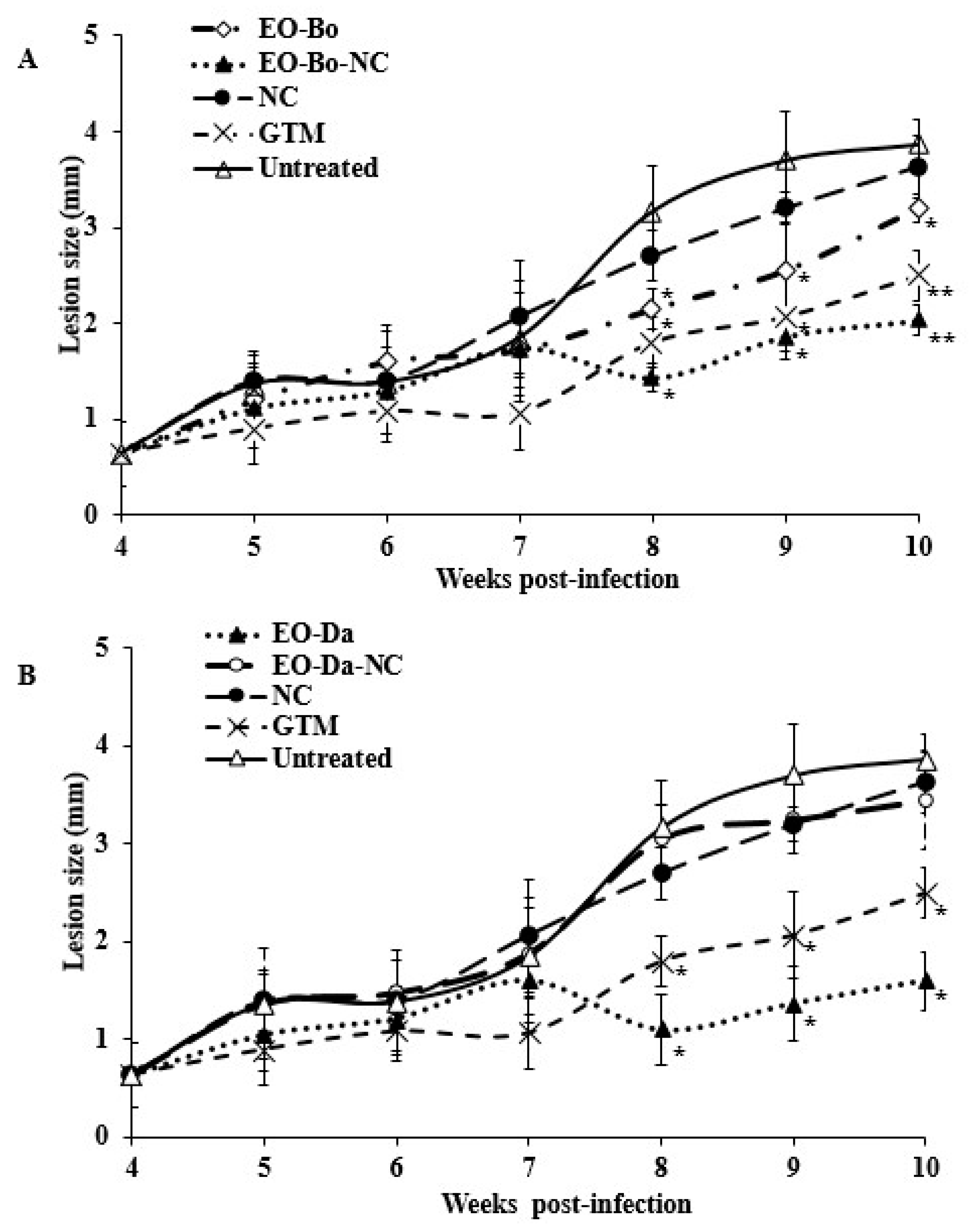
2.2. In Vivo Effect on Experimental Cutaneous Leishmaniasis

In the in vivo assay, treatment of mice with the EO-Bo-NC and EO-Da-NC (30 mg/kg/intralesional route/every 4 days/total of 4 doses) did not cause mortality or weight loss greater than 10% (Table 2).
The lesion size was measured in order to follow the disease evolution. With respect to EO-Bo (Figure 1A), the administration of EO-Bo-NC resulted in a smaller lesion size (p < 0.05), which was comparable to the effects of GTM (p > 0.05). However, mice that received EO-Da-NC showed larger lesion sizes (p < 0.05) compared with EO-Da and were similar (p > 0.05) to animals treated with NC and untreated mice (Figure 1B). Additionally, after administration of EO-Bo-NC, EO-Da-NC or NC, no successful cure was exhibited, considering that no differences (p > 0.05) with respect to parasite load were displayed (Log10 = 3.33–3.93 parasite/g).

3. Discussion

Despite the efforts, the absence of antileishmanial vaccines in the near future makes the discovering of effective chemotherapies an urgent need, especially for poor endemic regions [22]. Plant-derived compounds have accordingly come to represent a limitless source of chemical entities [23]. In particular, EOs have demonstrated a broad pharmacological spectrum [24] and several evidences have highlighted their leishmanicidal potential [9,10,25]. In particular, EOs from Cuban plants like B. orellana [11] and D. ambrosioides [12] have demonstrated their inhibitory activity on the in vitro growth of L. amazonensis and their ability to control the development of experimental CL in BALB/c mice. The aim of the present work was to study the in vitro and in vivo antileishmanial activity of nanocochleate formulations containing EO-Bo and EO-Da.
In order to demonstrate if the nanocochlear presentation was able to enhance the in vitro antileishmanial effect of EOs, the inhibitory activity was assayed. However, both nanocochleate formulations showed lower antileishmanial activity (p < 0.05). Considering that cochleates frequently delay the release of the encapsulated active principle [26], the action of EO-Bo-NC and EO-Da-NC may have been greatly compromised owing to the shorter exposure time routine used in these experiments.
However, with respect to the in vitro cytotoxic effects, incubation with EO-Bo-NC decreased the mortality of peritoneal macrophage compared to EO-Bo (p < 0.05). Previous works have demonstrated that, depending on the drug, the intracellular accumulation may vary [27]. The chemical profile of the EO products differs in the number and type of molecular stereochemical structures [24]. Therefore, despite the similar properties of the EOs, the different composition of them [11,14] in combination with cochleate precursors may have contributed to the contrary results observed on host cells for EO-Bo-NC with respect to EO-Da-NC.
Although several authors have highlighted the positive contributions of drug delivery systems against leishmaniasis [20,26], only a few works have focused on the use of cochleates. Nevertheless, some studies have been found in scientific literature, including: (i) the oral administration of AmB-loaded cochleates in L. donovani infected mice [28,29], (ii) the encochleation of AmB against L. chagasi [30] and (iii) the proteoliposome-derived cochleates in a model of CL by L. major [31]. Recently, our research group evaluated the antileishmanial activity of the EO from Artemisia absinthium L. presented in a stable, tolerable and efficacious nanocochleate formulation [32].
Promising results were obtained after administration of EO-Bo-NC preparations in the model of CL in BALB/c mice. This effect could be attributed to the ability of the cochleate membrane to fuse with cellular and subcellular membranes, which facilitated the arrival of encochleated material to the cytosol or to parasitophorous vacuoles to directly attack the pathogen [33]. In this process, several events can take place after administration of encochleated bioactive molecules, including: (i) drug accumulation, (ii) drug delayed release in a sustained manner, (iii) cochleate and (sub)cellular membrane fusion and (iv) cochleate clearance through the reticuloendothelial system. These events can contribute to directing the antileishmanial agent to the parasite host cell location, and to reducing unwanted side effects, which is desirable for effective treatment of infection due to L. amazonensis [20,24,30].
Nevertheless, the encochleation of a complex mixture of bioactive compounds like EOs may lead to different delivery rates for the compounds encapsulated. The LBDSs represent a reservoir, where the release of EO components can mainly happen through different ways: (i) dissolution, (ii) desorption and/or (iii) diffusion [34]. Some studies have also demonstrated that the EO release can take place in two phases. In the first phase, the molecules liberated are those located in the external wall, while in the second phase, the freed compounds are those present in the internal space [35]. In general, the EOs can include up to 60 components at different concentrations [24]. This fact may contribute to creating a release competition among them, which can super retard the delivery of drugs of concern like ishwarane in EO-Bo or ascaridole in EO-Da. In this sense, the multilamellar aspect of cochleates facilitates the separate inclusion of lipophilic and hydrophilic compounds in the internal spaces [36].
Infection with L. amazonensis, a major cause of CL in South America, is often associated with non-healing dermal lesions in people. In mice, the progress to ulcerated wounds is attributed to the extreme anergic pathogenicity that distinguished L. amazonensis amastigotes replication [37]. In the reasoning of effective and less toxic therapeutic alternatives, several ideas can be managed, including: (i) the increase of drug exposure time with minor doses, (ii) the drug transport to host cytosol and/or (iii) to parasitophorous vacuole. In the design formulation process, a possible way to fulfill these requirements contemplates the use of LBDS-like nanocochleates.

4. Materials and Methods

4.1. Essential Oils from B. orellana and D. ambrosioides

Fruits of B. orellana were collected on January 2015 in front of the Institute of Tropical Medicine Pedro Kouri, La Lisa, Havana (Cuba), and a specimen was deposited at the National Botanical Garden, Havana, Cuba, with a voucher specimen of NGB-9600288. Plants of D. ambrosioides were collected on July 2014 in Finca la Quiruvina”, Caimito, Artemisa (Cuba), and the specimen was deposited at the Experimental Station of Medicinal Plants “Dr. Juan Tomás Roig” with a specimen number of No. 4639.
The EO was obtained from fresh seeds and aerial parts (manually crushed) of B. orellana and D. ambrosioides, respectively, by hydrodistillation using Clevenger-type equipment over a 4-h period. Chemical constituents of obtained EO were corroborated by gas chromatography coupled with mass spectrometry (GC-MS) as previously reported [11,14], and compared. EO-Bo and EO-Da were hermetically sealed and stored in the Natural Product Collection of the Institute of Tropical Medicine Pedro Kouri under standard conditions (4 °C and darkness). For the biological assays, the EOs were dissolved in dimethyl sulfoxide (DMSO) at 20 mg/mL.

4.2. Reference Drug

Glucantime® (GTM) from Rhône-Poulenc Rorer, Mexico, at a concentration of 30 mg/mL, was used as reference drug and dissolved in sterile saline solution to biological assays.

4.3. Nanocochleate Formulations

The dehydration–hydration modified method was employed to prepare nanocochleates (NC) containing EO-Bo or EO-Da, as previously described [32]. A suspension of opalescence aspect was obtained, in correspondence with the formation of small spherical vesicles like liposomes. The next step was filtering under sterile conditions through 0.2 μm, and enough CaCl2 was added to a final concentration of 10 mM in the formulation. For the preparation of empty cochleates (NC), a similar process was followed, but without the incorporation of the EO component. Lastly, the preparations were stored in amber flasks at 4 °C. In parallel, a sample of each NC was used to corroborate physical parameters, which was carried out by Electrophoretic Light Scattering, using Delsa™-Nano C (Beckman Coulter, Sweden), with a detector of 165 degrees. The particle size of EO-Bo-NC and EO-Da-NC varied between 52.3 and 96.1 nm. while that of the NC formulation was inferior to 40 nm. The polydispersion index of EO-Bo-NC and EO-Da-NC was between 0.325 and 0.335, whereas that of the NC preparation was between 0.44 and 0.52. Considering the difference of particle motility induced by different magnetic fields, the zeta potential for EO-Bo-NC and EO-Da-NC ranged from 40.4 to 41.2 mV and for NC between 31.1 and 31.3 mV. Finally, gas-chromatography with mass spectrometer detector demonstrated that no volatile components were identified in the supernatant of the NCs. From each formulation, a final volume of 3 mL was obtained, stored at 4 °C in an amber hermetically-sealed flask and used to carry out in vitro and in vivo studies.

4.4. Parasites

The strain MHOM/77BR/LTB0016 of L. amazonensis, donated by the Department of Immunology, Oswaldo Cruz Foundation (FIOCRUZ), Brazil, was used. Parasites were routinely isolated from an infected BALB/c mouse and directly cultivated in Schneider’s medium (Sigma-Aldrich, St. Louis, MO, USA) containing 10% heat-inactivated fetal bovine serum (HFBS, Sigma-Aldrich) and antibiotics (100 μg of streptomycin/mL and 100 U of penicillin/mL). Parasites were maintained as promastigotes at 26 °C with passages every 3 or 4 days and used between 5 and 10 in vitro passages to all experiments.

4.5. Animals

The animals were provided by The National Center of Laboratory Animals Production (CENPALAB), Cuba, and maintained under standard conditions. Forty-eight female BALB/c mice with a body weight from 20 to 22 g were used according to “Guideline on the Care and Use of Laboratory Animals”, which was approved by the Ethics Committee from the Institute of Tropical Medicine Pedro Kouri (CEI-IPK 14-12), Havana, Cuba.

4.6. Antiamastigote Assay

In order to evaluate the in vitro performance of EO-Bo-NC and EO-Da-NC, the antileishmanial activity facing intracellular amastigotes of L. amazonensis was evaluated according to Torres-Santos et al. [38] as follows. Briefly, peritoneal macrophages from normal BALB/c mice were obtained in RPMI medium (SIGMA, St. Louis, MO, USA) and antibiotics, seeded in 24-well Lab-Tek (Costar®, New York, NY, USA) plates and incubated at 37 °C and 5% CO2. After 2 h, free cells were removed, and a culture of stationary-phase L. amazonensis promastigotes was added at a 4:1 parasite/macrophage ratio in the same medium supplemented with 10% HFBS. The plate was incubated again for 4 h under the same conditions, and free parasites were discarded. After that, 1 mL of medium was added to the wells, and then 990 µL of medium containing 10 µL of products was added in the first wells. Serial dilutions 1:2 were performed. Untreated control, as well as cultures treated with NC or EO, were also included. The plate was incubated for an additional 48 h under the same conditions. The supernatant was then removed, the culture was washed, fixed with absolute methanol, stained with Giemsa and examined under a light microscope (Motic, Hong Kong, China) with immersion oil at 1000 ×. The infection rates were obtained by multiplying the percentage of infected macrophages by the number of amastigotes, counting in 25 macrophages per sample. Percent of inhibition of the infection rate in comparison to those of the controls was calculated. Two replicates were carried out to each product, and medium inhibitory concentration (IC50) was obtained from lineal regression curves. Results were expressed as medium of IC50 and standard deviation.

4.7. Cytotoxicity Assay

The cytotoxicity assay was carried out using the MTT method [39]. Briefly, peritoneal macrophages from BALB/c mice were collected, seeded at 3 × 10⁵ cells/mL and incubated in 96-well Lab-Tek (Greiner bio-one, Frichenhausen, Germany) at 5% CO2 and 37 °C. After 2 h, the medium was removed and 50 µL of fresh medium with 10% HFBS and antibiotics were added, with an additional 48 µL in the first wells and 2 µL of each product. An untreated control, as well as a culture treated with NC or EO alone, were also included. Then, five serial dilutions (1:2) were carried out, and an additional 50 µL of medium was added to each well. The plate was incubated at the same conditions for 48 h. Then, 15 µL of 3-[4,5-dimethylthiazol-2-yl]-2,5-diphenyltetrazolium bromide (MTT) (SIGMA, St. Louis, MO, USA) (5 mg/mL) was added to each well, and the plate was incubated under the same conditions. After 4 h, the formazan crystals were dissolved with 100 µL of DMSO, and the optical density was measured at 560 nm and 630 nm as a reference wavelength using a spectrophotometer (Sirio S Reader, 2.4-0, Seac and Radim Diagnostics, Calenzano, Italy). In each case, the medium cytotoxic concentration (CC50) was determined from lineal dose-response curves of three experiments. Results are expressed as means of CC50 with respective standard deviations.

4.8. In Vivo Studies

On day 0, female healthy BALB/c mice were infected by subcutaneous route in the right hind footpad with 5 × 10⁶ stationary-phase L. amazonensis promastigotes. After 4 weeks post-infection (p.i.), animals were randomly divided into seven groups of eight mice each, and the treatment was started. Products (EO-Bo, EO-Bo-NC, EO-Da, EO-Da-NC, NC and GTM) were administered by intralesional route at a dose of 30 mg/kg in a volume of 50 µL every 4 days to complete 4 administrations. An additional group did not receive any treatment, identified as untreated or control. Between 4 and 10 weeks p.i., deaths were registered daily, body weight was determined by group every 7 days, and the lesion size was determined weekly using a digital caliper, by measuring footpad swelling of the lesion diameter. Average lesion size for each group was calculated as the mean of the differences observed among infected and uninfected footpads. On week 6 and 10 p.i., three animals of each group were sacrificed by cervical dislocation, and parasite burden was determined, employing the culture microtitration method in 96-well plates according to Buffet et al. [40]. Briefly, a sample of the infected area in the footpad was excised, weighed and homogenized in 4 mL of Schneider’s medium. A serial four-fold dilution was prepared under sterile conditions in 96-well plates and was incubated at 26 °C for 7 days. Then, the plates were examined with an inverted microscope (Olympus, Japan). The final titer was defined as the last dilution for which the well contained at least one parasite. The parasite burden was calculated as the geometric mean of reciprocal titers from each duplicate/weight of homogenized cross-section × 400.

4.9. Statistical Analysis

Statistical differences, considered as p < 0.05, between the products in the in vitro assays, were determined using the Mann–Whitney test. In the mouse model, the lesion evolution and parasite load were processed by the Variance Analysis Test, accompanied by a Post Hoc Test (LDS test or planned comparison). The Statistica for Windows Program (Release 4.5, StatSoft, Inc. 1993) was used in all cases.

5. Conclusions

In conclusion, further studies aiming to explore the possible interactions occurring as a result of EO-Da encapsulation should be carried out in order to understand the moderated effects observed after EO-Da-NC treatment. Nevertheless, the present work demonstrated that the nanoencochleation of oily active principles like EO-Bo can be proposed for better control of leishmaniasis. Therefore, these encouraging findings may become a new starting point for the development of alternatives for CL treatment.

Author Contributions

Conceptualization, B.T. and L.M.; Investigation, L.M., B.T., A.P., R.C.A., R.S. and L.M.; supervision, L.M.; Writing—Original draft, L.M., W.N.S. and L.M.; Writing—Review and editing, L.M., B.T., A.P., R.C.A., R.S., W.N.S. and L.M.

Funding

This research received no external funding.

Acknowledgments

We are grateful to the Department of Immunology, Oswaldo Cruz Foundation (FIOCRUZ), Brazil, for the generous donation of the Leishmania amazonensis parasites. This study was performed as part of the activities of the Research Network Natural Products against Neglected Diseases (ResNet NPND) (http://www.resnetnpnd.org/Start/).

Conflicts of Interest

The authors declare no conflicts of interest.

References

  1. Steverding, D. The history of leishmaniasis. Parasit. Vectors 2017, 10, 82. [Google Scholar] [CrossRef] [PubMed]
  2. Kevric, I.; Cappel, M.A.; Keeling, J.H. New world and old world leishmania infections: A practical review. Dermatol. Clin. 2015, 33, 579–593. [Google Scholar] [CrossRef] [PubMed]
  3. Torres-Guerrero, E.; Quintanilla-Cedillo, M.R.; Ruiz-Esmenjaud, J.; Arenas, R. Leishmaniasis: A review. F1000Research 2017, 6, 750. [Google Scholar] [CrossRef] [PubMed]
  4. Monzote, L. Antileishmanial patents antileishmanial current drugs and relevant patents. Recent Pat. Antiinfect. Drug Discov. 2011, 6, 1–26. [Google Scholar] [CrossRef]
  5. Andrews, K.T.; Fisher, G.; Skinner-Adams, T.S. Drug repurposing and human parasitic protozoan diseases. Int. J. Parasitol. Drugs Drug Resist. 2014, 4, 95–111. [Google Scholar] [CrossRef] [PubMed]
  6. Lehnhardt Pires, C.; Rodrigues, S.D.; Bristot, D.; Gaeta, H.H.; de Oliveira Toyama, D.; Farias, W.R.L.; Toyama, M.H. Evaluation of macroalgae sulfated polysaccharides on the Leishmania (L.) amazonensis promastigote. Mar. Drugs 2013, 11, 934–943. [Google Scholar] [CrossRef]
  7. Armeli Minicante, S.; Michelet, S.; Bruno, F.; Castelli, G.; Vitale, F.; Sfriso, A.; Morabito, M.; Genovese, G. Bioactivity of phycocolloids against the mediterranean protozoan Leishmania infantum: An inceptive study. Sustainability 2016, 8, 1131. [Google Scholar] [CrossRef]
  8. Ogungbe, I.V.; Singh, M.; Setzer, W.N. Antileishmanial natural products from plants. Stud. Nat. Prod. Chem. 2012, 36, 331–381. [Google Scholar]
  9. Rodrigues, I.A.; Mazotto, A.M.; Cardoso, V.; Alves, R.L.; Amaral, A.C.F.; de Andrade Silva, J.R.; Pinheiro, A.S.; Vermelho, A.B. Natural products: Insights into Leishmaniasis inflammatory response. Mediat. Inflamm. 2015, 2015, 1–12. [Google Scholar] [CrossRef]
  10. Monzote, L.; Herrera, I.; Satyal, P.; Setzer, W.N. In-Vitro evaluation of 52 commercially-available essential oils against Leishmania amazonensis. Molecules 2019, 24, 1248. [Google Scholar] [CrossRef]
  11. Monzote, L.; Garcia, M.; Scull, R.; Cuellar, A.; Setzer, W.N. Antileishmanial activity of the essential oil from Bixa orellana. Phyther. Res. 2014, 28, 753–758. [Google Scholar] [CrossRef] [PubMed]
  12. Monzote, L.; García, M.; Pastor, J.; Gil, L.; Scull, R.; Maes, L.; Cos, P.; Gille, L. Essential oil from Chenopodium ambrosioides and main components: Activity against Leishmania, their mitochondria and other microorganisms. Exp. Parasitol. 2014, 136, 20–26. [Google Scholar] [CrossRef] [PubMed]
  13. Monzote, L.; García, M.; Montalvo, A.M.; Linares, R.; Scull, R. Effect of oral treatment with the essential oil from Chenopodium ambrosioides against cutaneous leishmaniasis in BALB/c mice, caused by Leishmania amazonensis. Complement. Med. Res. 2009, 16, 334–338. [Google Scholar] [CrossRef] [PubMed]
  14. Monzote, L.; Pastor, J.; Scull, R.; Gille, L. Antileishmanial activity of essential oil from Chenopodium ambrosioides and its main components against experimental cutaneous leishmaniasis in BALB/c mice. Phytomedicine 2014, 21, 1048–1052. [Google Scholar] [CrossRef]
  15. Turek, C.; Stintzing, F.C. Stability of essential oils: A review. Compr. Rev. Food Sci. Food Saf. 2013, 12, 40–53. [Google Scholar] [CrossRef]
  16. Martinho, N.; Damgé, C.; Reis, C.P. Recent advances in drug delivery systems. J. Biomater. Nanobiotechnol. 2011, 2, 510–526. [Google Scholar] [CrossRef]
  17. Papahadjopoulos, D.; Vail, W.J.; Jacobson, K.; Poste, G. Cochleate lipid cylinders: Formation by fusion of unilamellar lipid vesicles. Biochim. Biophys. Acta (BBA)-Biomembr. 1975, 394, 483–491. [Google Scholar] [CrossRef]
  18. Delmarre, D.; Lu, R.; Tatton, N.; Krause-Elsmore, S.; Gould-Fogerite, S.; Mannino, R.J. Formulation of hydrophobic drugs into cochleate delivery vehicles: A simplified protocol & formulation kit. Drug Deliv. Technol. 2004, 4, 64–69. [Google Scholar]
  19. Rawat, M.; Singh, D.; Saraf, S.; Saraf, S. Lipid carriers: A versatile delivery vehicle for proteins and peptides. Yakugaku Zasshi 2008, 128, 269–280. [Google Scholar] [CrossRef]
  20. Romero, E.L.; Morilla, M.J. Drug delivery systems against leishmaniasis? Still an open question. Expert Opin. Drug Deliv. 2008, 5, 805–823. [Google Scholar] [CrossRef]
  21. Smith, D.M.; Simon, J.K.; Baker, J.R. Applications of nanotechnology for immunology. Nat. Rev. Immunol. 2013, 13, 592–605. [Google Scholar] [CrossRef] [PubMed]
  22. Iborra, S.; Solana, J.C.; Requena, J.M.; Soto, M. Vaccine candidates against Leishmania under current research. Expert Rev. Vaccines 2018, 17, 323–334. [Google Scholar] [CrossRef] [PubMed]
  23. Oryan, A. Plant-derived compounds in treatment of leishmaniasis. Iran. J. Vet. Res. 2015, 16, 1–19. [Google Scholar] [PubMed]
  24. Bilia, A.R.; Guccione, C.; Isacchi, B.; Righeschi, C.; Firenzuoli, F.; Bergonzi, M.C. Essential oils loaded in nanosystems: A developing strategy for a successful therapeutic approach. Evidence-Based Complement. Altern. Med. 2014, 2014, 651593. [Google Scholar] [CrossRef]
  25. Islamuddin, M.; Chouhan, G.; Want, M.Y.; Tyagi, M.; Abdin, M.Z.; Sahal, D.; Afrin, F. Leishmanicidal activities of Artemisia annua leaf essential oil against visceral Leishmaniasis. Front. Microbiol. 2014, 5, 626. [Google Scholar] [CrossRef]
  26. Kayser, O.; Olbrich, C.; Croft, S.L.; Kiderlen, A.F. Formulation and biopharmaceutical issues in the development of drug delivery systems for antiparasitic drugs. Parasitol. Res. 2003, 90, S63–S70. [Google Scholar] [CrossRef]
  27. Maurin, M.; Raoult, D. Bartonella (Rochalimaea) quintana infections. Clin. Microbiol. Rev. 1996, 9, 273–292. [Google Scholar] [CrossRef]
  28. Santangelo, R.; Paderu, P.; Delmas, G.; Chen, Z.W.; Mannino, R.; Zarif, L.; Perlin, D.S. Efficacy of oral cochleate-amphotericin B in a mouse model of systemic candidiasis. Antimicrob. Agents Chemother. 2000, 44, 2356–2360. [Google Scholar] [CrossRef]
  29. Thornton, S.J.; Wasan, K.M. The reformulation of amphotericin B for oral administration to treat systemic fungal infections and visceral leishmaniasis. Expert Opin. Drug Deliv. 2009, 6, 271–284. [Google Scholar] [CrossRef]
  30. Sesana, A.M.; Monti-Rocha, R.; Vinhas, S.A.; Morais, C.G.; Dietze, R.; Lemos, E.M. In vitro activity of amphotericin B cochleates against Leishmania chagasi. Mem. Inst. Oswaldo Cruz 2011, 106, 251–253. [Google Scholar] [CrossRef]
  31. Pérez, O.; Bracho, G.; Lastre, M.; Mora, N.; Del Campo, J.; Gil, D.; Zayas, C.; Acevedo, R.; González, D.; López, J.A.; et al. Novel adjuvant based on a proteoliposome-derived cochleate structure containing native lipopolysaccharide as a pathogen-associated molecular pattern. Immunol. Cell Biol. 2004, 82, 603–610. [Google Scholar] [CrossRef] [PubMed]
  32. Tamargo, B.; Monzote, L.; Piñón, A.; Machín, L.; García, M.; Scull, R.; Setzer, W.N. In vitro and in vivo evaluation of essential oil from Artemisia absinthium L. formulated in nanocochleates against cutaneous leishmaniasis. Medicines 2017, 4, 38. [Google Scholar] [CrossRef] [PubMed]
  33. Syed, U.M.; Woo, A.F.; Plakogiannis, F.; Jin, T.; Zhu, H. Cochleates bridged by drug molecules. Int. J. Pharm. 2008, 363, 118–125. [Google Scholar] [CrossRef]
  34. Soppimath, K.S.; Aminabhavi, T.M.; Kulkarni, A.R.; Rudzinski, W.E. Biodegradable polymeric microparticles as drug delivery devices. J. Control. Release 2015, 49, 187–190. [Google Scholar]
  35. Gomes, C.; Moreira, R.G.; Castell-Perez, E. Poly (DL-lactide-co-glycolide) (PLGA) nanoparticles with entrapped trans-cinnamaldehyde and eugenol for antimicrobial delivery applications. J. Food Sci. 2011, 76, N16–N24. [Google Scholar] [CrossRef] [PubMed]
  36. Bhosale, R.R.; Ghodake, P.P.; Mane, A.N.; Ghadge, A.A. Nanocochleates: A novel carrier for drug transfer. J. Sci. Innov. Res. 2013, 2, 964–969. [Google Scholar]
  37. Soong, L.; Henard, C.A.; Melby, P.C. Immunopathogenesis of non-healing American cutaneous leishmaniasis and progressive visceral leishmaniasis. Semin. Immunopathol. 2012, 34, 735–751. [Google Scholar] [CrossRef] [Green Version]
  38. Torres-Santos, E.C.; Moreira, D.L.; Kaplan, M.A.C.; Meirelles, M.N.; Rossi-Bergmann, B. Selective effect of 2′,6′-dihydroxy-4′-methoxychalcone isolated from Piper aduncum on Leishmania amazonensis. Antimicrob. Agents Chemother. 1999, 43, 1234–1241. [Google Scholar] [CrossRef] [Green Version]
  39. Sladowski, D.; Steer, S.J.; Clothier, R.H.; Balls, M. An improved MTT assay. J. Immunol. Methods 1993, 157, 203–207. [Google Scholar] [CrossRef]
  40. Buffet, P.A.; Sulahian, A.; Garin, Y.J.F.; Nassar, N.; Derouin, F. Culture microtitration: A sensitive method for quantifying Leishmania infantum in tissues of infected mice. Antimicrob. Agents Chemother. 1995, 39, 2167–2168. [Google Scholar] [CrossRef] [Green Version]
Sample Availability: Samples are no longer available.
Figure 1. Effect of treatment with essential oil from Bixa orellana L. (A) and Dysphania ambrosioides (L.) Mosyakin & Clemants (B), in comparison with their nanocochleate formulations. BALB/c mice were infected with L. amazonensis, and 30 days post-infection the treatment was started with 4 doses by intralesional route at 30 mg/kg every four days. The results are expressed as the mean of lesion size in infected area ± standard deviation. EO-Bo: Essential oil from B. orellana; EO-Bo-NC: Essential oil from B. orellana formulated in nanocochleates. EO-Da: Essential oil from D. ambrosioides. EO-Da-NC: Essential oil from D. ambrosioides formulated in nanocochleates. NC: Empty nanocochleates. GTM: Glucantime® used as reference drug. *: Displays statistical differences (p < 0.05) compared to untreated control and animals treated with NC. **: Displays statistical differences (p < 0.05) compared to animals treated with the respective EO.
Molecules 24 04222 g001
Table 1. In vitro antileishmanial activity (IC50) and cytotoxic effect (CC50) of the essential oils from Bixa orellana L. and Dysphania ambrosioides (L.) Mosyakin & Clemants and their nanocochleate formulations.
ProductsIC50a ± SDb (μg/mL)CC50c ± SDb (μg/mL)Selectivity Index
EO-Bo8.5 ± 0.861.8 ± 5.97
EO-Bo-NC15.4 ± 1.3*94.6 ± 2.2*6
EO-Da4.9 ± 1.157.9 ± 3.712
EO-Da-NC>60*46.9 ± 4.4*-
GTM11.0 ± 3.4>1500>136
NC~25% of infection at maximum volume tested.~70% of mortality at maximum volume tested.-
a IC50: Concentration of product that caused 50% of inhibition growth. b SD: Standard deviation. c CC50: Concentration of product that caused 50% of macrophage mortality. EO-Bo: Essential oil from B. orellana. EO-Bo-NC: Essential oil from B. orellana formulated in nanocochleates. EO-Da: Essential oil from D. ambrosioides. EO-Da-NC: Essential oil from D. ambrosioides formulated in nanocochleates. GTM: Glucantime®. NC: Empty nanocochleates. *: Displays statistical differences (p < 0.05) compared to respective EO.
Table 2. Variations of body weight of animals infected with L. amazonensis and divided by groups of mice (n = 8) treated with the essential oils from Bixa orellana L. and Dysphania ambrosioides (L.) Mosyakin & Clemants and their nanocochleate formulations.
Group of Animals (Number of Animals)Average ofVariation of Body Weight (%)a
Initial Weight (g)5 w.p.i.6 w.p.i.7 w.p.i.8 w.p.i.9 w.p.i.10 w.p.i.
EO-Bo (8)19.2+8.1+9.4+10.7+12.7+13.2+16.4
EO-Bo-NC (8)19.0+3.5+4.6+7.9+8.9+9.3+9.5
EO-Da (8)19.3+3.1+3.7+3.5+7.3+5.9+11.8
EO-Da-NC (8)19.5+0.3+3.1+3.3+10.7+12.2+11.2
NC (8)19.7−2.1+0.3+4.6+8.0+8.6+8.6
GTM (8)19.2+2.9+6.3+5.5+8.6+8.4+9.5
Untreated (8)19.3+4.0+3.7+5.8+6.4+6.3+8.4
a Positive number represents an increase of body weight, while negative number indicats a decrease of body weight, with respect to initial body weight at week 4 post-infection. EO-Bo: Essential oil from B. orellana. EO-Bo-NC: Essential oil from B. orellana formulated in nanocochleates. EO-Da: Essential oil from D. ambrosioides. EO-Da-NC: Essential oil from D. ambrosioides in nanocochleates. NC: Empty nanocochleates. GTM: Glucantime® used as reference drug. w.p.i.: Weeks post infection.

