Article

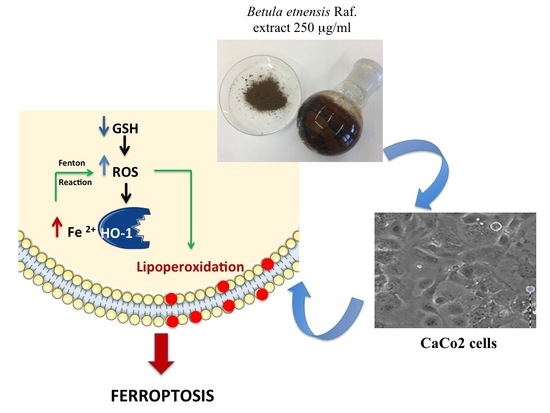
Betula etnensis Raf. (Betulaceae) Extract Induced HO-1 Expression and Ferroptosis Cell Death in Human Colon Cancer Cells

by
Giuseppe Antonio Malfa
1,†,
Barbara Tomasello
1,†,
Rosaria Acquaviva
1,*,
Carlo Genovese
2,
Alfonsina La Mantia
1,
Francesco Paolo Cammarata
3,4,
Monica Ragusa
5,
Marcella Renis
1 and
Claudia Di Giacomo
1
1
Department of Drug Science, Section of Biochemistry, University of Catania, Viale A. Doria 6, 95125 Catania, Italy
2
Department of Biomedical and Biotechnological Sciences, Microbiology Section University of Catania, 95125 Catania, Italy
3
Institute of Bioimaging and Molecular Physiology, National Council of Research (IBFM-CNR), 90015 Cefalù (PA), Italy
4
National Institute of Nuclear Physics, South National Laboratory (LNS-INFN), 95125 Catania, Italy
5
Department of Experimental and Clinical Medicine, University Magna Graecia of Catanzaro, 88100 Catanzaro, Italy
*
Author to whom correspondence should be addressed.
These authors contributed equally to this work.
Int. J. Mol. Sci. 2019, 20(11), 2723; https://doi.org/10.3390/ijms20112723
Submission received: 30 April 2019 / Revised: 23 May 2019 / Accepted: 31 May 2019 / Published: 3 June 2019
(This article belongs to the Special Issue Protective and Detrimental Role of Heme Oxygenase-1)

Abstract

:
Betula etnensis Raf. (Birch Etna) belonging to the Betulaceae family grows on the eastern slope of Etna. Many bioactive compounds present in Betula species are considered promising anticancer agents. In this study, we evaluated the effects of B. etnensis Raf. bark methanolic extract on a human colon cancer cell line (CaCo2). In order to elucidate the mechanisms of action of the extract, cellular redox status, cell cycle, and heme oxygenase-1 (HO-1) expression in ferroptosis induction were evaluated. Cell viability and proliferation were tested by tetrazolium (MTT) assayand cell cycle analysis, while cell death was evaluated by annexin V test and lactic dehydrogenase (LDH) release. Cellular redox status was assessed by measuring thiol groups (RSH) content, reactive oxygen species (ROS) production, lipid hydroperoxide (LOOH) levels and (γ-glutamylcysteine synthetase) γ-GCS and HO-1 expressions. The extract significantly reduced cell viability of CaCo2, inducing necrotic cell death in a concentration-depending manner. In addition, an increase in ROS levels and a decrease of RSH content without modulation in γ-GCS expression were detected, with an augmentation in LOOH levels and drastic increase in HO-1 expression. These results suggest that the B. etnensis Raf. extract promotes an oxidative cellular microenvironment resulting in CaCo2 cell death by ferroptosis mediated by HO-1 hyper-expression.

Graphical Abstract

1. Introduction

After prostate cancer in men, breast cancer in women and lung cancer, the colorectal cancer (CRC) represents the second leading cause of death in the Western world in both males and females. The association between nutrition and colon cancer has extensively been investigating by many studies but is controversial because of the diet shows a causal and protective role in the CRC development [1,2].
It is well known that the consumption of vegetables is correlated with a low incidence of cancer and, in particular, is effective in the prevention and reduction of CRC risk [3,4]. A plausible reason might be plant foodstuff is a good source of fibers, folate vitamins, various antioxidants, and other bioactive compounds including polyphenols, terpenoids, saponins, and carotenoids [5,6,7]. Besides their well-known anti-oxidant activities, they have been reported to be anti-mutagenic and/or anti-carcinogenic and to possess several other biological activities. In addition, natural antioxidants are known to have a dual face, behaving as pro-oxidant compounds after reacting directly with reactive oxygen species (ROS) in the presence of transition metal ions, such as copper and iron. This increase of cellular ROS to cytotoxic level may generate secondary oxidative damage and induce a selective killing of cancer cells by a variety of ways including ferroptosis, a non-apoptotic form of cell death characterized by the high expression of heme oxygenase-1 (HO-1) and accumulation of lipid hydroperoxides (LOOH) [8]. The role of heme oxygenase-1 (HO-1) in cancer biology is poorly understood. In fact, HO-1 has been described as survival molecule because of its anti-apoptotic and pro-angiogenic effects in several cancer types and its modulation can be induced by several natural compounds such as polyphenols and terpenoids [5].
Today potential usefulness of natural compounds as anti-cancer agents has to be ascribed prevalently to improve patient quality of life and to support conventional chemotherapy or radiotherapy. In the genus Betula (Betulaceae) there belongs common trees and shrubs of the boreal and north temperate zones. The bioactivities of Betula species are well documented, in fact the Betula was largely used in human and veterinary medicine to treat various diseases. Betula compounds have a wide variety of properties including anticancer, anti-inflammatory, and immunomodulatory beyond being antioxidant [9,10,11].
Currently, of particular interest are anticancer activities showed in vitro by some constituents isolated from these plants, which displayed cytotoxic effects on neuroblastoma, melanoma, medulloblastoma, and Ewing’s sarcoma cells [10,12]. In particular, it has been reported that betulinic acid may be considered a promising anticancer agent [10,13]. Commonly known as Birch Etna, B. etnensis Raf. is a medium-sized deciduous tree typically reaching 5–20 m tall, which belongs to the family Betulaceae. It grows on Mt. Etna volcano, at an altitude between 1200 and 2000 m and its ivory colored bark, in particular in the young branches, is rich of resinous chemicals secreted by the numerous glands present [14]. Polyphenols, terpenoids, betulin, betulinic acid, and ursolic acid are the main constituents present in the B. etnensis Raf. bark. As for as our knowledge no literature data are present on biological activities of B. etnensis Raf.
In the present study, we evaluated, for the first time, the effects of B. etnensis Raf. bark methanolic extract on a human colon cancer cell line (CaCo2). In order to elucidate the mechanisms of action of this extract, cell viability, annnexin, lactic dehydrogenase (LDH) release, ROS production, thiol groups (RSH) content, lipid peroxidation (LOOH) levels, cell cycle, HO-1, and γ-GCS protein expression were evaluated.

2. Results

2.1. MTT Bioassay

Methanolic extract of bark from B. etnensis Raf. (5, 50, 250, or 500 µg/mL) was able to significantly reduce in a dose dependent manner cell viability (Figure 1); because 250 and 500 µg/mL of methanolic extract showed similar effects, in the other subsequent experiments we omitted 500 µg/mL of extract.

2.2. LDH Release

As shown in Figure 2, after 72 h of incubation with methanolic extract of bark from B. etnensis Raf. a statistically significant increase in LDH release was observed in CaCo2 cells treated with 50 and 250 µg/mL of extract.

2.3. Annexin V and Dead Cell Evaluation

A slight non-significant increase in the percentage of total apoptotic cells from 9.20 ± 1.25% to 11.75 ± 0.40% after treatment with 50–250 µg/mL of the extract was observed (Table 1). The annexin-V/7-AAD results confirm that most of antiproliferative activity of B. etnensis Raf. observed by viability assay is mediated by necrosis.

2.4. ROS Levels

Data reported in Figure 3 demonstrate that exposure of CaCo2 cells to several concentrations of B. etnensis Raf. methanolic extracts resulted in a significant increase in radical species, as revealed by fluorescence intensity.

2.5. LOOH Levels

Figure 4 shows that the addition of methanolic extract of B. etnensis Raf. at 5, 50, and 250 μg/mL for 72 h induced a significant and dose-dependent increase in LOOH levels with respect to untreated CaCo2 cells.

2.6. Total Thiol Groups

Treatment of cells with 50–250 µg/mL methanolic extract of B. etnensis Raf. resulted in a significant reduction in RSH levels (Figure 5). Instead, the lowest concentration of methanolic extract of B. etnensis Raf. did not alter the levels of RSH groups with respect to the control.

2.7. γ-GCS Determination

No significant change in γ-GCS expression was observed in CaCo2 cells treated with the extract of B. etnensis Raf. with respect to untreated cells (Figure 6).

2.8. HO-1 by ELISA

Results reported in Figure 7 show that the addition of 250 μg/mL of extract to CaCo2 cells, caused a significant increase in HO-1 protein expression (Figure 7).

2.9. Cell Cycle Analysis

When CaCo2 cells were treated with increasing concentrations of B. etnensis Raf. for 72 h, a dose dependent cell cycle arrest at G0/G1 and S phases were evident with concomitant marked decrease in the G2/M phase. After the treatment with 50 μg/mL of B. etnensis Raf., cell population in G0/G1 and S phases increased by ~5.14% and ~16% respectively, whereas cells in G2/M were decreased by ~21% (Figure 8). The same trend was observed with 250 μg/mL of extract.

3. Discussion

Tumors of the digestive tract, particularly CRC, are among the most common forms of cancer, with thousands of deaths worldwide per year. Chemotherapy and radiation therapy are the main treatments with significant side effects. Among cancers, CRC is the most responsive to dietary modification, in fact, several studies demonstrated that approximatively 75% of all sporadic cases of CRC are directly influenced by diet and that dietary modification is a feasible strategy for reducing CRC risk [5,15,16]. The consumption of fruit and vegetables and herbs is associated with a low incidence of cancer [7,17] and this may be partly due to the presence of several bioactive natural compounds in plants [6].
Previous studies have shown that betulinic acid is cytotoxic for different types of human cancer cells [10,13,18]. In this study, we evaluated the anti-cancer activity of B. etnensis Raf. methanolic extract in CaCo2 cells.
In order to evaluate cytotoxic effects of the methanolic extract, we first conducted an MTT test. The results, showed in Figure 1, indicate that cell viability is significantly reduced by the extract. Cell viability reaches a plateau at 250 μg/mL with a 70% decrease and the same result was obtained with 500 μg/mL so the highest concentration of treatment used in next experiments was 250 μg/mL of B. etnensis Raf. methanolic extract.
In consideration of the results obtained on cell viability, we performed LDH assay in medium to evaluate cell membrane disruption which is accompanied by necrotic cells death. Results showed a dose-dependent necrotic effect with a maximum at 250 μg/mL in line with results obtained by MTT assay (Figure 2). These results were confirmed by annexin V levels, in fact, treatment with B. etnensis Raf. methanolic extract did not induce apoptotic cell death (Table 1). In order to verify the role of oxidative stress in the mechanism of cell death, the determination of ROS and LOOH was performed. Clearly, the treatments with B. etnensis Raf. extract induced an increase in ROS levels in CaCo2 tumor cell line, indicating that the extract does not act as an antioxidant agent, but rather exerts its cytotoxic effect as a pro-oxidant with concomitant increase lipid peroxidation particularly evident at 250 μg/mL (Figure 3 and Figure 4).
This result suggests that the extract may exert its activity by destabilizing the cellular redox balance so involving intracellular factors probable targets of ROS. Certainly the high production of ROS is a key factor that contributes to carcinogenesis. Within a neoplastic cell the persistent oxidative stress may be responsible for the activation of growth factor pathways and the increased resistance to apoptosis [19]. In fact, ROS have been shown to act as second messengers [20,21], stimulating the transduction of intracellular signals [22,23].
Recently, it has been suggested that GSH, as well as the main thiol responsible for maintaining the intracellular redox state, through thiol/disulfide exchange reactions, may be involved in the redox regulation of this type of signal [20,21,22,23,24].
In our study, the treatment of CaCo2 cells with the extract causes a dose-dependent reduction of intracellular RSH levels (Figure 5). To further confirm the involvement of the antioxidant defense systems in the mechanism of action of the extract, we also evaluated the expression of γ-GCS, a key enzyme in glutathione synthesis the expression of which in cancer cells is involved in tumor aggressiveness and chemo-resistance. The Western blot analysis of γ-GCS showed that the extract does not significantly influence the expression of the protein at all concentrations tested, suggesting that decreased RSH levels are most due to an inhibition of GSH synthesis and to a depletion for excessive ROS reactions (Figure 6). All these results confirms the persistence of a pro-oxidative imbalance induced by B. etnensis Raf. methanolic extract.
To further understand the mechanism of action of the extract, under the same experimental conditions, the expression of HO-1, one of the main cellular defense mechanisms against oxidative stress was evaluated. The role of HO-1 in tumors is still not very clear [25]. Results obtained in the present study show that the B. etnensis Raf. extract may exert a dual effect on HO-1 expression: at low concentration (5–50 μg/mL) it reduced HO-1 expression whereas, at higher concentrations (250 μg/mL) induced a significant increase in HO-1 expression (Figure 7). The reported inverse correlation between GSH levels and HO-1 expression is only confirmed at the highest concentration of the extract [26].
The HO-1 effect in cancer cells is not yet clear, but it is wildly documented that an HO-1 over-expression confers resistance to chemotherapy and radiation therapy. This protective effect could be due to its reaction products such as CO or biliverdin/bilirubin [27,28]. In spite of its implication in tumor initiation, angiogenesis, and metastasis, excessively increased expression of HO-1 in tumor cells may lead to cell death through a process called ferroptosis [29,30,31,32] In fact, a lot of evidence showed that HO-1 induces ferroptosis through an increase of ROS production mediated by iron accumulation [33,34,35] and accompanied by augmentation lipid peroxidation and glutathione depletion. Results obtained in the present study, at the highest concentration of the extract, may suggest a ferroptotic cell death.
The antiproliferative effect of extract was also demonstrated by the analysis of the cytometric flow, in fact the treatment induced cell cycle arrest at G0/G1 and S phases and a concomitant decrease in the G2/M phase.
Results obtained in the present research demonstrated that methanolic extract of B. etnensis Raf. by inducing ROS production and decreasing antioxidant cellular defense, elicited an imbalance in intracellular redox status. This effect, in turn, leads to a higher lipid peroxidation and a drastic rise in HO-1 activity, letting us to hypothesize the induction of ferroptosis, a non-programmed cell death.

4. Materials and Methods

4.1. Chemicals

All solvents, chemicals and reference compounds were purchased from Sigma-Aldrich (Milano, Italy) except as otherwise specified.

4.2. Plant Collection and Preparation of Extract

B. etnensis Raf. bark was collected in the area around Linguaglossa (Catania, Italy) in November 2018. The specimen was obtained and authenticated by botanist R. Acquaviva, Department of Drug Science, Section of Biochemistry, University of Catania, Italy. A voucher specimen of the plant (No. 36/03) was deposited in the herbarium of the same Department. Dried bark from B. etnensis Raf. (50 g) was extracted at 80 °C in 70% methanol for 4 h. The extract was then filtered and evaporated to dryness under reduced pressure with a rotatory evaporator. The values of total phenolic and flavonoid content were 45 µM ± 0.05 gallic acid and 3.80 µM ± 0.09 catechin.

4.3. Cell Culture and Treatments

CaCo2 colon rectal cancer cells were obtained from ATCC cell bank (Rockville, MD, USA), were propagated in DMEM (Gibco BRL, Life Technologies, city, country) supplemented with 10% fetal calf serum, 1% sodium pyruvate, 1% L-glutamine solution, and 1% streptomycin/penicillin.
After 24 h of incubation in a humidified atmosphere of 5% CO2 at 37 °C to allow cell attachment, the cells were treated for 72 h with different concentrations (5, 50, 100, 250 or 500 µg/mL) of the methanolic extract, previously dissolved in the minimum amount of dimethyl sulfoxide (DMSO) and diluted with medium.

4.4. MTT Bioassay

MTT assay was performed to assess cell viability on a 96 multiwell plate (8 × 10³ cells/well). This assay measures the conversion of tetrazolium salt to yield colored formazan in the presence of metabolic activity. The amount of formazan is proportional to the number of living cells [36]. The optical density was measured with a microplate spectrophotometer reader (Titertek Multiskan, Flow Laboratories, Helsinki, Finland) at λ = 570 nm. Results are expressed as percentage cell viability respect to control (untreated cells).

4.5. Lactic Dehydrogenase Release

LDH release was measured to evaluate cell necrosis as a result of cell membrane disruption. LDH activity was measured spectrophotometrically in the culture medium and in the cellular lysates, at λ = 340 nm by analyzing NADH reduction [5]. The percentage of LDH release was calculated from the total sum of the enzymatic activity present in the cellular lysate and of that in the culture medium. Results are expressed as percentage in LDH released.

4.6. Annexin V and Dead Cell Evaluation

The percentage of cells undergoing apoptosis after treatment with B. etnensis Raf. was evaluated by Muse™ Annexin V & Dead cell kit (Catalog No. MCH100105, Millipore, Milan, Italy) according to the manufacture’s protocol. Briefly for cells staining, 100 µL of the Muse™ Annexin V & Dead Cell Reagent was added to 100 µL of cell suspension, mixed thoroughly by vortexing and incubated for 20 min at room temperature in the dark. Then samples were analyzed by Muse™ Cell Analyzer (Millipore).

4.7. Reactive Oxygen Species Assay

Dichlorofluorescein diacetate (DCFH-DA) assay was performed to quantify ROS levels as previously described [37]. The fluorescence intensity was detected by fluorescence spectrophotometry (excitation, λ = 488 nm; emission, λ = 525 nm). Results are expressed as fluorescence intensity/mg protein and, for each sample the total protein content was determined using the Sinergy HTBiotech instrument by measuring the absorbance difference at λ = 280 and λ = 260. Protein content was determined using the Sinergy HTBiotech instrument by measuring the absorbance difference at λ = 280 and λ = 260.

4.8. Determination of Lipid Hydroperoxide Levels

LOOH levels were evaluated by oxidation of Fe+2 to Fe+3 that in the presence of xylenol orange forms a Fe3+-xylenol orange complex which can be measured spectrophotometrically at λ = 560 nm [38]. Results are expressed as percentage increase respect to control (untreated cells).

4.9. Total Thiol Group Determination

RSH groups were measured by using a spectrophotometric assay as previously described [39]. Results are expressed in nmol/mg protein.

4.10. Western Blotting

CaCo2 cells were harvested using cell lysis buffer; cell lysates were collected for Western blot analysis and the level of γ-GCS expression was visualized by immunoblotting with primary antibody against γ-GCS as previously described [5]. The anti- γ-GCS was used in 1:1000 dilution in TBST solution containing 5% not fat milk and detected with secondary antibody conjugated to horseradish peroxidase and Pierce ECL Plus substrate solution (Thermo Scientific, Rockford, IL, USA). Beta-actin, was used as a loading control to normalize the expression level of γ-GCS protein. The result was expressed in arbitrary densitometric units (ADU).

4.11. HO-1 Protein Expression

HO-1 protein concentration in cellular lysates was measured using a commercial HO-1 ELISA kit, according to the manufacturers’ instructions. A standard curve generated with purified HO-1 was used to calculate HO-1 concentration from the absorbance at λ = 450 nm detected for each sample [40]. Detection limits were 0.78–25 ng/mL as reported by the manufacturer. Results are expressed as ng/mg protein.

4.12. Cell Cycle Analysis

Distribution of cell cycle was evaluated by flow cytometry using Muse™ Cell Cycle Kit (Catalog No. MCH100106, Millipore, Milan, Italy) according to the manufacture’s guidelines. Briefly, CaCo2 cells were treated with different concentration of B. etnensis Raf. (5, 50, 250 μg/mL) for 72 h. After incubation, the cells were harvest, washed with PBS and fixed with ice-cold 70% ethanol at −20 °C for 24 h. Then 200 μL of cell suspension (1 × 106 cells) was washed with PBS, stained with 200 μL of Muse™ Cell Cycle Reagent for 30 min in the dark at 37 °C and subsequently analyzed by Muse™ Cell Analyzer (Millipore).

4.13. Statistical Analysis

One-way analysis of variance (ANOVA) followed by Bonferroni’s t-test was performed in order to estimate significant differences among groups. Data were reported as mean values ± SD and differences among groups were considered to be significant at p < 0.005.

5. Conclusions

These findings confirm the growing body of evidence on the bioactivities of B. etnensis Raf. and its potential impact on cancer therapy and human health. Our data demonstrate that exposure of CaCo2 cancer cells to B. etnensis Raf. extract decreased cell proliferation and induced necrotic death, suggesting that this extract may represent a subject for further studies on drug discovery, also helpful for cancer integrative medicine.
Moreover, our results show a prospective application of B. etnensis Raf. in the HO-1 mediated ferroptosis as a chemotherapeutic strategy against tumor. Further research should be carried out to better clarify the involvement of HO-1 in the mechanism of action of this promising anticancer medicinal plant.

Author Contributions

Conceptualization, writing original draft, R.A., G.A.M., B.T., and C.D.G.; Collect plant, authentication, preparation extract, R.A., G.A.M.; Methodology and investigation, B.T., G.A.M., A.L.M., F.P.C., and C.G.; Project administration, R.A. and G.A.M.; Formal analysis, M.R.; Writing–review & editing, C.D.G. and M.R.; Funding acquisition, R.A.; All authors read and approved the final manuscript.

Funding

Work was supported by University of Catania and Association Mani Amiche onlus.

Acknowledgments

The authors would like to thank M. Wilkinson (Research Assistant) for proofreading the manuscript and Associazione Mani Amiche Onlus.

Conflicts of Interest

The authors declare no conflict of interest.

Abbreviations

CRCcolorectal cancer
DCFH-DA2′,7′-dichlorofluorescein diacetate
DMSOdimethyl sulfoxide
HO-1Heme oxygenase-1
LDHLactic dehydrogenase
ROSReactive oxygen species
LOOHLipid hydroperoxide
RSHThiol groups
γ-GCSγ-glutamylcysteine synthetase

References

  1. Thanikachalam, K.; Khan, G. Colorectal Cancer and Nutrition. Nutrients 2019, 11, 164. [Google Scholar] [CrossRef] [PubMed]
  2. Haggar, F.A.; Boushey, R.P. Colorectal cancer epidemiology: Incidence, mortality, survival, and risk factors. Clin. Colon Rectal. Surg. 2009, 22, 191–197. [Google Scholar] [CrossRef] [PubMed]
  3. Chao, A.; Thun, M.J.; Connell, C.J.; McCullough, M.L.; Jacobs, E.J.; Flanders, W.D.; Rodriguez, C.; Sinha, R.; Calle, E.E. Meat consumption and risk of colorectal cancer. JAMA 2005, 293, 172–182. [Google Scholar] [CrossRef] [PubMed]
  4. Bultman, S.J. Interplay between diet, gut microbiota, epigenetic events, and colorectal cancer. Mol. Nutr. Food Res. 2017, 61. [Google Scholar] [CrossRef] [PubMed]
  5. Acquaviva, R.; Sorrenti, V.; Santangelo, R.; Cardile, V.; Tomasello, B.; Malfa, G.; Vanella, L.; Amodeo, A.; Mastrojeni, S.; Pugliese, M.; et al. Effects of extract of Celtis aetnensis (Tornab.) Strobl twigs in human colon cancer cell cultures. Oncol. Rep. 2016, 36, 2298–2304. [Google Scholar] [CrossRef] [PubMed]
  6. Weisburger, J.H. Nutritional approach to cancer prevention with emphasis on vitamins, antioxidants, and carotenoids. Am. J. Clin. Nutr. 1991, 53, S226–S237. [Google Scholar] [CrossRef] [PubMed]
  7. Nakamura, Y.; Torikai, K.; Ohto, Y.; Murakami, A.; Tanaka, T.; Ohigashi, H. A simple phenolic antioxidant protocatechuic acid enhances tumor promotion and oxidative stress in female ICR mouse skin: Dose-and timing-dependent enhancement and involvement of bioactivation by tyrosinase. Carcinogenesis 2000, 21, 1899–1907. [Google Scholar] [CrossRef]
  8. Chiang, S.K.; Chen, S.E.; Chang, L.C. A dual role of Heme Oxygenase-1 in cancer cells. Int. J. Mol. Sci. 2018, 20, 39. [Google Scholar] [CrossRef]
  9. Acquaviva, R.; Menichini, F.; Ragusa, S.; Genovese, C.; Amodeo, A.; Tundis, R.; Loizzo, M.R.; Iauk, L. Antimicrobial and antioxidant properties of Betula aetnensis Rafin. (Betulaceae) leaves extract. Nat. Prod. Res 2013, 27, 475–479. [Google Scholar] [CrossRef]
  10. Rastogi, S.; Pandey, M.M.; Kumar Singh Rawat, A. Medicinal plants of the genus Betula-traditional uses and a phytochemical-pharmacological review. J. Ethnopharmacol. 2015, 159, 62–83. [Google Scholar] [CrossRef]
  11. Rapp, O.A.; Pashinskii, V.G.; Chuchalin, V.S. Comparative study of pharmacological activity of birch bark ethanol extracts. Pharm. Chem. J. 1999, 33, 1–3. [Google Scholar] [CrossRef]
  12. Coates, E.M.; Popa, G.; Gill, C.I.; McCann, M.J.; McDougall, G.J.; Stewart, D.; Rowland, I. Colon-available raspberry polyphenols exhibit anti-cancer effects on in vitro models of colon cancer. J. Carcinog. 2007, 6, 4. [Google Scholar] [CrossRef]
  13. Mshvildadze, V.; Legault, J.; Lavoie, S.; Gauthier, C.; Pichette, A. Anticancer diarylheptanoid glycosides from the inner bark of Betula papyrifera. Phytochemistry 2007, 68, 2531–2536. [Google Scholar] [CrossRef] [PubMed]
  14. Pignatti, S. Flora d’Italia, EFIZIO, Edagricole, New Business Media Italia, 2017, Vol. II, pp. 676–677. Available online: https://books.google.it/books?id=AqFexQEACAAJ&hl=it&source=gbs_book_other_versions (accessed on 31 May 2019).
  15. Johnson, I. New approaches to the role of diet in the prevention of cancers of the alimentary tract. Mutat. Res. 2004, 551, 9–28. [Google Scholar] [CrossRef] [PubMed]
  16. Bruce, W.R.; Giacca, A.; Medline, A. Possible mechanisms relating diet and risk of colon cancer. Cancer Epidemiol. Biomark. Prev. 2000, 9, 1271–1279. [Google Scholar]
  17. Shibata, A.; Paganini-Hill, A.; Ross, R.K.; Henderson, B.E. Intake of vegetables, fruits, beta-carotene, vitamin C and vitamin supplements and cancer incidence among the elderly: A prospective study. Br. J. Cancer 1992, 66, 673–679. [Google Scholar] [CrossRef] [PubMed]
  18. Foo, J.B.; Saiful Yazan, L.; Tor, Y.S.; Wibowo, A.; Ismail, N.; How, C.W.; Armania, N.; Loh, S.P.; Ismail, I.S.; Cheah, Y.K.; et al. Induction of cell cycle arrest and apoptosis by betulinic acid-rich fraction from Dillenia suffruticosa root in MCF-7 cells involved p53/p21 and mitochondrial signalling pathway. J. Ethnopharmacol. 2015, 166, 270–278. [Google Scholar] [CrossRef]
  19. Brown, N.S.; Bicknell, R. Hypoxia and oxidative stress in breast cancer. Oxidative stress: Its effects on the growth, metastatic potential and response to therapy of breast cancer. Breast Cancer Res. 2001, 3, 323–327. [Google Scholar] [CrossRef]
  20. Bae, Y.S.; Kang, S.W.; Seo, M.S.; Baines, I.C.; Tekle, E.; Chock, P.B.; Rhee, S.G. Epidermal growth factor (EGF)-induced generation of hydrogen peroxide. Role in EGF receptor-mediated tyrosine phosphorylation. J. Biol. Chem. 1997, 272, 217–221. [Google Scholar] [CrossRef]
  21. Hardwick, J.S.; Sefton, B.M. The activated form of the lck tyrosine protein kinase in cells exposed to hydrogen peroxide is phosphorylated at both Tyr-394 and Tyr-505. J. Biol. Chem. 1997, 272, 25429–25432. [Google Scholar] [CrossRef]
  22. Sundersan, M.; Yu, Z.X.; Ferrans, V.J.; Irani, K.; Finkel, T. Requirement for generation of H2O2 for platelet-derived growth factor signal transduction. Science 1995, 270, 296–299. [Google Scholar] [CrossRef]
  23. Devary, Y.; Gottlieb, R.A.; Smeal, T.; Karin, M. The mammalian ultraviolet response is triggered by activation of Src tyrosine kinases. Cell 1992, 71, 1081–1091. [Google Scholar] [CrossRef]
  24. Lanceta, L.; Li, C.; Choi, A.M.; Eaton, J.W. Heme oxygenase-1 overexpression alters intracellular iron distribution. Biochem. J. 2013, 449, 189–194. [Google Scholar] [CrossRef]
  25. Jozkowicz, A.; Was, H.; Dulak, J. Heme Oxygenase-1 in tumors: Is it a false friend? Antioxid. Redox Signal. 2007, 9, 2099–2118. [Google Scholar] [CrossRef] [PubMed]
  26. Di Giacomo, C.; Acquaviva, R.; Piva, A.; Sorrenti, V.; Vanella, L.; Piva, G.; Casadei, G.; La Fauci, L.; Ritieni, A.; Bognanno, M.; et al. Protective effect of cyanidin 3-O-beta-D-glucoside on ochratoxin A-mediated damage in the rat. Br. J. Nutr. 2007, 98, 937–943. [Google Scholar] [CrossRef]
  27. Nakasa, K.; Kitayama, M.; Fukuda, H.; Kimura, K.; Yanagawa, T.; Ishii, T.; Nakashima, K.; Yamada, K. Oxidative stress-related proteins A170 and heme oxygense-1 are differently induced in the rat cerebellum under kainate-mediated excitotoxicity. Neurosci. Lett. 2000, 282, 57–60. [Google Scholar] [CrossRef]
  28. Busserolles, J.; Megias, J.; Terencio, M.C.; Alcaraz, M.J. Heme oxygenase-1 inhibits apoptosis in Caco-2 cells via activation of Akt pathway. Int. J. Biochem. Cell. Biol. 2006, 38, 1510–1517. [Google Scholar] [CrossRef]
  29. Ryter, S.W.; Alam, J.; Choi, A.M. Heme oxygenase-1/carbon monoxide: From basic science to therapeutic applications. Physiol. Rev. 2006, 86, 583–650. [Google Scholar] [CrossRef] [PubMed]
  30. Loboda, A.; Jozkowicz, A.; Dulak, J. HO-1/CO system in tumor growth, angiogenesis and metabolism-Targeting HO-1 as an anti-tumor therapy. Vascul. Pharmacol. 2015, 74, 11–22. [Google Scholar] [CrossRef]
  31. Loboda, A.; Damulewicz, M.; Pyza, E.; Jozkowicz, A.; Dulak, J. Role of Nrf2/HO-1 system in development, oxidative stress response and diseases: An evolutionarily conserved mechanism. Cell. Mol. Life Sci. 2016, 73, 3221–3247. [Google Scholar] [CrossRef]
  32. Nitti, M.; Piras, S.; Marinari, U.M.; Moretta, L.; Pronzato, M.A.; Furfaro, A.L. HO-1 induction in cancer progression: A matter of cell adaptation. Antioxidants 2017, 6, 29. [Google Scholar] [CrossRef] [PubMed]
  33. Kwon, M.Y.; Park, E.; Lee, S.J.; Chung, S.W. Heme oxygenase-1 accelerates Erastin induced ferroptotic cell death. Oncotarget 2018, 416, 124–137. [Google Scholar] [CrossRef] [PubMed]
  34. Chang, L.C.; Chiang, S.K.; Chen, S.E.; Yu, Y.L.; Chou, R.H.; Chang, W.C. Heme oxygenase-1 mediates BAY 11-7085 induced ferroptosis. Cancer Lett. 2018, 416, 124–137. [Google Scholar] [CrossRef] [PubMed]
  35. Hassannia, B.; Wiernicki, B.; Ingold, I.; Qu, F.; Van Herck, S.; Tyurina, Y.Y.; Bayır, H.; Abhari, B.A.; Angeli, J.P.F.; Choi, S.M.; et al. Nano-targeted induction of dual ferroptotic mechanisms eradicates high-risk neuroblastoma. J. Clin. Investig. 2018, 128, 3341–3355. [Google Scholar] [CrossRef]
  36. Chillemi, R.; Cardullo, N.; Greco, V.; Malfa, G.; Tomasello, B.; Sciuto, S. Synthesis of amphiphilic resveratrol lipoconjugates and evaluation of their anticancer activity towards neuroblastoma SH-SY5Y cell line. Eur. J. Med. Chem. 2015, 96, 467–481. [Google Scholar] [CrossRef]
  37. Malfa, G.A.; Tomasello, B.; Sinatra, F.; Villaggio, G.; Amenta, F.; Avola, R.; Renis, M. Reactive response evaluation of primary human astrocytes after methylmercury exposure. J. Neurosci. Res. 2014, 92, 95–103. [Google Scholar] [CrossRef]
  38. Acquaviva, R.; Di Giacomo, C.; Vanella, L.; Santangelo, R.; Sorrenti, V.; Barbagallo, I.; Genovese, C.; Mastrojeni, S.; Ragusa, S.; Iauk, L. Antioxidant activity of extracts of Momordica foetida Schumach. et Thonn. Molecules 2013, 18, 3241–3249. [Google Scholar] [CrossRef]
  39. Acquaviva, R.; Di Giacomo, C.; Sorrenti, V.; Galvano, F.; Santangelo, R.; Cardile, V.; Gangia, S.; D’Orazio, N.; Abraham, N.G.; Vanella, L. Antiproliferative effect of oleuropein in prostate cell lines. Int. J. Oncol. 2012, 41, 31–38. [Google Scholar] [Green Version]
  40. Li Volti, G.; Galvano, F.; Frigiola, A.; Guccione, S.; Di Giacomo, C.; Forte, S.; Tringali, G.; Caruso, M.; Adekoya, O.A.; Gazzolo, D. Potential immunoregulatory role of heme oxygenase-1 in human milk: A combined biochemical and molecular modeling approach. J. Nutr. Biochem. 2010, 21, 865–871. [Google Scholar] [CrossRef]
Figure 1. Cell viability in human colon cancer cell line (CaCo2) cells untreated and treated for 72 h with methanolic extract of B. etnensis Raf. at different concentrations (5–500 µg/mL). Values are the mean ± SD of four experiments in triplicate. * Significant vs. untreated control cells: p < 0.001.
Ijms 20 02723 g001
Figure 2. Lactic dehydrogenase (LDH) released in CaCo2 cells untreated and treated for 72 h with methanolic extract of B. etnensis. Raf. at different concentrations (5–250 µg/mL). Values are the mean ± SD of four experiments in triplicate. * Significant vs. untreated control cells: p < 0.001.
Ijms 20 02723 g002
Figure 3. Intracellular oxidants in CaCo2 cells untreated and treated for 72 h with methanolic extract of B. etnensis Raf. at different concentrations (5–250 µg/mL). Values are the mean ± SD of four experiments in triplicate. * Significant vs. untreated control cells: p < 0.001.
Ijms 20 02723 g003
Figure 4. LOOH levels in CaCo2 cells untreated and treated for 72 h with methanolic extract of B. etnensis Raf. at different concentrations (5–250 µg/mL). Values are the mean ± SD of four experiments in triplicate. * Significant vs. untreated control cells: p < 0.001.
Ijms 20 02723 g004
Figure 5. Thiol groups in CaCo2 cells untreated and treated for 72 h with methanolic extract of B. etnensis Raf. at different concentrations (5–250 µg/mL). Values are the mean ± SD of four experiments in triplicate. * Significant vs. untreated control cells: p < 0.001.
Ijms 20 02723 g005
Figure 6. Immunoblotting of (γ-glutamylcysteine synthetase) γ-GCS levels in CaCo2 cells untreated and treated for 72 h with methanolic extract of B. etnensis Raf. at different concentrations (5–250 µg/mL). Values are the mean ± SD of four experiments performed in triplicate. * Significant vs. untreated control cells: p < 0.001.
Ijms 20 02723 g006
Figure 7. Heme oxygenase-1 (HO-1) levels in CaCo2 cells untreated and treated for 72 h with methanolic extract of B. etnensis Raf. at different concentrations (5–250 µg/mL). Values are the mean ± SD of four experiments in triplicate. * Significant vs. untreated control cells: p < 0.001.
Ijms 20 02723 g007
Figure 8. B. etnensis Raf. induces a cell cycle arrest at both G0/G1 and S phases in CaCo2 cells. The panel shows bar graph representing quantified values of the flow cytometry data. The graphs show the mean ± SD of four independent cell cycle experiments. (*) For values that are significantly different from the untreated control. * Significant vs. untreated control cells: p < 0.001.
Ijms 20 02723 g008
Table 1. Annexin V in CaCo2 cells untreated and treated for 72 h with methanolic extract of B. etnensis Raf. at different concentrations (5–250 µg/mL). Values are the mean ± SD of four experiments in triplicate. * Significant vs. untreated control cells: p < 0.001.
LiveEarly ApoptosisLate ApoptosisDead CellsTotal Apoptosis
CTRL96.0 ± 0.045%0%1.64 ± 0.09%2.36 ± 0.06%1.64 ± 0.05%
5 µg/mL92.30 ± 0.055%0%2.1 ± 0.035%4.9 ± 0.065%2.1 ± 0.08%
50 µg/mL84.70 ± 0.08% *0%9.20 ± 0.09%6.10 ± 0.1%9.20 ± 1.25%
250 µg/mL4.0 ± 0.084% *1.10 ± 0.085%10.65 ± 0.075%84.25 ± 0.5%11.75 ± 0.40%

